Embed Size (px)
Citation preview
7
Robotic Thymectomy
Victor Tomulescu Fundeni Clinical Institute, Center of General Surgery and
Liver Transplant “Dan Setlacec” Romania
1. Introduction
After the great success of minimally invasive approach in abdominal surgery, thoracoscopic surgery has gained a broad acceptance for diagnostic and therapeutic procedures due to the fact that permits good exposure of the pleural cavity, enables extensive dissection and combines the advantages of minimal invasive surgery with little tissue trauma, short recovery, less pain and improved cosmetic results.
Although thoracoscopic surgery brings clear benefits to the patients, surgeons face distinct disadvantages: working through fixed entry points limits maneuverability of the instruments inside the body cavity, looking at a two-dimensional screen. Surgeons are handicapped by the loss of the visual depth perception, the need for a human assistant to hold and move the camera makes surgeons lose the independent ability of controlling the operation field (Vasilescu C and Popescu I 2008, 103:9-11).
Telemanipulator robots have been developed in response to limitations placed on the surgeon by endoscopic surgery: reduced visual quality and control, reduced dexterity related to the instrumentation, ergonomics.
Of the two advanced surgical robotic systems that are used for thoracic and abdominal surgery only “da Vinci” remains since the 2003 acquisition of Computer Motion by Intuitive Surgical and the corporate decision to stop production of the Zeus system.
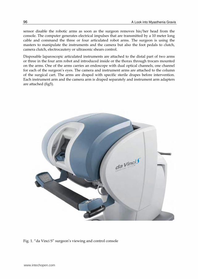
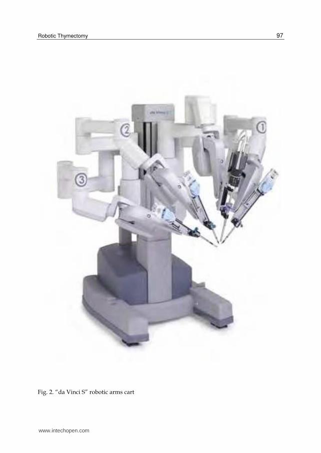
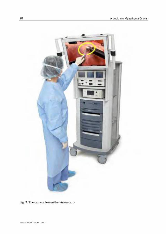
The Da Vinci system consists of three primary components: the surgeon’s viewing and control console (fig 1),a movable cart with three or four articulated robot arms (fig2) and the camera tower(the vision cart) (fig3).
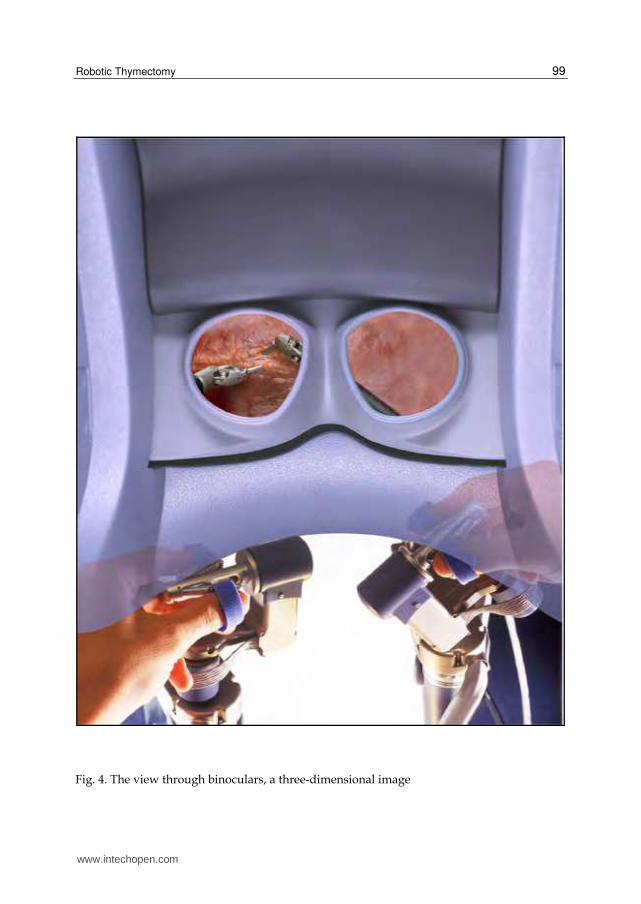
The console is located outside the sterile field. The surgeon is seated in front of the console and manipulates handles that are similar to “joysticks” while viewing a high-resolution, truly three-dimensional image of the surgical field through binoculars (fig 4). Manipulation of the handles transmits electronic signals to the computer, which can control and modify the movement of instrument tips by downscaling the movements, by eliminating physiologic tremor, and by adjusting grip strength applied to the tools. The masters are aligned with instrument tips, making the movement of the instruments natural and intuitive. The surgeon can also monitor the status of the instruments and arms without removing the head from the console through the various messages that are displayed on the display. In fact the sensation is that messages appear in the operation field. An infrared
www.intechopen.com
A Look into Myasthenia Gravis
96
sensor disable the robotic arms as soon as the surgeon removes his/her head from the console. The computer generates electrical impulses that are transmitted by a 10 meter long cable and command the three or four articulated robot arms. The surgeon is using the masters to manipulate the instruments and the camera but also the foot pedals to clutch, camera clutch, electrocautery or ultrasonic shears control.
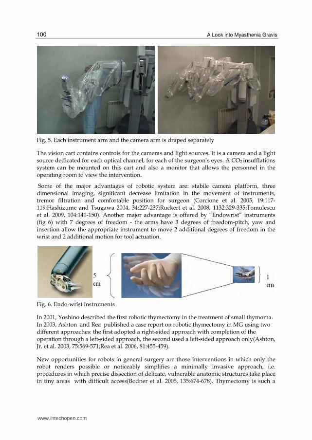
Disposable laparoscopic articulated instruments are attached to the distal part of two arms or three in the four arm robot and introduced inside or the thorax through trocars mounted on the arms. One of the arms carries an endoscope with dual optical channels, one channel for each of the surgeon’s eyes. The camera and instrument arms are attached to the column of the surgical cart. The arms are draped with specific sterile drapes before intervention. Each instrument arm and the camera arm is draped separately and instrument arm adapters are attached (fig5).
Fig. 1. “da Vinci S” surgeon’s viewing and control console
www.intechopen.com
Robotic Thymectomy
97
Fig. 2. “da Vinci S” robotic arms cart
www.intechopen.com
A Look into Myasthenia Gravis
98
Fig. 3. The camera tower(the vision cart)
www.intechopen.com
Robotic Thymectomy
99
Fig. 4. The view through binoculars, a three-dimensional image
www.intechopen.com
A Look into Myasthenia Gravis
100
Fig. 5. Each instrument arm and the camera arm is draped separately
The vision cart contains controls for the cameras and light sources. It is a camera and a light source dedicated for each optical channel, for each of the surgeon’s eyes. A CO2 insufflations system can be mounted on this cart and also a monitor that allows the personnel in the operating room to view the intervention.
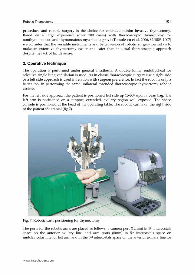
Some of the major advantages of robotic system are: stabile camera platform, three dimensional imaging, significant decrease limitation in the movement of instruments, tremor filtration and comfortable position for surgeon (Corcione et al. 2005, 19:117-119;Hashizume and Tsugawa 2004, 34:227-237;Ruckert et al. 2008, 1132:329-335;Tomulescu et al. 2009, 104:141-150). Another major advantage is offered by “Endowrist” instruments (fig 6) with 7 degrees of freedom - the arms have 3 degrees of freedom-pitch, yaw and insertion allow the appropriate instrument to move 2 additional degrees of freedom in the wrist and 2 additional motion for tool actuation.
Fig. 6. Endo-wrist instruments
In 2001, Yoshino described the first robotic thymectomy in the treatment of small thymoma. In 2003, Ashton and Rea published a case report on robotic thymectomy in MG using two different approaches: the first adopted a right-sided approach with completion of the operation through a left-sided approach, the second used a left-sided approach only(Ashton, Jr. et al. 2003, 75:569-571;Rea et al. 2006, 81:455-459).
New opportunities for robots in general surgery are those interventions in which only the robot renders possible or noticeably simplifies a minimally invasive approach, i.e. procedures in which precise dissection of delicate, vulnerable anatomic structures take place in tiny areas with difficult access(Bodner et al. 2005, 135:674-678). Thymectomy is such a
www.intechopen.com
Robotic Thymectomy
101
procedure and robotic surgery is the choice for extended minim invasive thymectomy. Based on a large experience (over 300 cases) with thoracoscopic thymectomy for nonthymomatous and thymomatous myasthenia gravis(Tomulescu et al. 2006, 82:1003-1007) we consider that the versatile instruments and better vision of robotic surgery permit us to make an extensive thymectomy easier and safer than in usual thoracoscopic approach despite the lack of tactile sense.
2. Operative technique
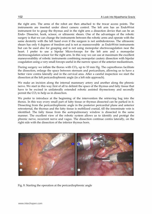
The operation is performed under general anesthesia. A double lumen endotracheal for selective single lung ventilation is used. As in classic thoracoscopic surgery use a right side or a left side approach is used in relation with surgeon preference. In fact the robot is only a better tool in performing the same unilateral extended thoracoscopic thymectomy robotic assisted.
For the left side approach the patient is positioned left side up 15-30o upon a bean bag. The left arm is positioned on a support, extended, axillary region well exposed. The video console is positioned at the head of the operating table. The robotic cart is on the right side of the patient 45o cranial (fig 7).
Fig. 7. Robotic carts positioning for thymectomy
The ports for the robotic arms are placed as follows: a camera port (12mm) in 5th intercostals space on the anterior axillary line, and arm ports (8mm) in 5th intercostals space on midclavicular line for left arm and in the 3rd intercostals space on the anterior axillary line for
www.intechopen.com
A Look into Myasthenia Gravis
102
the right arm. The arms of the robot are then attached to the trocar access points. The instruments are inserted under direct camera control. The left arm has an EndoWrist instrument for to grasp the thymus and in the right arm a dissection device that can be an Endo- Dissector, hook, scissor, or ultrasonic shears. One of the advantages of the robotic surgery is that we can change the instruments between the robotic arms and operate with the same dexterity with the left hand even if the surgeon is not ambidexterous. The ultrasonic shears has only 4 degrees of freedom and is not as maneuverable as EndoWrist instruments but can be used also for grasping and is not using monopolar electrocoagulation near the heart. I prefer to use a bipolar Micro-forceps for the left arm and a monopolar electrocoagulation scissor for the right arm. In this way we can use at maximum the excellent maneuverability of robotic instruments combining monopolar cautery dissection with bipolar coagulation using a very small forceps useful in the narrow space of the anterior mediastinum.
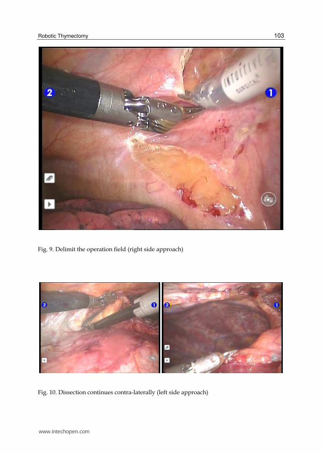
During surgery we inflate the thorax with CO2, up to 10 mm Hg. The capnothorax facilitate the dissection, enlarge the space between sternum and pericardium, allowing us to have a better view contra laterally and in the cervical area. After a careful inspection we start the dissection at the left pericardiophrenic angle (in a left side approach).
We make an incision along the internal mammary artery and another along the phrenic nerve. We start in this way first of all to delimit the space of the thymus and fatty tissue that have to be excised in unilaterally extended robotic assisted thymectomy and secondly permit the CO2 to help us in dissection.
We prefer to introduce at the beginning of the intervention the retrieving bag into the thorax. In this way every small part of fatty tissue or thymus dissected can be parked in it. Dissecting from the pericardiophrenic angle in the posterior pericardial plane and anterior retrosternal, the thymus and the fatty tissue is mobilized cranial, till the innominate vein is identified. The fatty tissue from the aortopulmonary window is dissected in the same manner. The excellent view of the robotic system allows us to identify and protégé the phrenic nerve, recurrent nerve and vagus. The dissection continue contra laterally, on the right side with the dissection of the inferior thymus horn.
Fig. 8. Starting the operation at the pericardiophrenic angle
www.intechopen.com
Robotic Thymectomy
103
Fig. 9. Delimit the operation field (right side approach)
Fig. 10. Dissection continues contra-laterally (left side approach)
www.intechopen.com
A Look into Myasthenia Gravis
104
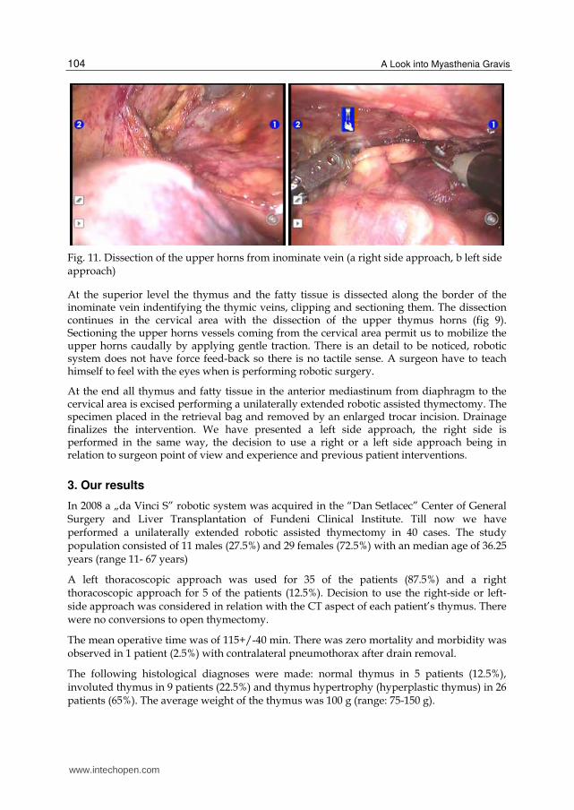
Fig. 11. Dissection of the upper horns from inominate vein (a right side approach, b left side approach)
At the superior level the thymus and the fatty tissue is dissected along the border of the inominate vein indentifying the thymic veins, clipping and sectioning them. The dissection continues in the cervical area with the dissection of the upper thymus horns (fig 9). Sectioning the upper horns vessels coming from the cervical area permit us to mobilize the upper horns caudally by applying gentle traction. There is an detail to be noticed, robotic system does not have force feed-back so there is no tactile sense. A surgeon have to teach himself to feel with the eyes when is performing robotic surgery.
At the end all thymus and fatty tissue in the anterior mediastinum from diaphragm to the cervical area is excised performing a unilaterally extended robotic assisted thymectomy. The specimen placed in the retrieval bag and removed by an enlarged trocar incision. Drainage finalizes the intervention. We have presented a left side approach, the right side is performed in the same way, the decision to use a right or a left side approach being in relation to surgeon point of view and experience and previous patient interventions.
3. Our results
In 2008 a „da Vinci S” robotic system was acquired in the “Dan Setlacec” Center of General Surgery and Liver Transplantation of Fundeni Clinical Institute. Till now we have performed a unilaterally extended robotic assisted thymectomy in 40 cases. The study population consisted of 11 males (27.5%) and 29 females (72.5%) with an median age of 36.25 years (range 11- 67 years)
A left thoracoscopic approach was used for 35 of the patients (87.5%) and a right thoracoscopic approach for 5 of the patients (12.5%). Decision to use the right-side or left-side approach was considered in relation with the CT aspect of each patient’s thymus. There were no conversions to open thymectomy.
The mean operative time was of 115+/-40 min. There was zero mortality and morbidity was observed in 1 patient (2.5%) with contralateral pneumothorax after drain removal.
The following histological diagnoses were made: normal thymus in 5 patients (12.5%), involuted thymus in 9 patients (22.5%) and thymus hypertrophy (hyperplastic thymus) in 26 patients (65%). The average weight of the thymus was 100 g (range: 75-150 g).
www.intechopen.com
Robotic Thymectomy
105
The mean length of hospitalization was 3.25 days (range: 2-5 days)
4. Conclusions
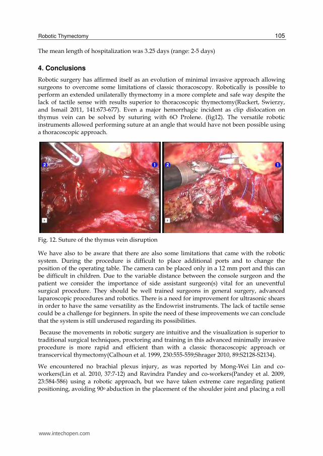
Robotic surgery has affirmed itself as an evolution of minimal invasive approach allowing surgeons to overcome some limitations of classic thoracoscopy. Robotically is possible to perform an extended unilaterally thymectomy in a more complete and safe way despite the lack of tactile sense with results superior to thoracoscopic thymectomy(Ruckert, Swierzy, and Ismail 2011, 141:673-677). Even a major hemorrhagic incident as clip dislocation on thymus vein can be solved by suturing with 6O Prolene. (fig12). The versatile robotic instruments allowed performing suture at an angle that would have not been possible using a thoracoscopic approach.
Fig. 12. Suture of the thymus vein disruption
We have also to be aware that there are also some limitations that came with the robotic system. During the procedure is difficult to place additional ports and to change the position of the operating table. The camera can be placed only in a 12 mm port and this can be difficult in children. Due to the variable distance between the console surgeon and the patient we consider the importance of side assistant surgeon(s) vital for an uneventful surgical procedure. They should be well trained surgeons in general surgery, advanced laparoscopic procedures and robotics. There is a need for improvement for ultrasonic shears in order to have the same versatility as the Endowrist instruments. The lack of tactile sense could be a challenge for beginners. In spite the need of these improvements we can conclude that the system is still underused regarding its possibilities.
Because the movements in robotic surgery are intuitive and the visualization is superior to traditional surgical techniques, proctoring and training in this advanced minimally invasive procedure is more rapid and efficient than with a classic thoracoscopic approach or transcervical thymectomy(Calhoun et al. 1999, 230:555-559;Shrager 2010, 89:S2128-S2134).
We encountered no brachial plexus injury, as was reported by Mong-Wei Lin and co-workers(Lin et al. 2010, 37:7-12) and Ravindra Pandey and co-workers(Pandey et al. 2009, 23:584-586) using a robotic approach, but we have taken extreme care regarding patient positioning, avoiding 90o abduction in the placement of the shoulder joint and placing a roll
www.intechopen.com
A Look into Myasthenia Gravis
106
under the arm and axilla in these patients. The anaesthesiologist and the assistant surgeon were constantly looking for any injury that the robotic arms could potentially cause. Continuous communication between the console surgeon and the other two members of the surgical-anaesthesiology team and careful procedure planning are the keys of good results with robotic surgery.
5. References
Ashton RC, Jr., McGinnis KM, Connery CP, Swistel DG, Ewing DR, and Derose JJ, Jr. 2003. Totally endoscopic robotic thymectomy for myasthenia gravis. Ann. Thorac. Surg. 75 (2): 569-571.
Bodner J, Augustin F, Wykypiel H, Fish J, Muehlmann G, Wetscher G, and Schmid T. 2005. The da Vinci robotic system for general surgical applications: a critical interim appraisal. Swiss. Med. Wkly. 135 (45-46): 674-678.
Calhoun RF, Ritter JH, Guthrie TJ, Pestronk A, Meyers BF, Patterson GA, Pohl MS, and Cooper JD. 1999. Results of transcervical thymectomy for myasthenia gravis in 100 consecutive patients. Ann. Surg. 230 (4): 555-559.
Corcione F, Esposito C, Cuccurullo D, Settembre A, Miranda N, Amato F, Pirozzi F, and Caiazzo P. 2005. Advantages and limits of robot-assisted laparoscopic surgery: preliminary experience. Surg. Endosc. 19 (1): 117-119.
Hashizume M, and Tsugawa K. 2004. Robotic surgery and cancer: the present state, problems and future vision. Jpn. J. Clin. Oncol. 34 (5): 227-237.
Lin MW, Chang YL, Huang PM, and Lee YC. 2010. Thymectomy for non-thymomatous myasthenia gravis: a comparison of surgical methods and analysis of prognostic factors. Eur. J. Cardiothorac. Surg. 37 (1): 7-12.
Pandey R, Elakkumanan LB, Garg R, Jyoti B, Mukund C, Chandralekha, Punj J, and Vanlal D. 2009. Brachial plexus injury after robotic-assisted thoracoscopic thymectomy. J. Cardiothorac. Vasc. Anesth. 23 (4): 584-586.
Rea F, Marulli G, Bortolotti L, Feltracco P, Zuin A, and Sartori F. 2006. Experience with the "da Vinci" robotic system for thymectomy in patients with myasthenia gravis: report of 33 cases. Ann. Thorac. Surg. 81 (2): 455-459.
Ruckert JC, Ismail M, Swierzy M, Sobel H, Rogalla P, Meisel A, Wernecke KD, Ruckert RI, and Muller JM. 2008. Thoracoscopic thymectomy with the da Vinci robotic system for myasthenia gravis. Ann. N. Y. Acad. Sci. 1132: 329-335.
Ruckert JC, Swierzy M, and Ismail M. 2011. Comparison of robotic and nonrobotic thoracoscopic thymectomy: a cohort study. J. Thorac. Cardiovasc. Surg. 141 (3): 673-677.
Shrager JB. 2010. Extended transcervical thymectomy: the ultimate minimally invasive approach. Ann. Thorac. Surg. 89 (6): S2128-S2134.
Tomulescu V, Ion V, Kosa A, Sgarbura O, and Popescu I. 2006. Thoracoscopic thymectomy mid-term results. Ann. Thorac. Surg. 82 (3): 1003-1007.
Tomulescu V, Stanciulea O, Balescu I, Vasile S, Tudor S, Gheorghe C, Vasilescu C, and Popescu I. 2009. First year experience of robotic-assisted laparoscopic surgery with 153 cases in a general surgery department: indications, technique and results. Chirurgia. (Bucur. ) 104 (2): 141-150.
Vasilescu C, and Popescu I. 2008. Chirurgia robotica - problemele inceputului; posbilitati si perspective. Chirurgia. (Bucur. ) 103 (1): 9-11.
www.intechopen.com
A Look into Myasthenia GravisEdited by Dr. Joseph A. Pruitt
ISBN 978-953-307-821-2Hard cover, 106 pagesPublisher InTechPublished online 20, January, 2012Published in print edition January, 2012
InTech EuropeUniversity Campus STeP Ri Slavka Krautzeka 83/A 51000 Rijeka, Croatia Phone: +385 (51) 770 447 Fax: +385 (51) 686 166www.intechopen.com
InTech ChinaUnit 405, Office Block, Hotel Equatorial Shanghai No.65, Yan An Road (West), Shanghai, 200040, China
Phone: +86-21-62489820 Fax: +86-21-62489821
Myasthenia gravis is presently an incurable antibody-mediated autoimmune disorder characterized bygeneralized voluntary skeletal muscle weakness. The cause of the weakness is a defect at the neuromuscularjunction level, in which autoimmune antibodies block the receptors responsible for initiating muscularcontraction. Literally translated from its Latin and Greek etymological roots, myasthenia gravis means "gravemuscle weakness". Fortunately, advances in modern medicine have resulted in a reduction of the truly "grave"outcomes for those inflicted but, without a cure, the gravity surrounding the disease remains
How to referenceIn order to correctly reference this scholarly work, feel free to copy and paste the following:
Victor Tomulescu (2012). Robotic Thymectomy, A Look into Myasthenia Gravis, Dr. Joseph A. Pruitt (Ed.),ISBN: 978-953-307-821-2, InTech, Available from: http://www.intechopen.com/books/a-look-into-myasthenia-gravis/robotic-thymectomy