Embed Size (px)
Citation preview

© 2018 Journal of Minimal Access Surgery | Published by Wolters Kluwer - Medknow 1
Robotic buccal mucosa graft ureteroplasty (inlay and onlay) for upper ureteric stricture: Point of technique
Arvind P. Ganpule, Abhishek G. Singh, Mohammed Rafiqul Islam, Parag Sonawane, Ravindra B. Sabnis, Mahesh R. Desai
Departments of Urology, Muljibhai Patel Urological Hospital, Nadiad, Gujarat, India
How I Do It
INTRODUCTION
Incidence of ureteral strictures is increasing, with increase in the number of ureteric interventions. Various management options are available to the urologist, depending on the length and site of the ureteric stricture segment. Management of complex long‑segment ureteral strictures using endourological techniques yields poorer results as compared to surgical reconstruction. Minimally invasive buccal mucosal graft (BMG) ureteroplasty can be performed in such cases using robotic platform.[1‑3] Principle of surgery remains the same as in any other reconstruction, that is creation of water‑tight, tension‑free and well‑vascularised anastomosis. Robotic platform allows the urologist to perform these steps successfully through magnified view, three‑dimensional field of vision, with increased dexterity and the ability to precisely place
sutures using EndoWrist technology.[4] The buccal mucosa because of its inherent properties has been widely used in urethral strictures and the same can be useful for ureteric strictures.[5,6] The literature is replete with number of case reports describing this procedure with the open surgical approach.[7,8] There are a few case reports in which the robotic platform is used for the treatment of complex ureteral strictures using the BMG as a tube or an onlay flap.[1‑3] We describe the use of the BMG as onlay and inlay graft instead of the conventional onlay or tube in ureteral stricture reconstruction, using a robotic platform.
Pre‑operative evaluationPatient is evaluated by detailed history taking, physical examination, complete serum chemistries and a computed tomography intravenous urography (CT‑IVU). The
Ureteral stricture resulting from chronic inflammations such as tuberculosis, recurrent stone disease and multiple endourological interventions are complex in nature; these may lead to severe adhesions to surrounding structures. Endourological management of these cases is difficult with poorer outcomes. In such situations, reconstructive surgical corrections remain a reliable option. We describe the technique of onlay and inlay buccal mucosal graft ureteroplasty using a robotic platform in management of complex ureteral strictures.
Keywords: Inlay buccal mucosal graft ureteroplasty, onlay buccal mucosal graft ureteroplasty, ureteral stricture
Abstract
Address for correspondence: Dr. Arvind P. Ganpule, Department of Urology, Muljibhai Patel Urological Hospital, Nadiad, Gujarat, India. E‑mail: [email protected] Received: 22.09.2017, Accepted: 07.12.2017
Access this article onlineQuick Response Code:
Website:www.journalofmas.com
DOI:10.4103/jmas.JMAS_188_17
How to cite this article: Ganpule AP, Singh AG, Islam MR, Sonawane P, Sabnis RB, Desai MR. Robotic buccal mucosa graft ureteroplasty (inlay and onlay) for upper ureteric stricture: Point of technique. J Min Access Surg 0;0:0.
This is an open access article distributed under the terms of the Creative Commons Attribution‑NonCommercial‑ShareAlike 3.0 License, which allows others to remix, tweak, and build upon the work non‑commercially, as long as the author is credited and the new creations are licensed under the identical terms.
For reprints contact: [email protected]
[Downloaded free from http://www.journalofmas.com on Monday, May 7, 2018, IP: 210.212.153.193]

Ganpule, et al.: Robotic buccal mucosa graft ureteroplasty (inlay and onlay) for upper ureteric stricture
2 Journal of Minimal Access Surgery | Volume XX | Issue XX | Month 2017
pre‑operative imaging helps to assess the exact site and length of the stricture. The CT‑IVU, apart from assessing the location of stricture, also helps in assessing the surrounding anatomy of the stricture segment. If a nephrostomy has been preplaced, an antegrade and retrograde contrast study is necessary to assess the length of the stricture. It is important to know the length of stricture as this will decide the length of BMG to be harvested. The length of the graft required will be at least 2 cm more than the length of the stricture as there will be a grey zone of unhealthy tissue beyond the white zone of stricture, beyond which will be the normal pink and healthy mucosa.
Positioning and portsUnder general anaesthesia, endotracheal intubation is done transnasally using Ring, Adair and Elwyn north‑pole tube and the mouth is separately prepared and draped for harvesting the BMG. The patient is placed in lithotomy position for antegrade and retrograde dye study [Figure 1]. Antegrade and retrograde dye study is done to localise the ureteric stricture [Figure 1]. A 6F ureteric catheter over a guide wire is placed in the ureter retrogradely up to the stricture; another 6F ureteric catheter over a guide wire is negotiated through the percutaneous nephrostomy (PCN) up to the stricture segment antegradely. The purpose of performing this contrast study and placing ureteric catheters is to identify the ureter and the exact location of the stricture as in most of the cases, the ureter is encased in dense adhesions intraoperatively. Stricture identification at times is one of the most challenging tasks in minimally invasive surgery as there is no tactile feedback as opposed to open surgery where the surgeon can palpate and identify the stricture.
The patient is now placed in partial lithotomy position with ipsilateral leg slightly flexed at knee joint and higher level than contralateral leg; contralateral leg is extended and contralateral lateral tilt is given by supporting at back side of shoulder and flank with rectangular pillows [Figure 2].
Co2 insufflations are done by inserting Veress needle in ipsilateral iliac fossa, and after insufflation of abdomen, ports’ placement is planned. Four ports should be inserted; among them, two are 12 mm, one for camera and another for the assistant. Rest two ports are 8 mm robotic trocars, for robotic arms. Site of the stricture is marked on skin during the dye study, and camera port is placed in front of the stricture segment pararectally. Robotic ports are placed 8–10 cm cranial and caudal to the camera port in the midclavicular line. Assistant port was placed between camera and left robotic port.
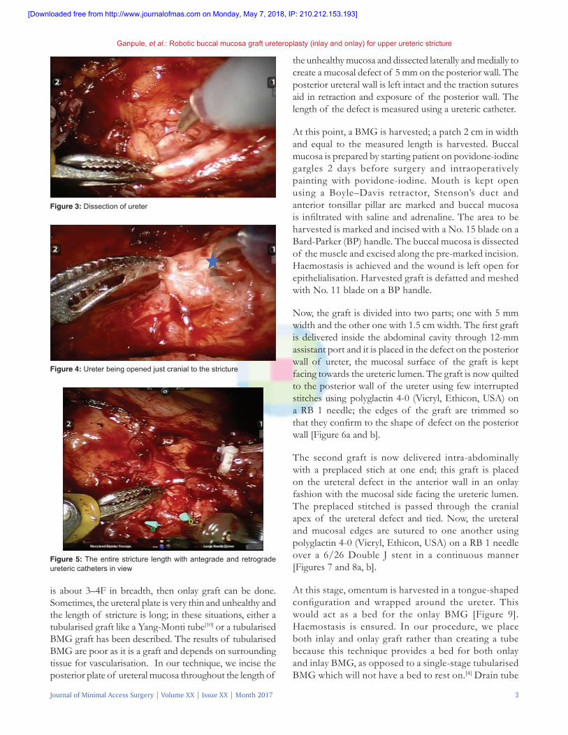
Operative stepsAfter docking the robot, incision is made along the white line of Toldt and colon is mobilised and reflected medially. After entering the retroperitoneum, the ureter is identified by sharp and blunt dissection [Figures 3 and 4]. The ureter is usually densely adherent to the surrounding structures and difficult to identify. In such situations, the ureteric catheters which are placed through antegrade and retrograde route help to identify the exact site of the stricture [Figure 5]. At this point, the patient side assistant jiggles the guide wire within the ureteric catheter; this movement of the guide wires within the ureteric catheters help in identifying the exact location of the upper and lower end of stricture.
Peroperatively, various techniques were used to identify the segment of ureteric stricture like filling the nephrostomy with saline so that upper ureter gets distended; identification of ureteric catheters and recently near‑infrared fluorescence image has been described.[9]
Stricture segment is slowly and patiently dissected; this area may be adherent to posterior structures including the iliac vessels and may be difficult to mobilise. In these situations, without circumferential mobilisation of the ureter, only the anterior wall of ureter is incised along the length of the stricture till healthy pink ureteric mucosa is seen on both sides of the stricture [Figure 5]. At the midpoint of the length of incision, two stay stitches are taken opposite to each other from the incised edges. This exposes the posterior wall i.e the ureteral plate. If the ureteral plate
Figure 1: Patient positioning Figure 2: (a and b) Antegrade dye study delineating the strictureba
[Downloaded free from http://www.journalofmas.com on Monday, May 7, 2018, IP: 210.212.153.193]
Ganpule, et al.: Robotic buccal mucosa graft ureteroplasty (inlay and onlay) for upper ureteric stricture
Journal of Minimal Access Surgery | Volume XX | Issue XX | Month 2017 3
is about 3–4F in breadth, then onlay graft can be done. Sometimes, the ureteral plate is very thin and unhealthy and the length of stricture is long; in these situations, either a tubularised graft like a Yang‑Monti tube[10] or a tubularised BMG graft has been described. The results of tubularised BMG are poor as it is a graft and depends on surrounding tissue for vascularisation. In our technique, we incise the posterior plate of ureteral mucosa throughout the length of
the unhealthy mucosa and dissected laterally and medially to create a mucosal defect of 5 mm on the posterior wall. The posterior ureteral wall is left intact and the traction sutures aid in retraction and exposure of the posterior wall. The length of the defect is measured using a ureteric catheter.
At this point, a BMG is harvested; a patch 2 cm in width and equal to the measured length is harvested. Buccal mucosa is prepared by starting patient on povidone‑iodine gargles 2 days before surgery and intraoperatively painting with povidone‑iodine. Mouth is kept open using a Boyle–Davis retractor, Stenson’s duct and anterior tonsillar pillar are marked and buccal mucosa is infiltrated with saline and adrenaline. The area to be harvested is marked and incised with a No. 15 blade on a Bard‑Parker (BP) handle. The buccal mucosa is dissected of the muscle and excised along the pre‑marked incision. Haemostasis is achieved and the wound is left open for epithelialisation. Harvested graft is defatted and meshed with No. 11 blade on a BP handle.
Now, the graft is divided into two parts; one with 5 mm width and the other one with 1.5 cm width. The first graft is delivered inside the abdominal cavity through 12‑mm assistant port and it is placed in the defect on the posterior wall of ureter, the mucosal surface of the graft is kept facing towards the ureteric lumen. The graft is now quilted to the posterior wall of the ureter using few interrupted stitches using polyglactin 4‑0 (Vicryl, Ethicon, USA) on a RB 1 needle; the edges of the graft are trimmed so that they confirm to the shape of defect on the posterior wall [Figure 6a and b].
The second graft is now delivered intra‑abdominally with a preplaced stich at one end; this graft is placed on the ureteral defect in the anterior wall in an onlay fashion with the mucosal side facing the ureteric lumen. The preplaced stitched is passed through the cranial apex of the ureteral defect and tied. Now, the ureteral and mucosal edges are sutured to one another using polyglactin 4‑0 (Vicryl, Ethicon, USA) on a RB 1 needle over a 6/26 Double J stent in a continuous manner [Figures 7 and 8a, b].
At this stage, omentum is harvested in a tongue‑shaped configuration and wrapped around the ureter. This would act as a bed for the onlay BMG [Figure 9]. Haemostasis is ensured. In our procedure, we place both inlay and onlay graft rather than creating a tube because this technique provides a bed for both onlay and inlay BMG, as opposed to a single‑stage tubularised BMG which will not have a bed to rest on.[4] Drain tube
Figure 5: The entire stricture length with antegrade and retrograde ureteric catheters in view
Figure 4: Ureter being opened just cranial to the stricture
Figure 3: Dissection of ureter
[Downloaded free from http://www.journalofmas.com on Monday, May 7, 2018, IP: 210.212.153.193]

Ganpule, et al.: Robotic buccal mucosa graft ureteroplasty (inlay and onlay) for upper ureteric stricture
4 Journal of Minimal Access Surgery | Volume XX | Issue XX | Month 2017
is kept in situ in all cases.
Post‑operative careThe patient is given injectable antibiotics for 5 days; PCN is kept open for 5 days then it is clamped, and if the drain output does not increase, PCN is removed on the post‑operative day (POD) 6. Foley catheter is removed on POD 6, and if drain output does not increase, drain is removed on POD 7. Double J stent is kept in situ for 6 weeks. Stent is removed after 6 weeks and the patient is followed up on ultrasound imaging. A contrast imaging is done at 3 months to assess the repair.
CONCLUSION
Robotic BMG inlay and onlay ureteroplasty is a novel technique in the management of ureteral stricture. The dual graft offers an advantage of placing the graft on a robust backing thus increasing the chance of vascularisation and uptake of the graft. It has a low potential for morbidity as it does not involve bowel anastomosis. Although the result of this procedure is encouraging with 3‑month follow‑up, more cases and longer follow‑up are needed for better perfection of this procedure.
AcknowledgmentWe would like to thank Mr. Sanjay Bhagat, Theatre technician for helping us develop this technique.
Financial support and sponsorshipNil.
Conflicts of interestThere are no conflicts of interest.
REFERENCES
1. Marien T, Bjurlin MA, Wynia B, Bilbily M, Rao G, Zhao LC, et al. Outcomes of robotic‑assisted laparoscopic upper urinary tract reconstruction: 250 consecutive patients. BJU Int 2015;116:604‑11.
2. Zhao LC, Yamaguchi Y, Bryk DJ, Adelstein SA, Stifelman MD. Robot‑assisted ureteral reconstruction using buccal mucosa. Urology 2015;86:634‑8.
3. Arora S, Campbell L, Tourojman M, Pucheril D, Jones LR, Rogers C, et al. Robotic buccal mucosal graft ureteroplasty for complex ureteral stricture. Urology 2017;110:257‑8.
4. Yohannes P, Rotariu P, Pinto P, Smith AD, Lee BR. Comparison of robotic versus laparoscopic skills: Is there a difference in the learning curve? Urology 2002;60:39‑45.
5. Mundy AR, Andrich DE. Urethral strictures. BJU Int 2011;107:6‑26.6. Hudak SJ, Lubahn JD, Kulkarni S, Morey AF. Single‑stage reconstruction
of complex anterior urethral strictures using overlapping dorsal and ventral buccal mucosal grafts. BJU Int 2012;110:592‑6.
7. Agrawal V, Dassi V, Andankar MG. Buccal mucosal graft onlay repair for a ureteric ischemic injury following a pyeloplasty. Indian J Urol 2010;26:120‑2.
8. Kroepfl D, Loewen H, Klevecka V, Musch M. Treatment of long
Figure 7: Double J stent being passed after inlay graft placement
Figure 9: Omentum being rapped around the graft and ureter
Figure 6: (a) Placement of the buccal mucosal graft in an inlay fashion, (b) quilting of the buccal mucosal graft in an inlay fashion
ba
Figure 8: (a) Apical stitch of the onlay graft being taken, (b) anterior wall of the onlay graft being sutured in a continuous fashion
ba
[Downloaded free from http://www.journalofmas.com on Monday, May 7, 2018, IP: 210.212.153.193]

Ganpule, et al.: Robotic buccal mucosa graft ureteroplasty (inlay and onlay) for upper ureteric stricture
Journal of Minimal Access Surgery | Volume XX | Issue XX | Month 2017 5
ureteric strictures with buccal mucosal grafts. BJU Int 2010;105:1452‑5.9. Bjurlin MA, Gan M, McClintock TR, Volpe A, Borofsky MS, Mottrie A,
et al. Near‑infrared fluorescence imaging: Emerging applications in
robotic upper urinary tract surgery. Eur Urol 2014;65:793‑801.10. Ghoneim MA, Ali‑El‑Dein B. Replacing the ureter by an ileal tube,
using the yang‑monti procedure. BJU Int 2005;95:455‑70.
[Downloaded free from http://www.journalofmas.com on Monday, May 7, 2018, IP: 210.212.153.193]