Embed Size (px)
Citation preview

Robotic Assisted Versus Pure MicrosurgicalVasectomy Reversal: Technique and ProspectiveDatabase Control TrialSijo J. Parekattil, M.D. 1, 2 Ahmet Gudeloglu, M.D. 2 Jamin Brahmbhatt, M.D. 2
Jessica Wharton, A.R.N.P. 2 Karen B. Priola 2
1Division of Robotics and Urology, Department of Urology, WinterHaven Hospital, University of Florida, Winter Haven, Florida
2Department of Urology, Winter Haven Hospital & University ofFlorida, Winter Haven, Florida
J Reconstr Microsurg
Address for correspondence and reprint requests Sijo J. Parekattil,M.D., Director of Robotics and Urology, Department of Urology,Winter Haven Hospital, University of Florida, 200 Avenue, F, N.E.Winter Haven, FL 33881(e-mail: [email protected]).
Vasectomy is considered one of the most reliable family-planning methods currently available because of its simplici-ty, effectiveness, and low morbidity rate.1 An estimated 40 to60 million men worldwide rely on this method of contracep-tion. About 2% of these men undergo a reversal operationwithin the first 10 years after the vasectomy because of adesire to become fertile again (usually due to a new relation-ship).1,2 Vasectomy reversal is a technically demanding pro-cedure. Since the introduction of the operatingmicroscope in1975 for this kind of surgery, there has been an improvementin success rates.3,4 The use of the operative microscope toachieve greater fertility rates and vas patency after vaso-vasostomy has become a standard for the microsurgeon whotreats male infertility.4 However, this technique requires agreat degree of microsurgical training and a skilled surgicalassistant.
The development of robotic assisted procedures in severalsurgical fields continues to expand.5–9 There are severalpotential benefits: a stable, ergonomic, scalable control sys-temwith three-dimensional visualization andmagnification;elimination of tremor with simultaneous ability to controlthree instruments and a camera; ability to integrate up tothree visual inputs simultaneously in the surgeon console,similar to a fighter pilot cockpit. All these features mayprovide surgeons an advantage when performing complexmicrosurgical procedures. The use of robotic assistance forvasectomy reversal could potentially provide the microsur-geon with improved visualization, decreased fatigue andobviate the need for a skilled surgical assistant. This studypresents the current technique and outcomes for roboticassisted microsurgical vasovasostomy (RAVV) and vasoepidi-dymostomy (RAVE).
Keywords
► vasovasostomy► vasoepididymostomy► robotic► vasectomy► reversal
Abstract Microsurgical vasectomy reversal is a technically demanding procedure. Previousstudies have shown the possible benefit of robotic assistance during such procedures.Our goal was to compare robotic assisted vasovasostomy and vasoepididymostomy tostandard microsurgical vasovasostomy (MVV) and vasoepididymostomy (MVE). The useof robotic assistance for vasectomy reversal may provide the microsurgeon withimproved visualization, elimination of tremor, and decreased fatigue and obviate theneed for a skilled microsurgical assistant. This study provides the first clinical prospec-tive control trial of robotic assisted versus pure microsurgical vasectomy reversal. Theuse of robotic assistance in microsurgical vasovasostomy and vasoepididymostomymayhave benefit over MVV and MVE with regards to decreasing operative duration andimproving the rate of recovery of postoperative total motile sperm counts based on ourstudy.
receivedFebruary 26, 2012acceptedMarch 21, 2012
Copyright © 2012 by Thieme MedicalPublishers, Inc., 333 Seventh Avenue,New York, NY 10001, USA.Tel: +1(212) 584-4662.
DOI http://dx.doi.org/10.1055/s-0032-1315788.ISSN 0743-684X.
Methods
Robotic Surgical Platform and Operative SetupIntuitive Surgical (Sunnyvale, CA) offers a four-arm da Vincitype Si robotic systemwith high-definition (HD) digital visualmagnification (up to 10 to 15 � ). The left and right arms areloaded with black diamond microneedle drivers (IntuitiveSurgical, Inc., Sunnyvale, CA). The additional fourth armprovides the microsurgeon with one additional tool such asmicro Potts scissors (Intuitive Surgical, Inc., Sunnyvale, CA).
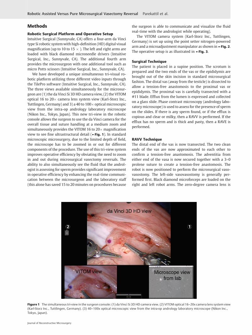
We have developed a unique simultaneous tri-visual ro-botic platform utilizing three different video inputs throughthe TilePro software (Intuitive Surgical, Inc., Sunnyvale, CA).The three views available simultaneously for the microsur-geon are (1) the da Vinci Si 3DHD camera view, (2) the VITOMoptical 16 to 20� camera lens system view (Karl-Storz Inc.,Tuttlingen, Germany) and 3) a 40 to 100� optical microscopicview from the intra-op andrology laboratory microscope(Nikon Inc., Tokyo, Japan). This new tri-view in the roboticconsole allows the surgeon to use the da Vinci camera for theoverall tissue and suture handling at a medium zoom andsimultaneously provides the VITOM 16 to 20�magnificationview to see fine ultrastructural detail (►Fig. 1). In standardmicroscopic microsurgery, due to the limited depth of field,the microscope has to be zoomed in or out for differentcomponents of the procedure. The use of this tri-view systemimproves operative efficiency by obviating the need to zoomin and out during microsurgical vasectomy reversals. Theability to also simultaneously see the fluid that the androl-ogist is assessing for sperm provides significant improvementin operative efficiency by enhancing the real-time communi-cation between the microsurgeon and the laboratory staff(this alone has saved 15 to 20minutes on procedures because
the surgeon is able to communicate and visualize the fluidreal-time with the andrologist while operating).
The VITOM camera system (Karl-Storz Inc., Tuttlingen,Germany) is set up using the point setter nitrogen-poweredarm and amicroadjustment manipulator as shown in►Fig. 2.The operative setup is as illustrated in ►Fig. 3.
Surgical TechniqueThe patient is placed in a supine position. The scrotum isprepared and the two ends of the vas or the epididymis arebrought out of the skin incision in standard microsurgicalfashion. The distal vas (away from the testicle) is dissected toallow a tension-free anastomosis to the proximal vas orepididymis. The proximal vas is carefully transected with a#11 blade. Efflux from the lumen is expressed and collectedon a glass slide. Phase contrast microscopy (andrology labo-ratorymicroscope) is used to assess for the presence of spermon the slides. If there is any sperm found, or if the efflux iscopious and clear or milky, then a RAVV is performed. If theefflux has no sperm and is thick and pasty, then a RAVE isperformed.
RAVV TechniqueThe distal end of the vas is now transected. The two cleanends of the vas are now approximated to each other toconfirm a tension-free anastomosis. The adventitia fromeither end of the vasa is now secured together with a 3–0prolene suture to create a tension-free anastomosis. Therobot is now positioned to perform the microsurgical vaso-vasostomy. The left-side vasovasostomy is generally per-formed first. Black diamond microforceps are loaded on theright and left robot arms. The zero-degree camera lens is
Figure 1 The simultaneous tri-view in the surgeon console: (1) da Vinci Si 3D HD camera view. (2) VITOM optical 16–20x camera lens system view(Karl-Storz Inc., Tuttlingen, Germany). (3) 40–100x optical microscopic view from the intra-op andrology laboratory microscope (Nikon Inc.,Tokyo, Japan).
Journal of Reconstructive Microsurgery
Robotic Assisted Versus Pure Microsurgical Vasectomy Reversal Parekattil et al.

loaded onto the robot camera arm. The micro Potts scissorsare loaded onto the fourth robot arm. The two ends of the vasare placed over a ¼-inch Penrose drain. The assistantirrigates the field with saline using a 10-mL syringe with an18-gauge angiocatheter tip. Weck sponge sticks are used todry the field.
The assistant now passes the 9–0 nylon suture that is keptin its inner packaging to the surgical field. The suture isgrasped using the black diamond right-hand grasper and cutto �2 inches length using the micro Potts scissors (left-handfourth arm). The 9–0 nylon suture is held and manipulatedusing the black diamond forceps in both left and right arms as
needle drivers. The posterior muscularis layer of the two endsof the vas is now approximated (►Fig. 4). The suture is cutusing the micro Potts scissors.
Two or three double-armed 10–0 nylon sutures are nowplaced to re-anastomose the posterior mucosal lumen of thevas (►Fig. 5). The sutures are placed inside out to ensure goodmucosal approximation. All sutures are placed before they aretied.
Three double-armed 10–0 nylon sutures are used to closethe anteriormucosal lumen of the vas (►Fig. 6). Five to six 9–0nylon sutures are used to approximate the anterior muscu-laris layer of the vas (►Fig. 7). The Penrose drain is gentlyremoved fromunder the repair. The vas is placed back into thescrotal cavity. The same procedure is now performed on thecontralateral right side by repositioning the robot away fromthe patient to the right scrotum.
The dartos layer is closed using a running 3–0 chromicsuture. The skin is closed using a 5–0 vicryl running suture.Bacitracin ointment is applied over the incision. Fluff dressingwith athletic scrotal support is applied. An ice pack iscarefully applied to the scrotum in the recovery room.
RAVE TechniqueThe RAVE procedure starts from above if there is no sperm inthe fluid from the proximal vas and the fluid is thick andpasty. The scrotal incision is enlarged by another 1 to 2 cminferiorly. The testicle is delivered and the tunica is incised toexpose the epididymis. The adventitial layer of the epididy-mis is incised above the level of epididymal obstruction (blue/gray zone with dilated epididymal tubules above this area). A3–0 prolene suture is utilized to approximate the adventitia ofthe epididymis to themuscularis of the vas to prevent tensionbetween the anastomosis. The robot is now positioned toperform the microsurgical vasoepididymostomy (MVE) as
Figure 2 Operative setup for the robotic platform with the additional VITOM lens camera system and point setter nitrogen powered arm(5th arm).
Figure 3 General layout of the operative room equipment for roboticassisted microsurgical vasectomy reversal.
Journal of Reconstructive Microsurgery
Robotic Assisted Versus Pure Microsurgical Vasectomy Reversal Parekattil et al.

described earlier. The black diamondmicroforceps are loadedon the right and left robot arms. The zero-degree camera lensis loaded onto the robot camera arm. An ophthalmologicmicroblade is held in the fourth arm with black diamondmicroforceps, or a Potts scissor may be used in the fourth arm.Two 10–0 nylon double-armed suture needles are placedlongitudinally through a single epididymal tubule to exposethe tubule (►Fig. 8). This tubule is then incised longitudinallyusing the microblade between the two suture needles tocreate a lumen in the tubule. Alternatively, the tubule may be
incisedwith a Potts scissor in the fourth robotic arm. Thefluidis then aspirated (►Fig. 9) and examined under a separatephase contrast microscope for the presence of sperm (an-drology laboratory microscope).
The two double-armed 10–0 nylon needles in the epidid-ymal tubule are advanced through, and then all four of theneedles are brought inside out on the vas mucosal lumen toinvolute the epididymal tubule lumen into the vas lumen(►Fig. 10). Five to six 9–0 nylon sutures are placed circum-ferentially to approximate the muscularis of the vas to the
Figure 4 Placement of two posterior 9–0 nylon vas deferens muscularis anastomosis sutures during robotic assisted microsurgicalvasovasostomy.
Figure 5 Placement of two or three posterior vasal mucosal lumen 10–0 nylon double-armed sutures during robotic assisted microsurgicalvasovasostomy.
Journal of Reconstructive Microsurgery
Robotic Assisted Versus Pure Microsurgical Vasectomy Reversal Parekattil et al.

adventitia of the epididymal tubule (►Fig. 11). The testicleand anastomosis are carefully delivered back into the scro-tum. The dartos layer is closed using a running 3–0 chromicsuture. The skin is closed using a 4–0 chromic running suture.Bacitracin ointment is applied over the incision. Fluff dressingwith athletic scrotal support is applied. An ice pack iscarefully applied to the scrotum in the recovery room.
Clinical Study Design & MethodsWe designed an institutional review board (IRB)–approvedprospective database based control study to compare RAVV
and RAVE to standard microsurgical vasovasostomy (MVV)and MVE. Between August 2007 and February 2012, 155vasectomy reversal cases performed by a single fellowship-trained microsurgeon were reviewed. The primary end pointwas operative duration. The secondary end point was totalmotile sperm count at 2, 5, 9, and 12 months postoperatively.Case breakdown was as such: 110 with robotic assistance, 45puremicrosurgical. 66 cases bilateral RAVV, 44 cases RAVE onat least one side, 28 cases bilateral MVV, and 17 cases MVE onat least one side. Selection of approach (robotic versus puremicroscopic) was based on patient choice. Preoperative
Figure 6 Placement of three anterior vasal mucosal lumen 10–0 nylon double-armed sutures during robotic assisted microsurgicalvasovasostomy.
Figure 7 Placement of five or six anterior and circumferential vas deferens muscularis 9–0 nylon sutures during robotic assisted microsurgicalvasovasostomy.
Journal of Reconstructive Microsurgery
Robotic Assisted Versus Pure Microsurgical Vasectomy Reversal Parekattil et al.

patient characteristics were similar in both groups. The samesuture materials and suturing techniques (two-layer 10–0and 9–0 nylon anastomosis for RAVV; 10–0 nylon double-armed longitudinal intussusception technique for RAVE)were used in both approaches.
Results
Median clinical follow-up was 17 months (range 1 to52 months). Median duration from vasectomy in the RAVVgroup was 7 years (range 1 to 21 years) and 6.5 years (range
1 to 19 years) in the MVV group (p ¼ 0.3). Median age of thepatients in the RAVV group was 41 and 39 in the MVV group(p ¼ 0.4).
A patency of 96% was achieved in the RAVV cases and 80%inMVV (>1million sperm/ejaculate). Therewas a statisticallysignificant difference in patency rates between the twogroups (p ¼ 0.02). Pregnancy rates (within 1 year postop)did not differ significantly for the two groups: 65% for theRAVV and 55% for the MVV. Operative duration (skin to skin)started at 150 to 180minutes initially for the first 10 cases forRAVV, but median operative duration was significantly
Figure 8 Two 10–0 nylon double-armed suture needles are placed longitudinally through a single epididymal tubule during robotic assistedmicrosurgical vasoepididymostomy.
Figure 9 Robotic assisted microsurgical vasoepididymostomy: Epididymal tubule incised with Potts Scissor or ophthalmologic blade and thenfluid aspirated for microscopic examination.
Journal of Reconstructive Microsurgery
Robotic Assisted Versus Pure Microsurgical Vasectomy Reversal Parekattil et al.

decreased in RAVV at 97 minutes (range 40 to 180 minutes)compared with MVV at 120 minutes (range 60 to 180 mi-nutes), p ¼ 0.0003. RAVE at 120minutes (range 60 to 180mi-nutes) was significantly faster than MVE at 150 minutes(range 120 to 240 minutes), p ¼ 0.0008. Suture breakageand needle bending reduced significantly after the first10 RAVVcases. Mean postoperative total motile sperm countswere not significantly higher in RAVV/RAVE versus MVV/MVE, but the rate of postoperative sperm count recovery was
significantly greater in RAVV/RAVE. ►Fig. 12 illustrates thepostoperative mean sperms counts in both groups.
Discussion
The robotic technique in this initial study appears to be safe,with comparable outcomes to the standard microsurgicalapproach. Subjectively, there appeared to be ergonomicadvantages to using the robotic system over the microscopic
Figure 10 Robotic assisted microsurgical vasoepididymostomy: The two double armed 10–0 nylon needles in the epididymal tubule areadvanced through and then all four of the needles are brought inside out on the vas mucosal lumen to involute the epididymal tubule lumen intothe vas lumen.
Figure 11 Robotic assisted microsurgical vasoepididymostomy: Five to six 9–0 nylon sutures are placed circumferentially to approximate themuscularis of the vas to the adventitia of the epididymal tubule.
Journal of Reconstructive Microsurgery
Robotic Assisted Versus Pure Microsurgical Vasectomy Reversal Parekattil et al.

platform. Objectively, there appears to be increased operativeefficiency with RAVV compared with MVV. Despite the sur-geon’s previous extensive background in microsurgical androbotic surgery (fellowships in each discipline), there was alearning curve associated with robotic microsurgery. Theoperative duration, number of suture breaks and needlebends decreased rapidly after the first 10 cases.
The robotic reversals initially had longer operative dura-tions (150 to 180 minutes) than standard microsurgicalreversals (usually 120 to 150 minutes depending on whethervasoepididymostomy is needed). There was an additional 30to 60 minutes to prepare the robot at the beginning of thecase: this time significantly decreasedwith experience, as theoperating room staff became more familiar with our setup.The duration of preparation for the robot (prior to the case) isroutinely �20 to 25 minutes now (this is similar to the timethe staff takes to prepare the microscope for pure microsur-gical cases). This learning curve in the initial robotic caseswasalso observed Kuang et al.10,11 Six years ago, Schiff et al12,13
showed in a prospective randomized control trial in ananimal model that robotic assistance could significantlyreduce operative duration in RAVV compared with MVV.Our study confirms these findings in a prospective humantrial.
Previous studies have shown that patency rate variesbetween 97% and 71% depending on the interval betweenvasectomy and reversal procedure.14 Our patient seriesbreakdown in terms of years from vasectomy was as followsfor RAVV and MVV respectively: <3 years from vasectomy,8.3% and 10.7%; 3 to 8 years, 46.7% and 60.7%; 9 to 14 years,26.7% and 17.9%; and �15 years, 18.3% and 10.7%. Based onthese expected patency rates, the duration-adjusted expectedpatency rates for our two groupswould be 83.2% for RAVVand85.5% for MVV. In our study the RAVV patency rate was 96%and the MVV patency rate was 80%. Although there was astatistically significant difference in patency rates in thisstudy (p ¼ 0.02) between RAVV over MVV, there may besome confounding variables. The surgeon did start perform-ing these procedures just after completing a fellowship, andmost of the MVV cases were performed earlier in the seriesand the RAVV cases were performed almost exclusively in the
later part of the series. There may be an inherent learning-curve bias to better outcomes as the series matures and thismay overamplify the difference between RAVV over MVVpatency rates. However, this does support the notion thatRAVV may allow a surgeon to achieve outcomes similar tovery mature retrospective case reviews of well-establishedmicrosurgeons in a shorter amount of time. The series ofRAVV cases presented in this study are the first 110 roboticcases performed by this surgeon, and the patency outcomesclosely match those of microsurgeons who have performedseveral hundred MVV cases.
Mean postoperative total motile sperm counts were notsignificantly higher in RAVV/RAVE versus MVV/MVE. Howev-er, the rate of sperm return to the ejaculate after surgery wasgreater in RAVV/RAVE. For RAVV, the mean rate of return ofsperm to the ejaculate postop was 13 million motile spermper month (the slope of the mean sperm counts 2, 5, 9, and12months postop). ForMVV, themean rate of return of spermto the ejaculate postopwas 3millionmotile spermpermonth.
The improved operative efficiency and eliminated need fora skilled assistant in RAVV/RAVE has reduced the cost of thisreversal procedure to less than the regional average cost forMVV/MVE. The total out-of-pocket cost of the robotic reversalprocedure for the patient at our facility (hospital setting)including operating room, anesthesia, and surgeon fees is$5,600. The use of robotic assistance has allowed the micro-surgeon to go from performing two to three microsurgicalcases to five to six such cases in the same time period due todecreased surgeon fatigue and improved surgical efficiency.
Conclusion
The use of robotic assistance in microsurgical vasovasostomyand vasoepididymostomy may have potential benefit overMVV and MVEwith regards to decreasing operative durationand improving the rate of recovery of postoperative totalmotile sperm counts. The advantages of a stablemicrosurgicalplatform, ergonomic surgeon instrument controls, elimina-tion of tremor, magnified immersive 3D vision, and simulta-neous tri-view ability all contribute to improved surgicalefficiency. Further evaluation and longer follow-up is neededto assess its clinical potential and the true cost-benefit ratio.However, the preliminary results are quite promising.
NoteNone of the authors have any disclosures.
References1 Schwingl PJ, Guess HA. Safety and effectiveness of vasectomy.
Fertil Steril 2000;73:923–9362 Dohle GR, Diemer T, Kopa Z, Krausz C, Giwercman A, Jungwirth A;
European Association of Urology Working Group on Male Infertil-ity. European Association of Urology guidelines on vasectomy. EurUrol 2012;61:159–163
3 Silber SJ. Microsurgery in clinical urology. Urology 1975;6:150–153
Figure 12 Postoperative mean total motile sperm counts perejaculate in the robotic assisted microsurgical vasovasostomy andstandard microsurgical vasovasostomy groups.
Journal of Reconstructive Microsurgery
Robotic Assisted Versus Pure Microsurgical Vasectomy Reversal Parekattil et al.

4 Owen ER. Microsurgical vasovasostomy: a reliable vasectomyreversal. Aust N Z J Surg 1977;47:305–309
5 Yates DR, Vaessen C, Roupret M. From Leonardo to da Vinci: thehistory of robot-assisted surgery in urology. BJU Int 2011;108:1708–1713, discussion 1714
6 Bourla DH, Hubschman JP, Culjat M, Tsirbas A, Gupta A, SchwartzSD. Feasibility study of intraocular robotic surgery with the daVinci surgical system. Retina 2008;28:154–158
7 Casale P. Robotic pediatric urology. Expert Rev Med Devices2008;5:59–64
8 Berber E, Siperstein A. Robotic transaxillary total thyroidectomyusing a unilateral approach. Surg Laparosc Endosc Percutan Tech2011;21:207–210
9 Lehr EJ, Rodriguez E, Chitwood WR. Robotic cardiac surgery. CurrOpin Anaesthesiol 2011;24:77–85
10 Kuang W, Shin PR, Matin S, Thomas AJ Jr. Initial evaluation ofrobotic technology for microsurgical vasovasostomy. J Urol2004;171:300–303
11 Kuang W, Shin PR, Oder M, Thomas AJ Jr. Robotic-assisted vaso-vasostomy: a two-layer technique in an animal model. Urology2005;65:811–814
12 Schiff J, Li PS, Goldstein M. Robotic microsurgical vasovasostomyand vasoepididymostomy: a prospective randomized study in a ratmodel. J Urol 2004;171:1720–1725
13 Schiff J, Li PS, Goldstein M. Robotic microsurgical vasovasostomyand vasoepididymostomy in rats. Int J Med Robot 2005;1:122–126
14 Belker AM, Thomas AJ Jr, Fuchs EF, Konnak JW, Sharlip ID. Results of1,469 microsurgical vasectomy reversals by the VasovasostomyStudy Group. J Urol Nurs 1992;11:93–111
Journal of Reconstructive Microsurgery
Robotic Assisted Versus Pure Microsurgical Vasectomy Reversal Parekattil et al.