Embed Size (px)
Citation preview

PhD Program in Translational and Molecular Medicine DIMET
XXII Cycle, Academic Year 2008-2009
University of Milano-Bicocca School of Medicine and Faculty of Science
DEFECTS IN NEURONAL DIFFERENTIATION AND
AXONAL CONNECTIVITY IN MICE MUTANT IN THE
SOX2 TRANSCRIPTION FACTOR GENE:
IN VITRO AND IN VIVO STUDIES
Roberta Caccia
Matr. No. 708294

Coordinator: Prof. Andrea Biondi
Tutor: Prof. Silvia K. Nicolis
The research presented in this thesis was performed at the Department
of Biotechnology and Biosciences, University of Milano-Bicocca, in
the laboratory of genetics headed by Prof. Silvia K. Nicolis

3
TABLE OF CONTENTS
CHAPTER 1 General Introduction.......................................................... 5
1. The development of Central Nervous System in vertebrates. 7
2. Cortical and thalamic development........................................ 8
2.1 Organization of cortex..................................................... 9
2.2 Organization of thalamus .............................................. 11
3. Neuronal differentiation and axon pathfinding .................... 12
3.1 Neuronal differentiation and migration......................... 12
3.2 Axon pathfinding .......................................................... 15
4. The Sox transcription factors family.................................... 17
4.1 The SoxB1 subgroup..................................................... 18
4.2 The Sox2 gene............................................................... 20
5. The Emx2 gene .................................................................... 23
SCOPE OF THE THESIS................................................................. 26
References................................................................................. 28
CHAPTER 2 Impaired generation of mature neurons by neural stem
cells from hypomorphic Sox2 mutants............................ 37
CHAPTER 3 Emx2 is a dose-dependent negative regulator of Sox2
telencephalic enhancers ................................................. 103
CHAPTER 4 Abnormal development of corticothalamic connections in
Sox2 hypomorphic and conditional knock out mice ..... 153

4
CHAPTER 5 Conclusions and future perspectives ............................. 201
1. Sox2 is required for NSC maintenance.............................. 203
2. Sox2 is required for the differentiation of GABAergic
neurons ............................................................................... 204
3. Sox2 is required for the correct development of
corticothalamic axons......................................................... 205
4. Emx2 acts as a regulator of Sox2....................................... 209
5. Sox2 and human diseases................................................... 210
5.1 Sox2 deficiency and epilepsy in humans and mice..... 210
5.2 Are abnormalities in axon guidance involved in motor
coordination defects present in Sox2 mutant patients?211
5.3 Sox2 and cell therapy.................................................. 211
References............................................................................... 213

5
CHAPTER 1
GENERAL INTRODUCTION

6

7
1. The development of Central Nervous System in
vertebrates
The vertebrate Central Nervous System (CNS) originates from the
ectoderm, which is one of the three primordial embryonic layers
together with the mesoderm and endoderm. These germinal layers are
derived from the process of gastrulation that occurs at early stages
during the embryogenesis, at about 6.5 days postcoitum (dpc).
In particular, at the end of gastrulation, the ectoderm differentiates in
two different tissues: the epithelial ectoderm (or epiblast) that gives
rise to the epidermis, and the neural ectoderm (or neuroblast) which
gives rise to the nervous system.
The neural ectoderm extends along the dorso-medial embryonic
region and differentiates, in the course of gestation, in the neural plate.
The neural plate margins are subsequently raised to form the neural
folds. The fusion of neural folds leads to the formation of the neural
tube, the cavity of which is significantly larger in the more cephalic
region. This process is called neurulation.
Since the early stages of CNS development, an antero-posterior and
dorso-ventral regional identity is established. This is the first step for
the subsequent development of the CNS. Before the fusion of neural
folds is already possible to distinguish two different regions of the
encephalon: the prosencephalic region (more rostrally) and the
deuterencephalic region (more caudally). Through the expansion and
the appearance of constrictions, primitive encephalon divides in three
vesicles that give rise to the different portions of the brain: the
prosencephalic vesicle, the midbrain vesicle and the romboencephalic

8
vesicle. Further development requires a subdivision of these vesicles:
the prosencephalon divides in the telencephalon (rostrally) and in the
diencephalon (caudally). The latter continues in the midbrain,
followed by metencephalon and mielencephalon, which are derived
from the romboencephalic vesicle and extend to form the spinal cord.
The telencephalic vesicle originates two lateral vesicles: the cerebral
hemispheres. The ventral part of the telencephalon forms the corpus
striatum. The dorsal telencephalon develops into archipallium, in
mammalian called hippocampus, paleopallium and neopallium, which
develop enormously to form cerebral cortex.
The diencephalon is anatomically divided in three areas:
epithalamus, thalamus and hypothalamus along the dorso-ventral axis.
Among these three structures, the thalamus is divided in a dorsal part,
that processes sensory input, and a ventral part, that processes motor
input.
2. Cortical and thalamic development
During the embryonic development, the forebrain is divided into two
major regions, the telencephalon (rostral) and the diencephalon
(caudal). The telencephalon will give the cerebral cortex; from the
diencephalon develops the thalamic structure.
The cerebral cortex of mammalian brain is a complex, highly
organized structure divided into discrete subdivisions (or areas) that
process particular aspects of sensation, movement, and cognition. The
cortex contains hundreds of different neuronal cell types and diverse
range of glia (Peters and Jones, 1984).

9
The mechanism that control neocortical regionalization involves a
rich array of signals, with interplay between intrinsic mechanisms,
such as differential gene expression autonomous in neocortex, and
extrinsic mechanisms, such as input from thalamocortical afferent.
The thalamus is a structure that contains multiple sensory nuclei and
serves as relay station in which specific thalamic nuclei receive and
project set of fibers to targeted cortical areas.
Sense organs and subcortical motor centers send input to one or
more thalamic nuclei, and these nuclei have well defined reciprocal
connections with the cortical regions where the sensory information
are processed. The reciprocal connections have area and lamina
specificity, highly conserved among species. Most of the thalamic
input terminates in layer IV of the neocortex. Neurons of layer V, VI
and subplate of each area send corticofugal projections to the
corresponding thalamic nuclei.
2.1 Organization of cortex
At early stage of development, there is an expansion of
neuroepithelium in the dorso-lateral wall of rostral neural tube. The
layer adjacent the ventricle is named Ventricular Zone (VZ). The
cortex, or pallium, develops from a morphological uniform VZ
located in dorso-caudal part of the telencephalic vesicle.
The vertebrate CNS contains a great diversity of neurons and glial
cells, which are generated in the embryonic neural tube at specific
times and positions. Patterning centres, located at the perimeter of the
dorsal telencephalon, produce morphogenetic diffusible molecules,
which establish the differential expression of transcription factors that
specify the area identity of cortical progenitors (Ragsdale and Grove,

10
2001). Signals of morphogenetic molecules are translated into
transcription factor codes for regional specification, which leads to
neurogenesis of the diversity of cell types in each brain region
(Guillemot, 2007 a-b).
The first postmitotic neurons are accumulated below the pial surface
forming a new layer called preplate. As the development proceed,
between the VZ and the preplate forms an additional proliferative
layer named Subventricular Zone (SVZ) (Bayer and Altman, 1991).
Subsequently, at embryonic day 12 (E12), neurons generated in
VZ/SVZ migrate using radial glia as scaffold, to form the Cortical
Plate (CP) which splits the preplate into a superficial Marginal Zone
(MZ) and a deep subplate. The later born neurons arrive at the cortical
plate and migrate over the earliest born neuron forming the superior
layers of cortex. So, the cortical plate differentiates in a deep to
superficial (inside-out) pattern, forming layers from VI to II of the
adult neocortex. (Bayer and Altman, 1991; Anderson et al., 2002; Xu
et al., 2004). The subplate disappears after birth incorporated by layer
VI (Allendoerfer and Shatz, 1994).The other five layers derived from
the cortical plate. The cortex becomes also patterned along antero-
posterior and medio-lateral axes (Bayer and Altman, 1991).
The cortex, also named pallium, is divided into Medial Pallium
(MP), Dorsal Pallium (DP), Lateral Pallium (LP) and Ventral Pallium
(VP), which give rise respectively to the hippocampus, neocortex,
olfactory/piriform cortex and claustrum and part of amygdale (Puelles
and Rubenstein, 2003). Each of these domains is subdivided into
subdomains, such as the functional areas of the neocortex.

11
The neocortex is the largest region in the mammalian cerebral
cortex. This is the part that shows the most extensive expansion and
specialization during evolution. (Northcutt and Kaas, 1995; Krubitzer
and Huffman, 2000).
Cortical areas differ by location, molecular property, histological
organization, pattern of connectivity and function. Rostral region
regulate motor and executive functions, caudal regions process
somatosensory, auditory and visual input. These different cortical
areas have a precise connectivity with nuclei in the dorsal thalamus.
In the mature cortex are distinguishable two broad classes of cortical
neurons: the interneurons, that make local connections, and projection
neurons, that extend axons to distant intracortical, subcortical and
subcerebral targets.
2.2 Organization of thalamus
The thalamus develops from a progenitor region in the diencephalon:
this region can be divided into three transverse domains: the
Presumptive Pretectum (p1), the Presumptive Thalamus (p2) and the
Presumptive Prethalamus (p3) (Rubenstein et al., 1994; Puelles and
Rubenstein, 2003). In the alar plate of diencephalon resides the Zona
Limitans Intrathalamica (ZLI) that divides p2 and p3 and functions as
organizer (Vieira et al., 2005): indeed it expresses Shh, which is a key
signal for patterning the thalamus. Additionally, Wnts expression is
required for establishment of thalamic regional identities (Braun et al.,
2003; Zhou et al., 2004) and Fgf8 controls the patterning of thalamic
and prethalamic nuclei (Kataoka and Shimogori, 2008).
Thalamus and cortex develop synchronously. The majority of
thalamic neurons are born between embryonic day E13 and E18,

12
(Altman and Bayer, 1979) which coincides with the period of
neurogenesis in the cortex.
The thalamic nuclei are generated between E10.5 and E15.5 (Altman
and Bayer, 1988), and are completely defined by gene expression at
E15.5 (Nakagawa and O’Leary, 2001). They became morphologically
distinguishable postnatally.
The Ventrobasal Nucleus (VB) is connected to Somatosensory (S1)
and Motor (M1) Cortex, and the Dorsal Lateral Geniculate Nucleus
(dLGN) is connected to the Visual Cortex (V1).
The Lateral Geniculate Nucleus (LGN) is generated between E12
and E14 (Lund and Mustari, 1977). Thalamus and hypothalamus can
be distinguished after E12. On E13 begins to developing dorsal and
ventral thalamus.
Only dorsal thalamic neurons are connected with the cortex. In
addition to cerebral projection, thalamic regions send axons to
striatum, amygdale, olfactory tuberculum, piriform cortex and
hippocampus.
3. Neuronal differentiation and axon pathfinding
3.1 Neuronal differentiation and migration
The process that leads to the formation of the mature neurons is
named neurogenesis, and consists of a progressive differentiation of
the cells in the three main cell-types of the mature nervous tissue:
astrocytes, neurons and oligodendrocytes.
All the cells that form the mature nervous tissue are derived from
neural precursors, undifferentiated cells with high proliferative
capacity. During differentiation, these cells give rise to neural

13
progenitors, a committed cell-type with a more restricted
differentiation potential and with a limited regenerative capacity, that
lead to the different cell-types of mature CNS through a process of
maturation.
The differentiation reflects a qualitative change of the features (i.e.
the acquisition of functional properties and the expression of specific
genes by the cell), while the maturation leads to an increasing in the
levels of specific genes expression.
During the differentiation process and the subsequent maturation
process the cells migrate from the VZ of the neural tube to their final
destination, giving rise to the specific functional areas of the CNS.
Two types of migration are described: radial migration of excitatory
neuron precursors and tangential migration of interneurons (see
below).
The differentiation and patterning of the neural tube occurs by
patterning centres that impart positional information. These neural
centres produce signalling molecules which are able to impart regional
identity to the various embryonic areas. These signalling molecules
act according to a gradient, then, neural precursors respond differently
to different concentrations of the signal undergoing to a region-
specific specialization. The cells that will become part of the same
defined area, will express the same specific genes that confer them
characteristics closely related to the regional specificity.
Neuronal migration is the method by which neurons leave their birth
place and reach their final position in the brain. In the neocortex are
present two principal models of neuronal migration: the radial
14
migration of projection neurons ant the tangential migration of
GABAergic neurons.
Radial migration. At the E12 the first postmitotic neurons begin to
migrate radially outward from the VZ of the dorsal telencephalon
along the ventricular-pial axis, and form the several structures that
will give rise to the mature cortex in a well-described inside-out
pattern. Radial glia fibers serve as scaffold for migrating cells. Most
of the cells derived from dorsal proliferative zones will become
pyramidal projection neurons.
Projection neurons are excitatory, glutamatergic neurons with a
particular pyramidal morphology. There are three types of progenitors
that give rise to this class of neurons residing in VZ/SVZ:
neuroepithelial cells, radial glia and intermediate progenitors.
Neuroepithelial cells are the earlier cells forming a single sheet of
cells, which progressively transform in radial glia. The radial glia
contributes to cortical neurogenesis (Malatesta et al., 2000; Noctor et
al., 2001; Malatesta et al., 2003) generating pyramidal neurons at the
apical surface of ventricular zone or producing intermediate
progenitors (Noctor et al., 2004). The intermediate progenitors
migrate to SVZ where they undergo a symmetric division producing
two neurons (Noctor et al., 2004; Miyata et al., 2004), probably
addressed to upper cortical layers. So, the production of mature
neurons is tightly controlled in time from E11.5 to E17.5 (Rakic,
1974; Caviness and Takahashi, 1995), and postmitotic neurons
position themselves in the developing neocortex through defined
neurogenic gradients.

15
Tangential migration. At the beginning of neurogenesis, the
proliferative zones of ventral telencephalon (the Ganglionic
Eminences, GE) generate neurons which migrate tangentially into
developing cortex to constitute most of GABAergic inhibitory
interneurons (Anderson et al., 1997).
Interneurons are inhibitory neurons containing GABA (γ-
amminobutirric acid). They comprise the 20-30% of cortical neurons.
GABAergic interneurons derived from VZ/SVZ of the ventral
(subcortical) telencephalon. The Medial Ganglionic Eminence (MGE)
is the primary source of cortical interneurons (Anderson et al., 2001;
Wichterle et al., 2001), but also Lateral Ganglionic Eminence (LGE)
and Caudal Ganglion Eminence (CGE) give rise to cortical
interneurons (Anderson et al., 2001; Jimenez et al., 2002; Nery et al.,
2002). Within this group are recognizable subpopulations expressing
distinct calcium-binding proteins (parvalbumin, calretinin, calbindin).
Has been demonstrated that different interneurons subgroups have
distinct spatial and temporal origin (Kubota et al, 1994; Gonchar and
Burkhalter, 1997). Calretinin expressing interneurons originate within
CGE, somatostatin and parvalbumin expressing interneurons derive
from MGE.
Interneuron maturation in completed postnatally (Gao et al., 2000)
3.2 Axon pathfinding
In mice, between E13 and E18, neocortex and dorsal thalamus start
to link with each other through reciprocal connections.
Corticothalamic and thalamocortical connections have area and
lamina specificity. The thalamocortical fibers run from VB to layer IV

16
of S1, and from dLGN to layer IV of V1. Neurons in layer VI of the
same areas project to respective thalamic nuclei.
Thalamocortical and corticothalamic projections have to cross
several boundary zone to reach their final target, like Diencephalic-
Telencephalic (DTB) and Pallial-Subpallial Boundaries (PSPB),
which are demarcated by distinct molecular properties (Puelles et al.,
2000).
The developing thalamocortical axons proceed ventrally from the
dorsal thalamus and then turn dorsolaterally at the DTB to enter the
Internal Capsula (IC) at E13. Then they advanced rapidly and pause
before cross the PSPB at E15.
Projection originated from the preplate in the neocortex pause at
PSPB at E14 (Molnár and Cordery, 1999). Projections from different
cortical region arrive at this zone according the cortical developmental
gradient, but the front of corticofugal projection lines up along PSPB
(Molnár and Cordery, 1999). After crossing the PSPB corticofugal
projections enter the IC, and in the region thalamocortical and
corticofugal fibers interact and become dependent on each other,
advancing intimately associated towards their targets (Molnár et al.,
1998).
Certain macromolecules, diffusible or membrane bound, generated
in specific region of the developing brain seem to participate in
establishing the correct thalamocortical connectivity (Barbe and
Levitt, 1992; Suzuki et al., 1997; Gao et al., 1998; Donoghue and
Rakic, 1999). The release of attractive and repulsive factors, and axon
guidance molecules, guide the growing axons though the forebrain

17
and help the projection to reach their specific target (O‘Leary and
Nakagawa, 2002).
Some of these molecules have different function at different
developmental stages, acting both attractive and repulsive guidance
cues during the projection patterning.
Growing axons interact with cells resident on the future path of
thalamocortical connectivity (like perirhinal cortex, thalamic reticular
nucleus or ganglionic eminence); these cells contribute to guide
projection along their trajectory (McConnell et al., 1989; De Carlos
and O’Leary, 1992; Mitrofanis and Baker, 1993; Molnár et al., 1998).
Further, is necessary for thalamic axons to form an intimate
relationship with the scaffold of preplate axons, and vice versa
(Stoykova and Gruss, 1994; Hevner et al., 2002; Jones et al., 2002)
In mammals, the fibers arrive at the appropriate cortical region
around E18.5, before their ultimate target neurons are born (Rakic,
1976; Shatz and Luskin, 1986; Molnár and Cordery, 1999), and they
have to wait two or three days before they can establish their final
connections.
4. The Sox transcription factors family
Sox genes encode a wide group of transcription factors (TFs) that
play key roles in the regulation of embryonic development and in the
determination of the cell fate (Kamachi et al., 2000). In fact, Sox
proteins are expressed in various phases of embryonic development
and cell differentiation.
All Sox proteins interact with DNA through the HMG domain
(High-Mobility Group domain), allowing them to function as

18
transcription factors. The HMG domain encodes a 79-amino acid
protein motif that binds the minor groove of DNA in a sequence-
specific manner.
Initially, Sox genes were identified on the basis of their grade of
similarity to the HMG domain of Sry (sex-determining region of Y
chromosome) gene, which encodes for the mammalian testis-
determining factor. Approximately, 26 vertebrate Sox (sry-related
HMG box) genes have been identified and are classified into 7
subgroups (A-G) based on sequence identity of their HMG domain
(Pevny and Placzek, 2005). The class comprising SOX1, SOX2 and
SOX3, share greater than 90% amino acid residue identity in the
HMG-DNA binding domain and are classified as subgroup B1.
During the embryogenesis, the early onset of the expression of SoxB1
genes, directly correlates first, with ectodermal cells that are
competent to acquire a neural fate, and second, with the commitment
of cells to a neural fate. These data suggest a role for SoxB1
transcription factors in establishing neural fate during the
embryogenesis (Pevny and Placzek, 2005).
4.1 The SoxB1 subgroup
The SoxB1 genes, Sox1, Sox2 and Sox3 are expressed throughout
cells that are competent to form the neural primordium, and then
become restricted to cells that are committed to a neural identity.
Sox1 is involved in neural determination, since the onset of its
expression appears to coincide with the induction of neural ectoderm
(Pevny et al., 1998).
In chick embryos, Sox3 is initially expressed throughout ectoderm
that is competent to form nervous tissue before neural induction.

19
Sox2 expression marks neural primordial cells at various stages of
development. Furthermore, its expression highly correlated with the
multipotent neural stem cell state (see below). Because Sox2 is
expressed uniformly in the early neural tube, it is regarded as a “pan-
neural” marker in early embryonic stages. Another important aspect of
Sox2 regulation is that its expression in the CNS is first activated upon
neural induction elicited by signals from the organizer (Fernandez-
Garre et al., 2002; Streit et al., 1997). Therefore, initiation of Sox2
expression must be an essential part of the mechanism of neural
induction (Uchikawa et al., 2003).
After neural induction, Sox1, Sox2 and Sox3 are co-expressed in
proliferating neural precursors along the entire antero-posterior axis of
the developing embryo, and are detected in neurogenic regions in the
postnatal and adult CNS (Pevny and Placzek, 2005). Their expression
is modified by signalling molecules involved in neural induction.
Several evidences underline that SoxB1 factors are required for the
maintenance of neural progenitor identity. First, two independent
studies in chick embryos, have shown that SoxB1 proteins have a role
in maintaining the undifferentiated state of neural progenitors (Bylund
et al., 2003; Graham et al., 2003). Specifically, over-expression of
SOX2 and/or SOX3 (by in ovo electroporation of chicken neural tube)
inhibits neuronal differentiation of neural progenitors and causes them
to retain their undifferentiated properties, including the ability to
proliferate and express progenitor markers. Conversely, expression of
a dominant negative form of SOX2 and/or SOX3 (interfering with the
endogenous genes function) in neural progenitors results in their
premature exit from the cell cycle and the onset of neuronal

20
differentiation, with the consequent exhaustion of neural progenitors
pool. In a second study in rat embryos, investigating the molecular
mechanisms regulating the conversion of Oligodendrocytes Precursors
(OPCs) into multipotent Neural Stem-Like Cells (NSLCs), identified
Sox2 as a key player in this process (Kondo and Raff, 2004). The
conversion of OPCs into NSLCs directly depends on the reactivation
of Sox2 expression, while inhibition of Sox2 expression results in
premature exit from the cell cycle and neuronal differentiation of
OPCs (Kondo and Raff, 2004).
SoxB1 factors must be key players in the timing of differentiation
from a proliferating neural progenitor to a postmitotic neuron,
regulating self-renewal, proliferation and crucial steps in several
differentiation events.
4.2 The Sox2 gene
Sox2 is one of the earliest transcription factors expressed in the
developing neural tube and is highly conserved among different
species. This gene is composed by a single exon that encodes for a 2.4
Kb transcript. The encoded protein includes three main regions: an N-
terminal hydrophobic region; a central region containing the HMG-
DNA binding domain (by which the protein interacts with DNA and
which is also the major interface for protein-protein interactions); an
activation domain close to the C-terminus.
During mouse embryonic development, Sox2 expression is first
detected in totipotent cells at the morula stage (2.5 dpc) and in the
blastocyst inner cell mass (3.5 dpc). Later, Sox2 expression persists
throughout the epiblast (the embryonic ectoderm, 6 dpc) and after
gastrulation becomes restricted to the presumptive neuroectoderm, and

21
then in all the neural tube from the earliest stages of its development
(neural plate, 7-7.5 dpc). In the following days of the embryonic
development (by 9 dpc) Sox2 is expressed uniformly in the early
neural tube (Avilion et al., 2003); it is regarded as an embryonic “pan-
neural” marker. This pan-neural Sox2 expression results from the
combined actions of many regulatory enhancers, each functioning in a
specific area of the brain. These transcriptional enhancers correspond
to extragenic sequence blocks widely conserved between different
species (including chicken, mouse and human) and arranged
colinearly in the different genomes (Uchikawa et al., 2003; 2004).
Mutant mice carrying Sox2-null mutation in homozygosis, failed to
survive shortly after implantation (Avilion et al., 2003) because of the
progressive loss of pluripotent stem cells of the epiblast. In vitro
studies shown that Sox2, at early stages, is required to maintain cells
of the epiblast in an undifferentiated state. In fact, in its absence
pluripotent cells of the epiblast cease to proliferate and self-renew,
and change their identity becoming trophoblast cells.
As the embryonic development proceeds, Sox2 expression is
uniformly present in neurogenic regions: the neural plate and,
thereafter, the entire neural tube. In the differentiating neural tube,
Sox2 expression persist in the proliferating ventricular zone, and is
diminished proceeding to the outer layers, where differentiation takes
place (Ferri et al., 2004). In the adult brain, high-levels of Sox2
expression are seen in the two main adult neurogenic regions:
a) the subventricular zone (SVZ) of the lateral ventricle, from
where expression extends along the entire rostral migratory

22
stream (RMS), along which dividing precursors migrate to the
olfactory bulb;
b) the germinative layer of the hippocampus dentate gyrus.
In vitro cultures experiments, showed that, the ventricular zone cell
population that expresses Sox2, in both embryos and adult mice,
includes cells with functional properties of neural stem cells, i.e. self-
renewal and multipotentiality (Zappone et al., 2000; Ferri et al., 2004).
These results highlight that Sox2 function is related to important
aspects of the biology of, at least, two types of stem cells: epiblast
stem cells and neural stem cells.
In addition to neural proliferation/maintenance defects, adult Sox2
deficient mice, in which Sox2 expression is decreased by about 70%,
(Sox2 “knockdown” mutants) exhibit important cerebral
malformations (parenchymal and ventricle enlargement, circling
behaviour and epilepsy) and neuronal abnormalities (degeneration and
cytoplasmic protein aggregates) features common to different human
diseases (Ferri et al., 2004). These observations suggest a role for
Sox2 also in the maturation and survival of embryonic and adult
neurons.
In vitro differentiation studies on neural stem cells cultured from
embryonic and adult brains of Sox2 “knockdown” mutants, was
observed that mutant cells produce reduced numbers of mature
neurons (in particular GABAergic neurons), but generate normal glia.
Most of the cells belonging to the neuronal lineage failed to progress
to mature neurons showing morphological abnormalities. In vitro
over-expression of Sox2 (by lentiviral infections) in neural cells at
early, but not late, stages of differentiation, rescued the neuronal

23
maturation defects of mutant cells. Further, Sox2 over-expression
suppresses the endogenous GFAP gene, a marker of glial
differentiation. These results propose that Sox2 is required in early
differentiating neuronal cells, for maturation and for suppression of
alternative lineage markers (Cavallaro et al., 2008).
5. The Emx2 gene
The transcription factor Emx2, is one of the genes implicated in the
process of “cortical arealization”, which leads to the definition of the
various areas composing the developing cerebral cortex (Mallamaci et
al., 2000 a-b). Emx2 is a homeobox-containing TF. The homeobox
sequence encodes a DNA-binding motif present in numerous proteins
that regulate gene expression during development (Taylor, 1998).
Functionally the homeobox proteins act as transcriptional regulators,
targeting responsive genes via interaction between the homeodomain,
regulatory sequences, and other cofactors.
Emx2 is expressed in dorsal telencephalon from early embryonic
stages (8.5 dpc). Emx2 is expressed by progenitor cells in a low
rostro-lateral to high caudo-medial gradient across the germinative
ventricular zone of the cerebral cortex (Bishop et al., 2000; 2002). Its
expression is maintained in adult brain neurogenic regions, the SVZ of
the lateral ventricle and the hippocampus Dentate Gyrus (DG)
(Gangemi et al., 2001; Galli et al., 2002). In Emx2-/- brains, there was
a selective reduction of cortical areas with more caudo-medial
identities, together with an expansion of rostro-lateral territories.
Emx2-/- brains have a reduction in the size of the cerebral hemispheres
and the olfactory bulbs. In particular, the hippocampus is greatly

24
reduced in size and the dentate gyrus is completely absent (Pellegrini
et al., 1996; Yoshida et al., 1997). Emx2 mutant embryos also have an
abnormally thick VZ in the medial embryonic cortex, and a thinner,
less developed cortical plate, possibly due to a delay in cortical
neurogenesis or a failure of cells to leave the cell cycle and migrate
away from the VZ (Tole et al., 2000). These data suggest a dual role
for the Emx2 gene: a more general effect on the patterning of
forebrain regions and a more specific role in proliferation and/or
specification of precursor cells of the medial cortex.
Emx2 expression is restricted to the proliferating precursors of the
ventricular zone of the developing cerebral cortex and the adult brain,
and is down-regulated in post-mitotic cortical neurons (Gulisano et al.,
1996, Gangemi et al., 2001, Galli et al., 2002).
Emx2 regulates the proliferation of adult neural stem cells in a
negative fashion, probably by diminishing their capacity for self-
maintenance (Galli et al., 2002). Emx2 could be involved in pushing
neural stem cells toward an asymmetric mode of cell division,
increasing the proportion of more mature precursors in the cell
population (Gangemi et al., 2001). Taken together these data suggest
that Emx2 may be involved in the transition between neural stem cells
and more mature precursors that migrate out of the ventricular zone
(Gangemi et al., 2006). Again, the comparison of the expression
profile of cultured neurospheres derived from wild-type and Emx2-
null brain, confirmed a role for Emx2 in regulating the differentiation
and migration properties of neural precursor cells.
The expression pattern of Emx2 and the defects observed in Emx2
mutant mice point to a complex regulatory role of this TF. The altered

25
lamination of the cortex indicates an impairment of neural migration,
and the thickening of the ventricular zone suggests that a defective or
delayed maturation of less mature precursor cells may be responsible
for an intrinsic inability to respond to migratory cues. Under these
circumstances, the higher proliferating Emx2 null cells remain in the
VZ, leading to an expansion of this area, together with a reduction of
the cortical areas (Gangemi et al., 2006).
The knowledge of target for Emx2 is limited to very few genes.
Different studies revealed that the spatially restricted expression of
Wnt1 in the developing CNS requires Emx2 control (Iler et al., 1995;
Ligon et al., 2003). The Wnt1 gene encodes signalling molecules that
plays a crucial role in the establishment of the appropriate boundaries
during CNS patterning (Iler et al., 1995). Emx2 is a direct repressor of
Wnt1 in the developing mammalian telencephalon acting via direct
binding to regulatory sequences located in the Wnt1 3’ enhancer.
Emx2 could be a more general transcriptional repressor of its target
genes, acting by different mechanisms. In fact, there are evidences
that Emx2 represses also the activity of the FGF8 promoter induced
by the transcription factor SP8, but without binding to the FGF8
promoter itself, whereas via protein to protein interaction with SP8
(Sahara et al., 2007; Zembrzycki et al., 2007).

26
SCOPE OF THE THESIS
The general aim of my PhD research was the study of the role of the
Sox2 gene in neuronal differentiation and maturation and in the
creation of axonal networking.
First I participated to work (Cavallaro et al., 2008, presented in
Chapter 2) in which we performed in vitro differentiation studies on
neural stem cells cultured from embryonic and adult Sox2
“knockdown” mutant brains, expressing reduced levels of Sox2. We
demonstrated that Sox2 deficiency causes impaired neuronal final
differentiation. In particular, I contributed to this work studying ex
vivo cultures of neurons explanted from newborn mice cortex. By
immunofluorescences I found that the neuronal population explanted
from mutant brains revealed a reduction in number of cells positive
for GABAergic markers. These results, together with the in vivo
observation of a reduced number and abnormal arborization of
GABAergic neurons in adult cortex, suggest a role for Sox2 in
differentiation of at least one neuronal subpopulation: the GABAergic
inhibitory neurons.
In the second part of this work (Mariani et al., submitted, presented
in chapter 3) I contributed to the study of interactions between Sox2
and others transcription factors in vivo. The study on Sox2
“knockdown” mutants had revealed that in postnatal hippocampus the
population of neural stem cells (NSC) is significantly reduced. Emx2
mutant mice show delayed hippocampal development, and in vitro,
mutant Emx2-/- NSC show increased proliferation in long term
neurosphere cultures. By the study of double mutant mice expressing

27
reduced levels of both Sox2 and Emx2 we found that Emx2 deficiency
counteracts (at least in part) the effects of Sox2 deficiency on neural
stem cell proliferation ability in the postnatal hippocampus, and also
rescued other brain morphological abnormalities of Sox2-deficient
mutants. The parallel study of double mutant mice expressing reduced
levels of both Sox2 and Pax6 showed no differences as compared with
the Sox2 “knockdown” alone. This work allowed to conclude that
Emx2 may controls NSC decision, acting like Sox2 negative
modulator, and a reduction of 50% in Emx2 expression can restore
Sox2 controlled functions, at least with respect to NSC.
The goal of my main project (ongoing work, presented in chapter 4)
is to study the ability of projection neurons to reach their specific
target in Sox2 mutant brains. Previous work had demonstrated that
loss of Sox2 causes defective maturation of cortical GABAergic
interneurons. Projection neurons are another subset of cortical
neurons, included in the glutamatergic neurons family. This work
shows that a reduction or ablation of Sox2 expression leads to
abnormalities in corticofugal axonal growth. Corticothalamic
projection neurons are not able to reach their thalamic nuclei target,
independently by the cortical area from which they start. Also, I
demonstrated that the defect does not appear to reside in a cortical role
of Sox2, as in a cortical specific involvement in differentiation of
projection neurons. The role of Sox2 deficiency in thalamus (where
Sox2 is expressed in neurons), in particular with respect to the
possibility of altered patterning or altered expression of
attracting/repulsive cues, remain to be investigate.

28
References
Allendoerfer, K. L. and Shatz, C. J. (1994). The subplate, a transient neocortical structure: its role in the development of connections between thalamus and cortex. Annu Rev Neurosci 17, 185-218.
Altman, J. and Bayer, S. A. (1979). Development of the diencephalon in the rat. V. Thymidine-radiographic observations on internuclear and intranuclear gradients in the thalamus. J Comp Neurol 188, 473-499.
Altman, J. and Bayer, S. A. (1988). Development of the rat thalamus: III. Time and site of origin and settling pattern of neurons of the reticular nucleus. J Comp Neurol 275, 406-428.
Anderson, S. A., Eisenstat, D. D., Shi, L. and Rubenstein, J. L. (1997). Interneuron migration from basal forebrain to neocortex: dependence on Dlx genes. Science 278, 474-476.
Anderson, S. A., Marin, O., Horn, C., Jennings, K. and Rubenstein, J. L. (2001). Distinct cortical migrations from the medial and lateral ganglionic eminences. Development 128, 353-363.
Anderson, S. A., Kaznowski, C. E., Horn, C., Rubenstein, J. L. and McConnell, S. K. (2002). Distinct origins of neocortical projection neurons and interneurons in vivo. Cereb Cortex 12, 702-709.
Avilion, A. A., Nicolis, S. K., Pevny, L. H., Perez, L., Vivian, N. and Lovell-Badge, R. (2003). Multipotent cell lineages in early mouse development depend on SOX2 function. Genes Dev 17, 126-140.
Barbe, M. F. and Levitt, P. (1992). Attraction of specific thalamic input by cerebral grafts depends on the molecular identity of the implant. Proc Natl Acad Sci U S A 89, 3706-3710.
Bayer, S. A. and Altman, J. (1991). Neocortical Development (Raven, New York,)

29
Bishop, K. M., Goudreau, G. and O'Leary, D. D. (2000). Regulation of area identity in the mammalian neocortex by Emx2 and Pax6. Science 288, 344-349.
Bishop, K. M., Rubenstein, J. L. and O'Leary, D. D. (2002). Distinct actions of Emx1, Emx2, and Pax6 in regulating the specification of areas in the developing neocortex. J Neurosci 22, 7627-7638.
Braun, M. M., Etheridge, A., Bernard, A., Robertson, C. P. and Roelink, H. (2003). Wnt signaling is required at distinct stages of development for the induction of the posterior forebrain. Development 130, 5579-5587.
Bylund, M., Andersson, E., Novitch, B. G. and Muhr, J. (2003). Vertebrate neurogenesis is counteracted by Sox1-3 activity. Nat Neurosci 6, 1162-1168.
Cavallaro, M., Mariani, J., Lancini, C., Latorre, E., Caccia, R., Gullo, F., Valotta, M., DeBiasi, S., Spinardi, L., Ronchi, A., Wanke, E., Brunelli, S., Favaro, R., Ottolenghi, S. and Nicolis S. K. (2008). Impaired generation of mature neurons by neural stem cells from hypomorphic Sox2 mutants. Development 135, 541-557.
Caviness, V. S., Jr. and Takahashi, T. (1995). Proliferative events in the cerebral ventricular zone. Brain Dev 17, 159-163.
De Carlos, J. A. and O'Leary, D. D. (1992). Growth and targeting of subplate axons and establishment of major cortical pathways. J Neurosci 12, 1194-1211.
Donoghue, M. J. and Rakic, P. (1999). Molecular evidence for the early specification of presumptive functional domains in the embryonic primate cerebral cortex. J Neurosci 19, 5967-5979.
Fernandez-Garre, P., Rodriguez-Gallardo, L., Gallego-Diaz, V., Alvarez, I. S. and Puelles, L. (2002). Fate map of the chicken neural plate at stage 4. Development 129, 2807-2822.

30
Ferri, A. L., Cavallaro, M., Braida, D., Di Cristofano, A., Canta, A., Vezzani, A., Ottolenghi, S., Pandolfi, P. P., Sala, M., DeBiasi, S. and Nicolis S. K. (2004). Sox2 deficiency causes neurodegeneration and impaired neurogenesis in the adult mouse brain. Development 131, 3805-3819.
Galli, R., Fiocco, R., De Filippis, L., Muzio, L., Gritti, A., Mercurio, S., Broccoli, V., Pellegrini, M., Mallamaci, A. and Vescovi, A. L. (2002). Emx2 regulates the proliferation of stem cells of the adult mammalian central nervous system. Development 129, 1633-1644.
Gangemi, R. M., Daga, A., Marubbi, D., Rosatto, N., Capra, M. C. and Corte, G. (2001). Emx2 in adult neural precursor cells. Mech Dev 109, 323-329.
Gangemi, R. M., Daga, A., Muzio, L., Marubbi, D., Cocozza, S., Perera, M., Verardo, S., Bordo, D., Griffero, F., Capra, M. C., Mallamaci A. and Corte G. (2006). Effects of Emx2 inactivation on the gene expression profile of neural precursors. Eur J Neurosci 23, 325-334.
Gao, P. P., Yue, Y., Zhang, J. H., Cerretti, D. P., Levitt, P. and Zhou, R. (1998). Regulation of thalamic neurite outgrowth by the Eph ligand ephrin-A5: implications in the development of thalamocortical projections. Proc Natl Acad Sci U S A 95, 5329-5334.
Gao, W. J., Wormington, A. B., Newman, D. E. and Pallas, S. L. (2000). Development of inhibitory circuitry in visual and auditory cortex of postnatal ferrets: immunocytochemical localization of calbindin- and parvalbumin-containing neurons. J Comp Neurol 422, 140-157.
Gonchar, Y. and Burkhalter, A. (1997). Three distinct families of GABAergic neurons in rat visual cortex. Cereb Cortex 7, 347-358.
Graham, V., Khudyakov, J., Ellis, P. and Pevny, L. (2003). SOX2 functions to maintain neural progenitor identity. Neuron 39, 749-765.

31
Guillemot, F. (2007a). Spatial and temporal specification of neural fates by transcription factor codes. Development 134, 3771-3780.
Guillemot, F. (2007b). Cell fate specification in the mammalian telencephalon. Prog Neurobiol 83, 37-52.
Gulisano, M., Broccoli, V., Pardini, C. and Boncinelli, E. (1996). Emx1 and Emx2 show different patterns of expression during proliferation and differentiation of the developing cerebral cortex in the mouse. Eur J Neurosci 8, 1037-1050.
Hevner, R. F., Miyashita-Lin, E. and Rubenstein, J. L. (2002). Cortical and thalamic axon pathfinding defects in Tbr1, Gbx2, and Pax6 mutant mice: evidence that cortical and thalamic axons interact and guide each other. J Comp Neurol 447, 8-17.
Iler, N., Rowitch, D. H., Echelard, Y., McMahon, A. P. and Abate-Shen, C. (1995). A single homeodomain binding site restricts spatial expression of Wnt-1 in the developing brain. Mech Dev 53, 87-96.
Jimenez, D., Lopez-Mascaraque, L. M., Valverde, F. and De Carlos, J. A. (2002). Tangential migration in neocortical development. Dev Biol 244, 155-169.
Jones, L., Lopez-Bendito, G., Gruss, P., Stoykova, A. and Molnar, Z. (2002). Pax6 is required for the normal development of the forebrain axonal connections. Development 129, 5041-5052.
Kamachi, Y., Uchikawa, M. and Kondoh, H. (2000). Pairing SOX off: with partners in the regulation of embryonic development. Trends Genet 16, 182-187.
Kataoka, A. and Shimogori, T. (2008). Fgf8 controls regional identity in the developing thalamus. Development 135, 2873-2881.
Kondo, T. and Raff, M. (2004). Chromatin remodeling and histone modification in the conversion of oligodendrocyte precursors to neural stem cells. Genes Dev 18, 2963-2972.

32
Krubitzer, L. and Huffman, K. J. (2000). Arealization of the neocortex in mammals: genetic and epigenetic contributions to the phenotype. Brain Behav Evol 55, 322-335.
Kubota, Y., Hattori, R. and Yui, Y. (1994). Three distinct subpopulations of GABAergic neurons in rat frontal agranular cortex. Brain Res 649, 159-173.
Ligon, K. L., Echelard, Y., Assimacopoulos, S., Danielian, P. S., Kaing, S., Grove, E. A., McMahon, A. P. and Rowitch, D. H. (2003). Loss of Emx2 function leads to ectopic expression of Wnt1 in the developing telencephalon and cortical dysplasia. Development 130, 2275-2287.
Lund, R. D. and Mustari, M. J. (1977). Development of the geniculocortical pathway in rats. J Comp Neurol 173, 289-306.
Malatesta, P., Hartfuss, E. and Gotz, M. (2000). Isolation of radial glial cells by fluorescent-activated cell sorting reveals a neuronal lineage. Development 127, 5253-5263.
Malatesta, P., Hack, M. A., Hartfuss, E., Kettenmann, H., Klinkert, W., Kirchhoff, F. and Gotz, M. (2003). Neuronal or glial progeny: regional differences in radial glia fate. Neuron 37, 751-764.
Mallamaci, A., Mercurio, S., Muzio, L., Cecchi, C., Pardini, C. L., Gruss, P. and Boncinelli, E. (2000a). The lack of Emx2 causes impairment of Reelin signaling and defects of neuronal migration in the developing cerebral cortex. J Neurosci 20, 1109-1118.
Mallamaci, A., Muzio, L., Chan, C. H., Parnavelas, J. and Boncinelli, E. (2000b). Area identity shifts in the early cerebral cortex of Emx2-/- mutant mice. Nat Neurosci 3, 679-686.
McConnell, S. K., Ghosh, A. and Shatz, C. J. (1989). Subplate neurons pioneer the first axon pathway from the cerebral cortex. Science 245, 978-982.
Mitrofanis, J. and Baker, G. E. (1993). Development of the thalamic reticular and perireticular nuclei in rats and their relationship to the

33
course of growing corticofugal and corticopetal axons. J Comp Neurol 338, 575-587.
Miyata, T., Kawaguchi, A., Saito, K., Kawano, M., Muto, T. and Ogawa, M. (2004). Asymmetric production of surface-dividing and non-surface-dividing cortical progenitor cells. Development 131, 3133-3145.
Molnar, Z., Adams, R. and Blakemore, C. (1998). Mechanisms underlying the early establishment of thalamocortical connections in the rat. J Neurosci 18, 5723-5745.
Molnar, Z. and Cordery, P. (1999). Connections between cells of the internal capsule, thalamus, and cerebral cortex in embryonic rat. J Comp Neurol 413, 1-25.
Nakagawa, Y. and O'Leary, D. D. (2001). Combinatorial expression patterns of LIM-homeodomain and other regulatory genes parcellate developing thalamus. J Neurosci 21, 2711-2725.
Nery, S., Fishell, G. and Corbin, J. G. (2002). The caudal ganglionic eminence is a source of distinct cortical and subcortical cell populations. Nat Neurosci 5, 1279-1287.
Noctor, S. C., Flint, A. C., Weissman, T. A., Dammerman, R. S. and Kriegstein, A. R. (2001). Neurons derived from radial glial cells establish radial units in neocortex. Nature 409, 714-720.
Noctor, S. C., Martinez-Cerdeno, V., Ivic, L. and Kriegstein, A. R. (2004). Cortical neurons arise in symmetric and asymmetric division zones and migrate through specific phases. Nat Neurosci 7, 136-144.
Northcutt, R. G. and Kaas, J. H. (1995). The emergence and evolution of mammalian neocortex. Trends Neurosci 18, 373-379.
O'Leary, D. D. and Nakagawa, Y. (2002). Patterning centers, regulatory genes and extrinsic mechanisms controlling arealization of the neocortex. Curr Opin Neurobiol 12, 14-25.

34
Pellegrini, M., Mansouri, A., Simeone, A., Boncinelli, E. and Gruss, P. (1996). Dentate gyrus formation requires Emx2. Development 122, 3893-3898.
Peters, A. and Jones, E. G. (1984). Cellular Components of the Cerebral Cortex (Plenum, New York),
Pevny, L. H., Sockanathan, S., Placzek, M. and Lovell-Badge, R. (1998). A role for SOX1 in neural determination. Development 125, 1967-1978.
Pevny, L. and Placzek, M. (2005). SOX genes and neural progenitor identity. Curr Opin Neurobiol 15, 7-13.
Puelles, L., Kuwana, E., Puelles, E., Bulfone, A., Shimamura, K., Keleher, J., Smiga, S. and Rubenstein, J. L. (2000). Pallial and subpallial derivatives in the embryonic chick and mouse telencephalon, traced by the expression of the genes Dlx-2, Emx-1, Nkx-2.1, Pax-6, and Tbr-1. J Comp Neurol 424, 409-438.
Puelles, L. and Rubenstein, J. L. (2003). Forebrain gene expression domains and the evolving prosomeric model. Trends Neurosci 26, 469-476.
Ragsdale, C. W. and Grove, E. A. (2001). Patterning the mammalian cerebral cortex. Curr Opin Neurobiol 11, 50-58.
Rakic, P. (1974). Neurons in rhesus monkey visual cortex: systematic relation between time of origin and eventual disposition. Science 183, 425-427.
Rakic, P. (1976). Prenatal genesis of connections subserving ocular dominance in the rhesus monkey. Nature 261, 467-471.
Rubenstein, J. L., Martinez, S., Shimamura, K. and Puelles, L. (1994). The embryonic vertebrate forebrain: the prosomeric model. Science 266, 578-580.
Sahara, S., Kawakami, Y., Izpisua Belmonte, J. C. and O'Leary, D. D. (2007). Sp8 exhibits reciprocal induction with Fgf8 but has an

35
opposing effect on anterior-posterior cortical area patterning. Neural Dev 2, 10.
Shatz, C. J. and Luskin, M. B. (1986). The relationship between the geniculocortical afferents and their cortical target cells during development of the cat's primary visual cortex. J Neurosci 6, 3655-3668.
Stoykova, A. and Gruss, P. (1994). Roles of Pax-genes in developing and adult brain as suggested by expression patterns. J Neurosci 14, 1395-1412.
Streit, A., Sockanathan, S., Perez, L., Rex, M., Scotting, P. J., Sharpe, P. T., Lovell-Badge, R. and Stern, C. D. (1997). Preventing the loss of competence for neural induction: HGF/SF, L5 and Sox-2. Development 124, 1191-1202.
Suzuki, S. C., Inoue, T., Kimura, Y., Tanaka, T. and Takeichi, M. (1997). Neuronal circuits are subdivided by differential expression of type-II classic cadherins in postnatal mouse brains. Mol Cell Neurosci 9, 433-447.
Taylor, H. S. (1998). A regulatory element of the empty spiracles homeobox gene is composed of three distinct conserved regions that bind regulatory proteins. Mol Reprod Dev 49, 246-253.
Tole, S., Goudreau, G., Assimacopoulos, S. and Grove, E. A. (2000). Emx2 is required for growth of the hippocampus but not for hippocampal field specification. J Neurosci 20, 2618-2625.
Uchikawa, M., Ishida, Y., Takemoto, T., Kamachi, Y. and Kondoh, H. (2003). Functional analysis of chicken Sox2 enhancers highlights an array of diverse regulatory elements that are conserved in mammals. Dev Cell 4, 509-519.
Uchikawa, M., Takemoto, T., Kamachi, Y. and Kondoh, H. (2004). Efficient identification of regulatory sequences in the chicken genome by a powerful combination of embryo electroporation and genome comparison. Mech Dev 121, 1145-1158.

36
Vieira, C., Garda, A. L., Shimamura, K. and Martinez, S. (2005). Thalamic development induced by Shh in the chick embryo. Dev Biol 284, 351-363.
Wichterle, H., Turnbull, D. H., Nery, S., Fishell, G. and Alvarez-Buylla, A. (2001). In utero fate mapping reveals distinct migratory pathways and fates of neurons born in the mammalian basal forebrain. Development 128, 3759-3771.
Xu, Q., Cobos, I., De La, C. r. E., Rubenstein, J. L. and Anderson, S. A. (2004). Origins of cortical interneuron subtypes. J Neurosci 24, 2612-2622.
Yoshida, M., Suda, Y., Matsuo, I., Miyamoto, N., Takeda, N., Kuratani, S. and Aizawa, S. (1997). Emx1 and Emx2 functions in development of dorsal telencephalon. Development 124, 101-111.
Zappone, M. V., Galli, R., Catena, R., Meani, N., De Biasi, S., Mattei, E., Tiveron, C., Vescovi, A. L., Lovell-Badge, R., Ottolenghi, S. and Nicolis S. K. (2000). Sox2 regulatory sequences direct expression of a (beta)-geo transgene to telencephalic neural stem cells and precursors of the mouse embryo, revealing regionalization of gene expression in CNS stem cells. Development 127, 2367-2382.
Zembrzycki, A., Griesel, G., Stoykova, A. and Mansouri, A. (2007). Genetic interplay between the transcription factors Sp8 and Emx2 in the patterning of the forebrain. Neural Dev 2, 8.
Zhou, C. J., Pinson, K. I. and Pleasure, S. J. (2004). Severe defects in dorsal thalamic development in low-density lipoprotein receptor-related protein-6 mutants. J Neurosci 24, 7632-7639.

37
CHAPTER 2
IMPAIRED GENERATION OF MATURE
NEURONS BY NEURAL STEM CELLS FROM
HYPOMORPHIC SOX2 MUTANTS
Cavallaro M., Mariani J., Lancini C., Latorre E., Caccia R., Gullo F.,
Valotta M., DeBiasi S., Spinardi L., Ronchi A., Wanke E., Brunelli S.,
Favaro R., Ottolenghi S.and Nicolis S.K.
Development 135, 541-557 (2008)

38

39
Impaired generation of mature neurons by neural
stem cells from hypomorphic Sox2 mutants
Maurizio Cavallaro1#, Jessica Mariani1#, Cesare Lancini1#, Elisa Latorre1, Roberta
Caccia1, Francesca Gullo1, Menella Valotta1, Silvia DeBiasi2, Laura Spinardi1,3,
Antonella Ronchi1, Enzo Wanke1, Silvia Brunelli4,5, Rebecca Favaro1, Sergio
Ottolenghi1 and Silvia K. Nicolis1
1Dipartimento di Biotecnologie e Bioscienze, Università di Milano-Bicocca, piazza
della Scienza 2, 20126 Milano, Italy 2Dipartimento di Scienze Biomolecolari e Biotecnologie, Università degli Studi di
Milano, via Celoria 26, 20133 Milano, Italy 3Direzione Scientifica Fondazione IRCCS Ospedale Maggiore Policlinico,
Mangiagalli e Regina Elena, Via Francesco Sforza 28, 20122 Milano, Italy 4Dip. di Medicina Sperimentale, Facoltà di Medicina, Università degli Studi di
Milano-Bicocca, Via Cadore, 48 - 20052 Monza, Italy 5Stem Cell Research Institute, DIBIT H San Raffaele, Via Olgettina 58, 20132
Milano,
Italy
#These authors contributed equally to this work
Abstract
The transcription factor Sox2 is active in neural stem cells, and Sox2
“knockdown” mice show defects in neural stem/progenitor cells in the
hippocampus and eye, and possibly some neurons. In humans,
heterozygous Sox2 deficiency is associated with eye abnormalities,
hippocampal malformation and epilepsy. To better understand the role
of Sox2, we performed in vitro differentiation studies on neural stem
cells cultured from embryonic and adult brains of “knockdown”
40
mutants. Sox2 expression is high in undifferentiated cells, and
declines with differentiation, but remains visible in at least some of
the mature neurons. In mutant cells, neuronal, but not astroglial
differentiation, was profoundly affected. β-Tubulin-positive cells were
abundant, but most failed to progress to more mature neurons, and
showed morphological abnormalities. Overexpression of Sox2 in
neural cells at early, but not late, stages of differentiation, rescued the
neuronal maturation defect. In addition, it suppressed GFAP
expression in glial cells. Our results show an in vitro requirement for
Sox2 in early differentiating neuronal lineage cells, for maturation and
for suppression of alternative lineage markers. Finally, we examined
newly generated neurons from Sox2 “knockdown” newborn and adult
mice. GABAergic neurons were greatly diminished in newborn mouse
cortex and in the adult olfactory bulb, and some showed abnormal
morphology and migration properties. GABA deficiency represents a
plausible explanation for the epilepsy observed in some of the
knockdown mice, as well as in SOX2-deficient individuals.
Introduction
Sox genes (Gubbay et al., 1990) encode transcription factors that
regulate critical developmental decisions (Kamachi et al., 2000;
Wilson and Koopman, 2002; Wegner and Stolt, 2005). In mouse,
Sox2 is expressed in, and essential for, multipotent stem cells of the
blastocyst inner cell mass, and its ablation causes early embryonic
lethality (Avilion et al., 2003).
In the nervous system, Sox2 is expressed, and is functionally
important, at the earliest developmental stages, in both chick and
41
Xenopus (Kamachi et al., 2000; Pevny and Placzek, 2005; Wegner and
Stolt, 2005). In humans, Sox2 neural expression is conserved, and
heterozygous SOX2 mutations cause hippocampal defects, forebrain
abnormalities and anophtalmia (Fantes et al., 2003; Sisodiya et al.,
2006; Kelberman et al., 2006). In the mouse nervous system, Sox2 is
expressed in stem cells and early precursors, and in few mature
neurons (Zappone et al., 2000; Ferri et al., 2004). Adult Sox2-
deficient mice, in which Sox2 expression is decreased by about 70%,
exhibit neural stem/precursor cell proliferative defects in the
hippocampus and periventricular zone (Ferri et al., 2004). Moreover,
neurons containing neurofilament/ubiquitin-positive aggregates are
observed, together with dead neurons, in thalamic and striatal
parenchyma, which are already substantially reduced in size at early
developmental stages. These observations point to a possible role for
Sox2 in the maturation and/or survival of embryonic and adult
neurons. In these mutant mice, abnormalities of ependyma and
choroid plexi (the source of growth and trophic factors/signalling
molecules) (Lim et al., 2000) were also observed (Ferri et al., 2004).
This raises the issue of whether neuronal defects observed in vivo
represent an intrinsic defect, or a response to abnormalities in the
environment.
We performed in vitro differentiation studies on neurosphere-derived
neural cells. Neural stem cells from Sox2-deficient mice produce
reduced numbers of mature neurons, but generate normal glia. Normal
Sox2 levels are required at early differentiation stages. In vivo, subsets
of GABAergic neurons are affected.

42
Materials and Methods
Neural stem cell culture and differentiation
Neurosphere cultures were derived from adult or E14.5 mouse
forebrain (Zappone et al., 2000; Ferri et al., 2004). For differentiation,
neurospheres were dissociated to single cells, and plated onto
MATRIGEL (Becton-Dickinson)-coated chambered slides (LabTec,
Nunc) at 1-5 x 10⁴ cells/cm2 (Zappone et al., 2000; Gritti et al., 1996,
Gritti et al. 2001), with bFGF only as mitogen. After 3 days, the
medium was changed to neural stem cell medium without bFGF,
supplemented with 1% foetal calf serum (FCS). After further six days
(differentiation day 9), cells were analyzed by immunocytochemistry.
Immunocytochemistry and immunohistochemistry
Immunocytochemistry was as described by Zappone et al. (Zappone
et al., 2000). For single-cell Sox2 immunofluorecence quantitation,
see Fig. S2 in the supplementary material. Apoptosis was assayed by
the DedEnd Fluorimetric TUNEL system (Promega).
Immunohistochemistry and BrdU labeling were as in Ferri et al. (Ferri
et al., 2004); in the latter, sacrifice was 3 days after the last injection.
Five olfactory bulb sections (20 μm; 1 every 16) were counted per
animal.
Antibodies
Primary antibodies were: mouse anti-β-tubulin III (Covance 1:500),
rabbit anti-β-tubulin III (Covance 1:2000), rabbit anti-calretinin
(Chemicon 1:1000; 1:500 for immunohistochemistry), rabbit anti-
connexin 43 (Sigma 1:2000), rabbit anti-GABA (Sigma 1:2000),

43
mouse anti-GALC (Chemicon 1:200), mouse anti-GFAP (Sigma
1:400), rabbit anti-GFAP (Zymed 1:100), mouse anti-GFP (Molecular
Probes 1:100), rabbit anti-GFP (Molecular Probes 1:300), mouse anti-
MAP2 (Biomeda 1:100), mouse anti-MAP2 (Immunological Sciences
1:200), rabbit anti-MAP2 (Chemicon 1:1000), mouse anti-nestin
(Chemicon 1:200), mouse anti-NeuN (Zymed 1:100 or Chemicon 1:
400, for immunohistochemistry), mouse anti PSA-NCAM (AbCys
1:800), rabbit anti-Sox2 (Chemicon 1:200 or 1:500 for
immunohistochemistry), mouse anti-Sox2 (R&D 1:10 or 1:50 for
immunohistochemistry), rabbit anti-S100 (DakoCytomation, 1:400)
and mouse anti-RC2 [Developmental Hybridoma Bank (ascites fluid)
1:250]. Secondary antibodies were: anti rabbit or anti mouse Alexa
488 (green) or Alexa 594 (red) (Molecular Probes 1:1000-1:2000), anti
rabbit or anti mouse FITC or TRITC (Jackson 1:200).
For immunofluorescence, 4% paraformaldehyde-fixed cells were
pre-incubated with 10% FCS, 0.2% Triton X-100 in PBS for 30-60
minutes at room temperature, than the primary antibody was added (in
10% FCS in PBS) and left overnight at 4°C (or 1 hour at 37°C); cells
were washed in PBS, the secondary antibody was added (in 10% FCS
in PBS) for 1 hour at room temperature, followed by wash in PBS,
DAPI nuclear counterstaining (4-8 minutes), and mounting in
Fluorsave. Cells immunopositive for the various markers were counted
under a fluorescence microscope; a minimum of 3000 total cells
distributed on five fields was evaluated. Negative controls (equal cell
samples treated the same way but omitting the primary antibody) were
always performed in parallel for each reported experiment, and gave
no signal.

44
RT-PCR
DNAse-treated RNA was reverse transcribed and assayed by PCR
for Sox2 as described by Zappone et al. (Zappone et al., 2000). Results
were normalized using 18S RNA primers:
5'TTTCGGAACTGAGGCCATGATTAAG3'
and 5'AGTTTCAGCTTTGCAACCATACTCC3'.
Chromatin immunoprecipitation (ChiP), electrophoresis mobility
shift (EMSA) and transfections
For ChIP, see Weinmann and Farnham (Weinmann and Farnham,
2002). Antibodies were anti-Sox2 (R&D) and rabbit anti-SV40 large-
T (Santa Cruz). Primers for GFAP upstream region were
5'AAAGAATTCCCTGTGTTAGTCAGGGTTCTCTAG3' and
5'AAACTCGAGTACAGTGAAT- GGGTAATAAAAATA3'. For
SRR2 and nestin primers, see Miyagi et al. (Miyagi et al., 2006). For
EMSA, see Catena et al. (Catena et al., 2004). Oligonucleotides are
shown in Fig. 9.
For P19 transfection, the 0.6 Gfap region (Fig. 9; amplified with
above ChIP primers) was cloned upstream to the TK promoter in the
TK-luciferase vector (Miyagi et al., 2006). P19 cells (5x10⁵), plated
the previous day in 3 cm dishes, were transfected with 0.5 µg
luciferase reporter and 0.5 µg Sox2 expression vector (the CMV-
Sox2-GFP lentiviral genome described below, or the same empty
vector) using Lipofectamine 2000 (Invitrogen). Lysates were assayed
for luciferase (Promega-E1980 kit) after 24 hours.

45
Sox2 lentiviral transduction
The Sox2 cDNA (XhoI-Bsu36I 1.3kb fragment) was cloned into the
pRRLsin.PPT.CMV.NTRiresGFPpre lentiviral vector (Brunelli et al.,
2007), between the CMV promoter and the IRES-GFP. The same
vector, empty or carrying a Cre gene, was used as negative control
(with comparable results). Lentiviruses were prepared as described by
Brunelli et al. (Brunelli et al., 2007). Cells were transduced at MOI
100 at day 1 or 4 (Fig.1A) overnight. The following day the medium
was changed to proliferation (day 1 transductions) or differentiation
medium (day 4 transductions), and differentiation continued to day 9.
Primary cultures of cortical neurons
P0 Cortical neurons (Wagenaar et al., 2005, Li et al., 2005) were
plated on polyethyleneimine-laminin-coated slides at 106 cells/ml.
After 3hours, the plating medium was replaced with Neurobasal
medium with B27, 1mM glutamine, 5ng/ml bFGF. The culture was
maintained for 4-10 hours, prior to fixation with 4%
paraformaldehyde.
Results
In vitro differentiation of normal and mutant neurospheres
Neurosphere cultures were derived from the subventricular zone
(SVZ) of adult normal and Sox2-hypomorphic mice, carrying a null
allele (Sox2β-geo) together with a “knockdown” allele (Sox2ΔENH) (Ferri
et al., 2004). The null allele is a “knock-in”, where the β-geo gene
replaces Sox2. In the “knockdown” allele an upstream Sox2 enhancer

46
is deleted. The level of Sox2 mRNA in Sox2β-geo/ΔENH neurosphere
cultures is 25-30% of the wild type (Ferri et al., 2004).
In vitro, the growth (Zappone et al., 2000) of undifferentiated
cultures (measured as numbers of total cells, or neurospheres) from
mutant mice was not significantly different from that of normal
controls (not shown).
Differentiation was carried out according to Gritti et al. (Gritti et al.,
1996; Gritti et al., 2001) (Fig.1A). Undifferentiated neurospheres,
dissociated to single cells, were made to adhere to slides, in the
presence of bFGF. After 3 days, bFGF was removed, and 1% FCS
was added, leading to differentiation within 9 days from initial plating.
We studied differentiation of neurons and glia, as well as Sox2
expression, during this time window. For Sox2 evaluation, we used
mouse monoclonal (R&D) and rabbit polyclonal (Chemicon)
antibodies, of which we carefully confirmed the specificity (Fig.1B;
see Fig. S1 in the supplementary material) by testing wild-type cells
versus Sox2 conditionally deleted (null) cells.
Sox2 expression during in vitro NSC differentiation
In undifferentiated neurospheres, Sox2 is expressed, together with
nestin (a marker of undifferentiated precursors) in virtually all cells
(not shown). In differentiating cells, Sox2 is expressed at variable
levels (dim to bright) in most cells until day 9, although the bright
population was much reduced after differentiation day 1 (Fig.1C; see
Fig. S2 in the supplementary material); nestin colocalized with Sox2
at day 1 (Fig.1C) but disappeared in most cells by day 3 (see Fig. S4 in
the supplementary material). This result is mirrored by a 80%

47
reduction of Sox2 mRNA in differentiated cells (Fig.1D). In mutant
cells, at the beginning of differentiation, Sox2 mRNA (Ferri et al.,
2004) and protein (Fig.1E) are lower than in normal cells, as expected.
By single-cell immunofluorescence, at day 1, the Sox2-bright
population is much decreased in mutant cells; between days 5 and 9,
the difference between normal and mutant cells is progressively
reduced (see Fig. S2 in the supplementary material).
β-Tubulin-positive cells (neuronal lineage) appear towards day 5,
and persist until day 9; MAP2, a more differentiated marker, is well
visible at day 9. Neuronal lineage cells express relatively high levels
of Sox2 (Fig. 2A,B); however, not all Sox2-bright cells expressed
these markers. Similarly, the few GALC-expressing cells
(oligodendrocytes) clearly retained Sox2 expression (Fig. 2C).
However, the predominant population of (GFAP-positive) astroglia
exhibited little Sox2-fluorescence (however, glial nuclei are more
expanded than other nuclei, and thus may tend to be less Sox2 bright)
(Fig. 2D). As in wild-type cultures, most mutant MAP2-positive (Fig.
2B) and β-tubulin- and GALC-positive cells (see Fig. S2 in the
supplementary material and data not shown) retained significant,
though slightly decreased (see Fig. S2C in the supplementary
material), Sox2 expression.
Sox2 mutant neural stem cells generate morphologically
immature β-tubulin III-positive neurons
In cultures from normal adults, most neuronal cells show mature
morphology, with extensive arborization, at differentiation day 9 (Fig.
3A,B, left). However, in mutant cultures, β-tubulin-positive cells with

48
developed arborization were very rare (Fig. 3A,B, right) and most
(undeveloped) β-tubulin-positive cells showed much weaker staining
(Fig. 3A). Thus, although the total number of β-tubulin-positive cells
is similar between normal and mutant cultures, the absolute number of
morphologically “mature” mutant neurons is strikingly decreased (see
Table S1 in the supplementary material; Fig. 3).
Sox2 is important for the in vitro generation of mature neurons,
but not of glia
The immature morphology of mutant β-tubulin-positive cells
correlates with impaired expression of mature neuronal markers (Fig.
4). In normal cells, most β-tubulin-positive cells were positive for
NeuN (80%) or MAP2 (60%) (Fig. 4, see Table S1 in the
supplementary material), whereas in the mutant, cells positive for β-
tubulin/NeuN, β-tubulin/MAP2 and PSA-NCAM were strikingly
decreased (Fig. 4). We obtained similar results using cultures from
E14.5 forebrains (not shown).
Differentiated neuronal cells express the GABA neurotransmitter
(Fig. 5) (Gritti et al., 1996; Gritti et al., 2001), and Ca2+-binding
proteins (calretinin and calbindin), which define inhibitory neurons
and their different subpopulations (Wonders and Anderson, 2006;
Levitt et al., 2004; Makram et al., 2004). We evaluated, at day 9, the
number of cells expressing GABA or calretinin as a proportion of β-
tubulin or MAP2-positive cells (Fig. 5; see Table S1 in the
supplementary material). Only cells giving strong signals, covering
cell body and processes, were scored positive. In both embryonic and
adult cultures from normal mice, most of the strong β-tubulin- or
MAP2-positive cells were also GABA positive (Fig. 5; see Table S1

49
in the supplementary material); a few GABA-positive cells (10-15%
of the GABA-positive population) were MAP2 negative. In the
mutant, most of the (rare, see Table S1 in the supplementary material)
MAP2- and (well-developed) β-tubulin-positive cells were also
GABA positive, as in the normal cells, but absolute numbers were
reduced by more than ten times (Fig. 5); in addition, many GABA-
positive cells were MAP2 negative (Fig. 5). Similarly, calretinin
expression in the normal cells was frequent in MAP2-positive cells
(30-40%), whereas in the mutant it was very rare (Fig. 5; see Table S1
in the supplementary material).
We further studied differentiation into GFAP-positive astroglia, and
GALC-positive oligodendroglia. Contrary to results with neuronal
differentiation, GFAP-positive cells with mature astroglia morphology
were detected in similar proportions in cultures from normal and
mutant cells (not shown and see Table S1 in the supplementary
material).
Unexpectedly, in mutant cultures, some (~30%) of the β-tubulin-
positive cells also showed clear, although quite low, GFAP expression
(Fig. 6). These cells often showed some neuron-like arborization (Fig.
6, rows 2, 3), but it was not as developed as in wild type β-tubulin-
positive cells; however, these cells were obviously distinguished from
normal astrocytes, which were highly GFAP-positive (but β-tubulin-
negative) and morphologically well developed (Fig. 6, row 4). In
normal cultures, we never observed such cells, although a very low
proportion of β-tubulin-positive cells (~3%) showed double staining
(Fig. 6, top, arrowhead); these cells, however, were very poorly
developed, and might represent an early maturation stage.

50
Interestingly, β-tubulin/GFAP double-positive cells were observed in
differentiated cultures of glioblastoma multiforme neural stem cells
(Galli et al., 2004; Lee et al., 2006a). Notably, these cells aberrantly
express Sox2 (Hemmati et al., 2003; Lee et al., 2006a; Nicolis, 2007;
Pomeroy et al., 2002). Finally, oligodendrocytes were slightly reduced
(not shown; see Table S1 in the supplementary material).
The observed results are neither caused by differentiation delay nor
by increased apoptosis of mutant cells, as indicated by normal kinetics
of nestin and β-tubulin expression and by TUNEL assays (see Fig. S4
in the supplementary material). In conclusion, Sox2 is important
mainly in neuronal, but not in astroglial differentiation.
High levels of Sox2 are required at early, but not late stages of
neural differentiation
As shown above, Sox2-mutant cells show significantly lower levels
of Sox2 than normal cells at the onset of differentiation (Fig. 1E, see
Fig. S2 in the supplementary material); but not at later stages (see Fig.
S2A-C in the supplementary material).
To evaluate if restoration of Sox2 levels might rescue the
differentiation defect of mutant cells, we used a Sox2-IRES-GFP
lentiviral construct. We transduced mutant cells at the end of day 1
after plating (Fig. 1A); after 16 hours, we washed the well to remove
the virus, adding fresh medium to allow differentiation to proceed
until day 9. Control cells were treated similarly, without virus or with
control virus expressing only GFP. In an alternative experiment, cells
were transduced at day 4, after the switch from mitogen-containing
medium to mitogen-free, serum-containing medium. A high
proportion (75-80%) of the cells were transduced, expressing GFP and

51
Sox2 (Fig. 7A). Transduction at day 1 did not change the overall
number of β-tubulin-positive cells, but resulted in a dramatic increase
in the proportion of well-arborized β-tubulin-positive cells (Fig.
7B,C,D), and of cells expressing the more mature MAP2 marker (Fig.
7C,D).
Importantly, well-arborized morphology in β-tubulin or MAP2-
positive cells was observed almost exclusively in efficiently
transduced (i.e. GFP-positive) cells (Fig. 7C; arrowheads). Most of the
untransduced (GFP-negative) β-tubulin-positive cells showed poor
arborization (Fig. 7C; arrow). This latter result represents an
“internal” control, indicating that the rescue of the normal phenotype
is due to viral-dependent expression, but not to any “environmental”
change (caused by the transduction procedure) affecting the efficiency
of differentiation. Moreover, control virus expressing GFP but not
Sox2 had no effect (Fig. 7B,D). In contrast to the results obtained
when the virus was transduced at day 1, no significant effect of Sox2
transduction was observed at day 4 (Fig. 7B,D). Thus, appropriate
Sox2 levels are required at a crucial early stage of differentiation.
Ectopic Sox2 represses GFAP expression in differentiating cells
We further examined the astroglia population from cultures
transduced with the Sox2-GFP-expressing lentivirus. Unexpectedly,
cells expressing high levels of GFP (thus presumably of Sox2) showed
reduced or no GFAP expression, while retaining astroglia morphology
(Fig. 8A, left) and expression of astrocyte markers S100 and connexin
43 (Fig. 8B; see Fig. S3 in the supplementary material); by contrast,
cells that had not been transduced showed the expected astroglia
morphology with high GFAP expression (Fig. 8A, left). The loss of

52
GFAP expression is not due to toxicity from high levels of GFP, as
cells transduced with a GFP-lentivirus without the Sox2 gene were not
affected (Fig. 8A, right). Furthermore, the inhibitory effect of excess
Sox2 levels on GFAP expression was observed both when the virus
was added at day 1 and at day 4 (Fig. 8A).
This surprising result prompted an investigation of the possibility
that Sox2 might directly affect GFAP expression. Upstream to the
GFAP promoter (Morita et al., 1997; Kuzmanovic et al., 2003) lies a
region containing three potential consensus Sox2-binding sites
(conserved between mouse and man) (Fig. 8C). We cloned this region
upstream to the thymidine kinase (TK) minimal promoter, linked to a
luciferase reporter, and transfected this construct into P19 embryonic
carcinoma cells, together with a Sox2 expression vector or, as control,
the same vector without Sox2. The upstream promoter region
stimulated luciferase activity by twofold in the absence of Sox2;
however, the stimulation was abolished by Sox2 overexpression (Fig.
8D). This suggests that Sox2, expressed at high levels, is a repressor at
this regulatory element.
In gel shift analysis (Fig. 8E), recombinant Sox2 (expressed in COS
cells) or endogenous Sox2 from P19 cells (Fig. 8E left panels, lanes 1,
4) forms a retarded complex with a GFAP probe containing the two
upstream putative Sox2 sites. This complex has mobility similar to
that formed on a bona fide Sox2-binding site from an Oct4 gene
enhancer (Chew et al., 2005) (Fig. 8E, left panels, Oct4 probe, lanes 2,
5). The complex was abolished by mutation of the Sox2 sites of the
probe (MutGfap, lanes 3, 6) and by competition with excess
unlabelled Oct4 (not shown) and wild-type, but not mutant, GFAP

53
oligonucleotide (Fig. 8E, right). Furthermore, in in vivo chromatin
immunoprecipitation (ChIP) experiments, an anti-Sox2 antibody
specifically precipitates the upstream GFAP regulatory region in
chromatin from both P19 (which express Sox2) and embryonic
(E12.5) neural tube cells (Fig. 8F). Control experiments with other
Sox2-binding sequences (SRR2 and nestin) indicate that the anti-Sox2
antibody correctly precipitates these chromatin regions in P19 and
spinal cord cells, respectively, although SRR2 is not precipitated in
spinal cord cells, as expected (Miyagi et al., 2006). These experiments,
which demonstrate binding of Sox2 to the GFAP upstream region in
vivo and in vitro, and Sox2-dependent transcriptional inhibition (Fig.
8C-F), demonstrate that the repression of GFAP by Sox2 shown in
differentiating neural cells (Fig. 8A) may be mediated, at least in part,
by direct Sox2 regulation of transcription.
In vivo analysis of neurons in mutant mice
In vitro studies provided three main observations: (1) mutant cells
show impaired neuronal maturation, with cells exhibiting abnormal
morphologies; (2) GABAergic markers are significantly reduced; and
(3) Sox2 levels are higher in early than in more differentiated neural
cells, but significant Sox2 protein is retained in many neurons.
To analyze in vivo neuronal differentiation, we examined cortical
neurons of newborn mice and newly generated rostral migratory
stream (RMS) neurons. P0 cortical neurons derive from embryonic
radial glia, and had only a few days to mature since their terminal cell
division. Neurons, made to adhere to slides, were stained for neuronal
markers. Most cells were positive for β-tubulin and MAP2 at variable
intensities and had comparable levels of staining between normal and

54
mutant brains (see Fig. S5 in the supplementary material). However,
GABA-positive and calretinin-positive cells were decreased by 50-
60% in mutant cortical cells (Fig. 9A-C), confirming a defect, in
mutant brain in vivo, of at least one class of mature neurons: the
GABAergic neurons.
Cortical GABAergic neurons originate from precursors in the
ganglionic eminences, which migrate after terminal division by
tangential routes (Makram et al., 2004; Wonders and Anderson,
2006). In normal E17.5 embryos, we found several calretinin-positive
(i.e. GABAergic) cells within the cortical plate (Fig. 10A-D), whereas
in mutant embryos calretinin-positive cells were detected along
subcortical fiber bundles but were very scarce or absent in the cortical
plate (Fig. 10E-H). This migration abnormality might be part of the
suggested differentiation defect. GABA staining at the same stage
reveals a disorganized labeling pattern of GABAergic neurons in the
mutant (Fig. 10I-N). GABAergic cells which reach their final
destination in the cortex progressively develop postnatally into several
more mature interneurons subtypes, which include calretinin-positive
ones (Markram et al., 2004; Wonders and Anderson, 2006). In adult
mutant cortex, calretinin-positive cells showed significant
abnormalities, such as reduced dendritic and axonal arborizations (Fig.
11). In conclusion, a subpopulation of embryonically generated
neurons (GABAergic neurons) is not only decreased in numbers in
postnatal cortex, but also shows significant morphological
abnormalities in embryo and adult.
In adult mouse, stem cells within the SVZ generate neurons (many
of them GABAergic) that migrate to the olfactory bulb, where they

55
complete differentiation with the expression of mature markers (NeuN
in all neurons, calretinin and calbindin in GABAergic neurons
subclasses) (Doetsch, 2003; Lledo et al., 2006). We administered
BrdU to adult mice, and measured the proportion of NeuN-positive
cells within the BrdU-positive population in the olfactory bulb. The
newly generated neurons (BrdU/NeuN-positive cells) are substantially
(∼40%) decreased in granule (GL) and in periglomerular (PGL) layers
of mutant mice (Fig. 12A), indicating a significant maturation defect.
Does this maturation defect result in reduced steady-state levels of
GABAergic neurons? Calretinin-positive cells are strongly decreased
(40%) within the most external (periglomerular) layer, where mature
calretinin-positive cells reside (Fig. 12B). This suggests that mutant
cells destined to develop as calretinin-positive cells in the
periglomerular layer may fail to reach it and/or complete their
maturation. Additionally, calretinin-positive cells in the external
layers of the olfactory bulb showed an important decrease in their
degree of arborization (Fig. 12C).
Discussion
In mouse, Sox2 deficiency causes defects in adult hippocampal and
subventricular zone stem/progenitor cells, decreased neurogenesis and
neuronal defects (Ferri et al., 2004). Here, we show that normal Sox2
levels are essential for proper neuronal differentiation in vitro and, in
vivo, for at least one class of neuron, the GABAergic neuron.

56
Sox2 is expressed in differentiating neural cells in vitro
In vitro, Sox2 expression is high in undifferentiated cells,
significantly declines during differentiation, but is not completely
extinguished in many cells (Figs 1, 2). The observed Sox2 expression
is not due to antibody crossreactions, as shown by control
experiments, using Sox2-null neural cells. (Fig. 1B; see Fig. S1 in the
supplementary material), and by RT-PCR (Fig. 1D). This agrees with
Bani-Yaghoub et al. (Bani-Yaghoub et al., 2006), who showed
significant Sox2 expression in P3 cortex (glia and neurons), relative to
high levels in embryonic cortex (mostly neural precursors).
Both in vitro and in vivo, Sox2 expression is decreased in the
mutant, although much more in early than in more mature cells (Fig.
1E; see Figs S2, S5 in the supplementary material). It is possible that
the enhancer that is deleted in the knockdown allele may be less
relevant in mature cells, allowing some compensation. Notably, in
vivo (Ferri et al., 2004) (see Fig. S5 in the supplementary material)
Sox2 expression is maintained in subsets of differentiated neurons,
within P0 cortical neurons, in adult SVZ-generated precursors/neurons
in the olfactory bulb and in other cells. In the mutant, Sox2 is already
decreased within early precursors, but much less significantly in
neurons (see Fig. S5 in the supplementary material), in agreement
with the in vitro observations.
Sox2 is important at early stages of neuronal differentiation in
vitro
In vitro, Sox2-deficient cells exhibit a striking differentiation defect,
characterized by abnormal morphology and decreased expression of
mature differentiation markers. As the defect is apparent at

57
differentiation day 5 (Fig. 3C), Sox2 is already required at early
stages. This is confirmed by the in vitro rescue experiment with a
Sox2-expressing lentivirus (Fig. 7). Sox2 overexpression in mutant
cells at the onset of differentiation is necessary to rescue the well-
arborized β-tubulin-positive, MAP2-positive phenotype observed in
normal, but not mutant cells. However, late expression does not rescue
the phenotype (Fig. 7). Preliminary data (in preparation) indicate that
neurons originate only from cells that are still dividing at early
differentiation stages (day 2, but not day 4); moreover, progenitors at
early, but not late stages, express transcription factors known to be
involved in neuronal differentiation. Correct expression of Sox2 at
early stages may be required to establish a downstream transcriptional
program for differentiation, perhaps by generating a “poised”
chromatin structure at loci crucial for subsequent neuronal
development (as exemplified for Sox2 itself in ES cells) (Boyer et al.,
2005; Boyer et al., 2006a; Boyer et al., 2006b; Szutoriz and Dillon,
2005; Azuara et al., 2006; Bernstein et al., 2006; Lee et al., 2006b).
When such a program is compromised by insufficient Sox2 levels, as
in the mutant, all successive maturation steps (from β-tubulin to
MAP2/NeuN expression) would be altered. Indeed, clearly decreased
levels of Sox2 are found, in the mutant, at early, but not at late, stages
of neurogenesis. (Fig. 1E; see Figs S2, S5 in the supplementary
material).
The rescue experiment, while highlighting an essential role of Sox2
in early cells, does not rule out additional, but not yet demonstrated,
roles of Sox2 at later stages, as suggested by the presence of Sox2 in
well-developed MAP2-positive cells in vitro (Fig. 2) and a few

58
neurons in vivo (see Fig. S5 in the supplementary material) (Ferri et
al., 2004).
In the mutant, some cells with poorly developed neuronal
morphology co-express a neuronal (β-tubulin) with a glial (GFAP)
marker (Fig. 6). In neuronal committed cells, Sox2 might act to
repress part of a gliogenic transcription program. Indeed, Sox2 binds
to the GFAP promoter in vitro and in vivo (Fig. 8E,F); moreover,
when overexpressed, it silences the endogenous GFAP activity in
differentiating neural cells (Fig. 8A), and inhibits a co-transfected
GFAP promoter-driven reporter transgene (Fig. 8D). Thus, at least
part of the Sox2-dependent inhibition of GFAP is explained by a
direct repressor activity of Sox2.
We hypothesize that Sox2 has a dual role in neural cell
differentiation; in early precursors committing themselves to
neurogenesis, it “programs” later neuronal differentiation events,
while repressing some alternative (glial-specific) transcription
programs. In cells undergoing gliogenesis, its decline would allow
proper glial-specific gene expression. Similar models have been
proposed for other differentiation systems (Enver and Greaves, 1998;
Hu et al., 1997; Laslo et al., 2006; Mikkola et al., 2002; Nutt et al.,
1999). In mutant neural precursors, Sox2 levels would be too low to
upregulate the neuronal differentiation program efficiently and/or to
switch-off the glial program.
Different roles for Sox2 in stem and in differentiating cells?
An important role of Sox2 in neural stem/precursor cells
proliferation/maintenance was identified previously (Graham et al.,
2003; Bylund et al., 2003; Ferri et al., 2004). This is consistent with

59
the high level of Sox2 detected in such cells (Fig. 1B-E; see Fig. S5 in
the supplementary material). Our present results point to an additional
role of Sox2 in differentiated cells. Sox2 might participate in different
networks of transcription factors in stem versus differentiating cells. A
precedent exists for Oct4, a factor co-expressed with Sox2 in ES cells,
the levels of which affect both pluripotency and differentiation (Niwa
et al., 2000).
Graham et al. (Graham et al., 2003) and Bylund et al. (Bylund et al.,
2003) showed that increasing Sox2 levels in normal chick embryo
neural tube prevents their initial (day 1) differentiation into β-tubulin-
positive cells and maintains their self-renewal. Bani-Yaghoub et al.
(Bani-Yaghoub et al., 2006) obtained similar results in embryonic
neural precursors in vitro. These results are apparently at variance
with our observation that Sox2 overexpression in Sox2-mutant cells
increases their differentiation (Fig. 7).
Several important differences in species, cellular models, stages and
differentiation techniques may explain these discrepancies. In
particular, we transduced Sox2 in cells that had previously been
induced to initiate differentiation by adherence to matrigel, whereas
the above-mentioned authors overexpressed Sox2 in proliferating
early precursors prior to their entry into differentiation. Furthermore,
most importantly, we overexpressed Sox2 in mutant cells that already
have an abnormally low Sox2 level, whereas the above authors
overexpressed Sox2 in wild-type cells expressing the physiological
level of Sox2. Thus, the rescue we observe may simply reflect the
reestablishment of Sox2 levels appropriate for differentiation in cells
that already entered the differentiation pathway; the fact that the

60
majority, but not all, of the transduced cells were rescued may indicate
the need for a critical Sox2 level, that is neither too low (as in some
transduced cells, Fig. 7A) nor too high. By contrast, their results may
be due to Sox2 levels too high to allow entry of stem and early
precursor cells into the differentiation pathway.
Sox2 overexpression in mutant cells did not change the balance
between neuronal (as measured by β-tubulin expression) and glial
cells. Rather, it modulated their differentiated characteristics
(increased neuronal maturation, decreased glial GFAP expression).
Thus, Sox2 does not control the choice between neuronal and glial
differentiation.
In vivo defects in a subset of neuronal cells
In agreement with in vitro neural defects, we detect, in vivo,
significant abnormalities of a subset of neurons, GABAergic neurons.
These are decreased by 40-60% in P0 cortical cells and in the
olfactory bulb, indicating that both embryonic and adult genesis of
this neuronal type is compromised (Figs 9, 12). Additionally, we
detect morphological abnormalities in embryonic GABAergic
neurons, during their migration to the cortex from the ganglionic
eminences, and in early postnatal cortex (Figs 10, 11), as well as, to a
lower extent, in newly generated calretinin-positive cells in the adult
olfactory bulb (Fig. 12C). These results confirm the in vitro results
(Figs 3, 4 and 5) and extend preliminary in vivo evidence of loss of
neural parenchyma and reduced maturation of postnatal neurons (Ferri
et al., 2004).
From a quantitative point of view, the overall population in the P0
cortex and postnatal olfactory bulb is not as deeply affected as in the

61
in vitro experiments. We suggest several, not mutually exclusive,
explanations for this discrepancy.
First, only selected neuronal populations may be vulnerable to low
Sox2 dosage; these might be more represented in vitro than in vivo.
Indeed, in vivo, among the neuron types tested, only the GABAergic
subset is detectably compromised; significantly, in our in vitro system,
the majority of differentiated neurons are of this type (Fig. 5) (see
Gritti et al., 2001; Conti et al., 2005).
Second, in vitro stem cells may differ to some extent from in vivo
stem cells. Indeed, most bona fide in vivo stem cells are in a low
cycling state, and are a radial glia cell type (Doetsch, 2003), whereas
in vitro stem cells are highly proliferating. Moreover, many in vitro
stem cells actually arise from more differentiated in vivo precursors
(transit-amplifying progenitors, astroglia and oligodendrocytes),
which have been reprogrammed in vitro to a stem cell status by
growth factor stimulation (Doetsch et al., 2002). Interestingly,
reprogramming of oligodendrocyte precursors to stem cells requires
Sox2 reactivation (Kondo and Raff, 2004); thus, Sox2 mutant neural
stem cells might have been “reprogrammed” less efficiently than wild-
type cells.
Third, in vitro culture conditions, while allowing efficient
differentiation of normal neural stem cells, might be subtly deficient
relative to the in vivo environment. This might exaggerate the
proportion of mutant Sox2 cells that fail to undergo appropriate
differentiation in vitro. Indeed, in vitro not all differentiated markers
are developed, and very few cells express appropriate

62
electrophysiological properties, in contrast to ex vivo neurons (Gritti
et al., 1996; Gritti et al., 2001).
Finally, cell selection effects normally operate in vivo, and only a
minority of post-migratory cells survive (Ferrer et al., 1992; Muotri
and Gage, 2006; Oppenheim, 1991). Abnormal neurons, that fail to
properly develop and establish connections, will probably be selected
against in vivo. The neuronal loss observed in vivo in specific brain
areas (striatum, thalamus), and the reduced cortical extension (Ferri et
al., 2004), might reflect these phenomena.
Conclusions
The in vitro culture system, by demonstrating a role for Sox2 in
neuronal differentiation, will allow the identification of early Sox2
targets important for neuronal differentiation, by functional rescue
experiments. Rare cases of Sox2 deficiency in man are characterized
by hippocampal abnormalities, epilepsy, eye and pituitary defects
(Fantes et al., 2003; Ragge et al., 2005; Sisodiya et al., 2006;
Kelberman et al., 2006), also reported in mutant mice (Ferri et al.,
2004; Taranova et al., 2006). Loss of GABAergic inhibitory neurons
leads to epilepsy in mouse and man (Noebels, 2003; Cobos et al.,
2005). Our observation of GABAergic neuron deficiency in mouse
points to a plausible cellular basis for epilepsy in humans with SOX2
mutations. Other neuronal subsets remain to be tested for their Sox2
requirement.

63
Acknowledgements
We thank Akihiko Okuda for the TK-luciferase vector, Anna Ferri
and Valentina Tosetti for help with experiments, and Daniela Santoni
for animal care. This work was supported by grants from Telethon
(GGP05122), Fondazione Cariplo (2004-1503 and NOBEL) and
MIUR (Cofin 2005; FAR 2004-6) to S.K.N.

64
Figures

65
Figure 1 – Sox2 expression during in vitro neural stem cell differentiation. (A) In vitro neural stem cell differentiation scheme. (B) Specificity of the anti-Sox2 antibodies used in immunocytochemistry. Differentiation day 1 and 9 of wild-type

66
(wt) and Sox2 conditionally deleted (null) cells are shown. Left, R&D antibody; right, Chemicon antibody (see also Fig. S1 in the supplementary material). A clear nuclear signal is visible in wild-type, but not in Sox2-null, cells. A slight cytoplasmic staining can be seen with the rabbit antibody (Chemicon) in wild-type and null cells, thus likely representing a nonspecific background. (C) Sox2 and nestin immunofluorescence on differentiation day 1. We used Chemicon’s anti-Sox2 antibody, confirming with R&D antibody. (D) RT-PCR of Sox2 expression in undifferentiated neurospheres (Undiff. NSC), day 9 differentiated cells (diff. NSC) and P0 cortical cells. Top: cDNA dilutions from undifferentiated NSC (0.1, 0.25, 0.5, 1) allow an estimate of Sox2 expression levels in differentiated (diff. NSC) and cortical cells. Bottom: 18S RNA PCR, for normalization. (E) Western blot of Sox2 (R&D antibody) in normal (+/+) and mutant (MUT) undifferentiated neurospheres. Upper band: ubiquitous CP2 transcription factor (loading control). Sox2 protein in the mutant is 15-25% of normal by densitometry.

67
Figure 2 – Immunofluorescence for Sox2, neuronal and glial markers at differentiation day 9. (A) Sox2 and β-tubulin in normal cells. β-Tubulin-expressing cells show relatively high Sox2 positivity. (B) Sox2 and MAP2. Top: normal; bottom: mutant. MAP2-positive cells show significant Sox2 levels in both normal and mutant. (C) Sox2 and GALC, marking oligodendrocytes. (D) Sox2 and GFAP.

68
Figure 3 – β-Tubulin-positive cells are abnormal in differentiated Sox2 mutant cell cultures from adult mouse. (A) β-Tubulin immunofluorescence of normal (left) and

69
mutant (right) day 9-differentiated cells. Bottom: DAPI. Many of the mutant poorly arborized, less intensely stained cells are barely visible in this low-magnification image. (B) Higher magnification of normal and mutant β-tubulin staining. In mutant, the arrowhead indicates a cell with well-developed neuronal morphology and long arborizations; arrows indicate abnormal cells with short processes and often weak β-tubulin staining typical of the mutant. (C) Time course of β-tubulin expression during differentiation. “Mut, well developed” indicates cells with long arborizations (B, wt or arrowhead in mutant); “mut, total”: total β-tubulin-positive cells (including those indicated by arrows in B, mut). The abnormal phenotype is already observed at day 5, the earliest stage when significant numbers of β-tubulin-positive cells appear.
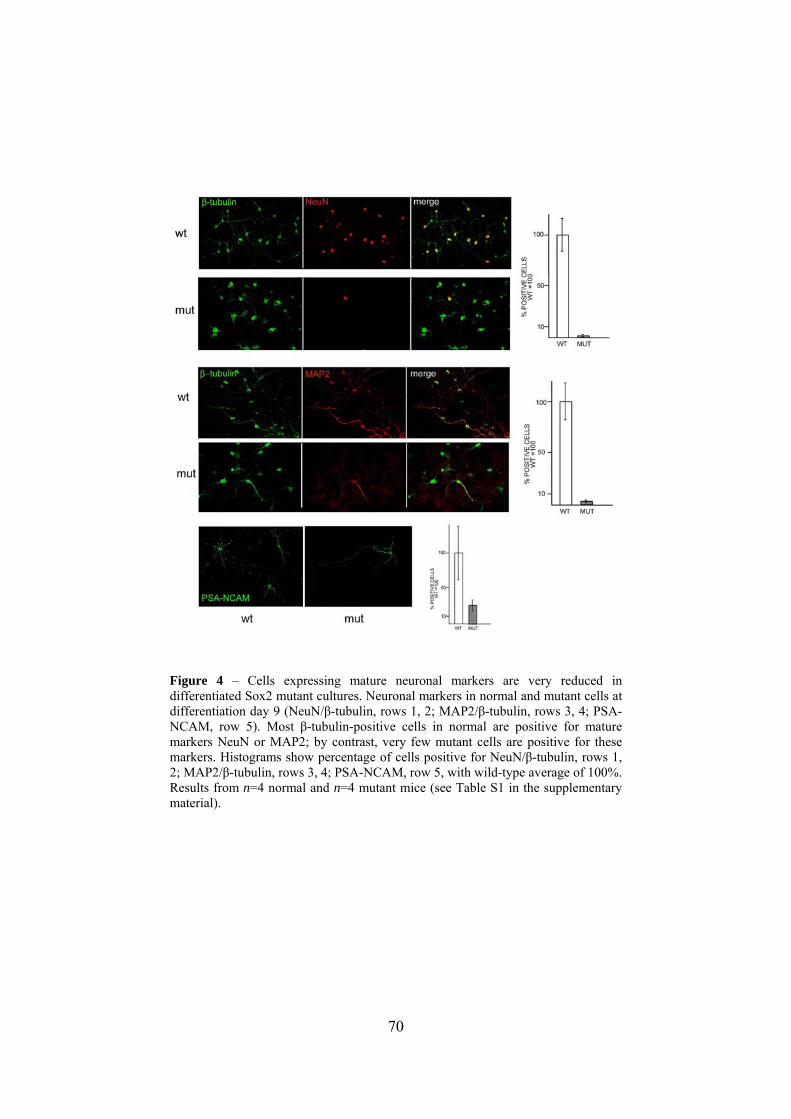
70
Figure 4 – Cells expressing mature neuronal markers are very reduced in differentiated Sox2 mutant cultures. Neuronal markers in normal and mutant cells at differentiation day 9 (NeuN/β-tubulin, rows 1, 2; MAP2/β-tubulin, rows 3, 4; PSA-NCAM, row 5). Most β-tubulin-positive cells in normal are positive for mature markers NeuN or MAP2; by contrast, very few mutant cells are positive for these markers. Histograms show percentage of cells positive for NeuN/β-tubulin, rows 1, 2; MAP2/β-tubulin, rows 3, 4; PSA-NCAM, row 5, with wild-type average of 100%. Results from n=4 normal and n=4 mutant mice (see Table S1 in the supplementary material).

71
Figure 5 – Cells expressing GABAergic markers are very reduced in differentiated Sox2 mutant cultures. Double-immunofluorescence with general neuronal markers (β-tubulin, rows 1, 2; MAP2, rows 3, 6; red), GABA (rows 1-4) and calretinin (5-6), in normal and mutant day 9-differentiated cultures. Histograms: percentage of positive cells, with wild-type average of 100%. Most β-tubulin-positive cells in normal (top) are GABA positive. In mutant (second row), two immature-looking β-tubulin-positive cells are very weakly GABA positive (or negative) (arrows), in contrast to the adjacent well-arborized GABA-positive cell. In normal cultures, most GABA- and virtually all calretinin-positive cells (rows 3, 5) express the mature

72
neuronal marker MAP2; these cells are extremely reduced in mutant cultures (rows 4, 6 and histogram). Results from n=4 normal and n=4 mutant mice (see Table S1 in the supplementary material).

73
Figure 6 Co-expression of neuronal and glial markers in individual cells in Sox2 mutant cultures. Double-immunofluorescence (β-tubulin and GFAP) of normal (wt) and mutant (mut) day 9-differentiated cells. Typical wild-type neurons (β-tubulin positive) show extensive arborization, are closely associated with glia (which are GFAP positive), and are GFAP negative (top row). Rare cells with a very undifferentiated morphology are weakly positive for both markers (top, arrowhead). In mutant, various arborized cells are positive for both β-tubulin and GFAP (second row, arrowhead; third row, two arborized cells). Well-developed astrocytes are GFAP positive, but β-tubulin negative (arrows, rows 2, 4). In mutant, some intensely β-tubulin stained cells with neuronal morphology are also present (fourth row, arrowhead); these cells are GFAP-negative, as in wild type.
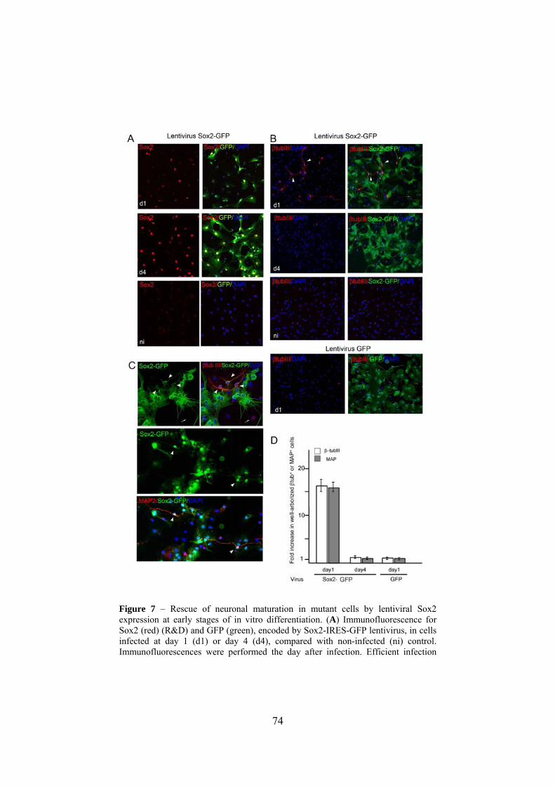
74
Figure 7 – Rescue of neuronal maturation in mutant cells by lentiviral Sox2 expression at early stages of in vitro differentiation. (A) Immunofluorescence for Sox2 (red) (R&D) and GFP (green), encoded by Sox2-IRES-GFP lentivirus, in cells infected at day 1 (d1) or day 4 (d4), compared with non-infected (ni) control. Immunofluorescences were performed the day after infection. Efficient infection

75
(high proportion of GFP-positive cells) is coupled to clear Sox2 overexpression, which is observed at variable levels in transduced cells. (B) β-tubulin- and GFP immunofluorescence, at differentiation day 9, of mutant cells transduced with Sox2-GFP lentivirus at day 1 (d1), or day 4 (d4), compared with non-infected (ni) control, or the control infected with GFP-only transducing virus. Abundant well-arborized β-tubulin-positive cells (arrowheads indicate two of them) are observed in cultures transduced at day 1 with the Sox2-expressing virus, but not in cells transduced at day 4, or in controls. (C) GFP (green) and β-tubulin (red, top) or MAP2 (red, bottom) immunofluorescence shows that well-arborized neuronal cells (arrowheads) are always double-positive for the neuronal marker and for GFP, indicating that they derive from a Sox2-transduced cell. By contrast, some poorly developed neuronal cells (arrow) are not green, thus presumably originating from non-transduced cells. (D) Fold-increase in numbers of MAP2-positive and well-arborized β-tubulin-positive cells in mutant cells infected with Sox2-lentivirus at differentiation day 1, when compared with infection at day 4, or with control virus (day 1) expressing GFP but not Sox2. Values represent fold increase in numbers of MAP2-positive or well-arborized β-tubulin-positive cells (arrowheads in B,C for examples) relative to non-infected control. In day 1 transduced cells, numbers of well-arborized β-tubulin-positive and of MAP2-positive cells were 3.7% and 4.3%, respectively. In a parallel experiment using wild-type control cells mock-treated in the same way with a non-Sox2-expressing virus, the corresponding values were 5.7 and 6.2%. Data from two experiments in duplicate.

76
Figure 8 – Sox2 regulates GFAP expression and directly interacts with upstream regulatory DNA sequences of the GFAP gene in vitro and in neural cells chromatin. (A) Sox2 overexpression in differentiating cells represses endogenous GFAP expression. Double immunofluorescence (confocal microscopy) of day 9-differentiated cells transduced with Sox2-expressing lentivirus (Sox2-GFP; left) or

77
control lentivirus (GFP; right) at day 1 (d1) or 4 (d4), with antibodies against GFP (green, revealing Sox2-IRES-GFP, or GFP for control virus), and the astroglial marker GFAP (red). Sox2-lentivirus-transduced cells show no, or very little, GFAP expression, whereas strongly GFAP-positive cells in the same field are Sox2-GFP-negative (left). By contrast, in cells transduced with control virus, GFP and GFAP colocalize within most cells. (B) Double immunofluorescence for GFAP and astrocytic markers S-100 (left) or connexin 43 (CX43; right) (Nagy and Rash, 2000) in differentiation day 9 cells; not transduced (nt) or day 1 transduced with Sox2-GFP-expressing lentivirus (d1). Virtually all cells positive for GFAP co-express S-100 or CX43 in non-transduced cells. In Sox2-transduced cells, numerous cells can be seen which have low or absent GFAP expression; and are positive for S-100 (left) or for CX43 (right), confirming their astroglial identity. (C) Putative Sox2-binding sites within a 0.6 kb region (0.6GFAP) just upstream to a previously investigated 2.5 kb GFAP promoter/enhancer. The sequence highlights the Sox2 consensus sequences investigated (red). Gfap is the oligonucleotide used in EMSA experiments in E; MutGfap is its mutated version (nucleotide substitutions in green). CDS: coding sequence. (D) Co-transfection experiments in P19 cells. Activity of a luciferase reporter gene driven by the 0.6 GFAP region linked to a TK minimal promoter (0.6GfapTK), or by the TK promoter only (TK), when co-transfected with Sox2 expression vector, or control “empty” vector (as indicated). Asterisk indicates a statistically significant difference (paired t-test, P<0.005). Results are average of n=4 transfections in duplicate. (E) EMSA with probes (indicated below the panels) encompassing the Sox2 consensus binding sites in the 0.6 GFAP region (Gfap), or the same probe mutated as in 8B (MutGfap), or a control probe carrying a Sox2-binding site from an Oct4 gene enhancer (Oct4). Nuclear extracts (P19; SOX2/COS, COS cells transfected with Sox2 expression vector; COS, untransfected COS cells), and competitor oligonucleotides with the molar excesses used for the competition experiments in the right panel, are indicated above the figure. (F) ChIP with anti-SOX2 antibodies of the 0.6 Gfap region in P19 and E12.5 spinal cord cell chromatin, compared with control SRR2 (which is bound by Sox2 in P19, but not in E12.5 spinal cord cell chromatin) (Miyagi et al., 2006) or nestin (bound by Sox2 in P19 and E12.5 spinal cord cell chromatin) (Tanaka et al., 2004; Miyagi et al., 2006) regulatory regions. The anti-Sox2 antibody precipitates both GFAP and SRR2 chromatin in P19 cells, but only GFAP chromatin in spinal cord cells, as expected. Antibodies are indicated above the panels; cell types and amplified DNA regions are indicated below the panels. Arrowheads indicate the positions of PCR bands corresponding to amplified target regions. Low-intensity diffused bands at the bottom are non-reacted primers. Results are representative of three experiments. unrel, unrelated control antibody against SV40 large-T antigen; Input chrom, input chromatin (not immunoprecipitated) - a positive control for the PCR reaction.

78
Figure 9 – Neurons expressing GABAergic markers are reduced in Sox2 mutant neonatal brains. (A,B) GABA (A) and calretinin (B) immunofluorescence of P0 cortical neurons (normal, left; mutant, right). Lower panels are counterstained with DAPI. (C) Percentage of GABA- or calretinin-positive cells in normal or mutant P0 cortical neurons. Results from n=3 normal and n=3 mutant mice.

79
Figure 10 – Abnormal calretinin- and GABA-positive neurons in E17.5 mutant brain. Calretinin (A-H) or GABA (I-N) immunohistochemistry in sections from normal (A-D,I-K) and mutant (E-H,L-N) forebrains. (A,E,I,L) General views of normal and mutant forebrain sections (dorsal region). Lower panels show progressively more enlarged details. (B,F,J,M) Details of the cortical region. The boxed regions in B and F are shown in C,D and G,H, respectively. Arrows in B indicate calretinin-positive neurons that reached the more external cortical layers following migration. Neurons in these positions are much rarer in the corresponding mutant section (F). C shows neurons that reached deep layers of the cortical plate; in the corresponding region of the mutant (G), no cells are seen. (D) Subcortical fiber bundles (along which calretinin-positive cells migrate from ganglionic eminences to cortex at earlier stages); no cells are seen here in the wild type. In the corresponding region of the mutant (H), calretinin-positive cells are still seen along this migratory route. (K,N) Enlarged details of J and M. In mutant (N), general disorganization of the GABA-positive neurons and of their arborizations is seen. V, ventricle; VZ, ventricular zone; CP, cortical plate.

80
Figure 11 – Decreased frequency and arborization of calretinin-positive neurons in adult mutant somatosensory cortex. (A,C) Calretinin immunohistochemistry reveals lower frequency of calretinin-positive neurons in mutant (C) versus wild-type (A) mice. (B,D) Higher magnification shows reduction of dendritic arborizations and of axonal varicosities (the swellings where transmitter-containing vesicles accumulate) in calretinin-positive neurons (asterisks) of mutant (D) versus wild-type (B) brains. Insets in B show, on the left, two vertically oriented varicose processes (arrows) and on the right a highly ramified calretinin-positive neuron (asterisk). Inset in D shows a poorly ramified calretinin-positive neuron (asterisk) with a vertically oriented smooth process (arrow). Original magnifications: A,C 940x ; B,D 2400x; insets 3200x.

81
Figure 12 – Impaired neuronal maturation in adult olfactory bulb of Sox2 mutant mice. (A) Immunofluorescence of BrdU/NeuN-double positive (red and green, yellow in overlay; first row) and BrdU-single-positive (red only; second row) cells in olfactory bulb sections. Histograms: percentage of BrdU/NeuN double-positive cells within the total BrdU-positive population in normal (WT) and mutant (MUT) olfactory bulb, in the entire bulb (TOT) or specifically in the granule layer (GL) and periglomerular layer (PGL) neuronal populations. Results from wild-type (n=4) and mutant mice (n=6). (B) Calretinin-positive cells (green) in olfactory bulb. Histograms: quantitation of calretinin-positive cells in normal (WT) and mutant (MUT) olfactory bulb within the periglomerular layer (four wild type, six mutants). (C) Confocal microscopy of calretinin-positive cells in the olfactory bulb reveals very limited arborization of mutant (mut) cells compared with wild type (wt). This morphology was clearly detected in two out of the four mutant mice analyzed.

82
Supplementary figures

83
84
Supplementary Figure 1 – Evaluation of anti-Sox2 antibodies by immunocytochemistry, immunohistochemistry and western blot analysis of wild-type and Sox2-null neural cells, and of recombinant Sox proteins by western blot. We evaluated the Sox2 specificity of two commercial antibodies (R&D, mouse monoclonal; Chemicon, rabbit polyclonal). Sox2-null neural cells, obtained by in vivo nestin-driven Cre-mediated deletion (R.F. et al., unpublished), were compared with wild-type cells. Both antibodies gave clear nuclear staining in most of the wild-type cells, but failed to show any reactivity with nuclei of Sox2-null cells. (A) Dissociated neurospheres allowed to attach to a slide were probed with the indicated antibodies at the beginning (day 1) or at the end (day 9) of the differentiation protocol described in Fig. 1. With both antibodies, a clear nuclear signal is visible in wild-type, but not in Sox2-null cells. Expression decreases with differentiation, but is still clearly detected in day 9 differentiated cells. A slight cytoplasmic staining can be seen with the rabbit antibody (Chemicon) at both day 1 and day 9, in wild type and null cells, thus likely representing a nonspecific background. Secondary antibodies only (bottom panels) yield no signal. (B) In vivo, neither antibody stains nuclei in brain sections of mutant null newborn mice. Immunohistochemistry with both mouse (left panels) and rabbit (right panels) anti-Sox2 antibodies detects abundant nuclear Sox2 expression in wild-type (wt), but not in Sox2-deleted (null) ventricular zone at P0. Some background staining seen in the null mouse sections does not localize to nuclei. (C) Western blot studies with the R&D antibody, confirming that it does not crossreact with any proteins in undifferentiated neurosphere lysates of Sox2-null cells, even in the presence of a large excess of protein and with long exposures. Proteins from neurosphere cultures of wild-type (+/+), Sox2 heterozygous (+/−) and Sox2-deleted (−/−) mice were probed with anti-Sox2 antibody. Positions of Sox2 and CP2 (ubiquitous nuclear protein, as loading control) are indicated. Left panels: two different exposures of a filter probed with anti-Sox2 and anti-CP2 antibodies. Genotypes are indicated above the lanes. The longer (top) exposure shows failure of the antibody to detect any non-specific signal in the −/− sample; the lower (shorter) exposure allows better comparison of the CP2 signal, demonstrating that equal amounts of extracts were loaded in all lanes. Middle panel: the same filter probed with the Sox2 antibody, prior to re-probing with the CP2 antibody. No signal is seen in the Sox2-null (−/−) extract, even with this long (1 minute) exposure. Asterisks indicate the expected position of the Sox1 (*) and Sox3 (**) transcription factors, which are expressed in the same cells at normal levels (see D). Right panels: progressive dilutions (1/10, 1/20) of the amount of extract (1 corresponds to the amount loaded in the +/+ lane of the upper left and middle panels) still yield a clearly visible Sox2 signal, even when the same filters exposed for only 6 seconds (lower panel), instead of 1 minute (top panel). Thus, a 10-fold overexposure of an amount of extract 20-fold in excess to that required for Sox2 detection, still does not yield any non-specific signal. (D) RT-PCR analysis of expression of SoxB family members Sox1 and Sox3 (co-expressed with Sox2 in neural precursors), in wild-type and Sox2-null neurosphere cultures. Samples shown were taken from the PCR reactions at 25, 30, 35 and 40 cycles for both wild-type and null. Expression levels of Sox1 and Sox3 are similar between wild-type and Sox2-null cells. −, control reaction with reverse transcriptase-negative null control (40 cycles); M, marker. (E,F) Lack of cross-reaction of the anti-Sox2 antibodies with recombinant Sox1, Sox3 and Sox6. NIH3T3 (E) or HeLa (F) cells were transfected with CMV promoter-driven expression vectors (pCDNA3) for Sox2, or

85
Sox1, Sox3 and Sox6. Cell extracts were probed with R&D anti-Sox2 antibody. The Sox1, Sox3 (E) and Sox6 (F) positions are indicated beside the panels. Although Sox2 was easily detected, no reactivity was obtained with extracts from cells transfected with the other Sox expression vectors. In conclusion, anti-Sox2 antibodies do not significantly crossreact with protein present in neural cells at various differentiation stages. The staining experiments reported in the paper were always performed with both antibodies (as indicated in figures), with essentially identical results. When quantitation of the staining was required, the R&D antibody was used.

86
Supplementary Figure 2 – Evaluation of Sox2 immunofluorescence at the single-cell level. To evaluate Sox2 immunofluorescence at the single-cell level, digital images of Sox2 immunofluorescence-labeled nuclei were acquired, and individual nuclei were delimited and evaluated (on the monochromatic image taken on the appropriate fluorescence channel) with the image-processing algorithm of the Region Of Interest (ROI) program provided with the Leica TCS2 Confocal
87
Microscope (Leica Microsystems), or the ImageJ.exe processing and analysis program (http://rsb.info.nih.gov/ij/), and expressed in arbitrary units as the sum of the background-subtracted pixel values within each ROI (nucleus). Background levels were established measuring nuclei of Sox2-null cells (see Fig. S1) or of cells treated with secondary antibody only (B), giving comparable values. The ratios between positive signals and internal background (measured on five different positions within each field) were plotted and statistical significances were assessed by nonparametric tests (heteroskedastic ANOVA, T-test; *P<0,05). (A) Examples of Sox2 immunofluorescence of normal and mutant cells at day 1 (left) or day 9 (right) of in vitro differentiation. In day 1 cells, a Sox2-bright cell population is seen in the normal, which is very reduced in the mutant. At day 9, fluorescence levels are very similar between wild type and mutant. (B) Evaluation of Sox2 immunofluorescence (R&D antibody) at the single-cell level in wild type (WT) and mutant (MUT) cells, on the overall population at days 1, 5 and 9 of in vitro differentiation (as indicated). Each dot represents the Sox2 fluorescence level of a single cell nucleus; each vertical dot series represents the values within an individual microscope field evaluated (see Materials and methods below). “II Ab” indicates nuclear fluorescence values obtained with the secondary antibody only; the “0” level was set just above the highest values obtained with this negative control, as shown in B (the same applies to C and D). Red dots identify the β-tubulin-positive cells within the samples shown (see also C). At least 500 nuclei per differentiation day per genotype were quantitated, within at least six different fields. The asterisk indicates a significant difference at day 1, but not at days 5 and 9, between wild-type and mutant Sox2 fluorescence distributions (one-way ANOVA, P<0.03; two-tailed t-test, P<0.001). (C,D) Evaluation of Sox2 immunofluorescence within the β-tubulin-positive cell population at day 9 of in vitro differentiation (C) or in in vivo differentiated P0 cortical cells (D), in normal (WT) and mutant (MUT). Fluorescence levels are indicated as explained in B. Examples of Sox2/β-tubulin-double-positive cells in differentiation day 9 cells and P0 cortical neurons are shown in Fig. 2A, Fig. S5B, respectively. In the in vitro-differentiated β-tubulin positive cells (C), the Sox2 level was slightly, but significantly, decreased in mutants (two-tailed t-test, P<0.01). This is at variance with the analysis reported in Fig. S2B for the overall population, where most cells are glia. A comparison between normal and mutant MAP2-positive cells for Sox2 expression was not performed, owing to the rarity of MAP2-positive cells in the mutant (see text). In D, the data document a slight (statistically non-significant) difference between the wild- type and the mutant (two-tailed t-test, P<0.34). At least 200 nuclei from β-tubulin-positive cells were analyzed in C and D, for n=2 wild type and n=2 mutants.

88
Supplementary Figure 3 – Expression of astrocytic markers S-100 and connexin 43 (CX43) (Nagy and Rash, 2000) in GFAP-positive in vitro differentiated

89
astrocytes (untransduced, or day 1 transduced with Sox2-expressing lentivirus). (A) Double immunofluorescence for GFAP and S-100 (top panels) or CX43 (bottom panels) in differentiation day 9 cells, untransduced (left) or transduced with Sox2-GFP-expressing lentivirus (right). Virtually all cells positive for GFAP co-express S-100 (top panels) or CX43 (bottom panels) in untransduced cells. In Sox2-transduced cells, numerous cells can be seen which have low or absent GFAP expression (see Fig. 9) and are positive for S-100 (top) or for CX43 (bottom), confirming their astroglial identity (arrows indicate examples). (B) Double immunofluorescence for GFP (marking cells transduced with the Sox2-GFP-expressing lentivirus) and for S-100 (top) or CX43 (bottom). The vast majority of Sox2-transduced cells (where downregulation of endogenous GFAP is observed, see Fig. 8) express S-100 (top panels) and CX43 (bottom panels), consistent with an astrocytic identity. S-100 may be somewhat reduced in occasional Sox2-transduced cells. No fluorescence signal is observed in Sox2-GFP virus-transduced cells prior to antibody staining (lower right image, indicating that GFP endogenous green fluorescence is not detected in cells after fixation), nor with secondary antibodies only (not shown). Images are by non-confocal microscopy; see also Fig. 8 for confocal images of GFAP/S-100 and GFAP/CX43 immunofluorescence.

90
Supplementary Figure 4 – The block in neuronal maturation in Sox2 mutant cultures is not associated with apoptosis, nor with persistence of undifferentiated cells characteristics (nestin positivity). (A) Apoptosis between initial β-tubulin expression and MAP2/NeuN activation can be ruled out. In fact, between day 5 and 9, ~15% of the cells show TUNEL positivity (green), both in normal and mutant; however, >98% of β-tubulin-positive cells (red) do not show TUNEL positivity. Shown are differentiation day 7 mutant cells. Furthermore, the total number of cells in mutant cultures at day 9, and the number of β-tubulin-positive cells were comparable between normal and mutant cells (see Table S1 in the supplementary material; data not shown), indicating that the maturation block is not associated with, or dependent on, apoptotic cell death. Numbers of Ki67-positive (dividing) cells were also similar (not shown). (B) Time course of nestin expression. The kinetics of decrease of the number of cells positive to nestin (a marker of the undifferentiated state) is very similar between wild-type and mutant cultures. Note that β-tubulin appeared at day 5 in mutant, as in normal cells (see Fig. 3C). Thus, initial differentiation steps are not significantly delayed in mutant cells.

91

92
Supplementary Figure 5 – Sox2 expression in the lateral ventricle (A), and in regions of neuronal differentiation (within the neonatal cortex, B,C, and in adult olfactory bulb, D), in normal and mutant mice. (A) Left: Sox2 (red) (Chemicon) and RC2 (green, a radial glia marker) (Merkle et al., 2004) immunofluorescence on sections of P0 lateral ventricle (P0 LV) of normal (wt) and mutant (mut) mice (confocal microscopy). Arrowheads: examples of Sox2/RC2 double-positive cells. Right: Sox2 (green) (Chemicon) and GFAP (red) immunofluorescence in adult lateral ventricle (LV) of wild type (wt) and mutant (mut). (B,C) Immunofluorescence of isolated P0 cortical neurons from normal (wt) and mutant (mut) brains with Sox2 (R&D) and β-tubulin (B) or MAP2 (C) antibodies (confocal microscopy). A large proportion of β-tubulin or MAP2-stained neurons are clearly Sox2-positive.Within the MAP2-positive population, the intensity of Sox2 staining inversely correlates with that of differentiated marker, and the most strongly MAP2-labeled cells are completely devoid of Sox2. Arrowheads: examples of Sox2/β-tubulin or Sox2/MAP2 double-positive cells. Sox2/MAP2 double-positive cells are generally weakly positive for both markers. Arrows indicate strongly MAP2-positive cells (generally Sox2-negative). Asterisks indicate strongly Sox2-positive cells (generally MAP2-weakly positive or negative). (D) Immunofluorescence analysis of Sox2 expression in the olfactory bulb. Top: Low-magnification image of an olfactory bulb section (DAPI nuclear staining); white boxes highlight the regions of the rostral migratory stream (RMS) and, more externally, sections of the peripheral layers where terminal neuronal differentiation is completed: the granule layer (GL) and periglomerular layer (PGL). Lower panels show higher magnifications of these regions (as indicated) analyzed in wild-type (wt) and mutant (mut), with the indicated antibodies In the RMS, Sox2 is expressed in numerous cells, many of which are positive for PSA-NCAM (Ferri et al., 2004), a marker of transit-amplifying progenitors (Doetsch, 2003; Lledo et al., 2006). In the differentiated peripheral layers, some weakly Sox2-positive cells are still visible; they are rare in the GL, but more numerous in the PGL, where calretinin-positive neurons differentiate 14-20 days after their birth (Lledo et al., 2006). Here, however, few if any calretinin or NeuN-positive cells show Sox2. In the mutant, the number of Sox2-positive cells is diminished, as expected on the basis of the observations on the SVZ. Arrowheads in GL indicate Sox2-positive NeuN-negative cells. Arrowhead in PGL indicates cell appearing weakly positive for Sox2 and calretinin.

93
Supplementary Table 1: expression of lineage-specific markers in differentiated neural stem cells from Sox2-deficient mice
WT MUT
ß-tubulina
with well-developed neuronal
morphology, extensive
arborization
13,2% ± 1,5%
1,3% ± 0,9%
Poorly developed, limited
arborization, generally less
intenslely stained
<0,5%
18,9% ± 1,9%
NeuNb 11,4% ± 1,9% 0,25% ± 0,12%
MAP2b 7,9% ± 1,4% 0,26% ± 0,1%
PSA-NCAM 3,8% ± 1,5% 1% ± 0,4%
GABAc 8,9% ± 1,9% 0,8% ± 0,4%
CALRETININd 3,1 % ± 0.7% <0,1%
GFAP 60% ± 1,3% 58% ± 2,3%
GALC 3% ± 0,8% 2,5% ± 1%
These data were obtained from differentiation of neural stem cells from adult brain (similar data were obtained with E14.5 embryonic cells, not shown). In one set of experiments ß-tubulin, NeuN, MAP2, PSA-NCAM, GFAP and GAL-C were evaluated in slides from differentiated cultures obtained from n=4 wt and n=4 mutant mice; MAP2 and NeuN were counted in double immunofluorescence labellings with ß-tubulin. GABA and calretinin were evaluated in a separate experiment, in which n=2 wt and n=2 mutants (already assayed for the markers above) were differentiated, and assayed by double labelling with ß-tubulin or MAP2 (similar percentages of ß-tubulin and MAP2-positive cells were obtained in all these experiments). The total number of cells at the end of differentiation was always very similar between wild type and mutant. a: see Fig. 2 for the different appearance of ß-tubulin-positive cells in the mutant; b: NeuN and MAP2-positive cells are also ß-tubulin-positive in double immunofluorescence labellings; c: GABA-bright cells are indicated. GABA-bright cells were nearly always MAP-2 positive in double immunofluorescence labellings in the wild type (see Fig. 4). A dimmer GABA positivity was observed in most ß-tubulin-positive cells in the wild type, though not (or much less) in the mutant (see Fig.4); d: CALRETININ-positive cells were essentially always MAP2-positive in double immunofluorescence labellings; they constituted about 38% of the total MAP2-positive cells.

94
References
Avilion,A.A., Nicolis,S.K., Pevny,L.H., Perez,L., Vivian,N., and Lovell-Badge,R. (2003). Multipotent cell lineages in early mouse development depend on SOX2 function. Genes Dev., 17 , 126-140.
Azuara,V., Perry,P., Sauer,S., Spivakov,M., Jorgensen,H.F., John,R.M., Gouti,M., Casanova,M., Warnes,G., Merkenschlager,M., and Fisher,A.G. (2006). Chromatin signatures of pluripotent cell lines. Nat.Cell Biol., 8, 532-538.
Bani-Yaghoub,M., Tremblay,R.G., Lei,J.X., Zhang,D., Zurakowski,B., Sandhu,J.K., Smith,B., Ribecco-Lutkiewicz,M., Kennedy,J., Walker,P.R., and Sikorska,M. (2006). Role of Sox2 in the development of the mouse neocortex. Dev.Biol., 295, 52-66.
Bernstein,B.E., Mikkelsen,T.S., Xie,X., Kamal,M., Huebert,D.J., Cuff,J., Fry,B., Meissner,A., Wernig,M., Plath,K., Jaenisch,R., Wagschal,A., Feil,R., Schreiber,S.L., and Lander,E.S. (2006). A bivalent chromatin structure marks key developmental genes in embryonic stem cells. Cell, 125, 315-326.
Boyer,L.A., Lee,T.I., Cole,M.F., Johnstone,S.E., Levine,S.S., Zucker,J.P., Guenther,M.G., Kumar,R.M., Murray,H.L., Jenner,R.G., Gifford,D.K., Melton,D.A., Jaenisch,R., and Young,R.A. (2005). Core transcriptional regulatory circuitry in human embryonic stem cells. Cell, 122, 947-956.
Boyer,L.A., Plath,K., Zeitlinger,J., Brambrink,T., Medeiros,L.A., Lee,T.I., Levine,S.S., Wernig,M., Tajonar,A., Ray,M.K., Bell,G.W., Otte,A.P., Vidal,M., Gifford,D.K., Young,R.A., and Jaenisch,R. (2006a). Polycomb complexes repress developmental regulators in murine embryonic stem cells. Nature, 441, 349-353.
Boyer,L.A., Mathur,D., and Jaenisch,R. (2006b). Molecular control of pluripotency. Curr.Opin.Genet.Dev., 16, 455-462.
Brunelli,S., F.Relaix, S.Baesso, M.Buckingham, and G.Cossu. (2007). Beta catenin-independent activation of MyoD in presomitic

95
mesoderm requires PKC and depends on Pax3 transcriptional activity. Dev.Biol. 304, 604-614.
Bylund,M., E.Andersson, B.G.Novitch, and J.Muhr. (2003). Vertebrate neurogenesis is counteracted by Sox1-3 activity. Nat.Neurosci. 6, 1162-1168.
Catena,R., Tiveron,C., Ronchi,A., Porta,S., Ferri,A., Tatangelo,L., Cavallaro,M., Favaro,R., Ottolenghi,S., Reinbold,R., Schöler,H. and Nicolis, S.K. (2004). Conserved POU binding DNA sites in the Sox2 upstream enhancer regulate gene expression in embryonic and neural stem cells. J.Biol.Chem. 279, 41856-41857.
Chew,J.L., Y.H.Loh, W.Zhang, X.Chen, W.L.Tam, L.S.Yeap, P.Li, Y.S.Ang, B.Lim, P.Robson, and H.H.Ng. (2005). Reciprocal transcriptional regulation of Pou5f1 and Sox2 via the Oct4/Sox2 complex in embryonic stem cells. Mol.Cell Biol. 25, 6031-6046.
Cobos,I., Calcagnotto,M.E., Vilaythong,A.J., Thwin,M.T., Noebels,J.L., Baraban,S.C., and Rubenstein,J.L. (2005). Mice lacking Dlx1 show subtype-specific loss of interneurons, reduced inhibition and epilepsy. Nat.Neurosci., 8, 1059-1068.
Conti,L., Pollard,S., Gorba,T., Reitano,E., Toselli,M., Biella,G., Sun,Y., Sanzone,S., Ying,Q.-L., Cattaneo,E. and Smith,A. (2005). Niche-independent symmetrical self-renewal of a mammalian tissue stem cell. PloS Biol., 3, e283
Doetsch,F., L.Petreanu, I.Caille, J.M.Garcia-Verdugo, and A.Alvarez-Buylla. (2002). EGF converts transit-amplifying neurogenic precursors in the adult brain into multipotent stem cells. Neuron 36, 1021-1034.
Doetsch,F. 2003. The glial identity of neural stem cells. Nat.Neurosci. 6, 1127-1134.
Enver,T. and Greaves,M. (1998). Loops, lineage, and leukemia. Cell, 94, 9-12.

96
Fantes,J., Ragge,N.K., Lynch,S.A., McGill,N.I., Collin,J.R., Howard-Peebles,P.N., Hayward,C., Vivian,A.J., Williamson,K., van,H., V, and FitzPatrick,D.R. (2003). Mutations in SOX2 cause anophthalmia. Nat.Genet., 33, 461-463.
Ferrer,I., Soriano,E., del Rio,J.A., Alcantara,S., and Auladell,C. (1992). Cell death and removal in the cerebral cortex during development. Prog.Neurobiol., 39, 1-43.
Ferri,A.L., Cavallaro,M., Braida,D., Di Cristofano,A., Canta,A., Vezzani,A., Ottolenghi,S., Pandolfi,P.P., Sala,M., DeBiasi,S., and Nicolis,S.K. (2004). Sox2 deficiency causes neurodegeneration and impaired neurogenesis in the adult mouse brain. Development, 131, 3805-3819.
Galli,R., Binda,E., Orfanelli,U., Cipelletti,B., Gritti,A., De Vitis,S., Fiocco,R., Foroni,C., Dimeco,F., and Vescovi,A. (2004). Isolation and characterization of tumorigenic, stem-like neural precursors from human glioblastoma. Cancer Res., 64, 7011-7021.
Graham,V., J.Khudyakov, P.Ellis, and L.Pevny. (2003). SOX2 functions to maintain neural progenitor identity. Neuron 39, 749-765.
Gritti,A., Parati,E.A., Cova,L., Frolichsthal,P., Galli,R., Wanke,E., Faravelli,L., Morassutti,D.J., Roisen,F., Nickel,D.D., and Vescovi,A.L. (1996). Multipotential stem cells from the adult mouse brain proliferate and self-renew in response to basic fibroblast growth factor. J.Neurosci., 16, 1091-1100.
Gritti,A., Galli,R. and Vescovi,A.L. (2001). Cultures of stem cells of the Central Nervous system. Protocols for Neural Stem Cell culture, 3rd ed., Ed. S. Fedoroff and A. Richardson, Humana Press, Inc., Totowa, NJ, USA.
Gubbay,J., Collignon,J., Koopman,P., Capel,B., Economou,A., Munsterberg,A., Vivian,N., Goodfellow,P., and Lovell-Badge,R. (1990). A gene mapping to the sex-determining region of the mouse Y chromosome is a member of a novel family of embryonically expressed genes. Nature, 346, 245-250.

97
Guillemot,F. (2005). Cellular and molecular control of neurogenesis in the mammalian telencephalon. Curr.Opin.Cell Biol., 17, 639-647.
Hemmati,H.D., Nakano,I., Lazareff,J.A., Masterman-Smith,M., Geschwind,D.H., Bronner-Fraser,M., and Kornblum,H.I. (2003). Cancerous stem cells can arise from pediatric brain tumors. Proc.Natl.Acad.Sci.U.S.A, 100, 15178-15183
Hu,M., Krause,D., Greaves,M., Sharkis,S., Dexter,M., Heyworth,C., and Enver,T. (1997). Multilineage gene expression precedes commitment in the hemopoietic system. Genes Dev., 11, 774-785.
Kamachi,Y., Uchikawa,M., and Kondoh,H. (2000). Pairing SOX off: with partners in the regulation of embryonic development. Trends Genet., 16, 182-187.
Kelberman,D., Rizzoti,K., Avilion,A., Bitner-Glindzicz,M., Cianfarani,S., Collins,J., Chong,W.K., Kirk,J.M., Achermann,J.C., Ross,R., Carmignac,D., Lovell-Badge,R., Robinson,I.C., and Dattani,M.T. (2006). Mutations within Sox2/SOX2 are associated with abnormalities in the hypothalamo-pituitary-gonadal axis in mice and humans. J.Clin.Invest, 116, 2442-2455.
Kondo,T. and M.Raff. (2004). Chromatin remodeling and histone modification in the conversion of oligodendrocyte precursors to neural stem cells. Genes Dev. 18, 2963-2972.
Kuzmanovic,M., V.J.Dudley, and V.P.Sarthy. (2003). GFAP promoter drives Muller cell-specific expression in transgenic mice. Invest Ophthalmol.Vis.Sci, 44, 3606-3613.
Laslo,P., Spooner,C.J., Warmflash,A., Lancki,D.W., Lee,H.J., Sciammas,R., Gantner,B.N., Dinner,A.R., and Singh,H. (2006). Multilineage transcriptional priming and determination of alternate hematopoietic cell fates. Cell, 126, 755-766.
Lee,J., Kotliarova,S., Kotliarov,Y., Li,A., Su,Q., Donin,N.M., Pastorino,S., Purow,B.W., Christopher,N., Zhang,W., Park,J.K.,
98
and Fine,H.A. (2006). Tumor stem cells derived from glioblastomas cultured in bFGF and EGF more closely mirror the phenotype and genotype of primary tumors than do serum-cultured cell lines. Cancer Cell, 9, 391-403.
Lee,T.I., Jenner,R.G., Boyer,L.A., Guenther,M.G., Levine,S.S., Kumar,R.M., Chevalier,B., Johnstone,S.E., Cole,M.F., Isono,K., Koseki,H., Fuchikami,T., Abe,K., Murray,H.L., Zucker,J.P., Yuan,B., Bell,G.W., Herbolsheimer,E., Hannett,N.M., Sun,K., Odom,D.T., Otte,A.P., Volkert,T.L., Bartel,D.P., Melton,D.A., Gifford,D.K., Jaenisch,R., and Young,R.A. (2006). Control of developmental regulators by Polycomb in human embryonic stem cells. Cell, 125, 301-313.
Levitt,P., K.L.Eagleson, and E.M.Powell. (2004). Regulation of neocortical interneuron development and the implications for neurodevelopmental disorders. Trends Neurosci. 27, 400-406.
Li,D., Marks,J.D., Schumacker,P.T., Young,R.M., and Brorson,J.R. (2005). Physiological hypoxia promotes survival of cultured cortical neurons. Eur.J.Neurosci., 22, 1319-1326.
Lim,D.A., Tramontin,A.D., Trevejo,J.M., Herrera,D.G., Garcia-Verdugo,J.M., and Alvarez-Buylla,A. (2000). Noggin antagonizes BMP signaling to create a niche for adult neurogenesis. Neuron, 28, 713-726.
Lledo,P.M., M.Alonso, and M.S.Grubb. (2006). Adult neurogenesis and functional plasticity in neuronal circuits. Nat.Rev.Neurosci. 7, 179-193.
Markram,H., M.Toledo-Rodriguez, Y.Wang, A.Gupta, G.Silberberg, and C.Wu. (2004). Interneurons of the neocortical inhibitory system. Nat.Rev.Neurosci. 5, 793-807.
Merkle,F.T., A.D.Tramontin, J.M.Garcia-Verdugo, and A.Alvarez-Buylla. (2004). Radial glia give rise to adult neural stem cells in the subventricular zone. Proc.Natl.Acad.Sci.U.S.A 101, 17528-17532.

99
Mikkola,I., Heavey,B., Horcher,M., and Busslinger,M. (2002). Reversion of B cell commitment upon loss of Pax5 expression. Science, 297, 110-113.
Miyagi,S., M.Nishimoto, T.Saito, M.Ninomiya, K.Sawamoto, H.Okano, M.Muramatsu, H.Oguro, A.Iwama, and A.Okuda. (2006). The Sox2 regulatory region 2 functions as a neural stem cell-specific enhancer in the telencephalon. J.Biol.Chem, 281, 13374-13381.
Morita,N., K.Nakahira, H.Baba, H.Akita, T.Kumada, M.Ogawa, K.Nakajima, M.Kawata, K.Mikoshiba, and K.Ikenaka. (1997). Astrocytic lineage analysis by detection of GFAP promoter activity in vitro. Dev.Neurosci, 19, 210-218.
Muotri,A.R. and Gage,F.H. (2006). Generation of neuronal variability and complexity. Nature, 441, 1087-1093.
Nagy,J.I. and Rash, J.E. (2000). Connexins and gap junctions of astrocytes and oligodendrocytes in the CNS. Brain Res Rev. 32, 29-44.
Nicolis, S.K. (2007). Cancer stem cells and “stemness” genes in neuro-oncology. Neurobiology of disease, 25, 217-229.
Niwa,H., Miyazaki,J., and Smith,A. (2000). Quantitative expression of Oct 3/4 defines differentiation, dedifferentiation or self-renewal of ES cells. Nat. Genetics 24, 372-376.
Noebels,J.L. (2003). The biology of epilepsy genes. Annu.Rev.Neurosci., 26, 599-625.
Nutt,S.L., Heavey,B., Rolink,A.G., and Busslinger,M. (1999). Commitment to the B-lymphoid lineage depends on the transcription factor Pax5. Nature, 401, 556-562.
Oppenheim,R.W. (1991). Cell death during development of the nervous system. Annu.Rev.Neurosci., 14, 453-501.
Pevny,L. and Placzek,M. (2005). SOX genes and neural progenitor identity. Curr.Opin.Neurobiol., 15, 7-13.

100
Pomeroy,S.L., Tamayo,P., Gaasenbeek,M., Sturla,L.M., Angelo,M., McLaughlin,M.E., Kim,J.Y., Goumnerova,L.C., Black,P.M., Lau,C., Allen,J.C., Zagzag,D., Olson,J.M., Curran,T., Wetmore,C., Biegel,J.A., Poggio,T., Mukherjee,S., Rifkin,R., Califano,A., Stolovitzky,G., Louis,D.N., Mesirov,J.P., Lander,E.S., and Golub,T.R. (2002). Prediction of central nervous system embryonal tumour outcome based on gene expression. Nature, 415, 436-442.
Ragge,N.K., Lorenz,B., Schneider,A., Bushby,K., de Sanctis,L., de Sanctis,U., Salt,A., Collin,J.R., Vivian,A.J., Free,S.L., Thompson,P., Williamson,K.A., Sisodiya,S.M., van,H., V, and FitzPatrick,D.R. (2005). SOX2 anophthalmia syndrome. Am.J.Med.Genet.A, 135, 1-7.
Sisodiya,S.M., Ragge,N.K., Cavalleri,G.L., Hever,A., Lorenz,B., Schneider,A., Williamson,K.A., Stevens,J.M., Free,S.L., Thompson,P.J., van,H., V, and FitzPatrick,D.R. (2006). Role of SOX2 mutations in human hippocampal malformations and epilepsy. Epilepsia, 47, 534-542.
Sur,M. and Rubenstein,J.L. (2005). Patterning and plasticity of the cerebral cortex. Science, 310, 805-810.
Szutorisz,H. and Dillon,N. (2005). The epigenetic basis for embryonic stem cell pluripotency. Bioessays, 27, 1286-1293.
Tanaka,S., Y.Kamachi, A.Tanouchi, H.Hamada, N.Jing, and H.Kondoh. (2004). Interplay of SOX and POU factors in regulation of the Nestin gene in neural primordial cells. Mol.Cell Biol. 24, 8834-8846.
Taranova,O.V., Magness,S.T., Fagan,B.M., Wu,Y., Surzenko,N., Hutton,S.R., and Pevny,L.H. (2006). SOX2 is a dose-dependent regulator of retinal neural progenitor competence. Genes Dev., 20, 1187-1202.
Wagenaar,D.A., Madhavan,R., Pine,J., and Potter,S.M. (2005). Controlling bursting in cortical cultures with closed-loop multi-electrode stimulation. J.Neurosci., 25, 680-688.

101
Wegner,M. and Stolt,C.C. (2005). From stem cells to neurons and glia: a Soxist's view of neural development. Trends Neurosci., 28, 583-588.
Weinmann,A.S. and P.J.Farnham. (2002). Identification of unknown target genes of human transcription factors using chromatin immunoprecipitation. Methods, 26, 37-47.
Wells,J. and P.J.Farnham. (2002). Characterizing transcription factor binding sites using formaldehyde crosslinking and immunoprecipitation. Methods, 26, 48-56.
Wilson,M. and Koopman,P. (2002). Matching SOX: partner proteins and co-factors of the SOX family of transcriptional regulators. Curr.Opin.Genet.Dev., 12, 441-446.
Wonders,C.P. and S.A.Anderson. (2006). The origin and specification of cortical interneurons. Nat.Rev.Neurosci. 7, 687-696.
Zappone,M.V., Galli,R., Catena,R., Meani,N., De Biasi,S., Mattei,E., Tiveron,C., Vescovi,A.L., Lovell-Badge,R., Ottolenghi,S., and Nicolis,S.K. (2000). Sox2 regulatory sequences direct expression of a (beta)-geo transgene to telencephalic neural stem cells and precursors of the mouse embryo, revealing regionalization of gene expression in CNS stem cells. Development, 127, 2367-2382.

102

103
CHAPTER 3
EMX2 IS A DOSE-DEPENDENT NEGATIVE
REGULATOR OF SOX2 TELENCEPHALIC
ENHANCERS
J. Mariani, C. Lancini, G.Vaccari, R. Favaro, A. Ferri, D. Tonoli, E.
Latorre, R. Caccia, A. Ronchi, S. Ottolenghi, S. Miyagi, G. Corte, A.
Okuda, V. Zappavigna and S.K. Nicolis
Stem Cell submitted

104

105
Emx2 is a dose-dependent negative regulator of
Sox2 telencephalic enhancers
J. Mariani1, C. Lancini1, G.Vaccari2, R. Favaro1, A. Ferri1, D. Tonoli1, E. Latorre1,
R. Caccia1, A. Ronchi1, S. Ottolenghi1, S. Miyagi3, G. Corte4, A. Okuda3, V.
Zappavigna2 and S.K. Nicolis1
1 Department of Biotechnology and Biosciences, University of Milano-Bicocca,
Piazza della Scienza 2, 20126 Milano, Italy
2 Department of Animal Biology, University of Modena and Reggio Emilia, Via G.
Campi 213/d, Modena 41100, Italy
3 Division of Developmental Biology, Research Center for Genomic Medicine,
Saitama Medical School, Saitama 350-1241, Japan
4 Department of Biology, Biology and Genetics, University of Genova; IST –
National Institute for Cancer Research, Genova, Italy
Authors contribution:
J. Mariani: Collection and assembly of data, data analysis and interpretation
C. Lancini: Collection and assembly of data, data analysis and interpretation
G.Vaccari: Collection and assembly of data, data analysis and interpretation
R. Favaro: Collection and assembly of data, data analysis and interpretation
A. Ferri: Collection and assembly of data, data analysis and interpretation
D. Tonoli: Collection and assembly of data, data analysis and interpretation
E. Latorre: Collection and assembly of data, data analysis and interpretation
R. Caccia: Collection and assembly of data, data analysis and interpretation
A. Ronchi: Collection and assembly of data, data analysis and interpretation
S. Ottolenghi: Conception and design, manuscript writing, final approval of
manuscript
S. Miyagi: Transgenic mouse lines, final approval of manuscript
G. Corte: Collection and assembly of data, data analysis and interpretation, Emx2
antibodies, final approval of manuscript

106
A. Okuda: Conception and design, transgenic mouse lines, final approval of
manuscript
V. Zappavigna: Conception and design, collection and assembly of data, data
analysis and interpretation, final approval of manuscript
S.K. Nicolis: Conception and design, transgenic and Sox2-mutant mouse lines,
manuscript writing, financial support, final approval of manuscript
Author for correspondence: Silvia K. Nicolis
Department of Biological Sciences and Biotechnology
University of Milano-Bicocca
Piazza della Scienza, 2, 20126 Milano, Italy
phone: +39 02 6448 3339 (office), 3315 (lab)
FAX: +39 02 6448 3565
e-mail: [email protected]
This work was supported by Telethon (GGP05122), EEC (STEMBRIDGE),
Fondazione Cariplo, Associazione Italiana Ricerca sul Cancro (AIRC), Fondazione
Banca del Monte di Lombardia, MIUR (Cofin) and FAR 2004-7 grants to S.K.N.,
and MIUR-PRIN to G.C.
Keywords: Neural stem cells, gene regulation, transcription factors, brain
development, human inherited neurological disease
Abstract
The transcription factor Sox2 is essential for neural stem cells (NSC)
maintenance in the hippocampus and in vitro. The transcription factor
Emx2 is also critical for proper hippocampal development, and its loss
causes an unbalance between NSC self renewal and commitment to
differentiation in vitro. In a search for “modifier” genes affecting the
Sox2 deficiency phenotype in mouse, we observed that loss of a single

107
Emx2 allele substantially increased the telencephalic LacZ transgenic
expression driven by the 5’ or 3’ enhancer of Sox2. In vitro
electrophoresis mobility shift assays, protein to protein interaction and
transfection studies indicated that Emx2 represses 5’ and 3’ Sox2
enhancer activities. Emx2 bound to overlapping Emx2/POU binding
sites, preventing binding of the POU transcriptional activator Brn2 to
its target sequence. In addition, Emx2 directly interacted with Brn2
without binding to DNA, sequestering it. Loss of a single Emx2 allele
increased Sox2 levels in the medial telencephalic wall, including the
hippocampal primordium.
In hypomorphic Sox2 mutants, retaining a single copy of a “weak”
Sox2 allele, loss of a single Emx2 allele resulted in a substantial
rescue of hippocampal radial glia stem cells and of neurogenesis,
indicating that Emx2 functionally interacts with Sox2 at the stem cell
level. These data show that Emx2 negatively modulates Sox2
expression, and may thus control important aspects of NSC function
in development.
Introduction
The transcription factor Sox2 is essential in pluripotent stem cells of
the blastocyst inner cell mass [1]. Sox-2 is also highly expressed in
neural stem cells (NSC) and in their early progeny, and repressed
upon differentiation [2-6]. The decreased expression of Sox2 in a
mouse hypomorphic Sox2 mutant causes important brain and
neurologic defects [5, 7], which mimic significant aspects of the
pathology of Sox2-deficient patients [8, 9]. In this hypomorphic
mutant, we combined the deletion of one Sox2 allele (Sox2β-geo knock-
108
in) with the deletion, on the other allele, of an upstream enhancer of
Sox2 (Sox2∆Enh), important for its expression in telencephalic NSC [2,
5, 10]. In the hypomorphic mutant, Sox2 expression is 25-30% as that
of the wild type; this mutant shows hippocampal stem cells loss,
corpus callosum interruption, parenchymal loss in striatum and
thalamus, decreased numbers of GABAergic neurons and neurological
defects, including epilepsy [5, 7]. Recently [11], we showed that Sox2
embryonic deletion leads to complete perinatal loss of hippocampal
stem cells. NSC from the forebrain of such mutants become rapidly
exhausted in in vitro neurosphere culture.
The Emx2 transcription factor is expressed in the developing dorsal
telencephalon, including prospective hippocampus and cerebral
cortex, from early embryogenesis [12, 13]. Its expression is
maintained postnatally in adult brain neurogenic regions, the
subventricular zone (SVZ) and hippocampus dentate gyrus (DG)[14,
15].
Emx2 inactivation in mouse causes delayed hippocampal
development, with reduced cerebral cortex and abnormal specification
of cortical areas at birth [reviewed in 13,16-18]. In vitro, mutant
Emx2-/- NSC show increased proliferation in long term neurosphere
cultures [15].
Following our description of brain abnormalities in hypomorphic
Sox2 mutants, we wished to investigate possible effects of “modifier
genes” on the Sox2 hypomorphic phenotype.
A common aspect of the defects in Sox2 and Emx2 mutants is the
abnormal hippocampal development [5, 11, 13, 16]. Moreover, NSC

109
from both Sox2-/- and Emx2-/- mutants exhibit important (opposite)
abnormalities in in vitro culture [11, 15]. Therefore, we looked for
genetic interactions between Sox2 and Emx2 in double mutants in
which the Sox2 hypomorphic genotype (Sox2β-geo/∆Enh)[5] was
combined with loss of a single Emx2 allele. This significantly
ameliorated the brain phenotype of Sox2 hypomorphic mice (Suppl.
Fig.1) This suggested that Emx2 may play antagonistic roles to Sox2,
possibly by negatively modulating its activity.
We report that Emx2 is a direct transcriptional repressor of Sox2.
Loss of a single Emx2 allele substantially rescues the number of
hippocampal NSC in the dentate gyrus of hypomorphic Sox2 mutants.
Thus, Emx2 functionally interacts with Sox2 at the stem cell level.
Results
Emx2 represses transgenic and knock-in Sox2-LacZ reporters
We crossed Sox2β-geo/+, Emx2+/- double heterozygotes with
homozygous Sox2 knock-down (Sox2∆Enh/∆Enh) mice, obtaining double
mutants in which the Sox2 hypomorphic genotype, Sox2β-geo/∆Enh [5],
was combined with the loss of a single Emx2 allele. The brain
phenotype of these double mutants was significantly ameliorated
relative to Sox2 hypomorphic mice from the same litter, in which both
Emx2 alleles were still present (Suppl. Fig. 1). This suggested that
Emx2 might transcriptionally repress Sox2, or somehow antagonize it.
To evaluate the effect of Emx2 in Sox2 regulation, we crossed mice
carrying Sox2-lacZ transgenic or knock-in reporters to Emx2 +/- mice.
The Sox2-β-geo transgene [2] is driven by 5.7 kb of the Sox2

110
promoter/enhancer, and its neural expression is progressively confined
to the telencephalon, after E11.5. The SRR2 transgene [19, 20]) is
driven by the tk-promoter linked to an enhancer normally located
immediately 3’ to the Sox2 coding region (these mouse lines are
denominated 5’ and 3’ enhancer lines, respectively). In the knock-in
line, a Sox2β-geo construct [2], was inserted by homologous
recombination into the Sox2 locus, allowing regulation of a properly
integrated construct; note, however, that this knock-in lacks the 3’
enhancer, that is part of the region replaced with β-geo.
Breeding with Emx2-mutant mice, we obtained E14.5 progeny
consisting of embryos carrying the transgene in the heterozygous
state, together with the three possible Emx2 genotypes (wild type,+/+;
heterozygote, +/-; homozygote, -/-).
For each construct, loss of one Emx2 allele is associated to
significantly increased LacZ expression both dorsally and ventrally
(Fig. 1A); a further strong increase is observed in Emx2-/- mice (note,
however, that the Emx2-/- brain is abnormal, as expected [13]).
We confirmed these results by beta-galactosidase staining of brain
sections (Fig. 1B). The 5’enhancer construct is expressed in dorsal and
medial areas of the telencephalic ventricular zone and, ventrally, along
the medial ganglionic eminence , whereas the 3’ enhancer construct is
more active in ventrolateral areas. In Emx2+/- heterozygotes, the
respective domains of expression were more intensely stained, both
anteriorly and posteriorly; additionally, the extension of the LacZ-
positive region was somewhat increased towards the midline, in mice
carrying the 3’enhancer construct (arrows). In Sox2β-geo knock-in ;
Emx2+/-; heterozygotes, LacZ expression was similarly increased

111
LacZ in medial and ventral regions (arrows), where the residual
5’enhancer is active. As expected, homozygous Emx2-/- mutants
showed increased LacZ expression, although matching the different
areas is problematic due to morphological abnormalities.
These results indicate that Emx2 represses, in vivo, the activities of
both the 5’ and 3’ enhancers of Sox2.
Emx2 transfection in Sox2-positive P19 teratocarcinoma cells
represses reporter genes driven by the 5’ or 3’ Sox2 enhancer
The 5’- and 3’-enhancers “core”elements were defined in vivo by
transgenic assays and, in vitro, by transfection in Embryonic Stem
(ES) Cells [19-21]. Both elements contain POU sites, known to be
functionally important in ES and brain cells [19-21], which bind
specific transcription factors (Oct4 in ES , Brn1 and Brn2 in neural
cells) [19-21]. In transgenic mice, approximately 400 nucleotides of
the 5’ enhancer are sufficient for full activity [2]. This enhancer
contains, in addition to the two POU sites, several ATTA sites
(referred to as ATTA-1 to ATTA-6, Fig.2A), which represent the core
of potential homeobox transcription factor-binding motifs [21],
including Emx2. The more 5’ POU site is combined with ATTA-3 site
within a single overlapping sequence. The 3’ enhancer similarly
contains several ATTA sites, together with a previously characterized
POU-binding element [19](Fig. 2A).
To evaluate the role of Emx2 in the control of Sox2 expression, we
transfected into P19 teratocarcinoma cells a luciferase reporter gene,
driven by the minimal tk promoter linked to the core 5’Sox2 enhancer,
in the absence or presence of an Emx2-expression vector. P19 cells

112
express Sox2 at high levels, but are negative for both Emx2 and the
putative Sox2 activators [21] Brn1 and Brn2.
Emx2 cotransfection strongly repressed the activity of the enhancer,
to a level just above that of the control enhancer-less tk-luciferase
vector (Fig. 2B). Cotransfection with a vector expressing Otx2, a
related homeobox gene, or with empty vector gave no significant
repression. Similarly, Emx2 strongly repressed the activity of the
3’Sox2 telencephalic enhancer [19, 20], when assayed with both a full
size and a “core” enhancer [19] construct (Fig. 2C). The repression
caused by Emx2 was dose-dependent for both the 5 and 3’ enhancers
(Fig. 2D).
To identify the site where Emx2 binds to repress transcription, we
mutated, in different combinations, each of six sites characterized by
the ATTA sequence in the 5’enhancer. Unexpectedly, all the
mutations strongly decreased the activity (in the absence of
cotransfected Emx2)(Fig. 2E); the simultaneous mutation of five out
of six sites (1/2/4/5/6, leaving only ATTA-3), essentially abolished the
activity of the core enhancer (Fig. 2E).In these experiments, Emx2
cotransfection further reduced the residual activity of the mutants to
the background level corresponding to the activity of the tk-promoter-
luciferase construct.
These experiments suggest that the mutation of the ATTA sites
destroys the binding of some (yet unidentified) activator protein. In
contrast, as the repressive Emx2 activity is not abolished by any of the
mutations, Emx2 either binds to other unidentified sites, or somehow
antagonizes the activator at each of the defined sites.

113
Emx2 binds a composite POU/Emx2 binding-site (ATTA-3), and
inhibits the binding of Brn2 to the same site
We characterized by electrophoretic mobility shift assays (EMSA)
the binding of recombinant Emx2 to all of the ATTA sites in the core
5’ enhancer. ATTA-3 resembles (Fig. 3A) one of the few
characterised Emx2-binding sites, that of the Wnt1 gene [23, 24];
furthermore, a similar site is located in the 3’ enhancer (ATTA-4) just
upstream to the already studied [18, 19], functionally important, POU
site. In EMSA, recombinant Emx2 (Suppl.Fig.2, panel A) bound to
the Wnt-1 oligonucleotide (originally characterized only by foot-
printing) generating a complex, that was supershifted by an anti-Emx2
antibody (Suppl.Fig.2, panel B). Similarly, ATTA-3 generated with
Emx2 a strong retarded band (Fig. 3B, lanes 3-4; Fig. 2C, lane 21);
two different mutations of ATTA-3 abolished Emx2 binding (Fig. 3C,
lanes 11 and 16, versus lane 21). Further, the ATTA-3/Emx2 binding
was efficiently competed by excess unlabelled Wnt-1 or ATTA-3
oligonucleotides, with similar kinetics. (Suppl. Fig. 2B).
An oligonucleotide including the combined ATTA/POU site
(ATTA-3) binds [21, 23] the ES cell factor OCT4 and its brain
homologues Brn1 and Brn2.As Emx2 inhibits the activity of Sox2
telencephalic enhancers in brain (Fig. 1), we asked if Emx2 binding to
the POU sites in brain cells might interfere with the binding of Brn
factors. Brn2 bound, as expected, the composite POU/ATTA-site 3
(ATTA-3) of the 5’enhancer, that was shown to bind Emx2 (Fig. 3B,
lanes 5,6). When Brn2 and Emx2 were added together, no ternary
Emx2-Brn2-probe complex was detected, suggesting that the binding
was mutually exclusive. Addition of anti-Emx2 antibody caused the

114
loss of the Emx2 band and its supershift, but did not affect the Brn2
band (Fig. 3B, lanes 7,8). Importantly, Brn2 binding was abolished
(Fig. 3C, lanes 12 and 17 as compared to lane 22) by the same
mutations that cause loss of Emx2 binding.
Adding increasing amounts of Emx2, in the presence of a fixed
amount of Brn2, proportionally increased Emx2 binding, whereas
Brn2 binding was strongly decreased. (Fig. 3D lanes 5-7). The
repression of Brn2 binding was observed already at relatively low
levels of added Emx2 (and Emx2 binding), and under conditions of a
large excess of labelled oligonucleotide; this suggests that the
repression of Brn2 binding is not simply the result of a direct
competition on the same DNA molecule, but rather entails other
indirect mechanisms (see below).
We performed similar experiments using the 3’ enhancer. Again,
3’enhancer ATTA-4 site (Fig. 3A) bound both Brn2 and Emx2 (Fig.
3E), and addition of Emx2 greatly decreased the binding of Brn2 (Fig.
3E, lanes 4,5). Similarly to the 5’ site, mutation of this site abolished
the binding of both Emx2 and Brn2 (not shown).
Emx2 inhibits Brn2 binding to ATTA sites 1,2 without directly
binding to DNA
The ATTA motif is part of a large number of core sequences of
distinct transcription factor-binding motifs, which are difficult to
identify purely on the basis of the DNA sequence. As the POU/ATTA
sequence (ATTA-3) binds both Oct4 and Brn1/Brn2 [21], and other
sequences containing an ATTA motif bind Brn1 and Brn2 ([26- 28];
see Fig. 3A), we tested all ATTA sites in the 5’ enhancer for binding
to these factors. Brn2 bound (Fig. 4A) an oligonucleotide containing

115
both sites 1 and 2 (ATTA-1/2), whereas Emx2 did not bind (the weak
band migrating slightly faster than Brn2 in lane 3, arrowhead, is due to
a protein contained in the TNT extract used for Brn2 synthesis, see
lane 2). Mutation of the conserved TT doublet in the ATTA motif
abolished Brn2 binding, leaving only the fast TNT-derived band
(lanes 10-11). The Brn2 band was almost completely ablated by
addition of anti-Brn2 antibody (lanes 3,4). Finally, excess unlabeled
ATTA-1/2 oligonucleotide competed the binding of the previously
validated Brn2-binding site, ATTA-3 in the 5’ enhancer ([21] and
present paper) as efficiently as unlabelled ATTA-3 site
oligonucleotide did (Fig.4B, lanes 4,5, versus lane 3). In contrast, a
mutated ATTA-1/2 site oligonucleotide failed to compete (lane 6). We
conclude that ATTA-1/2 site is a genuine Brn2-binding site.
As shown in Fig. 3D, Emx2 might inhibit the binding of Brn2 to the
POU/ATTA site (ATTA-3) oligonucleotide both by direct DNA
binding and by other indirect mechanisms. We tested the effects of
Emx2 addition to the ATTA-1/2 site oligonucleotide, in the presence
of Brn2. Emx2 addition (Fig. 4A, lane 5) almost completely abolished
Brn2 binding, already at low Emx2 concentrations. Similar or higher
amounts of the hematopoietic transcription factors GATA-1 and
GATA-2 did not interfere with Brn2 binding (Fig. 4A, lanes 6,7).
In additional experiments (Fig. 4C) Emx2 prevented Brn2 binding,
in a dose-dependent fashion, to two independently characterized Brn2-
binding sites (Fig. 3A), those in the Delta and Nestin genes neural
enhancers [26, 28].
Finally, we asked if “endogenous” Brn2 from neural stem/progenitor
cells behaves as recombinant Brn2. We used nuclear extracts from a

116
murine adult hippocampal stem/progenitor cell line (AHP)[29], which
coexpresses Sox2, Brn2 and Emx2 in a substantial proportion of cells
(Suppl. Fig. 3), and from neurosphere cells. The ATTA 3 site is
known to bind endogenous Brn2 [21]. The ATTA1/2 site generated,
with AHP nuclear extracts, strong retarded bands (arrows) of mobility
similar to that observed with the ATTA-3 site oligonucleotide (Fig.
4D). Both bands were supershifted by anti-Brn antibodies, but not
anti-GATA-1 antibody; excess unlabeled ATTA1/2 oligonucleotide
(but not its mutated version) efficiently competed, at low
concentration, the binding to ATTA-3 labelled oligonucletide.
Endogenous Emx2 is low in AHP and neurosphere nuclear extracts
(Suppl. Fig. 2A and not shown), and did not generate a retarded band
with either the canonical Wnt1 Emx2-binding sequence, or the ATTA-
3 oligonucleotide. However, when recombinant Emx2 was added to
nuclear extracts, the binding of Brn2 to ATTA-3 site was strongly
inhibited already at low concentrations (similar results, not shown,
with the ATTA1/2 site); upon addition of larger Emx2 amounts, the
expected Emx2 band appeared (Fig. 4E). Addition of (control)
GATA-1 protein had no effect. Thus, Emx2 antagonizes the binding
of endogenous Brn2 to the ATTA-3 site.
Overall, the experiments reported above (Figs. 3,4) demonstrate that
Emx2 prevents the binding of transcription factors (in this case Brn2)
to their cognate motifs via mechanisms independent of its binding to
DNA; one possible mechanism might be protein to protein interaction
between Emx2 and Brn2. In a GST-pull down assay, a GST-Emx2
fusion protein retained in vitro synthesized Brn2 (Fig. 4F). We
conclude that Emx2 and Brn2 proteins are able to physically interact.

117
Emx2 functionally antagonizes Brn2
POU factors, including Oct4 and neural transcription factors Brn1
and Brn2, were characterized as activators of the Sox2 3’ enhancer in
co-transfection experiments, and the mutation of the POU/ATTA site
(ATTA-3 site) in the 5’enhancer [21] or of the POU site in the
3’enhancer [19,20] substantially decreased the activity of Sox2
transgenic constructs, suggesting that Brn1 and Brn2 factors may be
positive regulators of Sox2 transcription in the brain.
To evaluate the respective roles of Brn2 and Emx2 in transfection
experiments we linked to the minimal tk-promoter the ATTA-1/2 or
the POU/ATTA (ATTA-3) site (the latter as a trimer) from the
5’enhancer. We transfected the construct into P19 cells in the presence
of different amounts of Brn2-and/or Emx2 expression vectors (Fig.
5). In the absence of Emx2, Brn2 strongly stimulated the activity of
the ATTA-1/2 construct in a dose-dependent way and, to a lesser
extent, that of the ATTA-3 construct (Fig. 5A,C). The Brn2-dependent
stimulation of the ATTA-1/2 construct was repressed to basal levels
(just above the level of the tk-luc reporter, lane 9 versus lanes 1 and
2), by cotransfection of progressively increasing amounts of the
Emx2-expression vector (Fig. 5B). Cotransfection of control “
empty” vector, instead of Emx2- expression vector, yielded a slight
repression only at the highest tested levels, ensuring specificity of the
Emx2 repression observed (Fig. 5B, lanes 10-13). Similarly, on the
ATTA-3 construct, Brn2-dependent stimulation was repressed by
Emx2 (Fig. 5C). Thus, Brn2 is an activator at both the ATTA-3 (as
previously shown in vivo and in vitro, [21]) and the ATTA-1/2 sites,
and Emx2 represses the transcriptional activity at the same sites,

118
antagonizing Brn2-dependent stimulation. As Emx2 does not bind to
ATTA-1/2 site sequences (Fig. 4A), this repression is caused by
mechanisms that do not strictly require Emx2 binding to the DNA.
The somewhat lower effect of Emx2 in the Brn2-dependent system, as
compared to the drastic effect observed with the full “core” element
(in the absence of cotransfected Brn2)(Fig. 2), probably reflects the
modest enhancer activity of the individual ATTA sites in isolation, as
compared with the cooperative activity of the multiple sites active in
the full enhancer (Fig. 2).
Emx2 binds to the 5’enhancer in vivo
To ascertain if Emx2 interacts in brain cells with the Sox2 regulatory
elements, we performed in vitro Chromatin Immunoprecipitation
(ChIP) with anti-Emx2 antibodies, using chromatin from embryonic
telencephalon (E14.5), from wild type and Emx2-null (negative
control) embryos. A fragment comprising the ATTA-3 and the
adjacent ATTA-1/2 sites was bound by Emx2 in wild type chromatin,
but not in Emx2-null chromatin (Fig. 6). No binding was detected in
an adjacent region B, comprising ATTA-5 and 6 sites, and lying 3’ to
the bound DNA region. We conclude that Emx2 likely functionally
interacts with the Sox2 regulatory region in vivo.
Loss of a single Emx2 allele significantly rescues the hippocampal
NSC deficiency of hypomorphic Sox2 mutant mice
To ascertain if the Emx2-dependent inhibition of Sox2 expression,
demonstrated in vitro, has any in vivo effects on Sox2-dependent
brain phenotypes, we selected for further studies the hippocampus
neural stem/progenitor cells of the hypomorphic Sox2β-geo/∆Enh mutant

119
[5, 7], that expresses Sox2 (from the single residual knock-down
allele) at low levels. In these mice, postnatal neurogenesis is strongly
diminished, particularly in the hippocampus. In particular, the number
of nestin/GFAP double-positive radial glia cells (a stem/progenitor
cell expressing Sox2 [5, 6]) is drastically decreased [5].
In Sox2 hypomorphic mutants, heterozygosis for a mutated Emx2
allele was sufficient to substantially rescue the number of
GFAP/nestin stem/progenitor cells from about 20% to 60% of wild
type levels (Fig. 7A,B); additionally, the radial glia was converted
from a thin, poorly-developed appearance typical of cells of the
hypomorphic mutant, to quasi-normal morphology (Fig. 7A). In
agreement, BrdU incorporation (Fig. 7B) was substantially increased
to 45% of wild type levels in Sox2β-geo/∆Enh; Emx2+/-, versus about
30% in Sox2β-geo/∆Enh; Emx2+/+ controls (even if loss of a single Emx2
allele, per se, causes some decrease of BrdU incorporation, Fig. 6B,
Discussion, and [30]).
To interpret this result, we examined Sox2 expression in wild type
mice in the prospective hippocampal area during development. In this
area, Sox2, Brn2 and Emx2 are coexpressed in a large proportion of
cells (Fig. 7C). At E 15.5, both the medial and lateral walls of the
hippocampus expressed Sox2; however the medial wall of the lateral
ventricle, from which the hippocampus will originate, expressed Sox2
at comparatively lower levels than the lateral wall (Fig. 7D). On the
other hand, the Emx2 level was higher in the medial as compared to
the lateral wall (Fig.7D, see also refs.13, 17]), pointing to an inverse
relation between Sox2 and Emx2 expression.

120
In Emx2+/- heterozygotes we noted a significant upregulation of
Sox2 expression in the medial telencephalic, relative to the lateral
wall, when compared to wild type mice (Fig. 7D). This suggests that,
within the area from which the hippocampus will arise, Emx2
negatively modulates Sox2 levels. This result is consistent with the
possibility that the loss of a single Emx2 allele in Sox2 hypomorphic
/Emx2+/-double mutants contributes, by upregulating the deficient
Sox2 expression, to the observed radial glia rescue.
Discussion
We studied the effect of Emx2, a transcription factor involved in
hippocampal growth and in cortex patterning, on the expression of
Sox2, a transcription factor critical for NSC maintenance. In spite of
the importance of Emx2 in brain development, very few direct target
genes (Wnt1 and possibly FGF8) are known [17, 18, 24, 25, 31-33].
In vivo and in vitro experiments show that Emx2 negatively regulates
Sox2 at defined enhancer sites. Our results, together with data of the
literature, suggest that Emx2 may control NSC decisions, at least in
part by regulating Sox2 levels.
Emx2 negatively modulates Sox2 expression in the telencephalon
by a direct action on Sox2 telencephalic enhancers
Sox2 neural expression is regulated by multiple enhancers, active at
specific locations [2, 5, 19-21, 34]. In mouse, the best characterized
enhancers are the 5’ and 3’ Sox2 enhancers studied here [2, 19-21,
35]. Both enhancers direct transgenic reporter gene expression to the

121
telencephalon, the 5’ enhancer being more active in dorso-medial
regions, and the 3’ enhancer in ventro-lateral regions.
Emx2 is expressed in the dorsal telencephalon according to a
posterior medial to anterior lateral concentration gradient, that
intercepts the Sox2 expression domain [18, 21, 30, 31].
At the cellular level, Sox2 and Emx2 expression domains
substantially overlap within the ventricular zone [5, 30]. In particular,
in the late embryo, both genes are active in the prospective
hippocampal domain; at this stage, in the lateral ventricle, regions of
high Sox2 expression show relatively lower Emx2, and regions of
high Emx2 expression have lower Sox2 levels (Fig. 7D).
Coexpression of Sox2 and Emx2 is also observed in adult
hippocampal cells in vivo (Supplementary Fig. 4), and in adult
hippocampal AHP cells (Supplementary Fig. 3).
Loss of either one or both copies of Emx2 greatly increases the
expression of transgenes driven by the 5’ or the 3’ Sox2 enhancers
(Fig. 1); we observed a similar result with the Sox2β-geo knock-in
allele, that retains the 5’ enhancer (but has lost the 3’ enhancer [19,
20]), within the full Sox2 locus. We propose that this effect of Emx2
deficiency depends on direct effects of Emx2 on Sox2 regulatory
regions.
To interpret at the molecular level these in vivo data, we performed
EMSA and transfection experiments, mainly with a cell line (P19)
that, although non-neural, expresses Sox2 and can be manipulated by
transfection to express Brn2 and/or Emx2 (absent in the basal state).
Based on these data, we propose two different mechanisms whereby
Emx2 might downregulate Sox2 enhancer activity (Figs. 3-5, Suppl.

122
Fig. 5). First, it can directly bind to 5’ (ATTA-3) and 3’enhancer
(ATTA-4) sites (Fig. 3); these sites resemble the Emx2-binding site
which represses Wnt1 in the developing telencephalon (Fig. 3A; see
[25]). ATTA-3 and ATTA-4 are also bound by the POU factors Brn1
and Brn2 ([21] and Fig. 3), that were previously implicated in Sox2
regulation on the basis of transfection, transgenic and ChIP
experiments [19-21]. As mutations at the ATTA-3 site abolish the
binding of both Emx2 and Brn2, it is likely that their binding is
mutually exclusive; indeed, we did not detect in EMSA experiments
(even at high concentration of protein relative to probe, not shown)
any band of mobility slower than that of Brn2, that might suggest the
formation of a ternary complex of DNA with both factors. Therefore,
Emx2 might directly prevent Brn2 activity at these sites by binding to
the overlapping Emx2-Brn2 DNA motifs.
Additionally, Emx2 may repress the Sox2 enhancers by
antagonizing the binding to DNA of transcription factors, likely
through protein to protein interaction, without direct DNA binding. In
fact, the binding of Brn2 to ATTA-sites in Sox2 enhancers and to
other previously described and validated Brn2 sites [21, 26, 28] is
prevented by Emx2 addition, in the absence of any binding of Emx2
itself to the same sequences (Fig. 4). Thus, Emx2 might antagonize
Brn2 by sequestering it, preventing its binding. Evidence in favour of
this mechanisms is provided by GST pull-down experiments showing
that Brn2 and Emx2 may physically interact (Fig. 4D). Emx2
represses SP8 trancription factor-dependent activity of the FGF8
promoter without binding to the promoter itself [32]; moreover, Emx2
and SP8 proteins physically interact [33]. Our data extend these

123
observations, pointing to Emx2-dependent modulation of Brn2
activity via protein to protein interaction. It is worth noting that the
binding sequence recognized by Brn2 in our experiments is a rather
degenerate one, centred on an ATTA motif that is potentially
recognized by many transcription factors [22]. Presently, we cannot
rule out that, in addition to Brn2, other transcription factors,
particularly the Brn1 homolog or Oct6, might bind to this sequence,
and could thus be antagonized by Emx2.
Additional data suggest that these mechanisms do operate in vivo. In
fact, Emx2 binds to a fragment comprising the POU/ATTA-site
(ATTA-3) in nuclei from normal telencephalon, in ChIP experiments
(Fig. 6). This fragment lies within a 120 bp DNA region that mediates
POU site-dependent reporter gene expression in the telencephalon of
transgenic embryos [21].
In conclusion, we propose that Emx2 contributes to the regulation of
Sox2 expression by antagonizing Brn2 (and possibly other activators
able to bind the ATTA core sequence, [22]). The mechanism provides
a wide scope for modulation, depending on the affinities of Emx2 for
its DNA target and or protein interactors, and on the relative ratios
between Emx2 and brain transcription factors at different locations.
Loss of a single Emx2 allele significantly antagonizes the
hippocampal NSC loss in Sox2 hypomorphic mutants
Sox2 hypomorphic, Sox2 conditional-null and Emx2 homozygous
mutants all show severe hippocampal defects, indicating that separate
Sox2 and Emx2 activities are required for hippocampal development
[5, 11, 13]. In addition to its essential role in hippocampal
development, Emx2 has antagonistc functions towards Sox2, as

124
demonstrated by the increased Sox2 expression observed in the medial
lateral ventricle wall, including the prospective hippocampus, upon
the loss of a single Emx2 allele (Figs. 1 and 7). An important question
is whether the loss of a single Emx2 allele (and the resulting
moderate Sox2 overexpression) has any phenotypic consequences on
Sox2-dependent functions.
Sox2 is critically required for NSC in the hippocampus. Embryonic
deletion of Sox2 (by E12.5) does not immediately result in NSC loss,
but this becomes evident at later stages, starting by P2 and resulting
in complete ablation of hippocampal neurogenesis and dentate gyrus
severe hypoplasia by P7 [11]. In adult Sox2 hypomorphic (Sox2β-
geo/∆Enh) mutants, the number of nestin/GFAP radial glia cells (a neural
stem/progenitor cell type expressing Sox2 [5, 6, 36] in the
hippocampus is importantly decreased ([5] and Fig. 7, present paper).
Our experiments show that loss of a single Emx2 allele (that, by
itself, has little phenotypic effects [13, 17, 31]) slightly raises the
number of nestin/GFAP radial glia cells in Sox2 wild type mice (Fig.
7); importantly, however, in Sox2 hypomorphic mutants, the loss of a
single Emx2 allele strongly increases the number of nestin/GFAP
radial glia cells, as well as, to a lesser extent, BrdU incorporation
(note that heterozygous Emx2 deficiency, per se, decreases BrdU
incorporation (Fig. 7)(see also [30]). This demonstrates that Emx2
deficiency critically affects at least one well characterized Sox2-
dependent phenotype. There may be several mechanisms for this
effect. One possibility, suggested by the effect of the deletion of a
single Emx2 allele on Sox2 expression (Figs. 1 and 7) is that Emx2
deficiency (Emx2+/-), by raising the activity of the single

125
“knockdown” Sox2 allele in the hypomorphic mutant, may contribute
to a better embryonic/perinatal development of hippocampal NSC
and thus to the rescue of the nestin/GFAP hippocampal stem cells
(Fig. 7A). Note that the gap in Sox2 expression level between the
severely affected hypomorphic mutant (25-30% of normal) and the
essentially normal Sox2 heterozygote (about 65% of normal Sox2
activity, [5,7]) is relatively small, suggesting that limited derepression
of the Sox2 knockdown allele due to Emx2 deficiency might be
sufficient to reach a threshold level adequate to improve stem cell
maintenance.
Although it remains possible that other activities of Emx2 besides
that on Sox2 regulation contribute to the observed results, our
interpretation is in keeping with suggestions [15] that Emx2 functions
at the level of the decision of the NSC between self renewal
(symmetrical division) and commitment to differentiation
(asymmetrical division). In fact, in neurosphere long term cultures of
Emx2-/- mutants, the growth rate and the proportion of symmetrical
stem cell divisions were increased relative to wild type cells [15].
Thus, the decision between self-renewal (which requires adequate
Sox2 levels, [11] and commitment to differentiation (linked to Sox2
downregulation [7]) might be influenced by the level of Emx2
expression at least in part through Sox2 regulation.
Perspectives
The defective hippocampal development, together with the
significant decrease in cortex growth and patterning defects in Emx2
homozygous mutants [17, 31] are the result of complex mechanisms.

126
Although a direct patterning activity of Emx2 was demonstrated by
transgenic Emx2 overexpression [37], the cortex growth deficiency,
failure of hippocampal development and, to a lesser extent, patterning
activity, are explained, in part, by indirect mechanisms, such as
changes in gradients of diffusible factors [17, 30, 38].
The identification of Sox2 as a potential target of Emx2 repressive
action, together with strong evidence that Sox2 controls NSC
maintenance, suggests that Emx2 gradients might affect Sox2 levels in
different developing cortical regions, thus helping control the balance
between NSC self-renewal and commitment to differentiation. Here,
we limited our study of Sox2-dependent functions (Fig. 7) to
heterozygous Emx2 mutants, which retain normal brain morphology.
Future studies may address the role of complete Emx2 deficiency in
relation to Sox2-dependent phenotypes.
Materials and Methods (see also Online Methods)
Mouse lines and immmunohistochemistry
Mouse lines were described: 5’ and 3’ enhancer-reporter, refs. 2, 19-
21; Sox2-hypomorphic (Sox2 ∆Enh) and null (Sox2 β-geo) mutant alleles,
ref.5; Emx2 null mutant, ref.13.
X-gal staining, immunohistochemistry (IHC) and histology were as
reported [5]. For anti-Emx2 IHC, see ref.14; for anti-Brn2 IHC, a
SantaCruz goat antibody [21] was used (1:100).
Reporter constructs and transfection
The 400 bp Sox2 5’ telencephalic enhancer [21] and its PCR-
mutated versions were cloned into the pGL3-based luciferase reporter,

127
upstream to a 215bp minimal tk promoter (5’enh-tk-luc). Luciferase
reporters for 3’enhancer activity were described [18, 19]; their core
sequence was as in [40], Fig 3. Exponentially growing P19 cells were
transfected with Lipofectamine 2000 (Invitrogen) and luciferase
activity assayed after 24 hrs.
Recombinant protein expression and purification
Recombinant Emx2, Brn2, GATA1 and GATA2 were produced in
the reticulocyte lysate system (TNT, Promega). For GST-pull-down
experiments, Emx2 (or CP2 control, [41]) cDNAs, cloned in
pGEX2T, were expressed in Escherichia coli BL21ce. Purified
proteins (1 μg of total protein, as GST-Emx2, GST-CP2 and GST-
only resins) were used for GST-pulldown of 35S Brn2-containing TNT
reaction as in [39].
Electrophoretic mobility shift assay (EMSA) and Chromatin
Immunoprecipitation (ChIP)
EMSA (ref.42) utilized TNT-produced proteins or nuclear extracts;
ChIP was as in [6].
Acknowledgements
We thank Dieter Chichung Lie and Esra Karaca for providing the
AHP cell line and for advice on their culture and transfection, and
Annalisa Canta for help with histology.

128
Figures
Figure 1 – Emx2 deficiency increases activity of Sox2 telencephalic enhancers-driven lacZ transgenes. (A) X-gal stained E15.5 brains carrying beta-geo transgenes driven by the 5’ Sox2 telencephalic enhancer (left) or by the 3’ enhancer (right), of Emx2+/+, Emx2+/-, or Emx2-/- genotype, as indicated. Dorsal (top row), ventral (middle row) and lateral (bottom row) views are shown. Increased X-gal staining is seen, most clearly in dorsal views, in Emx2+/- as compared to Emx2+/+ brains, and in Emx2-/- as compared to Emx2+/- brains. In the 5’ enhancer-transgenic brains, an X-gal-positive spot on the ventral telencephalic vesicles, visibile in the ventral (arrow) and lateral views, has comparable intensity in Emx2+/+ and Emx2+/- brains, acting as an internal control for staining. Overall, 7/7 Emx2+/- transgenic embryos (5’ construct, E15.5) showed increased lacZ expression relative to Emx2+/+ from the same litter (4 embryos). Similarly, 7/8 Emx2+/- embryos carrying the 3’ transgene

129
showed increased lacZ activity relative to Emx2+/+ controls (4 embryos). Homozygous Emx2-/- 5’ transgenic embryos were always (7/7) more intensely stained than their control heterozygotes (Emx2+/-) littermates (11 embryos); 7/7 of the Emx2-/- 3’ transgenics were more stained than their Emx2+/- heterozygous controls (10 embryos). (B, C) X-gal stained brain coronal sections of 5’ or 3’ enhancer-lacZ transgenic forebrains (B), and of Sox2β-geo knock-in heterozygous brains (C), of Emx2+/+ (top row). Emx2+/- (middle) and Emx2-/- (bottom) genotype. Arrow in B (3’ enhancer) points to some dorsal expansion of X-gal staining signal in Emx2+/-, as compared to Emx2+/+ brain. Arrows in C point to the medial telencephalic wall (including the prospective hippocampus) and the medial ganglionic eminence, where increased X-gal staining is clearly visibile in Emx2+/- brains as compared to Emx2+/+.
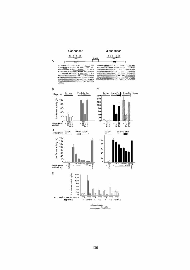
130

131
Figure 2 – Emx2 represses the activity of the 5’ and 3’ Sox2 telencephalic enhancers in transfection assays. (A) 5’ and 3’ Sox2 telencephalic enhancers. Numbered squares: ATTA sites, underlined and bold in the sequences below. Boxed bold sequences: POU sites [18-20] in 5’ and 3’ enhancers (B,C) Cotransfection of 5’ or 3’ enhancer-driven (black bars, full enhancer; striped bars, “core” enhancer) tk-luciferase vectors, or “empty” tk-luciferase vector (white bars), with Emx2 or Otx2 expression vectors, or with “empty” vector. The mean activity of the enhancer-driven constructs (with no cotransfected expression vector) is set = 100% luciferase activity. (D) Co-transfection of 5’ and 3’-enh. luciferase constructs with increasing amounts of Emx2-expression vector. (E) Luciferase activity of 5’ enhancer constructs carrying mutations in the indicated ATTA sites, and their response to co-transfection of the Emx2 expression vector (500 ng).

132
Figure 3 – Emx2 binds to ATTA sites within the Sox2 5’ and 3’ enhancers, and antagonizes binding of the activator Brn2. (A) ATTA sequences binding Emx2 and/or Brn2. Lowermost line: Brn2/POU consensus based on TFBS cluster and our data. Letter size is proportional to nucleotide frequency. The spacer (n) is 2-3 nucleotides in previously validated sites [25, 27]. For the interaction of a POU factor with its binding site, and spacer length, see [37]. Boxed sequences are homologies to the Brn2 consensus. Underlined sequences correspond to the previously reported Emx2 binding sequence (footprint) in the Wnt1 enhancer [23, 24], and to

133
homologous sequences within the 5’ and 3’ Sox2 enhancers. (B) EMSA with an ATTA-3 site probe (5’ enhancer) and recombinant Emx2 and Brn2 proteins (as indicated above the lanes). Anti-Emx2 antibody was added in lane 8. Asterisk: supershifted band. (C) EMSA with wild type (lanes 19-23) and two different mutated (lanes 9-13; 14-18) ATTA-3 site probes (5’ enhancer). (D)Addition of increasing amounts of Emx2 (lanes 5-7) to ATTA-3 site probe (5’ enhancer) together with a fixed amount of Brn2 (as in lane 4). An Emx2 retarded band appears, while the Brn2 band progressively disappears. (E) EMSA with a probe from the 3’ enhancer ATTA-4 site, showing ability to bind Emx2 or Brn2. Addition of Emx2 together with Brn2 (lane 5) antagonizes Brn2 binding. Asterisks indicate bands supershifted by antibodies (lanes 6,7).

134
Figure 4 – Emx2 antagonizes the binding of Brn2 to ATTA-1/2 sites in the 5’ enhancer, and to previously characterized Brn2 binding sites in other neural enhancers. (A) EMSA with a probe containing ATTA sites 1 and 2 (5’ enhancer); added recombinant proteins, and Brn2 antibody, are indicated above the lanes. The

135
probe binds recombinant Brn2 (arrow), but not Emx2 (TNT- arrowhead indicates a non-specific band seen also with TNT extract only). Addition of Emx2 antagonizes Brn2 binding (lane 5). No antagonism is seen upon addition of GATA1 or GATA2 (lanes 6,7). (B) EMSA with an ATTA-3 site probe (a previously validated Brn2 binding site in the 5’ enhancer [19-21]; binding of Brn2 is efficiently competed by wild type non-labelled ATTA-1/2 sites oligonucleotide (lane 5), but not by its mutated version (lane 6). Competition is as efficient as with the “self” oligonucleotide (lane 4). (C) EMSA with probes containing previously validated Brn2 binding sites in the nestin and Delta-1 enhancers. Brn2 binding (arrow) is antagonized by simultaneous Emx2 addition in a dose-dependent way. Asterisk: Brn2 antibody-supershifted band. (D) EMSA with ATTA-1/2 site probe and nuclear extracts from AHP neural cells. Two complexes are generated (arrows) with both ATTA-3 (lane 1, “+” as in [21]) and ATTA-1/2 (lane 2), which are supershifted by anti-Brn2 (lane 3), but not anti-GATA1 antibodies (lane 4). Binding of Brn2 to ATTA-1/2 is efficiently competed by unlabelled ATTA-3 (lane 8), by “self” ATTA-1/2 (lane 5), but not by mutated ATTA-1/2 (lanes 6,7) oligonucletides. (E) EMSA with ATTA-3 probe and nuclear extracts from AHP cells. Added recombinant proteins (Emx2, GATA-1) are indicated above the lanes.The Brn2 retarded complex (lane 1, arrow) (see also [21] and panel D) is sharply decreased following addition of Emx2 (lanes 2-4), but not of control GATA-1 (lane 5).The lower, Emx2-containing complex, is progressively increased in parallel with the addition of Emx2. This complex has the same mobility of that generated by direct binding of recombinant Emx2 to the ATTA-3 probe (lane 6). (F) Emx2 and Brn2 directly interact in a GST pulldown assay. Brn2 is retained by GST-Emx2, but not by GST-CP2 control resin (which gives a weak signal equivalent to that seen with the “empty” resin (GST).

136
Figure 5 – Emx2 represses Brn2-transactivated ATTA-1/2 and ATTA-3 sites – tk luciferase reporter constructs in a dose-dependent way. (A) Brn2 dose-dependent

137
transactivation of ATTA-1/2 sites (5’ enhancer). (B,C) Emx2 dose-dependent repression of Brn2-dependent transactivation of ATTA-1/2 sites construct (B) and of ATTA site 3 construct (C). In A, luciferase activity is expressed in arbitrary units, where 1 is the activity of the tk luc reporter; in B and C, 100% luciferase activity is set to the maximum observed activity. The horizontal line in A and B represents the background activity of the ATTA-1/2 site construct in the absence of cotransfected Brn2.

138
Figure 6 – Emx2 is bound to the Sox2 enhancer in vivo. ChIP with anti-Emx2 antibodies of E14.5 embryonic brain chromatin from wild type and Emx2-/- control embryos. Region A, containing ATTA-3 site is immunoprecipitated from wild type, but not Emx2-null chromatin. The previously described Wnt1 enhancer containing an Emx2 binding site [24] is used as a control (Wnt1), and is similarly precipitated from wild type, but not mutant, chromatin. Antibodies used are indicated below the lanes. Input: input chromatin. IgG: anti-IgG control antibodies. Emx2: anti-Emx2 antibodies.

139
Figure 7 – Emx2 deficiency (Emx2+/-) rescues GFAP/nestin stem cells impairment in the hippocampus of Sox2-deficient (Sox2β-geo/∆Enh) mutant mice. (A) GFAP/nestin double immunofluorescence of hippocampus dentate gyrus in the indicated genotypes. GFAP/nestin-positive cells, strongly depleted in Sox2-hypomorphic (Sox2β-geo/∆Enh) mutants, recover to a significant extent in Sox2β-geo/∆Enh ;Emx2+/- double mutants (asterisks mark vessels, showing non-specific fluorescence). (B) GFAP/nestin-positive cells and BrdU-positive cells (n=8 mice per genotype). Wild

140
type is set = 100%. (C) triple immunofluorescence (confocal microscopy) with anti Sox2 (green), anti Emx2 (red) and anti Brn2 (blue) on E15.5 telencephalic sections detects extensive coexpression of Sox2, Emx2 and Brn2 in the ventricular zone. The image shows an area within the medial telencephalic wall, that approximately corresponds to the region boxed in D. (D) double immunofluorescence with anti Emx2 (red) and anti Sox2 (green) antibodies on E15.5 telencephalic sections (confocal microscopy), in wild type (Emx2+/+, top) and Emx2+/- heterozygotes (two different mice/genotype). In Emx2+/- brains, compared to Emx2+/+ controls, an increase in the intensity of Sox2 staining is seen in the medial telencephalic wall (comprising the prospective hippocampus), as compared with the outer/lateral wall within the same section.
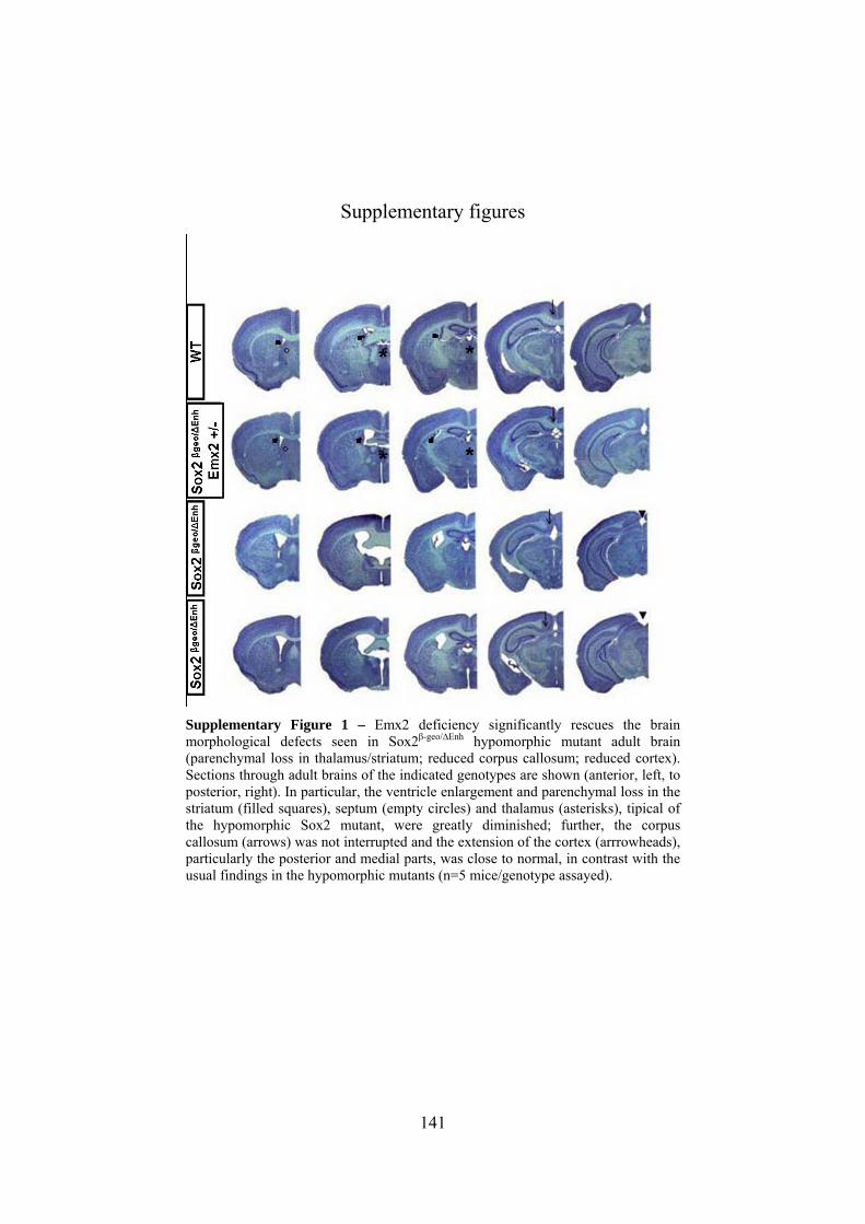
141
Supplementary figures
Supplementary Figure 1 – Emx2 deficiency significantly rescues the brain morphological defects seen in Sox2β-geo/∆Enh hypomorphic mutant adult brain (parenchymal loss in thalamus/striatum; reduced corpus callosum; reduced cortex). Sections through adult brains of the indicated genotypes are shown (anterior, left, to posterior, right). In particular, the ventricle enlargement and parenchymal loss in the striatum (filled squares), septum (empty circles) and thalamus (asterisks), tipical of the hypomorphic Sox2 mutant, were greatly diminished; further, the corpus callosum (arrows) was not interrupted and the extension of the cortex (arrrowheads), particularly the posterior and medial parts, was close to normal, in contrast with the usual findings in the hypomorphic mutants (n=5 mice/genotype assayed).

142
Supplementary Figure 2 – Emx2 binds to the Wnt1 and Sox2 (ATTA-3) enhancers. (A) Western blot showing Emx2 recombinant protein produced in P19 cells transfected with Emx2 expression vector (lane 1), in the TNT in vitro system (lane 2), compared to extracts from the hippocampal AHP cell line (lanes 3,4). Note that Emx2 levels are much lower in nuclear (lane 4) than in total extracts (lane 3)(same cell numbers used). (B) EMSA with recombinant Emx2 and probes containing the sites in the Wnt1 3’ enhancer (lanes 1,2), or the ATTA-3 site in the

143
Sox2 5’ enhancer (lanes 3-10)(see Fig. 3A for sequences). Added competitor oligonucleotides and antibodies are indicated above the lanes. Two bands are generated by Emx2 binding to the Wnt1 probe (lane 1, arrowheads), consistent with the Wnt1 site being a double site (Fig. 3A); both bands are supershifted by anti-Emx2 antibody (lane 2, asterisk indicates the supershifted band). The ATTA-3 probe generates with Emx2 a complex (arrow), which is supershifted by anti-Emx2 antibodies (lane 4, asterisk), and is competed by unlabelled ATTA-3 oligonucleotide (50-100 molar excess)(lanes 5,6), but not by mutant ATTA-3 (lanes 7,8). Unlabelled Wnt1 oligonucleotide competes binding to the ATTA-3 probe as efficiently as ATTA-3 itself (lanes 9,10; compare to lanes 5,6). Equivalent data were obtained with neurosphere extracts (not shown).

144
Supplementary Figure 3 – AHP cells coexpress Sox2, Emx2 and Brn2. Triple immunofluorescence with anti-Sox2, Emx2 and Brn2 antibodies of the AHP line detects coexpression of all three proteins in numerous cells. Note the presence of cytoplasmic as well as nuclear Emx2.

145
Supplementary Figure 4 – (A) Emx2 (brown, antibody staining) is coexpressed with Sox2 (Sox2β-geo, blue, X-gal staining) in cells of the DG SGZ (arrows point to examples of double-positive cells). (B) Emx2 (green, immunofluorescence, confocal microscopy) is expressed in GFAP-positive (red) radial glia cells in the DG (arrows), as seen for Sox2 [5]. (C) Emx2 (green, immunofluorescence, confocal microscopy) is expressed in BrdU-positive cells (red, antiBrdU antibody) at the basis of the DG, as previously seen for Sox2 [5].

146
Supplementary Figure 5 – A hypothesis for the dose-dependent negative modulation of the Sox2 neural enhancer by Emx2. The Sox2 5’ enhancer ( “1-2” and “3” are tthe ATTA-1/2 and ATTA-3 transcription factor binding sites) is bound by POU activators Oct4 (in ES cells) and Brn1/2 (in neural stem/progenitor cells, NSC)([19-21] and present work). Emx2 antagonizes Brn2 function in two ways: preventing Brn2 binding to DNA (to both ATTA-1/2 and ATTA-3 sites) by protein-to-protein interaction, and by direct binding to DNA (to ATTA-3 site), to a sequence overlapping that recognized by Brn2. This mechanism operates on multiple Brn activator binding sites (the two sites ATTA-1/2 and 3, represented here; possibly to all six ATTA sites in the enhancer, see Fig. 2). Hence, at high Brn2/Emx2 ratios (top), Brn2 is bound to DNA at all sites (1-2 and 3), and the enhancer is fully active; at higher Emx2 concentrations relative to Brn2, some sites (1-2, or 3) are no longer bound by Brn2, and Emx2 is bound to some of them (site 3), giving rise to intermediate levels of activity (middle); at higher Emx2 concentrations, the Brn activator is no longer bound, leading to low activity (bottom).
147
Disclosure of potential conflicts of interest
The authors indicate no potential conflicts of interest.
References
1 Avilion AA, Nicolis SK, Pevny LH et al. Multipotent cell
lineages in early mouse development depend on SOX2 function.
Genes Dev 2003;17:126-140.
2 Zappone MV, Galli R, Catena R et al. Sox2 regulatory sequences
direct expression of a (beta)-geo transgene to telencephalic neural
stem cells and precursors of the mouse embryo, revealing
regionalization of gene expression in CNS stem cells.
Development 2000;127:2367-2382.
3 Graham V, Khudyakov J, Ellis P et al. SOX2 functions to
maintain neural progenitor identity. Neuron 2003;39:749-765.
4 Ellis P, Fagan BM, Magness ST et al. SOX2, a persistent marker
for multipotential neural stem cells derived from embryonic stem
cells, the embryo or the adult. Dev Neurosci 2004;26:148-165.
5 Ferri AL, Cavallaro M, Braida D et al. Sox2 deficiency causes
neurodegeneration and impaired neurogenesis in the adult mouse
brain. Development 2004;131:3805-3819.
6 Suh H, Consiglio A, Ray J et al. In vivo fate analysis reveals the
multipotent and self-renewal capacities of Sox2+ neural stem
cells in the adult hippocampus. Cell Stem Cell 2007;1:515-528.
7 Cavallaro M, Mariani J, Lancini C et al. Impaired generation of
mature neurons by neural stem cells from hypomorphic Sox2
mutants. Development 2008;135:541-557.
148
8 Sisodiya SM, Ragge NK, Cavalleri GL et al. Role of SOX2
mutations in human hippocampal malformations and epilepsy.
Epilepsia 2006;47:534-542.
9 Kelberman D, de Castro SC, Huang S et al. SOX2 plays a critical
role in the pituitary, forebrain, and eye during human embryonic
development. J Clin Endocrinol Metab 2008;93:1865-1873.
10 Foshay KM, Gallicano GI Regulation of Sox2 by STAT3 initiates
commitment to the neural precursor cell fate. Stem Cells Dev.
2008;17:269-78.
11 Favaro R, Valotta M, Ferri ALM et al. Hippocampal development
and neural stem cell maintenance require Sox2-dependent
regulation of Shh. Nat Neurosci 2009;12:1248-1256.
12 Simeone A, Gulisano M, Acampora D et al. Two vertebrate
homeobox genes related to the Drosophila empty spiracles gene
are expressed in the embryonic cerebral cortex. EMBO J
1992;11:2541-2550.
13 Pellegrini M, Mansouri A, Simeone A et al. Dentate gyrus
formation requires Emx2. Development 1996;122:3893-3898.
14 Gangemi RM, Daga A, Marubbi D et al. Emx2 in adult neural
precursor cells. Mech Dev 2001;109:323-329.
15 Galli R, Fiocco R, De Filippis L et al. Emx2 regulates the
proliferation of stem cells of the adult mammalian central nervous
system. Development 2002;129:1633-1644.
16 Roelink H. Hippocampus formation: an intriguing collaboration.
Curr Biol 2000;10:R279-R281.

149
17 Rash BG, Grove EA. Area and layer patterning in the developing
cerebral cortex. Curr Opin Neurobiol 2006;16:25-34.
18 O'Leary DD, Chou SJ, Sahara S. Area patterning of the
mammalian cortex. Neuron 2007;56:252-269.
19 Miyagi S, Saito T, Mizutani K et al. The Sox-2 regulatory regions
display their activities in two distinct types of multipotent stem
cells. Mol Cell Biol 2004;24:4207-4220.
20 Miyagi S, Nishimoto M, Saito T et al. The Sox2 regulatory region
2 functions as a neural stem cell-specific enhancer in the
telencephalon. J Biol Chem 2006;281:13374-13381.
21 Catena R, Tiveron C, Ronchi A et al. Conserved POU binding
DNA sites in the Sox2 upstream enhancer regulate gene
expression in embryonic and neural stem cells. J Biol Chem
2004;279:41846-41857.
22 Noyes MB, Christensen RG, Wakabayashi A et al. Analysis of
homeodomain specificities allows the family-wide prediction of
preferred recognition sites. Cell 2008;133:1277-1289.
23 McEvilly RJ, de Diaz MO, Schonemann MD et al.
Transcriptional regulation of cortical neuron migration by POU
domain factors. Science 2002;295:1528-1532.
24 Iler N, Rowitch DH, Echelard Y et al. A single homeodomain
binding site restricts spatial expression of Wnt-1 in the
developing brain. Mech Dev 1995;53:87-96.
25 Ligon KL, Echelard Y, Assimacopoulos S et al. Loss of Emx2
function leads to ectopic expression of Wnt1 in the developing

150
telencephalon and cortical dysplasia. Development
2003;130:2275-2287.
26 Josephson R, Muller T, Pickel J et al. POU transcription factors
control expression of CNS stem cell-specific genes. Development
1998;125:3087-3100.
27 Tanaka S, Kamachi Y, Tanouchi A et al. Interplay of SOX and
POU factors in regulation of the Nestin gene in neural primordial
cells. Mol Cell Biol 2004;24:8834-8846.
28 Castro DS, Skowronska-Krawczyk D, Armant O et al. Proneural
bHLH and Brn proteins coregulate a neurogenic program through
cooperative binding to a conserved DNA motif. Dev Cell
2006;11:831-844.
29 Lie DC, Colamarino SA, Song HJ, Désiré L, Mira H, Consiglio
A, Lein ES, Jessberger S, Lansford H, Dearie AR, Gage FH. Wnt
signalling regulates adult hippocampal neurogenesis. Nature
2005;437:1370-5.
30 Muzio L, Soria JM, Pannese M et al. A mutually stimulating loop
involving emx2 and canonical wnt signalling specifically
promotes expansion of occipital cortex and hippocampus. Cereb
Cortex 2005;15:2021-2028.
31 O'Leary DD, Sahara S. Genetic regulation of arealization of the
neocortex. Curr Opin Neurobiol 2008;18:90-100.
32 Sahara S, Kawakami Y, Izpisua Belmonte JC et al. Sp8 exhibits
reciprocal induction with Fgf8 but has an opposing effect on
anterior-posterior cortical area patterning. Neural Dev 2007;2:10.

151
33 Zembrzycki A, Griesel G, Stoykova A et al. Genetic interplay
between the transcription factors Sp8 and Emx2 in the patterning
of the forebrain. Neural Dev 2007;2:8.
34 Uchikawa M, Ishida Y, Takemoto T et al. Functional analysis of
chicken Sox2 enhancers highlights an array of diverse regulatory
elements that are conserved in mammals. Dev Cell 2003;4:509-
519.
35 Takanaga H, Tsuchida-Straeten N, Nishide K et al. Gli2 is a novel
regulator of sox2 expression in telencephalic neuroepithelial cells.
STEM CELLS 2009;27:165-174.
36 Doetsch F. The glial identity of neural stem cells. Nat Neurosci
2003;6:1127-1134.
37 Hamasaki T, Leingartner A, Ringstedt T et al. EMX2 regulates
sizes and positioning of the primary sensory and motor areas in
neocortex by direct specification of cortical progenitors. Neuron
2004;43:359-372.
38 Sur M, Rubenstein JL. Patterning and plasticity of the cerebral
cortex. Science 2005;310:805-810.
39 Scully KM, Jacobson EM, Jepsen K et al. Allosteric effects of Pit-
1 DNA sites on long-term repression in cell type specification.
Science 2000;290:1127-1131.
40 Tomioka M, Nishimoto M, Miyagi S et al. Identification of Sox-2
regulatory region which is under the control of Oct-3/4-Sox-2
complex. Nucleic Acids Res 2002;30:3202-3213.

152
41 Bose F, Fugazza C, Casalgrandi M et al. Functional interaction of
CP2 with GATA-1 in the regulation of erythroid promoters. Mol
Cell Biol 2006;26:3942-3954.
42 Di Rocco G, Gavalas A, Popperl H et al. The recruitment of
SOX/OCT complexes and the differential activity of HOXA1 and
HOXB1 modulate the Hoxb1 auto-regulatory enhancer function. J
Biol Chem 2001;276:20506-20515.
153
CHAPTER 4
ABNORMAL DEVELOPMENT OF
CORTICOTHALAMIC CONNECTIONS IN
SOX2 HYPOMORPHIC AND
CONDITIONAL KNOCK OUT MICE
Caccia R., Broccoli V. and Nicolis S.K.
On going work

154

155
Abnormal development of corticothalamic
connections in Sox2 hypomorphic and conditional
knock out mice
R. Caccia1, V. Broccoli2 and S.K. Nicolis1 1Department of Biotechnology and Biosciences, University of Milano-Bicocca,
Piazza della Scienza 2, 20126 Milano, Italy 2Stem Cell Research Institute, DIBIT H San Raffaele, Via Olgettina 58, 20132
Milano,
Introduction
The neocortex, the largest region of the cerebral cortex, is divided in
different functionally area. Besides, the cortex presents also a laminar
morphology, with six recognisable layers.
Sense organs, excluded olfactory organs, send the sensory input to
one or more thalamic nuclei. These nuclei have well defined and
reciprocal connections with specific cortical regions that process the
information. The connections have area and lamina specificity.
The corticothalamic projection neurons are a part of cortical neurons.
They are generated in the ventricular/subventricular zone of the lateral
ventricle and migrate to form the cerebral cortex in an “inside out”
pattern to form layers from VI to II. The populations of neurons
located in the subplate and in the layer VI send long distance
projections to link the cortex and the rest of central nervous system.
Thalamic nuclei are generated between E10.5 and E15.5.
Cortex and thalamus develop synchronously, and start to form
connections around E13.5. Corticothalamic and thalamocortical
156
connections have to cross several zone to reach their ultimate target.
These comprise pallial-subpallial boundary (PSPB) and diencephalic-
thelencephalic boundary (DTB). These zones act as barrier zone and
corridor for elongating neurons.
Thalamocortical projections proceed ventrally towards the ventral
thalamus, then turn dorsolaterally at DTB and arrive to the internal
capsula (IC) at E13.5: and then pause. Projections from the neocortex
arrive at PSPB at E14.5 (projections from different regions reach the
boundary asynchronously, according to the cortical developmental
gradient) and pause. After entering in the IC, at E15.5 corticothalamic
and thalmocortical axons interact then proceed associated with each
other towards their targets.
Previous work performed in our laboratory demonstrated reduced
cortical size and parenchymal loss with cell death in the thalamus of
Sox2βgeo/Δenh mutants (Ferri et al. 2004). We hypothesized that
thalamocortical and/or corticothalamic connections may be affected in
mice deficient for the Sox2 transcription factor. Here, we investigate
corticothalamic connections in four different mouse strains mutant in
Sox2: a knockdown mouse strain, expressing reduced amount of Sox2
an three different strains with complete deletion of Sox2 in different
regions of brain (the whole brain, the cortex only and the dorsal
thalamus only).
We find that Sox2 is required for the development of corticothalamic
axons after E12.5. we demonstrated that the defect does not reside in
the developing neurons; more probably, the environment surrounding
growing axons have a defect in program of expression of molecules
involved in axon guidance.

157
Results
Corticothalamic projection abnormalities exist in
Sox2βgeo/Δenh “knockdown” mutant mouse
Sox2βgeo/Δenh mice carry a regulatory (Sox2Δenh) mutation together
with a null (Sox2βgeo) mutation; these “knockdown” mice express 20-
30% of the normal amount of Sox2 in the developing brain. (Ferri et
al., 2004)
To obtain our experimental model, we crossed mice carrying the null
mutation with mice homozygous or heterozygous for the Sox2
regulatory mutation (Fig. 1). Mice carrying only the regulatory
mutation (Sox2Δenh/+), which do not show any phenotypical
abnormality (Ferri et al., 2004), were considered as controls.
To study if cortical neurons are able to develop and form correct
interactions in Sox2βgeo/Δenh mice, we analyzed embryonic brains at
E18.5. At this stage of embryonic development, the axonal projections
have finished to grow and are establishing final synaptic contacts with
their specific target.
To visualize the pattern of axon elongation we used DiI crystals (1,
1-dioctadecyl –3, 3, 3’, 3’-tetramethylindocarbocyanine perchlorate).
DiI is a fluorescent dye able to bind the plasma membrane by
hydrophobic interaction with its lypophilic portion. DiI crystals are
placed by manual insertion in specific points of brain. The molecules
of dye diffuse along the biological membranes localized near the site
of implant, including the membrane of the long axonal projections.
DiI crystals were implanted, in separate experiments, in the three
major functional areas in the neocortex. The Primary Somatosensory

158
Cortex (S1) is located in the medial region of brain, and sends
projections that will synapse onto the Ventrobasal Nucelus (VB) of
the thalamus. The Primary Motor Cortex (M1), the more rostral region
of the neocortex, also sends its axons towards the VB nucleus, but its
final target is more rostral than that of S1. The more caudal region of
cortex is the Primary Visual Cortex (V1), which sends its projections
to the dorsal Lateral Geniculate Nucleus (dLGN).
In the wild type, as expected, fibers start to grow from their specific
area in the neocortex (Fig. 2). The outgrowth of axons begins around
E13.5. They elongate first ventrally to arrive at PSPB, then turn
towards the midline of brain and arrive to the IC. After exiting the IC
and crossing the DTB (that happens at E15.5) they turn their trajectory
towards the dorsal thalamus, where dorsal thalamic nuclei reside (Fig.
2).
In all mutant brains studied, the initial tract of corticofugal
projections is normal: the axons start to grow towards the ventral part
of the telencephalon and turn towards the midline to reach in the IC.
Subsequently, however, abnormalities become apparent in mutant
brains.
In three out of four mutant brains examined for the connections
between M1 and medial VB nucleus, the axons exiting the IC and
reaching their target are reduced in number (Fig. 3; Table 1). In the
fourth brain analyzed, as well in all controls, the progression and the
number of corticofugal axonal projections reaching the VB is
comparable and normal (Fig. 3).
Similarly, five out of eight mutant brains implanted in S1 show
reduced numbers of axons exiting the IC and arriving to the VB (Fig.

159
4). The other three mutant brains do not show significant anomalies
(Fig. 4).
Similar observations are made about the implants in V1. In two out
of seven mutant brains, the initial tract of projections is normal, but
the fibers that cross the boundary between telencephalon and
diencephalon and reach the dLGN are very reduced in number
compared to wild type brains. In the other five brains, no significant
differences were observed between wild type and mutant in the
number of axons reaching dLGN (Fig. 5).
In summary, in Sox2βgeo/Δenh mutant brains important abnormalities
are present in the development of corticothalamic axons. These
abnormalities do not affect the correct routing of projection neurons
along the initial tract of their trajectory, but rather affect the number of
axons that arrive to their final target, which is severely reduced in
some mutants, more mildly in others. Out of nineteen mutant brains
implanted, about 50% (10/19) show a substantial depletion in the
numbers of axons crossing the DTB and reaching their specific
nucleus (Table 1; Figs. 3-5). In particular, we observe this depletion in
75% (3/4) of the brains implanted in M1, in 65% (5/8) of brains
implanted in S1 and in 28% (2/7) of brains implanted in V1.
The severity of abnormalities in these brains is variable, from almost
total absence of axons reaching the thalamus, to a milder phenotype,.
(Fig. 6)

160
Abnormal corticothalamic connections in the
Sox2βgeo/flox;Nestincre mutant mouse
We then investigated a mouse model in which complete ablation of
Sox2 could be obtained by the action of a Cre recombinase.
This model carries a Sox2 null allele (Sox2βgeo) together with a Sox2
allele flanked by two loxP sites (Sox2flox; Favaro et al., 2009), the
substrate of Cre recombinase; a transgene specifically expressed in the
developing central nervous system is also present, in which the
expression of the Cre recombinase gene is driven by the regulatory
regions of the Nestin gene, (Medina et al., 2004) The deletion of the
Sox2flox gene is complete by E12.5 in the whole brain, including
cortex and thalamus (Favaro et al., 2009). The mating plan is
presented in Figure 7.
The analysis of projection neurons in these mutants was also
performed on E18.5 brains, using the DiI tracer. The implants of DiI
crystals was made always in the three major functional areas of the
neocortex (M1, S1 and V1). We implanted four mutant brains in M1,
five mutant brains in S1 and seven mutant brains in V1, with their
respective controls.
In all wild type brains, the projection axons show the expected
pattern (Fig. 8; Table 2). In the mutant brains(Sox2βgeo/flox;Nestincre),
the initial tract of axonal development is normal, with the correct turn
of trajectory towards the ventral area first, and the midline later, as
previously seen in the hypomorphic (Sox2βgeo/Δenh) brains (not shown).
However, all sixteen mutant brains analyzed show that the axons
exiting the IC and crossing the telencephalon-diencephalon boundary
are extremely reduced in number (Fig. 8; Table 2).

161
In conclusion, Sox2 is required for correct axon pathfinding after day
E12.5. Complete ablation of Sox2 by E12.5 leads to important
abnormalities in all the mice studied; Sox2 reduction (“knockdown”
model) causes similar abnormalities, but with reduced penetrance
(50%) and greater variability.
Cortical specific deletion of Sox2 does not lead to
abnormalities in axonal pathfinding
There are two possible explanations for the abnormal growth of
axons in mice lacking Sox2 protein:
• Sox2 expression is required in the cortex for the birth and
maturation of cortical projection neurons
• Sox2 expression is required in the thalamus, to produce
molecular signals required for the correct elongation and
pathfinding of corticothalamic axons
To investigate if the problem resides in the cortex or in the thalamus
we deleted Sox2flox by specific cortical, or thalamic, Cre-expressing
mice.
To obtain the ablation of Sox2 gene expression in neocortex only,
we crossed (Fig. 9) mice carrying the Sox2flox allele with mice
carrying a Cre recombinase gene inserted into the Emx1 locus by
homologous recombination, downstream to an IRES (Internal
Ribosome Entry Site) element in the gene 3’-UTR. (Gorski et al.,
2002). In this “knock-in” construct, Cre is inserted into the Emx1
locus in a way that does not affect the normal expression of the Emx1
gene, and is expressed according to the Emx1 expression pattern (the
cortex only from E9.5)(Gorski .et al., 2002).

162
By E12.5 (with timing similar to Necstincre), Sox2 expression is
completely ablated specifically in the neocortex, whereas it is not
affected in other regions of the brain, including the thalamus (Fig. 10).
Also in this case DiI crystals were placed in the three major
functional cortical areas, M1, S1 and V1, of E18.5 brains. The
implants were made in two mutant brains in M1, six mutant brains in
S1 and six mutant brains in V1, and an equal number of wild type
control brains (Table 3).
No one of the implanted mutant (Sox2βgeo/flox;Emx1IREScre) brains
shows significant differences in the numbers of axons reaching their
final target as compared to wild type (Sox2flox/+) control brains. In
both mutant and control brains the fasciculation of fibers starts from
neocortex and proceeds to the ventral region, turns towards the
midline, passes the IC crossing the DTB, so reaching the final target
(Fig. 11).
Hence, deletion of Sox2 restricted to the neocortex does not seem to
affect the development and routing of corticothalamic projection
neurons.
Finally, we attempted a thalamic specific deletion. We used mice
carrying the Cre recombinase inserted (“knock in”) in the locus of the
RORα gene. RORα is expressed as early as E12.5 in dorsal thalamus
by presumptive ventroposterior neurons (Nakagawa and O’Leary,
2003). We expected to see the deletion of Sox2 around E14.5.
However, in Sox2βgeo/flox;RORαcre mice Sox2 expression is still
present at E15.5 (Fig. 12) in amount comparable to controls. Zhou and
colleagues (Zhou et al., 2008) have seen that in RORαcre mice, cre
expression was restricted to a subset of thalamic dorsal cells. It is

163
possible that this partial expression of cre recombinase does not
permit a ablation of Sox2.
Indeed we did not observe abnormalities in the mutant brains
carrying the RorαIREScre (data non shown).
Discussion
In this study we show that Sox2 is required for correct elongation of
corticothalamic axonal connections. In brains in which Sox2
expression is decreased (“knockdown” mutants), or conditionally
ablated from day E12.5 (Sox2flox/flox;Nestincre mutants) the initial
tract of axonal projections navigating through the ventral
telencephalon is unaffected, and the fibers are able to reach the
internal capsula. The second tract of growth is abnormal: axons able to
exit the internal capsula and make the correct turning towards the
dorsal thalamus are very reduced in number.
Sox2 is an important transcription factor expressed in the central
nervous system from the beginning of its development, and the
complete knock out mouse is early embryonic lethal (Avilion et al.
2003). Previous studies performed in our laboratory have utilized as
model the Sox2βgeo/Δenh hypomorphyc mice, which expresses 20-30%
of the normal amount of Sox2; in these mutants brains show several
abnormalities, including decreased cortical size, defects in
neurogenesis and parenchymal reduction and cell death in thalamus
(Ferri et al. 2004). To better understand the physiological role of Sox2
we also generated conditional mutant mice, in which Sox2 is flanked
by loxP sites (Sox2flox) and can be ablated by driven expression of Cre
recombinases. Using a Cre recombinase driven by a regulatory region

164
specific to the developing central nervous system (Nestincre), also
these mice show brain abnormalities including reduced hippocampus,
a moderate lateral ventricle enlargement and slight size reduction of
the posterior ventrolateral cortex (Favaro et al. 2009). On the basis of
the defects found in neurons, we started to study the networking of
long range development of corticothalamic axons in mouse brain.
Sox2 is required for correct development of thalamic tract of
corticothalamic projection neurons
DiI labeling experiments in E18.5 brains indicate that the lack of
Sox2 results in failure of corticofugal projections to grow into the
thalamus, but is not accompanied by a misrouting of the fibers; rather,
few fibers exit the internal capsula and cross the diencephalic-
telencephalic boundary. Most of the projections seem to stall into the
internal capsula without exiting. Preliminary data suggest that
thalamocortical connections are not affected in Sox2 mutant brains
(data not shown).
The aberrant development of the terminal thalamic projection tract,
present as a constant character in conditional mutant mice, shows
greater variability in hypomorphic mice. Probably the incomplete
ablation of Sox2 gene products in “knockdown” mutants can explain
the wide spectrum of phenotypes observed, suggesting that small
differences in amounts of Sox2 can be sufficient to elicit great
variability in the development of corticofugal projections.
In contrast, the phenotype of Nestincre conditional knock out mice is
more similar in all mutant brains analyzed, with a consistent reduction
in number of axons reaching their target. This is an evidence that Sox2
is important for the correct development of corticofugal axons after

165
E12.5. Corticofugal axons begin their growth towards thalamic targets
around E13.5, and complete the process at E18.5. So, the total loss of
SOX2 protein occurs before the beginning of axonal development.
Axon guidance is a complex process involving many molecules.
Different genes encoding transcription factors, nuclear receptors, cell
adhesion molecules, axon guidance receptors and ligands were
described (reviewed in Lopez-Bendito and Molnar 2003).
Several hypothesis can be made to explain the abnormalities in
corticofugal projections development.
A first possibility is a cell autonomous defect due to an abnormal
differentiation program of projection neurons. The growth cone is
programmed to respond to specific cues and the environment is
specified to produce them. Axons might lack ability to respond to
normal cues along the elongation pathway. Notably, in vivo Sox2
expression is maintained in a subset of differentiated neurons,
including cortical pyramidal neurons (Ferri et al. 2004, Cavallaro et al.
2008) (it remains to be elucidated if this Sox2 positive population
comprises the corticofugal projection neurons). In Sox2 hypomorphic
cells, neuronal differentiation is impaired, with cells exhibiting a not
developed arborization; this immature morphology correlates with
impaired expression of some mature neuronal markers (Cavallaro et
al. 2008). Lentiviral Sox2 transduction experiment in Sox2-deficient
mutant cells differentiating in vitro showed that Sox2 is required at
early stages of differentiation, not at later stages. Probably, at early
stages Sox2 establish a downstream transcriptional program for a
correct differentiation. Normal axons are able to growth towards the
right synaptic partner because they express several specific molecular

166
receptors on their growth cone. It is possible that the mutant projection
neurons are able to initially grow towards ventral telencephalon, but
are unable to respond to later stimuli, because the lack of Sox2 causes
a defective transcriptional program leading to the non expression of
some particular receptors involved in guidance in the thalamic tract.
The growing axons also express several cell adhesion molecules on
their surface. These molecules bind to similar proteins on nearby cells.
It has been demonstrated that corticofugal and thalamocortical fibers
interact physically and proceed dependent on each other (Molnar et al.
1995, 1998). This interaction happens in the internal capsula. Errors in
pathfinding of both corticofugal and thalamocortical connections were
described in mice with mutations in transcription factors Tbr1, Gbx2
and Pax6 (Stoykova and Gruss, 1994; Hevner et al., 2002; Jones et al.,
2002). Because we have seen that the defects appears only after axons
growing into the subpallium, and entering the internal capsula, another
possibility is that there is misexpression of one or more of these cell
adhesion molecules.
Alternatively, pathfinding defects at the level of the internal capsula
could be caused by abnormal development of sourrounding
subpallium cells. Sox2 expression was found in sparse mature neurons
in the striatum (Ferri et al. 2004). The striatum is a major forebrain
nucleus that integrates cortical and thalamic afferents. Spiny
projection neurons, a subset of striatal neurons, reside in dorsal
striatum, and receive glutamatergic projection from cerebral cortex,
which form well defined synapses (Wolf, 1998). We do not know if
these are the neurons expressing Sox2, but it is possible that the region

167
of residence, overlapping the region of internal capsula, can contribute
to regulate the growth of projection neurons.
Another possible explanation is a defective growth of axons due to
anomal expression of thalamic attractive/repulsive cues. Sox2 is an
important transcription factor expressed in developing and postmitotic
thalamus, including dorsal thalamic nuclei (Vue et al. 2007). The area
of Sox2 expression in dorsal thalamus overlaps the region of residence
of thalamic nuclei. It is possible that Sox2 could be involved in
regulating the production of one or more terminal guidance cues or,
more in general, in the patterning of the thalamus.
Several studies have demonstrated that the diffusible molecule Sonic
Hedgehog is involved in the guidance of commissural axons (Charron
et al. 2003, Okada et al. 2006) acting by regulating the attractive
Netrin1 signal. Recent work (Parra and Zou 2010) demonstrates that
Shh is also involved in the repulsive response to semaphorine of
commissural axons. Shh expression is present along the axial midline
not only in the spinal cord, but also in the forebrain. Rostrally, Shh is
expressed in ventral forebrain. In previous work we demonstrated that
Shh is a direct target of Sox2 and the complete ablation of Sox2 gene
expression from E12.5 causes a progressive reduction of Shh
expression in telencephalon and diencephalon, but not in midbrain
(Favaro et al. 2009). Shh expression along the midline of
diencephalon, reduced in Sox2 conditional knock out mice, could be
involved in the response of growing axons that normally leads axonal
projections to turn towards the dorsal thalamus.
Shh is also a well known signaling center in the developing
diencephalon, that patterns the thalamus in mice (Ishibashi and

168
McMahon 2002). Additionally, other signaling molecules involved in
thalamic pattern are Wnts, required for establishing regional thalamic
identitites (Braun et al 2003, Zhou et al. 2004) and Fgf8 that controls
the pattern of thalamic and prethalamic nuclei along the
anteroposterios axis (Kataoka and Shimogori, 2008); Sox2 can be
involved in regulating directly or indirectly the development of dorsal
thalamus by acting on the expression of these genes .
Sox2 deficient projection neurons do not show growth defects
To elucidate if the defect resides in the neurons resident in the cortex
or in signalling molecules of the thalamus, we generated mice in
which the expression of Sox2 is ablated specifically in the cortex.
To obtain cortical specific deletion we used mice in which the Cre
recombinase is driven by the Emx1 regulatory regions (Gorski et al.
2002). Emx1 is expressed in the cerebral cortex from E9.5.
Immunohistochemistry shows that by E12.5 the ablation of Sox2
protein is complete in the cerebral cortex, but is not altered in other
regions of brain, including prospective dorsal thalamus (Fig.10).
Because the deletion in the neural tube in conditional knock out mice
previously studied (Nestincre, Favaro et al. 2009) is also complete at
E12.5, the timing of gene ablation is correct to perform this analysis.
DiI labeling experiments in E18.5 brains deleted specifically in the
cortex reveals absence of abnormalities in corticofugal projection. The
connections are normal, both in routing and abundance (Fig.11; Table
3).
The deletion of Sox2 gene restricted to the cortical region does not
lead to an abnormal phenotype comparable to that seen with
Nestincre, despite the fact that the timing of deletion is at least as

169
early (see above). This suggests that neurons born in the ventricular
zone and resident in the cortex are able to elongate their projections
towards the final target also in the absence of Sox2 in the region of
origin. So, after E12.5, Sox2 is not necessary for the creation of a
functional growth cone and a correctly elongating axon.
An approach to delete Sox2 in thalamus
To obtain thalamic specific deletion I begun to work with mice
carryng the Cre recombinase driven by regulatory element of RORα.
RORα gene is expressed as early as E12.5 in the presumptive
thalamus and cerebellum (Nakagawa and O’Leary 2003).
Immunohistochemistry on E15.5 brains (the time of exiting of axons
from the internal capsula) revealed that Sox2 protein is still present in
the dorsal thalamus at levels undistinguishable from wild type.
Moreover, the thalamic nuclei develop between E10.5 and E15.5 in
mice (Altman and Bayer 1988). So, this deletion, also if happened
later than the time points we analyzed, is not useful for this analysis:
the nuclei are generated and are presumably already “programmed”;
the projections are already routed towards their target and have almost
finished to grow.
Conclusions
In summary, we found that the thalamic tract of corticofugal axonal
growth is dependent on the expression of Sox2 at midgestation (after
E12.5). Sox2 expression is not necessary in the cortex after E12.5 for
normal development of projections. We hypothesize that Sox2
expression is needed in the thalamus where it would be involved in the
mechanism of correct elongation of axons.

170
In the future, we would like to better investigate the problem of
elongation of cortical projection neurons in the thalamic tract. The
projection neurons elongate, after crossing the DTB, towards the
midline, then turn towards dorsal thalamic nuclei. Since Shh is
expressed in the ventral midline, and is greatly reduced in NestinCre-
deleted Sox2 mutant mice (Favaro et al., 2009), we may try to delete
Sox2 using a Cre transgene under the control of SBE2 (Shh brain
enhancer-2). The SBE2 element is active in the hypothalamus, and
partially in the dorsal thalamus, of transgenic mouse embryos (Jeong
et al., 2006); hence, we could drive Sox2 deletion along the midline of
diencephalon, the region of expression of Shh. This experiments can
give us a first answer about the potential role of Sox2 in axon
guidance via Shh regulation.
It will be very important to perform in situ hybridization analysis to
study the expression of specific axon guidance molecules (like netrin1
and semaphorins) in different regions and at different stages of the
developing thalamus.
Materials and Methods
Animals
The generation of Sox2βgeo allele and Sox2Δenh allele has been
described (Zappone et al., 2000; Avilion et al., 2003; Ferri et al.,
2004). Hypomorphic experimental mice embryos were derived from
intercrosses of heterozygous mice carrying a null Sox2 allele
(Sox2βgeo/+) with mice carrying regulatory mutant allele in
heterozygosis or homozygosis (Sox2Δenh/+ or Sox2Δenh/Δenh). Generation
of Sox2flox allele has been described (Favaro et al., 2009). Conditional

171
knock out mutants were obtained through two generation of crossing.
First, mice carrying Sox2βgeo allele were crossed with mice carrying
Nestincre transgene (Medina et al, 2004) to obtain double
heterozygotes. Experimental mice were obtained crossing Sox2flox/flox
mice with Sox2βgeo/+;Nestincre mice. Regional specific knock out
mice were derived in two generations: mice carrying Sox2βgeo allele
were crossed with mice carrying Emx1IREcre (Gorski et al., 2002) to
obtain double heterozygotes. Sox2βgeo/+;Emx1IREScre mice were then
intercrossed with Sox2flox/flox mice to obtain experimental animals.
The day of vaginal plug is consider E0.5. Foetuses were removed and
anaesthetized by hypothermia before decapitation. Brains were
dissected at stages E18.5 for tracing with carbocyanine dyes, and
E15.5 for immunohistochemistry. Whole embryos was collected at
E12.5 for immunohistochemistry. All brain and embryos were fixed at
4°C in 4% (wt/vol) paraformaldehyde in phosphate buffered saline
(PBS). Experimental procedures involving mice were approved by the
Italian Ministry of Health.
Genotyping
Screening of embryos was carried out by allele specific PCR.
Tracing with carbocyanine dye
Brains at E18.5 were fixed 48h at 4°C in 4% paraformaldehyde. To
label corticofugal fibers, small holes were made into the cerebral
cortex in three different sites: rostral, medial and caudal. Single
crystals of DiI (1,1’-dioctadecyl-3,3,3’,3’-tetramethyl-
indocarbocyanine perchlorate, Molecular Probes) were placed into
them by using a tungsten wire under a binocular dissecting

172
microscope. Brains were incubated in 4% paraformaldehyde at room
temperature at dark for 24h and in 2% paraformaldehyde at room
temperature at dark for 5-6 weeks. The brains were then washed with
PBS and embedded in 4% agarose and were sectioned in 200 μm
coronal slices by a vibratome (LEICA). Tissue was counterstained
with DAPI (4’,6-diamidino-2-phenylindole) 5μg/ml, washed in PBS
and coverslipped in FluorSave reagent (345789, Calbiochem). Slices
were analysed by a fluorescent microscope: all images were collected
on a Zeiss Axioplan 2 microscope and processed with Adobe
Photoshop 7.0 software (Adobe Systems).
Immunohistochemistry
E12.5 embryos and E15.5 dissected brains were fixed overnight at 4°C
in 4% (wt/vol) paraformaldehyde in PBS, cryoprotected with sucrose
30% in PBS and cryostat sectioned onto slides (SuperFrost Plus). For
Sox2 immunohistochemistry antigen unmasking was carried out by
boiling sections in 0.01 M citric acid and 0.01 M sodium citrate for 3
min in a microwave, before blocking. Sections were then washed in
PBS and blocked with FBS 1% in PBS 1h at room temperature. After
extensively washing in PBS sections were incubated overnight at 4°C
with primary antibody (mouse antibody to SOX2, 1:50, R&D
MAB2018) diluted in 1% FBS in PBS, extensively washed in PBS
and then incubated for 1h at room temperature with a secondary
antibody conjugated with a fluorochrome (goat antimouse IgG Alexa
546, 1:500, Molecular Probes). Slides are counterstained with DAPI
and mounted in PBS. Section were analysed with fluorescent
microscope. All images were collected on a Zeiss Axioplan 2

173
microscope and processed with Adobe Photoshop 7.0 software
(Adobe Systems).
Acknowledgements
We thank Rebecca Favaro for help with breeding and genotyping
conditional mice, and Anna Ferri for help with fluorescent imaging.
We are very grateful to Kevin Jones and Dennis O’Leary for let we
use their tissue-specific deleter mice.
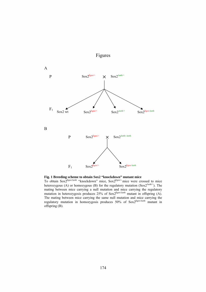
174
Figures
A
B
Fig. 1 Breeding scheme to obtain Sox2 “knockdown” mutant mice To obtain Sox2βgeo/Δenh “knockdown” mice, Sox2βgeo/+ mice were crossed to mice heterozygous (A) or homozygous (B) for the regulatory mutation (Sox2Δenh/+). The mating between mice carrying a null mutation and mice carrying the regulatory mutation in heterozygosis produces 25% of Sox2βgeo/Δenh mutant in offspring (A). The mating between mice carrying the same null mutation and mice carrying the regulatory mutation in homozygosis produces 50% of Sox2βgeo/Δenh mutant in offspring (B).
P Sox2βgeo/+ Sox2Δenh/+
Sox2βgeo/+ Sox2Δenh/+ Sox2 wt Sox2βgeo/Δenh F1
F1
P Sox2βgeo/+ Sox2Δenh/ Δenh
Sox2βgeo/+ Sox2βgeo/Δenh

175
E18.5 Sox2+/+

176
Fig. 2 normal outgrowth pattern of fibers labeled with DiI in the forebrain E 18.5 coronal sections (A more rostral to D more caudal), implanted with DiI crystals and counterstained with DAPI. In E18.5 Sox2+/+ brains axons projecting from cortex show a normal pattern of elongation. (A) Axons leave the neocortex (Ncx) and elongate towards the ventral telencephalon, then (B) turn towards the midline and enter the internal capsula (IC); axons exiting the IC cross the diencephalic-telencephalic boundary (DTB, white outline) turning towards the dorsal thalamus to reach the appropriate thalamic nucleus (C,D), highlighted with yellow outline(ventrobasal nucleus, VB) and red outline (dorsal Lateral geniculate nucleus, dLGN). Arrows indicate normal routing of axonal growth.
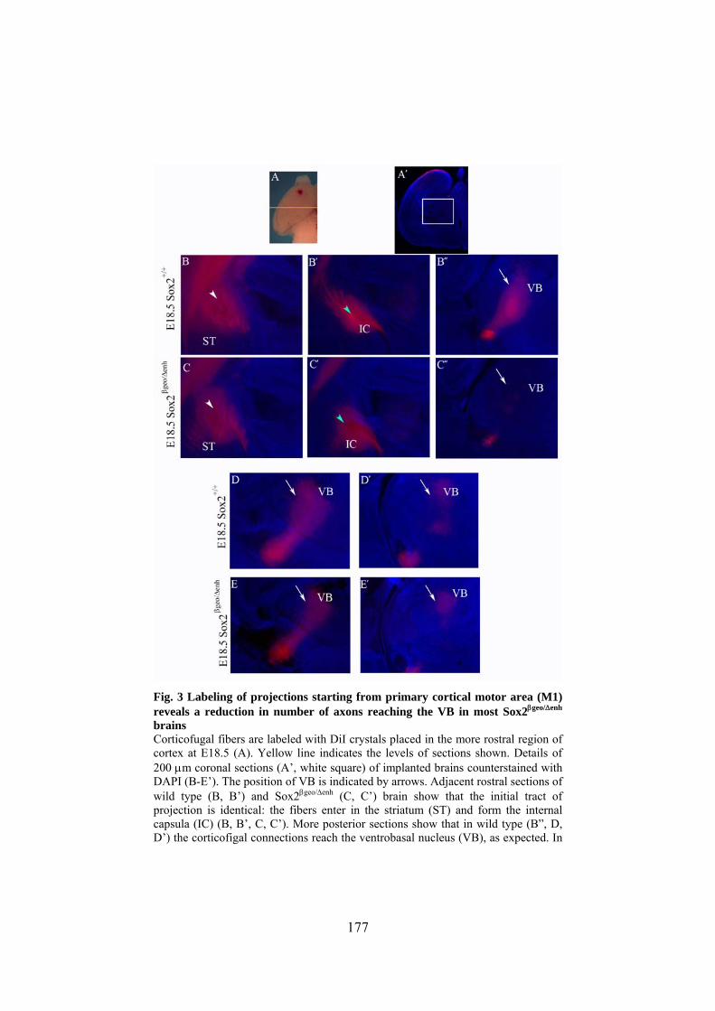
177
Fig. 3 Labeling of projections starting from primary cortical motor area (M1) reveals a reduction in number of axons reaching the VB in most Sox2βgeo/Δenh brains Corticofugal fibers are labeled with DiI crystals placed in the more rostral region of cortex at E18.5 (A). Yellow line indicates the levels of sections shown. Details of 200 μm coronal sections (A’, white square) of implanted brains counterstained with DAPI (B-E’). The position of VB is indicated by arrows. Adjacent rostral sections of wild type (B, B’) and Sox2βgeo/Δenh (C, C’) brain show that the initial tract of projection is identical: the fibers enter in the striatum (ST) and form the internal capsula (IC) (B, B’, C, C’). More posterior sections show that in wild type (B”, D, D’) the corticofigal connections reach the ventrobasal nucleus (VB), as expected. In

178
Sox2βgeo/Δenh (C”) brain the number of fibers able to exit the IC and turn towards the VB is extremely reduced. In one single case (E, E’), Sox2βgeo/Δenh brain shows that fibers reaching the VB in numbers comparable as the wild type.

179
*
*
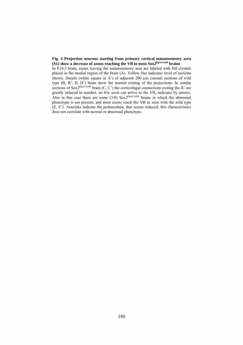
180
Fig. 4 Projection neurons starting from primary cortical somatosensory area (S1) show a decrease of axons reaching the VB in most Sox2βgeo/Δenh brains In E18.5 brain, axons leaving the somatosensory area are labeled with DiI crystals placed in the medial region of the brain (A). Yellow line indicates level of sections shown. Details (white square in A’) of adjacent 200 μm coronal sections of wild type (B, B’, D, D’) brain show the normal routing of the projections. In similar sections of Sox2βgeo/Δenh brain (C, C’) the corticofugal connections exiting the IC are greatly reduced in number, an few axon can arrive to the VB, indicates by arrows. Also in this case there are some (3/8) Sox2βgeo/Δenh brains in which the abnormal phenotype is not present, and most axons reach the VB as seen with the wild type (E, E’). Asterisks indicate the pedunculum, that seems reduced; this characteristics does not correlate with normal or abnormal phenotype.

181

182
Fig. 5 DiI placement in the primary cortical visual area (V1) reveals a depletion in the numbers of axons reaching the dLGN in Sox2βgeo/Δenh brains Placement of DiI crystals in the more caudal region of the neocortex at E18.5 (A) labels the corticofugal projections that reach the dorsal lateral geniculate nucleus (dLGN). Yellow line indicates the level of sections shown. Details (white square in A’) of 200 μm section evidentiate the normal pattern of axonal fasciculation in wild type brain (B, B’, D, D’), counterstained with DAPI. In Sox2βgeo/Δenh brain a clear reduction of axon numbers exiting the IC and reaching the dLGN is observed (arrows in C, C’). In several Sox2βgeo/Δenh brains the abnormal phenotype is not present and the number of axons reaching the dorsal thalamus is comparable to that in wild type brain (E, E’). White circles indicate the site of DiI crystals placement ; the diffused red colour is the fluorescent dye that labels all the membranes next to the implant site.

183

184
Fig. 6 DiI labeling of projection neurons in Sox2βgeo/Δenh brain demonstrates abnormalities in axonal projections of variable severity. Details (white square in A) of 200 μm section of implanted brains. DiI crystals placed in the medial region of Sox2+/+ brains (B, B’)show that axons leaving this cortical region follow the expected route of growth and do not show abnormalities. Implant of DiI crystals in the same region of Sox2βgeo/Δenh brain (B-D’) leads to a variable axonal phenotype; this mutant genotype shows variability ranging from a wild type-like phenotype(B, B’), to a partial reduction in the number of axons entering the dorsal thalamus (C, C’), to a quite complete absence of axons reaching the appropriate nucleus (D, D’). Arrows indicate the ventrobasal nucleus (VB).

185
Table 1 Summary of total Sox2βgeo/Δenh versus Sox2+/+ brains analyzed by DiI implantation
DiI brain implants
wild typea Sox2βgeo/Δenh
Axonal projection pattern Site of
implant
Normal Abnormalb Normal Abnormalb Anterior
(M1) 4 0 1 3c
Medial (S1) 8 0 3 5d
Posterior (V1) 7 0 5 2e
TOTAL 19 0 9 10 a: Sox2+/+ and Sox2Δenh/+ are both identified as wild type b: brains showing a visible reduction or absence of axonal projections reaching the
appropriate target in thalamus are defined as abnormal c: two out of three brains show severe phenotype(fibres are absent, as in fig. 6, E,
E’), one shows a milder phenotype (as in figure 6, D, D’) d: two out of five brains show severe phenotype; three show a milder mile
phenotype e: all show severe phenotype

186
Fig. 7 Breeding scheme to obtain Sox2 conditional knock out mutant mice To obtain Sox2βgeo/flox; tgNestincre conditional mutant mice two generations of breeding were required. First, a Sox2βgeo/+ mouse was crossed to a mouse carrying the Nestincre transgene. This mating allows to obtain 25% of offspring with both Sox2βgeo/+ genotype and Nestincre transgene (orange box). Crossing the Sox2βgeo/+; tgNestincre mouse to a mouse carrying the Sox2 allele flanked by the loxP sites in homozygosis produces 25% of Sox2βgeo/flox; tgNestincre mouse (red box), the conditional knock out mouse in which Sox2 is completely absent in the entire neural tube from E12.5
Sox2βgeo/+
tg Nestincre Sox2flox/flox
P
F1 Sox2 wt Sox2βgeo/+
Sox2βgeo/flox
Sox2+/+ tg Nestincre
Sox2flox/+ tg Nestincre
Sox2flox/+ Sox2βgeo/flox
tg Nestincre
Sox2βgeo/+ Sox2+/+
tg Nestincre
F2

187

188
Fig. 8 DiI labeling of corticofugal fibers in Nestincre Sox2 deleted mice demonstrates a depletion in projections reaching the appropriate thalamic nucleus Three sites of placement of DiI crystals in E18.5 brains label the corticofugal projections directed to the ventrobasal nucleus (VB, A, D) and dorsal lateral geniculate nucleus (dLGN, G). Details of 200 μm adiacent coronal sections counterstained with DAPI of Sox2flox/+ brains (B, B’, E. E’, H, H’) show the axons arriving to the appropriate nucleus as expected. DiI labeled axons in 200 μm adiacent coronal section counterstained with DAPI of Sox2βgeo/flox; tgNestincre brain (C, C’, F, F’, I, I’), show a severe reduction in number of fibers reaching their appropriate thalamic nucleus in all brains analyzed. Crystals of DiI placed in the rostral (C, C’) or medial (F, F’) region, like the implants in caudal region (I, I’), show that the axons entering the thalamus and turning towards the dorsal thalamus are fewer in comparison with the Sox2flox/+brains. White arrows indicate the ventrobasal nucleus (VB) and light blue arrows indicate the dorsal lateral genciulate nucleus (dLGN).

189
Table 2 Summary of Sox2βgeo/floxNestincre brains analyzed versus Sox2flox/+ brains
DiI brain implants
Sox2flox/+ Sox2βgeo/flox; Nestincre
Axonal projection pattern Site of
implant
Normal Abnormala Normal Abnormala Anterior
(M1) 4 0 0 4
Medial (S1) 5 0 0 5
Posterior (V1) 7 0 0 7
TOTAL 16b 0 0 16c a: brains showing a visible reduction or absence of axonal projections reaching the
appropriate target in thalamus are defined as abnormal b: all the control brains analyzed show that axons leaving the cortical region
follow the expected route of growth c: all the mutant brains analyzed show severe reduction of axons reaching the
appropriate thalamic nucleus.
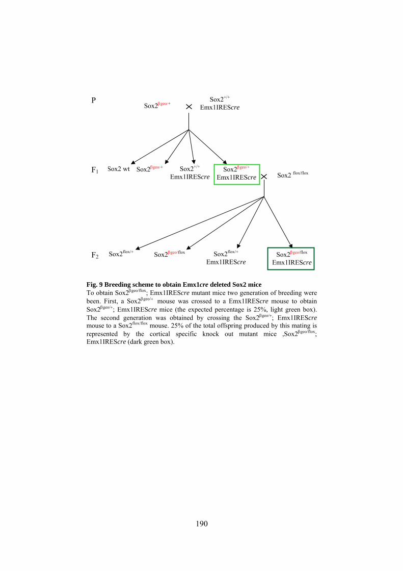
190
Fig. 9 Breeding scheme to obtain Emx1cre deleted Sox2 mice To obtain Sox2βgeo/flox; Emx1IREScre mutant mice two generation of breeding were been. First, a Sox2βgeo/+ mouse was crossed to a Emx1IREScre mouse to obtain Sox2βgeo/+; Emx1IREScre mice (the expected percentage is 25%, light green box). The second generation was obtained by crossing the Sox2βgeo/+; Emx1IREScre mouse to a Sox2flox/flox mouse. 25% of the total offspring produced by this mating is represented by the cortical specific knock out mutant mice ,Sox2βgeo/flox; Emx1IREScre (dark green box).
Sox2βgeo/+
Emx1IREScre Sox2 flox/flox
P
Sox2 wt
Sox2βgeo/flox Sox2flox/+ Emx1IREScre
Sox2flox/+ Sox2βgeo/flox
Emx1IREScre
Sox2βgeo/+ Sox2+/+
Emx1IREScre
Sox2+/+ Emx1IREScre
Sox2βgeo/+ F1
F2

191
Fig. 10 Emx1IREScre deletes Sox2 gene in developing cortex, but not in other cerebral regions, by E12.5. Immunofluorescence with anti-Sox2 antoibodies (red) of 20 μm coronal slices of E12.5 brains, counterstained with DAPI (blue). In Sox2flox/+ brain the expression of Sox2 protein is detectable in ventricular/subventricular zone of lateral ventricle and in the presumptive thalamus (A, A’). In Sox2βgeo/flox;Emx1IREScre brain the expression of Sox2 protein is no longer detectable specifically in the dorsal telencephalon (B, B’), but is not affected in the ventral telencephalon (arrows indicate the boundary of expression between dorsal and ventral telencephalon). The espression of Sox2 in the thalamus is not compromised by the action of Cre recombinase (light blue arrowheads in A’ and B’ indicate the thalamic eminence expressing Sox2).
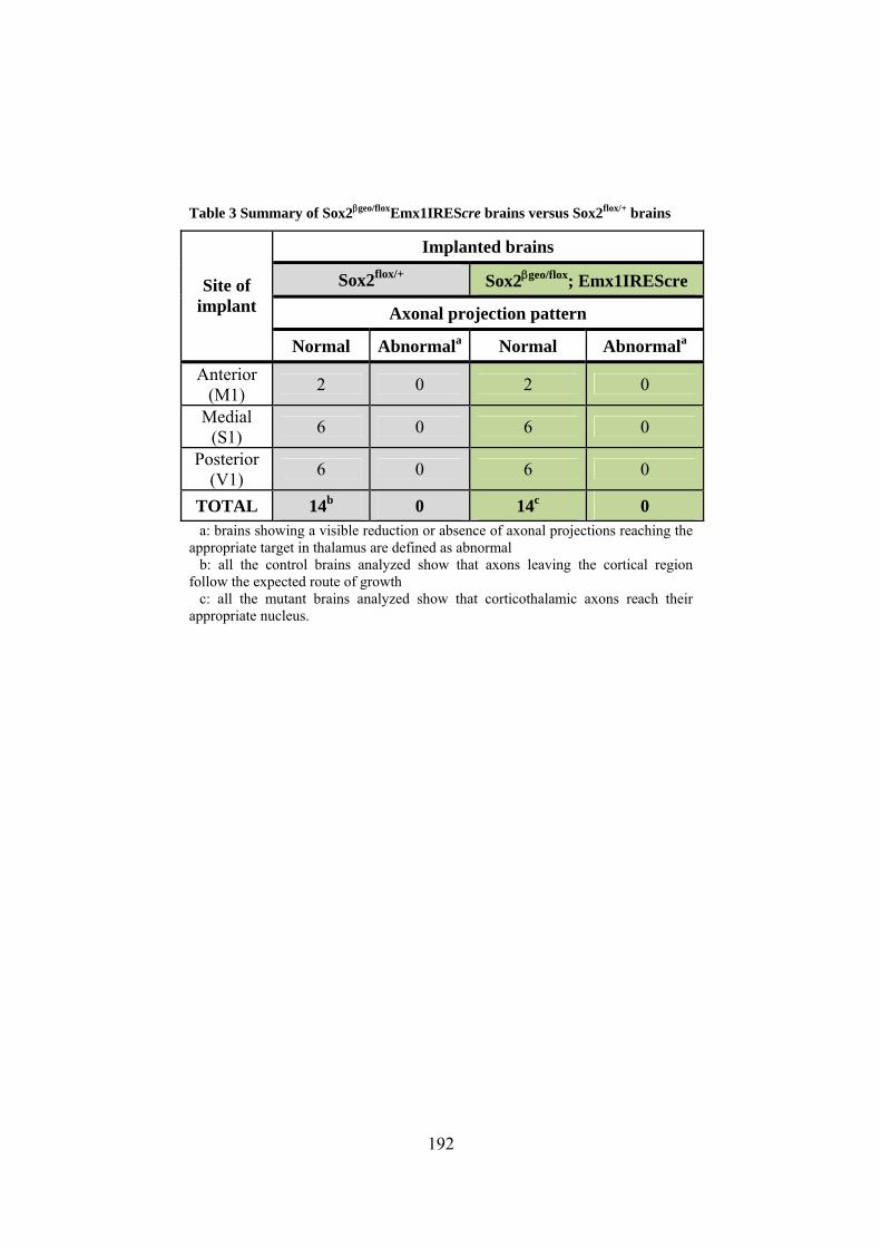
192
Table 3 Summary of Sox2βgeo/floxEmx1IREScre brains versus Sox2flox/+ brains
Implanted brains
Sox2flox/+ Sox2βgeo/flox; Emx1IREScre
Axonal projection pattern Site of
implant
Normal Abnormala Normal Abnormala Anterior
(M1) 2 0 2 0
Medial (S1) 6 0 6 0
Posterior (V1) 6 0 6 0
TOTAL 14b 0 14c 0 a: brains showing a visible reduction or absence of axonal projections reaching the
appropriate target in thalamus are defined as abnormal b: all the control brains analyzed show that axons leaving the cortical region
follow the expected route of growth c: all the mutant brains analyzed show that corticothalamic axons reach their
appropriate nucleus.

193

194
Fig. 11 Corticofugal axons labeled with DiI are not abnormal in Sox2βgeo/flox;Emx1IREScre brain DiI crystals were placed in the three major functional regions of cortex (motor cortex, A, somatosensory corte, D, visual cortex, G) in E18.5 brains to label the corticofugal prejections. (C, C’F, F’, I, I’) 200 μm adjacent coronal sections of braina lacking Sox2 specifically in the cortex show that the DiI stained fibers do not show abnormalities if compared with their Sox2flox/+ controls (B. B’, E, E’, H, H’). The axons arriving from rostral (B, B’, C, C’) and medial (E, E’, F, F’) region project correctly towards the VB both in Sox2flox/+ and in Sox2 mutant brain. Also the projections from the caudal region (H, H’, I, I’) show the same pattern of elongation in Sox2flox/+ and in Sox2 cortical null brain. White arrows indicate ventrobasal nucleus (VB), light blue arrows indicate dorsal lateral geniculate nucleus (dLGN).

195
Fig.12 RORαIREScre does not delete Sox2flox in dorsal thalamus up to E15.5 Immunofluorescence with anti-Sox2 antoibodies (red) of 20 μm coronal slices of E15.5 brains, counterstained with DAPI (blue). Staining of Sox2flox/+ (A) and Sox2βgeo/flox; RORαcre (B) show that expression of Sox2 is in dorsal thalamus is not affected by the expression of Cre recombinase (B, asterisks indicate the ventrobasal nucleus, the circles indicate the dorsal lateral geniculate nucleus; the triangle indicates aspecific red staining)

196
References
Altman, J. and Bayer, S. A. (1988). Development of the rat thalamus: III. Time and site of origin and settling pattern of neurons of the reticular nucleus. J Comp Neurol 275, 406-428.
Avilion, A. A., Nicolis, S. K., Pevny, L. H., Perez, L., Vivian, N. and Lovell-Badge, R. (2003). Multipotent cell lineages in early mouse development depend on SOX2 function. Genes Dev 17, 126-140.
Braun, M. M., Etheridge, A., Bernard, A., Robertson, C. P. and Roelink, H. (2003). Wnt signaling is required at distinct stages of development for the induction of the posterior forebrain. Development 130, 5579-5587.
Cavallaro, M., Mariani, J., Lancini, C., Latorre, E., Caccia, R., Gullo, F., Valotta, M., DeBiasi, S., Spinardi, L., Ronchi, A. et al. (2008). Impaired generation of mature neurons by neural stem cells from hypomorphic Sox2 mutants. Development 135, 541-557.
Charron, F., Stein, E., Jeong, J., McMahon, A. P. and Tessier-Lavigne, M. (2003). The morphogen sonic hedgehog is an axonal chemoattractant that collaborates with netrin-1 in midline axon guidance. Cell 113, 11-23.
Favaro, R., Valotta, M., Ferri, A. L., Latorre, E., Mariani, J., Giachino, C., Lancini, C., Tosetti, V., Ottolenghi, S., Taylor, V. et al. (2009). Hippocampal development and neural stem cell maintenance require Sox2-dependent regulation of Shh. Nat Neurosci 12, 1248-1256.
Ferri, A. L., Cavallaro, M., Braida, D., Di Cristofano, A., Canta, A., Vezzani, A., Ottolenghi, S., Pandolfi, P. P., Sala, M., DeBiasi, S. et al. (2004). Sox2 deficiency causes neurodegeneration and impaired neurogenesis in the adult mouse brain. Development 131, 3805-3819.

197
Gorski, J. A., Talley, T., Qiu, M., Puelles, L., Rubenstein, J. L. and Jones, K. R. (2002). Cortical excitatory neurons and glia, but not GABAergic neurons, are produced in the Emx1-expressing lineage. J Neurosci 22, 6309-6314.
Hevner, R. F., Miyashita-Lin, E. and Rubenstein, J. L. (2002). Cortical and thalamic axon pathfinding defects in Tbr1, Gbx2, and Pax6 mutant mice: evidence that cortical and thalamic axons interact and guide each other. J Comp Neurol 447, 8-17.
Ishibashi, M. and McMahon, A. P. (2002). A sonic hedgehog-dependent signaling relay regulates growth of diencephalic and mesencephalic primordia in the early mouse embryo. Development 129, 4807-4819.
Jeong, Y., El-Jaick, K., Roessler, E., Muenke, M. and Epstein, D. J. (2006). A functional screen for sonic hedgehog regulatory elements across a 1 Mb interval identifies long-range ventral forebrain enhancers. Development 133, 761-772.
Jones, L., Lopez-Bendito, G., Gruss, P., Stoykova, A. and Molnar, Z. (2002). Pax6 is required for the normal development of the forebrain axonal connections. Development 129, 5041-5052.
Kataoka, A. and Shimogori, T. (2008). Fgf8 controls regional identity in the developing thalamus. Development 135, 2873-2881.
Lopez-Bendito, G. and Molnar, Z. (2003). Thalamocortical development: how are we going to get there?. Nat Rev Neurosci 4, 276-289.
Medina, D. L., Sciarretta, C., Calella, A. M., Von Bohlen, U. n. H. O., Unsicker, K. and Minichiello, L. (2004). TrkB regulates neocortex formation through the Shc/PLCgamma-mediated control of neuronal migration. EMBO J 23, 3803-3814.
Molnar, Z., Adams, R. and Blakemore, C. (1998). Mechanisms underlying the early establishment of thalamocortical connections in the rat. J Neurosci 18, 5723-5745.

198
Molnar, Z. and Blakemore, C. (1995). How do thalamic axons find their way to the cortex?. Trends Neurosci 18, 389-397.
Nakagawa, Y. and O'Leary, D. D. (2003). Dynamic patterned expression of orphan nuclear receptor genes RORalpha and RORbeta in developing mouse forebrain. Dev Neurosci 25, 234-244.
Okada, A., Charron, F., Morin, S., Shin, D. S., Wong, K., Fabre, P. J., Tessier-Lavigne, M. and McConnell, S. K. (2006). Boc is a receptor for sonic hedgehog in the guidance of commissural axons. Nature 444, 369-373.
Parra, L. M. and Zou, Y. (2010). Sonic hedgehog induces response of commissural axons to Semaphorin repulsion during midline crossing. Nat Neurosci 13, 29-35.
Stoykova, A. and Gruss, P. (1994). Roles of Pax-genes in developing and adult brain as suggested by expression patterns. J Neurosci 14, 1395-1412.
Vue, T. Y., Aaker, J., Taniguchi, A., Kazemzadeh, C., Skidmore, J. M., Martin, D. M., Martin, J. F., Treier, M. and Nakagawa, Y. (2007). Characterization of progenitor domains in the developing mouse thalamus. J Comp Neurol 505, 73-91.
Wolf, M. E. (1998). The role of excitatory amino acids in behavioral sensitization to psychomotor stimulants. Prog Neurobiol 54, 679-720.
Zappone, M. V., Galli, R., Catena, R., Meani, N., De Biasi, S., Mattei, E., Tiveron, C., Vescovi, A. L., Lovell-Badge, R., Ottolenghi, S. et al. (2000). Sox2 regulatory sequences direct expression of a (beta)-geo transgene to telencephalic neural stem cells and precursors of the mouse embryo, revealing regionalization of gene expression in CNS stem cells. Development 127, 2367-2382.
Zhou, C. J., Zhao, C. and Pleasure, S. J. (2004). Wnt signaling mutants have decreased dentate granule cell production and radial glial scaffolding abnormalities. J Neurosci 24, 121-126.

199
Zhou, L., Bar, I., Achouri, Y., Campbell, K., De Backer, O., Hebert, J. M., Jones, K., Kessaris, N., de Rouvroit, C. L., O'Leary, D. et al. (2008). Early forebrain wiring: genetic dissection using conditional Celsr3 mutant mice. Science 320, 946-949.

200

201
CHAPTER 5
CONCLUSIONS AND FUTURE PERSPECTIVES

202

203
1. Sox2 is required for NSC maintenance
Sox2 is a transcription factor expressed in, and essential for, the
multipotent stem cells of blastocyst inner cell mass. Its ablation causes
early embryonic lethality (Avilion et al., 2003). Later Sox2 is a marker
of nervous system from the beginning of its development. As
development proceeds, Sox2 expression is restricted to neural stem
cells and progenitors in the ventricular/subventricular zone (VZ/SVZ)
of developing brain and in neurogenic regions in adult brain, SVZ and
hippocampus dentate gyrus (Zappone et al., 2000; Ferri et al., 2004). It
is known that Sox2 is functionally essential for maintenance of
undifferentiated state of NSC (Graham et al., 2003; Ferri et al, 2004;
Cavallaro et al., 2008), and its expression is progressively
downregulated during the neuronal differentiation (Cavallaro et al.,
2008). A residual expression of Sox2 is retained in some populations
of differentiated neurons. Strikingly, it has been demonstrated that
Sox2 can reprogram terminally differentiated cells to induced
pluripotent stem (iPS) cells, acting together with three other
transcription factors (Takahashi and Yamanaka, 2006).In our
laboratory we investigated the role of Sox2 in developing brain and in
neural stem cells by in vivo and in vitro studies on animal models whit
reduced (hypomorphic mice, Ferri et al., 2004, Cavallaro et al., 2008)
or absent (conditional knock out mice, Favaro et al., 2009) Sox2
expression.
Reduced level of Sox2 expression causes depletion of stem and
progenitor cells and cerebral defects, including reduced neocortex size

204
and parenchymal loss. Moreover, this mice show some neurological
problems, like epilepsy and motor defects (Ferri et al., 2004).
2. Sox2 is required for the differentiation of GABAergic
neurons
Our study on neural stem cells derived from mice with reduced
expression of Sox2 showed that neuronal terminal differentiation is
specifically affected (Cavallaro et al., 2008). Sox2 deficient stem cells
cultures originate a normal number of cells expressing markers of
young neurons, even though with a poor morphology. This population,
however, is not immunoreactive for neuronal terminal differentiation
markers, such as MAP2 and NeuN. In particular, by study ex vivo and
in vivo, we have seen that in newborn mouse cortex and in adult
olfactory bulb GABAergic mature populations are greatly diminished
in number (40-60%). Additionally, we detect defective migration of
GABAergic neurons originated from precursors in ganglionic
eminence: these cells are detected along the subcortical fibre bundles
but are rare in cortical plate (Cavallaro et al., 2008).
So, we demonstrated that a normal level of Sox2 is required for
correct neuronal differentiation; mutant cells generate a reduced
number of mature neurons, in particular GABAergic neurons, but the
production of glia is not affected (Cavallaro et al., 2008). Sox2
overexpression at early, but not later, stages of differentiation in
cultured mutant cells is able to rescue the mutant phenotype. Neuron
progenitors express at early stages transcription factors known to be
involved in neuronal differentiation. We hypothesize that Sox2,
commits early precursor to neurogenesis establishing a downstream

205
transcriptional program for later neuronal differentiation events and
repressing alternative (glial) transcription programs. (Cavallaro et al.,
2008).
These data demonstrate a role of Sox2 in neuronal differentiation at
least for a subset of mature neurons, the GABAergic neurons.
3. Sox2 is required for the correct development of
corticothalamic axons
Another subset of mature neurons that are affected by the
reduction/absence of Sox2 is the population of cortical excitatory
projection neurons. Many different genes encoding transcription
factors, nuclear receptors, cell adhesion molecules, axon guidance
receptors and ligands are involved in the mechanism of axon
pathfinding (reviewed in Lopez-Bendito and Molnar, 2003).
By studies on Sox2 mutant mice, we have seen that Sox2 is required
for the correct growth of corticothalamic axons after E12.5 (time of
complete Sox2 deletion driven by Nestincre transgene, Favaro et al.,
2009). The Sox2 absence leads to an aberrant growth of axons,
without misrouting of fibers: the corticofigsl projections arrive in the
striatum without evident problems, then, they seem stall into the
internal capsula and axonal growth in thalamus is absent,.
Normal axons are able to grow because express specific molecular
receptors on their surface and growth cone. Besides, the growing of
axons involved also several cell adhesion molecules that bind to
similar proteins on nearby cells. Corticothalamic and thalamocortical
projection interact physically in the internal capsula, then proceed
dependent on each other (the “handshake hypothesis”, Molnar and
206
Blakemore, 1995; Molnar et al., 1998; Hevner et al., 2002). We have
seen that in Sox2 mutant mice the defects appears only after
corticothalamic axons growing into the subpallium and entering the
internal capsula, whereas seem that thalamocortical fibres are not
affected, as seen in preliminary experiments. A first hypothesis to
explane the abnormal corticothalamic growth is a defective expression
of one or more adhesion molecules on corticothalamic axons surface.
This can lead the axons to lack the ability to interact with
thalamocortical fibres. This defect can be attributed to a cell specific
altered differentiation program which does not allow progenitors
differentiate and express correct molecule on their surface.
On the basis of previous evidences for the role of Sox2 in correct
neuronal differentiation, we first investigated the possibilitiy of a cell
autonomous defect, dues to a misregulation of a “differentiation
program” established by Sox2, that leads to the lack of capacity to
response to environmental stimuli.
Cortical projection neurons originate from progenitors expressing
Emx1 (Britanova et al., 2006). We have ablated Sox2 specifically in
the compartment of cells expressing Emx1, using an Emx1IREScre
deleter mouse. The timing of deletion is very similar to that one of the
Nestincre, with a complete dorsal telencephalic ablation by E12.5
This deletion does not affect the correct development of
corticothalamic projections, ruling out the hypothesis of a cell
autonomous differentiating defect of projection neurons.
Several other explanations are possible to clarify this defect.
The internal capsula resides in the dorsal striatum. The striatum is
the region where cortical and thalamic afferents are integrated. Spiny

207
projection neurons reside in dorsal striatum, and receive and contact
glutamatergic projection from cerebral cortex, which form well
defined synapses (Wolf, 1998). In this region Sox2 is expressed in
sparse neurons (Ferri et al., 2004).
Errors in pathfinding of both corticofugal and thalamocortical
connections were described in mice with mutations in transcription
factors Tbr1, Gbx2 and Pax6 (Stoykova and Gruss 1994; Hevner et al.
2002; Jones et al., 2002).
Mechanisms of guidance in IC are still poorly defined, but is known
that this region expresses some guidance molecules, like Netrin1
(Métin et al., 1997; Richards et al., 1997), Ephrin-A (Dufour et al.,
2003), Semaphorin 3A (Bagnard et al., 2001) and Semaphorin 6A
(Garel et al., 2002). Additional positional cues are found at the DTB.
Genetic defects affecting this region can stop axons traveling in either
direction, or lead to misrouting (Garel and Rubenstein, 2004; Hevner
et al. 2002).
It is possible that Sox2 controls the expression of signaling
molecules in IC, or, more in general, in the striatal region, along the
path of projections growth. In Situ Hybridization studies can elucidate
if there is a variability in expression of striatal guidance cues between
normal and Sox2 mutant mice.
Sox2 is expressed also in the dorsal thalamus, in territory including
the region of thalamic nuclei (Vue et al., 2007).
Dorsal thalamusis the final target of corticothalamic projections. It is
possible that the lack of Sox2 affects the expression of diffusible
molecules involved in guidance events.

208
Sonic Hedgehog (Shh) is described acting in the pathway of
guidance of commissural axons. It is involved both in attractive
Netrin1 signaling (Charron et al., 2003; Okada et al., 2006) and in the
repulsive Semaphorins signaling (Parra and Zou, 2010) in spinal cord.
Besides, both Netrin1 and Sempahorins are involved as guidance cues
for corticofugal axons (Metin et al., 1997; Bagnard et al., 1998). We
demonstrated that Shh is a direct target of Sox2 (Favaro et al., 2009).
Shh expression is detectable in the midline of ventral forebrain. In
mice lacking Sox2, the expression of Shh is reduced in ventral
foirebrain, but not in midbrain (Favaro et al., 2009). Shh in ventral
forebrain could be involved in the pathway of expression of some
guidance cue, like Netrin1 and Semaphorins. Sox2 would be involved
in the same pathway, regulating the expression of Shh.
Zona limitans intrathalamica (ZLI) is a neuroepithelial domain that
separates preumptive prethalamus from presumptive thalamus during
thalamic development (Larsen et al., 2001) and functions as secondary
organizer (Vieira et al., 2005). ZLI is a source of Shh, known to be an
important signaling molecule in the patterning of thalamus in mice
(Ishibashi and McMahon, 2002). Other diffusible factors involved in
normal development of thalamus are Wnts , that contributes to
establishment of regional thalamic identities (Braun et al., 2003; Zhou
et al., 2004). Additionaly, Fgf8, which is expressed in ZLI, controls
the patterning of thalamic and prethalamic nuclei (Kataoka and
Shimogori, 2008). By In Situ Hybridization or Immunohistochemistry
analysis is possible to investigate if Sox2 deletion causes defective
expression of gene involved in thalamic patterning.
209
The thalamic nuclei are generated between E10.5 and E15.5 (Altman
and Bayer, 1988). E15.5 is the earliest stage in which individual
thalamic nuclei are defined by gene expression pattern (Nakagawa and
O’Leary, 2001; Kataoka and Shimogori, 2008). In mutant brains,
thalamic nuclei, at E18.5, show normal morphology, but remains the
possibility that Sox2 deficiency causes alterations in their molecular
identity by defective differentiation of dorsal thalamic neurons. It is
interesting to perform the molecular chatacterization of thalamic
nuclei in Sox2 deficient mice, by analysis of gene expression.
4. Emx2 acts as a regulator of Sox2
The identification of Emx2 as direct transcriptional repressor of
Sox2 expression during brain development, together with strong
evidences that Sox2 controls stem cell maintenance, suggest that
Emx2 gradients might affect Sox2 levels in different cortical regions,
controlling the balance between self-renewal and commitment to
differentiation of stem cells. Thus, Emx2 may control NSC decisions,
at least in part, by regulating Sox2 levels.
Emx2 seems to antagonize Sox2 expression by direct transcriptional
repression of the two Sox2 telencephalic enhancers (Sox2 5’ and 3’
regulatory elements) both in vivo and in vitro.
The “core” elements of both the Sox2 5’ and 3’ enhancers contain
POU sites, known to bind different positive regulators of their
transcriptional activity in different cell types. Probably at later stages
of development, Emx2 might repress transcription at these sites by
negatively affecting the activators (by directly binding to the same
sites or via protein to protein interaction) to regulate differentiation of

210
neural stem/progenitor cells and cortical patterning, thus allowing the
downregulation of Sox2 expression in differentiating cells.
The ablation of Emx2 expression in neural stem cells enhances their
rate of proliferation, and it is possible that Emx2 deficiency
counteracts the effects of Sox2 deficiency on neural stem cells
proliferation ability and neuronal differentiation, probably
antagonizing the defect by rescuing Sox2 levels.
5. Sox2 and human diseases
In human, Sox2 deficiency is a rare condition found in patients with
microphtalmia (small eyes) or anophtalmia (no eyes) (Fantes et al.,
2003). Moreover, these patients show others important neural defects,
including abnormalities in hippocampus and corpus callosum,
epilepsy, pituitary defects and motor problems (Ragge et al., 2005;
Sisodiya et al., 2006; Kelberman et al., 2006).
5.1 Sox2 deficiency and epilepsy in humans and mice
Genetic distruption of homeobox genes related to specification,
regionalization and terminal differentiation results in epileptic
phenotype. Sox2 have a role in neuronal terminal differentiation
(Cavallaro et al., 2008) The Sox2 mutant mice reproduce several
different characteristics of neurological diseases present in Sox2
deficient patients. In particular epilepsy and hippocampal defects are
mirrored in both mutant mice generated in our laboratory (Ferri et al.,
2004; Favaro et al., 2009).
Loss of GABAergic inhibitory neurons leads to epilepsy in mouse
and man (Noebels, 2003; Cobos et al., 2005). The finding that

211
GABAergic inhibitory neurons show defective migration and are
reduced in Sox2 mutant cortex, represents a plausible cellular basis for
epilepsy in humans with Sox2 mutation (Cavallaro et al., 2008).
The in vitro culture system allowed to identify that Sox2 is important
at early, not at later stages, of neuronal differentiation; moreover, this
system will allow the identification of Sox2 target important for
neuronal differentiation, by rescue experiments.
5.2 Are abnormalities in axon guidance involved in motor
coordination defects present in Sox2 mutant patients?
Another characteristic present in Sox2 deficient patients is motor
coordination problems (Sisodiya et al., 2006). Similar behavioural
defects are present also in Sox2 mutant mice (Ferri et al., 2004).
Loss of projection neurons, in Otx1 mutant mice, leads to a
rearrangement of local circuitry characterized by excess of excitation
(Sancini et al., 2001). The sense organs send to cortex several
complex informations. Corticothalamic axons projecting in the
thalamus act as feedback system, that plays a crucial role in
modulating the thalamic responses required to perform the complex
information processing and integration that underlie mammalian
behaviors (Jones, 2002; Alitto and Usrey, 2003; Temereanca and
Simons, 2004). The reduction/absence of these connections, present
also in Sox2 mutants, can leads to a lack of negative feedback,
resulting in excess of motor response to environmental stimuli.
5.3 Sox2 and cell therapy
As Sox2 plays pivotal roles in controlling neural stem cells self-
renewal/proliferation and differentiation (Ferri et al., 2004; Cavallaro

212
et al., 2008; Favaro et al., 2009), its study will be useful for
elucidating such mechanisms that are of particular relevance for the
improvement of stem-cell-based approaches.
Elucidating the molecular mechanisms which govern proliferation
and differentiation of NSC give great hope for the treatment of
neurological disorders. Different subtypes of differentiated neurones
can be generated in vitro from stem cells of various sources including
reprogrammed somatic cells (iPS). The transplantation of in vitro
generated neurones, instead of undifferentiated NSC, have shown a
major, long-lasting improvement in some patients (Rossi and
Cattaneo, 2002; Lindvall and Kokaia, 2006). However, effective
strategies must be developed to isolate, enrich and propagate
homogeneous populations of NSCs, and to identify the molecules and
mechanisms that are required for their proper integration and
differentiation into the injured brain.

213
References
Alitto, H. J. and Usrey, W. M. (2003). Corticothalamic feedback and sensory processing. Curr Opin Neurobiol 13, 440-445.
Altman, J. and Bayer, S. A. (1988). Development of the rat thalamus: III. Time and site of origin and settling pattern of neurons of the reticular nucleus. J Comp Neurol 275, 406-428.
Avilion, A. A., Nicolis, S. K., Pevny, L. H., Perez, L., Vivian, N. and Lovell-Badge, R. (2003). Multipotent cell lineages in early mouse development depend on SOX2 function. Genes Dev 17, 126-140.
Bagnard, D., Lohrum, M., Uziel, D., Puschel, A. W. and Bolz, J. (1998). Semaphorins act as attractive and repulsive guidance signals during the development of cortical projections. Development 125, 5043-5053.
Bagnard, D., Chounlamountri, N., Puschel, A. W. and Bolz, J. (2001). Axonal surface molecules act in combination with semaphorin 3a during the establishment of corticothalamic projections. Cereb Cortex 11, 278-285.
Braun, M. M., Etheridge, A., Bernard, A., Robertson, C. P. and Roelink, H. (2003). Wnt signaling is required at distinct stages of development for the induction of the posterior forebrain. Development 130, 5579-5587.
Britanova, O., Alifragis, P., Junek, S., Jones, K., Gruss, P. and Tarabykin, V. (2006). A novel mode of tangential migration of cortical projection neurons. Dev Biol 298, 299-311.
Cavallaro, M., Mariani, J., Lancini, C., Latorre, E., Caccia, R., Gullo, F., Valotta, M., DeBiasi, S., Spinardi, L., Ronchi, A. et al. (2008). Impaired generation of mature neurons by neural stem cells from hypomorphic Sox2 mutants. Development 135, 541-557.
Charron, F., Stein, E., Jeong, J., McMahon, A. P. and Tessier-Lavigne, M. (2003). The morphogen sonic hedgehog is an axonal chemoattractant that collaborates with netrin-1 in midline axon guidance. Cell 113, 11-23.

214
Cobos,I., Calcagnotto,M.E., Vilaythong,A.J., Thwin,M.T., Noebels,J.L., Baraban,S.C., and Rubenstein,J.L. (2005). Mice lacking Dlx1 show subtype-specific loss of interneurons, reduced inhibition and epilepsy. Nat.Neurosci., 8, 1059-1068.
Dufour, A., Seibt, J., Passante, L., Depaepe, V., Ciossek, T., Frisen, J., Kullander, K., Flanagan, J. G., Polleux, F. and Vanderhaeghen, P. (2003). Area specificity and topography of thalamocortical projections are controlled by ephrin/Eph genes. Neuron 39, 453-465.
Fantes, J., Ragge, N. K., Lynch, S. A., McGill, N. I., Collin, J. R., Howard-Peebles, P. N., Hayward, C., Vivian, A. J., Williamson, K., van Heyningen, V. et al. (2003). Mutations in SOX2 cause anophthalmia. Nat Genet 33, 461-463.
Favaro, R., Valotta, M., Ferri, A. L., Latorre, E., Mariani, J., Giachino, C., Lancini, C., Tosetti, V., Ottolenghi, S., Taylor, V. et al. (2009). Hippocampal development and neural stem cell maintenance require Sox2-dependent regulation of Shh. Nat Neurosci 12, 1248-1256.
Ferri, A. L., Cavallaro, M., Braida, D., Di Cristofano, A., Canta, A., Vezzani, A., Ottolenghi, S., Pandolfi, P. P., Sala, M., DeBiasi, S. et al. (2004). Sox2 deficiency causes neurodegeneration and impaired neurogenesis in the adult mouse brain. Development 131, 3805-3819.
Garel, S., Yun, K., Grosschedl, R. and Rubenstein, J. L. (2002). The early topography of thalamocortical projections is shifted in Ebf1 and Dlx1/2 mutant mice. Development 129, 5621-5634.
Garel, S. and Rubenstein, J. L. (2004). Intermediate targets in formation of topographic projections: inputs from the thalamocortical system. Trends Neurosci 27, 533-539.
Graham, V., Khudyakov, J., Ellis, P. and Pevny, L. (2003). SOX2 functions to maintain neural progenitor identity. Neuron 39, 749-765.
Hevner, R. F., Miyashita-Lin, E. and Rubenstein, J. L. (2002). Cortical and thalamic axon pathfinding defects in Tbr1, Gbx2, and Pax6 mutant mice: evidence that cortical and thalamic axons interact and guide each other. J Comp Neurol 447, 8-17.

215
Ishibashi, M. and McMahon, A. P. (2002). A sonic hedgehog-dependent signaling relay regulates growth of diencephalic and mesencephalic primordia in the early mouse embryo. Development 129, 4807-4819.
Jones, E. G. (2002). Thalamic circuitry and thalamocortical synchrony. Philos Trans R Soc Lond B Biol Sci 357, 1659-1673.
Jones, L., Lopez-Bendito, G., Gruss, P., Stoykova, A. and Molnar, Z. (2002). Pax6 is required for the normal development of the forebrain axonal connections. Development 129, 5041-5052.
Kataoka, A. and Shimogori, T. (2008). Fgf8 controls regional identity in the developing thalamus. Development 135, 2873-2881.
Kelberman, D., Rizzoti, K., Avilion, A., Bitner-Glindzicz, M., Cianfarani, S., Collins, J., Chong, W. K., Kirk, J. M., Achermann, J. C., Ross, R. et al. (2006). Mutations within Sox2/SOX2 are associated with abnormalities in the hypothalamo-pituitary-gonadal axis in mice and humans. J Clin Invest 116, 2442-2455.
Larsen, C. W., Zeltser, L. M. and Lumsden, A. (2001). Boundary formation and compartition in the avian diencephalon. J Neurosci 21, 4699-4711.
Lindvall, O. and Kokaia, Z. (2006). Stem cells for the treatment of neurological disorders. Nature 441, 1094-1096.
Lopez-Bendito, G. and Molnar, Z. (2003). Thalamocortical development: how are we going to get there?. Nat Rev Neurosci 4, 276-289.
Metin, C., Deleglise, D., Serafini, T., Kennedy, T. E. and Tessier-Lavigne, M. (1997). A role for netrin-1 in the guidance of cortical efferents. Development 124, 5063-5074.
Molnar, Z. and Blakemore, C. (1995). How do thalamic axons find their way to the cortex?. Trends Neurosci 18, 389-397.
Molnar, Z., Adams, R. and Blakemore, C. (1998). Mechanisms underlying the early establishment of thalamocortical connections in the rat. J Neurosci 18, 5723-5745.
Nakagawa, Y. and O'Leary, D. D. (2001). Combinatorial expression patterns of LIM-homeodomain and other regulatory genes parcellate developing thalamus. J Neurosci 21, 2711-2725.

216
Noebels,J.L. (2003). The biology of epilepsy genes. Annu.Rev.Neurosci., 26, 599-625.
Okada, A., Charron, F., Morin, S., Shin, D. S., Wong, K., Fabre, P. J., Tessier-Lavigne, M. and McConnell, S. K. (2006). Boc is a receptor for sonic hedgehog in the guidance of commissural axons. Nature 444, 369-373.
Parra, L. M. and Zou, Y. (2010). Sonic hedgehog induces response of commissural axons to Semaphorin repulsion during midline crossing. Nat Neurosci 13, 29-35.
Ragge, N. K., Lorenz, B., Schneider, A., Bushby, K., de Sanctis, L., de Sanctis, U., Salt, A., Collin, J. R., Vivian, A. J., Free, S. L. et al. (2005). SOX2 anophthalmia syndrome. Am J Med Genet A 135, 1-7; discussion 8.
Richards, L. J., Koester, S. E., Tuttle, R. and O'Leary, D. D. (1997). Directed growth of early cortical axons is influenced by a chemoattractant released from an intermediate target. J Neurosci 17, 2445-2458.
Rossi, F. and Cattaneo, E. (2002). Opinion: neural stem cell therapy for neurological diseases: dreams and reality. Nat Rev Neurosci 3, 401-409.
Sancini, G., Franceschetti, S., Lavazza, T., Panzica, F., Cipelletti, B., Frassoni, C., Spreafico, R., Acampora, D. and Avanzini, G. (2001). Potentially epileptogenic dysfunction of cortical NMDA- and GABA-mediated neurotransmission in Otx1-/- mice. Eur J Neurosci 14, 1065-1074.
Sisodiya, S. M., Ragge, N. K., Cavalleri, G. L., Hever, A., Lorenz, B., Schneider, A., Williamson, K. A., Stevens, J. M., Free, S. L., Thompson, P. J. et al. (2006). Role of SOX2 mutations in human hippocampal malformations and epilepsy. Epilepsia 47, 534-542.
Stoykova, A. and Gruss, P. (1994). Roles of Pax-genes in developing and adult brain as suggested by expression patterns. J Neurosci 14, 1395-1412.
Takahashi, K. and Yamanaka, S. (2006). Induction of pluripotent stem cells from mouse embryonic and adult fibroblast cultures by defined factors. Cell 126, 663-676.

217
Temereanca, S. and Simons, D. J. (2004). Functional topography of corticothalamic feedback enhances thalamic spatial response tuning in the somatosensory whisker/barrel system. Neuron 41, 639-651.
Vieira, C., Garda, A. L., Shimamura, K. and Martinez, S. (2005). Thalamic development induced by Shh in the chick embryo. Dev Biol 284, 351-363.
Vue, T. Y., Aaker, J., Taniguchi, A., Kazemzadeh, C., Skidmore, J. M., Martin, D. M., Martin, J. F., Treier, M. and Nakagawa, Y. (2007). Characterization of progenitor domains in the developing mouse thalamus. J Comp Neurol 505, 73-91.
Wolf, M. E. (1998). The role of excitatory amino acids in behavioral sensitization to psychomotor stimulants. Prog Neurobiol 54, 679-720.
Zappone, M. V., Galli, R., Catena, R., Meani, N., De Biasi, S., Mattei, E., Tiveron, C., Vescovi, A. L., Lovell-Badge, R., Ottolenghi, S. et al. (2000). Sox2 regulatory sequences direct expression of a (beta)-geo transgene to telencephalic neural stem cells and precursors of the mouse embryo, revealing regionalization of gene expression in CNS stem cells. Development 127, 2367-2382.
Zhou, C. J., Zhao, C. and Pleasure, S. J. (2004). Wnt signaling mutants have decreased dentate granule cell production and radial glial scaffolding abnormalities. J Neurosci 24, 121-126.

218
I would like to thank Daniela Santoni for all the information she
shared with me about the animal care.

219
Università degli Studi di Milano-Bicocca
CONSULTAZIONE TESI DI DOTTORATO DI RICERCA La sottoscritta Roberta Caccia, n° matricola 708294, nata a Legnano (MI), il 04/01/1975, autrice della tesi di DOTTORATO dal titolo:
“DEFECTS IN NEURONAL DIFFERENTIATION AND AXONAL CONNECTIVITY IN MICE MUTANT IN THE
SOX2 TRANSCRIPTION FACTOR GENE: IN VITRO AND IN VIVO STUDIES”
AUTORIZZA La consultazione della tesi stessa, fatto divieto di riprodurre, in tutto o in parte, quanto in essa contenuto. Data, 19/03/2010 Firma


![A5* Ordine di Partenza · 1 90 etoile della caccia ita saglietto lorenza s ita 2°gr. f baio [acolord / fila della caccia / ] [propr. soc.sp.dilet. la caccia coop.soc.agr.onlus] caccia](https://img.dokumen.tips/doc/110x75/6085ddc91dddd02e872d16a8/a5-ordine-di-1-90-etoile-della-caccia-ita-saglietto-lorenza-s-ita-2gr-f-baio.jpg)