Embed Size (px)
Citation preview

RNA synthesis/Transcription IBiochemistry 302
February 4, 2005Bob Kelm
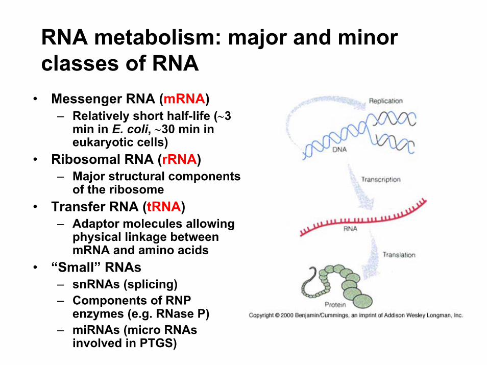
RNA metabolism: major and minor classes of RNA• Messenger RNA (mRNA)
– Relatively short half-life (∼3 min in E. coli, ∼30 min in eukaryotic cells)
• Ribosomal RNA (rRNA)– Major structural components
of the ribosome• Transfer RNA (tRNA)
– Adaptor molecules allowing physical linkage between mRNA and amino acids
• “Small” RNAs – snRNAs (splicing)– Components of RNP
enzymes (e.g. RNase P)– miRNAs (micro RNAs
involved in PTGS)

Overview of RNA polymerases• Prokaryotes
– Single processive RNA polymerase (technically, primase is a RNAP too).
– Inhibited by rifampicin (binds RNAP β subunit & blocks path of RNA chain elongation)
• Eukaryotes– Three processive RNAPs– Differential sensitivity to
inhibition by α-amanitin• RNA Pol I (resistant)
→ rRNA • RNA Pol II (low conc)
→ mRNA• RNA Pol III (high conc)
→ tRNA plus 5S rRNA
Fig. 26.4
Note: α-amanitin, a non-competitive inhibitor, stops the translocation of RNAP along the DNA template after the formation of the first phospho-diester bond.

Features of RNA vs DNA synthesis• Similarities to DNA synthesis
– Synthesis of ribonucleotide chain is template-dependent.– Substrates are nucleoside triphosphates (rNTPs).– Direction of chain growth is 5′→3′.– Same chemical mechanism applies (base-pairing of incoming
rNTP, 3′ OH attack, loss of PPi).– Highly processive enzyme
• Differences from DNA synthesis– One DNA strand is transcribed per gene w/o a primer.– Only certain genes are transcribed at any given time.– Kinetics favor “slow” transcription of multiple genes. (Vmax ∼50
nt/s for RNA Pol vs ∼103/s for DNA Pol III; ∼3000 RNA Pol/cell vs ∼10 DNA Pol III complexes/cell)
– Less accurate ∼10-5 vs 10-10
– Cofactor-mediated proofreading
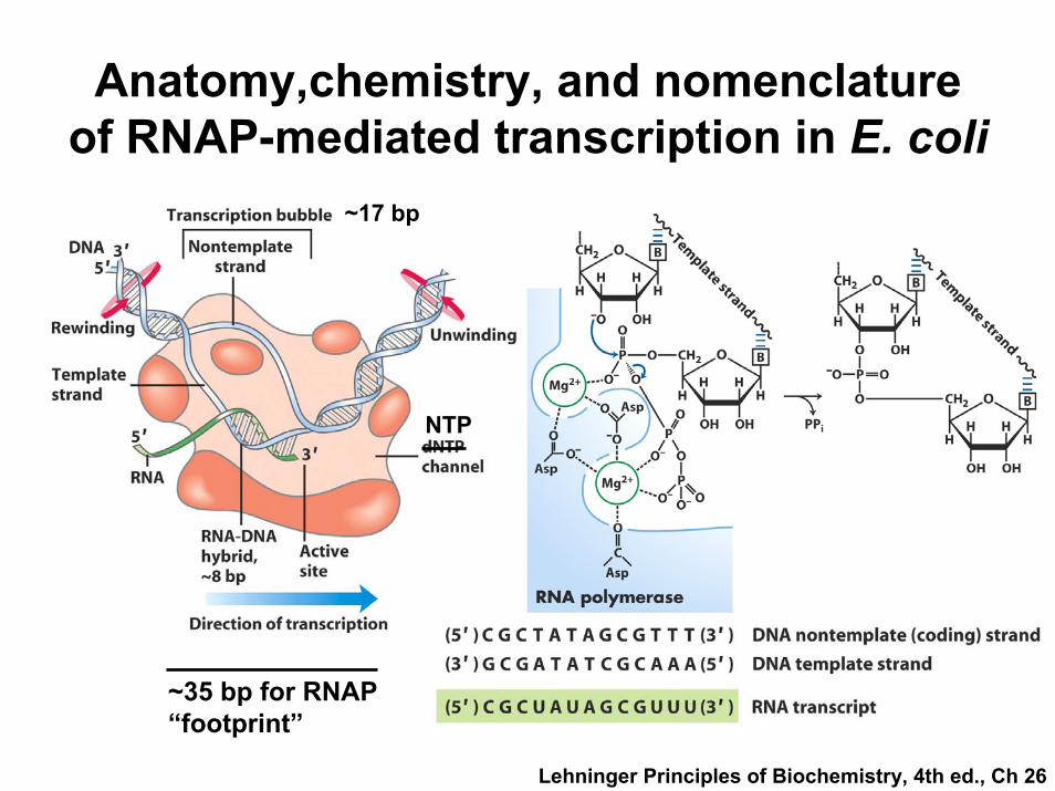
Anatomy,chemistry, and nomenclature of RNAP-mediated transcription in E. coli
~17 bp
NTP
~35 bp for RNAP “footprint”
Lehninger Principles of Biochemistry, 4th ed., Ch 26

Biochemical features of E. coli RNAP
Core RNAP
*
contains part of active site
holoenzyme assembly
sliding clamp
• 450 kDa enzyme containing six subunits• Two Mg2+ and one Zn2+ required (chemistry and clamping)• No independent 3′→5′ exonuclease activity but may have
kinetic proofreading capabilities• Two binding sites for ribonucleotides
– Initiation site binds only purine rNTPs (GTP or ATP) with Kd = 100 µM…most mRNAs start with purine on 5′ end.
– Elongation site binds any of 4 rNTPs with Kd = 10 µM.

σ factors: regulatory factors which direct transcription of certain genes
• Assist RNAP in binding DNA at the proper site for initiation of transcription – the promoter.
• Different sigma factors orchestrate transcription of different classes of genes. – Heat shock (σ35) – Other stress responses– Metabolic enzymes (σ70, most abundant)
• Not required for core RNA polymerase activity.
Transcription like replication can be construed to occur in distinct steps
• Initiation (requires special signals)– RNAP recognizes the promoter, binds to DNA,
and starts transcription.– Highly regulated
• Elongation– RNAP tracks down the length of the gene
synthesizing RNA along the way. • Termination (requires special signals)– Transcription stops then RNAP and the
nascent mRNA dissociate.

Features of initiation phase in E. coli 1:RNAP binding and sliding (electrostatic interaction)
2:Formation of closed complex (–55 to –5, Ka∼107-108 M−1,T½~10 s)
3:Formation of open complex (–10 to –1, Ka∼1012 M−1, T½~15s to 20 min), temp-dependent, stable
4:Mg2+-dependent conformational change (–12 to +2), add 1st nt
5:Promoter clearance: RNAP moves away from promoter
6:Release of σ after first 8-9 nts & continuation of elongation (now cannot be inhibited by rifampicin)
7,8:Pausing → Termination
Signal for specific DNA-binding seen by σ factor
Fig. 26-6
Transcription initiation: key role of the gene promoter• RNAP binding sequence: −70 to +30 in E. coli• DNA sequence specifying start site and basal rate
of transcription– Constitutive: Specify that a gene product will be
transcribed at a constant rate (e.g. genes involved in metabolic control)
– Inducible or regulated: Specify transcription of certain genes in response to external signals (requires additional protein-DNA interactions)
• Promoter recognition by RNAP: rate limiting for transcription (structure → frequency of initiation)
• Promoters: exhibit certain consensus sequences
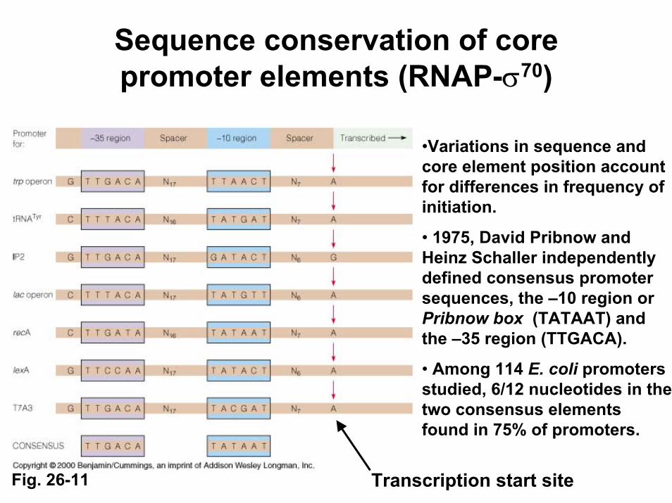
Sequence conservation of core promoter elements (RNAP-σ70)
•Variations in sequence and core element position account for differences in frequency of initiation.
• 1975, David Pribnow and Heinz Schaller independently defined consensus promoter sequences, the –10 region or Pribnow box (TATAAT) and the –35 region (TTGACA).
• Among 114 E. coli promoters studied, 6/12 nucleotides in the two consensus elements found in 75% of promoters.
Transcription start siteFig. 26-11

Genetic evidence for functional importance of core promoter elements(naturally-occurring and site-directed mutations)
• The more closely core elements resemble the consensus, the more efficient the promoter at initiating transcription.
• ↑Mutations: those toward the consensus sequence.
• ↓Mutations: those away from the consensus sequence.
• Spacing (optimal 17 bp) between core consensus sequences is important.
Fig. 26-12
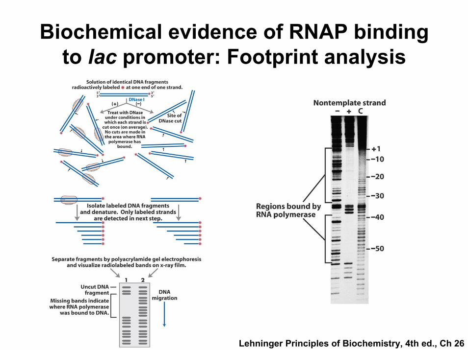
Biochemical evidence of RNAP binding to lac promoter: Footprint analysis
Lehninger Principles of Biochemistry, 4th ed., Ch 26
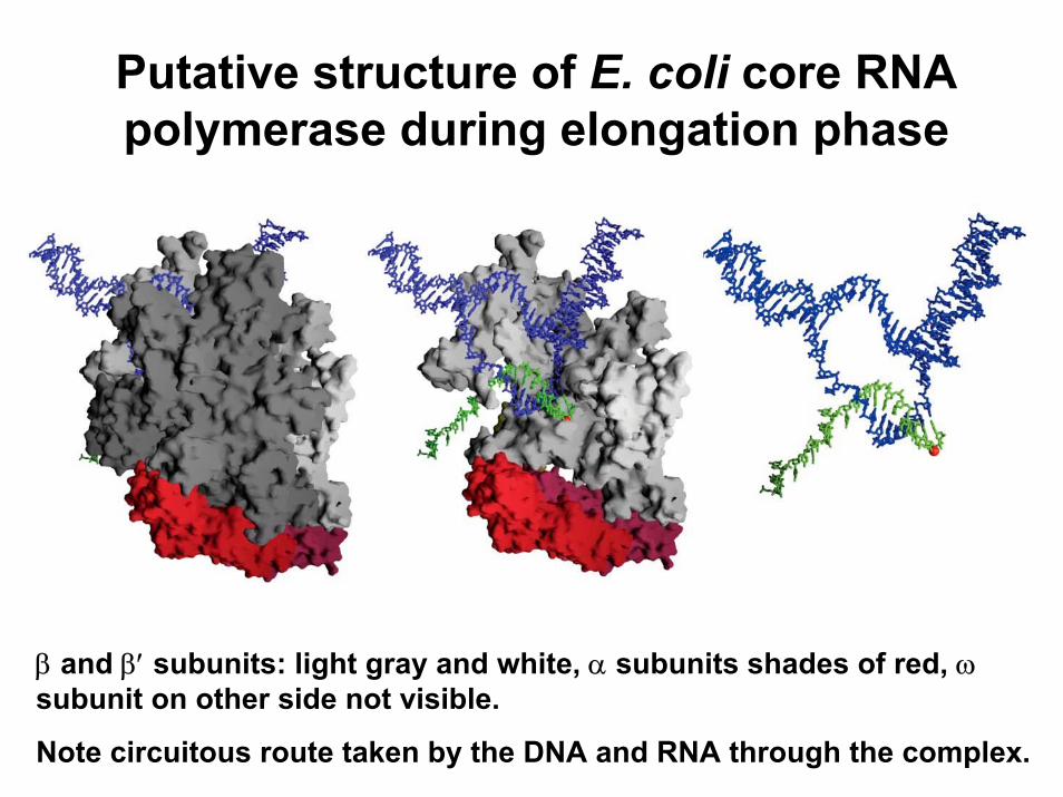
Putative structure of E. coli core RNA polymerase during elongation phase
β and β′ subunits: light gray and white, α subunits shades of red, ωsubunit on other side not visible.
Note circuitous route taken by the DNA and RNA through the complex.

Transcription elongation: a detailed view
• Elongation complexes are stabilized by contact between specific regions/residues of β/β′and the growing RNA chain (RBS), heteroduplex (HBS), or “downstream” DNA (DBS).
• Core RNAP moves along the DNA template simultaneously unwinding DNA ahead and rewinding the template behind. Zn2+-binding domain of β′subunit is the sliding clamp. RNAP activity requires Mg2+. Formation of 5′ RNA hairpin may be a signal for termination.
Fig. 26-9

But elongation of ternary complex often proceeds discontinuously….
• Transcription “bubble” model implies continuous movement but RNAP may pause at difficult to “read” sites (e.g. high G/C content).• Resolution of pause sites may involve backtracking to create a RNA 3′ end which is displaced from the active site. • GreA and GreB bind transiently to RNAP active site and stimulate its intrinsic transcript (i.e. RNA) hydrolysis activity creating a new base-paired 3′ end.Fig. 26-10
“backtracked” RNAP
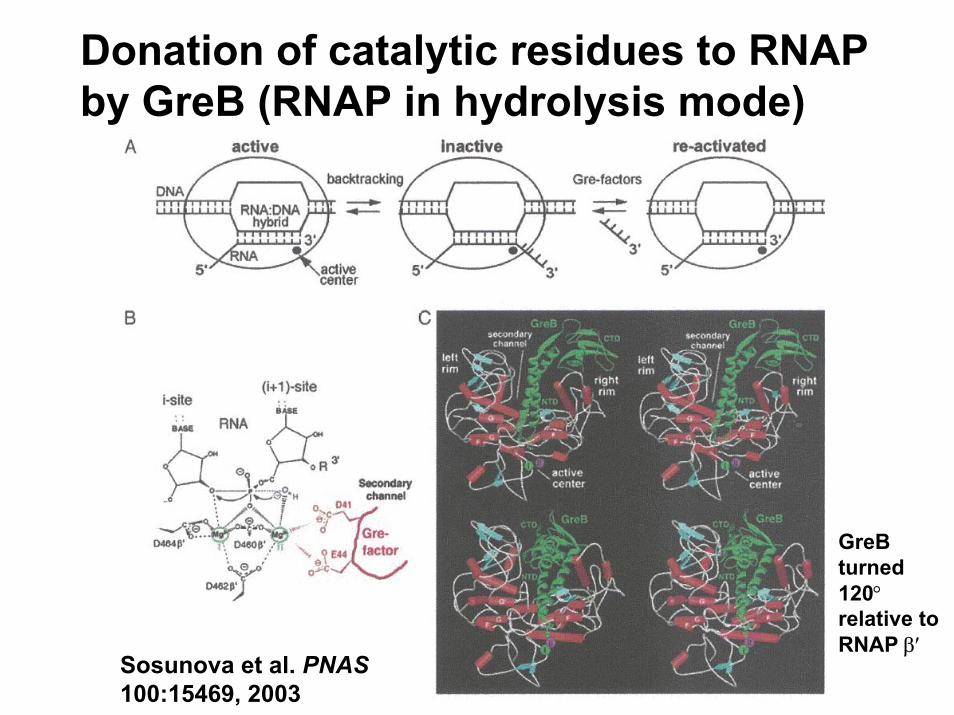
Donation of catalytic residues to RNAP by GreB (RNAP in hydrolysis mode)
Sosunova et al. PNAS100:15469, 2003
GreB turned 120°relative to RNAP β′

Termination of transcription: another process controlled by signals in DNA
• Termination signals are similar to signals that promote pausing– High G/C content (tend to form stem-loop structure)– Palindromic sequences that de-stabilize the DNA/RNA
heteroduplex
• Two types of termination mechanisms– Factor independent: Dyad symmetry followed by
poly A sequence - intrastrand stem loop followed by rU:dA that destabilizes RNA/template
– Factor (ρ, rho) dependent: Rho protein (RNA-dependent ATPase) destabilizes the RNA-DNA duplex.
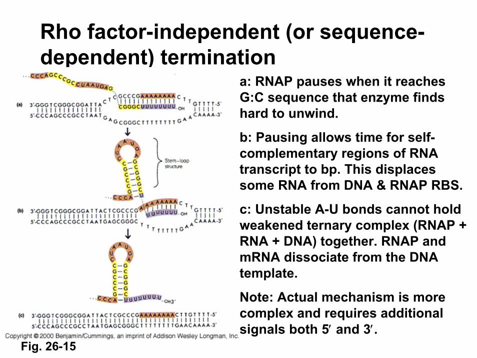
Rho factor-independent (or sequence-dependent) termination
a: RNAP pauses when it reaches G:C sequence that enzyme finds hard to unwind.
b: Pausing allows time for self-complementary regions of RNA transcript to bp. This displaces some RNA from DNA & RNAP RBS.
c: Unstable A-U bonds cannot hold weakened ternary complex (RNAP + RNA + DNA) together. RNAP and mRNA dissociate from the DNA template.
Note: Actual mechanism is more complex and requires additional signals both 5′ and 3′.
Fig. 26-15

Rho-dependent termination…less frequent and more complex
1: Rho (ρ) protein binds as a homohexamer to RNA at a CA-rich site (rut for rho utilization) near 3′ end and slides toward paused RNAP. 2: RNA-DNA helicase and ATPase activity of Rho unwinds RNA away from template DNA. 3: Template and transcript dissociate.Note: An additional protein, NusA, may be required for RNAP pausing. NusA binds to core RNAP after σ has dissociated. Fig. 26-16
NusA = N utilization substance