Embed Size (px)
Citation preview
Nephrol Dial Transplant (2014) 0: 1–6doi: 10.1093/ndt/gfu258
Original Article
Rituximab-associated agranulocytosis in children withrefractory idiopathic nephrotic syndrome: case seriesand review of literature
Koichi Kamei, Masaki Takahashi, Masaki Fuyama, Ken Saida, Hiroyuki Machida, Mai Sato, Masao Ogura
and Shuichi Ito
Department of Nephrology and Rheumatology, National Center for Child Health and Development, Tokyo, Japan
Correspondence and offprint requests to: Koichi Kamei; E-mail: [email protected]
ABSTRACT
Background. Agranulocytosis has been reported as a delayed-onset complication of rituximab treatment. However, the exactincidence and risk factors of this complication in patients withnephrotic syndrome remain unknown.Methods. Records of 213 rituximab treatments for 114 pa-tients with refractory nephrotic syndrome between February2006 and April 2013 were reviewed to identify episodes ofagranulocytosis (defined as an absolute neutrophil count of<500 mm3).Results. Eleven episodes of agranulocytosis were detected in11 patients. Median time of onset of agranulocytosis was 66days (range, 54–161 days) after rituximab treatment. Nine pa-tients experienced acute infections and received antibiotics.All but one patient received granulocyte colony-stimulatingfactor. Agranulocytosis resolved in all cases within a median of3 days. The incidence of agranulocytosis was 9.6% in total pa-tients and 5.2% in all treatments. Median age of the 11 patientswho developed agranulocytosis was 6.4 years at the first rituxi-mab treatment, significantly younger than the median age ofthe 103 patients who did not (median, 12.5 years; P = 0.0009).Five patients received re-treatment with rituximab. No recur-rence of agranulocytosis was observed in any patient.Conclusions. It is important to pay extra attention to this clin-ically serious delayed-onset complication as it may be accom-panied by life-threatening infections such as sepsis. Furtherclinical studies are needed to clarify its pathogenesis.
Keywords: agranulocytosis, B cell, infection, nephrotic syndrome,rituximab
INTRODUCTION
Rituximab is a chimeric monoclonal antibody directed againstthe cell surface antigen CD20 expressed on B lymphocytes. Asit has been proven to be effective in preventing relapses [1–7],it is increasingly being used in the treatment of patients withsteroid-dependent nephrotic syndrome. While transient infu-sion-related side effects such as cough, sore throat, dyspneaand fever may occur, the toxicity of rituximab is relatively mildand well tolerated by most patients.
Nonetheless, agranulocytosis, a severe form neutropenia,has been reported to be a delayed-onset complication of rituxi-mab treatment in patients with lymphoma [8–17] and auto-immune diseases [18–21]. Agranulocytosis usually occurs1–6 months after rituximab administration and is often self-limiting. However, life-threatening infections can sometimesemerge. To date, there has been a case report of agranulocyto-sis associated with rituximab in nephrotic syndrome [22]. Theexact incidence and risk factors of this complication in pa-tients with nephrotic syndrome remain unknown. Herein, weanalyzed all cases of agranulocytosis associated with rituximabtreatment for refractory nephrotic syndrome in our center toevaluate the incidence and clinical characteristics of this com-plication.
MATERIALS AND METHODS
Records of 213 rituximab treatments administered betweenFebruary 2006 and April 2013 for 114 patients with refractory
© The Author 2014. Published by Oxford University Presson behalf of ERA-EDTA. All rights reserved.
1
NDT Advance Access published August 1, 2014 at U
niversity of South Australia on A
ugust 10, 2014http://ndt.oxfordjournals.org/
Dow
nloaded from
nephrotic syndrome were reviewed to identify episodes ofagranulocytosis, defined as Grade 4 neutropenia (an absoluteneutrophil count of <500 mm3) by the Common TerminologyCriteria for Adverse Events (CTCAE) Ver. 4.0. Study protocolwas based on the Declaration of Helsinki and was conductedwith the approval of the off-label use of rituximab obtainedfrom the ethical committee of our center (#645).
Indication of rituximab treatment was refractory steroid-dependent nephrotic syndrome (steroid dependence underimmunosuppressive agents) or refractory steroid-resistantnephrotic syndrome (failure to go into remission despite acombination of cyclosporine and methylprednisolone pulsetherapy). Rituximab was administered at a single dose of 375mg/m2 for 205 treatments, two doses of 375 mg/m2 for onetreatment (2-week interval for each infusion) and four dosesof 375 mg/m2 (once a week) for seven treatments. A total of235 doses of rituximab were administered. To minimize infu-sion reactions, patients received intravenous methylpredniso-lone (1–1.5 mg/kg), oral acetaminophen (10 mg/kg, amaximum of 300 mg) and chlorpheniramine maleate (0.04mg/kg, a maximum of 2 mg), 30 min prior to rituximab infu-sion. All patients were admitted to our center and were moni-tored for at least 24 h after rituximab treatment for infusionreactions. Complete blood counts and CD19+ B-cell countswere performed at least once a month until B-cell recovery.When a patient presented with fever at an emergency visit,procedures such as complete blood counts, C-reactive proteintest, chest X-ray, urinalysis, blood and urine culture were im-mediately performed. Rituximab-associated agranulocytosiswas defined as an episode during B-cell depletion where CD19+B-cell count was <1% of total lymphocytes. B-cell recovery wasdefined as CD19+ B-cell count of equal to or >1% of total lym-phocytes. Since January 2012, sulfamethoxazole/trimethoprimhas been used at a dose of 5 mg/kg of trimethoprim onceevery 2 days for B-cell depletion with prophylaxis of pneumo-cystis jirovecii and bacterial infection.
The data were analyzed with JMP version 9.0 (SAS instituteJapan Ltd, Tokyo, Japan). The Mann–Whitney U-test was
used for continuous values and the Fisher’s exact test for cat-egorical values. Statistical significance was established atP < 0.05.
RESULTS
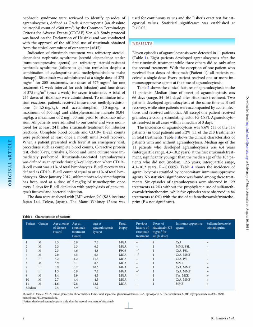
Eleven episodes of agranulocytosis were detected in 11 patients(Table 1). Eight patients developed agranulocytosis after thefirst rituximab treatment while three others did so only afterthe second treatment. With the exception of one patient whoreceived four doses of rituximab (Patient 1), all patients re-ceived a single dose. Every patient received one or more im-munosuppressive agents at the time of agranulocytosis.
Table 2 shows the clinical features of agranulocytosis in the11 patients. Median time of onset of agranulocytosis was66 days (range, 54–161 days) after rituximab treatment. Twopatients developed agranulocytosis at the same time as B-cellrecovery, while nine patients were accompanied by acute infec-tions and received antibiotics. All except one patient receivedgranulocyte colony-stimulating factor (G-CSF). Agranulocyto-sis resolved in all cases within a median of 3 days.
The incidence of agranulocytosis was 9.6% (11 of the 114patients) in total patients and 5.2% (11 of the 213 treatments)in total treatments. Table 3 shows the clinical characteristics ofpatients with and without agranulocytosis. Median age of the11 patients who developed agranulocytosis was 6.4 years(interquartile range, 4.3–10.2 years) at the first rituximab treat-ment; significantly younger than the median age of the 103 pa-tients who did not (median, 12.5 years; interquartile range,4.3–10.2 years; P = 0.0009). Table 4 shows the incidence ofagranulocytosis stratified by concomitant immunosuppressiveagents. No statistical significance was found among these treat-ments. Six episodes of agranulocytosis were observed in 129treatments (4.7%) without the prophylactic use of sulfameth-oxazole/trimethoprim, while five episodes were observed in 84treatments (6.0%) with the use of sulfamethoxazole/trimetho-prim (P = not significant).
Table 1. Characteristics of patients
Patient Gender Age at onsetof disease(years)
Age atrituximabtreatment(years)
Age atagranulocytosis(years)
Renalbiopsy
Previoushistory ofrituximabtreatment
Doses ofrituximab (375mg/m2 forsingle dose)
Immunosuppressiveagents
Sulfamethoxazole/trimethoprim
1 M 2.5 6.9 7.3 MGA – 4 CsA –2 M 2.3 6.3 6.5 MGA – 1 MMF, PSL –3 M 2.3 4.6 4.8 FSGS +a 1 CsA, PSL –4 M 2.0 6.5 6.6 MGA +a 1 CsA, MMF –5 F 8.2 11.2 11.3 MGA – 1 CsA, PSL –6 M 6.9 8.1 8.6 MGA – 1 MMF –7 F 3.9 10.2 10.6 MGA – 1 CsA, MMF +8 F 1.3 6.9 7.2 MGA +a 1 CsA, MMF +9 M 1.4 3.9 4.3 MGA – 1 Tac, MZR +10 M 2.7 4.4 4.5 MGA – 1 CsA, MMF +11 M 11.6 12.8 13.1 MGA – 1 MMF +Median 2.5 6.9 7.2
M, male; F, female; MGA, minor glomerular abnormalities; FSGS, focal segmental glomerulosclerosis; CsA, cyclosporin A; Tac, tacrolimus; MMF, mycophenolate mofetil; MZR,mizoribine; PSL, prednisolone.aPatient developed agranulocytosis only after the second treatment of rituximab.
ORIG
INALARTIC
LE
2 K. Kamei et al.
at University of South A
ustralia on August 10, 2014
http://ndt.oxfordjournals.org/D
ownloaded from
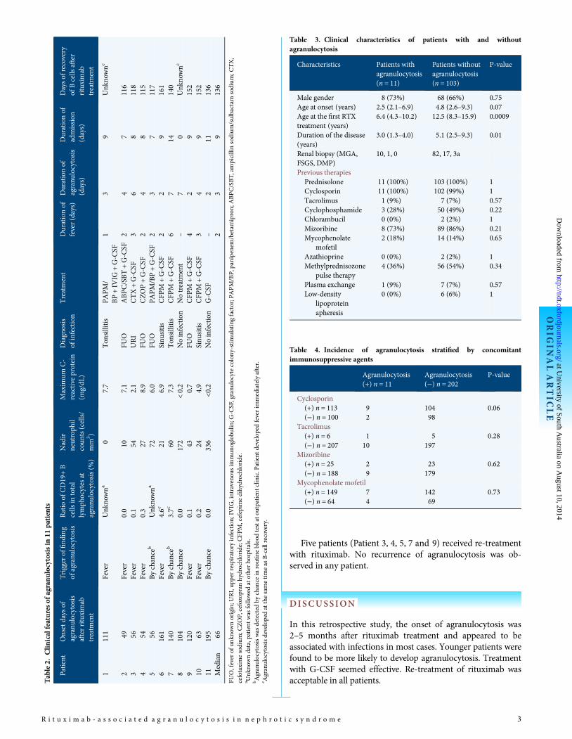
Five patients (Patient 3, 4, 5, 7 and 9) received re-treatmentwith rituximab. No recurrence of agranulocytosis was ob-served in any patient.
DISCUSSION
In this retrospective study, the onset of agranulocytosis was2–5 months after rituximab treatment and appeared to beassociated with infections in most cases. Younger patients werefound to be more likely to develop agranulocytosis. Treatmentwith G-CSF seemed effective. Re-treatment of rituximab wasacceptable in all patients.T
able2.
Clinicalfeatures
ofagranulocytosisin
11patients
Patient
Onsetdays
ofagranu
locytosis
afterrituximab
treatm
ent
Triggerof
find
ing
ofagranu
locytosis
Ratioof
CD19+B
cells
intotal
lymph
ocytes
atagranu
locytosis(%
)
Nadir
neutroph
ilcoun
ts(cells/
mm
3 )
Maxim
umC-
reactive
protein
(mg/dL
)
Diagnosis
ofinfection
Treatment
Durationof
fever(days)
Durationof
agranu
locytosis
(days)
Durationof
admission
(days)
Daysof
recovery
ofBcells
after
rituximab
treatm
ent
1111
Fever
Unk
nowna
07.7
Ton
sillitis
PAPM/
BP+IV
IG+G-C
SF1
39
Unk
nownc
249
Fever
0.0
107.1
FUO
ABPC/SBT+G-C
SF2
47
116
356
Fever
0.1
542.1
URI
CTX+G-C
SF3
68
118
454
Fever
0.3
278.9
FUO
CZOP+G-C
SF2
48
115
556
Bychance
bUnk
nowna
726.0
FUO
PAPM/BP+G-C
SF2
37
117
6161
Fever
4.6c
216.9
Sinu
sitis
CFP
M+G-C
SF2
29
161
7140
Bychance
b3.7c
607.3
Ton
sillitis
CFP
M+G-C
SF6
714
140
8104
Bychance
0.0
172
<0.2
Noinfection
Notreatm
ent
–7
0Unk
nownc
9120
Fever
0.1
430.7
FUO
CFP
M+G-C
SF4
29
152
1063
Fever
0.2
244.9
Sinu
sitis
CFP
M+G-C
SF3
49
152
11195
Bychance
0.0
336
<0.2
Noinfection
G-C
SF–
211
136
Median
662
39
136
FUO,feverof
unkn
ownorigin;U
RI,up
perrespiratoryinfection;IV
IG,intraveno
usim
mun
oglobu
lin;G
-CSF,granu
locytecolony-stimulatingfactor;PAPM/BP,panipenem/betam
ipron;ABPC/SBT,ampicillinsodium
/sulbactam
sodium
;CTX,
cefotaximesodium
;CZOP,cefozopran
hydrochloride;CFP
M,cefepim
edihydrochloride.
a Unk
nowndata,patient
was
followed
atotherho
spitals.
bAgranulocytosiswas
detected
bychance
inroutinebloodtestatou
tpatient
clinic.Patient
developedfeverim
mediatelyafter.
c Agranulocytosisdevelopedatthesametimeas
B-cellrecovery.
Table 3. Clinical characteristics of patients with and withoutagranulocytosis
Characteristics Patients withagranulocytosis(n = 11)
Patients withoutagranulocytosis(n = 103)
P-value
Male gender 8 (73%) 68 (66%) 0.75Age at onset (years) 2.5 (2.1–6.9) 4.8 (2.6–9.3) 0.07Age at the first RTXtreatment (years)
6.4 (4.3–10.2) 12.5 (8.3–15.9) 0.0009
Duration of the disease(years)
3.0 (1.3–4.0) 5.1 (2.5–9.3) 0.01
Renal biopsy (MGA,FSGS, DMP)
10, 1, 0 82, 17, 3a
Previous therapiesPrednisolone 11 (100%) 103 (100%) 1Cyclosporin 11 (100%) 102 (99%) 1Tacrolimus 1 (9%) 7 (7%) 0.57Cyclophosphamide 3 (28%) 50 (49%) 0.22Chlorambucil 0 (0%) 2 (2%) 1Mizoribine 8 (73%) 89 (86%) 0.21Mycophenolate
mofetil2 (18%) 14 (14%) 0.65
Azathioprine 0 (0%) 2 (2%) 1Methylprednisozone
pulse therapy4 (36%) 56 (54%) 0.34
Plasma exchange 1 (9%) 7 (7%) 0.57Low-density
lipoproteinapheresis
0 (0%) 6 (6%) 1
Table 4. Incidence of agranulocytosis stratified by concomitantimmunosuppressive agents
Agranulocytosis(+) n = 11
Agranulocytosis(−) n = 202
P-value
Cyclosporin(+) n = 113 9 104 0.06(−) n = 100 2 98
Tacrolimus(+) n = 6 1 5 0.28(−) n = 207 10 197
Mizoribine(+) n = 25 2 23 0.62(−) n = 188 9 179
Mycophenolate mofetil(+) n = 149 7 142 0.73(−) n = 64 4 69
ORIG
INALARTIC
LE
R i t u x i m a b - a s s o c i a t e d a g r a n u l o c y t o s i s i n n e p h r o t i c s y n d r o m e 3
at University of South A
ustralia on August 10, 2014
http://ndt.oxfordjournals.org/D
ownloaded from

Table 5 shows previous reports of agranulocytosis asso-ciated with rituximab treatment in patients with non-Hodgkinlymphoma [8–17] and autoimmune diseases [18–21]. The in-cidence of agranulocytosis was reported as 6.9 and 3.2%, re-spectively. The higher incidence in patients with lymphomawas probably due to the influence of other chemotherapies. Incomparison, the incidence in our study is higher than that ofpatients with autoimmune diseases, but lower than that of pa-tients with lymphoma.
The risk factors for agranulocytosis after rituximab treat-ment have so far been investigated mainly in lymphoma pa-tients. Autologous stem-cell transplantation [9, 13], acquiredimmunodeficiency syndrome-related lymphoma [10], previ-ous cytotoxic treatment [11], intensive chemotherapy [13],intensive radiotherapy [13], advanced stage of lymphoma(3 or 4) [13], multiple doses of rituximab (>4) [11] and FcγRgenotypes [16] have all been reported to be risk factors. Inour study, a young age at time of rituximab treatment seemedto be risk factor. In our study, even though one patient re-ceived four doses of rituximab while the others received asingle dose, the number of rituximab may not have been aninfluencing factor in the development of agranulocytosis.Further investigation is needed to verify this.
While we used G-CSF for our patients, we are aware that itsefficacy for rituximab-induced agranulocytosis remains question-able. However, a systematic review of 980 case reports of drug-induced agranulocytosis did demonstrate that the use of hemato-poietic growth factors resulted in a shorter median duration ofneutropenia, and that patients treated with growth factors hadsignificantly lower proportion of infections or fatal complications[23]. A recent review article proposed that the use G-CSF mustbe considered on a case-by-case basis [24]. The authors suggestedthat in patients presenting with Grade 4 neutropenia or asso-ciated factors that may confer poor prognosis, such as agedpeople, absolute neutrophil count of <100 /mm3, clinical evi-dence of bacteremia or septic shock and severe comorbiditiessuch as renal failure, the use of G-CSF may be justifiable.
To date, there are few reports on re-administration ofrituximab. One case series from Israel reported a rituximab
re-treatment for late-onset neutropenia resulted in recurrentepisodes of agranulocytosis in one patient [25], while anothercase series reported six patients with re-treatment but hadno such recurrence [21]. In our study, none of the patientsexperienced recurrence of agranulocytosis. Moreover, 3 of the11 patients developed agranulocytosis only after the second ri-tuximab treatment. Nonetheless, further investigation is requiredto confirm the safety of rituximab re-treatment for patients witha history of rituximab-associated agranulocytosis.
The exact mechanism of neutropenia associated with rituxi-mab remains poorly understood. Several hypotheses have beenproposed, such as the transient production of autoantibodiesagainst neutrophils during immune reconstitution [26], hyper-proliferation of large granular T cells in the bone marrow[27, 28] or viral infection during dysfunctional humoral im-munity [29, 30]. In previous reports, neutrophil maturationarrest was revealed by bone marrow aspiration. A direct toxicityof rituximab is less likely since CD20 is not expressed on neu-trophils and their precursors. Dunleavy et al. [10] reported acorrelation between rapid B-cell recovery and perturbation ofstromal cell-derived factor-1 (SDF-1) and neutropenia. Anothercytokine influenced by rituximab administration is B-cell-activating factor (BAFF), which plays a role in human B-cellsurvival, expansion and development. Terrier et al. [31] re-ported that BAFF levels were undetectable prior to rituximabadministration, but increased after treatment and eventuallypeaked at a time interval that coincided with the occurrenceof neutropenia. The fact that two of our patients developedagranulocytosis almost at the same time as B-cell recovery maysupport these hypotheses, although it is noteworthy that neitherSDF-1 nor BAFF was analyzed. Some reports have shown thatFcγRIIIa polymorphism was highly associated with the develop-ment of late-onset neutropenia [16, 32]. Weissmann-Brenneret al. [33], who showed a suppression of colony-forming unitgrowth by plasma of patients with rituximab-induced neutro-penia, have proved that circulating antibodies in the plasmamay be responsible for leukopenia after rituximab treatment.Again, further investigation is necessary to confirm the exactpathogenesis.
Table 5. Previous reports of agranulocytosis associated with rituximab treatment
Reference Years ofpublication
Primary disease Number of patients of rituximabtreatment
Number of patients ofagranulocytosis
Incidence of agranulocytosis(%)
[8] 2003 NHL 59 8 14[9] 2004 NHL 39 6 15[10] 2005 NHL 76 6 8[11] 2006 NHL 77 10 13[12] 2006 NHL 54 3 6[13] 2006 NHL 107 10 9[14] 2008 NHL 113 6 5[15] 2009 NHL 121 4 3[16] 2010 NHL 80 2 3[17] 2012 NHL 160 6 4[18] 2007 AID 23 1 4[19] 2009 AID 65 2 3[20] 2011 AID 209 5 2[21] 2012 AID 138 6 4
ORIG
INALARTIC
LE
4 K. Kamei et al.
at University of South A
ustralia on August 10, 2014
http://ndt.oxfordjournals.org/D
ownloaded from
Agranulocytosis was detected in four of our patients bychance, supporting the theory of a primary mechanism.The other seven patients experienced fever prior to detection,suggesting a mechanism secondary to infections. On the otherhand, 32 events of bacterial infections were observed in the114 patients who received rituximab treatments but experi-enced no agranulocytosis. As such, we think that the cause ofagranulocytosis varies from patient to patient.
There are several limitations to our study. First, we do nothave information on the maturation of neutrophils as no bonemarrow aspirations were performed. Second, as the patientswere receiving one or more immunosuppressive agents at thetime of agranulocytosis, we cannot rule out the possibility thatthese agents may have an association with the condition.However, none of our patients developed agranulocytosisbefore rituximab use despite the use of a similar immunosup-pressive menu. This suggests that rituximab is likely to be themain cause of agranulocytosis. Third, the number of patientswas relatively small. We need to accumulate more clinical dataand information on this complication in future studies.
In conclusion, rituximab use is a potential cause of agran-ulocytosis for patients with refractory idiopathic nephroticsyndrome. As rituximab use is increasing in such patients, wehave to be vigilant of any potential clinically serious delayed-onset complication. When a patient with a history of rituxi-mab treatment suffers from fever, we should not rule out thepossibility of agranulocytosis as well as serious bacterial infec-tions such as sepsis. At the same time, it is necessary for pa-tients to recognize this complication and visit an emergencyclinic as soon as possible upon developing fever. The monitor-ing of complete blood counts and CD19+ B-cell counts at leastonce a month after rituximab use until B-cell recovery may behelpful for the early detection of agranulocytosis and preven-tion of serious infection. Further clinical studies are needed toelucidate the pathogenesis of delayed-onset postrituximabagranulocytosis.
ACKNOWLEDGEMENTS
The authors thank Drs K. Tanaka (Aomori), K. Tsuruga(Aomori), T. Watanabe (Gumma), Y. Owada (Tochigi),R. Hiramoto (Chiba), H. Eguchi (Chiba), M. Hisano (Chiba),C. Matsumura (Chiba), Y. Akioka (Tokyo), R. Hamada(Tokyo), T. Udagawa (Tokyo), M. Okada (Tokyo), M. Mizutani(Yamaguchi), M. Fujieda (Kochi), M. Ishihara (Kochi),Y. Kaku (Fukuoka), Y. Otsuka (Saga), H. Nagasako (Kagoshi-ma), A. Miyazono (Kagoshima), H. Yoshimura (Okinawa) andK. Iijima (Kobe) for their contributions to this study. Wewould also like to thank Dr J. Tang from the Department ofEducation for Clinical Research, National Center for ChildHealth and Development, for proofreading and editing themanuscript.
CONFLICT OF INTEREST STATEMENT
None declared.
REFERENCES
1. Sinha A, Bagga A. Rituximab therapy in nephrotic syndrome: implicationsfor patients’management. Nat Rev Nephrol 2013; 9: 154–169
2. Kamei K, Ito S, Nozu K et al. Single dose of rituximab for refractorysteroid-dependent nephrotic syndrome in children. Pediatr Nephrol 2009;24: 1321–1328
3. Gulati A, Sinha A, Jordan SC et al. Efficacy and safety of treatmentwith rituximab for difficult steroid-resistant and -dependent nephroticsyndrome: multicentric report. Clin J Am Soc Nephrol 2010; 5:2207–2212
4. Ravani P, Magnasco A, Edefonti A et al. Short-term effects of rituximabin children with steroid- and calcineurin-dependent nephrotic syn-drome: a randomized controlled trial. Clin J Am Soc Nephrol 2011; 6:1308–1315
5. Kemper MJ, Gellermann J, Habbig S et al. Long-term follow-up after ri-tuximab for steroid-dependent idiopathic nephrotic syndrome. NephrolDial Transplant 2012; 27: 1910–1915
6. Ito S, Kamei K, Ogura M et al. Survey of rituximab treatment for child-hood-onset refractory nephrotic syndrome. Pediatr Nephrol 2013; 28:257–264
7. Ravani P, Ponticelli A, Siciliano C et al. Rituximab is a safe and effectivelong-term treatment for children with steroid and calcineurin inhibitor-dependent idiopathic nephrotic syndrome. Kidney Int 2013; 84:1025–1033
8. Chaiwatanatorn K, Lee N, Grigg A et al. Delayed-onset neutropenia asso-ciated with rituximab therapy. Br J Haematol 2003; 121: 913–918
9. Lemieux B, Tartas S, Traulle C et al. Rituximab-related late-onset neutro-penia after autologous stem cell transplantation for aggressive non-Hodg-kin’s lymphoma. Bone Marrow Transplant 2004; 33: 921–923
10. Dunleavy K, Hakim F, Kim HK et al. B-cell recovery following rituximab-based therapy is associated with perturbations in stromal derived factor-1and granulocyte homeostasis. Blood 2005; 106: 795–802
11. Cattaneo C, Spedini P, Casari S et al. Delayed-onset peripheral blood cyto-penia after rituximab: frequency and risk factor assessment in a consecu-tive series of 77 treatments. Leuk Lymphoma 2006; 47: 1013–1017
12. Fukuno K, Tsurumi H, Ando N et al. Late-onset neutropenia in patientstreated with rituximab for non-Hodgkin’s lymphoma. Int J Hematol 2006;84: 242–247
13. Nitta E, Izutsu K, Sato T et al. A high incidence of late-onset neutropeniafollowing rituximab-containing chemotherapy as a primary treatment ofCD20-positive B-cell lymphoma: a single-institution study. Ann Oncol2007; 18: 364–369
14. Tesfa D, Gelius T, Sander B et al. Late-onset neutropenia associated withrituximab therapy: evidence for a maturation arrest at the (pro)myelocytestage of granulopoiesis. Med Oncol 2008; 25: 374–379
15. Lai GG, Lim ST, Tao M et al. Late-onset neutropenia following RCHOPchemotherapy in diffuse large B-cell lymphoma. Am J Hematol 2009; 84:414–417
16. Li SC, Chen YC, Evens AM et al. Rituximab-induced late-onset neutro-penia in newly diagnosed B-cell lymphoma correlates with Fc receptorFcγRIIIa 158(V/F) polymorphism. Am J Hematol 2010; 85: 810–812
17. Rozman S, Sonc M, Novakovic BJ. Late-onset neutropenia followingprimary treatment of diffuse large B-cell lymphoma with rituximab-con-taining therapy. Leuk Lymphoma 2012; 53: 1945–1948
18. Rios-Fernández R, Gutierrez-Salmerón MT, Callejas-Rubio JL et al. Late-onset neutropenia following rituximab treatment in patients with auto-immune diseases. Br J Dermatol 2007; 157: 1271–1273
19. Jones RB, Ferraro AJ, Chaudhry AN et al. A multicenter survey of rituxi-mab therapy for refractory antineutrophil cytoplasmic antibody-associatedvasculitis. Arthritis Rheum 2009; 60: 2156–2168
20. Tesfa D, Ajeganova S, Hägglund H et al. Late-onset neutropeniafollowing rituximab therapy in rheumatic diseases: association withB lymphocyte depletion and infections. Arthritis Rheum 2011; 63:2209–2214
21. Besada E, Koldingsnes W, Nossent J. Characteristics of late onset neutro-penia in rheumatologic patients treated with rituximab: a case review ana-lysis from a single center. QJM 2012; 105: 545–550
ORIG
INALARTIC
LE
R i t u x i m a b - a s s o c i a t e d a g r a n u l o c y t o s i s i n n e p h r o t i c s y n d r o m e 5
at University of South A
ustralia on August 10, 2014
http://ndt.oxfordjournals.org/D
ownloaded from
22. Kshirsagar AA, Reid CJ, Alamelu J et al. An unusual cause of severe per-sistent neutropenia in a child with nephrotic syndrome. BMJ Case Rep2013; 29; 1–3
23. Andersohn F, Konzen C, Garbe E. Systematic review: agranulocytosisinduced by nonchemotherapy drugs. Ann Intern Med 2007; 146: 657–665
24. Wolach O, Shpilberg O, Lahav M. Neutropenia after rituximab treatment:new insights on a late complication. Curr Opin Hematol 2012; 19: 32–38
25. Wolach O, Bairey O, Lahav M. Late-onset neutropenia after rituximabtreatment: case series and comprehensive review of the literature. Medi-cine (Baltimore) 2010; 89: 308–318
26. Voog E, Morschhauser F, Solal-Céligny P. Neutropenia in patients treatedwith rituximab. N Engl J Med 2003; 348: 2691–2694
27. Papadaki T, Stamatopoulos K, Stavroyianni N et al. Evidence for T-largegranular lymphocyte-mediated neutropenia in Rituximab-treated lymph-oma patients: report of two cases. Leuk Res 2002; 26: 597–600
28. Stamatopoulos K, Papadaki T, Pontikoglou C et al. Lymphocyte subpopu-lation imbalances, bone marrow hematopoiesis and histopathology in ri-tuximab-treated lymphoma patients with late-onset neutropenia.Leukemia 2008; 22: 1446–1449
29. Klepfish A, Rachmilevitch E, Schattner A. Parvovirus B19 reactivation pre-senting as neutropenia after rituximab treatment. Eur J Intern Med 2006;17: 505–507
30. Christopeit M, Haak U, Behre G. Late-onset neutropenia following viralbone marrow depression after rituximab therapy. Ann Hematol 2008; 87:761–762
31. Terrier B, Ittah M, Tourneur L et al. Late-onset neutropenia following ri-tuximab results from a hematopoietic lineage competition due to an exces-sive BAFF-induced B-cell recovery. Haematologica 2007; 92: e20–e23
32. Weng WK, Negrin RS, Lavori P et al. Immunoglobulin G Fc receptorFcγRIIIa 158 V/F polymorphism correlates with rituximab-induced neu-tropenia after autologous transplantation in patients with non-Hodgkin’slymphoma. J Clin Oncol 2010; 28: 279–284
33. Weissmann-Brenner A, Brenner B, Belyaeva I et al. Rituximab associatedneutropenia: description of three cases and an insight into the underlyingpathogenesis. Med Sci Monit 2011; 17: CS133–CS137
Received for publication: 27.3.2014; Accepted in revised form: 2.7.2014
ORIG
INALARTIC
LE
6 K. Kamei et al.
at University of South A
ustralia on August 10, 2014
http://ndt.oxfordjournals.org/D
ownloaded from