Embed Size (px)
Citation preview

Right Ventricular Failure -Anytime is a Bad Time
Eric R. Skipper, MD, FACS
Chief, Adult Cardiothoracic Surgery
Surgical Director Cardiac Transplantation
and Mechanical Assist
Sanger Heart and Vascular Institute
NCSRC Symposium 2012
34th Annual Meeting
September 28, 2012
Right Ventricular Failure -Anytime is a Bad Time
• No disclosures
• No conflicts of interest

Overview
• For decades, RV function was “under-valued”.• Basically..... a passive conduit for blood flow between the
systemic and pulmonary circulation
• Currently, we realize: • incidence of RV failure approximates that of LV failure
• RV failure can carry a worse prognosis than isolated LV failure
• Understanding both RV anatomy and physiology as well as the impact of RV dysfunction is essential in the management of these patients
Right Ventricular Anatomy / Physiology
• Receives blood from the right atrium (RA) and ejects blood into the pulmonary artery (PA)
• Divided into 2 sections• body or sinus - receives blood from the RA
• outflow tract or conus (infundibulum) - funnels blood into the PA
• Crista supraventricularis - muscular ridge separating the sinus region from the conus region

Right Ventricular Anatomy / Physiology
• Cross-sectional anatomy • Crescent-shaped
� concave free wall
� convex interventricular septum
• Thin walled
� 18% as much muscle mass as the LV
• Overall design
� accommodates increases in preload well
� does not tolerate significant increases in afterload
Right Ventricular Anatomy / Physiology
• In contrast to the LV, the RV:� pumps blood under much lower pressures through a
highly compliant vascular system ( ie: the pulmonary vascular bed)
� volume work, not pressure work
• RV ejection is dependent on:� contraction of it’s free wall
� contraction of the interventricular septum
� twisting, corkscrew action of the LV

Causes of Right Ventricular Failure
• Normal afterload• RV infarction
• Increased afterload• PE
• MV disease w pulmon htn
• Congenital heart disease
• OSA
• ARDS
• Following cardiac or thoracic surgery• Inflammatory effects of CPB
• Protamine
• Extensive lung resection
• LVAD
• Volume overload• ASD / VSD
Right Ventricular Failure with Normal RV Afterload• Myocardial Infarction - the most common senario
�RCA disease�Left Circumflex disease (left dominant)
• Within the past decade, the high mortality in this patient population has been recognized

Consequences of Increased RV Afterload
• Opening of the pulmonary valve in systole is delayed
• RV pressure-volume curve assumes the shape of a “LV curve” -> greater oxygen consumption
• RV dilates -> increases wall stress (a major determinant of O2 demand)
• RVEDP increases -> RCA blood flow becomes restricted to a diastolic phoenomenon
• O2 supply is decreased inspite of an increasing demand
• TV annulus dilates -> TR which causes further dilation
• RV hypertrophy occurs due to increased wall stress
• Cresentric shape is lost and IVS buldges into the LV
• LV filling and function is impaired
• Systemic and coronary perfusion pressures decrease
Right Ventricular Failure- Schematic Representation
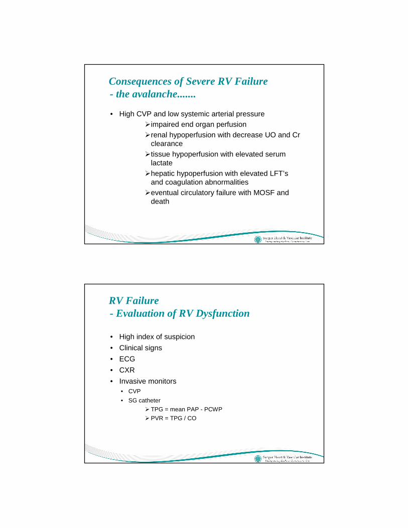
Consequences of Severe RV Failure- the avalanche.......
• High CVP and low systemic arterial pressure�impaired end organ perfusion�renal hypoperfusion with decrease UO and Cr
clearance�tissue hypoperfusion with elevated serum
lactate�hepatic hypoperfusion with elevated LFT’s
and coagulation abnormalities�eventual circulatory failure with MOSF and
death
RV Failure - Evaluation of RV Dysfunction
• High index of suspicion
• Clinical signs
• ECG
• CXR
• Invasive monitors• CVP
• SG catheter
� TPG = mean PAP - PCWP
�PVR = TPG / CO

RV Failure - Evaluation of RV Dysfunction • ECHO
• clues
� dilation
� hypertrophy
� contractility
� septal shift
� TR
• Specifically
�RV free wall motion
� free wall thickness (>15 mm)
� dilation
� TR
� tissue doppler tricuspid annular velocity
� hepatic venous flow pattern
Right Ventricular Failure- Perioperative Management
• Optimize preload
• Maintain AV synchrony ( ie: NSR)
• Attn to factors that can alter PVR�PaO2
�PCO2
� pH
�PCWP (LAP)
� airway pressure
� preload
» reduce with nitrates, diuretics, etc
�BP - not too low, not too high....just right

Right Ventricular Failure- Perioperative Management - When the Above Fails.....• Inotropes
• inhaled Nitric Oxode (iNO)
• inhaled Prostacyclin (iProstacyclin)
• Sildenafil
• Ventricular-Assist options
• “Creative Ventilator Tricks”• Collaboration is key
�Respiratory Therapist
�Pulmonary / Critical Care
Right Ventricular Failure- Inotropes• Dobutamine
�Beta 1 agonist with limited alpha 1 activity
• Phenylephrine / Norepinephrine � If arterial BP is low, vasoconstrictors may be beneficial
• Isoprenaline � pulmonary vasodilator
� use is limited by tachycardia
• Phosphodiesterase inhibitors� cAMP pathway
� vascular smooth muscle vasodilator
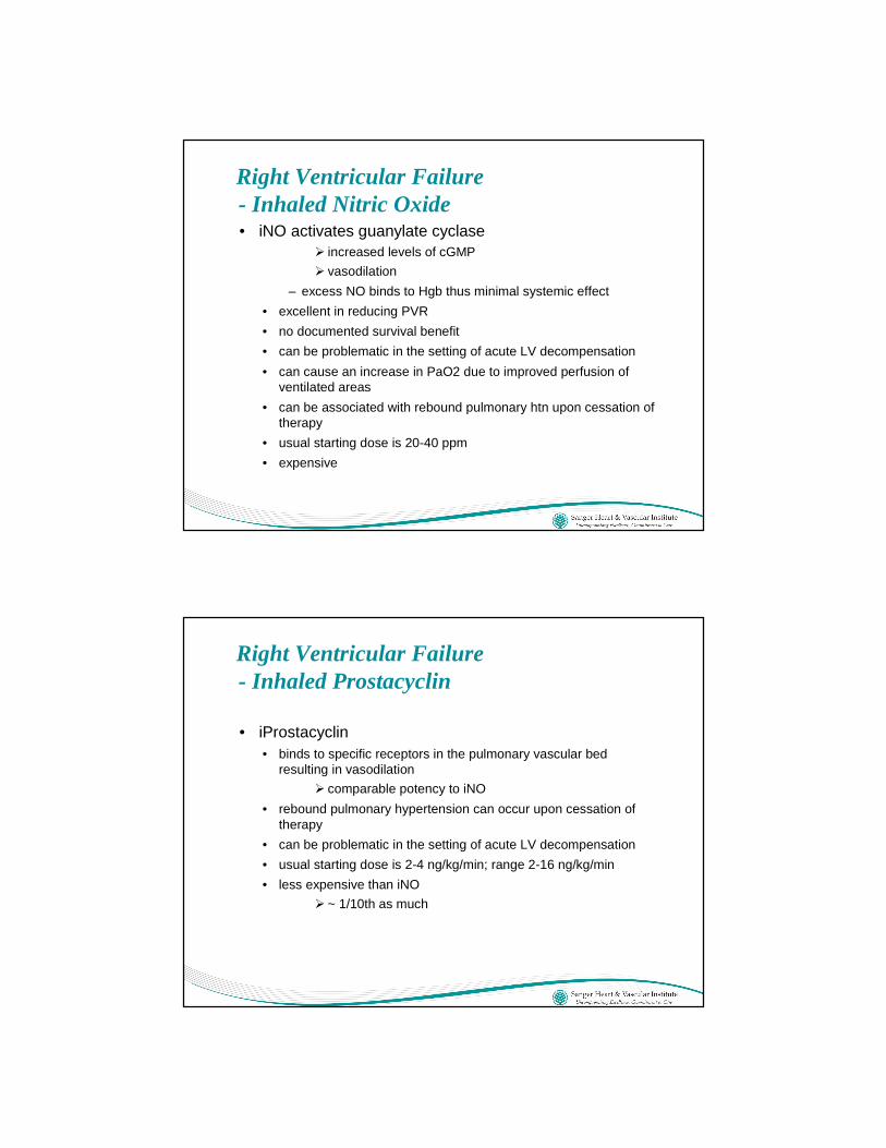
Right Ventricular Failure- Inhaled Nitric Oxide• iNO activates guanylate cyclase
� increased levels of cGMP
� vasodilation
– excess NO binds to Hgb thus minimal systemic effect
• excellent in reducing PVR
• no documented survival benefit
• can be problematic in the setting of acute LV decompensation
• can cause an increase in PaO2 due to improved perfusion of ventilated areas
• can be associated with rebound pulmonary htn upon cessation of therapy
• usual starting dose is 20-40 ppm
• expensive
Right Ventricular Failure- Inhaled Prostacyclin
• iProstacyclin • binds to specific receptors in the pulmonary vascular bed
resulting in vasodilation
� comparable potency to iNO
• rebound pulmonary hypertension can occur upon cessation of therapy
• can be problematic in the setting of acute LV decompensation
• usual starting dose is 2-4 ng/kg/min; range 2-16 ng/kg/min
• less expensive than iNO
� ~ 1/10th as much

Right Ventricular Failure- Sildenafil
• Sildenafil inhibits phosphodiesterase-5� the predominant isoform in pulmonary vascular smooth
muscle
• oral agent
• usual dose 20-40 mg po q 8 hrs
• can have a synergistic effect with iNO and iProstacyclin
• rapid onset of action
• a long-term treatment option
Right Ventricular Failure- Mechanical Circulatory Support• RVAD
� temporary
» percutaneous, transvenous (ie: Tandem)
» open (ie: Biomedicus, Centromag, etc)
� long-term
» Thoratec
• LVAD� temporary
» percutaneous (Impella 2.5; Tandem)
» open (Impella 5.0; Biomedicus, Centromag, etc)
� long-term
» Thoratec
• ECMO�Veno-arterial (VA)
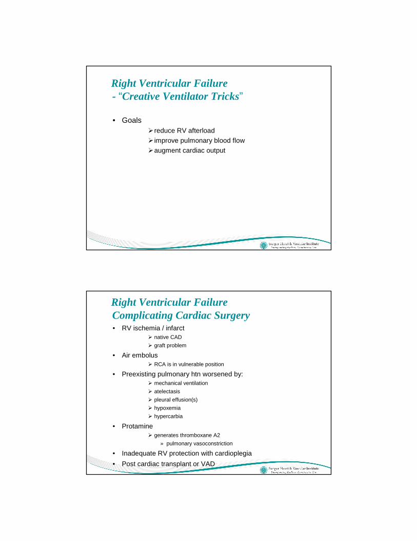
Right Ventricular Failure- “Creative Ventilator Tricks”
• Goals� reduce RV afterload
� improve pulmonary blood flow
�augment cardiac output
Right Ventricular Failure Complicating Cardiac Surgery• RV ischemia / infarct
� native CAD
� graft problem
• Air embolus� RCA is in vulnerable position
• Preexisting pulmonary htn worsened by:� mechanical ventilation
� atelectasis
� pleural effusion(s)
� hypoxemia
� hypercarbia
• Protamine� generates thromboxane A2
» pulmonary vasoconstriction
• Inadequate RV protection with cardioplegia
• Post cardiac transplant or VAD

Case #1
• 48 yo female • MVC
• Surgery for open femur fracture right LE
• Sudden intraop hypoxemia and hypotension
• TEE --> RV strain; RV dilation; mild PI; thrombus at PA bifurcation
• Continued hemodynamic deterioration - sys BP ~ 70; HR 140
• Strategies:
� IV primacor and levophed
�Emergent operative pulmonary artery embolectomy
» not candidate for lytics
�Consider iNO or iFlolan ??
Case #2• 78 yo female
• CAD, Htn, DM, DLE, COPD with bronchospasm, Asthma, large right pleural effusion, Obesity, PAD, prior right CEA.....
• CABG x 3 and mitral valve repair
• POD #1
• pO2 60 / pCO 50 on FiO2 .60 and 12 PEEP
• sys BP 100; CVP 25; CI 1.9
• UO 5-15 ml/hr
• HGB 8.5
• IV primacor and dopamine
• Strategies
�Reduce peep; increase FiO2; increase vent rate
� IV levophed or vasopressin; optimize IV primacor
�CXR / TEE
�Consider iNO or iFlolan

Case #3• 40 yo male
• Ascending aortic aneurysm involving aortic root; 3+ AI; LVEF 35%; dilated, moderately HK RV; small RCA; small LCx PDA branch
• Repair of asc ao aneur with valve-sparing aortic root replacement
• Small RCA orifice - preserved
• Nongraftable RCA br’s and LCx PDA
• No change in RV fxn on TEE
• Progressive pressor / vasoconstr requirements over 1st two POD’s
• Elevated CVP; normal PAP’s
• Low or borderline UO
• Rising creatinine and LFT’s
• TEE --> dilated, HK (near AK) RV; dilated RA; no effusion
• Strategies
� iNO or iFlolan
� off-load / rest RV --> temporary RVAD
Case # 4
• 37 yo male
• NICM with LVEF 10%; home IV primacor therapy; Htn; DLE; Obesity (BMI 40); Severe pulmonary htn; Deconditioned
• Admitted with low output state - CI 1.0; PAP’s 90/40; CVP 35
• IV Dobuta added; CI 1.3
• Diuretic tx --> CVP 28
• Strategies�Sildenafil
� iNO; iFlolan
� temporary lvad
� long-term lvad
» bridge-to-decision; possible OhTx

Thank you
Questions?