Embed Size (px)
Citation preview
Ann. rheum. Dis. (1970), 29, 609
Rheumatoid neuropathy
Clinical and electrophysiological features
M. ANNE CHAMBERLAIN AND F. E. BRUCKNER*Department of Rheumatology and Physical Medicine, Middlesex Hospital, London
It is now generally accepted that at least three distincttypes of peripheral neuropathy occur in associationwith rheumatoid arthritis:(a) Compression neuropathies often found with
early disease and associated with local jointchanges;
(b) A distal sensory neuropathy with a good prog-nosis;
(c) A severe, fulminating sensorimotor neuropathy.The present study, on the second and third types
of neuropathy, attempts to correlate their clinicalcharacteristics with nerve conduction studies andwith other features of the disease and its therapy.
Material and methodsCLINICAL ASPECTSFrom 1966 to 1969, 32 patients (12 male and 20 female)with classical or definite rheumatoid arthritis (Ropes,Bennett, Cobb, Jacox, and Jessar, 1959) were referred tous from routine clinics because of the presence of neuro-logical abnormalities. Their ages ranged from 35 years to77 years (mean 62).
Patients were examined yearly in a special reviewclinic. No attempt was made to influence therapy.
LABORATORY STUDIESThe following investigations were undertaken: haemo-globin, white cell count and erythrocyte sedimentationrate (Westergren); latex test and sheep cell agglutinationtitre; antinuclear factor, serum vitamin B12, and folicacid; oral glucose tolerance test or (rarely) blood glucose2 hours after a load of 50g. glucose.
RADIOLOGYRadiographs were taken of the hands, feet, chest, andcervical spine.
NERVE ANTIBODY STUDIESThe method used was essentially that used routinely fordetection of muscle antibodies (Roitt and Doniach, 1966)Sera from thirteen patients were incubated for 45 minutes
with unfixed frozen sections of chicken muscle. Themuscle was then washed for 14 hours and treated withfluorescein-conjugated anti-IgG. The specimen was thenre-washed and examined under the fluorescent microscopefor the presence of fluorescent nerve bundles.
ELECTROPHYSIOLOGYMuscles were sampled with a concentric needle electrodeto detect partial denervation. This was considered to bepresent when both spontaneous fibrillation and a diminu-tion in the interference pattern on maximum voluntaryactivity were found.Motor conduction velocities of the lateral popliteal
nerve 'were measured in all patients, as described byThomas, Sears, and Gilliatt (1959), recording the muscleresponse through a concentric needle electrode in theextensor digitorum brevis muscle. Similar studies on themedian nerve (recording from the abductor pollicisbrevis muscle) and medial popliteal nerve (recordingfrom the abductor hallucis longus muscle) were alsoundertaken in some patients by the methods describedin the same paper.Mixed afferent potential (MAP) were obtained by
recording from the lateral popliteal nerve through needleelectrodes inserted subcutaneously at the neck of thefibula, after stimulation of the anterior tibial nerve at theankle (Gilliatt, Goodman, and Willison, 1961). In theupper limbs, the index and minimus fingers were stimu-lated with ring electrodes and recordings were madefrom the median and ulnar nerves at the wrist (Dawson,1956). All nerve action potentials were recorded using thephotographic super-imposition of fifty tracings.
Subjects were examined in a warm room: where thetemperature of the limb was below 30°C the limb waswarmed before conduction studies were begun.
Results
Clinical groupings of rheumatoid neuropathyThe patients were divided into three groups:
Group 1 (a) Those with distal sensory signs only(19 patients)
(b) Those in whom distal sensory signspredominated (6 patients)
* Present address: St. George's Hospital, London, S.W.I.
copyright. on 31 July 2019 by guest. P
rotected byhttp://ard.bm
j.com/
Ann R
heum D
is: first published as 10.1136/ard.29.6.609 on 1 Novem
ber 1970. Dow
nloaded from
610 Annals of the Rheumatic Diseases
Group 2 Those in whom severe motor signs pre-
dominated (7 patients)
GROUP 1
Features of associated rheumatoid arthritisAll patients had classical or definite rheumatoidarthritis with mild or moderate disability. Thereappeared to be no relation between the time of onsetof the arthritis and the neuropathy. Characteristi-cally the latter followed the arthritis after severalyears, but the onset of the neuropathy was insiduousand difficult to determine with accuracy. The longestinterval between the onset of joint disease andsubsequent neuropathy was 24 years. In one patient,it preceded the symptoms of arthritis by one year,
and in another by 41 years.
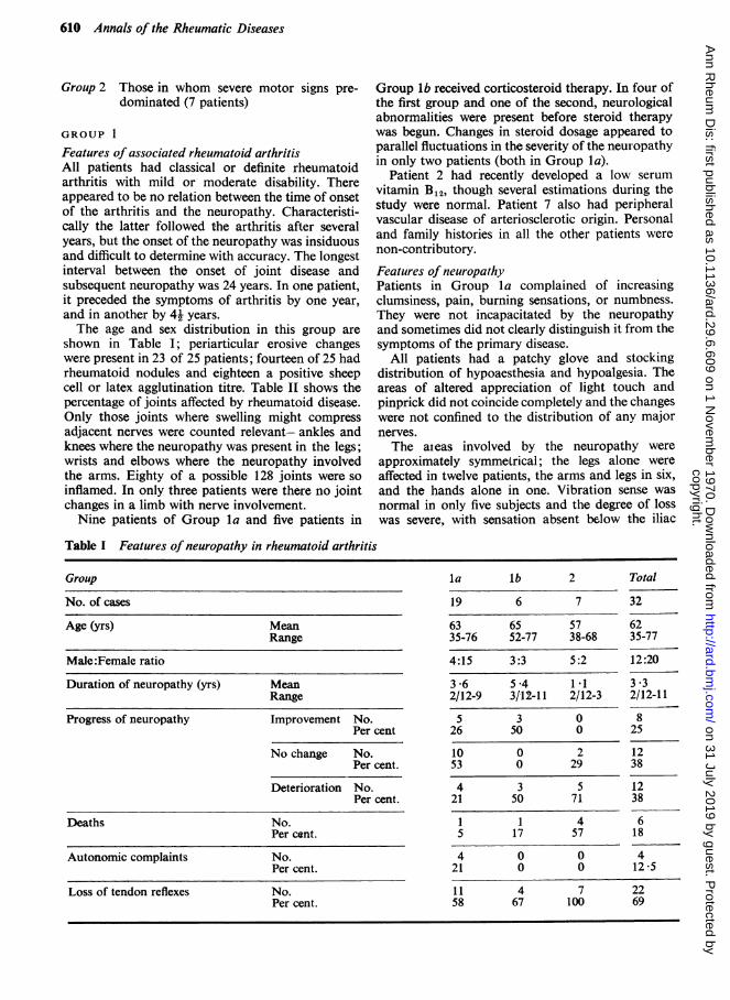
The age and sex distribution in this group are
shown in Table I; periarticular erosive changeswere present in 23 of 25 patients; fourteen of 25 hadrheumatoid nodules and eighteen a positive sheepcell or latex agglutination titre. Table II shows thepercentage of joints affected by rheumatoid disease.Only those joints where swelling might compress
adjacent nerves were counted relevant- ankles andknees where the neuropathy was present in the legs;wrists and elbows where the neuropathy involvedthe arms. Eighty of a possible 128 joints were so
inflamed. In only three patients were there no jointchanges in a limb with nerve involvement.Nine patients of Group la and five patients in
Group lb received corticosteroid therapy. In four ofthe first group and one of the second, neurologicalabnormalities were present before steroid therapywas begun. Changes in steroid dosage appeared toparallel fluctuations in the severity of the neuropathyin only two patients (both in Group la).
Patient 2 had recently developed a low serumvitamin B12, though several estimations during thestudy were normal. Patient 7 also had peripheralvascular disease of arteriosclerotic origin. Personaland family histories in all the other patients werenon-contributory.
Features of neuropathyPatients in Group la complained of increasingclumsiness, pain, burning sensations, or numbness.They were not incapacitated by the neuropathyand sometimes did not clearly distinguish it from thesymptoms of the primary disease.
All patients had a patchy glove and stockingdistribution of hypoaesthesia and hypoalgesia. Theareas of altered appreciation of light touch andpinprick did not coincide completely and the changeswere not confined to the distribution of any majornerves.The aieas involved by the neuropathy were
approximately symmelrical; the legs alone wereaffected in twelve patients, the arms and legs in six,and the hands alone in one. Vibration sense wasnormal in only five subjects and the degree of losswas severe, with sensation absent below the iliac
Table I Features of neuropathy in rheumatoid arthritis
Group
No. of cases
Age (yrs) MeanRange
Male:Female ratio
Duration of neuropathy (yrs) MeanRange
Progress of neuropathy Improvement No.Per cent
No change No.Per cent.
Deterioration No.Per cent.
Deaths No.Per cent.
Autonomic complaints No.Per cent.
Loss of tendon reflexes No.Per cent.
la lb 2 Total
19 6 7 32
63 65 57 6235-76 52-77 38-68 35-77
4:15 3:3 5:2
3-6 5-4 1 12/12-9 3/12-11 2/12-3
5 3 026 50 0
10 0 253 0 29
12:20
3 .32/12-11
825
1238
4 3 5 1221 50 71 38
1 1 4 65 17 57 18
4 0 0 421 0 0 12-5
11 4 7 2258 67 100 69
copyright. on 31 July 2019 by guest. P
rotected byhttp://ard.bm
j.com/
Ann R
heum D
is: first published as 10.1136/ard.29.6.609 on 1 Novem
ber 1970. Dow
nloaded from

Rheumatoid neuropathy 611
Table II Features of rheumatoid arthritis in patients with neuropathy
Group
No. of cases
Duration of arthritis Mean(yrs) Range
Duration before onset Meanof neuropathy (yrs) Range
Percentage of relevant* joints affected
Erosions No.Per cent.
Nodules No.Per cent.
Negative No.Per cent.
Weak Positive No.Per cent.
Strong Positive No.Per cent.
la lb 2
19 6 7
12'9 12-3 113-25 4/12-26 4-24
8-9 7.5 9.9-1 to +24 -4 5 to +13 -1 to +22
56 81 77
19 4 5/5100 67 100
11 3 758 50 100
210*5
1158
631 5
0
0
84
1
1
0/60
1/616
5/684
* Neuropathy of hands, relevant joints wrists and elbows* Neuropathy of legs, relevant joints ankles and knees
crests in five. Loss of tendon reflexes (most frequent-ly the achilles tendon reflex) was noted in elevensubjects. However, there was no disturbance ofbowel or bladder function and Raynaud's pheno-menon occurred in only one patient.
Three-quarters of the patients recovered partiallyor completely-that is, the areas of sensory lossretreated towards the fingers and toes and thesymptoms, if present, became less obtrusive-oftenwithin a few months. In the remaining patients,signs spread slowly proximally (but spared the trunk)or remained static.
Patients in Group lb experienced sensory changessimilar to those in Group la. Muscle wasting andweakness were minor or moderate and symmetrical,and involved the distal small muscles. The changeswere not confined to a myotome or the distributionof any major nerve.
Neurological signs in these patients remainedstationary during the time of study.
Nerve conduction studiesA total of eleven patients (8 in la; 3 in lb) wasstudied.Motor studies of the lateral popliteal nerve in
Group la were abnormal in half the patients. Distallatency at the ankle was not prolonged but the con-duction velocity of the fastest fibres was markedlyreduced in two patients and absent in another.The mixed afferent potential (MAP) was frequently
small, with a normal latency, or absent (see TableIII). One patient (Case 4) was studied before andafter the resolution of neurological symptoms; theMAP was absent on the first occasion and was ofdiminished (l,uv) amplitude on the second.
Table III Summary of electrophysiological findings in patients with rheumatoid arthritis
Lateral popliteal nerve Type ofneuropathy
la Sensory lb Mild mixed 2 Sensorimotor Total
Motor Partial denervation 1/8 1/3 0/6 2/17Complete denervation 1/8 0/3 6/6 7/17
Motor conduction velocity Within 2SD of mean 4/6 2/3 6/9Below 2SD 2/6 1/3 - 3/9
Mixed afferent potential Reduced/absent 5/7 0/1 3/3 8/11
Abnormalities in other nerves 4/7 1/2 2/3 7/12
SCATandlatextests
Total
32
12 44/12-268 *8-4*5 to +24
61
28/3093
2164
2/316 5
17/3155
12/3138 5
copyright. on 31 July 2019 by guest. P
rotected byhttp://ard.bm
j.com/
Ann R
heum D
is: first published as 10.1136/ard.29.6.609 on 1 Novem
ber 1970. Dow
nloaded from
612 Annals of the Rheumatic Diseases
In Group Ib, one patient (Case 9, see Weller,Bruckner, and Chamberlain, 1970) had normalfindings in all nerves surveyed, while another (Case10) had gross slowing of motor conduction in boththe lateral popliteal nerve, where there was greatdelay at the ankle, and in the median nerve. Thethird parient (Case 11) who developed neuritisbefore arthritis had normal motor findings in the leg.
Illustrative case historiesGroup la. A married woman aged 56 (Case 4) had sufferedfrom definite rheumatoid arthritis with moderate func-tional impairment for 13 years. Nodules, periarticularerosions, and involvement of most joints in a typicalpattern, were found. The sheep cell agglutination waspositive to a titre of 1: 80. There were no nailfold or eyelesions. She had been receiving prednislone for 12 years,initially 15 mg. daily and reducing to 7-5 mg. 2 yearsbefore entering the study.Burning paraesthesiae of the feet and fingers had been
present for 3 years. There was sensory loss to light touchand pinprick over both feet to the level of the ankles;vibration sense here was impaired and the ankle reflexeswere absent. Neither muscle weakness nor a history ofautonomic disturbance was found.
Spontaneous recovery continued slowly during thetime the patient was under observation. When she waslast seen (1969), only vibration sense and ankle reflexesremained impaired and she was symptom-free.
Spontaneous fibrillation was detected in the rigthextensor digitorum brevis muscle. Striking slowing ofmotor conduction was found in the right lateral poplitealnerve (22 4 m./sec.) and there was no MAP in this nervebefore recovery of sensation; after recovery it was of Il vamplitude.
Group lb. Case 10 (for full history, see Weller and others,1970). This patient's rheumatoid arthritis had similarcharacteristics to that of Case 4. Numbness of the fingersand toes began in 1964 and slowly progressed proximally.Moderate weakness of the intrinsic muscles of the handsand wrist extensors was present. Supinator and anklereflexes were absent.
Sensory and motor conduction studies of both lateralpopliteal nerves and the median and ulnar nerves of theright arm were normal.
GROUP 2-SEVERE SENSORIMOTOR NEUROPATHYFeatures of associated rheumatoid arthritisAll patients had classical rheumatoid arthritis withsevere disability. The mean duration of diseasebefore neuritis developed was 9 - 9 years, with a widerange (1 to 22 years).
There was a marked male predominance (5 males,2 females) and the mean age was lower than that ofthe other two groups.
All patients had periarticular erosions andrheumatoid nodules. All had a positive test forrheumatoid factor and, of the six in whom this wasquantitated, five had a titre of at least 1: 256. 77 percent. of 'relevant' joints were affected by arthritis.
Complications of rheumatoid disease were severeand frequent (see below).
Features of NeuropathyPeripheral motor manifestations with a dramaticonset (e.g. sudden foot drop) dominated the clinicalpicture, although coincidental asymmetrical sensorychanges were always found. The Achilles tendonreflexes were lost in all patients and vibration sen-sation was frequently impaired. Signs usually beganin the legs and commonly a picture of mononeuritismultiplex developed. The affected legs were oftencold and oedematous, and showed livido reticularis.Peripheral pulses were present.The neuropathy progressed relentlessly in most
patients. Subjects on azathioprine 100 mg. dailywere interesting as the drug appeared to halt pro-gression of the neuropathy, perhaps with slightimprovement in motor signs. However, in the onepatient re-tested, elecro-physiological evidence ofsevere denervation remained.
Illustrative case historyA man aged 62 had severe classical rheumatoid arthritisbeginning in 1962, which compelled him to give up workin 1967. The arthritis was sero-positive to a titre of I: 320,erosive, and nodular. Nailfold lesions, episcleritis,oedema, and leg ulceration were present.He had received prednisolone since 1964, a dose of 12 5
mg. daily being maintained during his last 3 years of life.Numbness of both hands and feet began in 1965 andwithin weeks a complete right foot drop developed andremained unchanged until the patient's death in 1968,after two myocardial infarctions; 9 months before deaththe patient had successfully undergone a small bowelresection following a superior mesenteric artery throm-bosis.
Nerve conduction studiesRemarkably uniform results were obtained in thesix patients studied. No response to nerve stimulationcould be recorded from the extensor digitorumbrevis muscle in any patient. Neither spontaneousfibrillation nor any motor unit potentials undervoluntary control were found. In one subject (BM:see Weller and others, 1970) the lateral poplitealnerve had been studied at the onset of foot drop(just before the nerve to the extensor digitorumbrevis muscle became inexcitable) when a singlemotor unit was found with a conduction velocity of36.5 m./sec. in the leg. In three patients attemptswere made to record from the adductor hallucislongus muscle, but there were no surviving motorunits in this muscle either.
In three of the patients in whom mixed afferentpotentials were searched for, none was found.The sensory action potential of the ulnar nerve
was studied in three of these patients; its amplitude
copyright. on 31 July 2019 by guest. P
rotected byhttp://ard.bm
j.com/
Ann R
heum D
is: first published as 10.1136/ard.29.6.609 on 1 Novem
ber 1970. Dow
nloaded from
Rheumatoid neuropathy 613
was diminished in one and the potential was absentin a second. This patient (Case 16) also had no mediannerve sensory action potential at this time. Thesesearches are limited, because these patients wereseverely ill. However, they do suggest that lesionsof the peripheral nerves may be widespread.
Nerve antibody studiesFluorescent staining of nerve bundles was foundfortuitously when the serum of a patient with earlyrheumatoid arthritis (without neurological abnorma-lities) was incubated with chicken muscle in a routinesearch for muscle antibodies. It was thus decided tostudy the sera of some of the patients in our groupwith peripheral neuropathies.
In none of the thirteen sera (11 from Group 1 and2 from Group 2) examined were antibodies to nervebundles found.
DiscussionCLINICAL ASSOCIATIONS BETWEENRHEUMATOID ARTHRITIS ANDNEUROPATHYThere is little difference between the two groups inthe duration of arthritis before neuropathy super-vened. Similarly, it is not possible to deduce any
definite relationships between joint swelling andperipheral nerve disease, as joint changes are sofrequently found in both groups.
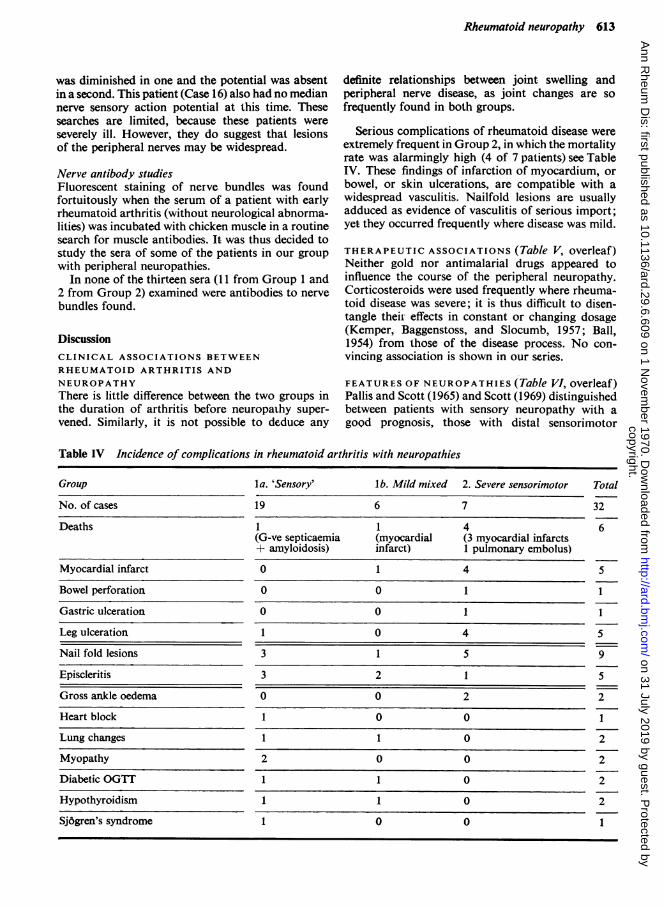
Serious complications of rheumatoid disease wereextremely frequent in Group 2, in which the mortalityrate was alarmingly high (4 of 7 patients) see TableIV. These findings of infarction of myocardium, orbowel, or skin ulcerations, are compatible with awidespread vasculitis. Nailfold lesions are usuallyadduced as evidence of vasculitis of serious import;yet they occurred frequently where disease was mild.
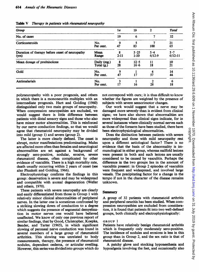
THERAPEUTIC ASSOCIATIONS (Table V, overleaf)Neither gold nor antimalarial drugs appeared toinfluence the course of the peripheral neuropathy.Corticosteroids were used frequently where rheuma-toid disease was severe; it is thus difficult to disen-tangle their effects in constant or changing dosage(Kemper, Baggenstoss, and Slocumb, 1957; Ball,1954) from those of the disease process. No con-vincing association is shown in our series.
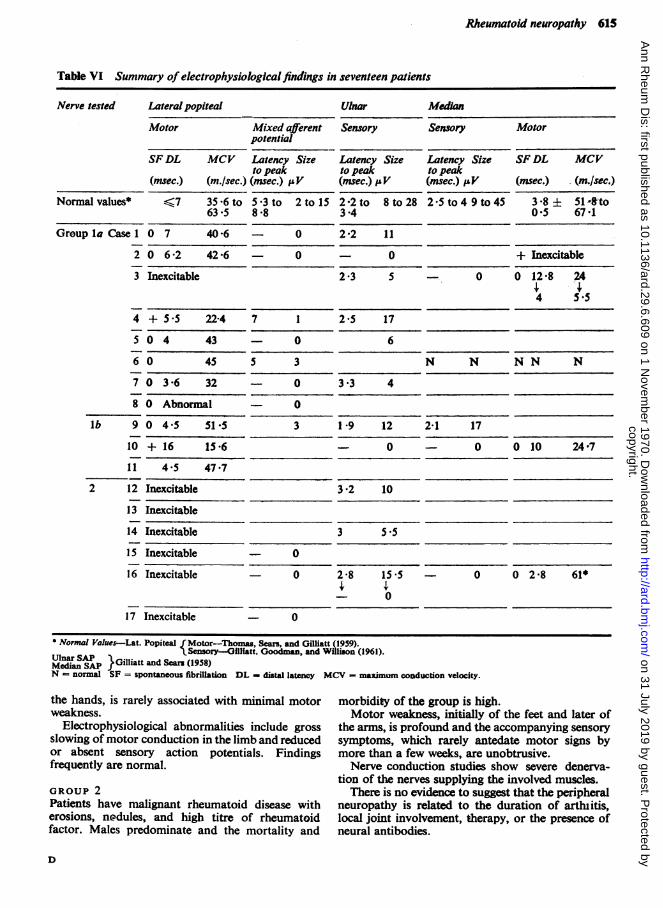
FEATURES OF NEUROPATHIES (Table VI, overleaf)Pallis and Scott (1965) and Scott (1969) distinguishedbetween patients with sensory neuropathy with agoQd prognosis, those with distal sensorimotor
Table IV Incidence of complications in rheumatoid arthritis with neuropathies
Group
No. of cases
Deaths
Myocardial infarct
Bowel perforation
Gastric ulceration
Leg ulceration
Nail fold lesions
Episcleritis
Gross ankle oedema
Heart block
Lung changes
Myopathy
Diabetic OGTT
Hypothyroidism
Sj6gren's syndrome
l a. 'Sensory' l b. Mild mixed 2. Severe sensorimotor
19 6 7
1 1 4(G-ve septicaemia (myocardial (3 myocardial infarcts+ amyloidosis) infarct) 1 pulmonary embolus)
0 1 4
o 0 1
O 0 1
1 0 4
3 1 5
3 2 1
0 0 2
1 0 0
1 1 0
2 0 0
1 1 0
1 1 0
1 0 0
Total
32
6
5
1
1
5
9
5
2
1
2
2
2
2
1
copyright. on 31 July 2019 by guest. P
rotected byhttp://ard.bm
j.com/
Ann R
heum D
is: first published as 10.1136/ard.29.6.609 on 1 Novem
ber 1970. Dow
nloaded from
614 Annals of the Rheumatic Diseases
Table V Therapy in patients with rheumatoid neuropathy
Group
No. of cases
Corticosteroids No.Per cent.
Duration of therapy before onset of neuropathy Mean(yrs) Range
Mean dosage of prednisolone Daily (mg.)Total (g.)
Gold No.Per cent.
Antimalarials No.Per cent.
polyneuropathy with a poor prognosis, and othersin which there is a mononeuritis multiplex with anintermediate prognosis. Hart and Golding (1960)distinguished only two main groups of neuropathy.When compression neuropathies are excluded, wewould suggest there is little difference betweenpatients with distal sensory signs and those who alsohave minor motor abnormalities. This is reinforcedby our nerve conduction findings, so that we wouldagree that rheumatoid neuropathy may be dividedinto mild (group 1) and severe (group 2).The latter is more clearly defined. The onset is
abrupt, motor manifestations predominating. Malesare affected more often than females and neurologicalabnormalities are set against a background ofstrongly sero-positive, nodular, erosive, severerheumatoid disease, often complicated by otherevidence of vasculitis. There is a high mortality rate,death usually occurring within 2 years of onset (seealso Plunkett and Golding, 1966).
Electrophysiology confirms the findings in thisgroup: denervation is severe and may be widespreadand compatible with axonal degeneration (Wellerand others, 1970).
These patients with severe neuropathy are clearlyand easily differentiated from those in Group 1 withrelatively minor clinical abnormalities of peripheralnerves. In the latter one is sometimes confronted bya striking slowing down of conduction to a degreewhich suggests the presence of segmental demyelina-tion in motor nerves one would have believedunaffected. We know of only one previous report ofsimilar findings, that by Good, Christopher, Koepke,Bender, and Tarter (1965), in which significantslowing of peroneal nerve conduction was found inseveral members of a large group of rheumatoidarthritics. This slowing was unrelated to bodymeasurements, therapy, the presence of rheumatoidnodules, dependent oedema, or articular swelling.However, this serieswas dividedinto groups which do
la lb 2
19 6 7
9 5 747 83 100
8 5*25 5.42-11 1-10 6/12-98 12 5 1128 16 f6 18
9 1 447 17 57
Total
32
2165
5 '76/12-11
1021
1444
3 1 2 617 16 28 18
not correspond with ours; it is thus difficult to knowwhether the figures are weighted by the presence ofsubjects with severe sensorimotor changes.Our work would suggest that a nerve may be
damaged more severely than is evident from clinicalsigns; we have also shown that abnormalities aremore widespread than clinical signs indicate, for inseveral instances where clinically normal nerves suchas those of the forearm have been studied, there havebeen electrophysiological abnormalities.Does the distinction between patients with severe
neuropathy and those with mild neuropathy restupon a different aetiological factor? There is noevidence that the basis of the abnormality is im-munological in either group, whereas nailfold lesionswere present in both and such lesions are usuallyconsidered to be caused by vasculitis. Perhaps thedifference in the two groups lies in the amount ofvasculitis present: in Group 2 episodes of vasculitiswere frequent and widespread, and involved largevessels. The precipitating factor for a change in thetempo if not in the character of the disease remainsunknown.
SummaryA group of 32 patients with rheumatoid arthritisand peripheral neuritis has been studied. When com-pression neuropathies are excluded from considera-tion, it is found that patients fit into two well-definedgroups, both clinically and electrophysiologically:
GROUP 1Patients have relatively benign rheumatoid arthritiswhich is frequently only moderately sero-positive.The incidence of nodules and erosions is less in thisgroup than in Group 2, and the sex ratio is that ofrheumatoid disease.A patchy glove and stocking hypoaesthesia and
hypoalgesia involving the feet, and occasionally also
copyright. on 31 July 2019 by guest. P
rotected byhttp://ard.bm
j.com/
Ann R
heum D
is: first published as 10.1136/ard.29.6.609 on 1 Novem
ber 1970. Dow
nloaded from
Rheumatoid neuropathy 615
Table VI Summary of electrophysiological findings in seventeen patients
Nerve tested Lateralpopiteal Ulnar Median
Motor Mixed afferent Sensory Sensory Motorpotential
SF DL MCV Latency Size Latency Size Latency Size SF DL MCVto peak to peak to peak
(msec.) (m./sec.) (msec.) jW (msec.) iV (msec.) .V (msec.) (m.Isec.)Normal values* 67 35 *6 to 5 *3 to 2 to 15 2*2to 8to28 2 *5 to 4 9 to 45 3 *8 514to
63 5 8*8 3*4 0'5 67*1Group la Case 1 0 7 40 6 0 2 2 11
2 0 6 2 42-6 0 - 0 + Inexcitable
3 Inexcitable 2*3 5 0 0 12 8 24
4 5 5
4 + 5 5 22-4 7 1 2 5 17
5 0 4 43 0 6
6 0 45 5 3 N N N N N
7 0 3*6 32 0 3X3 4
8 0 Abnormal - 0
lb 9 0 4 S 51*5 3 19 12 2-1 17
10 +16 15-6 0 - 0 010 24-7
I1 4 5 47*7
2 12 Inexcitable 3 -2 10
13 Inexcitable
14 Inexcitable 3 5.5
15 Inexcitable - 0
16 Inexcitable 0 2-8 15 - 0 0 2'8 61*
_ 0
17 Inexcitable 0
* Normal Values-Lat. Popiteal fMotor-Thomas, Sears, and Gilliatt (1959).Nn Sensory-Olllfatt, Goodman, and Wiflison (1961).
Median SAP }Gilliatt and Sears (1958)N - normal SF = spontaneous fibrilatlon DL - dist latency MCV = nmum conduction velocity.
the hands, is rarely associated with minimal motor morbidity of the group is high.weakness. Motor weakness, initially of the feet and later of
Electrophysiological abnormalities include gross the arms, is profound and the accompanying sensoryslowing of motor conduction in the limb and reduced symptoms, which rarely antedate motor signs byor absent sensory action potentials. Findings more than a few weeks, are unobtrusive.frequently are normal. Nerve conduction studies show severe denerva-
tion of the nerves supplying the involved muscles.GROUP 2 There is no evidence to suggest that the peripheralPatients have malignant rheumatoid disease with neuropathy is related to the duration of artblitis,erosions, nodules, and high titre of rheumatoid local joint involvement, therapy, or the presence offactor. Males predominate and the mortality and neural antibodies.
D
copyright. on 31 July 2019 by guest. P
rotected byhttp://ard.bm
j.com/
Ann R
heum D
is: first published as 10.1136/ard.29.6.609 on 1 Novem
ber 1970. Dow
nloaded from
616 Annals of the Rheumatic Diseases
We have pleasure in acknowledging the generous help Hospital; Dr. A. P. H. Randle, Dr. S. Meadows, Dr.given to us by many colleagues, including the following: M. Kremer, and Dr. R. A. Henson at the NationalDr. A. C. Boyle and Dr. M. Corbett of the Rheumatology Hospital for Nervous Diseases; Prof. I. Roitt and hisDepartment, Middlesex Hospital; Dr. P. M. Fullerton, department, who performed antibody studies for us.who helped us with electrophysiology; Mr. J. Andrew and We are also grateful for financial assistance from thehis senior registrars who performed sural nerve biopsies; Arthritis and Rheumatism Council.Dr. W. Tegner and Dr. M. Mason of the London
ReferencesBALL, J. (1954) Ann. rheum. Dis., 13, 277 (Rheumatoid arthritis and polyarteritis nodosa).DAWSON, G. D. (1956) J. Physiol. (Lond.), 131, 436 (The relative excitability and conduction velocity of sensory
and motor nerve fibres in man).GILLIATr, R. W., GOODMAN, H. V., AND WILLISON, R. G. (1961) J. NeuroL Neurosurg. Psychiat., 24, 305 (The
recording of lateral popliteal nerve action potentials in man).- , AND SEARS, T. A. (1958) J. Neurol. Neurosurg. Psychiat., 21, 109 (Sensory nerve action potentials in patients
with peripheral nerve lesions).GOOD, A. E., CRISTPHER, R. P., KOEPKE, G. H., BENDER, L. F., AND TARTER, M. E. (1965) Ann. intern. Med.,
63, 87 (Peripheral neuropathy associated with rheumatoid arthritis).HART, F. DUDLEY, AND GOLDING, J. R. (1960) Brit. med. J., 1, 1594 (Rheumatoid neuropathy).KEMPER, J. W., BAGGENSTOSS, A. H., AND SLOCUMiB, C. H. (1957) Ann. intern. Med., 46, 831 (The relationship
of therapy with cortisone to the incidence of vascular lesions in rheumatoid arthritis).PALLIS, C. A., AND Scorr, J. T. (1965) Brit. med. J., 1, 1141 (Peripheral neuropathy in rheumatoid arthritis).PLUNKErr, T. G., AND GOLDING, J. R. (1966) Ann. rheum. Dis., 25, 572 (Rheumatoid peripheral neuropathy).Rorrr, I. M., AND DONIACH, D. (1969) 'W.H.O. Manual of Immunological Techniques,' p. 1 (Autoimmune
serology: immunofluorescent tests for the detection of autoantibodies). W.H.O., Geneva.RoPEs, M. W., BENNErr, G. A., COBB, S., JAcox, R. F., AND JESSAR, R. A. (1959) Ann. rheum. Dis., 18, 49.Scorr, J. T. (1969) In "Textbook of the Rheumatic Diseases,"' ed. W. S. C. Copeman, 4th ed., p. 648. Livingstone,
Edinburgh.THOMAS, P. K., SEAS, T. A., AND GILLLIArr, R. W. (1959) J. Neurol. Neurosurg. Psychiat., 22, 175 (The range
of conduction velocity in normal motor nerve fibres to the small muscles of the hand and foot).WELLER, R. O., BRUCKNER, F. E., AND CAMBERLN, M. A. (1970) Ibid., 33, 592 (Rheumatoid neuropathy:
an histological and electrophysiological study).
copyright. on 31 July 2019 by guest. P
rotected byhttp://ard.bm
j.com/
Ann R
heum D
is: first published as 10.1136/ard.29.6.609 on 1 Novem
ber 1970. Dow
nloaded from