Embed Size (px)
Citation preview
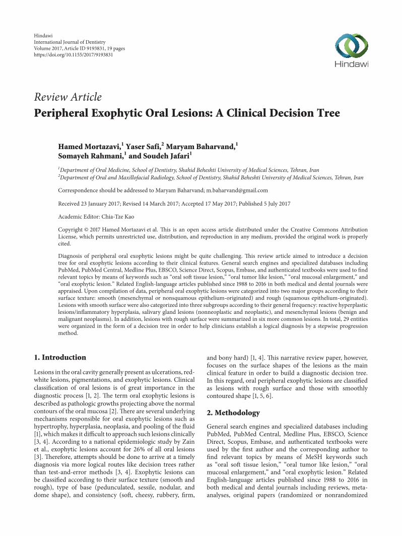
Review ArticlePeripheral Exophytic Oral Lesions: A Clinical Decision Tree
HamedMortazavi,1 Yaser Safi,2 Maryam Baharvand,1
Somayeh Rahmani,1 and Soudeh Jafari1
1Department of Oral Medicine, School of Dentistry, Shahid Beheshti University of Medical Sciences, Tehran, Iran2Department of Oral and Maxillofacial Radiology, School of Dentistry, Shahid Beheshti University of Medical Sciences, Tehran, Iran
Correspondence should be addressed to Maryam Baharvand; [email protected]
Received 23 January 2017; Revised 14 March 2017; Accepted 17 May 2017; Published 5 July 2017
Academic Editor: Chia-Tze Kao
Copyright © 2017 Hamed Mortazavi et al. This is an open access article distributed under the Creative Commons AttributionLicense, which permits unrestricted use, distribution, and reproduction in any medium, provided the original work is properlycited.
Diagnosis of peripheral oral exophytic lesions might be quite challenging. This review article aimed to introduce a decisiontree for oral exophytic lesions according to their clinical features. General search engines and specialized databases includingPubMed, PubMed Central, Medline Plus, EBSCO, Science Direct, Scopus, Embase, and authenticated textbooks were used to findrelevant topics by means of keywords such as “oral soft tissue lesion,” “oral tumor like lesion,” “oral mucosal enlargement,” and“oral exophytic lesion.” Related English-language articles published since 1988 to 2016 in both medical and dental journals wereappraised. Upon compilation of data, peripheral oral exophytic lesions were categorized into two major groups according to theirsurface texture: smooth (mesenchymal or nonsquamous epithelium-originated) and rough (squamous epithelium-originated).Lesions with smooth surface were also categorized into three subgroups according to their general frequency: reactive hyperplasticlesions/inflammatory hyperplasia, salivary gland lesions (nonneoplastic and neoplastic), and mesenchymal lesions (benign andmalignant neoplasms). In addition, lesions with rough surface were summarized in six more common lesions. In total, 29 entitieswere organized in the form of a decision tree in order to help clinicians establish a logical diagnosis by a stepwise progressionmethod.
1. Introduction
Lesions in the oral cavity generally present as ulcerations, red-white lesions, pigmentations, and exophytic lesions. Clinicalclassification of oral lesions is of great importance in thediagnostic process [1, 2]. The term oral exophytic lesions isdescribed as pathologic growths projecting above the normalcontours of the oral mucosa [2]. There are several underlyingmechanisms responsible for oral exophytic lesions such ashypertrophy, hyperplasia, neoplasia, and pooling of the fluid[1], whichmakes it difficult to approach such lesions clinically[3, 4]. According to a national epidemiologic study by Zainet al., exophytic lesions account for 26% of all oral lesions[3]. Therefore, attempts should be done to arrive at a timelydiagnosis via more logical routes like decision trees ratherthan test-and-error methods [3, 4]. Exophytic lesions canbe classified according to their surface texture (smooth andrough), type of base (pedunculated, sessile, nodular, anddome shape), and consistency (soft, cheesy, rubbery, firm,
and bony hard) [1, 4]. This narrative review paper, however,focuses on the surface shapes of the lesions as the mainclinical feature in order to build a diagnostic decision tree.In this regard, oral peripheral exophytic lesions are classifiedas lesions with rough surface and those with smoothlycontoured shape [1, 5, 6].
2. Methodology
General search engines and specialized databases includingPubMed, PubMed Central, Medline Plus, EBSCO, ScienceDirect, Scopus, Embase, and authenticated textbooks wereused by the first author and the corresponding author tofind relevant topics by means of MeSH keywords suchas “oral soft tissue lesion,” “oral tumor like lesion,” “oralmucosal enlargement,” and “oral exophytic lesion.” RelatedEnglish-language articles published since 1988 to 2016 inboth medical and dental journals including reviews, meta-analyses, original papers (randomized or nonrandomized
HindawiInternational Journal of DentistryVolume 2017, Article ID 9193831, 19 pageshttps://doi.org/10.1155/2017/9193831
2 International Journal of Dentistry
Medical
89 articled89
Medical data bases
150 relative articles
118 articles with full texts
78 articles was used
32 excluded(full texts unavailable)
40 excluded (not English or repetitive material)
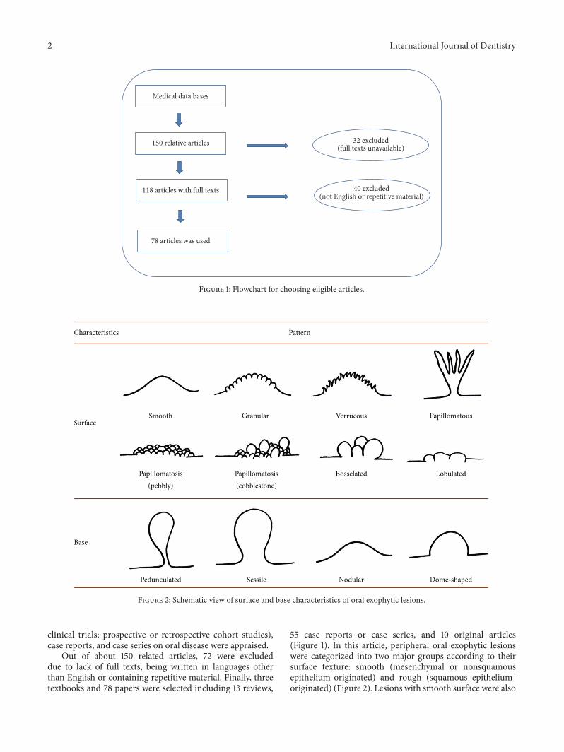
Figure 1: Flowchart for choosing eligible articles.
Characteristics
Surface
Base
Pattern
Smooth Granular Verrucous Papillomatous
Papillomatosis Papillomatosis(pebbly)
Bosselated Lobulated
Pedunculated Sessile Nodular Dome-shaped
(cobblestone)
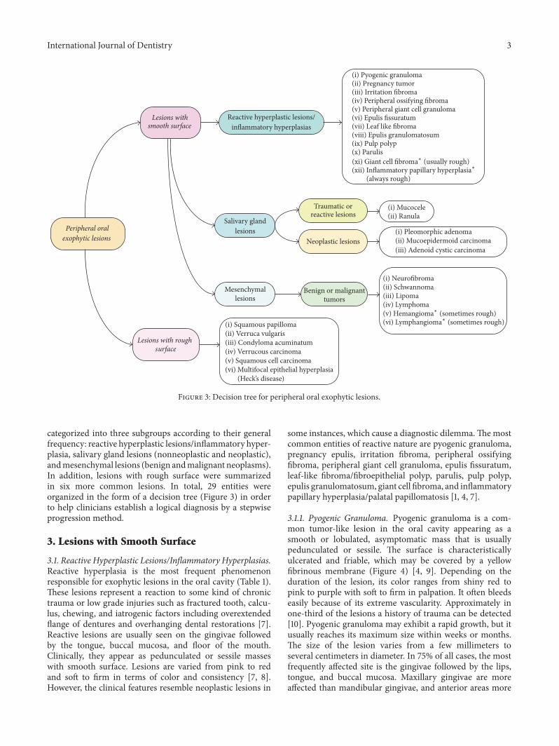
Figure 2: Schematic view of surface and base characteristics of oral exophytic lesions.
clinical trials; prospective or retrospective cohort studies),case reports, and case series on oral disease were appraised.
Out of about 150 related articles, 72 were excludeddue to lack of full texts, being written in languages otherthan English or containing repetitive material. Finally, threetextbooks and 78 papers were selected including 13 reviews,
55 case reports or case series, and 10 original articles(Figure 1). In this article, peripheral oral exophytic lesionswere categorized into two major groups according to theirsurface texture: smooth (mesenchymal or nonsquamousepithelium-originated) and rough (squamous epithelium-originated) (Figure 2). Lesions with smooth surface were also
International Journal of Dentistry 3
Lesions withsmooth surface
Peripheral oralexophytic lesions
Lesions with roughsurface
Reactive hyperplastic lesions/inflammatory hyperplasias
(i) Pyogenic granuloma(ii) Pregnancy tumor(iii) Irritation fibroma(iv) Peripheral ossifying fibroma(v) Peripheral giant cell granuloma(vi) Epulis fissuratum(vii) Leaf like fibroma(viii) Epulis granulomatosum(ix) Pulp polyp(x) Parulis
Salivary glandlesions
Mesenchymallesions
(i) Squamous papilloma(ii) Verruca vulgaris
(i) Mucocele(ii) Ranula
(i) Pleomorphic adenoma(ii) Mucoepidermoid carcinoma(iii) Adenoid cystic carcinoma
(i) Neurofibroma(ii) Schwannoma(iii) Lipoma(iv) Lymphoma
Traumatic orreactive lesions
Neoplastic lesions
Benign or malignanttumors
(iii) Condyloma acuminatum(iv) Verrucous carcinoma(v) Squamous cell carcinoma(vi) Multifocal epithelial hyperplasia
(Heck’s disease)
(vi) Lymphangiom;∗ (sometimes rough)
(v) Hemangiom;∗ (sometimes rough)
(always rough)(xii) Inflammatory papillary hyperplasi;∗(xi) Giant cell fibrom;
∗ (usually rough)
Figure 3: Decision tree for peripheral oral exophytic lesions.
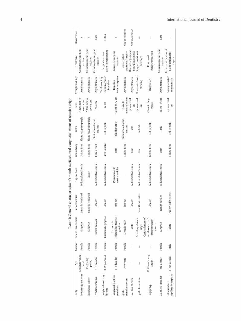
categorized into three subgroups according to their generalfrequency: reactive hyperplastic lesions/inflammatory hyper-plasia, salivary gland lesions (nonneoplastic and neoplastic),andmesenchymal lesions (benign andmalignant neoplasms).In addition, lesions with rough surface were summarizedin six more common lesions. In total, 29 entities wereorganized in the form of a decision tree (Figure 3) in orderto help clinicians establish a logical diagnosis by a stepwiseprogression method.
3. Lesions with Smooth Surface
3.1. Reactive Hyperplastic Lesions/Inflammatory Hyperplasias.Reactive hyperplasia is the most frequent phenomenonresponsible for exophytic lesions in the oral cavity (Table 1).These lesions represent a reaction to some kind of chronictrauma or low grade injuries such as fractured tooth, calcu-lus, chewing, and iatrogenic factors including overextendedflange of dentures and overhanging dental restorations [7].Reactive lesions are usually seen on the gingivae followedby the tongue, buccal mucosa, and floor of the mouth.Clinically, they appear as pedunculated or sessile masseswith smooth surface. Lesions are varied from pink to redand soft to firm in terms of color and consistency [7, 8].However, the clinical features resemble neoplastic lesions in
some instances, which cause a diagnostic dilemma.Themostcommon entities of reactive nature are pyogenic granuloma,pregnancy epulis, irritation fibroma, peripheral ossifyingfibroma, peripheral giant cell granuloma, epulis fissuratum,leaf-like fibroma/fibroepithelial polyp, parulis, pulp polyp,epulis granulomatosum, giant cell fibroma, and inflammatorypapillary hyperplasia/palatal papillomatosis [1, 4, 7].
3.1.1. Pyogenic Granuloma. Pyogenic granuloma is a com-mon tumor-like lesion in the oral cavity appearing as asmooth or lobulated, asymptomatic mass that is usuallypedunculated or sessile. The surface is characteristicallyulcerated and friable, which may be covered by a yellowfibrinous membrane (Figure 4) [4, 9]. Depending on theduration of the lesion, its color ranges from shiny red topink to purple with soft to firm in palpation. It often bleedseasily because of its extreme vascularity. Approximately inone-third of the lesions a history of trauma can be detected[10]. Pyogenic granuloma may exhibit a rapid growth, but itusually reaches its maximum size within weeks or months.The size of the lesion varies from a few millimeters toseveral centimeters in diameter. In 75% of all cases, the mostfrequently affected site is the gingivae followed by the lips,tongue, and buccal mucosa. Maxillary gingivae are moreaffected than mandibular gingivae, and anterior areas more
4 International Journal of Dentistry
Table1:Generalcharacteris
ticso
fsmoo
th-surfacedoralexop
hytic
lesio
nsof
reactiv
eorig
in.
Entity
Age
Gender
Siteof
involvem
ent
Surfa
cetexture
Type
ofbase
Con
sistency
Color
Size
Symptom
&sig
nTreatm
ent
Recurrence
Pyogenicgranulom
aCh
ildren/youn
gadult
Female
Gingivae
Smoo
th/lo
bulated
Pedu
nculated/sessile
Softto
firm
Shinyred/pink
/purple
Afewmm
toseveralcm
Asymptom
atic
Con
servatives
urgical
excisio
n+
Pregnancytumor
Pregnancy
perio
dFemale
Gingivae
Smoo
th/lo
bulated
Sessile
Softto
firm
Shinyred/pink
/purple
Afewmm
toseveralcm
Asymptom
atic
Con
servatives
urgical
excisio
n+
Irritationfib
roma
4–6decades
Female
Buccalmucosa
Smoo
thPedu
nculated/sessile
Firm
orsoft
Similartoadjacent
mucosa
≤1.5
cmAsymptom
atic
Con
servatives
urgical
excisio
nRa
re
Perip
heralossify
ing
fibroma
10–19yearso
ldFemale
Exclu
sively
ging
ivae
Smoo
thPedu
nculated/sessile
Firm
tohard
Redto
pink
<2c
mTo
othmob
ility
Toothmigratio
nBo
neloss
Surgicalexcisio
ndo
wnto
perio
steum
8–20%
Perip
heralgiant
cell
granulom
a5-6decades
Female
Exclu
sively
edentulous
ridge
&ging
ivae
Smoo
thPedu
nculated/
sessile/nod
ular
Firm
Bluish
purple
<2c
mor>2c
mBo
neloss
Root
resorptio
nCom
pletes
urgical
excisio
n+
Epulis
granulom
atosum
>40
years
Female
Dentalsocket
Smoo
th—
Softto
firm
Similartoadjacent
mucosa
<1cm
tomassiv
elesions
Asymptom
atic
Con
servative
treatment/surgery
Not
uncommon
Leaf-like
fibroma
——
Palate
Smoo
thPedu
nculated/sessile
Firm
Pink
Upto
several
cmAsymptom
atic
Denture
adjustm
ent
&surgicalremoval
Not
uncommon
E pulisfissuratum
——
Maxillaryalveolar
ridge
Smoo
th/ulcerative
Pedu
nculated/sessile
Firm
Redd
ishUpto
several
cmNon
tend
er/easily
bleeding
Surgicalexcisio
n&
curetta
ge—
Pulppo
lyp
Child
ren/youn
gadults
—
Cario
uslesio
nsof
decidu
ousteeth
&firstperm
anent
molars
Smoo
thPedu
nculated/sessile
Softto
firm
Redto
pink
<1cm
tolarge
masses
Disc
omfort
Root
canal
therapy/extractio
n—
Giant
cellFibrom
a3rddecade
Female
Gingivae
Roug
hsurfa
cePedu
nculated/sessile
Firm
Pink
<1cm
(ofte
n)Asymptom
atic
Con
servatives
urgical
excisio
nRa
re
Inflammatory
papillary
hyperplasia
3–5thdecades
Male
Palate
Pebb
ly/cob
blestone
—Softto
firm
Redto
pink
Upto
several
cmAsymptom
atic/
symptom
atic
Removalof
denturea
tnight/a
ntifu
ngals/
surgery
—
International Journal of Dentistry 5
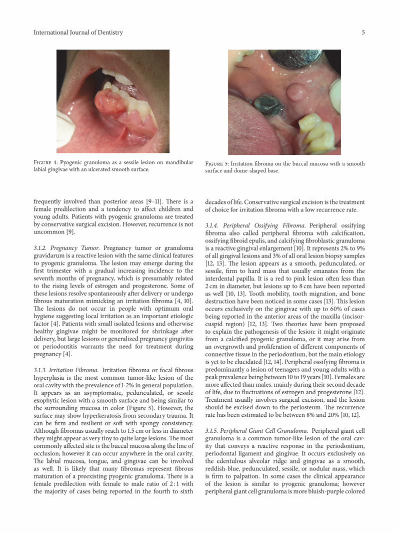
Figure 4: Pyogenic granuloma as a sessile lesion on mandibularlabial gingivae with an ulcerated smooth surface.
frequently involved than posterior areas [9–11]. There is afemale predilection and a tendency to affect children andyoung adults. Patients with pyogenic granuloma are treatedby conservative surgical excision. However, recurrence is notuncommon [9].
3.1.2. Pregnancy Tumor. Pregnancy tumor or granulomagravidarum is a reactive lesion with the same clinical featuresto pyogenic granuloma. The lesion may emerge during thefirst trimester with a gradual increasing incidence to theseventh months of pregnancy, which is presumably relatedto the rising levels of estrogen and progesterone. Some ofthese lesions resolve spontaneously after delivery or undergofibrous maturation mimicking an irritation fibroma [4, 10].The lesions do not occur in people with optimum oralhygiene suggesting local irritation as an important etiologicfactor [4]. Patients with small isolated lesions and otherwisehealthy gingivae might be monitored for shrinkage afterdelivery, but large lesions or generalized pregnancy gingivitisor periodontitis warrants the need for treatment duringpregnancy [4].
3.1.3. Irritation Fibroma. Irritation fibroma or focal fibroushyperplasia is the most common tumor-like lesion of theoral cavity with the prevalence of 1-2% in general population.It appears as an asymptomatic, pedunculated, or sessileexophytic lesion with a smooth surface and being similar tothe surrounding mucosa in color (Figure 5). However, thesurface may show hyperkeratosis from secondary trauma. Itcan be firm and resilient or soft with spongy consistency.Although fibromas usually reach to 1.5 cm or less in diametertheymight appear as very tiny to quite large lesions.Themostcommonly affected site is the buccal mucosa along the line ofocclusion; however it can occur anywhere in the oral cavity.The labial mucosa, tongue, and gingivae can be involvedas well. It is likely that many fibromas represent fibrousmaturation of a preexisting pyogenic granuloma. There is afemale predilection with female to male ratio of 2 : 1 withthe majority of cases being reported in the fourth to sixth
Figure 5: Irritation fibroma on the buccal mucosa with a smoothsurface and dome-shaped base.
decades of life. Conservative surgical excision is the treatmentof choice for irritation fibroma with a low recurrence rate.
3.1.4. Peripheral Ossifying Fibroma. Peripheral ossifyingfibroma also called peripheral fibroma with calcification,ossifying fibroid epulis, and calcifying fibroblastic granulomais a reactive gingival enlargement [10]. It represents 2% to 9%of all gingival lesions and 3% of all oral lesion biopsy samples[12, 13]. The lesion appears as a smooth, pedunculated, orsessile, firm to hard mass that usually emanates from theinterdental papilla. It is a red to pink lesion often less than2 cm in diameter, but lesions up to 8 cm have been reportedas well [10, 13]. Tooth mobility, tooth migration, and bonedestruction have been noticed in some cases [13]. This lesionoccurs exclusively on the gingivae with up to 60% of casesbeing reported in the anterior areas of the maxilla (incisor-cuspid region) [12, 13]. Two theories have been proposedto explain the pathogenesis of the lesion: it might originatefrom a calcified pyogenic granuloma, or it may arise froman overgrowth and proliferation of different components ofconnective tissue in the periodontium, but the main etiologyis yet to be elucidated [12, 14]. Peripheral ossifying fibroma ispredominantly a lesion of teenagers and young adults with apeak prevalence being between 10 to 19 years [10]. Females aremore affected than males, mainly during their second decadeof life, due to fluctuations of estrogen and progesterone [12].Treatment usually involves surgical excision, and the lesionshould be excised down to the periosteum. The recurrencerate has been estimated to be between 8% and 20% [10, 12].
3.1.5. Peripheral Giant Cell Granuloma. Peripheral giant cellgranuloma is a common tumor-like lesion of the oral cav-ity that conveys a reactive response in the periodontium,periodontal ligament and gingivae. It occurs exclusively onthe edentulous alveolar ridge and gingivae as a smooth,reddish-blue, pedunculated, sessile, or nodular mass, whichis firm to palpation. In some cases the clinical appearanceof the lesion is similar to pyogenic granuloma; howeverperipheral giant cell granuloma ismore bluish-purple colored
6 International Journal of Dentistry
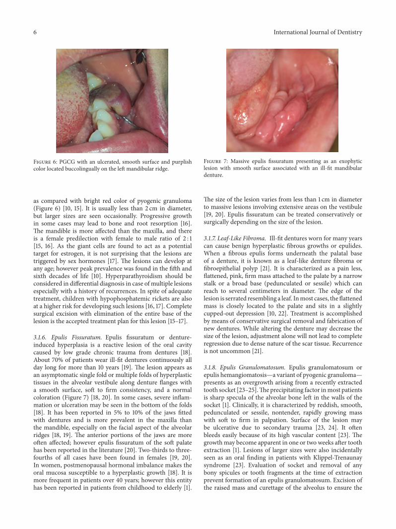
Figure 6: PGCG with an ulcerated, smooth surface and purplishcolor located buccolingually on the left mandibular ridge.
as compared with bright red color of pyogenic granuloma(Figure 6) [10, 15]. It is usually less than 2 cm in diameter,but larger sizes are seen occasionally. Progressive growthin some cases may lead to bone and root resorption [16].The mandible is more affected than the maxilla, and thereis a female predilection with female to male ratio of 2 : 1[15, 16]. As the giant cells are found to act as a potentialtarget for estrogen, it is not surprising that the lesions aretriggered by sex hormones [17]. The lesions can develop atany age; however peak prevalence was found in the fifth andsixth decades of life [10]. Hyperparathyroidism should beconsidered in differential diagnosis in case of multiple lesionsespecially with a history of recurrences. In spite of adequatetreatment, children with hypophosphatemic rickets are alsoat a higher risk for developing such lesions [16, 17]. Completesurgical excision with elimination of the entire base of thelesion is the accepted treatment plan for this lesion [15–17].
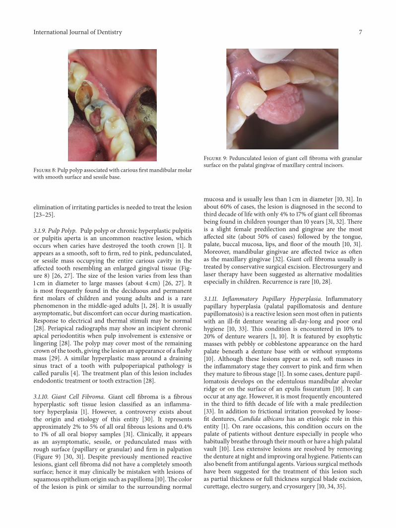
3.1.6. Epulis Fissuratum. Epulis fissuratum or denture-induced hyperplasia is a reactive lesion of the oral cavitycaused by low grade chronic trauma from dentures [18].About 70% of patients wear ill-fit dentures continuously allday long for more than 10 years [19]. The lesion appears asan asymptomatic single fold or multiple folds of hyperplastictissues in the alveolar vestibule along denture flanges witha smooth surface, soft to firm consistency, and a normalcoloration (Figure 7) [18, 20]. In some cases, severe inflam-mation or ulceration may be seen in the bottom of the folds[18]. It has been reported in 5% to 10% of the jaws fittedwith dentures and is more prevalent in the maxilla thanthe mandible, especially on the facial aspect of the alveolarridges [18, 19]. The anterior portions of the jaws are moreoften affected; however epulis fissuratum of the soft palatehas been reported in the literature [20]. Two-thirds to three-fourths of all cases have been found in females [19, 20].In women, postmenopausal hormonal imbalance makes theoral mucosa susceptible to a hyperplastic growth [18]. It ismore frequent in patients over 40 years; however this entityhas been reported in patients from childhood to elderly [1].
Figure 7: Massive epulis fissuratum presenting as an exophyticlesion with smooth surface associated with an ill-fit mandibulardenture.
The size of the lesion varies from less than 1 cm in diameterto massive lesions involving extensive areas on the vestibule[19, 20]. Epulis fissuratum can be treated conservatively orsurgically depending on the size of the lesion.
3.1.7. Leaf-Like Fibroma. Ill-fit dentures worn for many yearscan cause benign hyperplastic fibrous growths or epulides.When a fibrous epulis forms underneath the palatal baseof a denture, it is known as a leaf-like denture fibroma orfibroepithelial polyp [21]. It is characterized as a pain less,flattened, pink, firm mass attached to the palate by a narrowstalk or a broad base (pedunculated or sessile) which canreach to several centimeters in diameter. The edge of thelesion is serrated resembling a leaf. Inmost cases, the flattenedmass is closely located to the palate and sits in a slightlycupped-out depression [10, 22]. Treatment is accomplishedby means of conservative surgical removal and fabrication ofnew dentures. While altering the denture may decrease thesize of the lesion, adjustment alone will not lead to completeregression due to dense nature of the scar tissue. Recurrenceis not uncommon [21].
3.1.8. Epulis Granulomatosum. Epulis granulomatosum orepulis hemangiomatosis—a variant of pyogenic granuloma—presents as an overgrowth arising from a recently extractedtooth socket [23–25].Theprecipitating factor inmost patientsis sharp specula of the alveolar bone left in the walls of thesocket [1]. Clinically, it is characterized by reddish, smooth,pedunculated or sessile, nontender, rapidly growing masswith soft to firm in palpation. Surface of the lesion maybe ulcerative due to secondary trauma [23, 24]. It oftenbleeds easily because of its high vascular content [23]. Thegrowthmay become apparent in one or two weeks after toothextraction [1]. Lesions of larger sizes were also incidentallyseen as an oral finding in patients with Klippel-Trenaunaysyndrome [23]. Evaluation of socket and removal of anybony spicules or tooth fragments at the time of extractionprevent formation of an epulis granulomatosum. Excision ofthe raised mass and curettage of the alveolus to ensure the
International Journal of Dentistry 7
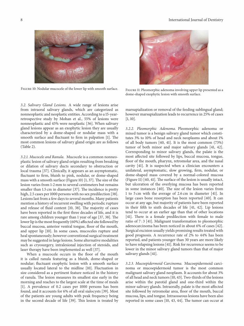
Figure 8: Pulp polyp associated with carious first mandibularmolarwith smooth surface and sessile base.
elimination of irritating particles is needed to treat the lesion[23–25].
3.1.9. Pulp Polyp. Pulp polyp or chronic hyperplastic pulpitisor pulpitis aperta is an uncommon reactive lesion, whichoccurs when caries have destroyed the tooth crown [1]. Itappears as a smooth, soft to firm, red to pink, pedunculated,or sessile mass occupying the entire carious cavity in theaffected tooth resembling an enlarged gingival tissue (Fig-ure 8) [26, 27]. The size of the lesion varies from less than1 cm in diameter to large masses (about 4 cm) [26, 27]. Itis most frequently found in the deciduous and permanentfirst molars of children and young adults and is a rarephenomenon in the middle-aged adults [1, 28]. It is usuallyasymptomatic, but discomfort can occur during mastication.Response to electrical and thermal stimuli may be normal[28]. Periapical radiographs may show an incipient chronicapical periodontitis when pulp involvement is extensive orlingering [28]. The polyp may cover most of the remainingcrown of the tooth, giving the lesion an appearance of a flashymass [29]. A similar hyperplastic mass around a drainingsinus tract of a tooth with pulpoperiapical pathology iscalled parulis [4]. The treatment plan of this lesion includesendodontic treatment or tooth extraction [28].
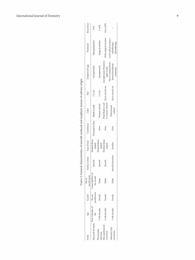
3.1.10. Giant Cell Fibroma. Giant cell fibroma is a fibroushyperplastic soft tissue lesion classified as an inflamma-tory hyperplasia [1]. However, a controversy exists aboutthe origin and etiology of this entity [30]. It representsapproximately 2% to 5% of all oral fibrous lesions and 0.4%to 1% of all oral biopsy samples [31]. Clinically, it appearsas an asymptomatic, sessile, or pedunculated mass withrough surface (papillary or granular) and firm in palpation(Figure 9) [30, 31]. Despite previously mentioned reactivelesions, giant cell fibroma did not have a completely smoothsurface; hence it may clinically be mistaken with lesions ofsquamous epithelium origin such as papilloma [10].The colorof the lesion is pink or similar to the surrounding normal
Figure 9: Pedunculated lesion of giant cell fibroma with granularsurface on the palatal gingivae of maxillary central incisors.
mucosa and is usually less than 1 cm in diameter [10, 31]. Inabout 60% of cases, the lesion is diagnosed in the second tothird decade of life with only 4% to 17% of giant cell fibromasbeing found in children younger than 10 years [31, 32]. Thereis a slight female predilection and gingivae are the mostaffected site (about 50% of cases) followed by the tongue,palate, buccal mucosa, lips, and floor of the mouth [10, 31].Moreover, mandibular gingivae are affected twice as oftenas the maxillary gingivae [32]. Giant cell fibroma usually istreated by conservative surgical excision. Electrosurgery andlaser therapy have been suggested as alternative modalitiesespecially in children. Recurrence is rare [10, 28].
3.1.11. Inflammatory Papillary Hyperplasia. Inflammatorypapillary hyperplasia (palatal papillomatosis and denturepapillomatosis) is a reactive lesion seenmost often in patientswith an ill-fit denture wearing all-day-long and poor oralhygiene [10, 33]. This condition is encountered in 10% to20% of denture wearers [1, 10]. It is featured by exophyticmasses with pebbly or cobblestone appearance on the hardpalate beneath a denture base with or without symptoms[10]. Although these lesions appear as red, soft masses inthe inflammatory stage they convert to pink and firm whenthey mature to fibrous stage [1]. In some cases, denture papil-lomatosis develops on the edentulous mandibular alveolarridge or on the surface of an epulis fissuratum [10]. It canoccur at any age. However, it is most frequently encounteredin the third to fifth decade of life with a male predilection[33]. In addition to frictional irritation provoked by loose-fit dentures, Candida albicans has an etiologic role in thisentity [1]. On rare occasions, this condition occurs on thepalate of patients without denture especially in people whohabitually breathe through their mouth or have a high palatalvault [10]. Less extensive lesions are resolved by removingthe denture at night and improving oral hygiene. Patients canalso benefit from antifungal agents. Various surgical methodshave been suggested for the treatment of this lesion suchas partial thickness or full thickness surgical blade excision,curettage, electro surgery, and cryosurgery [10, 34, 35].
8 International Journal of Dentistry
Figure 10: Nodular mucocele of the lower lip with smooth surface.
3.2. Salivary Gland Lesions. A wide range of lesions arisefrom intraoral salivary glands, which are categorized asnonneoplastic and neoplastic entities. According to a 15-year-retrospective study by Mohan et al., 55% of lesions werenonneoplastic and 45% were neoplastic [36]. When salivarygland lesions appear as an exophytic lesion they are usuallycharacterized by a dome-shaped or nodular mass with asmooth surface and fluctuant to firm in palpation [1]. Themost common lesions of salivary gland origin are as follows(Table 2).
3.2.1. Mucocele and Ranula. Mucocele is a common nonneo-plastic lesion of salivary gland origin resulting from breakingor dilation of salivary ducts secondary to obstruction orlocal trauma [37]. Clinically, it appears as an asymptomatic,fluctuant to firm, bluish to pink, nodular, or dome-shapedmass with a smooth surface (Figure 10) [1, 37].The size of thelesion varies from 1-2mm to several centimeters but remainssmaller than 1.5 cm in diameter [37]. The incidence is prettyhigh, 2.5 cases per 1000 persons with no sex predilection [38].Lesions last from a few days to several months. Many patientsmention a history of recurrent swelling with periodic ruptureand release of fluid content [10, 38]. The majority of caseshave been reported in the first three decades of life, and it israre among children younger than 1 year of age [37, 38]. Thelower lip is themost frequently (60%) affected site followed bybuccal mucosa, anterior ventral tongue, floor of the mouth,and upper lip [10]. In some cases, mucoceles rupture andheal spontaneously; however conventional surgical treatmentmay be suggested in large lesions. Some alternativemodalitiessuch as cryosurgery, intralesional injection of steroids, andlaser therapy have been implemented as well [37].
When a mucocele occurs in the floor of the mouthit is called ranula featuring as a bluish, dome-shaped ornodular; fluctuant exophytic lesion with a smooth surfaceusually located lateral to the midline [10]. Fluctuation insize considered as a pertinent feature noticed in the historyof ranula. The lesion measures its smallest size early in themorning and reaches to the largest scale at the time of meals[1]. A prevalence of 0.2 cases per 1000 persons has beenfound, and it accounts for 6% of all oral sialocysts [39]. Mostof the patients are young adults with peak frequency beingin the second decade of life [39]. This lesion is treated by
Figure 11: Pleomorphic adenoma involving upper lip presented as adome-shaped exophytic lesion with smooth surface.
marsupialization or removal of the feeding sublingual gland;however marsupialization leads to recurrence in 25% of cases[1, 10].
3.2.2. Pleomorphic Adenoma. Pleomorphic adenoma ormixed tumor is a benign salivary gland tumor which consti-tutes 3% to 10% of head and neck neoplasms and about 1%of all body tumors [40, 41]. It is the most common (73%)tumor of both minor and major salivary glands [41, 42].Corresponding to minor salivary glands, the palate is themost affected site followed by lips, buccal mucosa, tongue,floor of the mouth, pharynx, retromolar area, and the nasalcavity [41]. It is suspected when a clinician encounters aunilateral, asymptomatic, slow growing, firm, nodular, ordome-shaped mass covered by a normal-colored mucosa(Figure 11) [40, 41]. The surface of the lesion is usually intact,but ulceration of the overlying mucosa has been reportedin some instances [40]. The size of the lesion varies from1 to 5 cm with the average of 2.6 cm in diameter [41]. Inlarge cases bone resorption has been reported [40]. It canoccur at any age, but majority of patients have been reportedin their fifth to sixth decades of life [41, 42]. Lip lesionstend to occur at an earlier age than that of other locations[41]. There is a female predilection with female to maleratio of 7 : 3 [41]. Malignant transformation to pleomorphicadenocarcinoma has been noticed in about 6% of cases [42].Surgical excision usually yields promising results treated withgood prognosis. A recurrence rate of 2% to 44% has beenreported, and patients younger than 30 years are more likelyto have relapsing lesions [41]. Risk for recurrence seems to belower in the minor salivary gland tumors than that of majorsalivary glands [41].
3.2.3. Mucoepidermoid Carcinoma. Mucoepidermoid carci-noma or mucoepidermoid tumor is the most commonmalignant salivary gland neoplasm. It accounts for about 3%of all head and neck tumors [10, 43]. Two-thirds of the lesionsarise within the parotid gland and one-third within theminor salivary glands. Intraorally, palate is the most affectedsite followed by retromolar area, floor of the mouth, buccalmucosa, lips, and tongue. Intraosseous lesions have been alsoreported in some cases [10, 43, 44]. The tumor can occur at
International Journal of Dentistry 9
Table2:Generalcharacteris
ticso
fsmoo
th-surfacedoralexop
hytic
lesio
nsof
salivaryorigin.
Entity
Age
Gender
Siteof
involvem
ent
Surfa
cetexture
Type
ofbase
Con
sistency
Color
Size
Symptom
&sig
nTreatm
ent
Recurrence
Mucocele&
ranu
laFirst3
decadeso
flife
Nosex
predilection
Lower
lip/floo
rof
them
outh
Smoo
thNod
ular/dom
eshaped
Fluctuanttofirm
Bluish
topink
<1.5
cmAsymptom
atic
Marsupialization
25%
Pleomorph
icadenom
a5-6thdecades
Female
Palate
Smoo
thNod
ular/dom
eshaped
Firm
Normalcolored
1–5c
mAsymptom
atic
Surgicalexcisio
n2–44
%
Mucoepiderm
oid
carcinom
a3–6thdecades
Female
Palate
Smoo
thNod
ular/dom
eshaped
Firm
Pink
/bluish
tored
orno
rmalcolored
Upto
severalcm
Asymptom
atic/Painful
inhigh
Grades
Wides
urgicalexcision
Upto
60%
Adenoidcystic
carcinom
a5-6thdecades
Female
Palate
Smoo
th/ulcerative
Nod
ular
Firm
Pink
/normal
colored
Upto
severalcm
Pain
iscommon
/bon
edestr
uctio
n/dista
ntmetastasis
Localradicalexcisio
n+
radiotherapy/
chem
otherapy
—
10 International Journal of Dentistry
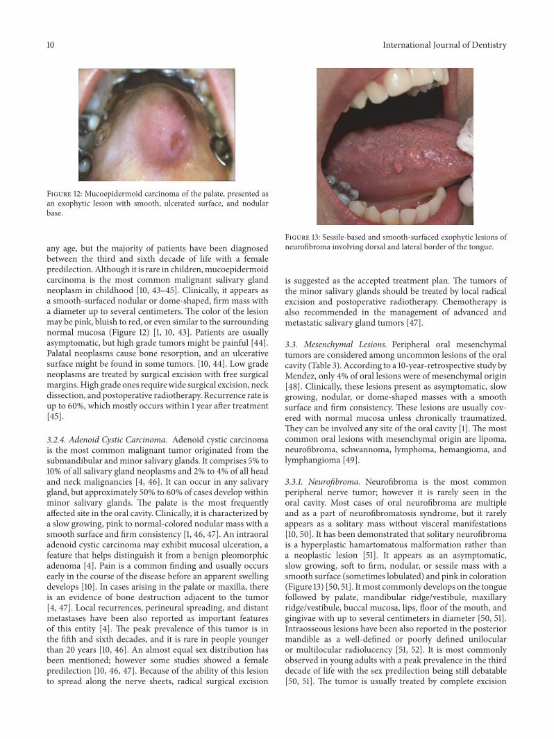
Figure 12: Mucoepidermoid carcinoma of the palate, presented asan exophytic lesion with smooth, ulcerated surface, and nodularbase.
any age, but the majority of patients have been diagnosedbetween the third and sixth decade of life with a femalepredilection. Although it is rare in children,mucoepidermoidcarcinoma is the most common malignant salivary glandneoplasm in childhood [10, 43–45]. Clinically, it appears asa smooth-surfaced nodular or dome-shaped, firm mass witha diameter up to several centimeters. The color of the lesionmay be pink, bluish to red, or even similar to the surroundingnormal mucosa (Figure 12) [1, 10, 43]. Patients are usuallyasymptomatic, but high grade tumors might be painful [44].Palatal neoplasms cause bone resorption, and an ulcerativesurface might be found in some tumors. [10, 44]. Low gradeneoplasms are treated by surgical excision with free surgicalmargins.High grade ones requirewide surgical excision, neckdissection, and postoperative radiotherapy. Recurrence rate isup to 60%, which mostly occurs within 1 year after treatment[45].
3.2.4. Adenoid Cystic Carcinoma. Adenoid cystic carcinomais the most common malignant tumor originated from thesubmandibular andminor salivary glands. It comprises 5% to10% of all salivary gland neoplasms and 2% to 4% of all headand neck malignancies [4, 46]. It can occur in any salivarygland, but approximately 50% to 60% of cases develop withinminor salivary glands. The palate is the most frequentlyaffected site in the oral cavity. Clinically, it is characterized bya slow growing, pink to normal-colored nodular mass with asmooth surface and firm consistency [1, 46, 47]. An intraoraladenoid cystic carcinoma may exhibit mucosal ulceration, afeature that helps distinguish it from a benign pleomorphicadenoma [4]. Pain is a common finding and usually occursearly in the course of the disease before an apparent swellingdevelops [10]. In cases arising in the palate or maxilla, thereis an evidence of bone destruction adjacent to the tumor[4, 47]. Local recurrences, perineural spreading, and distantmetastases have been also reported as important featuresof this entity [4]. The peak prevalence of this tumor is inthe fifth and sixth decades, and it is rare in people youngerthan 20 years [10, 46]. An almost equal sex distribution hasbeen mentioned; however some studies showed a femalepredilection [10, 46, 47]. Because of the ability of this lesionto spread along the nerve sheets, radical surgical excision
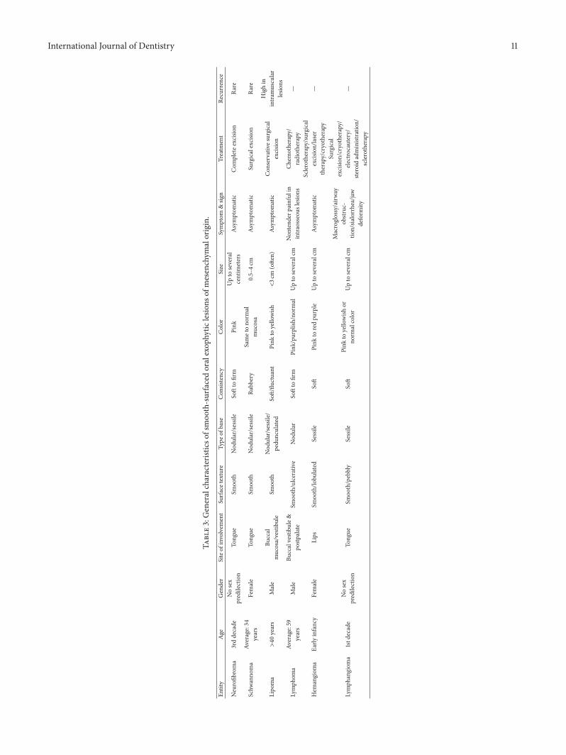
Figure 13: Sessile-based and smooth-surfaced exophytic lesions ofneurofibroma involving dorsal and lateral border of the tongue.
is suggested as the accepted treatment plan. The tumors ofthe minor salivary glands should be treated by local radicalexcision and postoperative radiotherapy. Chemotherapy isalso recommended in the management of advanced andmetastatic salivary gland tumors [47].
3.3. Mesenchymal Lesions. Peripheral oral mesenchymaltumors are considered among uncommon lesions of the oralcavity (Table 3). According to a 10-year-retrospective study byMendez, only 4% of oral lesions were of mesenchymal origin[48]. Clinically, these lesions present as asymptomatic, slowgrowing, nodular, or dome-shaped masses with a smoothsurface and firm consistency. These lesions are usually cov-ered with normal mucosa unless chronically traumatized.They can be involved any site of the oral cavity [1]. The mostcommon oral lesions with mesenchymal origin are lipoma,neurofibroma, schwannoma, lymphoma, hemangioma, andlymphangioma [49].
3.3.1. Neurofibroma. Neurofibroma is the most commonperipheral nerve tumor; however it is rarely seen in theoral cavity. Most cases of oral neurofibroma are multipleand as a part of neurofibromatosis syndrome, but it rarelyappears as a solitary mass without visceral manifestations[10, 50]. It has been demonstrated that solitary neurofibromais a hyperplastic hamartomatous malformation rather thana neoplastic lesion [51]. It appears as an asymptomatic,slow growing, soft to firm, nodular, or sessile mass with asmooth surface (sometimes lobulated) and pink in coloration(Figure 13) [50, 51]. Itmost commonly develops on the tonguefollowed by palate, mandibular ridge/vestibule, maxillaryridge/vestibule, buccal mucosa, lips, floor of the mouth, andgingivae with up to several centimeters in diameter [50, 51].Intraosseous lesions have been also reported in the posteriormandible as a well-defined or poorly defined unilocularor multilocular radiolucency [51, 52]. It is most commonlyobserved in young adults with a peak prevalence in the thirddecade of life with the sex predilection being still debatable[50, 51]. The tumor is usually treated by complete excision
International Journal of Dentistry 11
Table3:Generalcharacteris
ticso
fsmoo
th-surfacedoralexop
hytic
lesio
nsof
mesenchym
alorigin.
Entity
Age
Gender
Siteof
involvem
ent
Surfa
cetexture
Type
ofbase
Con
sistency
Color
Size
Symptom
&sig
nTreatm
ent
Recurrence
Neurofib
roma
3rddecade
Nosex
predilection
Tong
ueSm
ooth
Nod
ular/sessile
Softto
firm
Pink
Upto
several
centim
eters
Asymptom
atic
Com
pletee
xcision
Rare
Schw
anno
ma
Average:34
years
Female
Tong
ueSm
ooth
Nod
ular/sessile
Rubb
ery
Sametono
rmal
mucosa
0.5–4c
mAsymptom
atic
Surgicalexcisio
nRa
re
Lipo
ma
>40
years
Male
Buccal
mucosa/vestibu
leSm
ooth
Nod
ular/sessile/
pedu
nculated
Soft/flu
ctuant
Pink
toyello
wish
<3c
m(ofte
n)Asymptom
atic
Con
servatives
urgical
excisio
n
Highin
intram
uscular
lesio
ns
Lymph
oma
Average:59
years
Male
Buccalvestibu
le&
postp
alate
Smoo
th/ulcerative
Nod
ular
Softto
firm
Pink
/purplish
/normal
Upto
severalcm
Non
tend
erpainfulin
intraosseous
lesio
nsCh
emotherapy/
radiotherapy
—
Hem
angiom
aEa
rlyinfancy
Female
Lips
Smoo
th/lo
bulated
Sessile
Soft
Pink
toredpu
rple
Upto
severalcm
Asymptom
atic
Sclerotherapy/surgical
excisio
n/laser
therapy/cryotherapy
—
Lymph
angiom
a1std
ecade
Nosex
predilection
Tong
ueSm
ooth/pebbly
Sessile
Soft
Pink
toyello
wish
orno
rmalcolor
Upto
severalcm
Macroglossy/airw
ayob
struc-
tion/sia
lorrhea/jaw
deform
ity
Surgical
excisio
n/cryotherapy/
electrocautery/
steroid
administratio
n/sclerotherapy
—
12 International Journal of Dentistry
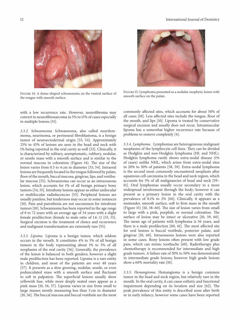
Figure 14: A dome-shaped schwannoma on the ventral surface ofthe tongue with smooth surface.
with a low recurrence rate. However, neurofibroma mayconvert to neurofibrosarcoma in 5% to 15% of cases especiallyin multiple lesions [51].
3.3.2. Schwannoma. Schwannoma, also called neurilem-moma, neurinoma, or perineural fibroblastoma, is a benigntumor of neuroectodermal origin [53, 54]. Approximately25% to 45% of lesions are seen in the head and neck with1% being reported in the oral cavity as well [53]. Clinically, itis characterized by solitary, asymptomatic, rubbery, nodular,or sessile mass with a smooth surface and is similar to thenormal mucosa in coloration (Figure 14). The size of thelesion varies from 0.5 to 4 cm in diameter [53, 54]. Intraorallesions are frequently located in the tongue followed by palate,floor of themouth, buccalmucosa, gingivae, lips, and vestibu-lar mucosa [55]. Schwannoma can occur as an intraosseouslesion, which accounts for 1% of all benign primary bonytumors [54, 55]. Intrabony lesions appear as either unilocularor multilocular radiolucencies [53]. Peripheral lesions areusually painless, but tenderness may occur in some instances[10]. Pain and paresthesia are not uncommon for intrabonytumors [10]. Schwannoma has been reported in the age rangeof 8 to 72 years with an average age of 34 years with a slightfemale predilection (female to male ratio of 1.6 : 1) [53, 55].Surgical excision is the treatment of choice and recurrenceand malignant transformation are extremely rare [55].
3.3.3. Lipoma. Lipoma is a benign tumor, which seldomoccurs in the mouth. It constitutes 4% to 5% of all benigntumors in the body representing about 1% to 5% of allneoplasms of the oral cavity [56]. Generally, the prevalenceof the lesion is balanced in both genders; however a slightmale predilection has been reported. Lipoma is a rare entityin children, and most of the patients are over 40 years[57]. It presents as a slow growing, nodular, sessile, or evenpedunculated mass with a smooth surface and fluctuantto soft in palpation. The superficial lesions usually showyellowish hue, while more deeply seated ones appear as apink mass [10, 56, 57]. Lipoma varies in size from small tolarge masses mostly measuring less than 3 cm in diameter[10, 56].The buccal mucosa and buccal vestibule are the most
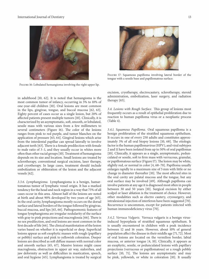
Figure 15: Lymphoma presented as a nodular exophytic lesion withsmooth surface on the palate.
commonly affected sites, which accounts for about 50% ofall cases [10]. Less affected sites include the tongue, floor ofthe mouth, and lips [10]. Lipoma is treated by conservativesurgical excision and usually does not recur. Intramuscularlipoma has a somewhat higher recurrence rate because ofproblems to remove completely [4].
3.3.4. Lymphoma. Lymphomas are heterogeneous malignantneoplasms of the lymphocyte cell lines. They can be dividedas Hodgkin and non-Hodgkin lymphoma (HL and NHL).Hodgkin lymphoma rarely shows extra-nodal disease (1%of cases) unlike NHL, which arises from extra-nodal sitesin 20% to 30% of patients [58, 59]. Extra-nodal lymphomais the second most commonly encountered neoplasm aftersquamous cell carcinoma in the head and neck region, whichaccounts for 5% of all malignancies of head and neck [59–61]. Oral lymphomas usually occur secondary to a morewidespread involvement through the body; however it canpresent as a primary lesion in the oral cavity with theprevalence of 0.1% to 2% [60]. Clinically, it appears as anontender, smooth surface, soft to firm mass in the mouth(Figure 15) [10, 58–60]. The size of lesion varies from smallto large with a pink, purplish, or normal coloration. Thesurface of lesion may be intact or ulcerative [10, 59, 60].The mean age of patients with lymphoma is 56 years, andthere is a male predilection [60, 61]. The most affected sitefor oral lesions is buccal vestibule, posterior palate, andgingivae [10, 60]. Intraosseous lesions were also reportedin some cases. Bony lesions often present with low gradepain, which can mimic toothache [60]. Radiotherapy pluschemotherapy is recommended for intermediate and highgrade tumors. A failure rate of 30% to 50% was demonstratedin intermediate grade lesions; however high grade lesionsshow a 60% mortality rate [10].
3.3.5. Hemangioma. Hemangioma is a benign commontumor in the head and neck region, but relatively rare in themouth. In the oral cavity, it can cause esthetic and functionalimpairment depending on its location and size [62]. Thepeak prevalence of this entity is described soon after birthor in early infancy; however some cases have been reported
International Journal of Dentistry 13
Figure 16: Lobulated hemangioma involving the right upper lip.
in adulthood [10, 62]. It is noted that hemangioma is themost common tumor of infancy, occurring in 5% to 10% ofone-year-old children [10]. Oral lesions are most commonin the lips, gingivae, tongue, and buccal mucosa [62, 63].Eighty percent of cases occur as a single lesion, but 20% ofaffected patients present multiple tumors [10]. Clinically, it ischaracterized by an asymptomatic, soft, smooth, or lobulated,sessile mass with various sizes from a few millimeters toseveral centimeters (Figure 16). The color of the lesionsranges from pink to red purple, and tumor blanches on theapplication of pressure [63, 64]. Gingival lesions which arisefrom the interdental papillae can spread laterally to involveadjacent teeth [63].There is a female predilection with femaleto male ratio of 3 : 1, and they usually occur in whites moreoften than other racial groups [10]. Treatment of hemangiomadepends on its size and location. Small lesions are treated bysclerotherapy, conventional surgical excision, laser therapy,and cryotherapy. In large cases, treatment should includeembolization or obliteration of the lesion and the adjacentvessels [62].
3.3.6. Lymphangioma. Lymphangioma is a benign, hamar-tomatous tumor of lymphatic vessel origin. It has a markedtendency for the head and neck region in a way that 75% of allcases occur in this area. Almost half of the lesions are notedat birth and about 90% developed by two years of age [65].In the oral cavity, lymphangiomamostly occurs on the dorsalsurface and lateral borders of the tongue followed by gingivae,buccal mucosa, and lips [65, 66]. Pathognomonic features oftongue lymphangioma are irregular nodularity of the surfacewith gray to pink projections and macroglossia [66]. There isno sex predilection, and oral lesions are most common in thefirst decade of life [66].The clinicalmanifestation of the lesionvaries based on whether it is superficial or deep. Superficiallesions appear as soft exophytic masses with rough (papillaryor pebbly) surface and pink to yellowish coloration. Deeperlesions are described as soft diffuse masses with normal colorand smooth surface [65, 67]. Massive lesions might causemacroglossia, obstruction in upper air way, sialorrhea, andjaw deformity as well as difficulties in mastication, speech,and oral hygiene [65]. Lymphangioma is treated by surgical
Figure 17: Squamous papilloma involving lateral border of thetongue with a sessile base and papillomatous surface.
excision, cryotherapy, electrocautery, sclerotherapy, steroidadministration, embolization, laser surgery, and radiationtherapy [65].
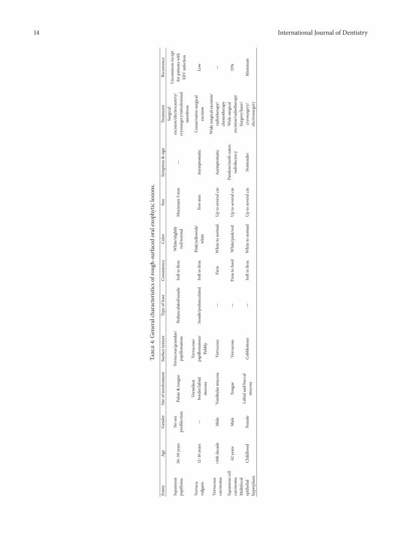
3.4. Lesions with Rough Surface. This group of lesions mostfrequently occurs as a result of epithelial proliferation due toreaction to human papilloma virus or a neoplastic process(Table 4).
3.4.1. Squamous Papilloma. Oral squamous papilloma is abenign proliferation of the stratified squamous epithelium.It occurs in one of every 250 adults and constitutes approx-imately 3% of all oral biopsy lesions [10, 68]. The etiologicfactor is the human papillomavirus (HPV), and viral subtypes2 and 11 have been isolated from up to 50% of oral papillomas[10]. Clinically, it appears as a single, asymptomatic, pedun-culated or sessile, soft to firm mass with verrucous, granular,or papillomatous surface (Figure 17).The lesionmay bewhite,slightly red, or normal in color [4, 68–70]. Papilloma usuallyenlarges rapidly to a maximum size of 5mm with little or nochange in diameter thereafter [10]. The most affected sites inthe oral cavity are palatal mucosa and the tongue, but anyoral surface may be involved [69]. Although papilloma caninvolve patients at any age it is diagnosedmost often in peoplebetween 30 and 50 years [10]. Surgical excision by eitherscalpel or laser ablation is the treatment of choice. However,other modalities such as electro cautery, cryosurgery, andintralesional injection of interferon have been suggested [70].Recurrence is uncommon, except for patients infected withhuman immunodeficiency virus [70].
3.4.2. Verruca Vulgaris. Verruca vulgaris is a benign virus-induced hyperplasia of stratified squamous epithelium. Itis usually encountered in children with a peak incidencebetween 12 and 16 years. However, about 10% of generalpopulation affect the disease in theirmiddle age [71, 72].Mostof oral lesions are located on the vermilion border, labialmucosa, or anterior tongue [4, 10]. Clinically, it appears asan exophytic, sessile, or pedunculated lesions with papillaryprojections (verrucous or papillomatous) or a rough pebblysurface [10, 71]. The lesions are asymptomatic and maybe pink, yellowish, or white in coloration [10]. It usually
14 International Journal of Dentistry
Table4:Generalcharacteris
ticso
frou
gh-surfacedoralexop
hytic
lesio
ns.
Entity
Age
Gender
Siteof
involvem
ent
Surfa
cetexture
Type
ofbase
Con
sistency
Color
Size
Symptom
&sig
nTreatm
ent
Recurrence
Squamou
spapillo
ma
30–50years
Nosex
predilection
Palate&tong
ueVe
rrucou
s/granular/
papillo
matou
sPedu
nculated/sessile
Softto
firm
White/slightly
red/no
rmal
Maxim
um5m
m—
Surgical
excisio
n/electro
cautery/
cryosurgery/intralesional
interfe
ron
Uncom
mon
except
forp
atientsw
ithHIV
infection
Verruca
vulgaris
12–16years
—Ve
rmilion
border/la
bial
mucosa
Verrucou
s/papillo
matou
s/Pebb
lySessile/pedun
culated
Softto
firm
Pink
/yellowish
/white
Fewmm
Asym
ptom
atic
Con
servatives
urgical
excisio
nLo
w
Verrucou
scarcinom
a>6thdecade
Male
Vestibu
larm
ucosa
Verrucou
s—
Firm
Whiteto
norm
alUpto
severalcm
Asym
ptom
atic
Wides
urgicalexcision
/radiotherapy/
chem
otherapy
—
Squamou
scell
carcinom
a62
years
Male
Tong
ueVe
rrucou
s—
Firm
tohard
White/pink/red
Upto
severalcm
Painless/m
oth-eaten
radiolucency
Wides
urgical
excisio
n/radiotherapy
33%
Multifocal
epith
elial
hyperplasia
Child
hood
Female
Labialandbu
ccal
mucosa
Cob
blestone
—Softto
firm
Whiteto
norm
alUpto
severalcm
Non
tend
erSurgery/laser/
cryosurgery/
electrosurgery
Minim
um

International Journal of Dentistry 15
Figure 18: Broad-based verrucous carcinoma involving maxillaryvestibular mucosa, with verrucous surface.
enlarges rapidly to its maximum size and remains constantfor months or years thereafter [71, 72]. Approximately 23%of lesions show spontaneous regression in two months andthe remaining lesions within 2 years [72]. Oral lesions areusually treated by conservative surgical excision. Meanwhile,laser therapy, cryotherapy, or electrosurgery has been alsorecommended. A small proportion of treated lesions showrecurrence [4, 10].
3.4.3. Verrucous Carcinoma. Verrucous carcinoma whichalso called Ackerman tumor, Buschke-Lowenstein tumor,florid oral papillomatosis, epithelioma cuniculatum, or snuffdipper’s cancer is a nonmetastasizing low grade variant oforal squamous cell carcinoma [68, 73].The incidence rate hasbeen estimated as one lesion per 1.000.000 of the populationeach year. The etiology of the lesion is not clear, but orallesions predominantly occur in patients habituated to arecachewing, alcohol drinking, smoking, and having poor oralhygiene. However, 15% to 51% of the lesions have been foundin people without these habits [10, 68]. The most commonaffected sites in the oral cavity are vestibular mucosa, buccalmucosa, gingivae, and the tongue [10, 68, 73]. There is a malepredilection with the majority of lesions being reported inthe sixth decade [4, 68, 73]. The lesion is featured by anasymptomatic, broad-based, well-circumscribed, thick, pinkto white plaque resembling a cauliflower with verruciformsurface (Figure 18) [10]. Similarly simultaneous oral and gen-ital lesions of larger sizes would suggest condyloma acumina-tum [10].Malignant transformation has been detected in 20%of the lesions [73]. Wide surgical excision, radiotherapy, andchemotherapy have been recommended for treatment.
3.4.4. Squamous Cell Carcinoma. Squamous cell carcinoma(SCC) accounts for 90% of all oral cancers and is consideredas the eighth most frequent cancer globally [74]. Males areaffected more frequently than females with a male to femaleratio of 3 : 1 [10]. The median age of diagnosis is 62 years.However, the incidence of oral SCC in persons younger than45 is increasing [75–77]. Tongue is the mostly affected sitein the oral cavity followed by floor of the mouth, gingivae,palate, retromolar area, buccal, and labial mucosa [10, 75–78].In young patients and those with congenital oral squamouscell carcinoma the tongue is the mostly affected site of
Figure 19: Exophytic SCCwith a verrucous, necrotic, and ulcerativesurface, extended from vestibule of the mandible to floor of themouth.
involvement as well [10].Warning signs of oral cancer includered-white lesions, ulcer lasting more than three weeks, painof the tongue due to its mobility and sensitive nature, lumpin the oral cavity or in the neck area, discomfort with speechor swallowing, mobile teeth in the absence of periodontitis,anesthesia, and earache without apparent disease. The mostfrequent complaints among oral cancer patients are swelling,pain, and ulceration [74]. Clinically, it can appear in variousforms such as a red or white plaque, a solitary chroniculcer or an exophytic mass [75]. Approximately 55% of thetongue lesions were exophytic-type.This type of lesions has arough surface (verrucous) and usually is irregular in shape(Figure 19). The lesion has a broad base and it is white,pink, or red in color. Ulceration may be present in largerfungating lesions with necrotic and multicolored surface. Inaddition, oral SCCs are painless and firm to hard in palpationand bleeding is not an early characteristic feature [1, 10, 75].However, in cases with destruction of the underlying bonepain may be reported, and a “moth-eaten radiolucency” withill-defined or ragged borders is found [10]. Despite early oralcancer whose manifestations are not definitive late-stage oralSCC show quite prominent signs such as a large mass withirregular margins, ulceration, nodularity, and fixation to thesurrounding tissues [74]. The treatment of intraoral SCC isguided by the clinical stage of the disease and consists ofwide surgical excision, radiation therapy, or a combinationof surgery and radiation therapy [10]. The recurrence rate isestimated about 33% within 2 to 96 months [79].
3.4.5. Multifocal Epithelial Hyperplasia. Multifocal epithe-lial hyperplasia, also known as focal epithelial hyperplasia,multifocal epithelial papilloma, virus epithelial hyperplasia,and Heck disease, is a virus-induced proliferation of oralsquamous epithelium [10]. Although this entity is usually achildhood condition other age groups may also be affected.The frequency of the lesion ranges from 0.002 to 35% indifferent populations and 70% of patients are in first twodecades of their life [79]. In addition, there is a femalepredilection with female to male ratio of 3 : 1 [79]. It appears
16 International Journal of Dentistry
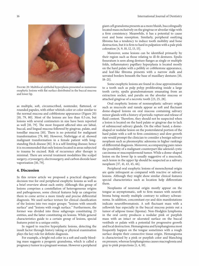
Figure 20: Multifocal epithelial hyperplasia presented as numerousexophytic lesions with flat surface distributed in the buccal mucosaand lower lip.
as multiple, soft, circumscribed, nontender, flattened, orrounded papules, with either whitish color or color similar tothe normal mucosa and cobblestone appearance (Figure 20)[10, 79, 80]. Most of the lesions are less than 0.5 cm, butlesions with several centimeters in size have been reportedas well [10, 79]. The most frequent affected sites are labial,buccal, and lingual mucosa followed by gingivae, palate, andtonsillar mucosa [10]. There is no potential for malignanttransformation [79, 80]. However, Niebrugge et al. showedmalignant transformation in a female patient with long-standing Heck disease [81]. It is a self-limiting disease; henceit is recommended that only lesions located in areas subjectedto trauma be excised. Risk of recurrence after therapy isminimal. There are several treatment modalities like scalpelsurgery, cryosurgery, electrosurgery, and carbon dioxide laservaporization [10, 79].
4. Discussion
In this review article we proposed a practical diagnosticdecision tree for oral peripheral exophytic lesions as well asa brief overview about each entity. Although this group oflesions comprises a constellation of heterogeneous originsand pathogeneses, some clinical features help us categorizethem to come arrive a more timely and precise differentialdiagnosis. We used surface texture for clinical classificationof the lesions into two major groups: “lesions with smoothsurface” and “lesions with rough surface.” Furthermore, theformer was divided into three subgroups constituting 23entities, and the latter constituting six lesions. While generalcharacteristics guide to a certain group of lesions, specialfeatures point to a unique entity.
In regard to reactive hyperplastic lesions, detecting theinsult factor through history taking or physical examinationplays the key role for definite diagnosis.
For example, a bright red color in a soft and easily bleed-ing mass suggests a pyogenic granuloma, which is called apregnancy tumor in a pregnantwoman.However a peripheral
giant cell granulomapresents as amore bluish, buccolinguallylocatedmass exclusively on the gingivae or alveolar ridgewitha firm consistency. Meanwhile, it has a potential to causeroot and bone resorption. Similarly, peripheral ossifyingfibroma has a tendency to induce tooth mobility and bonedestruction, but it is firm to hard in palpationwith a pale pinkcoloration [4, 9, 10, 12, 13, 15].
Moreover, some lesions can be identified primarily bytheir region such as those relating to ill-fit dentures. Epulisfissuratum is seen along denture flanges as single or multiplefolds, inflammatory papillary hyperplasia is located mostlyon the hard palate with a pebbly or cobblestone appearance,and leaf-like fibroma presents with a narrow stalk andserrated borders beneath the base of maxillary dentures [10,18–21].
Some exophytic lesions are found in close approximationto a tooth such as pulp polyp proliferating inside a largetooth cavity, epulis granulomatosum emanating from anextraction socket, and parulis on the alveolar mucosa orattached gingivae of a necrotic tooth [23, 25, 28].
Oral exophytic lesions of nonneoplastic salivary originsuch as mucocele and ranula appear as soft and fluctuantdome-shaped lesions on oral mucosa containing salivaryminor glands with a history of periodic rupture and release offluid content. Therefore, they should not be suspected whena lesion is located on the hard palate or gingivae due to lackof submucosal salivary glands. On the other hand, a dome-shaped or nodular lesion on the posterolateral portion of thehard palate with a soft to firm consistency and slow growthrate would prompt the clinician to consider a benign salivaryneoplasm such as pleomorphic adenoma in higher rankingsof differential diagnosis. Moreover, accompanying pain raisesthe possibility of a malignant counterpart like adenoid cysticcarcinoma ormucoepidermoid tumor.While a dome-shapedlesion on the lower lip is usually suggestive of a mucocele,such lesion in the upper lip should be suspected as a salivaryneoplasm [37, 41, 43, 45, 46].
Peripheral oral exophytic lesions of mesenchymal originare quite infrequent as compared with reactive or salivarylesions. Although they might show similar clinical featuresspecial characteristics such as location help differentiatethem.
Neoplasms of neuronal origin mostly appear on thetongue as asymptomatic, soft to firm masses with neurofi-broma being mostly multiple contrary to solitary schwan-noma. In addition, concomitant eye and skin manifestationsindicate neurofibromatosis. A soft fluctuant mass with ayellowish hue especially in the buccal mucosa points out atumor of adipose tissue (lipoma). Non-Hodgkin lymphomain the oral cavity produces a nodular pink or purplishmass with an intact or ulcerated surface on the buccalvestibule or palate with a potential for progressive growthand local destruction.Hemangioma and lymphangiomamostfrequently happen on the tongue sometimes with a roughsurface despite their connective tissue origin. Hemangiomais characterized by a pink or purplish color and blanchingon pressure, whereas lymphangioma causesmacroglossia andgray to pink projections [1, 4, 10].
International Journal of Dentistry 17
As a general rule, lesions originating from the epitheliumpresent with a rough surface. An exception in this regardis focal epithelial hyperplasia with numerous small flat-endpapules scattered on the oral mucosa most commonly inchildren and adolescence. Small finger-like projections onlesion surface are invariably seen in viral-induced lesionssuch as squamous papilloma, verruca vulgaris, and condy-loma acuminatum. Papilloma tends to occur as a single lesionrarely exceeding 1 cm in diameter. On the other hand, verrucavulgaris and Heck disease mostly appear in multiple formswith the former being coexisted with cutaneous lesions.Coincidence of large verruca vulgaris-form lesions in thegenitalia and oral cavity as well as high-risk sexual behaviorrefer to a condyloma acuminatum.
Squamous cell carcinoma when appears as a broad-basedexophytic lesion involves the lateral border of the tongue witha rough nonhomogenous or sometimes ulcerative or necroticsurface and a progressive potential to unlimited growth. Onthe other hand, its nonmetastasizing counterpart—verrucouscarcinoma—is mostly encountered on the vestibular or buc-cal mucosa in a snuff-dipper patient with a homogenouscauliflower appearance and an association with smokelesstobacco use.
5. Conclusion
We proposed a diagnostic decision tree regarding oralperipheral exophytic lesions divided into lesions with smoothsurface and rough-surfaced lesions. Upon confronting aperipheral exophyticmass in the oral cavity a clinician shouldconsider some features such as surface texture, shape of base,color, and consistency in order to categorize the lesion andprogress along the decision tree to reach a logistic differentialdiagnosis.
Conflicts of Interest
The authors declare that there are no conflicts of interestregarding the publication of this paper.
References
[1] N. K. Wood and P. W. Goaz, Differential Diagnosis of Oral AndMaxillofacial Lesions, Mosby, St. Louis, Mo, USA, 5th Editionedition, 1997.
[2] A. B. R. Santosh, D. Boyd, and K. K. Laxminarayana, “Proposedclinico-pathological classification for oral exophytic lesions,”Journal of Clinical and Diagnostic Research, vol. 9, no. 9, pp.ZE01–ZE08, 2015.
[3] R. B. Zain, N. Ikeda, I. A. Razak et al., “A national epidemio-logical survey of oral mucosal lesions in Malaysia,” CommunityDentistry and Oral Epidemiology, vol. 25, no. 5, pp. 377–383,1997.
[4] M. Glick, Burket’s Oral Medicine, People’s medical publishinghouse, 12th edition edition, 2015, 147-172, 175-188, 236-237.
[5] A. Bermejo-Fenoll and P. Lopez-Jornet, “Differential diagnosisof exophytic lesions of soft oral tissue,”Medicina Oral, PatologiaOral y Cirugia Bucal, vol. 10, no. 5, pp. 470-471, 2005.
[6] N. G. Nikitakis, “Oral soft tissue lesions: a guide to differentialdiagnosis part II: surface alterations,” Brazilian Journal of OralSciences, vol. 4, no. 13, pp. 707–715, 2005.
[7] V. Reddy, S. Saxena, S. Saxena, and M. Reddy, “Reactivehyperplastic lesions of the oral cavity: a ten year observationalstudy on North Indian population,” Journal of Clinical andExperimental Dentistry, vol. 4, no. 3, pp. e136–e140, 2012.
[8] H. Kadeh, S. Saravani, and M. Tajik, “Reactive hyperplasticlesions of the oral cavity,” Iranian Journal of Otorhinolaryngol-ogy, vol. 27, no. 79, pp. 137–144, 2015.
[9] H. Jafarzadeh, M. Sanatkhani, and N. Mohtasham, “Oral pyo-genic granuloma: a review,” Journal of Oral Science, vol. 48, no.4, pp. 167–175, 2006.
[10] B. W. Neville, D. D. Damm, C. M. Allen, and J. E. Bouquot,Oral andMaxillofacial Pathology, Saunders-Elsevier. China, 3rdedition, 2009.
[11] S. R. Gomes, Q. J. Shakir, P. V. Thaker, and J. K. Tavadia,“Pyogenic granuloma of the gingiva: a misnomer?: a casereport and review of literature,” Journal of Indian Society ofPeriodontology, vol. 17, no. 4, pp. 514–519, 2013.
[12] M. J. Franco-Barrera, M. G. Zavala-Cerna, R. Fernandez-Tamayo, I. Vivanco-Perez, N. M. Fernandez-Tamayo, and O.Torres-Bugarın, “An update on peripheral ossifying fibroma:case report and literature review,” Oral and MaxillofacialSurgery, vol. 20, no. 1, pp. 1–7, 2016.
[13] T. Farquhar, J. Maclellan, H. Dyment, and Anderson R. D.,“Peripheral ossifying fibroma: a case report,” Canadian DentalAssociation, vol. 47, no. 9, 809 pages, 2016, 812.
[14] M. B. Mishra, K. A. Bhishen, and S. Mishra, “Peripheralossifying fibroma,” Journal of Oral and Maxillofacial Pathology,vol. 15, no. 1, pp. 65–68, 2011.
[15] A. Nekouei, A. Eshghi, P. Jafarnejadi, and Z. Enshaei, “A reviewand report of peripheral giant cell granuloma in a 4-year-oldchild,” Case Reports in Dentistry, vol. 2016, Article ID 7536304,4 pages, 2016.
[16] C. M. Flaitz, “Peripheral giant cell granuloma: a potentiallyaggressive lesion in children,” Pediatric Dentistry, vol. 22, no. 3,pp. 232-233, 2000.
[17] G. S. Letterman, “Peripheral giant cell granuloma,” Plastic andReconstructive Surgery, vol. 46, no. 3, p. 320, 1970.
[18] K. Veena, H. Jagadishchandra, J. Sequria, S. Hameed, L. Chatra,and P. Shenai, “An extensive denture-induced hyperplasia ofmaxilla,” Annals of Medical and Health Sciences Research, vol.3, no. 5, p. 7, 2013.
[19] E. M. Canger, P. Celenk, and S. Kayipmaz, “Denture-relatedhyperplasia: A clinical study of a Turkish population group,”Brazilian Dental Journal, vol. 20, no. 3, pp. 243–248, 2009.
[20] H. Mortazavi, H. R. Khalighi, S. Jafari, and M. Baharvand,“Epulis fissuratum in the soft palate: Report of a case in a veryrare location,” Dental Hypotheses, vol. 7, no. 2, pp. 67–69, 2016.
[21] I. M. Brook and D. J. Lamb, “Surgical/prosthetic problems of alarge leaf fibroma,” Dental Update, vol. 15, no. 3, p. 126, 1988.
[22] N. G. Nikitakis and J. K. Brooks, “Sessile nodule on the palate.Leaflike denture fibroma,” General Dentistry, vol. 59, no. 1, pp.76-77, 2011.
[23] B. Manovijay, P. Rajathi, S. M. Fenn, and B. Sekar, “Recurrentepulis granulomatosa: a second look,” Journal of AdvancedClinical & Research Insights, vol. 2, pp. 140–142, 2015.
[24] S. Ghadimi, N. Chiniforush, M. Najafi, and S. Amiri, “Excisionof epulis granulomatosa with diode laser in 8 years old boy,”Journal of Lasers in Medical Sciences, vol. 6, no. 2, pp. 92–95,2015.
18 International Journal of Dentistry
[25] S. Palanivelu, P. Jayanthi, U. K. Rao, E. Joshua, and K. Ran-ganathan, “Rapidly enlargingmass following dental extraction,”Journal of Oral and Maxillofacial Pathology, vol. 15, no. 2, pp.223–227, 2011.
[26] J. Faryabi and S. Adhami, “Unusual presentation of chronichyperplastic pulpitis: a case report. Iran Endod J,” Winter, vol.2, no. 4, p. 156, 2008.
[27] N. S. A. Jabbar, J. M. Aldrigui, M. M. Braga, and M. T.Wanderley, “Pulp polyp in traumatized primary teeth: a case-control study,”Dental Traumatology, vol. 29, no. 5, pp. 360–364,2013.
[28] K. Anilkumar, S. Lingeswaran, G. Ari, R. Thyagarajan, and A.Logaranjani, “Management of chronic hyperplastic pulpitis inmandibular molars of middle aged adults: a multidisciplinaryapproach,” Journal of Clinical and Diagnostic Research, vol. 10,no. 1, pp. ZD23–ZD25, 2016.
[29] M. Gorduysus, Hyperplastic Pulpitis Development in a BridgeAbutment Tooth, vol. 32, Hacettepe Dis Hekimligi FakultesiDergisi, Ankara, Turkey, 1 edition, 2008, 35–37.
[30] H. R. Khalighi,M.Hamian, F.M. Abbas, and S. Farhadi, “Simul-taneous existence of giant cell fibroma and squamous papillomain the oral cavity,” Indian Journal of Medical Specialities, vol. 2,no. 2, 2011.
[31] N. G. Nikitakis, D. Emmanouil, M. P. Maroulakos, and M. V.Angelopoulou, “Giant cell fibroma in children: report of twocases and literature review,” Journal of Oral and MaxillofacialResearch, vol. 4, no. 1, article e5, pp. 1–7, 2013.
[32] V. K. K. Reddy, N. Kumar, P. Battepati, L. Samyuktha, and S. P.Nanga, “Giant cell fibroma in a paediatric patient: a rare casereport,” Case Reports in Dentistry, vol. 2015, Article ID 240374,3 pages, 2015.
[33] M. S. Thwaites, T. E. Jeter, and O. Ajagbe, “Inflammatorypapillary hyperplasia: review of literature and case reportinvolving a 10-year-old child,” Quintessence International, vol.21, no. 2, pp. 133–138, 1990.
[34] J. R. Antonelli, F. V. Panno, and A. Witko, “Inflammatorypapillary hyperplasia: supraperiosteal excision by the blade-loop technique.,” General dentistry, vol. 46, no. 4, pp. 390–397,1998.
[35] P. Infante-Cossio, R. Martinez-de-Fuentes, E. Torres-Carranza,and J. L. Gutierrez-Perez, “Inflammatory papillary hyperplasiaof the palate: treatment with carbon dioxide laser, followedby restoration with an implant-supported prosthesis,” BritishJournal of Oral andMaxillofacial Surgery, vol. 45, no. 8, pp. 658–660, 2007.
[36] H. Mohan, A. Tahlan, I. Mundi, R. P. S. Punia, and A.Dass, “Non-neoplastic salivary gland lesions: a 15-year study,”EuropeanArchives of Oto-Rhino-Laryngology, vol. 268, no. 8, pp.1187–1190, 2011.
[37] H. Mortazavi, M. Baharvand, S. Alirezaei, and R. Noor-Mohammadi, “Combination therapy in a large lower lip muco-cele: a non-invasive recommended technique,”Dental Hypothe-ses, vol. 5, no. 3, pp. 127–129, 2014.
[38] H. Mortazavi, H. R. Khalighi, M. Baharvand, M. Eshghpour,and R. Singh, “Bilateral symmetrical mucocele of the lower lip:report of a rare clinical presentation,” International Journal ofExperimental Dental Science, vol. 3, pp. 92–94, 2014.
[39] R. Prakash, B. B. Kushwaha, and S. Gautam, “Airway man-agement in a child with oral ranula,” International Journal ofBiomedical Research, vol. 5, no. 2, p. 148, 2014.
[40] S. Moghe, A. K. Pillai, S. Prabhu, S. Nahar, and U. K. Kartika,“Pleomorphic adenoma of the palate: report of a case,” Interna-tional Journal of Scientific Study, vol. 2, pp. 54–56, 2014.
[41] H. Mortazavi, S. Alirezaei, S. Azari-Marhabi, M. Baharvand,andM. Eshghpour, “Upper lip pleomorphic adenoma: compar-ison of reported cases between,” Journal of Dental materials andTechniques, vol. 2, no. 4, pp. 125–129, 1990.
[42] M. Rahnama, U.Orzedala-Koszel, L. Czupkallo, andM. Lobacz,“Pleomorphic adenoma of the palate: a case report and reviewof the literature,”Wspolczesna Onkologia, vol. 17, no. 1, pp. 103–106, 2013.
[43] S. J. Jarde, S. Das, S. Narayanswamy, A. Chatterjee, and C. Babu,“Mucoepidermoid carcinoma of the palate: a rare case report,”Journal of Indian Society of Periodontology, vol. 20, no. 2, pp.203–206, 2016.
[44] P. Ritwik, K. G. Cordell, and R. B. Brannon, “Minor salivarygland mucoepidermoid carcinoma in children and adolescents:a case series and review of the literature,” Journal ofMedical CaseReports, vol. 6, article no. 182, 2012.
[45] N. Shah, A. Mahjan, H. Patel, R. Shah, and S. Shah, “Mucoepi-dermoid carcinoma of palate: a case repor,” Scholars journal ofdental sciences, vol. 2, no. 3, pp. 222–224, 2015.
[46] K. Triantafillidou, J. Dimitrakopoulos, F. Iordanidis, and D.Koufogiannis, “Management of adenoid cystic carcinoma ofminor salivary glands,” Journal of Oral and MaxillofacialSurgery, vol. 64, no. 7, pp. 1114–1120, 2006.
[47] P. J. Bradley, “Adenoid cystic carcinoma of the head and neck:a review,” Current Opinion in Otolaryngology & Head and NeckSurgery, vol. 12, no. 2, pp. 127–132, 2004.
[48] M. Mendez, V. C. Carrard, A. N. Haas et al., “A 10-year studyof specimens submitted to oral pathology laboratory analysis:lesion occurrence and demographic features,” Brazilian OralResearch, vol. 26, no. 3, pp. 235–241, 2012.
[49] M. Ali and D. Sundaram, “Biopsied oral soft tissue lesions inKuwait: a six-year retrospective analysis,”Medical Principles andPractice, vol. 21, no. 6, pp. 569–575, 2012.
[50] P. Dalvi, K. Vandana, S. Prakash, and K. Mohan, “Solitaryneurofibroma of the gingiva: a rare case report,” Indian Journalof Multidisciplinary Dentistry, vol. 6, no. 2, p. 111, 2016.
[51] J. P. George and N. Sai Jyothsna, “Solitary neurofibroma: a rareoccurrence on gingiva,”General Dentistry, vol. 64, no. 3, pp. 28–31, 2016.
[52] T. Shimoyama, T. Kato, D. Nasu, T. Kaneko, N. Horie, and F.Ide, “Solitary neurofibroma of the oral mucosa: a previouslyundescribed variant of neurofibroma.,” Journal of oral science,vol. 44, no. 1, pp. 59–63, 2002.
[53] P. Prasanna Kumar and K. Meghashri, “Schwannoma of thehard palate: a case report and review of literature,” Journal ofOral Research and Review, vol. 3, no. 1, pp. 23–27, 2012.
[54] J.-M. Sanchis, C.-M. Navarro, J.-V. Bagan et al., “Intraoralschwannomas: presentation of a series of 12 cases,” Journal ofClinical and Experimental Dentistry, vol. 5, no. 4, pp. 192–196,2013.
[55] S. Parhar, H. Preet Singh, A. Nayyar, and A. S. Manchanda,“Intra-oral schwannoma: a case report,” Journal of Clinical andDiagnostic Research, vol. 8, no. 3, pp. 264-265, 2014.
[56] S. Kumaraswamy, N. Madan, R. Keerthi, and S. Shakti, “Lipo-mas of oral cavity: case reports with review of literature,” Journalof Maxillofacial and Oral Surgery, vol. 8, no. 4, pp. 394–397,2009.
International Journal of Dentistry 19
[57] R. Kaur, S. Kler, and A. Bhullar, “Intraoral lipoma: report of3 cases,” Dental Research Journal, vol. 8, no. 1, pp. 48–51, 2011,(Isfahan) Winter.
[58] K. Rana, V. Narula, E. K. Bhargava, R. Shankar, andN.Mahajan,“T-cell lymphoma of the oral cavity: case report,” Journal ofClinical andDiagnostic Research, vol. 9, no. 3, pp.MD03–MD04,2015.
[59] M. Malaguarnera, M. Giordano, C. Russo et al., “Lymphomaof cheek: a case report,” European Review for Medical andPharmacological Sciences, vol. 16, pp. 4–7, 2012.
[60] S. Alirezaei, M. Baharvand, B. Tavakoli, S. Sarikhani, and A. R.Mafi, “Advanced primary lymphoma of oral cavity: report of acase,” Open Journal of Stomatology, vol. 04, no. 03, pp. 109–114,2014.
[61] S. I. M. L. Queiroz, G. M. de Assis, V. D. Silvestre, A. R.Germano, and J. S. P. da Silva, “Treatment of oral hemangiomawith sclerotherapy: case report,” Jornal Vascular Brasileiro, vol.13, no. 3, 2014.
[62] A. Dilsiz, T. Aydin, and N. Gursan, “Capillary hemangioma asa rare benign tumor of the oral cavity: a case report,” CasesJournal, vol. 2, no. 9, article no. 8622, 2009.
[63] K. A. Kamala, L. Ashok, and G. P. Sujatha, “Cavernous heman-gioma of the tongue: a rare case report,” Contemporary ClinicalDentistry, vol. 5, no. 1, pp. 95–98, 2014.
[64] V.Usha, T. Sivasankari, S. Jeelani, G. S. Asokan, and J. Parthiban,“Lymphangioma of the tongue: a case report and review ofliterature,” Journal of Clinical andDiagnostic Research, vol. 8, no.9, pp. ZD12–ZD14, 2014.
[65] H. Bhayya, D. Pavani, M. L. Avinash Tejasvi, and P. Geetha,“Oral lymphangioma: a rare case report,”Contemporary ClinicalDentistry, vol. 6, no. 4, pp. 584–587, 2015.
[66] S. Sunil, D. Gopakumar, and B. Sreenivasan, “Oral lymphan-gioma: case reports and review of literature,” ContemporaryClinical Dentistry, vol. 3, no. 1, p. 116, 2012.
[67] H. Alan, S. Agacayak, G. Kavak, and A. Ozcan, “Verrucouscarcinoma and squamous cell papilloma of the oral cavity:report of two cases and review of literature,” European Journalof Dentistry, vol. 9, no. 3, pp. 453–456, 2015.
[68] L. A. Goodstein, A. Khan, J. Pinczewski, and V. N. Young,“Symptomatic squamous papilloma of the uvula: report of a caseand reviewof the literature,”Case Reports inOtolaryngology, vol.2012, pp. 1-2, 2012.
[69] P. P. Jaju, P. V. Suvarna, and R. S. Desai, “Squamous papilloma:case report and review of literature,” International Journal ofOral Science, vol. 2, no. 4, pp. 222–225, 2010.
[70] M. Nagaraj, “Verruca vulgaris of the tongue,” Journal of Max-illofacial and Oral Surgery, vol. 12, no. 3, pp. 329–332, 2013.
[71] A. Ural, S. Arslan, S. Ersoz, and B. Deger, “Verruca vulgaris ofthe tongue: a case report with literature review,” Bosnian Journalof Basic Medical Sciences, vol. 14, no. 3, pp. 136–138, 2014.
[72] H. Mortazavi, M. Baharvand, and M. Mehdipour, “Oral poten-tially malignant disorders: an overview of more than 20entities,” Journal of Dental Research, Dental Clinics, DentalProspects, vol. 8, no. 1, p. 14, 2014, Winter.
[73] M. Zargaran, N. Eshghyar, P. B. Vaziri, and H. Mortazavi,“Immunohistochemical evaluation of type IV collagen andlaminin-332 𝛾2 chain expression in well-differentiated oralsquamous cell carcinoma and oral verrucous carcinoma: a newrecommended cut-off,” Journal of Oral Pathology and Medicine,vol. 40, no. 2, pp. 167–173, 2011.
[74] A. van Zyl and BK. Bunn, “Clinical features of oral cancer,” inClinical features of oral cancer, vol. 67, p. 566, SADJ, Nov 2012.
[75] L. Feller and J. Lemmer, “Oral squamous cell carcinoma:epidemiology, clinical presentation and treatment,” Journal ofCancer Therapy, vol. 03, no. 04, pp. 263–268, 2012.
[76] H. Mortazavi, S. Hajian, E. Fadavi, S. Sabour, M. Baharvand,and S. Bakhtiari, “ABO blood groups in oral cancer: a first case-control study in a defined group of Iranian patients,” AsianPacific Journal of Cancer Prevention, vol. 15, no. 3, pp. 1415–1418,2014.
[77] N. Shamloo, A. Lotfi, H. R. Motazadian, H. Mortazavi, and M.Baharvand, “Squamous cell carcinoma as the most commonlesion of the tongue in Iranians: a 22-year retrospective study,”AsianPacific Journal of Cancer Prevention, vol. 17, no. 3, pp. 1415–1419, 2016.
[78] B. Wang, S. Zhang, K. Yue, and X.-D. Wang, “The recurrenceand survival of oral squamous cell carcinoma: a report of 275cases,” Chinese Journal of Cancer, vol. 32, no. 11, pp. 614–618,2013.
[79] N. Shamloo, H. Mortazavi, N. Taghavi, and M. Baharvand,“Multifocal epithelial hyperplasia: a forgotten condition in theelderly,” General Dentistry, vol. 64, no. 5, pp. 72–74, 2016.
[80] B. Ozden, K. Gunduz, O. Gunhan, and F. O. Ozden, “A casereport of focal epithelial hyperplasia (Heck’s disease) with PCRdetection of human papillomavirus,” Journal of Maxillofacialand Oral Surgery, vol. 10, no. 4, pp. 357–360, 2011.
[81] B. Niebrugge, E.-M. DeVilliers, K.-L. Gerlach, I. Franke, andH.Gollnick, “Demonstration of HPV 24 in long-standing Heck’sdisease with malignant transformation,” European Journal ofDermatology, vol. 9, no. 6, pp. 477–479, 1999.

Submit your manuscripts athttps://www.hindawi.com
Hindawi Publishing Corporationhttp://www.hindawi.com Volume 2014
Oral OncologyJournal of
DentistryInternational Journal of
Hindawi Publishing Corporationhttp://www.hindawi.com Volume 2014
Hindawi Publishing Corporationhttp://www.hindawi.com Volume 2014
International Journal of
Biomaterials
Hindawi Publishing Corporationhttp://www.hindawi.com Volume 2014
BioMed Research International
Hindawi Publishing Corporationhttp://www.hindawi.com Volume 2014
Case Reports in Dentistry
Hindawi Publishing Corporationhttp://www.hindawi.com Volume 2014
Oral ImplantsJournal of
Hindawi Publishing Corporationhttp://www.hindawi.com Volume 2014
Anesthesiology Research and Practice
Hindawi Publishing Corporationhttp://www.hindawi.com Volume 2014
Radiology Research and Practice
Environmental and Public Health
Journal of
Hindawi Publishing Corporationhttp://www.hindawi.com Volume 2014
The Scientific World JournalHindawi Publishing Corporation http://www.hindawi.com Volume 2014
Hindawi Publishing Corporationhttp://www.hindawi.com Volume 2014
Dental SurgeryJournal of
Drug DeliveryJournal of
Hindawi Publishing Corporationhttp://www.hindawi.com Volume 2014
Hindawi Publishing Corporationhttp://www.hindawi.com Volume 2014
Oral DiseasesJournal of
Hindawi Publishing Corporationhttp://www.hindawi.com Volume 2014
Computational and Mathematical Methods in Medicine
ScientificaHindawi Publishing Corporationhttp://www.hindawi.com Volume 2014
PainResearch and TreatmentHindawi Publishing Corporationhttp://www.hindawi.com Volume 2014
Preventive MedicineAdvances in
Hindawi Publishing Corporationhttp://www.hindawi.com Volume 2014
EndocrinologyInternational Journal of
Hindawi Publishing Corporationhttp://www.hindawi.com Volume 2014
Hindawi Publishing Corporationhttp://www.hindawi.com Volume 2014
OrthopedicsAdvances in