Embed Size (px)
Citation preview

The Pellino family: IRAK E3 ligaseswith emerging roles in innateimmune signallingPaul N. Moynagh
Institute of Immunology, National University of Ireland Maynooth, Maynooth, Co. Kildare, Ireland
Review
This review highlights the emerging roles of the Pellinofamily of E3 ubiquitin ligases as upstream mediators inToll-like receptor (TLR) pathways that lead to activationof MAP kinases and transcription factors. The functionalimportance of the Pellino family as RING-like-domain-containing proteins with intrinsic ubiquitin E3 ligaseactivity that can catalyse polyubiquitylation of the keyTLR signalling molecule IRAK1 is discussed in detail. Theimportance of Pellino proteins as novel targets for med-iating negative regulation of TLR signalling is alsoexplored. This new knowledge and understanding ofPellino biology begins to fill some long-standing voidsin our understanding of TLR signalling.
IntroductionToll-like receptors (TLRs) function as primary sensors ofconserved microbial structures known as pathogen-associ-ated molecular patterns (PAMPs) [1]. Ten TLRs have beenidentified in humans, each representing a transmembraneprotein with an ectodomain comprising leucine-richrepeats and a cytoplasmic Toll/interleukin (IL)-1 receptor(TIR) domain. TLRs 1, 2, 4, 5 and 6 are expressed on the cellsurface and their ectodomains bind PAMPs such as lipo-polysaccharide (LPS) and flagellin, which are exposed onthe exterior of the microbe. Other TLRs, such as TLR3,TLR7, TLR8 and TLR9, are found in intracellular endoso-mal compartments in which they serve to detect nucleicacids from invading microbes. The engagement of TLRectodomains by cognate ligands leads to the recruitmentof TIR-domain-containing adaptor proteins to the cyto-plasmic TIR domains of TLRs. This triggers activation oftranscription factors, such as nuclear factor (NF)-kB andinterferon regulatory factors (IRFs), and mitogen-acti-vated protein kinase (MAPK) cascades. These pathwayswork in a highly co-ordinated fashion to promote expres-sion of gene profiles that are tailored towards efficientremoval of the invading microbe.
The importance of Pellino proteins for TLR signallingMuch research effort has focused on the signal transductioncascades that are employed byTLRs in activating transcrip-tion factors and MAPK pathways. Ubiquitylation, a form ofpost-translational modification, is crucially importantin the regulation of proteins in these pathways and thereismuch interest in identifying the enzymes that control such
Corresponding author: Moynagh, P.N. ([email protected]).
1471-4906/$ – see front matter � 2008 Elsevier Ltd. All rights reserved. doi:10.1016/j.it.2008.10
ubiquitylation. The recent discovery of Pellino proteins as afamily of E3 ubiquitin ligases that catalyse polyubiquityla-tion of interleukin-1 receptor-associated kinase (IRAK)mol-ecules in the TLR signalling pathways and regulateactivation of NF-kB and MAPK cascades provides fascinat-ing insight into a new family of signalling molecules in TLRbiology. The three mammalian Pellino proteins are highlysimilar in primary structure (Figure 1a), with each proteinpossessing a C-terminal RING-like domain that confers E3ubiquitin ligase activity and an ability to promote polyubi-quitylation of IRAK-1. Pellino proteins also contain a phos-pho-threonine-binding forkhead-associated (FHA) domainthat might facilitate Pellino–IRAK-1 interactions(Figure 1b). Pellino proteins can also interact with otherTLR signalling molecules, such as TNF receptor-associatedfactor 6 (TRAF6) and transforming growth factor (TGF)-bactivated kinase 1 (TAK1), and the formation of such com-plexes, coupled with Pellino-mediated polyubiquitylation ofIRAK1, is probably a key upstream event in TLR signaltransduction cascades. This review aims to provide a com-prehensive overview of themolecular and functional roles ofPellino proteins in TLR biology, including detailed discus-sion of the emerging theme of Pellino proteins as targets forstrategies to negatively regulate TLR signalling. Given therole of Pellino proteins in regulating NF-kB andMAPKs, aninitial overviewofTLRsignalling in the context ofactivatingthese pathways serves as a valuable framework to discussthe functional roles of the Pellino family.
Overview of TIR-mediated activation of NF-kB andMAPKsTLRs and activation of NF-kB
The cytoplasmic TIR domain of the TLRs is key to initiat-ing all TLR signal transduction cascades. This domain isalso found in the IL-1 receptor (IL-1R) [2]. The engagementof the ectodomains of TLRs and IL-1R by their cognateligands leads to recruitment of soluble TIR-domain-con-taining adaptor proteins to their cytoplasmic domains.With the exception of TLR3, all TLRs and IL-1R recruitthe TIR adaptor myeloid differentiation factor 88 (MyD88)[3]. Although TLR3 and TLR4 can use other MyD88-independent pathways (for a review, see Ref. [4]), thisreview focuses on MyD88-dependent signalling cascades(Figure 2) given that research has largely highlightedPellino function in MyD88 pathways.
The associations of MyD88 with TLRs and IL-1R aremediated by homotypic TIR–TIR interactions. MyD88 also
.001 Available online 18 November 2008 33

Figure 1. Structure–function characteristics of mammalian Pellino proteins. (a) An alignment of the amino acid sequences of human Pellino1, 2 and 3 is indicated. Sequence
identity is represented by black shading and sequence similarity by grey shading. (b) Each Pellino protein has a C-terminal RING-like domain that confers E3 ubiquitin ligase
activity. Emerging reports indicate that Pellino2 contains a phosphothreonine-binding FHA domain in its N terminus that might facilitate interaction with phosphorylated
IRAK1 (see Note added in proof). The C-terminal regions of Pellino proteins can also interact with TRAF6 and TAK1 and the formation of these complexes, together with
Pellino-induced polyubiquitylation of IRAK-1, regulates activation of transcription factors and MAPK pathways.
Review Trends in Immunology Vol.30 No.1
contains a death domain that facilitates the subsequentrecruitment of death-domain-containing IRAK1and IRAK4to the receptor complex [2] where IRAK1 is phosphorylatedand activated by IRAK4 [5–7]. This is followed by intensiveautophosphorylation of IRAK1 [5,6], its interaction withTRAF6 and subsequent dissociation of IRAK1 and TRAF6as binding partners from the receptor complex [8–10].TRAF6 then interactswithTIFA (TRAF-interacting proteinwith a FHA domain), which promotes oligomerization andubiquitylation (Box 1) of the former [11]. In this process,TRAF6 interacts with the E2 ubiquitin-conjugating hetero-dimer UbcH13–Uev1a, resulting in covalent attachment ofLys63-linked polyubiquitin chains to TRAF6 [12]. TheIRAK1–TRAF6 complex interacts with another membranecomplex consisting of TAK1 and its pre-associated proteins,TAK1-binding protein (TAB)1, TAB2 and TAB3 [13,14],through the recognition of polyubiquitin chains on TRAF6
34
by highly conserved zinc finger domains in TAB2 andTAB3 [15] (Figure 2). Other reports indicate that TAB2can interact with unmodified TRAF6 [16] and facilitateits ubiquitylation [17]. TAK1 and TAB2 are subsequentlyphosphorylated aspart of themembrane complexand this isfollowed by translocation of a putative TRAF6–TAK1–
TAB1–TAB2–TAB3 complex to the cytosol, at whichTAK1 becomes activated by polyubiquitylated TRAF6[10,13]. By contrast, IRAK1 remains in the membraneand is subsequently degraded with kinetics that vary withdifferent TLR stimuli [10,18,19]. Active TAK1 promotesdownstream activation of the IkB-kinases (IKKs), IKKa
and IKKb [13,20]. The IKKs phosphorylate the NF-kBinhibitory IkB proteins, leading to polyubiquitylation and,ultimately, degradation of the latter by the 26S proteasome[21]. This facilitates the nuclear translocation of NF-kB andinduction of a wide range of TLR-responsive genes [22,23].
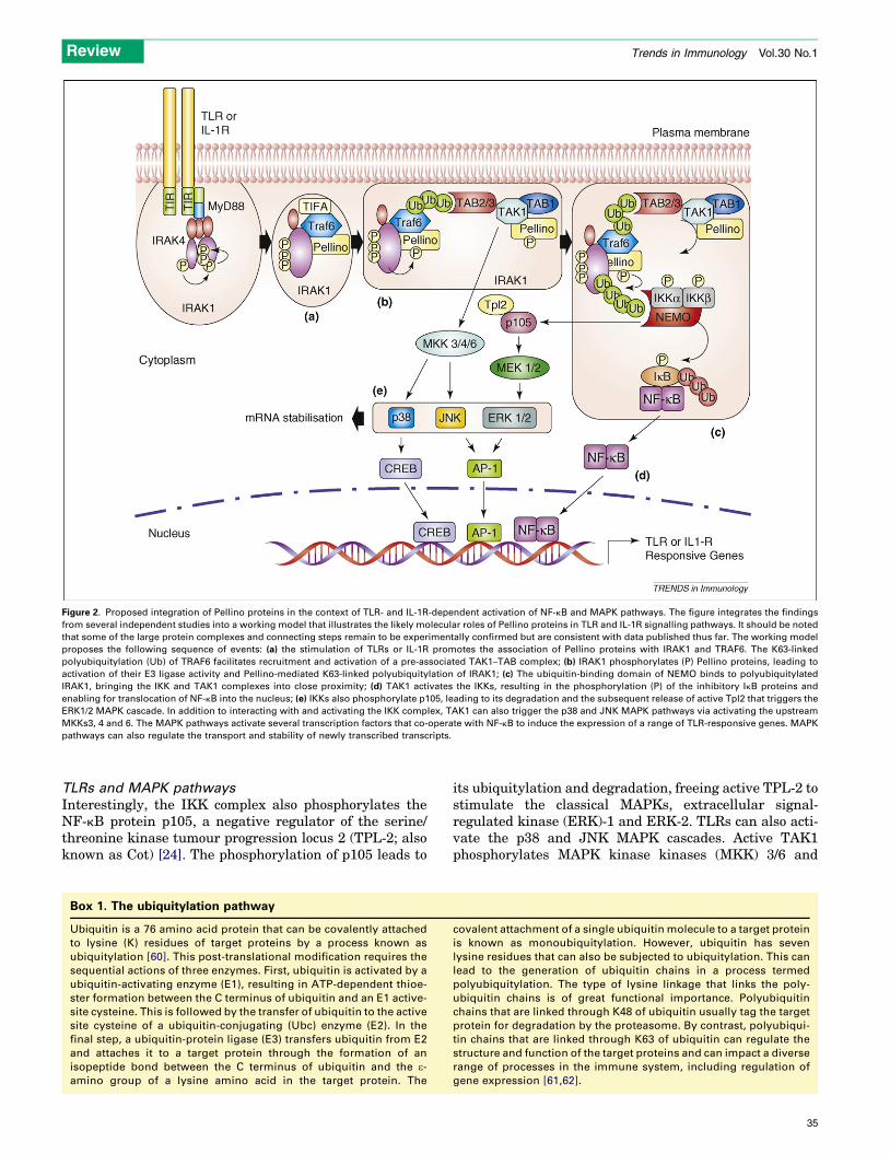
Figure 2. Proposed integration of Pellino proteins in the context of TLR- and IL-1R-dependent activation of NF-kB and MAPK pathways. The figure integrates the findings
from several independent studies into a working model that illustrates the likely molecular roles of Pellino proteins in TLR and IL-1R signalling pathways. It should be noted
that some of the large protein complexes and connecting steps remain to be experimentally confirmed but are consistent with data published thus far. The working model
proposes the following sequence of events: (a) the stimulation of TLRs or IL-1R promotes the association of Pellino proteins with IRAK1 and TRAF6. The K63-linked
polyubiquitylation (Ub) of TRAF6 facilitates recruitment and activation of a pre-associated TAK1–TAB complex; (b) IRAK1 phosphorylates (P) Pellino proteins, leading to
activation of their E3 ligase activity and Pellino-mediated K63-linked polyubiquitylation of IRAK1; (c) The ubiquitin-binding domain of NEMO binds to polyubiquitylated
IRAK1, bringing the IKK and TAK1 complexes into close proximity; (d) TAK1 activates the IKKs, resulting in the phosphorylation (P) of the inhibitory IkB proteins and
enabling for translocation of NF-kB into the nucleus; (e) IKKs also phosphorylate p105, leading to its degradation and the subsequent release of active Tpl2 that triggers the
ERK1/2 MAPK cascade. In addition to interacting with and activating the IKK complex, TAK1 can also trigger the p38 and JNK MAPK pathways via activating the upstream
MKKs3, 4 and 6. The MAPK pathways activate several transcription factors that co-operate with NF-kB to induce the expression of a range of TLR-responsive genes. MAPK
pathways can also regulate the transport and stability of newly transcribed transcripts.
Review Trends in Immunology Vol.30 No.1
TLRs and MAPK pathways
Interestingly, the IKK complex also phosphorylates theNF-kB protein p105, a negative regulator of the serine/threonine kinase tumour progression locus 2 (TPL-2; alsoknown as Cot) [24]. The phosphorylation of p105 leads to
Box 1. The ubiquitylation pathway
Ubiquitin is a 76 amino acid protein that can be covalently attached
to lysine (K) residues of target proteins by a process known as
ubiquitylation [60]. This post-translational modification requires the
sequential actions of three enzymes. First, ubiquitin is activated by a
ubiquitin-activating enzyme (E1), resulting in ATP-dependent thioe-
ster formation between the C terminus of ubiquitin and an E1 active-
site cysteine. This is followed by the transfer of ubiquitin to the active
site cysteine of a ubiquitin-conjugating (Ubc) enzyme (E2). In the
final step, a ubiquitin-protein ligase (E3) transfers ubiquitin from E2
and attaches it to a target protein through the formation of an
isopeptide bond between the C terminus of ubiquitin and the e-amino group of a lysine amino acid in the target protein. The
its ubiquitylation and degradation, freeing active TPL-2 tostimulate the classical MAPKs, extracellular signal-regulated kinase (ERK)-1 and ERK-2. TLRs can also acti-vate the p38 and JNK MAPK cascades. Active TAK1phosphorylates MAPK kinase kinases (MKK) 3/6 and
covalent attachment of a single ubiquitin molecule to a target protein
is known as monoubiquitylation. However, ubiquitin has seven
lysine residues that can also be subjected to ubiquitylation. This can
lead to the generation of ubiquitin chains in a process termed
polyubiquitylation. The type of lysine linkage that links the poly-
ubiquitin chains is of great functional importance. Polyubiquitin
chains that are linked through K48 of ubiquitin usually tag the target
protein for degradation by the proteasome. By contrast, polyubiqui-
tin chains that are linked through K63 of ubiquitin can regulate the
structure and function of the target proteins and can impact a diverse
range of processes in the immune system, including regulation of
gene expression [61,62].
35

Review Trends in Immunology Vol.30 No.1
MKK4 that, in turn, activate p38 and Jun N-terminalkinase (JNK), respectively [13]. Thus, TAK1 emerges asa central player in TLR regulation of NF-kB and MAPKs,given its ability to activate the IKKs and MKK3, 4 and 6(Figure 2). However, it should be noted that some reportsindicate that TLRs and IL-1R might activate these path-ways in a TAK1-independent manner. Thus, in mouseembryonic fibroblasts that lack TAB1, IL-1 is incapableof activating TAK1, yet can still activate IKKs and the p38and JNKMAPK pathways [25]. The mixed lineage kinases(MLK)-2 and MLK-3 might substitute for TAK1, at leastwith respect to activating the p38 and JNKMAPKs [26,27].Irrespective of the exact mechanism, the MAPK pathwayscan promote activation of the transcription factors cAMPresponse element-binding (CREB) and activator protein 1(AP-1) that cooperate with NF-kB at various gene promo-ters to increase gene transcription [28]. Furthermore,MAPKs can regulate the expression of these genes bypost-transcriptional mechanisms, including promotingthe nucleocytoplasmic transport and increasing thestability of newly transcribed transcripts.
IRAK polyubiquitylation and TLR/IL-1R signallingAlthough TAK1 promotes phosphorylation and activationof IKKa and IKKb [13,20], the exact mechanism by whichTAK1 and IKKs are brought into close proximity is notclearly understood. The above-mentioned model favoursthe dissociation of the multi-molecular TRAF6–TAK1–
TAB1–TAB2–TAB3 complex from IRAK1 at the mem-brane followed by its translocation to the cytosol, at whichTAK1 becomes active, TAB2 promotes assembly of TRAF6with the IKKs and TAK1 activates the IKKs [17]. Thus,IRAK1 remains at the membrane and the activation andassembly of TAK and the IKKs are spatially separatefrom IRAK1.
However, an alternativemodel has been proposed basedon very recent studies characterizing the ubiquitylation ofIRAK1. It is well known that LPS and IL-1 induce thepolyubiquitylation and degradation of IRAK1 and it waspreviously assumed that IRAK1 was modified by K48-linked polyubiquitylation that tagged it for proteolysisby the proteasome. However, two recent reports show thatTLR4 and IL-1R signalling leads to K63-linked and notK48-linked polyubiquitylation of IRAK1 [16,29], whichserves not to tag IRAK for degradation by the proteasomebut rather has an important role in TLR- or IL-1R-mediated activation of NF-kB. The studies show thatpolyubiquitylated IRAK recruits an IKK complex contain-ing catalytic IKKa and IKKb in association with theirregulatory subunit NF-kB essential modifier (NEMO; alsoknown as IKKg). NEMO lacks catalytic activity but isessential for activation of NF-kB [30]. It contains a ubiqui-tin-binding domain that specifically recognizes K63-linkedIRAK1, thus, facilitating IKK–IRAK complex formation aspart of TLR and IL-1R signalling [31,32]. IRAK is ubiqui-tylated on Lys134 and Lys180 and mutation of these sitesimpairs TLR- or IL-1R-mediated ubiquitylation of IRAK1,binding of IRAK to NEMO and NF-kB activation [29].These studies indicate a model in which TAK1 is recruitedto K63-linked polyubiquitin chains on TRAF6 and the IKKcomplex is recruited to K63-linked polyubiquitin chains on
36
IRAK1. This brings TAK1 and IKKs into close proximity,leading to TAK1-mediated phosphorylation and activationof the IKKs. Given this model, there is currently intenseinterest in identifying the E3 ubiquitin ligase(s) thatcatalyses polyubiquitylation of IRAK1. It has been pro-posed [29] that there is, at least, a partial involvement ofTRAF6 in the polyubiquitylation of IRAK1, but this hasbeen opposed by two other groups [16,33]. Although therole of TRAF6 as the E3 ligase for IRAK1 remains ambig-uous, the existence of a non-TRAF6 E3 ligase(s) that iscapable of catalysing K63-linked polyubiquitylation ofIRAK1 is widely agreed upon. Pellino proteins emergeas likely candidates to fill this role given their intrinsicE3 ubiquitin ligase activity and their ability to interactwith IRAK1.
Bi-directional communication in the IRAK–PellinocomplexPellino was first identified in Drosophila melanogaster byvirtue of its association with Pelle, the Drosophila ortho-logue of IRAK [34]. Three mammalian homologues (Pel-lino1, 2 and 3) have since been identified, with Pellino3being expressed in two spliced forms [35–39]. Given thatDrosophila Pellino was discovered based on its interactionwith Pelle, mammalian Pellino proteins have been charac-terized by their potential to interact with IRAKmolecules.Overexpression studies have shown that mammalian Pel-lino proteins interact with both IRAK1 and IRAK4[36,37,40–43]. Furthermore, a yeast two-hybrid screenusing IRAK4 as bait independently identified Pellino2 asa protein-binding partner [40]. Under physiological con-ditions, the Pellino–IRAK interactions are likely to bedependent on prior TLR or IL-1R stimulation becauseendogenous IRAK–Pellino associations are not observedin resting cells but manifest in response to stimulation byIL-1 [33,35–37,44] and LPS [44].
IRAK kinase activity and Pellino binding
Although not a substrate for the kinase activity of Pelle,Drosophila Pellino can only interact with the catalyticallyactive form of Pelle, indicating that it recognizes autopho-sphorylated Pelle [34]. Presently, there is disagreementon the importance of IRAK kinase activity for IRAK–
Pellino associations. Some groups have shown that IRAKkinase activity is dispensable for IRAK–Pellino inter-actions [40,42,45]. However, two other groups proposethat IRAK1–Pellino binding requires IRAK1 kinaseactivity [37,46], which is supported by the fact thatIRAK1b, a naturally occurring alternative splice variantof IRAK1 that is kinase inactive, fails to interact withPellino3 [37]. The basis to the opposing conclusions mightbe a result of interpretations being made exclusively onthe basis of overexpression studies. Future studies thatcharacterize endogenous associations of Pellino familymembers with naturally occurring catalytically inactivemembers of the IRAK family (e.g. IRAK1b, IRAK2and IRAK-M) might resolve some of the confusion. It isinteresting to note that a very recent report indicatesthat Pellino proteins have a cryptic FHA domainthat mediates interaction with phosphorylated IRAK1(see Note added in proof). Given that IRAK undergoes

Box 2. The RING domain
The classical RING-finger domain is characterized by a highly
conserved linear pattern of cysteine and histidine residues: Cys-
X2-Cys-X9–39-Cys-X1–3-His-X2–3-Cys/His-X2-Cys-X4–48-Cys-X2-Cys
[63]. The C3HC4 pattern defines a domain that can fold into a cross-
brace structure that is capable of binding two zinc atoms. This RING-
finger domain is usually a signature of proteins that have E3
ubiquitin ligase activity [64]. Intriguingly, Pellino proteins have a
pattern of cysteine and histidine residues that is highly related to the
RING domain. This RING-like domain in the Pellino family has the
pattern Cys-X1-His-X19-Cys-X2-Cys-X30-Cys-X1-His-X25-Cys-X2-Cys
and is abbreviated to CHC2CHC2 [43].
Review Trends in Immunology Vol.30 No.1
autophosphorylation, this study would favour a rolefor IRAK1 kinase activity in promoting IRAK1–Pellinointeractions.
IRAKs phosphorylate and enhance E3 ligase activity of
Pellino proteins
The association of Pellino proteins with IRAKs promotedinterest in the possibility of Pellino proteins being sub-strates for IRAK kinase activity. The co-expression ofIRAK1 and Pellino proteins leads to phosphorylation ofeach of the Pellino family [43], with in vitro kinase assaysdemonstrating that IRAK1 can directly phosphorylatePellino proteins [40,45]. These studies also show thatPellino1 and Pellino2 are substrates for IRAK4. Anappreciation of the consequence of IRAK-mediated phos-phorylation of the Pellino proteins was aided by an insightinto the functional properties of the Pellino family. Abioinformatic analysis had identified a RING-like domain(Box 2) in the C termini regions of the Pellino proteins,indicating that they might function as E3 ubiquitin ligases[38]. This was supported by a study showing that co-production of Pellino1 or Pellino2, but not Pellino3, withIRAK leads to IRAK polyubiquitylation [43]. Interestingly,two independent groups have shown that all three Pellinoproteins, including both splice variants of Pellino3, caninduce intense polyubiquitylation of co-expressed IRAK1[33,42]. In vitro ubiquitylation assays have shown thatrecombinant and endogenous forms of Pellino1–3 possessE3 ligase activity and, thus, Pellino proteins are likely todirectly catalyse polyubiquitylation of IRAK1 [33,42,45].Intriguingly, in an in vitro setting, the Pellino proteins arecapable of catalysing the formation of K63- and K48-linkedpolyubiquitin chains. The type of linkage is dictated by theE2 enzyme that collaborates with the Pellino E3 ligase tocatalyse polyubiquitylation. Each of the Pellino proteinscan combine with the E2 heterodimer UbcH13–Uev1a to
Figure 3. Bi-directional communication between IRAKs and Pellino proteins. The stimul
latter phosphorylates the Pellino proteins, leading to activation of their E3 ligase activi
IRAK. Polyubiquitylated IRAK can regulate several downstream pathways, including NF-
the Pellino proteins by an unknown mechanism, resulting in Pellino degradation.
catalyse K63-linked ubiquitylation. By contrast, detailedstudies using mass spectrometry show that Pellino1 cancombine with UbcH3 to catalyse formation of K48-linkedpolyubiquitin chains, whereas, in combination withUbcH4, UbcH5a or UbcH5b, it promotes formation ofK48- and K11-linked polyubiquitin chains [45]. However,cellular studies to date indicate that the co-expression ofPellino proteins with IRAK1 induces only K63-linked poly-ubiquitylation of IRAK1 [33,45]. It is possible that Pellinoproteins might be subject to regulatory processes in vivothat facilitate their interaction with different E2 enzymesand, hence, confer the ability to promote the formationof polyubiquitin chains that are linked by other lysineresidues.
It has already been mentioned that Pellino proteins arephosphorylated by IRAK1 and IRAK4 and the functionalconsequence of this phosphorylation is enhancement ofPellino E3 ligase activity [45]. A plausible model emergesin which TLRs or IL-1R promote IRAK–Pellino inter-actions, leading to IRAK-mediated phosphorylation ofthe Pellino proteins, enhancement of their E3 ligaseactivity and, ultimately, Pellino-mediated polyubiquityla-tion of the IRAK1 (Figure 3). Indeed, the suppression of
ation of TLRs or IL-1R promotes the association of Pellino proteins with IRAK1. The
ty. This leads to Pellino-mediated formation of K63-linked polyubiquitin chains on
kB and MAPK cascades. Kinase active IRAK can also promote polyubiquitylation of
37

Review Trends in Immunology Vol.30 No.1
endogenous Pellino3 by siRNAs causes a considerablereduction of IL-1-induced polyubiquitylation of IRAK1 [33].
Kinase-active IRAKs promote polyubiquitylation and
degradation of Pellino proteins
Interestingly, the IRAK–Pellino interactions also lead toreciprocal polyubiquitylation of the Pellino-binding part-ners [42] (Figure 3). The co-expression of kinase-activeIRAK1 or IRAK4 with Pellino proteins causes intensepolyubiquitylation of Pellino family members. The identityof the E3 ligase(s) that catalyses the IRAK-induced poly-ubiquitylation of the Pellino family remains to be eluci-dated, with reports differing on the relevant importance ofPellino E3 ligase activity in this process [42,45]. The poly-ubiquitylation of Pellino proteins is associated with sub-sequent Pellino degradation. This is a potentialmechanism for regulating TLR signalling and, indeed,the TLR4 ligand LPS induces degradation of Pellino3 inperipheral blood mononuclear cells [42]. Thus, a complexmodel of bi-directional communication is now emergingwith respect to IRAK–Pellino associations, in which IRAKsphosphorylate and activate the Pellino proteins, leading topolyubiquitylation of both binding partners (Figure 3). Thepolyubiquitylation of Pellino proteins leads to their degra-dation and might be a means to terminate TLR signalling,whereas the K63-linked polyubiquitylation of IRAK isprobably an important step in triggering downstreamsignalling (Figure 3). Indeed, as stated previously, a cur-rently favoured model for MyD88-dependent TLR signal-ling favours a scenario in which TAK1 is recruited to K63-linked polyubiquitin chains on TRAF6 and the IKK com-plex is recruited to K63-linked polyubiquitin chains onIRAK1. An important role for the E3 ligase activity ofPellino proteins in this model is supported by the findingthat wild-type Pellino2, but not a point-mutated form thatlacks a functional RING domain, promotes the recruitmentof NEMO to K63-linked polyubiquitylated IRAK [45].
Pellino proteins interact with other TLR and IL1-Rsignalling proteinsPellino proteins can also interact with other TLR signallingmolecules. None of the Pellino proteins have been reportedto interact with TLR or IL-1R complexes, however, onereport indicates that IL-1 signalling can promote the inter-action of overexpressedPellino1withMyD88 [44].However,other reports have failed to detect any basal interactionsbetween overexpressedMyD88 andPellino proteins [37,47].Many groups have described associations of each of thePellino proteins with TRAF6 and these interactions arestrongly promoted by IL-1R signalling [33,36,37,41,44,47].The nature of these interactions remains to be defined withno available data to date to indicate whether Pellinoproteins interact directly with TRAF-6 or indirectly as partof a larger protein complex. Pellino3 has also been reportedto interact with TAK1 [33,37,41]. Although an early studyused overexpressionanalyses to show that IL-1 can enhancethe Pellino3–TAK1 interaction [37], a more recent reportprovides strong support for a constitutive interaction be-tween Pellino3 and TAK1 [33]. Thus, Pellino3might be pre-associated with the TAK1–TAB complex and is broughtinto close association with IRAK-1 and TRAF-6 when
38
IL-1 signalling promotes the association of IRAK1–TRAF6with the TAK1 complex (Figure 1). This is also consistentwith the finding that the IL-1-induced association of TRAF6with theTAK1complexand the resultingactivationofTAK1precede the polyubiquitylation of IRAK1 and activation ofIKKs [16]. Interestingly, Pellino3 has also been reported tointeract with NF-kB-inducing kinase (NIK) [37], a kinasethat is involved in the activation of the alternative (NEMO-independent) NF-kB pathways by receptors, such as thelymphotoxin-b receptor [48]. This indicates that Pellinoproteins might also serve signalling roles in non-TLR andnon-IL-1R pathways.
Functional roles of Pellino proteins in TLR and IL-1Rsignalling pathwaysThe association of Pellino proteins with key signallingmolecules in the TLR and IL-1R pathways has promptedcharacterization of their involvement in mediating down-stream signalling cascades. Although each of the Pellinoproteins interact with common signalling molecules, suchas IRAKs and TRAF6, and each has E3 ubiquitin ligaseactivity that can promote polyubiquitylation of IRAK1,emerging data clearly show that the Pellino proteins arenot functionally redundant but instead display signs ofspecificity of function.
Pellino1
Thereare contrasting reports on theability of overexpressedPellino1 protein to induce a NF-kB-regulated reporter gene[10,44], however, these same groups have shown that sup-pression of endogenous Pellino1 by siRNAs impairs IL-1-induced activation of NF-kB in a human [36] and murine[44] cell line. Furthermore, suppression of Pellino1 stronglyinhibits the ability of IL-1 to induce the NF-kB-responsivegene IL-8 [36]. Interestingly, Pellino1 is not involved inregulating any of the MAPK pathways [41,47].
Pellino2
The functional role of Pellino2 in TLR or IL-1R signalling issomewhat controversial. Although overexpression of thePellino2 protein is incapable of driving expression of aNF-kB-regulated reporter gene, the suppression of Pellino2in murine embryonic fibroblast or macrophage cell linesinhibits LPS- and IL-1-dependent but not TNF-dependentactivation of NF-kB-regulated promoters [35,49]. Overex-pressionstudieshavealsobeenused tosuggest thatPellino2drives activation of the ERK1/2 and JNK pathways [47].However, some data [40] strongly question the specificity ofthe role of Pellino2 in the NF-kB and MAPK pathways andthe above-mentioned conclusions from reporter systems. Ithas been proposed that many of the findings can beexplained by a more generic role for Pellino2 in regulatingthe general transcription machinery rather than targetingspecific pathways. The generation of Pellino2-deficientmicewill undoubtedly resolve the present confusion.
Pellino3
Intriguingly, a recent report has proposed that Pellino3functions as a negative regulator of TAK1-mediated acti-vation ofNF-kB,with the inhibitory effects being dependenton a functional RING domain and on Pellino3-mediated

Review Trends in Immunology Vol.30 No.1
K63-linked polyubiquitylation of IRAK1 [33]. The authorspropose that IL-1 promotes both K48- and K63-linked poly-ubiquitylation of IRAK1 and Pellino3-mediated K63-linkedIRAK polyubiquitylation competes with K48-linked IRAKpolyubiquitylation for the same ubiquitylation site, K134 ofIRAK. Thus, Pellino3 blocks IRAK degradation and soinhibits IL-1-induced activation of NF-kB. However, thismodel requires a positive role for IRAK degradation in IL-1signalling and requires that IRAK degradation facilitatesdissociation of the TAK-1–TAB–TRAF6 complex from themembrane and its subsequent translocation to the cytosol,where it activates the IKK complex [33]. This differs fromthe model in which K63-linked polyubiquitylated TRAF6recruits the TAK1 complex and K63-linked polyubiquity-lated IRAK1 recruits the IKK complex, thus, bringingTAK1and IKK into close proximity and enabling IKK activation.Furthermore, in this model, it is proposed that K48-linkedpolyubiquitylation of IRAK1 leads to its proteasome-mediated degradation and Pellino3 prevents this degra-dation by instead catalysing K63-linked polyubiquitylationof IRAK [33]. This is consistent with a recent independentreport demonstrating K48- and K63-linked polyubiquityla-tion of IRAK1 and proteasome-mediated degradation ofIRAK1 in response to IL-1 signalling [50]. However, twoother groups contend that blockade of the proteolyticactivity of the proteasome fails to prevent IL-1-induceddegradation of IRAK1 [16,29] and this questions the import-ance of K48-linked polyubiquitylation for IRAK degra-dation. Furthermore, a mutant form of IRAK1 in whichLys134 is mutated to arginine, a residue that is not subjectto ubiquitylation, is still capable of triggering activation ofNF-kB [29]. Interestingly, this study demonstrates thatLys180, as well as Lys134, in IRAK1 is ubiquitylated inresponse to TLR and IL-1R signalling and the mutation ofboth residues is required to impair ubiquitylation of IRAK1and the ability of the latter to activate NF-kB. Although theexact mechanism underlying the inhibitory effects of Pel-lino3 on the NF-kB pathway remains to be fully delineatedand awaits the generation of Pellino3-deficient mice, theproposed negative regulatory role for Pellino3 is a fascinat-ing one and promotes it as an important regulator of TLRand IL-1R pathways.
Although transient overexpression of Pellino3 proteincauses activation of the JNK [33,37] and ERK MAPKpathways [37], the suppression of endogenous expressionof Pellino3 enhances IL-1-dependent activation of JNK[33], indicating a negative role for Pellino3 in the JNKpathway. However, Pellino3 has a positive role in acti-vation of p38 MAPK and is a mediator in IL-1-dependentactivation of p38 [41]. The stimulation of p38 by Pellino3 ismediated by TRAF6 and TAK1 and leads to activation ofthe p38 substrate,MAPK-activated protein kinase 2. Inter-estingly, Pellino3 is capable of exhibiting contrastingstimulatory and inhibitory effects on different pathwaysthat are downstream of TAK1. The molecular basis to suchcomplex effects remains to be defined.
Pellino proteins as novel targets for negative regulationof TLR signallingThe functional characterization of the Pellino family todate highlights their diverse and varying roles in innate
immune signalling. Intriguingly, some studies havedemonstrated that members of the Pellino family can betargeted by regulatory strategies to suppress TLR signal-ling [33,44,49]. Indeed, the role of Pellino3 as a directnegative regulator of innate immune signalling hasalready been described [33] (Figure 4a). However, twoadditional reports have identified Pellino1 and Pellino2as novel targets for other inhibitory molecules and path-ways [44,49] (Figure 4b,c). TGF-b1 has been characterizedas an anti-inflammatory cytokine that cross-talks with anddownregulates TLR pathways. Thus, TGF-b1-deficientmice experience heightened inflammatory responses andearly death and this is mainly mediated by abnormalactivation of the NF-kB pathway by TLR4 [51,52]. A recentstudy demonstrated that Pellino1 is targeted by TGF-b1 aspart of the manifestation of its anti-inflammatory inhibi-tory effects (Figure 4b). Thus, TGF-b1 induces the inhibi-tory Smad6 protein that subsequently binds to andsequesters Pellino1 and inhibits the ability of Pellino1 tointeract with IRAK1, MyD88 and TRAF6 [44]. It is pro-posed that the disruption of the Pellino1–IRAK–TRAF6signalling complex by Smad6 is responsible for the inhibi-tory effects of Smad6 on the LPS and IL-1-stimulatedactivation of NF-kB and induction of pro-inflammatorygenes.
Another study has shown Pellino2 to be targeted bysuppressor of cytokine signalling 3 (SOCS3) as part ofanother mechanism to downregulate TLR signalling [49](Figure 4c). SOCS3 is induced by LPS, inhibits LPS signal-ling and is required for the suppressive effects of IL-10 onthe TLR4 signalling pathway [53,54]. A recent study ident-ified B-cell lymphoma 10 (BCL10), an intracellular acti-vator of NF-kB, as a binding partner for Pellino2 andproposes the inhibitory effects of SOCS3 to be mediatedby blocking the interaction of BCL10 with Pellino2 [49]. Inthis study, BCL10 interacted with TLR4, associated withPellino2 and activated the NF-kB pathway in a Pellino2-dependent manner. The interaction of SOCS3 with BCL10blocked the BCL10–Pellino2 association, thereby leadingto inhibition of the downstream activation of NF-kB.
The direct role of Pellino3 as a negative regulator of TLRor IL-1R signalling, coupled with the targeting of Pellino1and Pellino2 by Smad6 and SOCS3, clearly highlightsthe Pellino family as key modulators of innate immunesignalling.
Concluding remarks and future perspectivesThe discovery of the Pellino family has provided newinsight into innate immunity and informed an understand-ing of previously unanswered questions in the field of TLRsignalling. The role of the kinase activity of IRAK1 hasremained an enigma since its original characterization, butPellino-related studies now strongly indicate that IRAK1and/or IRAK4 phosphorylate members of the Pellinofamily and enhance their E3 ligase activity, leading topolyubiquitylation of IRAK. Furthermore, the kinaseactivity of IRAK promotes the degradation of Pellinoproteins and this might be an important regulatory mech-anism that controls TLR signalling.
Although we await the generation of Pellino-deficientmice to define their physiological roles, studies to date
39

Figure 4. Targeting of Pellino proteins for negative regulation of TLR or IL-1R signalling. Several control systems have been identified that target Pellino proteins to
negatively regulate TLR and IL-1R signalling. (a) Pellino3 acts as a negative regulator of IL-1R-mediated activation of NF-kB by catalysing K63-linked polyubiquitylation of
IRAK1, leading to its stabilization. (b) TGF-b negatively regulates TLR or IL-1 signalling by inducing Smad6 that subsequently sequesters Pellino1 and, thus, inhibits
formation of the IRAK1–TRAF6–Pellino1 complex. This blocks TLR- or IL-1R-induced activation of the NF-kB pathway. (c) BCL10 associates with activated TLR4 and,
subsequently, interacts with Pellino2 to activate the NF-kB pathway. SOCS3 is induced by LPS and inhibits the NF-kB pathway by interacting with BCL10 and blocking the
BCL10–Pellino association.
Review Trends in Immunology Vol.30 No.1
support diverse roles for the family withmembers failing toshow functional redundancy. Furthermore, our currentknowledge of Pellino biology poses further intriguingquestions. Each Pellino member shares molecular charac-teristics, such as possessing E3 ligase activity, acting assubstrates for IRAKkinases and interactingwith commonsignalling molecules, and so we await understanding ofthe basis to their functional diversity. With this inmind, adetailed understanding of the mechanisms that controlPellino function is required. Indeed, studies have alreadyshown that Pellino proteins are subject to degradationand re-synthesis by TLR signals [42,55,56] and theirE3 ligase activity is regulated by IRAK-mediated phos-phorylation [45]. This indicates that Pellino proteinsare subject to multiple levels of regulation by TLRs,again emphasizing their probable key roles in innateimmunity.
Given the role of IRAK1 in promoting activation of IRFsin TLR7 and TLR9 signalling [57], it will be especiallyinteresting to explore the role of Pellino proteins in theseviral-sensing pathways. Much effort will also continue toinvestigate the functional consequence of polyubiquityla-tion of IRAK1 and probe the physiological importance ofPellino proteins in regulating such modification of IRAK1.Finally, it is worth mentioning that Pellino function might
40
not be restricted to the immune system. A Pellino homol-ogue in Ciona intestinalis (a primitive chordate) has a keyrole in notochord formation in early development [58,59]. Itwill be interesting to examine the role of the differentPellino proteins in the development of higher organisms.This is especially intriguing given that Pellino was firstcharacterized in the context of Drosophila Toll signallingand members of the Toll family also exhibit dualist func-tions in development and the innate immune system. Thisindicates that the Pellino family has co-evolved closelywith the Toll system, supporting the notion of key funda-mental roles for Pellino proteins in Toll-like receptor sig-nalling. Although the field of Pellino biology is in itsinfancy, it represents an area of enormous potential toinform our understanding of the workings of the innateimmune system.
Note added in proofA study published after this article had been acceptedrevealed for the first time that Pellino proteins contain acryptic FHA domain that mediates interaction with phos-phorylated IRAK1.
Lin et al., Pellino Proteins Contain a Cryptic FHADomain that Mediates Interaction with PhosphorylatedIRAK1, Structure (2008), doi:10.1016/j.str.2008.09.011
Review Trends in Immunology Vol.30 No.1
AcknowledgementsP.N.M. is funded by Science Foundation Ireland, Health Research Boardof Ireland and Enterprise Ireland.
References1 Janeway, C.A., Jr and Medzhitov, R. (2002) Innate immune
recognition. Annu. Rev. Immunol. 20, 197–2162 Martin, M.U. and Wesche, H. (2002) Summary and comparison of the
signaling mechanisms of the Toll/interleukin-1 receptor family.Biochim. Biophys. Acta 1592, 265–280
3 Medzhitov, R. et al. (1998)MyD88 is an adaptor protein in the hToll/IL-1 receptor family signaling pathways. Mol. Cell 2, 253–258
4 O’Neill, L.A. and Bowie, A.G. (2007) The family of five: TIR-domain-containing adaptors in Toll-like receptor signalling. Nat. Rev.Immunol. 7, 353–364
5 Li, S. et al. (2002) IRAK-4: a novel member of the IRAK family with theproperties of an IRAK-kinase. Proc. Natl. Acad. Sci. U. S. A. 99, 5567–
55726 Cao, Z. et al. (1996) IRAK: a kinase associated with the interleukin-1
receptor. Science 271, 1128–11317 Lye, E. et al. (2004) The role of interleukin 1 receptor-associated kinase-
4 (IRAK-4) kinase activity in IRAK-4-mediated signaling. J. Biol.Chem. 279, 40653–40658
8 Kollewe, C. et al. (2004) Sequential autophosphorylation steps in theinterleukin-1 receptor-associated kinase-1 regulate its availabilityas an adapter in interleukin-1 signaling. J. Biol. Chem. 279, 5227–
52369 Burns, K. et al. (2000) Tollip, a new component of the IL-1RI pathway,
links IRAK to the IL-1 receptor. Nat. Cell Biol. 2, 346–35110 Jiang, Z. et al. (2002) Interleukin-1 (IL-1) receptor-associated kinase-
dependent IL-1-induced signaling complexes phosphorylate TAK1 andTAB2 at the plasma membrane and activate TAK1 in the cytosol.Mol.Cell. Biol. 22, 7158–7167
11 Takatsuna, H. et al. (2003) Identification of TIFA as an adapter proteinthat links tumor necrosis factor receptor-associated factor 6 (TRAF6) tointerleukin-1 (IL-1) receptor-associated kinase-1 (IRAK-1) in IL-1receptor signaling. J. Biol. Chem. 278, 12144–12150
12 Deng, L. et al. (2000) Activation of the IkB kinase complex by TRAF6requires a dimeric ubiquitin-conjugating enzyme complex and a uniquepolyubiquitin chain. Cell 103, 351–361
13 Wang, C. et al. (2001) TAK1 is a ubiquitin-dependent kinase of MKKand IKK. Nature 412, 346–351
14 Cheung, P.C. et al. (2004) TAB3, a new binding partner of the proteinkinase TAK1. Biochem. J. 378, 27–34
15 Kanayama, A. et al. (2004) TAB2 andTAB3 activate theNF-kBpathwaythrough binding to polyubiquitin chains. Mol. Cell 15, 535–548
16 Windheim, M. et al. (2008) Interleukin-1 (IL-1) induces the Lys63-linked polyubiquitination of IL-1 receptor-associated kinase 1 tofacilitate NEMO binding and the activation of IkBa kinase. Mol.Cell. Biol. 28, 1783–1791
17 Kishida, S. et al. (2005) TAK1-binding protein 2 facilitatesubiquitination of TRAF6 and assembly of TRAF6 with IKK in theIL-1 signaling pathway. Genes Cells 10, 447–454
18 Jacinto, R. et al. (2002) Lipopolysaccharide- and lipoteichoic acid-induced tolerance and cross-tolerance: distinct alterations in IL-1receptor-associated kinase. J. Immunol. 168, 6136–6141
19 Yamin, T.T. and Miller, D.K. (1997) The interleukin-1 receptor-associated kinase is degraded by proteasomes following itsphosphorylation. J. Biol. Chem. 272, 21540–21547
20 Ninomiya-Tsuji, J. et al. (1999) The kinase TAK1 can activate the NIK-I kB as well as the MAP kinase cascade in the IL-1 signalling pathway.Nature 398, 252–256
21 Yi, A.K. and Krieg, A.M. (1998) CpG DNA rescue from anti-IgM-induced WEHI-231 B lymphoma apoptosis via modulation of IkBaand IkB b and sustained activation of nuclear factor-kB/c-Rel. J.Immunol. 160, 1240–1245
22 Medzhitov, R. et al. (1997) A human homologue of the Drosophila Tollprotein signals activation of adaptive immunity. Nature 388, 394–397
23 O’Neill, L.A. (2002) Signal transduction pathways activated by the IL-1receptor/toll-like receptor superfamily. Curr. Top. Microbiol. Immunol.270, 47–61
24 Beinke, S. et al. (2004) Lipopolysaccharide activation of the TPL-2/MEK/extracellular signal-regulated kinase mitogen-activated protein
kinase cascade is regulated by IkB kinase-induced proteolysis of NF-kB1 p105. Mol. Cell. Biol. 24, 9658–9667
25 Mendoza, H. et al. (2008) Roles for TAB1 in regulating the IL-1-dependent phosphorylation of the TAB3 regulatory subunit andactivity of the TAK1 complex. Biochem. J. 409, 711–722
26 Zhong, J. and Kyriakis, J.M. (2007) Dissection of a signaling pathway bywhich pathogen-associatedmolecular patterns recruit the JNK and p38MAPKs and trigger cytokine release. J. Biol. Chem. 282, 24246–24254
27 Zhong, J. and Kyriakis, J.M. (2004) Germinal center kinase is requiredfor optimal Jun N-terminal kinase activation by Toll-like receptoragonists and is regulated by the ubiquitin proteasome system andagonist-induced, TRAF6-dependent stabilization. Mol. Cell. Biol. 24,9165–9175
28 Dong, C. et al. (2002) MAP kinases in the immune response.Annu. Rev.Immunol. 20, 55–72
29 Conze, D.B. et al. (2008) Lys63-linked polyubiquitination of IRAK-1 isrequired for interleukin-1 receptor- and toll-like receptor-mediatedNF-kB activation. Mol. Cell. Biol. 28, 3538–3547
30 Rothwarf, D.M. et al. (1998) IKK-g is an essential regulatory subunit ofthe IkB kinase complex. Nature 395, 297–300
31 Ea, C.K. et al. (2006) Activation of IKK by TNFa requires site-specificubiquitination of RIP1 and polyubiquitin binding by NEMO. Mol. Cell22, 245–257
32 Wu, C.J. et al. (2006) Sensing of Lys 63-linked polyubiquitination byNEMO is a key event in NF-kB activation. Nat. Cell Biol. 8, 398–406
33 Xiao, H. et al. (2008) Pellino 3b negatively regulates interleukin-1-inducedTAK1-dependent NF kB activation. J. Biol. Chem. 283, 14654–14664
34 Grosshans, J. et al. (1999) Oligomerisation of Tube and Pelle leads tonuclear localisation of dorsal. Mech. Dev. 81, 127–138
35 Yu, K.Y. et al. (2002) Cutting edge: mouse pellino-2 modulates IL-1 andlipopolysaccharide signaling. J. Immunol. 169, 4075–4078
36 Jiang, Z. et al. (2003) Pellino 1 is required for interleukin-1 (IL-1)-mediated signaling through its interaction with the IL-1 receptor-associated kinase 4 (IRAK4)-IRAK-tumor necrosis factor receptor-associated factor 6 (TRAF6) complex. J. Biol. Chem. 278, 10952–10956
37 Jensen, L.E. and Whitehead, A.S. (2003) Pellino3, a novel member ofthe Pellino protein family, promotes activation of c-Jun and Elk-1 andmay act as a scaffolding protein. J. Immunol. 171, 1500–1506
38 Rich, T. et al. (2000) Pellino-related sequences from Caenorhabditiselegans and Homo sapiens. Immunogenetics 52, 145–149
39 Resch, K. et al. (2001) Assignment of homologous genes, Peli1/PELI1and Peli2/PELI2, for the Pelle adaptor protein Pellino to mousechromosomes 11 and 14 and human chromosomes 2p13.3 and14q21, respectively, by physical and radiation hybrid mapping.Cytogenet. Cell Genet. 92, 172–174
40 Strelow, A. et al. (2003) Characterization of Pellino2, a substrate ofIRAK1 and IRAK4. FEBS Lett. 547, 157–161
41 Butler, M.P. et al. (2005) Pellino3 is a novel upstream regulator of p38MAPK and activates CREB in a p38-dependent manner. J. Biol. Chem.280, 27759–27768
42 Butler, M.P. et al. (2007) Kinase-active interleukin-1 receptor-associated kinases promote polyubiquitination and degradation ofthe Pellino family: direct evidence for PELLINO proteins beingubiquitin-protein isopeptide ligases. J. Biol. Chem. 282, 29729–29737
43 Schauvliege, R. et al. (2006) Pellino proteins are more than scaffoldproteins in TLR/IL-1R signalling: a role as novel RING E3-ubiquitin-ligases. FEBS Lett. 580, 4697–4702
44 Choi, K.C. et al. (2006) Smad6 negatively regulates interleukin 1-receptor-Toll-like receptor signaling through direct interaction withthe adaptor Pellino-1. Nat. Immunol. 7, 1057–1065
45 Ordureau, A. et al. (2008) The IRAK-catalysed activation of the E3ligase function of Pellino isoforms induces the Lys63-linkedpolyubiquitination of IRAK1. Biochem. J. 409, 43–52
46 Schauvliege, R. et al. (2007) Pellino proteins: novel players in TLR andIL-1R signalling. J. Cell. Mol. Med. 11, 453–461
47 Jensen, L.E. andWhitehead, A.S. (2003) Pellino2 activates themitogenactivated protein kinase pathway. FEBS Lett. 545, 199–202
48 Dejardin, E. et al. (2002) The lymphotoxin-b receptor induces differentpatterns of gene expression via two NF-kB pathways. Immunity 17,525–535
49 Liu, Y. et al. (2004) BCL10 mediates lipopolysaccharide/toll-likereceptor-4 signaling through interaction with Pellino2. J. Biol.Chem. 279, 37436–37444
41

Review Trends in Immunology Vol.30 No.1
50 Newton, K. et al. (2008) Ubiquitin chain editing revealed bypolyubiquitin linkage-specific antibodies. Cell 134, 668–678
51 Kulkarni, A.B. et al. (1993) Transforming growth factor b 1 nullmutation in mice causes excessive inflammatory response and earlydeath. Proc. Natl. Acad. Sci. U. S. A. 90, 770–774
52 McCartney-Francis, N. et al. (2004) Aberrant Toll receptor expressionand endotoxin hypersensitivity in mice lacking a functional TGF-b 1signaling pathway. J. Immunol. 172, 3814–3821
53 Berlato, C. et al. (2002) Involvement of suppressor of cytokinesignaling-3 as a mediator of the inhibitory effects of IL-10 onlipopolysaccharide-induced macrophage activation. J. Immunol. 168,6404–6411
54 Stoiber, D. et al. (1999) Lipopolysaccharide induces inmacrophages thesynthesis of the suppressor of cytokine signaling 3 and suppressessignal transduction in response to the activating factor IFN-g. J.Immunol. 163, 2640–2647
55 Weighardt, H. et al. (2004) Identification of a TLR4- and TRIF-dependent activation program of dendritic cells. Eur. J. Immunol.34, 558–564
42
56 Sun, Y. et al. (2008) Toll-like receptor 4 signaling plays a role intriggering periodontal infection. FEMS Immunol. Med. Microbiol.52, 362–369
57 Moynagh, P.N. (2005) TLR signalling and activation of IRFs: revisitingold friends from the NF-kB pathway. Trends Immunol. 26, 469–476
58 Hotta, K. et al. (2007) Brachyury-downstream notochord genes andconvergent extension in Ciona intestinalis embryos. Dev. GrowthDiffer. 49, 373–382
59 Hotta, K. et al. (2000) Characterization of Brachyury-downstreamnotochord genes in the Ciona intestinalis embryo. Dev. Biol. 224, 69–80
60 Hershko, A. and Ciechanover, A. (1998) The ubiquitin system. Annu.Rev. Biochem. 67, 425–479
61 Liu, Y.C. et al. (2005) Immunity by ubiquitylation: a reversible processof modification. Nat. Rev. Immunol. 5, 941–952
62 Chen, Z.J. (2005) Ubiquitin signalling in the NF-kB pathway.Nat. CellBiol. 7, 758–765
63 Freemont, P.S. (2000) RING for destruction? Curr. Biol. 10, R84–R8764 Joazeiro, C.A. and Weissman, A.M. (2000) RING finger proteins:
mediators of ubiquitin ligase activity. Cell 102, 549–552