Embed Size (px)
Citation preview
a
d
A
0
d
Review
Principles of demineralization: Modern strategies for the isolation
of organic frameworks
Part I. Common definitions and history
Hermann Ehrlich a,*, Petros G. Koutsoukos b, Konstantinos D. Demadis c, Oleg S. Pokrovsky d
Max Bergmann Center of Biomaterials and Institute of Materials Science, Dresden University of Technology, Budapester Str. 27, D-01069 Dresden, Germanyb Laboratory of Inorganic and Analytical Chemistry, Department of Chemical Engineering, University of Patras, GR-265 04 Patras, Greece
c Crystal Engineering, Growth and Design Laboratory, Department of Chemistry, University of Crete, Voutes Campus, GR-71003 Heraklion, Crete, Greece
Laboratory of Mechanisms and Transfer in Geology, Observatory Midi-Pyrenees (OMP), UMR 5563, CNRS, 14 Avenue Edouard Belin, 31400 Toulouse, France
Received 13 December 2007; received in revised form 8 February 2008; accepted 10 February 2008
Dedicated to Professor Dr. Steve Weiner on the occasion of his 60th birthday.
www.elsevier.com/locate/micron
Available online at www.sciencedirect.com
Micron 39 (2008) 1062–1091
bstract
In contrast to biomineralization phenomena, that are among the most widely studied topics in modern material and earth science and
biomedicine, much less is systematized on modern view of demineralization. Biomineralized structures and tissues are composites, containing a
biologically produced organic matrix and nano- or microscale amorphous or crystalline minerals. Demineralization is the process of removing the
inorganic part, or the biominerals, that takes place in nature via either physiological or pathological pathways in organisms. In vitro
demineralization processes, used to obtain mechanistic information, consist in the isolation of the mineral phase of the composite biomaterials
from the organic matrix. Physiological and pathological demineralization include, for example, bone resorption mediated by osteoclasts.
Bioerosion, a more general term for the process of deterioration of the composite biomaterials represents chemical deterioration of the organic and
mineral phase followed by biological attack of the composite by microorganisms and enzymes. Bioerosional organisms are represented by
endolithic cyanobacteria, fungi, algae, plants, sponges, phoronids and polychaetes, mollusks, fish and echinoids.
In the history of demineralization studies, the driving force was based on problems of human health, mostly dental caries. In this paper we
summarize and integrate a number of events, discoveries, milestone papers and books on different aspect of demineralization during the last 400
years. Overall, demineralization is a rapidly growing and challenging aspect of various scientific disciplines such as astrobiology, paleoclimatol-
ogy, geomedicine, archaeology, geobiology, dentistry, histology, biotechnology, and others to mention just a few.
# 2008 Elsevier Ltd. All rights reserved.
Keywords: Biomineral; Dissolution; Decalcification; Desilicification; Bioerosion; Remineralization; Dental caries; Bone; Shell; Endoliths; History
Contents
1. Introduction . . . . . . . . . . . . . . . . . . . . . . . . . . . . . . . . . . . . . . . . . . . . . . . . . . . . . . . . . . . . . . . . . . . . . . . . . . . . . . . . 1063
2. Biominerals and biomineralization . . . . . . . . . . . . . . . . . . . . . . . . . . . . . . . . . . . . . . . . . . . . . . . . . . . . . . . . . . . . . . . . . 1063
3. Demineralization phenomena occurring in nature. . . . . . . . . . . . . . . . . . . . . . . . . . . . . . . . . . . . . . . . . . . . . . . . . . . . . . . 1074
4. Remineralization . . . . . . . . . . . . . . . . . . . . . . . . . . . . . . . . . . . . . . . . . . . . . . . . . . . . . . . . . . . . . . . . . . . . . . . . . . . . . 1077
5. History of demineralization . . . . . . . . . . . . . . . . . . . . . . . . . . . . . . . . . . . . . . . . . . . . . . . . . . . . . . . . . . . . . . . . . . . . . . 1079
6. Practical applications of demineralization . . . . . . . . . . . . . . . . . . . . . . . . . . . . . . . . . . . . . . . . . . . . . . . . . . . . . . . . . . . . 1081
7. Epilogue . . . . . . . . . . . . . . . . . . . . . . . . . . . . . . . . . . . . . . . . . . . . . . . . . . . . . . . . . . . . . . . . . . . . . . . . . . . . . . . . . . . 1082
Acknowledgements . . . . . . . . . . . . . . . . . . . . . . . . . . . . . . . . . . . . . . . . . . . . . . . . . . . . . . . . . . . . . . . . . . . . . . . . . . . 1083
References . . . . . . . . . . . . . . . . . . . . . . . . . . . . . . . . . . . . . . . . . . . . . . . . . . . . . . . . . . . . . . . . . . . . . . . . . . . . . . . . . 1083
* Corresponding author.
E-mail address: [email protected] (H. Ehrlich).
968-4328/$ – see front matter # 2008 Elsevier Ltd. All rights reserved.
oi:10.1016/j.micron.2008.02.004
H. Ehrlich et al. / Micron 39 (2008) 1062–1091 1063
1. Introduction
This is the first paper from a series on demineralization, and
contains common definitions, plus the history of this
phenomenon and its practical applications. The next two
papers will be dedicated to a detailed analysis of the principles
and mechanisms of decalcification (Part II) and desilicification
(Part III) relating to naturally occurring composites and
artificialy developed biomaterials.
One of the most troublesome points when dealing with
demineralization is making a distinction between naturally
occurring and in vitro, or laboratory or applied demineraliza-
tion. One action known as ‘‘quick and deep’’ demineralization
comes from a chemical point of view where very aggressive
chemical reagents have been used, which led to harsh
destruction of both mineral and organic phases and to
corresponding artifacts. For example, HF-based silica dissolu-
tion procedure could drastically change the structure of glass
sponge spicular proteins (Croce et al., 2004). As a consequence,
when this is the dissolution technique we can learn little about
the real nature of the organic matrix of the biomineral or the
actions of skeletal formation. The isolation of an organic
component from any natural biomineralized material whether
mineralized with calcium- or silica-containing compounds is
indispensable but in our minds the most efficient and effective
technique should be based not on fast dissolution of the
inorganic component, but instead on a slow, biomimetically
inspired process that would spare the organic component in the
biomineral-based naturally occurring composites and not result
in artifacts. Therefore we present here a discussion on demi-
neralization as a common phenomenon occurring in nature and
compare it to demineralization as a tool that has been widely
used in several different practical approaches.
We review the principles and concepts of demineralization
and their application in different fields of science, engineering
and medicine underscoring the principles and concepts that
arise from chemical and biologically mediated perspectives of
demineralization. After surveying the major types of biomin-
erals and biocomposites, the common definitions of biominer-
alization, demineralization and remineralization are discussed,
followed by a previously unpublished, detailed description of
the history of demineralization including decalcification and
desilicification phenomena, which occur in natural environ-
ments, in organisms and in the laboratory. The final chapter
describes how current knowledge of demineralization is
inspiring new directions for practical applications.
2. Biominerals and biomineralization
According to a modern definition (Skinner, 2005) ‘‘a mineral
is an element or compound, amorphous or crystalline, formed
through biogeochemical processes’’. This definition acknowl-
edges that some biominerals are barely crystalline but avoids a
discussion of ‘‘amorphous’’.
By the 1930s, there were approximately 10 different
minerals known to be present in living organisms. This
changed when Heinz Lowenstam published a paper (Low-
enstam, 1962) describing the presence of magnetite, a relatively
hard iron oxide that chemists supposed could only be formed at
very high pressures and temperatures. Lowenstam noticed that
limestone outcroppings near the ocean shore were being
undercut by the scrapping action of chitons, and he went on to
show that the surface of the lateral tooth of the chiton radula
was covered with magnetite. Since that discovery, numerous
biominerals with a range of chemical compositions have been
discovered. Most of them are listed in the book by Lowenstam
and Weiner (1989).
They list 38 ‘‘common’’ minerals found in metazoans; the
following cations Ba, Ca, Cu, Fe, K, Mn, Mg, Na, Ni, Pb, Si, Sr,
and Zn, occur as hydroxides, oxides, sulfates or sulfides,
carbonates and phosphates. Recently, it was reported (Skinner,
2005) that biominerals can be classified in the same framework
as minerals, by composition based on the anionic constituents,
and that there are representatives in many of the 78 mineral
classes listed in Dana’s New Mineralogy (Gaines et al., 1997).
All biominerals are common minerals, easily accommodated
in the usual definition of ‘‘mineral’’ but they may have distinct
morphologies and certainly do make unique contributions to
well-known life forms (Skinner, 2005). Minerals are com-
monly produced by bacteria, lower and higher plants,
and fungi. Among higher phyla most work has been on
Cnidaria, Mollusca, Arthropoda, Echinodermata, and Chor-
data (Wilt, 2005). Further, there are organisms whose hard
parts may be formed from one or several of the calcium
carbonate polymorphs: calcite, aragonite or vaterite. More-
over, the polymorphs may change between the larval and adult
forms (Skinner, 2005).
Biominerals may be deposited within the organism, and
within its immediate surroundings or environment, as a result of
the metabolism of the living creature (Skinner, 2000).
The variety of biomineralizers can best be expressed by the
fact that, approximately 128,000 species of molluscs (Krampitz
and Graser, 1988), about 800 species of corals (Frank and
Mokady, 2002), 5000 species of sponges including 500 species
of glass sponges (Hickman and Roberts, 1994), 700 species of
calcareous green, red and brown algae (Dawson, 1966), and
more than 300 species of deep-sea benthic foraminifera
(Holbourn et al., 2007) exist.
Vertebrate and particularly human biominerals can be
divided into two types (Skinner, 2000): those are
1. e
ssential, a normal part of the expected physiology of humansystems, such as the mineral matter found in bones (reviewed
by Skinner, 2005; Glimcher, 2006; Dorozhkin, 2007) and
teeth (reviewed by Robinson et al., 1995; Fincham et al.,
1999; Simmer and Hu, 2001).
There are 208 bones in the skeleton and 32 teeth in the
oral cavity of a normal adult (Skinner, 2000) and
2. u
nexpected, and undesired, or pathologic mineral depositsincluding
� pancreatic calculi (Jin et al., 2002) and stones (Multinger
et al., 1983);
� renal stones (Kageyama et al., 2001);
� kidney stones (Ryall et al., 2000; Khan et al., 2002);
H. Ehrlich et al. / Micron 39 (2008) 1062–10911064
� urinary calculi (Suto and Wooley, 1972), stones (Prien and
Prien, 1968; Rose, 1977; Williams et al., 2006) and
cystoliths (Saetre, 1954);
� gallstones (Been et al., 1979);
� bladder stones (Chaudhri et al., 2007);
� rhinoliths (calculus present in the nasal cavity) (Rasinger
et al., 1985; Cellikkanat et al., 1997; Vink et al., 2002;
Shaw, 2007);
� tonsilloliths (oropharyngeal concretions) (Cerny and
Bekarek, 1990; Mesolella et al., 2004);
� vaginoliths—vaginal calculi (Malik et al., 2006; Cetin-
kursun et al., 2001; Malhotra et al., 2004);
� cardiolytes (Gilinskaya et al., 2003);
� cutaneous calculi (Neild and Marsden, 1985; Tezuka,
1980; Moulik et al., 1974);
� enteroliths (Rudge, 1992; Lopez and Welch, 1991;
Pantongrag-Brown et al., 1996);
� sialoliths—salivary submandibular (Burstein et al., 1979)
and parotid gland stones (Thompson, 1973; Slomiany
et al., 1983);
� ptyaliths—calculus in a salivary glands (Anneroth et al.,
1975);
� dental calculi (Rabinowitz et al., 1969).
The formation of crystals in pathological mineralization
follows the same principles as normal calcifications (Magal-
haes et al., 2006).
Biogenic minerals have also been well-documented within
the plant kingdom (Franceschi and Horner, 1980; Franceschi
and Nakata, 2005). The most common phytocrystals are formed
from the calcium oxalate (Cox) hydrates, namely the calcium
oxalate monohydrate, and calcium oxalate dihydrate. Typically,
Cox crystals appear intracellularly in specialized cells called
crystal idioblasts. Extracellulars deposits are a characteristic
feature of numerous gymnosperm species and the ontogeny of
extracellular deposits in coniferous gymnosperms indicates
extracellular origin. However, within Plantae, the carbonate
biomineralization of marine and fresh water algae is replaced
by silica phytolith mineralization in the epidermis of some
vascular plants, especially grasses, sedges, and the sphenosid
genus Equisetum (Harrison, 1996; Knoll, 2003).
Another kind of organic–mineral composites discovered in
plants is known as cystoliths.
These formations are heavily calcified wall ingrowths that
occur in specialized cells called lithocysts in leaves, stems and
sometimes roots of species restricted to a few angiosperm
families, notably Moraceae, Urticaceae and Acanthaceae
(Metcalfe and Chalk, 1983).
Lithocyst are usually localized in the upper and/or lower
epidermis, and they are associated with many photosynthetic
cells in all plant species investigated, suggesting some
relationship between CaCO3 deposition in cystoliths and
photosynthesis (Okazaki et al., 1986). The cystolith is a
spindle-shaped body composed of concentric layers of long-
itudinally orientated cellulose microfibrils associated with
pectins and other cell wall polysaccharides. At maturity it is
heavily impregnated with calcium carbonate (Watt et al., 1987).
Silica-based biocomposites also occur widely in nature.
Why some organisms utilize silica rather than calcium
carbonate as a structural material is unknown (Mann, 1995).
Cell membranes of microorganisms might function as seed
crystals for Si precipitation, which is well known from
biogeosystems with Si supersaturation, e.g. geothermal springs
(Sommer et al., 2006). Due to their small size, bacteria as a
group have the highest surface area-to-volume ratio of any
group of living organisms and this, together with the presence
of charged chemical groups on their cell surface, is responsible
for the potent mineral nucleating ability of these cells (Douglas,
2005). Silicic acid which has been taken up from soil solution
(actively or passively) is precipitated primarily as amorphous
silica at cell walls, lumens, and in the intercellular voids of
plants (Ma et al., 2006). Silica-cuticle double layers and
silicacellulose double layers were observed on the surface of
leaves, stem and hulls (Yoshida, 1965). Some plant cystoliths
also contain silicon and are covered in a sheath of siliceous
material (Watt et al., 1987). Phytogenic silica in the form of
phytoliths is regarded as the major component of the biogenic
silica pool in soils, followed by diatoms’ skeletons and sponge
spicules (Clarke, 2003). Sliceous skeletons and spicules are
also known to be present in different Protozoa, e.g. radiolarians
(Hertwig, 1879; Cahon and Cahon, 1972), silicoflagellates
(Ehrenberg, 1830), sarcodines (Dujardin, 1841). In animals
siliceous skeletons are not only limited to glass sponges and
demosponges spicules (Uriz, 2006); there are minor occur-
rences such as the opalized mandibular blades of boreal
copepods (Sullivan et al., 1975) and micron-scale silica tablets
formed intracellularly in the epidermis of some brachiopod
larvae (Williams et al., 2001).
Thus, many biomineralized tissues are composite materials,
containing a biologically produced organic matrix and nano- or
microscale amorphous or crystalline minerals (De Stasio et al.,
2005). During the processes of biomineralization the organic
material acts variously as nucleator, cooperative modifier, and
matrix or mold for the mineral ions. The resulting tissue has
properties very different from those of the pure minerals
themselves. The stiff mineral prevents the organic matrix
(proteins, peptides, polysaccharides, lipids) from yielding,
while the organic matrix prevents the mineral from cracking
(Treccani et al., 2003).
Based on the highly regulated biological environments,
biomineralization can be classified as an intracellular,
intercellular, or extracellular process (Subburaman et al., 2006).
An understanding of how organisms select, localize, and
concentrate elements is gained by investigating biologically
controlled biomineralization processes. Such studies yield
information on how minerals are nucleated, spatially segre-
gated, their internal microstructure and bulk shape determined
and how inorganic/organic interfaces are controlled. The
organic matrix is generally assumed to play an important role in
crystal growth as well as contributing to the biomechanical
properties of the mineralized tissue formed (Weiner, 1984). In
many mineralized tissues, the organic matrix forms a two- or
three-dimensional structure onto which or into which the
crystal grows. The two categories of matrix components have
H. Ehrlich et al. / Micron 39 (2008) 1062–1091 1065
been called ‘‘framework’’ and ‘‘surface’’ constituents, for the
more hydrophobic–insoluble macromolecules and the more
acidic soluble macromolecules, respectively (Weiner et al.,
1983). A general mode of biomineralization that addresses
several levels of biomineral formation has been suggested by
Mann (2001). Good sources of information on these topics are
Table 1
History of demineralization
Year Events and discoveries
16th century
1552 ‘‘Tooth-worm’’ de-worming technique is first described
17th century
1670 ‘‘The Vain Speculation Undeceived by Senses. Response Letter A
Marine Objects that are Found in Different Inland Locations’’ is
18th century
1728 Pierre Fauchard writes the ‘‘Le Chirurgien Dentiste’’; he rejects
of dental caries and describes enamel hypoplasia as ‘‘an erosion
1754 Tooth-worm in ‘‘Onomatologia medica’’
1757 Images of tooth-worms are published
1766 Alexander Blackrie develops Blackrie’s Lixivium for dissolution
1771 ‘‘The Natural History of Human Teeth’’ is published
1777 First observations of stone deterioration through biological proce
1784 First description of morphology of the fish (Anarchichas lupus) t
the dentine as a variety of bone
19th century
1800 The history of teeth is published
1823 Discovery of chitin in cuticles
1826 First description of the sponge-like boring organism within the v
1829 ‘‘Anatomy, Physiology and Disorders of the Teeth’’ is published
1829 Amphibian dermal scales and osteoderms, description, demineral
fine structure studies start
1840 ‘‘Odontography’’ including classification of the different types of
1845 First description of endolithic boring foraminifera
1845 Review on demineralization of invertebrates skeletons using acid
1849 Studies on microscopic structure of the scales and dermal teeth o
1849 Studies on the ‘‘excavating powers of certain sponges’’
1850 Microscopic anatomy of human teeth
1852 Decalcification of mollusc shells using hydrochloric, acetic and f
1852 ‘‘Handbook of Human Tissue Study’’ is published
1854 First description of the accessory boring organ of molluscs
1856 Discovery of Sharpey’s fibers—as fibers which had perforated in
bone from the surrounding periosteum
1857 Discovery of ‘‘halisteresis’’ as the possibility of calcium loss by
its obligatory and simultaneous resorption
1859 First evidence of the presence of boring ‘‘unicellular fungi’’ (alg
of molluscs, balanids, corals and other animal groups
1859 Demineralization of spongin and chitin
1859 ‘‘The Physiological Anatomy and Physiology of Man’’ is publish
1859 Microscopic structure of the skeleton of osseous fishes is describ
1863 First report on demineralization of diatoms
1864 Desilicification of Hyalonema glass sponge
1864 Discovery of canaliculi boring by fungus in both recently deposi
1865 Crystal formations in plant cells are described
1866 ‘‘Overview of Crystalline Minerals in Table Form’’ is published
provided by Bauerlein, 2000; Weiner and Dove, 2003; Wilt
et al., 2003; Bouligand, 2004; Bauerlein et al., 2007 (see also
Table 1).
The active role of organic matrix in biomineralization is
fundamental because it represents a source of inspiration for
future nanotechnologies with a bottom-up approach (Sanchez
References
Boorde (1552)
bout the Petrified
published
Scilla (1670)
the toothworm theory
of the enamel’’
Cited by Hoffman-Axthelm (1981)
Von Haller (1754, 1756)
Schaffer (1757)
of kidney stones Blackrie (1766)
Hunter (1771, 1778)
sses Knight (1986); Liebig (1853)
eeth; author considers Andre (1784)
Schreger (1800); Lehner
and Plenk (1936)
Odier (1823)
alves of oysters Osler (1826)
Bell (1829)
ization and Mayer (1829a,b);
Cockerell (1912); Zylberberg
and Castanet (1980);
Zylberberg and Wake (1990)
dentine is published Baillere (1840)
Quenstedt (1845–1849);
Venec-Peyre (1996); Wisshak
and Ruggeberg (2006);
Bromley et al. (2007)
s and alkali Schmidt (1845)
f some fishes Williamson (1849)
Hancock (1849);
Nassonov (1883); Leidy (1889)
Czermak (1850)
ormic acids Leydolt and Sitzunsber
(1852, 1856); Schmidt
(1924, 1928)
Von Kolliker (1852)
Troschel (1854)
to lamellar Sharpey and Quain (1856)
living bone without Kilian (1857)
ae) in hard tissues Von Kolliker (1859a, 1860a,b)
Heintz (1859)
ed Todd and Bowmann (1859)
ed Von Kolliker (1859b)
Schultze (1863)
Von Kolliker (1864)
ted and fossil bones Wedl (1864)
Rosanoff (1865, 1867)
Butschli (1866)
Table 1 (Continued )
Year Events and discoveries References
1872 Fungoid sporangia with filamentous processes are found in shells of molluscs Stirrup (1872)
1872 Discovery of ‘‘spiculin’’, collagen and other organic components after demineralization
of calcareous sponge spicules
Haeckel (1872); von Ebner (1887);
Sollas (1885); Weinschenck (1905);
Travis et al. (1967); Ledger (1974);
Aizenberg et al. (1995, 1996);
Sethman and Worheide (2008)
1873 Discovery of osteodentine in the teeth of some of the lower vertebrates Heincke (1873)
1875 ‘‘Treatise on Dental Caries’’ is published Magitot (1875, 1867)
1877 Dissertation on studies of the origin of calcareous minerals in plants is published Melnikoff (1877)
1877 Discovery of plant cystoliths De Bary (1877); Molisch (1882);
Chareyre (1885)
1878 Thermochemical studies on water-containing salts Thomsen (1878)
1878 ‘‘On the Caries of the Teeth’’ is published Leber and Rottenstein (1878)
1880 ‘‘On the Action of a Lichen on a Limestone’’ is published Sollas (1880)
1881 Willoughby D. Miller finds that acid produced by microorganisms causes
caries of the enamel
Miller (1883)
1881 Studies on the skeleton of radiolaria Butschli (1881)
1882 Discovery of vasodentine in the teeth of pike (Esox lucius) Sternfeld (1882)
1883 Chemical theory of hard substrates’ dissolution by boring sponges first established Nassonov (1883);
Cotte (1902); Warburton
(1958); Cobb (1969)
1885 First observation of ‘‘an inner gelatinous uncalcified nucleus’’ after the decalcification
of ascidian spicules
Sluiter (1885)
1886 Dental caries is recognized as a process that may show ‘‘decalcification’’ Magitot (1886)
1887 Histology of the teeth Weil (1887)
1887 Mycelytes ossifragus—fungus producing bored channels in bone Roux (1887)
1888 Desilicification of glass sponge skeletons using HF and KOH Sollas (1888)
1889 Evidence of the presence of fungi-mediated bored channels in bone and teeth Schaffer (1889, 1890, 1894)
1889 Discovery of ‘‘hyalodentine’’—an osseous layer on elasmoid fish scales Hofer (1889); Meunier (1984)
1890 Chemo-parasitic theory of the etiology of dental caries Miller (1890)
1890 First evidence of the presence of carbonate-boring lichens Zahlbruckner (1890)
1890 First suggestion about the role of chemolithotrofic microorganisms in stone
deterioration
Muntz (1890)
1890 Discovery of cellulose, pectin and ‘‘callose’’ in plant cystoliths Mangin (1890a,b)
1890 Morphology of the fish scales including history of hardtissue is described Klaatsch (1890a,b);
Nickerson (1893)
1891 First suggestion that accessory boring organ secretes an acid, chemical theory of
the boring mechanism by molluscs
Schiemenz (1891); Turner (1953)
1891 Endolithic fungi in shells are recognized and described Bornet (1891);
Bornet and Flahaut (1889)
1892 Histological studies on coelencerates Schneider (1892a,b)
1892 ‘‘Botanische Mikrotechnik’’ (botanical microtechnique) is published Zimmermann (1892)
1897 Pathology of enamel Williams (1897); Black (1897)
1898 Winterberg shows that rabbits fed on oats can protect themselves against ingested
mineral acids by coupling these with ammonia
Winterberg (1898)
1899 Introduction of decalcified bone as a bone grafting material Senn (1899)
1899 Susceptibility and immunity in dental caries Black (1899)
20th century
1900 Studies on microstructure of artificial and natural silica (tabaschir, hydrophane, opal) Butschli (1900)
1901 Dissociation of calcium citrate Sabbatani (1901)
1901 Isolation of osseomucoid from ox bone Hawk and Gies (1901)
1902 Boring algae and disintegration of corals Duerden (1902)
1903 Review on demineralization of skeletons of lower invertebrates Von Fuerth (1903)
1906 Analysis of dentin and enamel of human teeth Hinkins (1906)
1906 Demineralization of Acantharia skeletons Butschli (1906a)
1906 Studies on influence of KOH on spicules of calcareous sponges Butschli (1906b)
1907 Studies on nature of the crystals isolated from crustaceans test and blood Butschli (1907)
1908 The solvent action of soil bacteria upon the insoluble phosphates of raw bone meal
and natural rock phosphates
Sackett et al. (1908)
1908 Reptilian osteoderms, description, demineralization and fine structure Otto (1908); Schmidt (1912);
Zylberberg and Castanet (1985)
1908 Demineralization of the fish otoliths and isolation of gelatinous organic matrix Immermann (1908);
Maier (1908); Lissner (1925)
1909 ‘‘A History of Dentistry’’ is published Prinz (1909)
1911 Bacterial-chemical study of dental caries Lothrop (1911)
H. Ehrlich et al. / Micron 39 (2008) 1062–10911066
Table 1 (Continued )
Year Events and discoveries References
1911 Fish scales, demineralization and fine structure, fish scale collagen Cockerell (1911);
Waterman (1970); Onozato
and Watabe (1979); Schonborner
et al. (1981); Zylberberg and
Meunier (1981);
Zylberberg et al. (1988)
1914 High ingestion of acid-forming foods appeared to cause decalcification Steenbock et al. (1914)
1914 The role of phosphoric esterase in decalcification Bergeim (1914)
1915 Kalklosende Algae Bachmann (1915)
1920 The origin, growth and fate of osteoclasts and their relation to bone resorption Arey (1920)
1921 Diaphanol (ClO2 in acetic acid) as demineralizing agent for animal hard tissues Schulze (1921)
1921 Development of decalcification solutions containing organic solvents Jenkins (1921); Scott and
Kyffin (1978)
1922 Use of hematoporphyrin for identification of decalcification in bone McCollum et al. (1922)
1922 Demineralization of marine invertebrates Clarke and Wheeler (1922)
1922 Coral sclerites as biocrystals Schmidt (1922a,b)
1922 Study on endolithic limestone lichens Fry (1922)
1923 Review on microchemistry of animal skeleton substances is published Schulze and Kunike (1923)
1923 Demineralization of plant encrustations Schmidt et al. (1923)
1924 Enamel and parasitic processes Faber (1924, 1928)
1924 Lichenes mediate biodeterioration of historical glass Mellor (1924)
1925 Review on demineralization properties of cellulose, chitin, conchin, spongin and cornein Kunike (1925)
1925 Classis ‘‘osteoclasis’’ hypothesis is proposed Pommer (1925)
1926 First studies on morphology of scleral ossicles (bony plates within vertebrates’ eyes) Yano (1926); Edinger (1929);
Lemmrich (1931); Franz-Odendaal
and Hall (2006); Franz-Odendaal
and Vickaryous (2006)
1926 Study on pathological chemistry of the teeth Toverud (1926)
1927 Bacteria as agents of chemical denudation Thiel (1927)
1928 ‘‘The Normal and Pathological Physiology of Bone’’ is published Leriche and Policard (1928)
1929 Use of magnesium citrate for decalcification of bone Kramer and Shipley (1929)
1930 Use of X-rays for determining when the decalcification is complete Hagens (1930)
1930 Microscopical observation of disorganized bone fibrils after decalcification Bodansky et al. (1930)
1930 ‘‘The Resorption of Bone’’ is published Jaffe (1930)
1931 X-ray and histological evidence of decalcification of bones Shelling (1931)
1932 Decalcifying action of ammonium chloride could be reduced by administration
of calcium salts
Jaffe et al. (1932)
1932 Studies on the cause and nature of dental caries Enright et al. (1932)
1932 Comparative study of histological preparations of bone with different
decalcifying fluids
Gooding and Stewart (1932)
1932 The participation of the carbonates of bone in the neutralization of ingested acid:
bone demineralization occurs in response to chronic acidosis
Irving and Chute (1932);
Bettice (1984)
1933 Decalcification of rats’ teeth using 3% HNO3 in 80% alcohol Templin and Steenbock (1933)
1933 The relationship of microorganisms to decay of stone Paine et al. (1933)
1933 The role of the parathyroid glands in deseasis associated with demineralization
of the human skeleton is discussed
Compere (1933)
1933 Dissolution of silica-containing plant cystoliths is described Freiserleben (1933)
1934 Demineralization of bone using 3% KOH in glycerol Crowell et al. (1934)
1935 Inorganic calcium and phosphate of blood appear to be in equilibrium with the bone salts Schmidt and Greenberg (1935)
1935 Ancient biosignatures Abel (1935)
1936 First review on boring (endolithic) algae is published Fremy (1936)
1936 Studies on bone tumors and osteolytic sarcomas started Geschickter and Copeland (1936);
McInnes and McCullough (1953);
Lesure (1958); Guise (2000);
Goltzman (2001)
1937 First postulation of the presence of ‘‘calcase’’—enzyme secreted by accessory boring
organ and responsible for demineralization of mollusc shells
Ankel (1937, 1938)
1937 Osteoporotic rat bone is produced by a diet containing calcium carbonate Harrison (1937)
1937 Calcium carbonate-dissolving algae Von Pia (1937)
1937 Chemical constitution of enamel and dentine Armstrong and Brekhus (1937)
1938 ‘‘The Dissociation of Some Calcium Salts’’ is published Greenwald (1938)
1938 Decalcification of crustaceans’ cuticles using 30% aqueous solution of sodium
hexamethaphosphate
Wilks (1938)
1939 Lactic acid associated with the caries process Miller and Muntz (1939)
1940 Preparation of the enamel organic matrix Diamond and Weinmann (1940)
H. Ehrlich et al. / Micron 39 (2008) 1062–1091 1067
Table 1 (Continued )
Year Events and discoveries References
1940 Plant cystolith skeletons are described and reviewed Wieler (1940)
1940 Histology and regeneration of the fish scale are described Neave (1940)
1941 Discovery of accessory boring organ by Muricidae and suggestion of
chemo-mechanical theory of penetration
Fretter (1941); Carriker (1943)
1943 Chemolysis of renal calculi by direct irrigation Suby and Albright (1943);
Keyser et al. (1947); Dretler
and Pfister (1983)
1944 ‘‘The Chemistry of Bone Formation’’ is published Kuyper (1944)
1945 Formic acid-sodium citrate decalcification of teeth and bones Morse (1945)
1945 The pH of the carious lesion Stephan (1945)
1945 X-ray study on mineral formations of plant, animal and human origin Brandenberger (1945)
1948 ‘‘An Improved Method of Decalcification’’ using formic acid is published Kristensen (1948)
1948 First evidence that microorganisms in rhizosphere can dissolve sparingly soluble
inorganic phosphate
Gerretsen (1948)
1949 Decalcification of the mother-of-pearl (nacre), isolation of organic components and
discovery of stratified membranes of conchiolin
Grogoire et al. (1949,
1950, 1954, 1955);
Gregoire (1957, 1959)
1949 Demineralization of enamel and isolation of eukeratin Block et al. (1949)
1950 Demineralization and classification of diseases in bones Haldeman (1950)
1950 Bacterial chemistry of dental plaques Stralfors (1950)
1951 EDTA (Versene) as organic chelating agent for demineralization of hard tissues Nikiforuk (1951); Sreebny
and Nikiforuk (1951); Nikiforuk
and Sreebny (1953)
1951 Collagen fibers of bony tissue in the electron microscope Huber and Roullier (1951)
1952 Acid-mediated demineralization of dental tissues for electron microscopy Albright et al. (1952); Scott (1952)
1952 EDTA-mediated demineralization of bone for electron microscopy Robinson and Watson (1952)
1953 Isolation of collagen from mammalian bone using dilute HCl Eastoe and Eastoe (1953)
1954 First report about the presence of amino acids in fossil bones and shells up
to approximately 350 M years old
Abelson (1954)
1954 Control of endpoint of decalcification by fluoroscopy Waerhaug (1954)
1954 The organic content of chalky enamel is described Stack (1954)
1954 Preparation of the Inorganic Matrix of Bone is described Williams and Irvine (1954)
1955 Demineralization against atherosclerosis; Chelation Therapy Clarke et al. (1955); Ernst (2000)
1955 Electron microscopy studies on normal and caries teeth Helmcke (1955)
1955 ‘‘Bone’’ is published McLean and Urist (1955)
1956 Discovery and study on organic matrix of urinary concretions Boyce and Sulkin (1956);
Boyce and Garvey (1956);
King and Boyce (1957);
Boyce et al. (1958); Boyce (1968)
1956 The basic factors of bone demineralization are published Morris and Benton (1956);
Benton and Morris (1956)
1956 Decalcification of serpulid worms’ tubes Hedley (1956); Bernhardt et al. (1985)
1956 A comparative histological study of fossil and recent bone tissue is published Enlow and Brown (1956)
1956 ‘‘General Anatomy and Histology of Bone’’ is published Bourne (1956)
1957 ‘‘A Histochemical Study of the Organic Matrix of Hen Egg-shells’’ is published Simkiss and Tyler (1957)
1957 Fluoridization of calcium carbonate microfossils Upshaw et al. (1957)
1958 A quantitative study of decalcification methods Vardenius and Alma (1958)
1958 ‘‘The Chemical Dynamics of Bone Mineral’’ is published Neuman and Neuman (1958)
1958 Study on nature and chemical analysis of ossicles—holothurian calcium
carbonate-containing sclerites
Hampton (1958)
1958 First evidence of the presence of collagen in human cementum as shown by
electron microscopy
Tonge and Boult (1958)
1959 Osteolytic bone is dissolved by aminopeptidase secreted by osteocytes Lipp (1959)
1960 ‘‘Specificity of the Molecular Structure of Organic Matrices
in Mineralization’’ is published
Glimcher (1960)
1960 ‘‘Histopatological Technic and Practical Histochemistry’’ is published Lillie and Fuller (1960)
1960 Rapid complexometric method for the estimation of calcium in bone, dentine and enamel Weatherell (1960)
1960 Method for studying the breakdown of synthetic and natural silicates by soil
bacteria is developed
Webley et al. (1960)
1961 The mechanism of silica dissolution from diatom walls is described Lewin (1961)
1961 An osteolytic mucor mycosis in a penguin is described Bigland et al. (1961)
1961 First report on amino acid composition of the organic matrix of decalcified
fetal bovine dental enamel
Glimcher et al. (1961)
1961 Report on the regular occurrence of demineralized collagen fibres at the resorbing
bone surface
Hancox and Boothroyd (1961)
H. Ehrlich et al. / Micron 39 (2008) 1062–10911068
Table 1 (Continued )
Year Events and discoveries References
1961 Decalcification of the sections of calcified tissue on the grids with potassium permanganate,
uranyl acetate, or phosphotungstic acid for electron microscopy
Dudley and Spiro (1961)
1962 Comparative studies of bone matrix in normal and osteoporotic bone Little et al. (1962)
1962 Kinetics of acid demineralization are described Gray (1962);
Birkedal-Hansen (1974)
1962 Decalcification of chicken egg shell and isolation of glycosaminoglycans Baker and Balch (1962);
Bronsch and Diamantstein
(1965); Heaney and Robinson
(1976); Nakano et al. (2001)
1963 Collagen and a cellulose-like substance in fossil dentine and bone Isaacs et al. (1963); Shackleford
and Wyckoff (1964);
Wyckoff et al. (1964);
Ho (1966); Pawlicki et al. (1966)
1963 ‘‘Principles of Bone Remodeling’’ is published Enlow (1963)
1963 ‘‘Mechanism of Hard Tissue Destruction’’ is published Soggnaes (1963)
1963 ‘‘Comparative Biology of Calcified Tissue’’ is published Moss (1963)
1964 Macromolecular organization of dentine matrix collagen Veis and Schlueter (1964)
1964 Lipids in demineralized dentine, proteolipids, phospholipids and lipids in demineralized
bone and kidney stone matrices
Dirksen and Ikels (1964);
Ennever et al. (1977);
Nefussi et al. (1992);
Khan et al. (1996);
Goldberg and Septier (2002)
1965 Intramuscular implantation of demineralized bone matrix elicits new bone formation,
discovery of Bone Morphogenetic Protein
Urist (1965);
Urist and Nogami (1970);
Urist et al. (1979)
1965 Phenomenon of focal calciolysis in exhumed bones is described Thurner et al. (1965)
1966 ‘‘Preparation of Decalcified Sections’’ is published Brain (1966)
1966 ‘‘Interactions in Electrolyte Solutions’’ is published Nancollas (1966)
1966 Kinetics of enamel dissolution Gray (1966)
1966 Bacteria can penetrate rock Myers and McCready (1966)
1966 Historadiographic studies on calciolysis as the initial stage of bone resorption Bohartirchuk (1966)
1967 ‘‘Structural and Chemical Organization of Teeth’’ is published Miles (1967)
1967 Discovery of the first acidic protein in vertebrate dentin Veis and Perry (1967)
1967 ‘‘Structural and Chemical Organization of Teeth’’ is published Miles (1967)
1967 Scanning Electron Microscopy studies of resorbing surfaces of dental hard tissues Boyde and Lester (1967)
1968 Isolation of proteins from modern and fossil molluscan shells Bricteux-Gregoir et al. (1968)
1968 Isolation of lipids and phospholipids from mineralized tissues of fish and other animals Shapiro (1968); Wuthier (1968)
1968 ‘‘Dentine and Pulp: Their Structure and Reaction’’ is published Symons (1968)
1969 Phosphoprotein phosphatase catalyzes the rapid demineralization of tooth enamel Kreitzmann et al. (1969, 1970)
1969 Fungi are considered to be agents of carbonate deterioration for the first time Krumbein (1969)
1969 Calcibiocavitology—the science dealing with the hollowing out of spaces in hard
calcareous substrata by organisms
Carriker and Smith (1969)
1969 Carbonic anhydrase is responsible for in vivo demineralization of the valves
of lamellibranches by molluscs
Chetail and Fournie (1969)
1969 Evidence of the chemical nature of the boring mechanism by Polydora
‘‘mud worm’’ in calcareous substrates
Haigler (1969);
Blake and Evans (1973);
Zottoli and Carriker (1974)
1970 ‘‘Biological Calcification: Cellular and Molecular Aspects’’ is published Schraer (1970)
1970 Fungal attack on rock: solubilization mechanisms Silverman and Munoz (1970)
1970 The demineralization in the bone of the Teleost fish can be produced in three
different ways: osteoclastic, osteolytic and halastatic
Lopez (1970)
1971 ‘‘The Metals of Life. The Solution Chemistry of Metal Ions in Biological
Systems’’ is published
Williams (1971)
1971 First ultrastructural study on osteodentin in the pike (Esox lucius) Herold (1971)
1972 Organic acids and chemical weathering Huang and Keller (1972)
1972 ‘‘Chemical Zoology’’ is published Florkin (1972)
1973 Uronic acid containing soluble intracrystalline polysaccharides isolated from
algal coccoliths for the first time
Westbroek et al. (1973)
1973 Studies on morphology and ultrastructure of shark enamel Reif (1973)
1974 ‘‘Handbook of Histopathology and Histochemical Techniques’’ is published Culling (1974)
1974 370 MYO devonian boring algae were described Kobluk and Risk (1974)
1974 Fungal osteoclasia: a model of bone resorption Marchiafava et al. (1974)
1974 Biodegradation and utilization of silica in nature Lauwers and Heinen (1974)
1975 Demineralization of bone matrix: observations using the electron microscope Thorogood and Gray (1975)
1975 ‘‘The Study of Trace Fossils’’ is published Frey (1975)
H. Ehrlich et al. / Micron 39 (2008) 1062–1091 1069
Table 1 (Continued )
Year Events and discoveries References
1975 Mineral-tetracycline reactions and tetracycines as demineralization agents in bone,
teeth and hard tissues
Skinner and Nalbandian (1975);
Wikesjo et al. (1986);
Sterrett et al. (1997)
1975 Decalcification techniques in electron microscopy Dietrich and Fontaine (1975)
1976 Isolation of 80 million year old mollusc shell proteins Weiner et al. (1976)
1976 Demineralization in forensic science Helfman and Bada (1976);
Waite et al. (1999)
1976 Oldest (Upper Silurian) organic remains of boring algae are found Kazmierczak and Golubic (1976)
1976 SEM study on dentin: demineralization results in shrinkage of the dentin structure Garberoglio and Brannstrom (1976)
1976 ‘‘Forensic Dentistry’’ is published Sopher (1976)
1977 Decalcified bone as a substrate for osteogenesis Nade and Burwell (1977)
1977 Caries and the remineralization phenomena Silverstone (1977)
1977 Phosphatic shell formation in brachiopod molluscs and isolation of their shell proteins Jope (1977); Watabe and Pan (1984)
1977 EDTA demineralization of calcium oxalate stones and discovery of a soluble
gamma-carboxyglutamic acid-containing protein in renal calculi
Lian et al. (1977);
Warpehoski et al. (1981)
1978 Dissolution of biominerals: a constant composition method Tomson and Nancollas (1978)
1978 Anatolepis—the earliest (520 MYA) presumed vertebrate known to possess
a mineralized skeleton is found
Repetski (1978); Smith et al. (1996)
1978 Osteoclast-mediated demineralization and molecular mechanisms of bone resorption Heersche (1978); Baron (1989);
Titelbaum (2000);
Vaananen et al. (2000);
Titelbaum (2007)
1978 Discovery of calcareous deposits in the renal sac of ascidians and isolation
of organic matrix from uric-acid-based spherulites
Saffo and Lowenstam (1978);
Lambert et al. (1998)
1978 Direct resorption of bone by cancer cells in vitro Eilon and Mundy (1978)
1978 Electron microscopy studies on demineralized osteodentine Kerebel et al. (1978)
1979 Creation of Mutvei’s solution as an ideal agent for the dissolution of biogenic carbonates Mutvei (1979); Schone et al. (2005)
1979 Discovery of aspartic acid-rich proteins in the soluble organic matrix of mollusc shell Weiner (1979)
1979 ‘‘The Chemistry of Silica—Solubility, Polymerization, Colloid and Surface Properties,
and Biochemistry’’ is published
Iler (1979)
1979 Demineralization of pancreatic stone and isolation of an acidic-rich phosphoglycoprotein De Caro et al. (1979);
Lohse et al. (1981);
Multinger et al. (1983)
1979 Etching cells of boring sponges can effect chemical dissolution of calcium carbonate
substrates by enzymic digestion via the lysosomal system and membranes of etching
cell processes
Pomponi (1979)
1980 ‘‘Skeletal Growth of Aquatic Organisms’’ is published Rhoads and Lutz (1980)
1980 ‘‘Biogeochemistry of Amino Acids’’ is published Hare et al. (1980)
1980 Demineralization of ganoid fish scales and isolation of ganoine—a superficial
hypermineralized layer that lacks collagenous fibers and is true enamel whose
organic matrix contains amelogenin
Meunier (1980);
Sire et al. (1987);
Daget et al. (2001)
1980 ‘‘Theory and Practice of Histotechnology’’ is published Sheenan and Hrapchak (1980)
1980 The implication of carbonic anhydrase in the physiological mechanism of penetration
of carbonate substrata by the marine burrowing sponge
Hatch (1980)
1980 Cytological mechanisms of calcium carbonate excavation by boring sponges are described Pomponi (1980)
1981 Desilicification techniques are discussed in ‘‘Silicon and Siliceous Structures
in Biological Systems’’
Simpson and Volcani (1981)
1981 ‘‘Biological Mineralization’’ is published Nancollas (1981)
1981 Symbiotic zooxanthellae enhance boring activity of host sponges Vacelet (1981); Hill (1996)
1982 ‘‘Biological Mineralization and Demineralization’’ is published Nancollas (1982)
1982 Antharctic cryptoendolithic microorganisms are described Friedmann (1982)
1982 Precambrian endoliths discovered Campbell (1982)
1982 Demineralization of fish otoliths: chemistry, composition, microstructure, organic
matrix proteins (OMP-1, Oltolin, zOtolin, otopetrin)
Watabe et al. (1982);
Campana and Neilson (1985);
Campana (1999); Murayama (2002);
Dauphin and Dufour (2003);
Hughes (2004); Murayama et al. (2005)
1982 Demineralization of fish scales and isolation of isopedine—a tissue consisting
of collagen fibrils organized into an orthogonal plywood-like structure
Meunier and Castanet (1982);
Meunier (1987)
1983 ‘‘Biomineralization and Biological Metal Accumulation’’ is published Westbroek and de Jong (1983)
1983 Demineralization of biomaterials: biodegradation that takes place by solution-driven
and cell-mediated processes
Klein and de Groot (1983);
Nagai and Takeshita (1984);
Freyssinet et al. (1993); Koerten
and van der Meulen (1999); Lu et al.
(2002); Xia and Triffitt (2006)
H. Ehrlich et al. / Micron 39 (2008) 1062–10911070
Table 1 (Continued )
Year Events and discoveries References
1983 Electron microscopy studies on fossil proteins in vertebrate calcified tissues Armstrong et al. (1983)
1984 ‘‘Calcium and its Role in Biology’’ is published Sigel (1984)
1984 Development of demineralization tests and methods for determining the
cariogenic potential of foods
Brudevold et al. (1984);
Imfeld (1994)
1984 Rapid nitric acid decalcification method Mawhinney et al. (1984)
1984 ‘‘Methods of Calcified Tissue Preparation’’ is published Dickson (1984)
1985 Chemical activity of lichens on mineral surfaces Jones and Wilson (1985)
1985 Rate of dissolution of carbonate sediments by microboring organisms is calculated Tudhope and Risk (1985)
1986 ‘‘Factors Relating to Demineralization and Remineralization of the Teeth’’ is published Leach (1986)
1986 Demineralization–remineralization phenomena and human dental decay Loesche (1986)
1986 Studies on organic matrix of the skeletal spicules of sea urchins and other echinodermates Benson et al. (1983, 1986);
Berman et al. (1990); Kilian and
Wilt (1996); Ameye et al. (1998);
Wilt (1999, 2002); Seto et al.
(2004); Bottjer et al. (2006)
1986 Demineralization of coccoliths and isolation of polysaccharides Kok et al. (1986)
1986 The microstructure of dentine in taxonomic and phylogenetic studies Hildebolt et al. (1986)
1987 The oldest microboring cyanobacteria are found in 1500 MYO rocks Zhang and Golubic (1987);
Golubic et al. (2005)
1987 Biogenic etching in amorphous and crystalline silicates Callot et al. (1987)
1987 ‘‘Biodeterioration of Constructional Materials’’ is published Morton (1987)
1987 Isolation of intricately patterned organic matrix from ascidian spicules
and investigation
of factors involved in the formation of amorphous calcium carbonate
Lambert and Lambert (1987);
Lambert (1992);
Aizenberg et al. (2002)
1987 Coupled diffusion as a basis for subsurface demineralization in dental caries Anderson and Elliott (1987,
1992, 2003); Anderson et al. (2004)
1987 Demineralization of human calcium oxalate renal stones and isolation of
nephrocalcin glycoprotein
Nakagawa et al. (1987)
1988 Review on dental anthropology is published Scott and Turner (1988)
1988 ‘‘The Testimony of Teeth: Forensic Aspects of Human Dentition’’ is published Rogers (1988)
1989 ‘‘Origin, Evolution and Modern Aspects of Biomineralization in Plants and
Animals’’, ‘‘On Biomineralization’’, ‘‘Biomineralization: Cell Biology and Mineral
Deposition’’, ‘‘ Biomineralization: Chemical and Biochemical
Perspectives’’ and ‘‘Skeletal Biomineralization: Patterns, Processes and
Evolutionary Trends’’ are published
Crick (1989); Lowenstam
and Weiner (1989); Simkiss
and Wilbur (1989);
Mann et al. (1989); Carter (1990)
1989 Demineralization of cholesterol gallstones and isolation of 30 kDa acidic protein
which regulates the precipitation and accretion of calcium salts
Shimizu et al. (1989)
1989 Osteoclastic bone resorption by a polarized vacuolar proton pump Blair et al. (1989)
1990 ‘‘Ultrastructure of Skeletal Tissues’’ is published Bonucci and Motto (1990)
1991 ‘‘Calcium Phosphates in Oral Biology and Medicine’’ is published Le Geros (1991)
1991 Bioerosion of coral reef—chemical approach Lazar and Loya (1991)
1991 Biologically mediated corrosion of synthetic glass Krumbein and Urzi (1991)
1991 ‘‘Mechanisms and Phylogeny of Mineralization in Biological Systems’’ is published Suga and Nakahara (1991)
1991 Perfusion of demineralization agents by the blood vessels could help to shorten
the decalcification process
Nilsson et al. (1991)
1991 Studies on calcium oxalate monohydrate renal uroliths and kinetics of their dissolution March et al. (1991);
Grases et al. (1995)
1992 Isolation of cement precursor and cement proteins from the concrete tubes
of sandcastle worms
Waite et al. (1992);
Zhao et al. (2005); Sun et al. (2007)
1993 Acidolysis, complexolysis, redoxolysis and mycelial metal accumulation as
main mechanisms of fungal-mediated mineral dissolution
Burgstaller and Schinner (1993);
Budford et al. (2003); Gadd (2007)
1993 Black fungal colonies induce decay phenomena of antique marbles Krumbein and Urci (1993)
1993 Discovery of DMP1—novel dentin matrix acidic phosphoprotein George et al. (1993); He et al. (2003)
1993 Decalcification of otoconia and isolation of organic matrix proteins
(otoconin-22, 90, calbindin D28K)
Pote et al. (1993); Davis et al.
(1995); Wang et al. (1998);
Verpy et al. (1999);
Merchan-Perez et al. (1999);
Thalman et al. (2001);
Thalmann et al. (2006);
Piscopo et al. (2004); Huss and
Dickman (2003)
1993 Discovery of hyaloine—highly mineralized tissue of the fish bony scutes
composed of thin vertical fibrils
Sire (1993); Sire and
Akimenko (2004)
1993 AFM appeared to offer a powerful new tool to directly evaluate demineralization
treatments for dentin
Marshall et al. (1993)
H. Ehrlich et al. / Micron 39 (2008) 1062–1091 1071

Table 1 (Continued )
Year Events and discoveries References
1993 The review on dentinogenesis is published Linde and Goldberg (1993)
1994 Demineralization of diatoms’ cell walls and isolation of frustulins (glycoproteins) Kroger et al. (1994, 1996, 1997)
1994 EDTA- and EGTA-based method for gentle decalcification of the algal cell walls Morse et al. (1994)
1994 Selective extractability of non-collagenous proteins from chicken bone is described Gerstenfeld et al. (1994)
1995 ‘‘Handbook of Metal–Ligand Interactions in Biological Fluids:
Bioorganic Chemistry’’ is published
Berthon (1995)
1995 Discovery of quartz dissolution by sponges Bavestrello et al. (1995)
1995 Demineralization of calcium oxalate crystals in plants and isolation of organic matrix Webb et al. (1995);
Bouropoulos et al. (2001);
Li et al. (2003); Nakata (2003)
1996 Mechanisms of microbially mediated mineral dissolution Ehrlich (1996)
1996 ‘‘Theory and Practice of Histological Techniques’’ is published Bancroft and Stevens (1996)
1996 Bacterial osteolytic factors and mechanism of bacterially induced bone destruction Nair et al. (1996)
1996 Biotechnological approach for chitin demineralization from shellfish waste
by lactic acid fermentation
Zakaria et al. (1996, 1998, 2005);
Jung et al. (2005)
1996 First evidence that protein-containing material is trapped within biologically
precipitated silica in plants
Harrison (1996);
Perry and Keeling-Tucker (2003)
1996 Mineralized tissue is shown to be important in buffering lactic acid during
anoxic submergence in reptiles and amphibians
Jackson et al.
(1996, 2000, 2003, 2007);
Warren and Jackson (2005);
Davis and Jackson (2007)
1996 Demineralization of enamel: gastric juice as erosive agent Bartlett et al. (1996);
Bartlett and Coward (2001)
1996 Review on Scanning Electron Microscopy of natural and demineralized bone is published Boyde and Jones (1996)
1996 An ion exchange method using Dowex ion exchange resin is developed and
applied for demineralization of biogenic minerals
Albeck et al. (1996);
Gotliv et al. (2003)
1997 Demineralization of molluscs’ shells and nacres and isolation of proteins (nacrein,
lustrin, perlustrin, pearlin, perlucin, perlwapin, aspein, perlinhibin, perlbikunin, mucoperlin,
prismalin, caspartin, calprismin), glycoproteins and acidic polysaccharides
Matsushiro et al. (1997);
Shen et al. (1997); Sudo et al. (1997);
Matsushiro (1999); Mann et al. (2000);
Weiss et al. (2000);
Miyashita et al. (2000);
Marin et al. (2000);
Gotliv et al. (2003);
Marxen et al. (2003);
Suzuki et al. (2004);
Tsukamoto et al. (2004);
Marin et al. (2005);
Marin and Luquet (2005);
Dauphin (2006); Marie et al. (2007)
1997 ‘‘Geomicrobiology: Interaction Between Microbes and Minerals’’ and ‘‘Biological
Impact on Mineral Dissolution’’ are published
Banfield and Nealson (1997);
Banfield et al. (1999)
1998 Silicates: principles of dissolution Dietzel (1998, 2000)
1998 Desilicification of demosponge spicules and isolation of silicatein filaments Shimizu et al. (1998);
Cha et al. (1999)
1998 ‘‘Decalcification of Bone: Literature Review and Practical Study of Various
Decalcifying Agents, Methods, and Their Effects on Bone Histology’’ is published
Callis and Sterchi (1998)
1998 Organic matrix-mediated remineralization process based on interaction between
self-assembled mussel adhesive protein vesicles and apatite
Shirkhanzadeh (1998)
1998 Qualitative and quantitative measurement of enamel demineralization using
AFM for the first time
Parker et al. (1998);
Finke et al. (2000)
1998 Digestive degradation of a king-sized theropod coprolite is described Chin et al. (1998)
1998 ‘‘Dental Anthropology: Fundamentals, Limits, and Prospects’’ is published Alt et al. (1998)
1999 Desilicification of diatoms and isolation of unusual phosphoproteins termed
silaffins and long chain polyamines
Kroger et al. (1999, 2002);
Poulsen et al. (2003, 2007);
Poulsen and Kroger (2004);
Sumper and Brunner (2006)
1999 Decalcification of bony samples by EDTA is highly recommended for application
in DNA in situ hybridization and comparative genomic hybridization techniques
Alers et al. (1999);
Yamamoto-Fukud et al. (2000);
Sarsfield et al. (2000);
Brown et al. (2002);
Gilbert et al. (2005)
1999 Kinetics of enamel demineralization in vitro are described Margolis et al. (1999)
21st century
2000 Phenomena of ‘‘dark decalcification’’ in coralline algae and soft corals Chisholm (2000); Tentori
and Allemand (2006)
H. Ehrlich et al. / Micron 39 (2008) 1062–10911072
Table 1 (Continued )
Year Events and discoveries References
2000 ‘‘ The Biomineralization of Nano- and Micro-structures’’ and ‘‘Biomineralization:
Principles and Concepts in Bioorganic Material Chemistry’’ are published
Bauerlein (2000); Mann (2001)
2000 Assessment of decalcifying protocols for detection of specific RNA Shibata et al. (2000)
2000 Review on phosphate-solubilizing fungi is published Whitelaw (2000)
2000 Demineralization of bone and calcium regulation during space flight Doty and Seagrave (2000)
2000 Similarities between the accessory boring organ, osteoclasts, and the mantle
of freshwater bivalves suggest that the mechanism for decalcification of calcareous
substrates is conserved
Clelland and Saleuddin (2000)
2000 Review: ‘‘The Chemistry of Enamel Caries’’ is published Robinson et al. (2000)
2001 Crystal dissolution stepwave model Lasaga and Luttge (2001)
2001 Method for estimation of the extent of endolithic tissue of the bioeroding sponges Schonberg (2001)
2001 Biotechnology on the rocks: chrysotile asbestos is converted into amorphous
material by chelating action of fungi and lichen metabolites
Fenoglio et al. (2001);
Martino et al. (2003);
Favero-Longo et al. (2005)
2001 Nanoindentation of dental enamel demineralization and
demineralization/remineralization cycles on human tooth enamel surfaces
Finke et al. (2001);
Barbour et al. (2003a,b);
Lippert et al. (2004a,b);
Barbour and Shellis (2007)
2001 ‘‘Adhesion-Decalcification Concept’’ relating to adhesion to and decalcification
of hydroxyapatite by carboxylic acids is published
Yoshida et al. (2001);
Yoshioka et al. (2002)
2002 ‘‘Geomicrobiology’’ is published Ehrlich (2002)
2002 Establishing of surface chemistry control on dissolution of all carbonate minerals;
possibility to predict the rates from chemical composition of solid and solution
Pokrovsky and Schott (2002)
2003 Nanosized particles: new understanding of demineralization, surface energetic control
in dissolution of crystallites and a new model for nanoscale enamel dissolution
are described
Tang et al. (2003,2004a);
Wang et al. (2005a,b, 2006a)
2003 Mineralization–demineralization cycle in terrestrial isopods and architecture
of organic matrix in sternal CaCO3 deposits
Fabritius and Ziegler (2003);
Ziegler et al. (2004, 2007);
Fabritius et al. (2005)
2003 The demineralization process inactivates infectious retrovirus in infected bone Swenson and Arnozky (2003)
2003 Silicase, an enzyme which degrades biogenous amorphous silica Schroeder et al. (2003)
2003 ‘‘The Experimental Determination of Solubilities’’ is published Tomkins and Hefter (2003)
2003 Discovery of AP7 and AP24—two aragonitic proteins isolated from nacre
of the red abalone
Michenfelder et al. (2003)
2003 The use of bacterial oxalate-degrading enzymes to coat urinary biomaterials
represents a novel paradigm to reduce biomaterial-related encrustation
Watterson et al. (2003)
2003 Discovery of bacteriomorphic nature of mineral formation in cardiolytes
(human heart valves)
Gilinskaya et al. (2003)
2003 ‘‘Silicon Biomineralization’’ is published Muller (2003)
2003 Review on palaeoecology and evolution of marine hard substrate communities
including bioerosion is published
Taylor and Wilson (2003)
2004 HF/HCl demineralization of a 3.5 billion year old Archean chert and isolation
of the organic matter
Derenne et al. (2004);
Skrzypczak et al. (2004, 2005)
2004 Biologically produced alginic acid affects calcite dissolution and determines
microbial deterioration of historic stone
Perry et al. (2004, 2005a);
McNamara and Mitchell (2005)
2004 Antarctic cryptoendolitic microorganisms could be suitable models for investigations
on extinct or extant life on Mars
Onofri et al. (2004, 2007)
2004 3.5 billion year old biosignatures discovered in Archean pillow lavas Furnes et al. (2004)
2004 Enamel dissolution and self-preservation of biominerals Tang et al. (2004b)
2004 The mineralization index as a new approach to the histomorphometric appraisal
of osteomalacia
Parfitt et al. (2004)
2004 Use of high-resolution spectroscopic and microscopic techniques to characterize
the organo-mineral cell walls of freshwater and marine diatoms
Gelabert et al. (2004)
2005 Demineralization of fossil hard tissues reveals the preservation of original tissues,
as well as apparent cells and blood vessels
Schweitzer et al. (2005a,b, 2007);
Asara et al. (2007)
2005 Desilicification of glass sponge spicules and the first evidence of the presence
of collagen and chitin in their skeletal formations
Ehrlich et al. (2005); Ehrlich et al.
(2006); Ehrlich and Worch (2007);
Ehrlich et al. (2007)
2005 Microbial interaction with silica and mineralogical footprints of microbial life Douglas (2005); Perry (2003)
2005 Discovery of asprich—a novel aspartic acid-rich protein family from mollusc
shell and acidic 8-kDa protein from aragonitic abalone shell nacre
Gotliv et al. (2005); Fu et al. (2005)
2005 Coralline alga: cell wall decalcification as part of epithelial cell replacement Pueschel et al. (2005)
2005 ‘‘Biominerals’’ is published Skinner (2005)
2005 EDTA-mediated calcite dissolution demonstrates that, after penetration through a
critical pit depth barrier, step velocity increases linearly with the pit depth
Perry et al. (2005b)
H. Ehrlich et al. / Micron 39 (2008) 1062–1091 1073
Table 1 (Continued )
Year Events and discoveries References
2005 Mechanism of classical crystal growth theory explain quartz and silicate
dissolution behavior
Dove et al. (2005)
2005 Biosilicified structure–function relationship is described Wang et al. (2005a)
2006 Plausible mechanism for the bioboring on carbonates proposed Garcia-Pichel (2006)
2006 Boring sponges: establishment of method for measurement of the rate of
chemical bioerosion
Zundelevich et al. (2006)
2006 Comparison of six different methods for extracting amino acids and proteins
from marine sediments
Nunn and Keil (2006)
2006 Modern review of methodologies for extracting plant-available and amorphous
silica from soils and aquatic sediments
Sauer et al. (2006)
2006 Acid-induced demineralization in vitro and dissolution kinetics of primary
and permanent tooth enamel
Wang et al. (2006a,b)
2006 Surface chemistry, solubility and dissolution kinetics of plant phytolites is described Fraysse et al. (2006)
2007 ‘‘Biomineralization-Medical aspects of Solubility’’ is published Konigsberger and Konigsberger (2007)
2007 ‘‘Function of Eggshell Matrix Proteins’’, ‘‘Biological Calcification: Normal
and Pathological Processes in the early Stages’’ and ‘‘Handbook of
Biomineralization’’ are published
Huopalahti et al. (2007);
Bonucci (2007); Bauerlein et al. (2007)
2007 Endolithic microborings on early Earth and applications to astrobiology McLoughlin et al. (2007)
2007 Osteoclasts have the ability to demineralize calcified elastin Simpson et al. (2007)
2007 Differentiating human bone from animal bone: a review of
histological methods is published
Hiller and Bell (2007)
2007 HCl-mediated demineralization and studies on homology and phylogeny of
chondrichthyan tooth enameloid
Gillis and Donoghue (2007)
2008 ‘‘Biomineralization: From Nature to Application’’ will be published Sigel et al. (2008)
‘‘Everything that a scientist does is a function of what others have done before him; the past is embodied in every conception and even in the possibility of its being
conceived at all’’ (Medawar, 1979).
H. Ehrlich et al. / Micron 39 (2008) 1062–10911074
et al., 2005; Lee and Choi, 2007). The building of discrete or
extended organic architectures in biomineralization often
involves hierarchical processing in which the molecular-based
construction of organic assemblies is used to provide frame-
works for the for the synthesis of organized inorganic materials
which in turn are exploited as prefabricated units in the
production of higher order complex microstructures (Lakes,
1993; Mann, 1995; Aizenberg et al., 2005; Meyers et al., 2006;
Fratzl, 2007; Pouget et al., 2007). Animal skeletons have been
appear to have been optimized by natural selection to
physically support and physiologically maintain diverse tissue
types encompassing a variety of functions.
Increased understanding of biomineralization has initiated
developments in biomimetic synthesis with the generation of
synthetic biomimetic materials fabricated according to biolo-
gical principles and processes of self-assembly and self-
organization (Green et al., 2002). Of course, the materials
chemistry aspects of biomineralization can be studied by
utilizing model systems, for optimizing the engineering of
materials with specialized function. Biocomposites show us
novel ways to construct useful materials. We are trying to
mimic the natural materials and processes when we design new
biomaterials. Therefore, demineralization as a tool is an
inevitable step in all modern strategies relating to investigations
of biomineralization phenomena and explore the biomimetic
potential of naturally occurring materials.
Although the biomineralization phenomena are probably
one of the most widely studied topics in modern materials
science, biomedicine and biomimetics, a review relating to
modern views on the basic information on demineralization and
its molecular mechanisms, including kinetic peculiarities,
needs our attention as it has been lacking up to now.
3. Demineralization phenomena occurring in nature
Biological mineralization and demineralization play a vital
part in our life and the environment around us (Liu and Lim,
2003). And it is the removal of the mineral component that
permits access to the organic matrix by extracellular organic
compounds produced by biological. The possibility of this kind
of attack, and cellular remodeling, that is in many respects
functionally similar to the chemical dissolution mechanisms of
demineralization.
Thus, demineralization is the process of removing minerals,
in the form of mineral ions, from biominerals that takes place
both in nature (physiological and pathological demineralization
in organisms and bioerosion), and in laboratories, where the
dissolution of mineral phases is determined by the practical
goals or pure scientific interest relating to the isolation and
investigation of organic matrix (Fig. 1). To investigate the
controlling mechanisms typically found in bioorganic materials
and matrices new techniques can be identified to mimic the
regeneration of these ‘‘hard’’ tissues which ensures that the
resulting bionanostructure and mechanical properties will be
the same as or very similar to those of the natural tissue (Liu and
Lim, 2003).
To understand the fundamental processes leading to
demineralization, we must first focus on the phenomena that
many natural systems have in common. At the very early stages
of tissue organization and mineral nucleation are the most
general needs, after which specific control of mineral processes
including dissolution would allow differentiation into char-
acteristics unique to each organism or organ. For example, in
vivo bone remodeling and tooth caries share the same first
step—dissolution of the mineral phase by the generation of low
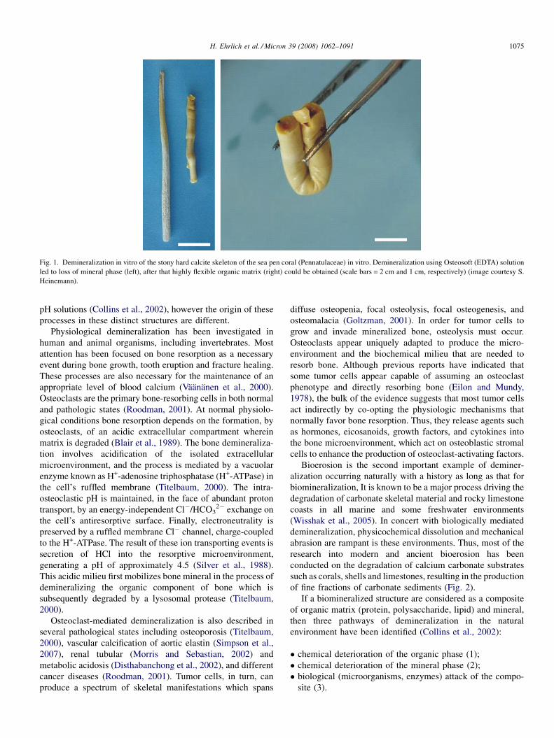
Fig. 1. Demineralization in vitro of the stony hard calcite skeleton of the sea pen coral (Pennatulaceae) in vitro. Demineralization using Osteosoft (EDTA) solution
led to loss of mineral phase (left), after that highly flexible organic matrix (right) could be obtained (scale bars = 2 cm and 1 cm, respectively) (image courtesy S.
Heinemann).
H. Ehrlich et al. / Micron 39 (2008) 1062–1091 1075
pH solutions (Collins et al., 2002), however the origin of these
processes in these distinct structures are different.
Physiological demineralization has been investigated in
human and animal organisms, including invertebrates. Most
attention has been focused on bone resorption as a necessary
event during bone growth, tooth eruption and fracture healing.
These processes are also necessary for the maintenance of an
appropriate level of blood calcium (Vaananen et al., 2000).
Osteoclasts are the primary bone-resorbing cells in both normal
and pathologic states (Roodman, 2001). At normal physiolo-
gical conditions bone resorption depends on the formation, by
osteoclasts, of an acidic extracellular compartment wherein
matrix is degraded (Blair et al., 1989). The bone demineraliza-
tion involves acidification of the isolated extracellular
microenvironment, and the process is mediated by a vacuolar
enzyme known as H+-adenosine triphosphatase (H+-ATPase) in
the cell’s ruffled membrane (Titelbaum, 2000). The intra-
osteoclastic pH is maintained, in the face of abundant proton
transport, by an energy-independent Cl�/HCO32� exchange on
the cell’s antiresorptive surface. Finally, electroneutrality is
preserved by a ruffled membrane Cl� channel, charge-coupled
to the H+-ATPase. The result of these ion transporting events is
secretion of HCl into the resorptive microenvironment,
generating a pH of approximately 4.5 (Silver et al., 1988).
This acidic milieu first mobilizes bone mineral in the process of
demineralizing the organic component of bone which is
subsequently degraded by a lysosomal protease (Titelbaum,
2000).
Osteoclast-mediated demineralization is also described in
several pathological states including osteoporosis (Titelbaum,
2000), vascular calcification of aortic elastin (Simpson et al.,
2007), renal tubular (Morris and Sebastian, 2002) and
metabolic acidosis (Disthabanchong et al., 2002), and different
cancer diseases (Roodman, 2001). Tumor cells, in turn, can
produce a spectrum of skeletal manifestations which spans
diffuse osteopenia, focal osteolysis, focal osteogenesis, and
osteomalacia (Goltzman, 2001). In order for tumor cells to
grow and invade mineralized bone, osteolysis must occur.
Osteoclasts appear uniquely adapted to produce the micro-
environment and the biochemical milieu that are needed to
resorb bone. Although previous reports have indicated that
some tumor cells appear capable of assuming an osteoclast
phenotype and directly resorbing bone (Eilon and Mundy,
1978), the bulk of the evidence suggests that most tumor cells
act indirectly by co-opting the physiologic mechanisms that
normally favor bone resorption. Thus, they release agents such
as hormones, eicosanoids, growth factors, and cytokines into
the bone microenvironment, which act on osteoblastic stromal
cells to enhance the production of osteoclast-activating factors.
Bioerosion is the second important example of deminer-
alization occurring naturally with a history as long as that for
biomineralization, It is known to be a major process driving the
degradation of carbonate skeletal material and rocky limestone
coasts in all marine and some freshwater environments
(Wisshak et al., 2005). In concert with biologically mediated
demineralization, physicochemical dissolution and mechanical
abrasion are rampant is these environments. Thus, most of the
research into modern and ancient bioerosion has been
conducted on the degradation of calcium carbonate substrates
such as corals, shells and limestones, resulting in the production
of fine fractions of carbonate sediments (Fig. 2).
If a biomineralized structure are considered as a composite
of organic matrix (protein, polysaccharide, lipid) and mineral,
then three pathways of demineralization in the natural
environment have been identified (Collins et al., 2002):
� c
hemical deterioration of the organic phase (1);� c
hemical deterioration of the mineral phase (2);� b
iological (microorganisms, enzymes) attack of the compo-site (3).
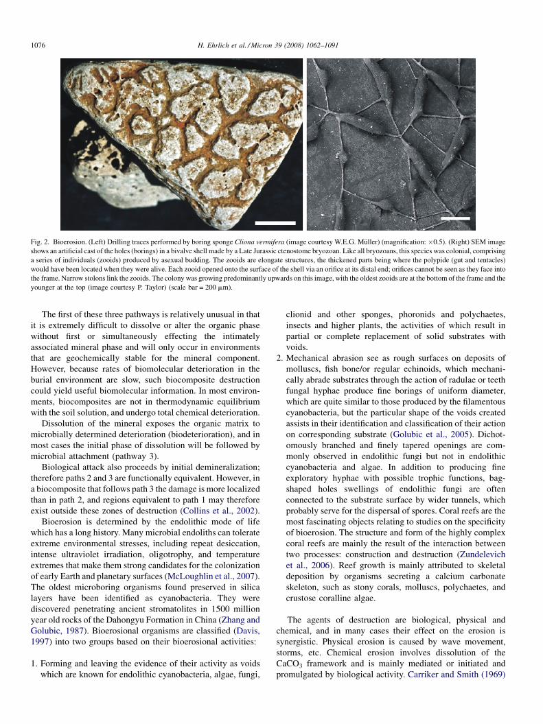
Fig. 2. Bioerosion. (Left) Drilling traces performed by boring sponge Cliona vermifera (image courtesy W.E.G. Muller) (magnification: �0.5). (Right) SEM image
shows an artificial cast of the holes (borings) in a bivalve shell made by a Late Jurassic ctenostome bryozoan. Like all bryozoans, this species was colonial, comprising
a series of individuals (zooids) produced by asexual budding. The zooids are elongate structures, the thickened parts being where the polypide (gut and tentacles)
would have been located when they were alive. Each zooid opened onto the surface of the shell via an orifice at its distal end; orifices cannot be seen as they face into
the frame. Narrow stolons link the zooids. The colony was growing predominantly upwards on this image, with the oldest zooids are at the bottom of the frame and the
younger at the top (image courtesy P. Taylor) (scale bar = 200 mm).
H. Ehrlich et al. / Micron 39 (2008) 1062–10911076
The first of these three pathways is relatively unusual in that
it is extremely difficult to dissolve or alter the organic phase
without first or simultaneously effecting the intimately
associated mineral phase and will only occur in environments
that are geochemically stable for the mineral component.
However, because rates of biomolecular deterioration in the
burial environment are slow, such biocomposite destruction
could yield useful biomolecular information. In most environ-
ments, biocomposites are not in thermodynamic equilibrium
with the soil solution, and undergo total chemical deterioration.
Dissolution of the mineral exposes the organic matrix to
microbially determined deterioration (biodeterioration), and in
most cases the initial phase of dissolution will be followed by
microbial attachment (pathway 3).
Biological attack also proceeds by initial demineralization;
therefore paths 2 and 3 are functionally equivalent. However, in
a biocomposite that follows path 3 the damage is more localized
than in path 2, and regions equivalent to path 1 may therefore
exist outside these zones of destruction (Collins et al., 2002).
Bioerosion is determined by the endolithic mode of life
which has a long history. Many microbial endoliths can tolerate
extreme environmental stresses, including repeat desiccation,
intense ultraviolet irradiation, oligotrophy, and temperature
extremes that make them strong candidates for the colonization
of early Earth and planetary surfaces (McLoughlin et al., 2007).
The oldest microboring organisms found preserved in silica
layers have been identified as cyanobacteria. They were
discovered penetrating ancient stromatolites in 1500 million
year old rocks of the Dahongyu Formation in China (Zhang and
Golubic, 1987). Bioerosional organisms are classified (Davis,
1997) into two groups based on their bioerosional activities:
1. F
orming and leaving the evidence of their activity as voidswhich are known for endolithic cyanobacteria, algae, fungi,
clionid and other sponges, phoronids and polychaetes,
insects and higher plants, the activities of which result in
partial or complete replacement of solid substrates with
voids.
2. M
echanical abrasion see as rough surfaces on deposits ofmolluscs, fish bone/or regular echinoids, which mechani-
cally abrade substrates through the action of radulae or teeth
fungal hyphae produce fine borings of uniform diameter,
which are quite similar to those produced by the filamentous
cyanobacteria, but the particular shape of the voids created
assists in their identification and classification of their action
on corresponding substrate (Golubic et al., 2005). Dichot-
omously branched and finely tapered openings are com-
monly observed in endolithic fungi but not in endolithic
cyanobacteria and algae. In addition to producing fine
exploratory hyphae with possible trophic functions, bag-
shaped holes swellings of endolithic fungi are often
connected to the substrate surface by wider tunnels, which
probably serve for the dispersal of spores. Coral reefs are the
most fascinating objects relating to studies on the specificity
of bioerosion. The structure and form of the highly complex
coral reefs are mainly the result of the interaction between
two processes: construction and destruction (Zundelevich
et al., 2006). Reef growth is mainly attributed to skeletal
deposition by organisms secreting a calcium carbonate
skeleton, such as stony corals, molluscs, polychaetes, and
crustose coralline algae.
The agents of destruction are biological, physical and
chemical, and in many cases their effect on the erosion is
synergistic. Physical erosion is caused by wave movement,
storms, etc. Chemical erosion involves dissolution of the
CaCO3 framework and is mainly mediated or initiated and
promulgated by biological activity. Carriker and Smith (1969)

H. Ehrlich et al. / Micron 39 (2008) 1062–1091 1077
demonstrated experimentally that chemical dissolution is
carried out by different penetrants created by the borers: acids,
chelating agents and specific enzymes (carbonic anhydrase) as
well as non-specific esterases, acid and alkaline phosphatases,
decarboxylases, and transaminases (Carriker and Smith, 1969).
Another example of naturally occurring demineralization
is the so-called phenomena of dark decalcification (Tentori
and Allemand, 2006). Kawaguti and Sakumoto (1948) noted
‘‘intake of Ca2+’’ in all scleractinian corals exposed to light
and ‘‘output of Ca2+’’ in all corals exposed to dark. They
argued that the skeleton formation was favored by the alkaline
pH (8.84–9.15) of the incubation medium, which was
assumed to be result of photosynthesis by zooxanthellae in
the coral; correspondingly, the drop in pH (8.00–7.80) in the
dark was the reason for the ‘‘resolution of the skeleton [sic]’’
(Kawaguti and Sakumoto, 1948). Chisholm (2000) observed
dark decalcification in coralline algae incubated at various
depths, and explained this as a result of previous light
exposure or the acidification caused by cell respiration. It is
also possible that the tissue recovery verified visually
underwater was overestimated and that decalcification was
due to tissue injury. Experiments on soft corals (Sarcophyton
sp. and Sinularia sp.) indicated that these corals also decalcify
in the dark (Tentori and Allemand, 2006). These authors
suggest that diurnal calcification–decalcification cycles
probably control coral sclerite size and shape.
Interestingly, the agents, principles and mechanisms of
chemical dissolution that are found or take place in natural
environments reported above seem to parallel to explanation of
demineralization processes in vitro. Better understanding of the
mechanisms leading to demineralization of biominerals in
natural environments and better characterization of reaction
conditions might allow us to more clearly identify specific
characteristics useful for the development of new techniques
based on gentle, biologically inspired demineralization.
4. Remineralization
Living forms appear to create specialized environments in
concert with the biomineralized tissues formations and
probably have been doing so since life first appeared (Skinner,
2005). Biomineralization and demineralization phenomena as
described above are only two parts of the mineral–organic
matrix circuit occurring in such specialized environments in
Fig. 3. Schematic view of ‘‘biomineralization–demineralization–remineraliza-
tion’’ cycle occurring in nature.
nature (Fig. 3). The third part of this biochemical cycle is
remineralization.
Remineralization is the process of restoring the solid
minerals – though the transfer of anions and cations – to a
nucleation sites where the lattices leading to mineral structures
are generated. This process follows demineralization and is
observed both in vivo in a host of natural environments. A
remineralization process in vitro has also been established.
De- and remineralization are critical to the formation of teeth,
caries and teeth erosion. Saliva, is a major destabilizer of erupted
teeth which may be affected by pH in the oral cavity (Lagerlof
and Olivery, 1994). Both demineralization and remineralization
occur on the surface of the tooth and can be considered as
dynamic processes, characterized by the flow of calcium and
phosphate out of and back into tooth enamel, which should be
balanced in order to prevent the progression of caries (Torrado
et al., 2004). Demineralization progressing to cavitation occurs if
the frequency and magnitude of acid production overwhelm the
repair process. This situation would occur in the case of frequent
eating or if the repair process is compromised by a reduction in
salivary flow (Loesche, 1986). Remineralization dominates if the
plaque acid production is restricted, as occurs with the ingestion
of low sucrose diets or the use of sugar substitutes for snacks
between meals. It involves carbon dioxide from breath and water
from saliva to create a mild, unstable carbonic acid that is at the
core of the natural remineralization process. Minerals in saliva
present from food are dissolved by the carbonic acids. In
addition, carbonic acid quickly and easily converts itself to
carbon dioxide and water under these conditions (Rantonen and
Meurman, 2000). When this happens, the dissolved mineral ions
precipitate out as solid mineral ions again, but not always as the
original mineral molecules. For example, fluoride also promotes
remineralization, and this may be the main mechanism by which
fluoride protects in turn tooth decay. When present in the liquid
phase during the remineralization of demineralized enamel,
fluoride will be incorporated into already existing crystalline
mineral and the enamel becomes more resistant to demineraliza-
tion (Lagerlof and Olivery, 1994).
Since natural remineralization is frequently inadequate to
maintain strong enamel, the natural remineralization process
needs to be augmented. When dentine is demineralized by the
caries organism, either in an experimental animal or human
being, it can be recalcified in vivo by local applications of a
preparation of calcium hydroxide (Eidelman et al., 1965). The
exogenous calcium ion that is applied to an exposed surface of
dentin produces an uptake of endogenous phosphate from the
fluids of the body. Crystal formation of apatite follows, and the
end result is complete remineralization of the matrix (Bang and
Urist, 1967).
Recently, artificial carious lesions in enamel have been
developed as a useful analogue for natural lesions when studying
de- and remineralization, and many different demineralizing
systems have been reported for their preparation (White, 1995;
Lynch et al., 2007). The essence of the remineralizing concept
might be achieved by simultaneously supplying calcium,
phosphate, and fluoride ions to the teeth in order to induce the
formation of calcium fluorapatite which remineralizes and
H. Ehrlich et al. / Micron 39 (2008) 1062–10911078
strengthens the tooth (Torrado et al., 2004). Therefore intensive
investigations into the remineralization potential of new tooth-
paste and fluid formulations are still in progress.
The demineralization–remineralization phenomenon, which
is an example of acid–base homeostasis mechanism in vivo,
occurs in different organisms. In molluscs (Mercenaria
mercenaria) under normal conditions, the animal becomes
anaerobic when the valves close (Crenshaw and Neff, 1969).
This period of anaerobiosis is accompanied by an accumulation
of succinic acid. This succinic acid is produced by the fixation
of CO2, however some of the CO2 fixed into succinate
originates from the shell carbonate when the shell is dissolved
to neutralize succinic acid. When a mollusc closes its valves,
the concentrations of calcium, total carbon dioxide, and
hydrogen ions increase. The increase in calcium was about
three times that of carbon dioxide. Correspondingly, the
remineralization mechanism starts when the mollusc opens its
valves.
Recently, mineralized tissue has been shown also to be
important in buffering lactic acid during anoxic submergence in
reptiles, specifically the skeleton and shell in different species
of turtles (Jackson, 2000), the skeleton and osteoderms in
caimans (Jackson et al., 2003), and the femur of amphibians
(Warren and Jackson, 2005). A turtle’s shell is the major
mineral reservoir of its body: over 99% of the total body
calcium, magnesium and phosphate, over 95% of the carbon
dioxide, and over 60% of the body’s sodium reside in the shell
and bone. It was shown (Jackson, 2000) that anoxic turtles
accumulate high levels of lactate in their blood.
To avoid fatal acidosis, turtles exploit buffer reserves in their
large mineralized shell. The shell acts by releasing calcium and
magnesium carbonates and by storing and buffering lactic acid.
Together with profound metabolic depression, shell buffering
permits survival without oxygen for several months at 3 8C.
Also in experiments in vitro with powdered turtle shell, it was
shown the shell alkalinized the solution, requiring acid titration
for pH-stat control, and the amount of titrated acid required
increased as solution pH fell. This is consistent with the
hypothesis that shell buffers help to neutralize circulating acids
and their release during anoxia is a passive consequence of acid
demineralization of the shell and bone. Additionally, one
intriguing conservation implication is that if mineralized tissues
proved vulnerable to demineralization in an acidic environ-
ment, then the tolerance of animals to anoxia or hypoxia and,
possibly, their metabolism and performance during anaerobic
periods could be compromised (Warren and Jackson, 2005).
Remineralization also plays an important role in natural
environments. The following experiments were recently
described by Le Cadre et al. (2003). To study the pH effects,
cultures with pH values ranging from normal (pH 7.9–8.3)
marine were prepared using hydrochloric acid to lower the pH to
7. Foraminifera Ammonia beccarii was collected and introduced
into these different environment scultures. Under neutral pH
(7.0) conditions, pseudopodial activities emission was reduced or
stopped. Then the animals external mineralized tissues, ‘‘the
test’’ became opaque as a result of superficial alteration, which is
the first stage of test decalcification. Decalcification progres-
sively extended over the whole test, first destroying the
mineralized areas of the last chambers, which contain less
tissue, i.e. are thinner. After 15 days, only interlocular walls were
preserved, giving the test a star-shaped characteristic of an
advanced stage of decalcification. If a specimen was maintained
in low pH conditions, the entire test was sometimes entirely
destroyed and only the cytoplasm, covered with the inner organic
layer, remained. On the other hand, if a specimen with a partially
dissolved test was placed in a solution at normal pH, it was able to
rebuild its test. Remineralization was somewhat different from
the original calcification and was accompanied, in most cases, by
morphological abnormalities (e.g. abnormal expansions, irre-
gular chamber sizes, wall with concave form).
These observations show that temporary acidification of the
environment, causing partial decalcification of the test, is able
to induce morphological abnormalities of foraminiferal tests
during recalcification. This acidification may be caused by the
anthropogenic impacts on a natural cause. In both cases,
deformation of foraminiferal tests yields information on
environmental characteristics of the area. Therefore the
observations on foraminiferal tests and their use as bioindi-
cators of pollution in coastal environments is now one pf the
areas under development in the discussion on climate change
(Le Cadre et al., 2003).
Similarly, effects of structural conformations were observed
in experiments with octocorals. Isolated spicules of the
gorgonian Leptogorgia virgulata were decalcified using 0.5 M
EDTA solution (pH 7.5) and exposed to an artificial seawater
solution to evaluate the ability of the spicules matrix to recalcify
(Watabe et al., 1986). Recalcification of the decalcified spicule
matrices occurred after 48 h in the artificial seawater solution
containing NaCl, CaCl2, KCl, MgSO4, MgCl2, and NaHCO3.
Decalcified matrices which retained the configuration of normal
spicules assumed a form upon recalcification similar to, but not
identical with, undecalcified spicules. Recalcification also
occurred in the use of decalcified matrices that did not retain
the form of normal spicules. Most of the recalcified matrices
showed reduced calcium content when compared with normal
spicules. The experiments demonstrated that completely
decalcified spicule matrices can initiate recalcification and
influence mineral form (Watabe et al., 1986).
Thus, morphological abnormalities observed in different
biomineral formations during and after remineralization might
be common phenomena, because also in case studies using high-
resolution transmission electron microscopy and relating to the
growth of apatite crystals in the remineralized enamel, similar
effects were obtained. The growth of newly formed crystals and
the regrowth of pre-existing enamel crystals occurred extensively
in remineralized enamel (Tohda et al., 1990). With advancing
growth, the crystals came into contact and fused with each other,
forming large crystals with hexagonal outlines. Various kinds of
crystalline defects, including edge dislocation, low-angle grain
boundary, and lattice shifting, were frequently detected between
the fusing crystals. These observations confirm the previous
suggestion that processes of de- and remineralization (shell, bone
resorption, caries) must be regarded as abnormal biomineraliza-
tion processes (Krampitz and Graser, 1988).

Fig. 4. Boring worms: the outer surface of the scallop Patinopecten yessoensis
(Jay) shell eroded (arrows) by polychaete worm Polydora brevipalpa (scale
bar = 1 cm) (image courtesy A.V. Silina).
H. Ehrlich et al. / Micron 39 (2008) 1062–1091 1079
A better understanding of the biomineralization–deminer-
alization–remineralization mechanisms at the molecular level
will result in more effective strategies for the development and
establishment of novel methods, tools and approaches for
science, engineering and medicine.
5. History of demineralization
To accommodate readers with little or no knowledge of
demineralization phenomena, we shall begin with a short
overview of the scientific history.
It is well known that each generation of scientists adds
details, largely re-explaining the same phenomena in new
terminology; only on occasion are completely new concepts
introduced (Mandel, 1983). In the case of demineralization, the
driving force determining progress in investigations was based
on problems of human health. Namely toothache stimulated
first of all the origin of the hypothesis and studies on dental
caries, a recently well-investigated example of demineraliza-
tion processes which take place in vivo. Dental caries is a
complex disease, the ‘‘cause’’ of which has received significant
research attention during the 19th and most of the 20th
centuries. Mostly via observational investigations, different
hypotheses on causes associated with dental caries were put
forth. The dominant theory at the beginning, the middle of the
19th century, was the ‘‘worm theory’’ (Ismail et al., 2001).
The oldest and most pervasive of all views on dental caries
depicts the tooth inhabited by a demon in the form of a worm
(Mandel, 1983). Most of our information on the origin of the
tooth worm is derived from an Assyrian tablet from the 7th
century B.C. that was found about 64 years ago (Weinberger,
1948).
The medical historians of ancient India, Egypt, Japan, and
China also make reference to the worm as the cause of
toothache. The legend of the worm is also found in the writings
of Homer, as well as the great surgeon of the middle ages, Guy
de Chauliac (1300–1368 A.D.), who still espoused the belief that
worms caused dental decay. The famous Flemish surgeon Jan
Yperman (who died about 1330) claimed to have observed that
the moving worms caused suppuration in the teeth (Gerabek,
1999).
The belief that a worm developed in a bad tooth, started to
pick upon the tooth structure immediately and then died as soon
as it came into contact with air can be traced back to Paracelsus
(1493–1541) (Gerabek, 1999).
The first full text on dental diseases was published in 1728
when Pierre Fauchard, a French surgeon, wrote ‘‘Le Chirurgien
Dentiste’’. Fauchard rejected the toothworm theory of dental
caries. Instead, he described enamel hypoplasia as ‘‘an erosion
of the enamel’’ and recommended that hypoplastic areas be
smoothed using files (Hoffman-Axthelm, 1981).
Although the toothworm theory is absolutely unacceptable
from the modern point of view, here we want to include a small
remark regarding the occurrence of worms in a natural
environment that really burrow into a variety of calcareous
substrata such as soft clays, mud, rock, coral reefs and
molluscan shells (Fig. 4). Most of them are boring polychaetous
annelids and the mechanisms (physical or chemical) of their
burrow expansion are still a matter of controversy (Martin and
Britayev, 1998).
By 1880, dental caries was defined as a ‘‘disintegration of
the tooth substance, molecule by molecule’’ (Black, 1880) and
a disease that was caused by the fermentation of ‘‘foods’’ inside
the mouth (MacPhee, 1935). The ‘‘tooth-worm’’ theory
disappeared as the microscope became more freely available,
and investigators began to note the teeming profusion of fungi,
and long Leptotrichia-type organisms on the tooth surface and
within the carious lesion (Mandel, 1983). In 1880 Weil wrote:
‘‘I regard it quite probable that this fungus Leptothrix buccalis
bores directly through it (enamel cuticle). The fungi now
proceed farther into enamel and force apart its prisms, gradually
breaking up its structure’’ (Weil, 1887).
Proponents of the chemical theory, such as Tomes (1859)
and Magitot (1867), suggested that caries was due to the solvent
action of acids generated by the fermentation of food. Dr. W.D.
Miller built on the earlier work of Leber and Rottenstein (1878)
and fully synthesized two older ideas into a new chemical-
parasitic theory of dental decay, generating the data that
advanced the speculation of a demonstration that the first step in
dental caries was the production of acid by microorganisms
fermenting the carbohydrates in the mouth.
Thus, only in 1881, studies by Miller found that acid
produced by microorganisms in the mouth caused destruction
of the enamel, and caries in dentin resulted from acidic
decalcification.
Bacteria were indicted as producing the acids that led to the
demineralization of enamel and dentin (Miller, 1883). Once an
opening had been made, the microorganisms could then enter
into the tooth substance and affect the organic structures (in the
dentine?).
Throughout the 20th century, many researches and dentists
recognized that dental caries is a product of the interplay of
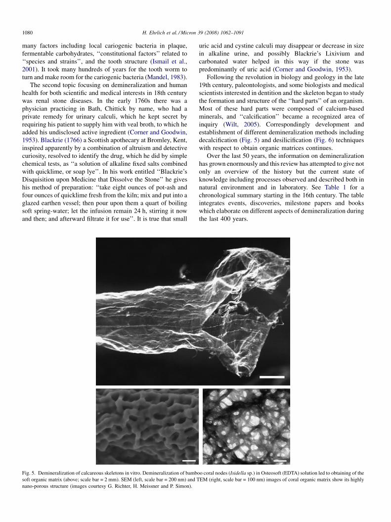
H. Ehrlich et al. / Micron 39 (2008) 1062–10911080
many factors including local cariogenic bacteria in plaque,
fermentable carbohydrates, ‘‘constitutional factors’’ related to
‘‘species and strains’’, and the tooth structure (Ismail et al.,
2001). It took many hundreds of years for the tooth worm to
turn and make room for the cariogenic bacteria (Mandel, 1983).
The second topic focusing on demineralization and human
health for both scientific and medical interests in 18th century
was renal stone diseases. In the early 1760s there was a
physician practicing in Bath, Chittick by name, who had a
private remedy for urinary calculi, which he kept secret by
requiring his patient to supply him with veal broth, to which he
added his undisclosed active ingredient (Corner and Goodwin,
1953). Blackrie (1766) a Scottish apothecary at Bromley, Kent,
inspired apparently by a combination of altruism and detective
curiosity, resolved to identify the drug, which he did by simple
chemical tests, as ‘‘a solution of alkaline fixed salts combined
with quicklime, or soap lye’’. In his work entitled ‘‘Blackrie’s
Disquisition upon Medicine that Dissolve the Stone’’ he gives
his method of preparation: ‘‘take eight ounces of pot-ash and
four ounces of quicklime fresh from the kiln; mix and put into a
glazed earthen vessel; then pour upon them a quart of boiling
soft spring-water; let the infusion remain 24 h, stirring it now
and then; and afterward filtrate it for use’’. It is true that small
Fig. 5. Demineralization of calcareous skeletons in vitro. Demineralization of bamb
soft organic matrix (above; scale bar = 2 mm). SEM (left, scale bar = 200 nm) and T
nano-porous structure (images courtesy G. Richter, H. Meissner and P. Simon).
uric acid and cystine calculi may disappear or decrease in size
in alkaline urine, and possibly Blackrie’s Lixivium and
carbonated water helped in this way if the stone was
predominantly of uric acid (Corner and Goodwin, 1953).
Following the revolution in biology and geology in the late
19th century, paleontologists, and some biologists and medical
scientists interested in dentition and the skeleton began to study
the formation and structure of the ‘‘hard parts’’ of an organism.
Most of these hard parts were composed of calcium-based
minerals, and ‘‘calcification’’ became a recognized area of
inquiry (Wilt, 2005). Correspondingly development and
establishment of different demineralization methods including
decalcification (Fig. 5) and desilicification (Fig. 6) techniques
with respect to obtain organic matrices continues.
Over the last 50 years, the information on demineralization
has grown enormously and this review has attempted to give not
only an overview of the history but the current state of
knowledge including processes observed and described both in
natural environment and in laboratory. See Table 1 for a
chronological summary starting in the 16th century. The table
integrates events, discoveries, milestone papers and books
which elaborate on different aspects of demineralization during
the last 400 years.
oo coral nodes (Isidella sp.) in Osteosoft (EDTA) solution led to obtaining of the
EM (right, scale bar = 100 nm) images of coral organic matrix show its highly

Fig. 6. Desilicification of natural silica-based glass sponge spicules. (Left) SEM image of collagenous fibrillar matrix obtained after alkali treatment of siliceous basal
spicules of Hyalonema sieboldii glass sponge (scale bar = 1 mm). (Right) High-resolution transmission electron microscopy (HRTEM) image of the fragment of glass
sponge collagen microfibril. The arrows indicate the presence of nanofibrillar structures the diameter if which correspond to 1.5 nm (image courtesy P. Simon).
H. Ehrlich et al. / Micron 39 (2008) 1062–1091 1081
6. Practical applications of demineralization
The data represented in the table shows evidence that
scientists from diverging fields, spanning mineralogy, zoology,
botany, biochemistry, biogeochemistry, histology, dentistry and
materials science, have made great efforts to understand the
demineralization mechanism in vivo, in natural environments,
and also to mimic the process in vitro using different techniques
based on a range of dissolution- and chelation-agents.
Additional corroboration regarding the importance of studies
on demineralization for the scientific community might be
found in the cited papers. More than 10% of articles relating to
the problems of demineralization are published in Nature and
Science. It must also be cautioned that because of the specificity
and vast volume of patent literature relating to demineralization
we did not enter information from that information pool for this
review.
In conclusion, demineralization is an ongoing, challenging
group of mechanisms and aspects applicable and used by
different scientific disciplines with their diverse directions
including the following:
� e
volutionary paleontology (Gould, 1970);� a
strobiology (McLoughlin et al., 2007);� b
iomolecular deterioration and survival of organic matter(Collins et al., 2002);
� g
eomycology (Gadd, 2007);� p
aleoclimatology (Schone and Giere, 2005);� s
eawater chemistry (Porter, 2007);� m
aterial chemistry (Dujardin and Mann, 2002);� g
eomedicine (Sahai, 2005);� p
aleobathymetry (Wisshak and Ruggeberg, 2006);� e
xobiology (Onofri et al., 2004);� o
steoarchaeology (Davis, 1997);� a
rchaeology and bioarcheology (Larsen, 2002);� h
istology and histotechnology (Callis and Sterchi, 1998);� fo
rensic dentistry and forensic science (Rogers, 1988);� d
emineralized bone allografts (Herold et al., 2002);� d
ental anthropology (Alt et al., 1998);� m
ineral dissolution (Nancollas, 1982; Wang et al., 2005b,2006a,b);
� b
io- and chemical weathering (Benzerara et al., 2005);� m
edical geology (Skinner and Berger, 2003);� c
alcibiocavitology and endolithic microborings (Carriker andSmith, 1969);
� b
ioerosion (Lazar and Loya, 1991; Garcia-Pichel, 2006);� r
emineralization of biomaterials (Liu et al., 2003);� s
hell repair and regeneration (Meenakshi et al., 1974; Palmer,1983);
� s
clerochronological studies (Schone et al., 2005);� b
ioremediation (Singh and Ward, 2004);� b
one diseases, osteoporosis, osteopenia, osteoclasia (Skinner,2000);
� b
iodeterioration (Morton, 1987);� ta
phonomy (Child, 1995);� a
rcheological bone chemistry (Pate, 1994);� p
aleohistory, paleoecology, paleontology (Pate, 1994; Taylorand Wilson, 2003);
� n
ormal and pathological remineralization (Robinson et al.,2000);
� b
iotechnology (Zakaria et al., 1996, 1998, 2005);� a
geing techniques (demineralization of vertebrae andotoliths) (Correia and Figueiredo, 1997).
We submit here some additional considerations responding
to important results which were obtained using demineraliza-
tion approaches in:
(a) M
odern science:Using different demineralization techniques and
approaches biologists can precisely reconstruct life-history
traits from growth structures (age, growth season, onset of
maturity, etc.), and paleoclimatologists and geochemists can
identify how much time is contained in each geochemical
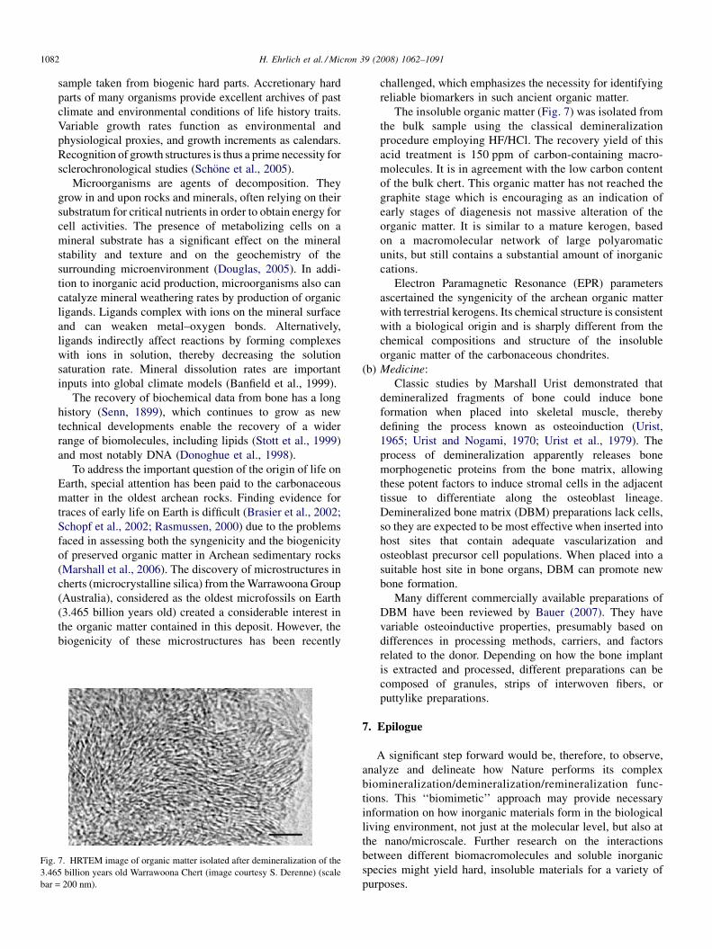
Fig.
3.46
bar =
H. Ehrlich et al. / Micron 39 (2008) 1062–10911082
sample taken from biogenic hard parts. Accretionary hard
parts of many organisms provide excellent archives of past
climate and environmental conditions of life history traits.
Variable growth rates function as environmental and
physiological proxies, and growth increments as calendars.
Recognition of growth structures is thus a prime necessity for
sclerochronological studies (Schone et al., 2005).
Microorganisms are agents of decomposition. They
grow in and upon rocks and minerals, often relying on their
substratum for critical nutrients in order to obtain energy for
cell activities. The presence of metabolizing cells on a
mineral substrate has a significant effect on the mineral
stability and texture and on the geochemistry of the
surrounding microenvironment (Douglas, 2005). In addi-
tion to inorganic acid production, microorganisms also can
catalyze mineral weathering rates by production of organic
ligands. Ligands complex with ions on the mineral surface
and can weaken metal–oxygen bonds. Alternatively,
ligands indirectly affect reactions by forming complexes
with ions in solution, thereby decreasing the solution
saturation rate. Mineral dissolution rates are important
inputs into global climate models (Banfield et al., 1999).
The recovery of biochemical data from bone has a long
history (Senn, 1899), which continues to grow as new
technical developments enable the recovery of a wider
range of biomolecules, including lipids (Stott et al., 1999)
and most notably DNA (Donoghue et al., 1998).
To address the important question of the origin of life on
Earth, special attention has been paid to the carbonaceous
matter in the oldest archean rocks. Finding evidence for
traces of early life on Earth is difficult (Brasier et al., 2002;
Schopf et al., 2002; Rasmussen, 2000) due to the problems
faced in assessing both the syngenicity and the biogenicity
of preserved organic matter in Archean sedimentary rocks
(Marshall et al., 2006). The discovery of microstructures in
cherts (microcrystalline silica) from the Warrawoona Group
(Australia), considered as the oldest microfossils on Earth
(3.465 billion years old) created a considerable interest in
the organic matter contained in this deposit. However, the
biogenicity of these microstructures has been recently
7. HRTEM image of organic matter isolated after demineralization of the
5 billion years old Warrawoona Chert (image courtesy S. Derenne) (scale
200 nm).
challenged, which emphasizes the necessity for identifying
reliable biomarkers in such ancient organic matter.
The insoluble organic matter (Fig. 7) was isolated from
the bulk sample using the classical demineralization
procedure employing HF/HCl. The recovery yield of this
acid treatment is 150 ppm of carbon-containing macro-
molecules. It is in agreement with the low carbon content
of the bulk chert. This organic matter has not reached the
graphite stage which is encouraging as an indication of
early stages of diagenesis not massive alteration of the
organic matter. It is similar to a mature kerogen, based
on a macromolecular network of large polyaromatic
units, but still contains a substantial amount of inorganic
cations.
Electron Paramagnetic Resonance (EPR) parameters
ascertained the syngenicity of the archean organic matter
with terrestrial kerogens. Its chemical structure is consistent
with a biological origin and is sharply different from the
chemical compositions and structure of the insoluble
organic matter of the carbonaceous chondrites.
(b) M
edicine:Classic studies by Marshall Urist demonstrated that
demineralized fragments of bone could induce bone
formation when placed into skeletal muscle, thereby
defining the process known as osteoinduction (Urist,
1965; Urist and Nogami, 1970; Urist et al., 1979). The
process of demineralization apparently releases bone
morphogenetic proteins from the bone matrix, allowing
these potent factors to induce stromal cells in the adjacent
tissue to differentiate along the osteoblast lineage.
Demineralized bone matrix (DBM) preparations lack cells,
so they are expected to be most effective when inserted into
host sites that contain adequate vascularization and
osteoblast precursor cell populations. When placed into a
suitable host site in bone organs, DBM can promote new
bone formation.
Many different commercially available preparations of
DBM have been reviewed by Bauer (2007). They have
variable osteoinductive properties, presumably based on
differences in processing methods, carriers, and factors
related to the donor. Depending on how the bone implant
is extracted and processed, different preparations can be
composed of granules, strips of interwoven fibers, or
puttylike preparations.
7. Epilogue
A significant step forward would be, therefore, to observe,
analyze and delineate how Nature performs its complex
biomineralization/demineralization/remineralization func-
tions. This ‘‘biomimetic’’ approach may provide necessary
information on how inorganic materials form in the biological
living environment, not just at the molecular level, but also at
the nano/microscale. Further research on the interactions
between different biomacromolecules and soluble inorganic
species might yield hard, insoluble materials for a variety of
purposes.
H. Ehrlich et al. / Micron 39 (2008) 1062–1091 1083
The three important bioprocesses, biomineralization/demi-
neralization/remineralization, are intimately connected together
in an admirable and intriguing fashion in Nature. And it is
important to use an integrated approach to study them as a whole
some adopting such a research strategy may reveal underlying
relationships that, otherwise, may remain unknown.
Understanding how these processes operate in a biological
environment can be a valuable guide to developing important
industrial processes. Nanoparticle synthesis and engineering is
a good example that exemplifies how industrial technologists
could learn from Nature (Demadis and Neofotistou, 2007;
Knecht and Wright, 2003). No matter the scientific field used to
approach these three processes many questions remain. These
are the true drivers for multidisciplinary research and such
efforts, thus far, have been amply rewarded and have led to
great advances. Meaningful progress in our understanding of
demineralization depends on continuous developments in areas
such as inorganic chemistry, materials science, biology,
biochemistry, analytical chemistry, just to mention a few and
the development of new tools that will allow for further and
deeper understanding of the complex processes occurring in
Nature.
Acknowledgements
Authors would like to acknowledge Prof. S. Weiner, Prof. M.
Ilan (Israel); Prof. M. Glimcher, Prof. H.C. Skinner, Prof. G.
Nancollas, Prof. D. Morse, Prof. N. Kroger, Prof. F. Wilt, Prof.
S. Golubic, Prof. G. Lambert, Prof. M. Schweitzer, Prof. R.
Thalmann, Prof. Henry L. Ehrlich, Prof. A. George, Prof W.
Goldberg, Prof. G. Marshall, Prof. A. Luttge, Prof. C. Pueschel,
Prof. C. McNamara, Prof. D. Jackson and G. Callis (USA);
Prof. G. Gadd, Prof. J. Elliott, Dr. P. Taylor, Dr. M. Barbour
(UK); Prof. Y. Dauphin, Prof. J. Vacelet, Prof. J. Luquet, Dr. S.
Derenne (France); Prof. D. Allemand (Monaco); Prof. G.
Bavestrello, Prof. S. Onofri, Prof. B. Fubini, Dr. S. Schiaparelli
(Italy); Prof. K.D. Jandt, Prof. M. Epple, Prof. W.E.G. Muller,
Prof. G. Worheide, Prof. E. Baeuerlein, Prof. B. Schone, Prof.
H. Schroder, Prof. M. Sumper, Prof. H. Worch, Prof. D. Raabe,
Prof. R. Kniep, Dr. P. Simon, Dr. D. Sauer, Dr. A. Ziegler, Dr.
C.H. Schonberg, Dr. H. Fabritius, Dr. M. Wisshak, (Germany);
Prof. A. Ereskovsky, Dr. L. Gilinskaya, Dr. A.V. Silina (Russia);
Prof. E. Konigsberger, Dr. M. Whitelaw-Weckert (Australia);
Prof. F. Grases, Prof. A. Checa (Spain); Prof. T. Franz-
Odendaal (Canada) for discussion and comments. We wish to
thank Timothy Douglas, Sascha Heinemann, Christiane Fisher,
Vasily V. Bazhenov and Irina Ehrlich for excellent technical
assistance.
This work was supported by German Research Foundation
(DFG) grant WO 494/17-1, and the General Secrtariat of
Science & Technology, Greece.
References
Abel, O., 1935. Vorzeitige Lebensspuren. Verlag von Gustav Fischer, Jena.
Abelson, P., 1954. Carnegie Inst. Washington Yearb. 53, 97–101.
Aizenberg, J., Hanson, J., Ilan, M., Leiserowitz, L., Koetzle, T.F., Addadi, L.,
Weiner, S., 1995. FASEB J. 9, 262–268.
Aizenberg, J., Ilan, M., Weiner, S., Addadi, L., 1996. Connective Tissue Res. 34,
255–261.
Aizenberg, J., Lambert, G., Weiner, S., Addadi, L., 2002. J. Am. Chem. Soc.
124, 32–39.
Aizenberg, J., Weaver, J.C., Thanawala, M.S., Sundar, V.C., Morse, D.E., Fratzl,
P., 2005. Science 309, 275–278.
Albeck, S., Weiner, S., Addadi, L., 1996. Chem. Eur. J. 2, 278–284.
Albright, J.T., Scott, D.B., Mosley, V.M., Wyckoff, W.G., 1952. J. Dent. Res. 31,
483–484.
Alers, J.C., Krijtenburg, P.J., Vissers, K.J., van Dekken, H., 1999. J. Hystochem.
Cytochem. 47 (5) 703–709.
Alt, K., Rosing, F.W., Teschler-Nicola, M., 1998. Dental Anthropology:
Fundamentals, Limits, and Prospects. Springer Verlag, Wien, New York.
Ameye, L., Compere, P., Dille, J., Dubois, P., 1998. Histochem. Cell. Biol. 110,
285–294.
Anderson, P., Bollet-Quivogne, F.R.G., Dowker, S.E.P., Elliott, J.C., 2004.
Arch. Oral Biol. 49, 199–207.
Anderson, P., Elliott, J.C., 1987. Caries Res. 21, 522–525.
Anderson, P., Elliott, J.C., 2003. Caries Res. 34, 33–40.
Anderson, P., Elliott, J.C., 1992. J. Dent. Res. 71, 1473–1481.
Andre, W., 1784. Philos. Trans. B 15, 540–541.
Ankel, W.E., 1937. Biol. Zentr. 57, 75–82.
Ankel, W.E., 1938. Zool. Anz. Suppl. 11, 223–295.
Anneroth, G., Eneroth, C.M., Isacsson, G., 1975. J. Oral Pathol. Med. 4,
266–272.
Arey, L.B., 1920. Am. J. Anat. 26, 315–347.
Armstrong, W.D., Brekhus, P.I., 1937. J. Biol. Chem. 120, 677–687.
Armstrong, W.G., Halstead, L.B., Reed, F.B., Wood, L., 1983. Philos. Trans. R.
Soc. Lond. B 301, 301–343.
Asara, J.M., Schweitzer, M.H., Freimark, L.M., Phillips, M., Cantley, L.C.,
2007. Science 316, 280–285.
Bachmann, E., 1915. Ber. Dtsch. Bot. Ges. 33, 45–47.
Baillere, H. (Ed.), 1840. Odontography; or a Treatise on the Comparative
Anatomy of the Teeth. H. Baillere Publisher, London, 655 pp. + atlas.
Baker, J.R., Balch, D.A., 1962. Biochem. J. 82, 352–361.
Bancroft, J.D., Stevens, A. (Eds.), 1996. Theory and Practice of Histological
Techniques. Churchill Livingstone, NY.
Banfield, J.E., Nealson, K.H. (Eds.), 1997. Geomicrobiology Interactions
between Microbes and Minerals, Reviews in Mineralogy, vol. 35. Miner-
alogical society, Washington, DC.
Banfield, J.F., Barker, W.W., Welch, S., Taunton, A., 1999. Proc. Natl. Acad.
Sci. U.S.A. 96, 3404–3411.
Bang, G., Urist, M.R., 1967. J. Dent. Res. 46, 722–730.
Barbour, M.E., Parker, D.M., Jandt, K.D., 2003a. Eur. J. Oral. Sci. 111, 258–
262.
Barbour, M.E., Parker, D.M., Jandt, K.D., 2003b. J. Colloid Interface Sci. 265,
9–14.
Barbour, M.E., Shellis, R.P., 2007. Phys. Med. Biol. 52, 899–910.
Baron, R., 1989. Anatomical Record 224, 317–324.
Bartlett, D.W., Coward, P.Y., 2001. J. Oral Rehabil. 28, 1045–1047.
Bartlett, D.W., Evans, D.F., Smith, B.G., 1996. J. Oral Rehabil. 23, 289–297.
Bauer, T.W., 2007. Arch. Pathol. Lab. Med. 131, 217–224.
Bauerlein, E. (Ed.), 2000. The Biomineralization of Nano- and Micro-struc-
tures. Wiley-VCH, Weinheim.
Bauerlein, E., Epple, M., Behrens, P. (Eds.), 2007. Handbook of Biominer-
alization. Wiley-VCH, Weinheim.
Bavestrello, G., Arillo, A., Benatti, U., Cerrano, C., Cattaneo-Vietti, R.,
Cortesogno, L., Gaggero, A., Giovine, M., Tonetti, M., Sara, M., 1995.
Nature 378, 374–376.
Been, J.M., Bills, P.M., Lewis, D., 1979. Gastroenterology 76 (3) 548–555.
Bell, T., 1829. Anatomy, Physiology and Disorders of the Teeth. Highley,
London.
Benson, S., Benson, N.C., Wilt, F., 1986. J. Cell. Biol. 102, 1878–1886.
Benson, S., Jones, E.M.E., Crise-Benson, N., Wilt, F., 1983. Exp. Cell Res. 148,
249–253.
Benton, R.S., Morris Jr., R.E., 1956. Am. J. Clin. Pathol. 26 (8) 882–898.
H. Ehrlich et al. / Micron 39 (2008) 1062–10911084
Benzerara, K., Yoon, T.H., Menguy, N., Tyliszczak, T., Brown Jr., G.E., 2005.
Proc. Natl. Acad. Sci. U.S.A. 102 (4) 979–982.
Bergeim, O., 1914. Proc. Soc. Exp. Biol. Med. XII, 22.
Berman, A., Addadi, L., Kvick, A., Leiserowitz, L., Nelson, M., Weiner, S.,
1990. Science 250, 664–667.
Bernhardt, A.M., Manyak, D.M., Wilbur, K.M., 1985. J. Moll. Stud. 51, 284–
289.
Berthon, G. (Ed.), 1995. Handbook of Metal–Ligand Interactions in Biological
Fluids: Bioorganic Chemistry. Dekker, New York.
Bettice, J.A., 1984. Am. J. Physiol. Renal Fluid Electrolyte Physiol. 247, F326–
F330.
Bigland, C.H., Graesser, F.E., Pennifold, K.S., 1961. Avian Dis. 5, 367–370.
Birkedal-Hansen, H., 1974. J. Histochem. Cytochem. 22, 434–441.
Black, G., 1880. Am. J. Dent. Sci. 14, 289–308.
Black, G.V., 1899. Dental Cosmos 41, 826–830.
Black, G.V., 1897. Dental Cosmos 39, 404.
Blackrie, A., 1766. A disquisition on medicines that dissolve the stone. In which
Dr. Chittick’s secret is considered and discovered. Printed for D. Wilson,
London.
Blair, H.C., Teitelbaum, S.L., Ghiselli, R., Gluck, S., 1989. Science 245, 855–
857.
Blake, J.A., Evans, J.W., 1973. Veliger 15, 235–249.
Block, R.J., Horwitt, M.K., Bolling, D., 1949. J. Dent. Res. 28, 518.
Bodansky, A., Blair, J.E., Jaffe, H.L., 1930. J. Biol. Chem. LXXXVIII (3) 629–
647.
Bohartirchuk, F., 1966. Am. J. Med. 41, 836–846.
Bonucci, E., 2007. Biological Calcification: Normal and Pathological Processes
in the Early Stages. Springer, Berlin, Heidelberg.
Bonucci, E., Motto, P.M. (Eds.), 1990. Ultrastructure of Skeletal Tissues.
Kluwer Academic, Boston.
Boorde, A., 1552. The Brevarie of Health. Thomas Eams Co., London.
Bornet, E., Flahaut, C., 1889. Soc. Bot. France Bull. 36, 147–176.
Bornet, M.E., 1891. J. Bot. 5, 397–400.
Bottjer, D.J., Davidson, E.H., Peterson, K.J., Cameron, R.A., 2006. Science
314, 956–960.
Bouligand, Y., 2004. Comptes Rendus Palevol. 3, 617–628.
Bourne, G.H. (Ed.), 1956. General Anatomy and Histology of Bone. Academic
Press, New York.
Bouropoulos, N., Weiner, S., Addadi, L., 2001. Chem. Eur. J. 7, 1881–1888.
Boyce, W.H., 1968. Am. J. Med. 45, 673–683.
Boyce, W.H., Garvey, F.K., 1956. J. Urol. 76, 213.
Boyce, W.H., Pool, C.S., Meschan, I., King, J.S., 1958. Acta Radiol. 50, 543.
Boyce, W.H., Sulkin, N.M., 1956. J. Clin. Invest. 35, 1067.
Boyde, A., Jones, S.J., 1996. Microsc. Res. Tech. 33, 92–120.
Boyde, A., Lester, K.S., 1967. Z. Zellforsch. Mikrosk. Anat. 83 (4) 538–548.
Brain, E.B., 1966. The Preparation of Decalcified Sections. Charles C. Thomas,
Springfield, IL.
Brandenberger, E., 1945. Vierteljahs. Naturforsch. Ges. 90, 250–259.
Brasier, M.D., Green, O.R., Jephcoat, A.P., Kleppe, A.T., Van Kranendonk, M.,
Lindsay, J.F., Steele, A., Grassineau, N.V., 2002. Nature 416, 76–81.
Bricteux-Gregoir, S., Florkin, M., Gregoire, C., 1968. Comp. Biochem. Physiol.
24 (2) 567–572.
Bromley, R.G., Wisshak, M., Glaub, I., Botquelen, A., 2007. Trace Fossils:
Concepts, Problems, Prospects. Elsevier, pp. 518–530.
Bronsch, K., Diamantstein, T., 1965. Nature 207, 635–636.
Brown, R.S.D., Edwards, J., Bartlett, J.W., Jones, C., Dogan, A., 2002. J.
Hystochem. Cytochem. 50 (1) 113–115.
Brudevold, F., Attarzadeh, F., Tehrani, A., Van Houte, J., Russo, J., 1984. Caries
Res. 18, 421–429.
Budford, E.P., Fomina, M., Gadd, G., 2003. Mineral. Mag. 67 (6) 1127–1153.
Burgstaller, W., Schinner, F., 1993. J. Biotechnol. 27, 91–116.
Burstein, L.S., Boskey, A.L., Tannerbaum, .P.J., Posner, A.S., Mandel, I.R.,
1979. J Oral Pathol. 8, 284–291.
Butschli, O., 1866. Ubersichtstabelle der krystallisierten Mineralien zum
Gebrauch bei krystallographischen Ubungen. Karlsruhe.
Butschli, O., 1907. Biol. Ztrbl. 27, 457–466.
Butschli, O., 1881. Z. wiss. Zool. 36, 485–540.
Butschli, O., 1906a. Zool. Anz. 29, 640–643.
Butschli, O., 1906b. Zool. Anz. 30, 784.
Butschli, O., 1900. Verh. d. Naturh. Med. Ver. Heidelberg NF. 6, pp. 287–348.
Cahon, J., Cahon, M., 1972. Arch. Protistenkd. 114, 51–64.
Callis, G., Sterchi, D., 1998. J. Histotechnol. 21, 49–58.
Callot, G., Marette, M., Pottier, L., Dubois, A., 1987. Nature 328, 147–149.
Campana, S.E., 1999. Mar. Ecol. Progr. Ser. 188, 263–297.
Campana, S.E., Neilson, J.D., 1985. Can. J. Fish. Aquat. Sci. 42, 1014–1032.
Campbell, S.E., 1982. Nature 299, 429–431.
Carriker, M.R., 1943. J. Morph. 73, 441–498.
Carriker, M.R., Smith, E.H., 1969. Am Zool. 9, 1011–1020.
Carter, J.G. (Ed.), 1990. Skeletal Biomineralization: Patterns, Processes and
Evolutionary Trends, vol. I. Van Nostrand Reinhold, New York.
Cellikkanat, S., Turgut, S., Ozcan, I., Balyan, F.R., Ozdem, C., 1997. Rhinology
35, 39–40.
Cerny, R., Bekarek, V., 1990. Acta Univ. Palacki Olomuc Fac. Med. 126, 267–
273.
Cetinkursun, S., Surer, I., Demirbag, S., Ozturk, H., 2001. Obstetrix Gynecol.
98, 978–979.
Cha, J.N., Shimizu, K., Zhou, Y., Christiansen, S.C., Chmelka, B.F., Stucky,
G.D., Morse, D.E., 1999. Proc. Natl. Acad. Sci. USA 96, 361–365.
Chaudhri, M., Watling, J., Khan, F., 2007. J. Radioanal. Nucl. Chem. 271, 713–
720.
Chareyre, K., 1885. Revue des Sciences Naturelles. Ed. Boehm et fils, Paris.
Chetail, M., Fournie, J., 1969. Am. Zool. 9, 983–990.
Child, A.M., 1995. Studies Conserv. 40, 19–30.
Chin, K., Tokaruyk, T.T., Erickson, G.M., Calk, L.C., 1998. Nature 393, 680–
682.
Chisholm, J., 2000. Limnol. Oceanogr. 45, 1476–1484.
Clarke, F.W., Wheeler, W.C., 1922. US Geol. Surv. Prof. Pa p124.
Clarke, N.E., Clarke, C.N., Mosher, R.E., 1955. Am. J. Med. Sci. 229, 142–149.
Clarke, J., 2003. Earth Sci. Rev. 60, 175–194.
Clelland, E.S., Saleuddin, A.S.M., 2000. Biol. Bull. 198, 272–283.
Cobb, W.R., 1969. Am. Zool. 9, 783–790.
Cockerell, T.D.A., 1911. Science 34, 126–127.
Cockerell, T.D.A., 1912. Science 36, 681.
Collins, M.J., Nielsen-Marsh, C.M., Hiller, J., Smith, C.I., Roberts, J.P.,
Prigodich, R.V., Wess, T.J., Csapo, J., Millard, A.R., Turner-Walker, G.,
2002. Archaeometry 44, 383–394.
Compere, E.L., 1933. J. Bone Joint Surg. Am. 15, 142–150.
Corner, G.W., Goodwin, W.E., 1953. J. Hist. Med. Allied. Sci. 8 (4) 359–377.
Correia, J.P., Figueiredo, I.M., 1997. Environ. Biol. Fishes 50, 225–230.
Cotte, J., 1902. C. R. Soc. Biol. 54, 636–637.
Crenshaw, M.A., Neff, J.M., 1969. Am. Zool. 9, 881–885.
Crick, R. (Ed.), 1989. Origin, Evolution and Modern Aspects of Biominer-
alization in Plants and Animals. Plenum, New York.
Croce, G., Frache, A., Milanesio, M., Marchese, L., Causa, M., Viterbo, D.,
Barbaglia, A., Bolis, V., Bavestrello, G., Cerrano, C., Benfatti, U., Pazzolini,
M., Giovine, M., Armenisch, M., 2004. Biophys. J. 86, 526–534.
Crowell, C.D., Hodge, H.C., Line, W.R., 1934. J. Dent. Res. 14, 251.
Culling, C.A.F., 1974. Handbook of Histopatology and Histochemical Tech-
niques. Butterworths, London.
Czermak, J., 1850. Z. Zool. 2, 297–322.
Daget, J., Gayet, M., Meunier, F.J., Sire, J.Y., 2001. Fish Fish. 2, 113–124.
Dauphin, Y., Dufour, E., 2003. Compar. Biochim. Physiol. Part A 134, 551–561.
Dauphin, Y., 2006. Zoology 109, 85–95.
Davis, E.C., Jackson, D.C., 2007. Compar. Biochem. Physiol. Part A 146, 299–
304.
Davis, P.G., 1997. Int. J. Osteoarchaeol. 7, 388–401.
Davis, R.L., Lopez, C.A., Mou, K., 1995. Ann. N.Y. Acad. Sci. 760, 279–295.
Dawson, E.Y., 1966. Marine Botany. Holt, Rinehart & Winston, New York.
de Bary, A., 1877. Vergleichende Anatomie der Vegetationsorgane der Phaner-
ogamen und Farne. Leipzig.
De Caro, A., Lohse, J., Sarles, H., 1979. Biochem. Biophys. Res. Commun. 87,
1176–1182.
Demadis, K.D., Neofotistou, E., 2007. Chem. Mater. 19, 581–587.
Derenne, S., Skrzypczak, A., Robert, F., Binet, L., Gorier, D., Rouzaud, J.-N.,
Clinard, C., 2004. Geophys. Res. Abstr. 6, 03612.
De Stasio, G., Schmitt, M.A., Gellman, S.H., 2005. Am. J. Sci. 305, 637–686.
H. Ehrlich et al. / Micron 39 (2008) 1062–1091 1085
Diamond, M., Weinmann, J.P., 1940. The Enamel of Human Teeth. Columbia
Univ. Press, New York.
Dickson, G.R., 1984. Chemical fixation and the preparation of calcified tissues
for transmission electron microscopy. In: Methods of Calcified Tissue
Preparation, Elsevier, Amsterdam, pp. 117–122.
Dietrich, H.F., Fontaine, A.R., 1975. Stain Technol. 50, 351–354.
Dietzel, M., 2000. Geochim. Cosmochim. Acta 64, 3275–3281.
Dietzel, M., 1998. Mineral. Mag. (Suppl. A) 62, 385–386.
Dirksen, T.R., Ikels, K.G., 1964. J. Dent. Res. 43, 246–251.
Disthabanchong, S., Matrin, K.J., McConkey, C.L., Gonzales, E.A., 2002.
Kidney Int. 62, 1171–1177.
Donoghue, H.D., Spigelman, M., Zias, J., Gernaey-Child, A.M., Minnikin,
D.E., 1998. Lett. Appl. Microbiol. 27, 265–269.
Dorozhkin, S.V., 2007. J. Mater. Sci. 42, 1061–1095.
Doty, S.E., Seagrave, R.C., 2000. Acta Astronaut. 46 (9) 591–604.
Douglas, S., 2005. Am. J. Sci. 305, 503–525.
Dove, P.M., Han, N., De Yoreo, J.J., 2005. Proc. Natl. Acad. Sci. U.S.A. 102,
15357–15362.
Dretler, S.P., Pfister, R.C., 1983. Ann. Rev. Med. 34, 359–366.
Dudley, H.R., Spiro, D., 1961. J. Biophys. Biochem. Cytol. 11, 627–649.
Duerden, J.E., 1902. Bull. Am. Mus. Nat. Hist. 16, 323–334.
Dujardin, F. 1841. Histoire naturelle des zoophytes infusoires. Libraire Ency-
clopaedique de Roret, Paris.
Dujardin, E., Mann, S., 2002. Adv. Mater. 14, 775–788.
Eastoe, J.E., Eastoe, B., 1953. J. Biol. Chem. 57, 453–459.
Edinger, T., 1929. Zool. Jahrb. Abt. Anat. 51, 163–226.
Ehrenberg, C.G., 1830. Abh. k. Akad. Wiss. Berlin 1–88.
Ehrlich, H.L., 2002. Geomicrobiology, fourth ed. Dekker, New York.
Ehrlich, H., Hanke, T., Simon, P., Goebel, C., Heinemann, S., Born, R., Worch,
H., 2005. BIOmaterialien 6 (4) 297–302.
Ehrlich, H., Krautter, M., Hanke, T., Simon, P., Knieb, C., Heinemann, S.,
Worch, H., 2007. J. Exp. Zool. (Mol. Dev. Evol.) 308 B, 347–356.
Ehrlich, H., Ereskovsky, A., Drozdov, A.L., Krylova, D., Hanke, T., Meissner,
H., Heinemann, S., Worch, H., 2006. Russ. J. Mar. Biol. 32, 186–193.
Ehrlich, H., Worch, H., 2007. In: Bauerlein (Eds.), Handbook of Biominer-
alization. Wiley-VCH, Weinheim.
Ehrlich, H.L., 1996. Chem. Geol. 132, 5–9.
Eidelman, E., Finn, S.B., Kuoourides, T., 1965. J. Dent. Child. 32, 218–225.
Eilon, G., Mundy, G.R., 1978. Nature 276, 726–728.
Enlow, D.H., 1963. Principles of Bone Remodelling. Charles C. Thomas,
Spriengfield.
Enlow, D.H., Brown, S.O., 1956. Tex. J. Sci. 8, 405–443.
Ennever, J., Boyan-Salyers, B.D., Riggan, L.J., 1977. J. Dent. Res. 56, 967–970.
Enright, J.J., Friesell, H.E., Trescher, M.O., 1932. J. Dent. Res. 12, 759–827.
Ernst, E., 2000. Am. Heart J. 140, 139–141.
Faber, F., 1928. Dt. Monatsschr. Zahnheilkd. 46, 695–699.
Faber, F., 1924. Vjschr. Zahnheilkd. 40, 197–208.
Fabritius, H., Walther, P., Ziegler, A., 2005. J. Struct. Biol. 150, 190–199.
Fabritius, H., Ziegler, A., 2003. J. Struct. Biol. 142, 281–291.
Fraysse, F., Pokrovsky, O.S., Schott, J., Meunier, J.-D., 2006. Geochim.
Cosmochim. Acta 70, 1939–1951.
Favero-Longo, S.E., Turci, F., Tomatis, M., Castelli, D., Bonfante, P., Hochella,
M.F., Piervittori, R., Fubini, B., 2005. J. Environ. Monit. 7, 764–766.
Fenoglio, I., Tomatis, M., Fubini, B., 2001. Chem. Commun. 2182–2183.
Fincham, A.G., Moradian-Oldak, J., Simmer, J.P., 1999. J. Struct. Biol. 126,
270–299.
Finke, M., Hughes, J.A., Parker, D.M., Jandt, K.D., 2001. Surf. Sci. 491 (3)
456–467.
Finke, M., Jandt, K.D., Parker, D.M., 2000. J. Dent. Res. 79, 604–609.
Florkin, B.T.S.M. (Ed.), 1972. Chemical Zoology. Academic Press, New York.
Franceschi, V.R., Horner, H.T., 1980. Bot. Rev. 46, 361–427.
Franceschi, V.R., Nakata, P., 2005. Annu. Rev. Plant Biol. 56, 41–71.
Frank, U., Mokady, O., 2002. Can. J. Zool. 80, 1723–1734.
Franz-Odendaal, T.A., Hall, B.K., 2006. J. Morphol. 267, 1326–1337.
Franz-Odendaal, T.A., Vickaryous, M.K., 2006. Dev. Dyn. 235, 1244–1255.
Fratzl, P., 2007. J. R. Soc. Interface 4, 637–642.
Freiserleben, R., 1933. Flora 27, 1–45.
Fremy, P., 1936. Mem. Soc. Nat. Sci. Natur. Math. Cherbourg 42, 275–300.
Fretter, V., 1941. J. Mar. Biol. Assoc. U.K. 25, 173–211.
Frey, R.W. (Ed.), 1975. The Study of Trace Fossils. Springer, New York.
Freyssinet, P., Rouquet, N., Tourenne, F., Hardy, D., Bonel, G., 1993. Cells
Mater. 3 (4) 383–394.
Friedmann, E.I., 1982. Science 215, 1045–1053.
Fry, E.J., 1922. Ann. Bot. XXXVI, 541–563.
Fu, G., Valiyaveettil, S., Wopenka, B., Morse, D.E., 2005. Biomacromolecules
6, 1289–1298.
Furnes, H., Banerjee, N.R., Muehlenbachs, K., Staudigel, H., de Wit, M., 2004.
Science 304, 578–581.
Gadd, G.M., 2007. Mycol. Res. III, 3–49.
Gaines, R.V., Skinner, H.C.W., Ford, E.E., Mason, B., Rosensweig, A., 1997.
Dana’s New Mineralogy. John Wiley & Sons, New York.
Garberoglio, R., Brannstrom, M., 1976. Arch. Oral Biol. 21, 355–362.
Garcia-Pichel, F., 2006. Sedimentary Geol. 185, 205–213.
Gelabert, A., Pokrovsky, O.S., Schott, J., Boudou, A., Fertet-Mazel, A.,
Mielczarski, E., Spalla, O., 2004. Geochim. Cosmochim. Acta 68, 4039–
4058.
George, A., Sabsay, B., Simonian, P.A., Veis, A., 1993. J. Biol. Chem. 268,
12624–12630.
Gerabek, W.E., 1999. Clin. Oral Invest. 3, 1–6.
Gerretsen, F.C., 1948. Plant Soil 1, 51–81.
Gerstenfeld, L.C., Feng, M., Gotoh, Y., Glimcher, M.J., 1994. Calcif. Tissue Int.
55, 230–235.
Geschickter, C.F., Copeland, M.M., 1936. Tumours of Bone, second ed.
American Journal of Cancer, New York.
Gilbert, M.T.P., Rudbeck, L., Willerslev, E., Hansen, A.J., Smith, C., Penkman,
K.E.H., Prangenberg, K., Nielsen-Marsh, C.M., Jans, M.E., Arthur, P.,
Lynnerup, N., Turner-Walker, G., Biddle, M., Kjolbye-Biddle, B., Collins,
M.J., 2005. J. Archeol. Sci. 32, 785–793.
Gilinskaya, L.G., Grigorieva, T.N., Okuneva, G.N., Vlasov, Y.A., 2003. J.
Struct. Chem. 44, 622–631.
Gillis, J.A., Donoghue, P.C.J., 2007. J. Morphol. 268, 33–49.
Glimcher, M., 1960. Specificity of the molecular structure of organic matrices in
mineralization. In: Sognaes, R.F. (Ed.), Calcification in Biological systems.
AAAS, Washington, DC, pp. 421–487.
Glimcher, M.J., Mechanic, G., Bonar, L.C., Daniel, E.J., 1961. J. Biol. Chem.
236, 3210–3213.
Glimcher, M.J., 2006. Rev. Mineral. Geochem. 64, 223–282.
Goldberg, M., Septier, D., 2002. Crit. Rev. Oral Biol. Med. 13, 276–290.
Goltzman, D., 2001. J. Clin. Invest. 107, 1219–1220.
Golubic, S., Radtke, G., Le Campion-Alsumard, T., 2005. Trends Microbiol. 13
(5) 229–235.
Gooding, H., Stewart, D.A., 1932. Lab. J. 7, 55.
Gotliv, B.A., Addadi, L., Weiner, S., 2003. ChemBioChem 4, 522–529.
Gotliv, B.A., Kessler, N., Sumerel, J.L., Morse, D.E., Tuross, N., Addadi, L.,
Weiner, S., 2005. ChemBioChem 6, 304–314.
Gould, S.J., 1970. Earth-Sci. Rev. 6, 77–119.
Grases, F., Sohnel, F., Garcia-Ferragut, L., Costa-Bauza, A., 1995. Scand. J.
Urol. Nephrol. 29, 421–428.
Gray, J.A., 1966. Arch. Oral Biol. 11, 397–421.
Gray, J.A., 1962. J. Dent. Res. 41, 633.
Green, D., Walsh, D., Mann, S., Oreffo, R.O.C., 2002. Bone 30, 810–815.
Greenwald, I., 1938. J. Biol. Chem. 437–452.
Gregoire, C., 1957. J. Biophys. Biochem. Cytol. 3, 797–806.
Gregoire, C., 1959. Nature 184, 1157–1158.
Grogoire, C., Duchateau, G., Florkin, M., 1955. Ann. Inst. Oceanogr. 31, 1.
Grogoire, C., Duchateau, G., Florkin, M., 1954. Experientia 10, 37–42.
Grogoire, C., Duchateau, G., Florkin, M., 1949. Arch. Int. Physiol. 57, 121.
Grogoire, C., Duchateau, G., Florkin, M., 1950. Arch. Int. Physiol. 58, 117.
Guise, T.A., 2000. Cancer Suppl. 88, 2892–2898.
Haeckel, E., 1872. Die Kalkschwamme. Eine monographie. Verlag von Georg
Reimer, Berlin.
Hagens, E.W., 1930. Arch. Otolaryng. 12, 14–17.
Haigler, S., 1969. Am. Zool. 9, 821–828.
Haldeman, K.O., 1950. J. Bone Joint. Surg. Am. 32, 596–600.
Hampton, J.S., 1958. Nature 189, 1608–1609.
Hancock, A., 1849. Ann. Mag. Nat. Hist. 17, 321–349.
H. Ehrlich et al. / Micron 39 (2008) 1062–10911086
Hancox, N.M., Boothroyd, P., 1961. J. Biophys. Biochem. Cytol. 1, 651–661.
Hare, P.E., Hoering, T.C., King, K. (Eds.), 1980. Biogeochemistry of Amino
Acids. John Wiley and Sons, New York.
Harrison (formerly Perry), C.C., 1996. Phytochemistry 4, 37–42.
Harrison, H.E., 1937. J. Biol. Chem. 120, 457.
Hatch, W.I., 1980. Biol. Bull. 159, 135–147.
Hawk, P.B., Gies, W.J., 1901. Am. J. Physiol. 5, 387.
He, G., Dahl, T., Veis, A., George, A., 2003. Nat. Mater. 2, 552–558.
Heaney, R.K., Robinson, D.S., 1976. Biochim. Biophys. Acta 451, 133–142.
Hedley, R.H., 1956. Quart. J. Microsc. Sci. 97, 411–419.
Heersche, J.N.M., 1978. Calcif. Tissue Res. 26, 81–84.
Heincke, F., 1873. Z. Zool. 23, 495–591.
Heintz, K., 1859. Journal fur Praktische Chemie 3, 169–172.
Helfman, P.M., Bada, L.L., 1976. Nature 262, 279–281.
Helmcke, J.-G., 1955. Dt. Zahnarztl. Z. 10, 1461–1478.
Herold, R.C., 1971. Z. Zellforsch. 112, 1–14.
Herold, R.W., Pashley, D.H., Cuenin, M.F., Niagro, F., Hokett, S.D., Peacock,
M.E., Mailhot, J., Borke, J., 2002. J. Periodontol. 73, 213–219.
Hertwig, R., 1879. Ges. Jena Denkschr. 2, 1–149.
Hickman, C.P., Roberts, L.S., 1994. Animal Diversity. Wm. C. Brown, Dubu-
que, IA.
Hildebolt, C.F., Bate, G., McKee, J.K., Conroy, G.C., 1986. Am. J. Phys.
Anthropol. 70, 39–46.
Hill, M.S., 1996. Mar. Biol. 125, 649–654.
Hiller, M.L., Bell, L.S., 2007. J. Forensic Sci. 52, 249–263.
Hinkins, J.E., 1906. Dent. Rev. 20, 1090.
Ho, T.Y., 1966. Comp. Biochem. Physiol. 18 (2) 353–358.
Hofer, B., 1889. Sitzber. Ges. Morph. Phys. Munchen 6, 103–118.
Hoffman-Axthelm, W., 1981. History of Dentistry. Quintessence Pub. Co.,
Chicago.
Holbourn, A., Henderson, A., Macleod, N., 2007. Paleobase: Deep-Sea Bentic
Foraminifera: Site Licence. Blackwell Science Ltd..
Huang, W.H., Keller, W.D., 1972. Nature 239, 149–151.
Huber, L., Roullier, C., 1951. Experientia 7 (9) 338–340.
Hughes, I., 2004. Dev. Biol. 276 (2) 391–402.
Hunter, J., 1778. The Natural History of Human Teeth, second ed. J. Johnson,
London.
Hunter, J., 1771. The Natural History of Human Teeth. J. Johnson, London.
Huopalahti, R., Lopez-Fandino, R., Anton, M., Schade, R., 2007. Function of
Eggshell Matrix Proteins. Springer, Berlin, Heidelberg.
Huss, D., Dickman, J.D., 2003. J. Neurosci. Methods 125, 129–136.
Iler, R.K., 1979. The Chemistry of Silica—Solubility, Polymerization, Colloid
and Surface Properties, and Biochemistry. Wiley-Interscience.
Imfeld, T., 1994. Adv. Dent. Res. 8 (2) 225–228.
Immermann, F., 1908. Wiss. Komm. Meeresunters. Abt. Helgoland Bd. viii (2)
1908.
Irving, L., Chute, A.L., 1932. J. Cell. Comp. Physiol. 2, 157–176.
Isaacs, W.A., Little, K., Currey, J.D., Tarlo, L.B., 1963. Nature 197, 192.
Ismail, A.I., Hasson, H., Sohn, W., 2001. J. Dent. Educ. 65, 953–959.
Jackson, D.C., Andrade, D.V., Abe, A.S., 2003. J. Exp. Biol. 206, 3601–3606.
Jackson, D.C., Crocker, C.E., Ultsch, G.R., 2000. Am. J. Physiol. 278, R1564–
R1571.
Jackson, D.C., 2000. News Physiol. Sci. 15, 181–185.
Jackson, D.C., Toney, V.I., Okamoto, S., 1996. Am. J. Physiol. 272, R409–
R416.
Jackson, D.C., Taylor, S.E., Asare, V.S., Villarnovo, D., Gall, J.M., Reese, S.A.,
2007. Am. J. Physiol. Regul. Integr. Comp. Physiol. 292, R1008–R1015.
Jaffe, H.L., Bodansky, A., Chandler, J.B., 1932. J. Exp. Med. 56, 823.
Jaffe, L., 1930. Arch. Surg. 20, 355–371.
Jenkins, E., 1921. J. Pathol. Bacteriol. 24, 166–167.
Jin, C.H., Naruse, S., Kitagawa, M., Ishiguro, H., Kondo, T., Hayakawa, S.,
Hayakawa, T., 2002. JOP J. Pancreas 3 (2) 54–61.
Jones, D., Wilson, M.J., 1985. Int. Biodeterior. 21, 99–104.
Jope, M., 1977. Am. Zool. 17, 133–140.
Jung, W.J., Kuk, J.H., Kim, K.Y., Park, R.D., 2005. Appl. Microbiol. Biotech-
nol. 67, 851–854.
Kageyama, S., Fujita, K., Ohtawara, Y., Takayama, T., Ushiyama, T., Suzuki,
K., 2001. BJU Int. 88, 644–648.
Kawaguti, S., Sakumoto, D., 1948. Bull. Oceanogr. Inst. Taiwan 4, 65–70.
Kazmierczak, J., Golubic, S., 1976. Nature 261, 404–406.
Kerebel, B., Le Cabellec, M.-T., Daculsi, G., Kerebel, L.-M., 1978. Cell Tissue
Res. 187, 135–146.
Keyser, L., Sherer, P., Claffey, L.W., 1947. Trans. Am. Assoc. Genitourinary
Surg. 39, 173–188.
Khan, S.R., Atmani, F., Glenton, P., Hou, Z.-C., Talham, D.R., Khurshid, M.,
1996. Calcif. Tissue Int. 59, 357–365.
Khan, S.R., Glenton, P.A., Backov, R., Talham, D.R., 2002. Kidney Int. 62,
2062–2072.
Kilian, C.E., Wilt, F., 1996. J. Biol. Chem. 271, 9150–9159.
Kilian, H.F., 1857. Das Halisteresische Becken. Bonn.
King, J.S., Boyce, W.H., 1957. Proc. Soc. Exp. Biol. Med. 95, 183.
Klaatsch, H., 1890a. Morphol. Jahrb. 16, 97–202.
Klaatsch, H., 1890b. Morphol. Jahrb. 16, 209–258.
Klein, C.P., de Groot, K., 1983. Biomaterials 6, 189–195.
Knecht, M.R., Wright, D.W., 2003. Chem. Commun. 3038–3039.
Knight, R.P., 1986. Expedition into Sicily (1777), London.
Knoll, A., 2003. Rev. Microbiol. Geochem. 54, 329–356.
Kobluk, D., Risk, M., 1974. Can. J. Earth Sci. 11, 1606–1610.
Koerten, H.K., van der Meulen, J., 1999. J. Biomed. Mater. Res. 44, 78–86.
Kok, D.J., Blomen, L.J., Westbroek, P., Bijvoet, O.L., 1986. Eur. J. Biochem.
158, 167–172.
Konigsberger, E., Konigsberger, L.C. (Eds.), 2007. Biomineralization—Med-
ical aspects of Solubility. John Wiley & Sons.
Kramer, A., Shipley, P.G., 1929. Handbook of Microscopical Technique. Paul
B. Hoebner, Inc., New York, p. 260.
Krampitz, G., Graser, G., 1988. Angew. Chem. Int. Ed. Engl. 27, 1145–1156.
Kreitzmann, S.N., Irving, S., Navia, J.M., Harris, R.S., 1969. Nature 223, 520.
Kreitzmann, S.N., Fritz, M.E., Saffir, A.J., 1970. Nature 228, 575–576.
Kristensen, H.K., 1948. Stain Technol. 23, 151–154.
Kroger, N., Bergsdorf, C., Sumper, M., 1996. Eur. J. Biochem. 126, 259–264.
Kroger, N., Bergsdorf, C., Sumper, M., 1994. EMBO J. 13, 4676–4683.
Kroger, N., Deutzmann, R., Sumper, M., 1999. Science 286, 1129–1132.
Kroger, N., Lehmann, G., Rachel, R., Sumper, M., 1997. Eur. J. Biochem. 250,
99–105.
Kroger, N., Lorenz, S., Brunner, E., Sumper, M., 2002. Science 298, 584–586.
Krumbein, W.E., Urci, C., 1993. Biologically induced decay phenomena of
antique marbles – some general considerations. In: Decroez, D., Chamy,
J., Zezza, F. (Eds.), The Conservation of Monuments in the Mediterra-
nean Basin, Proceedings of 2nd International Symposium, Musee d’Art et
d’ Historie Naturelle, Geneve, pp. 219–235.
Krumbein, W.E., Urzi, C.A., 1991. Geomicrobiol. J. 9, 139.
Krumbein, W.E., 1969. Geol. Rundsch. 58, 333–363.
Kunike, G., 1925. Zeitschrift fur Vergleichende Physilogie 2, 233–253.
Kuyper, A., 1944. J. Biol. Chem. 159 (2) 417–424.
Lagerlof, F., Olivery, A., 1994. Adv. Dent. Res. 8 (2) 229–238.
Lakes, R., 1993. Nature 361, 511–515.
Lambert, C., Lambert, G., Crundwell, G., Kantardjieff, K., 1998. J. Exp. Zool.
282, 323–331.
Lambert, G., Lambert, C., 1987. J. Morphol. 192, 145–159.
Lambert, G., 1992. Acta Zool. 73, 237–245.
Larsen, C.S., 2002. J. Archeol. Res. 10, 119–165.
Lasaga, A.C., Luttge, A., 2001. Science 291, 2400–2404.
Lauwers, A.M., Heinen, W., 1974. Arch. Microbiol. 95, 67–78.
Lazar, B., Loya, Y., 1991. Limnol. Oceanogr. 36, 377–383.
Le Cadre, V., Debenay, J.-P., Lesourd, M., 2003. J. Foraminiferal Res. 33, 1–9.
Le Geros, R.Z., 1991. Calcium Phosphates in Oral Biology and Medicine.
Karger, Basel.
Leach, S.A. (Ed.), 1986. Factors Relating to Demineralization and Reminer-
alization of the Teeth. IRL Press Ltd., Oxford.
Leber, T., Rottenstein, J., 1878. Uber d’Caries der Zahn. Churchill, London.
Ledger, P., 1974. Tissue Cell 6 (3) 385–389.
Lee, S.W., Choi, C.S., 2007. Micron 38, 58–64.
Lehner, J., Plenk, H., 1936. Die Zahne. In: Von Mollendorf, W. (Hrsg.),
Handbuch der mikroskopischen Anatomie des Menschen. Bd.5. Springer.
Berlin, pp. 449–708.
Leidy, J., 1889. Proc. Acad. Nat. Sci. Philadelphia 41, 70–75.

H. Ehrlich et al. / Micron 39 (2008) 1062–1091 1087
Lemmrich, W., 1931. Jenaische Z. Naturwiss 65, 513–586.
Leriche, R., Policard, A., 1928. The Normal and Patholigical Physiology of
Bone. J.V. Mosby, St. Louis.
Lesure, J., 1958. Rev. Med. Nancy 83, 278–280.
Lewin, J.C., 1961. Geochim. Cosmochim. Acta 21, 182–198.
Leydolt, F., Sitzunsber, K., 1852. Akad. Wissensch. Math. -naturwissensch Cl.
Wien 8, 261.
Leydolt, F., Sitzunsber, K., 1856. Akad. Wissensch. Math. -naturwissensch Cl.
Wien 19, 10.
Li, X., Zhang, D., Lynch-Holm, V.J., Okita, T.W., Franceschi, V., 2003. Plant
Physiol. 133, 549–559.
Lian, J.B., Prien Jr., L., Glimcher, M.J., Gallop, P.M., 1977. J. Clin. Invest. 59,
1151–1157.
Liebig, J., 1853. Ann. Chem. Pharm. 3, 113–115.
Lillie, R.D., Fuller, H.M., 1960. Histopatologic Technic and Practical
Histochemistry, fourth ed. McCraw-Hill Book Company, NY.
Linde, A., Goldberg, M., 1993. Crit. Rev. Oral Biol. Med. 4 (5) 679–728.
Lipp, W., 1959. J. Histochem. 7, 205.
Lippert, F., Parker, D., Jandt, K.D., 2004a. J. Colloid. Interface Sci. 280, 442–
448.
Lippert, F., Parker, D.M., Jandt, K.D., 2004b. Eur. J. Oral Sci. 112, 61–66.
Lissner, H., 1925. Berichte Deutsch. Wiss. Komm. Meeresf. N.F. Berlin.
Little, K., Kelly, M., Courts, A., 1962. J. Bone Joint Surg. 44B, 503.
Liu, Y.X., Hunziker, E.B., Layrolle, P., Van Blitterswijk, C., Calvert, P.D., de
Groot, K., 2003. J. Biomed. Mater. Res. Part A 67A, 1155–1162.
Liu, X.Y., Lim, S.W., 2003. J. Am. Chem. Soc. 125, 888–895.
Loesche, W.J., 1986. Microbiol. Rev. 50, 353–380.
Lohse, J., Vernie, H.J., Sarles, H., 1981. Digestion 21 (3) 125–132.
Lopez, E., 1970. Calcif. Tissue Res. (Suppl.) 4, 83.
Lopez, P.V., Welch, J.P., 1991. Dis. Colon. Rectum. 34, 941–944.
Lothrop, A., 1911. J. Allied Soc. 6, 297–391.
Lowenstam, H.A., 1962. Bull. Geol. Soc. Am. 13, 435–438.
Lowenstam, H.L., Weiner, S., 1989. On Biomineralization. Oxford University
Press, New York.
Lu, J., Descamps, M., Dejou, J., Koubi, G., Hardouin, P., Lemaitre, J., Proust,
J.P., 2002. J. Biomed. Mater. Res. 63, 408–412.
Lynch, R.J.M., Mony, U., ten Cate, J.M., 2007. Caries Res. 41, 257–262.
Ma, J.F., Tamai, K., Yamaji, N., Mitani, N., Konishi, S., Katsuhara, M., Ishiguro,
M., Murata, Y., Yano, M., 2006. Nature 440, 688–691.
MacPhee, G., 1935. Studies in the Aetiology of Dental Caries. John Bale, Sons
& Dainelsson, Ltd., London.
Magalhaes, M.C.F., Marques, P.A.A.P., Correia, R.N., 2006. Biomineraliza-
tion—Medical Aspects of Solubility. John Wiley & Sons, Ltd.
Magitot, E., 1886. Br. J. Dent. Sci. 29, 405–410.
Magitot, E., 1875. Treatise on Dental Caries. Houghton, Osgood and Co.,
Boston.
Magitot, E., 1867. Traite de la Caire Dentaire—recherches Experimentales et
Therapeutiques. J.B. Bailiere et Fils, Paris.
Maier, H.N., 1908. Wiss. Komm. Meeresunters. Abt. Helgoland. Bd viii, 1.
Malik, R., Pandya, V.K., Agrawal, G., 2006. IJRI 16, 825R–826R.
Malhotra, N., Kumar, S., Roy, K.K., Agarwal, R., Verma, V., 2004. J. Clin.
Ultrasound. 32, 204–206.
Mandel, I.D., 1983. J. Dent. Res. 62 (8) 926–929.
Mangin, L., 1890a. Bull. d 1 Soc. bot. de France 111, 120.
Mangin, L., 1890b. Bull. d 1 Soc. bot. de France 111, 644.
Mann, K., Weiss, I., Andre, S., Gabius, H., Fritz, M., 2000. Eur. J. Biochem.
267, 5257–5264.
Mann, S., 1995. J. Mater. Chem. 5 (7) 935–946.
Mann, S., 2001. Biomineralization: Principles and Concepts in Bioorganic
Materials Chemistry. Oxford University Press, Oxford, UK.
Mann, S., Webb, J., Williams, R.J.P., 1989. Biomineralization: Chemical and
Biochemical Perspectives. Wiley-VCH, Weinheim.
March, J.G., Costa-Bauza, A., Grases, F., 1991. J. Colloid. Interface Sci. 143,
581–585.
Marchiafava, V., Bonucci, E., Ascenzi, A., 1974. Calcif. Tissue Res. 14, 195–
210.
Margolis, H.C., Zhang, Y.P., Lee, C.Y., Kent Jr., R.L., Moreno, E.C., 1999. J.
Dent. Res. 78, 1326–1335.
Marie, B., Luquet, G., Pais De Barros, J.-P., Guichard, N., Morel, S., Alcaraz,
G., Bollache, L., Marin, F., 2007. FEBS J. 274, 2933–2945.
Marin, F., Amons, R., Guichard, M., Stigter, M., Hecker, A., Luquet, G.,
Layrolle, P., Alcaraz, G., Riondet, C., Westbroek, P., 2005. J. Biol. Chem.
280, 33895–33908.
Marin, F., Corstjens, P., de Gaulejac, B., de Vrind-de Jong, E., Westbroek, P.,
2000. J. Biol. Chem. 275, 20667–20675.
Marin, F., Luquet, G., 2005. Mater. Sci. Eng. C 25, 105–111.
Marshall, C.R., Allwood, A.C., Love, G.D., Summons, R., 2006. Gondwana
Res. 10, 393–395.
Marshall Jr., G.W., Balooch, M., Tench, R.J., Kinney, J.H., Marschall, S.J.,
1993. Dent. Mater. 9, 265–268.
Martin, D., Britayev, T.A., 1998. Oceanogr. Mar. Biol. Ann. Rev. 38, 217–
340.
Martino, E., Prandi, L., Fenoglio, I., Bonfante, P., Perotto, S., Fubini, B., 2003.
Angew. Chem. Int. Ed. 42, 219–222.
Marxen, J., Nimtz, M., Becker, W., Mann, K., 2003. Biochim. Biophys. Acta
1650, 92–98.
Matsushiro, A., Miyashita, T., Miyamoto, H., Hontsu, S., 1997. Mem. School
B.O.S.T. Kinki University 1, 24–29.
Matsushiro, A., 1999. Pearl protein (nacrein) and process for producing the
same, US Patent 5,968,772.
Mawhinney, W.H.B., Richardson, E., Malcolm, A.J., 1984. J. Clin. Pathol. 37,
1409–1415.
Mayer, A.F.T.C., 1829a. Isis 21, 694.
Mayer, A.F.T.C., 1829b. Z. Physiol. Herausgeb. Tiedemann Treviranus 1–9.
McCollum, E.V., Simmonds, N., Becker, J.E., Shipley, P.G., 1922. J. Biol.
Chem. liii, 293.
McInnes, J.D., McCullough, J.F., 1953. Canad. M.A.J. 68, 377–379.
McLean, F.C., Urist, M.R., 1955. Bone. The University of Chicago Press,
Chicago.
McLoughlin, N., Brasier, M.D., Wacey, D., Green, O.R., Perry, R.S., 2007.
Astrobiology 7, 10–25.
McNamara, C.J., Mitchell, R., 2005. Front. Ecol. Environ. 3 (8) 445–451.
Medawar, P.B., 1979. Advice to a Young Scientist. Harper and Row, New York.
Meenakshi, V.R., Martin, A.W., Wilbur, K.M., 1974. Mar. Biol. 27, 27–35.
Mellor, E., 1924. J. Soc. Glass Technol. 8, 182–186.
Melnikoff, P., 1877. Untersuchungen uber das Vorkommen des kohlensauren
Kalkes in Pflanzen. Diss. Bonn.
Merchan-Perez, A., Gil-Loyoza, P., Bartolome, M.V., Remezal, M., Fernandez,
P., Rodriguez, T., 1999. Histochem. Cell. Biol. 112, 125–130.
Mesolella, M., Cimmino, M., Di Martino, M., Criscuoli, G., Albanese, L.,
Gallient, V., 2004. Acta Otorhinolaryngol. Ital. 24, 302–307.
Metcalfe, C.R., Chalk, L., 1983. Anatomy of Dicotyledons, second ed., vol. 2.
Oxford University Press, Oxford.
Meunier, F., 1984. Am. Zool. 24, 953–964.
Meunier, F., 1980. Archives de Zoologie Experimentale et Generale 121,
279–295.
Meunier, F., Castanet, J., 1982. Zool. Scr. 11, 141–153.
Meunier, F., 1987. L’Annee Biologique 26, 201–233.
Meyers, M.A., Lin, A.Y.M., Seki, Y., Chen, P.-Y., Kad, B.K., Bodde, S., 2006.
JOM 58, 35–41.
Michenfelder, M., Fu, G., Lawrence, C., Weaver, J.C., Wustman, B.A., Taranto,
L., Evans, J.S., Morse, D.E., 2003. Biopolymers 70, 522–533.
Miles, A.E.W. (Ed.), 1967. Structural and Chemical Organization of Teeth.
Academic Press, New York.
Miller, B.F., Muntz, J.A., 1939. J. Dent. Res. 18, 259.
Miller, W., 1883. Am. J. Dent. Sci. 17, 77–130.
Miller, W.D., 1890. Micro-Organisms of the Human Mouth. S.S.White & Co.,
Philadelphia.
Miyashita, T., Takagi, R., Okushima, M., Nakano, S., Miyamoto, H., Nishi-
kawa, E., Matsushiro, A., 2000. Mar. Biotechnol. 2, 409–418.
Molisch, H., 1882. Osterreichische Bot. Zeitschr. 32.
Morris, R.C., Sebastian, A., 2002. J. Am. Soc. Nephrol. 13, 2186–2188.
Morris Jr., R.E., Benton, R.S., 1956. Am. J. Clin. Pathol. 26 (6) 579–595.
Morse, A.J., 1945. J. Dent. Res. 24, 143.
Morse, D.E., Morse, A.N.C., Raimondi, P., Hooker, N., 1994. Biol. Bull. 186,
172–181.
H. Ehrlich et al. / Micron 39 (2008) 1062–10911088
Morton, L.H.G. (Ed.), 1987. Biodeterioration of constructional materials.Pro-
ceedings of Summer Meeting of the Biodeterioration Society. Delf, The
Netherlands.
Moss, M.L. (Ed.), 1963. Comparative biology of calcified tissue. Ann. N.Y.
Acad. Sci. 109.
Moulik, B.K., Sinha, S.N., Walia, H.S., 1974. ANZ J. Surg. 44, 167–169.
Muller, W.E.G. (Ed.), 2003. Silicon Biomineralization: Biology–Biochemis-
try–Molecular Biology–Biotechnology. Springer Verlag.
Multinger, L., De Caro, A., Lombardo, D., Campese, D., Sarles, H., 1983.
Biochem. Biophys. Res. Commun. 110, 69–74.
Muntz, A., 1890. Compt. Rend. Acad. Sci. 110, 1370–1372.
Murayama, E., 2002. Studies on molecular mechanism of otolith formation in
salmonids. Ph.D. Dissertation. The University of Tokyo, Tokyo, Japan.
Murayama, E., Herbomel, P., Kawakami, A., Takeda, H., Nagasawa, H., 2005.
Mech. Dev. 122, 791–803.
Mutvei, H., 1979. Scand. Electron. Microsc. 2, 457–462.
Myers, G.E., McCready, R.G.L., 1966. Can. J. Microbiol. 12, 477–484.
Nade, S., Burwell, R.G., 1977. J. Bone Joint. Surg. 59(B), 189–196.
Nagai, N., Takeshita, N., 1984. Acta Histochem. Cytochem. 17 (6) 705.
Nair, S.P., Mghji, S., Wilson, M., Reddi, K., White, P., Henderson, B., 1996.
Infection Immun. 64, 2371–2380.
Nakagawa, Y., Achmed, M.A., Hall, S.L., Deganello, S., Coe, F.L., 1987. J.
Clin. Invest. 79, 1782–1787.
Nakano, T., Ikawa, N., Ozimek, L., 2001. Poult. Sci. 80, 681–684.
Nakata, P., 2003. Plant Sci. 164, 901–909.
Nancollas, G.H., 1966. Interactions in Electrolyte Solutions. Elsevier, Amster-
dam.
Nancollas, G.H. (Ed.), 1981. Biological Mineralization. North-Holland,
Amsterdam.
Nancollas, G.H. (Ed.), 1982. Biological Mineralization and Demineralization.
Dahlem Konf., Life Sci Rep, vol. 23. Springer-Verlag, Berlin.
Nassonov, N., 1883. Z. Wiss. Zool. 39, 295–308.
Neave, F., 1940. Quarterly J. Microsc. Sci. 81, 541–568.
Nefussi, J.-R., Septier, D., Sautier, J.-M., Forest, N., Goldberg, M., 1992. Calcif.
Tissue Int. 50, 273–282.
Neild, V.S., Marsden, R.A., 1985. Clin. Exp. Dermatol. 10, 398–401.
Neuman, W.F., Neuman, M.W., 1958. The Chemical Dynamics of Bone
Mineral. University of Chicago Press, Chicago.
Nickerson, W.S., 1893. Bull. Mus. Comp. Zool. 24, 115–139.
Nikiforuk, G., 1951. J. Res. 30, 472.
Nikiforuk, G., Sreebny, L., 1953. J. Dent. Res. 32, 859.
Nilsson, M., Hellstrom, S., Albiin, N., 1991. Histol. Histopathol. 6, 415–420.
Nunn, B.L., Keil, R.G., 2006. Mar. Chem. 98, 31–42.
Odier, A., 1823. Mem. Soc. Hist. Nat. Paris 1, 29.
Okazaki, M., Setoguchi, H., Aoki, H., Suga, A., 1986. J. Plant Res. 99, 281–
287.
Onofri, S., Selbmann, L., Zucconi, L., Pagano, S., 2004. Planet Space Sci. 52,
229–237.
Onofri, S., Zucconi, L., Tosi, S., 2007. Continental Antarctic Fungi. IHW-
Verlag, Eching.
Onozato, H., Watabe, N., 1979. Cell Tissue Res. 201, 409–422.
Osler, E., 1826. Philos. Trans. R. Soc. Lond. 116, 342–371.
Otto, H., 1908. Jena Zsch. Naturwiss. 44, 193–252.
Paine, S.G., Linggood, F.V., Schimmer, F., Thrupp, T.C., 1933. Philos. Trans. R.
Soc. Ser. B 222, 97–127.
Palmer, A.R., 1983. Mar. Biol. 75, 287–292.
Pantongrag-Brown, L., Levine, M.S., Buetow, P.C., Buck, J.L., Elsayed, A.M.,
1996. Am. J. Roentgenol. 167, 1447–1450.
Parfitt, A.M., Qiu, S., Rao, D.S., 2004. Bone 35, 320–325.
Parker, D.M., Finke, M., Addy, M., Hughes, J.A., Macdonald, E., Jandt, K.D.,
1998. J. Dent. Res. 77, 845–851.
Pate, F.D., 1994. J. Archaeol. Method Theory 1, 161–209.
Pawlicki, R., Dkorbel, A., Kubiak, H., 1966. Nature 211 (5049) 665–667.
Perry, C.C., 2003. Silicification: the processes by which organisms capture and
mineralize silica. In: Dove, P.M., De Yoreo, J.J., Weiner, S. (Eds.),
Biomineralization: Reviews in Mineralogy and Geochemistry, vol. 54.
pp. 291–327.
Perry, C.C., Keeling-Tucker, T., 2003. Colloid Polym. Sci. 281, 652–664.
Perry, T.D., Duckworth, O.W., McNamara, C., Martin, S.T., Mitchell, R., 2004.
Environ. Sci. Technol. 38, 3040–3046.
Perry, T.D., Klepac-Ceraj, V., Zhang, X.V., McNamara, C., Polz, M.F., Martin,
S.T., Berke, N., Mitchell, R., 2005a. Environ. Sci. Technol. 39, 8770–8775.
Perry, T.D., Owen, I.V., Duckworth, W., Kendall, T.A., Martin, S.T., Mitchell,
R., 2005b. J. Am. Chem. Soc. 127, 5744–5745.
Piscopo, M., Avallone, B., D’Angelo, L., Fascio, U., Balsamo, G., Marmo, F.,
2004. Hear. Res. 189, 76–82.
Pokrovsky, O.S., Schott, J., 2002. Environ. Sci. Technol. 36, 432–462.
Pommer, J., 1925. Deutsche Klein Chir. 136, 1.
Pomponi, S.A., 1980. Int. Rev. Cytol. 65, 301–319.
Pomponi, S.A., 1979. J. Mar. Biol Assoc. U.K. 59, 785–789.
Porter, S.M., 2007. Science 316, 1302.
Pote, K.G., Hauer III, C.R., Michel, H., Shabanowitz, J., Hunt, D.F., Kretsinger,
R.H., 1993. Biochemistry 32, 5017–5024.
Pouget, E., Dujardin, E., Cavalier, A., Moreac, A., Valery, C., Marchi-Artzner,
V., Weiss, T., Renault, A., Paternostre, M., Artzner, F., 2007. Nat. Mater. 6,
434–439.
Poulsen, N., Berne, C., Spain, J., Kroger, N., 2007. Angew. Chem. Int. Ed. 46,
1843–1846.
Poulsen, N., Kroger, N., 2004. J. Biol. Chem. 279, 42993–42999.
Poulsen, N., Sumper, M., Kroger, N., 2003. Proc. Natl. Acad. Sci. U.S.A. 100,
12075–12080.
Prien, E.L., Prien Jr., E.L., 1968. Am. J. Med. 45, 654–672.
Prinz, H., 1909. A History of Dentistry. Lea and Febiger, Philadelphia.
Pueschel, C., Judson, B.L., Wegeberg, S., 2005. Phycologia 44 (2) 156–162.
Quenstedt, F.A., 1845–1849. Petrefaktenkunde Deutschlands, 1. Abteilung 1,
Cephalopoden, Fues, Tubingen.
Rabinowitz, J.L., Korostoff, E., Cohen, D.W., Orlean, S., 1969. J. Dental Res.
48, 1216–1218.
Rantonen, P.J.F., Meurman, J.H., 2000. Acta Odont. Scand. 58, 160–165.
Rasinger, G.A., Brandtstatter, F., Auinger, A., 1985. HNO 33 (2) 65–69.
Rasmussen, B., 2000. Nature 405, 676–679.
Reif, W.E., 1973. Zool. Scr. 2, 231–250.
Repetski, J.E., 1978. Science 200, 529–531.
Rhoads, D.C., Lutz, R.A., 1980. Skeletal Growth of Aquatic Organisms.
Plenum, New York.
Robinson, C., Kirkham, J., Shore, R. (Eds.), 1995. Dental Enamel: Formation
to Destruction. CRC Press, Boca Raton, FL.
Robinson, C., Shore, R.C., Brookes, S.J., Strafford, S., Wood, S.R., Kirkham, J.,
2000. Crit. Rev. Oral Biol. Med. 11 (4) 481–495.
Robinson, R.A., Watson, M.L., 1952. Anat. Rec. 114, 383–409.
Rogers, S.L., 1988. The Testimony of Teeth: Forensic Aspects of Human
Dentition. Thomas Books, Spingfield.
Roodman, G.D., 2001. J. Clin. Oncol. 19, 3562–3571.
Rosanoff, S., 1865. Bot. Ztg. 329.
Rosanoff, S., 1867. Bot. Ztg. 41.
Rose, W., 1977. Proc. R. Soc. Med. 70, 517–520.
Roux, W., 1887. Z. wiss. Zool. 45, 227–254.
Rudge, F., 1992. Mil Med. 157, 98–100.
Ryall, R.L., Fleming, D.E., Grover, P.K., Chauvet, M., Dean, C.J., Marschall,
V.R., 2000. Mol. Urol. 4 (4) 391–402.
Sabbatani, L., 1901. Arch. Ital. Biol. 36, 397.
Sackett, W.G., Patten, A.J., Brown, C.W., 1908. Zentralblatt Bakteriol. 2, 688–
703.
Saetre, O., 1954. Nord Med. 51 (14) 483–484.
Saffo, M.B., Lowenstam, H.A., 1978. Science 200, 1166–1168.
Sahai, N., 2005. Am. J. Sci. 305, 661–672.
Sanchez, C., Arribat, H., Giraud-Guille, M.M., 2005. Nat. Mater. 4, 277–288.
Sarsfield, P., Wickham, C.L., Joyner, M.V., Ellard, S., Jones, D.B., Wilkins,
B.S., 2000. J. Clin. Pathol.: Mol. Pathol. 53, 336–337.
Sauer, D., Saccone, L., Conley, D.J., Herrmann, L., Sommer, M., 2006.
Biogeochemistry 80 (1) 89–110.
Schaffer, J., 1894. Anat. Anz. 10, 459–464.
Schaffer, J., 1889. S. -B. Akad. Wiss. Wien, math. -nat. Kl(III) 98, 319–382.
Schaffer, J., 1890. S. -B. Akad. Wiss. Wien, math. -nat. Kl(III) 99, 146–152.
Schaffer, J.C., 1757. Die eingebildeten Wurmer in Zahnen nebst dem vermeyn-
tlichen Hulfsmittel wider dieselben. Regensburg.

H. Ehrlich et al. / Micron 39 (2008) 1062–1091 1089
Schiemenz, P., 1891. Mitt. zool. Sta. Neapel 10, 153–169.
Schmidt, C., 1845. Zur vergleichenden Physiologie der wirbellosen Thiere.
Friedrich Vieweg und Sohn, Braunschweig.
Schmidt, C.L.A., Greenberg, D.M., 1935. Physiol. Rev. 15, 297.
Schmidt, E., Geisler, E., Arndt, P., Ihlow, F., 1923. Ber.d. dtsch Chem. Ges. 56.
Schmidt, W.J., 1924. Die Bausteine des Tierkorpers im polarisierten Lichte.
Verlag Fr, Cohen Bonn.
Schmidt, W.J., 1922a. Verhandl. Deutschen Zool. Gesellsch. E.V 27, 75–76.
Schmidt, W.J., 1922b. Arch. Entwicklungsmech. Organismen 51, 509–551.
Schmidt, W.J., 1912. I. Zschr. Wiss. Zool. 101, 140–252.
Schmidt, W.J., 1928. In: Pax, F., Arndt, W. (Eds.), Perlmutter und Perlen. Die
Rohstoffe des Tierreichs. Borntrager, Berlin.
Schneider, K., 1892b. Jenaische Zeitschrift fur Naturwissenschaft 27, 379–462.
Schneider, K., 1892a. Ann. Mag. Nat. History 6 (9 (no. 51)) 256–261.
Schonberg, C.H.L., 2001. Senckenbergiana maritima 31, 29–39.
Schonborner, A.A., Meunier, F.J., Castanet, J., 1981. Tissue Cell. Res. 13, 589–
597.
Schone, B.R., Giere, O., 2005. Deep-Sea Res. I 52, 1896–1910.
Schone, B.R., Dunca, E., Fiebig, J., Pfeiffer, M., 2005. PALAEO 228, 149–166.
Schopf, J.W., Kudryavtsev, A.B., Agresti, D.G., Wdowiak, T.J., Czaja, A.D.,
2002. Nature 416, 73–76.
Schraer, H. (Ed.), 1970. Biological Calcification: Cellular and Molecular
Aspects. Appleton-Century-Crofts, New York.
Schreger, C.H.T., 1800. Beitrage zur Geschichte der Zahne.
Schroeder, H.C., Krasko, A., Le Pennec, G., Adell, T., Wiens, M., Hassanein,
H., Muller, I.M., Muller, W.E.G., 2003. Prog. Mol. Subcell. Biol. 33, 249–
268.
Schultze, M., 1863. Die Structur der Diatomeenschale. In: Verhandlungen des
naturhistorischen Vereines der preussischen Rheinlande und Westphalens,
Max Cohen & Sohn, Bonn.
Schulze, P., 1921. Ein neues Verfahren zum Bleichen und Erweichen tierischer
Hartgebilde. Sitzungsber. D. Ges. Naturforsch. Freunde. Berlin.
Schulze, P., Kunike, G., 1923. Biol. Zentralbl. 5, 556.
Schweitzer, M.H., Suo, Z., Avci, R., Asara, J.M., Allen, M.A., Arce, F.T.,
Horner, J.R., 2007. Science 316, 277–280.
Schweitzer, M.H., Wittmeyer, J.L., Horner, J., 2005a. Science 308, 1456.
Schweitzer, M.H., Wittmeyer, J.L., Horner, J.R., Toporski, J.B., 2005b. Science
307, 1952.
Scilla, A., 1670. La vana specvlazione desinginatta dal senso. Lettera risponsiva
circa i corpi marini che petrificati si trouano in varij lioghi terrestri. Apresso
A. Calicchia Napoli.
Scott, D.B., 1952. Oral Surg. Oral Med. Oral Path. 5, 527–535.
Scott, G.R., Turner II, C.G., 1988. Ann. Rev. Anthropol. 17, 99–126.
Scott, J.E., Kyffin, T.W., 1978. Biochem. J. 169, 697–701.
Senn, N., 1899. Am. J. Med. Sci. 98, 219–243.
Sethman, I., Worheide, G., 2008. Micron 39, 209–228.
Seto, J., Zhang, Y., Hamilton, P., Wilt, F., 2004. J. Struct. Biol. 148, 123–130.
Shackleford, J.M., Wyckoff, R.W., 1964. J. Ultrastruct. Res. 11, 173–180.
Shapiro, I.M., 1968. The association of phospholipids with mineralized tissues.
Ph.D. Thesis. University of London.
Sharpey, W., Quain, J., 1856. In: Sharpey, W., Ellis, G.V. (Eds.), Elements of
Anatomy. sixth ed. Walton and Maberly, London.
Shaw, L.C.-K., 2007. Surg. Prac. 11, 48–50.
Sheenan, D.C., Hrapchak, B.B., 1980. Bone. In: Theory and Practice of
Histotechnology, second ed. Battelle Press, Columbus, OH.
Shelling, D.H., 1931. J. Biol. Chem. LXXXIX, 229–243.
Shen, X., Belcher, A.M., Hansma, P.K., Stucky, G.D., Morse, D.E., 1997. J.
Biol. Chem. 272, 32472–32481.
Shibata, Y., Fujita, S., Takahashi, H., Yamaguschi, A., Koji, T., 2000. Histo-
chem. Cell Biol. 113 (3) 153–159.
Shimizu, K., Cha, J., Stucky, G., Morse, D.E., 1998. Proc. Natl. Acad. Sci.
U.S.A. 95, 6234–6238.
Shimizu, S., Sabsay, B., Veis, A., Ostrow, J.D., Rege, R.V., Dawes, L.G., 1989.
J. Clin. Invest. 84, 1990–1996.
Shirkhanzadeh, M., 1998. J. Mater. Sci.-Mater. Med. 9, 503–508.
Sigel, H. (Ed.), 1984. Calcium and its Role in Biology. Dekker, New York.
Sigel, A., Sigel, H., Sigel, R.K.O. (Eds.), 2008. Biomineralization: From
Nature to Application. Wiley-VCH.
Silver, I.A., Murrills, R.J., Etherington, D.J., 1988. Exp. Cell. Res. 175, 266–
272.
Silverman, M.P., Munoz, E.F., 1970. Science 169, 985–987.
Silverstone, L.M., 1977. Caries Res 11 (Suppl. 1) 59–84.
Simkiss, K., Tyler, C., 1957. Q. J. Microsc. Sci. 98, 5–13.
Simkiss, K., Wilbur, K.M., 1989. Biomineralization: Cell Biology and Mineral
Deposition. Academic Press, San Diego.
Simmer, J.P., Hu, J.C.-C., 2001. J. Dent. Educ. 65, 896–905.
Simpson, C.L., Lindley, S., Eisenberg, C., Basalyga, D.M., Starcher, B.C.,
Simionescu, D.T., Vyavahare, N.R., 2007. Cardiovasc. Pathol. 16, 29–37.
Simpson, T.L., Volcani, B.E. (Eds.), 1981. Silicon and Siliceous Structures in
Biological Systems. Springer, New York.
Singh, A., Ward, O.P., 2004. Biodegradation and Bioremediation. Springer.
Sire, J.-Y., Geraudie, J., Meunier, F.J., Zylberberg, L., 1987. Am. J. Anat. 180,
391–402.
Sire, J.-Y., Akimenko, M.A., 2004. Int. J. Dev. Biol. 48, 233–247.
Sire, Y.-Y., 1993. J. Morphol. 215, 225–244.
Skinner, H.C.W., Nalbandian, J., 1975. Yale J. Biol. Med. 48, 377–397.
Skinner, H.C.W., 2000. Minerals and human health, in EMU Notesin
mineralogy. In: Varughan, D.J., Wogelius, R.A. (Eds.), Environmental
Mineralogy, vol. 2. Eotvos University Press, Budapest, Hungary, pp. 383–
412.
Skinner, H.C., 2005. Mineral. Mag. 69 (5) 621–641.
Skinner, H.C.W., Berger, A.R., 2003. Geology and Health: Closing the gap.
Oxford University Press, Oxford.
Skrzypczak, A., Derenne, S., Binet, L., Gourier, D., Robert, F., 2005. Lunar
Planet. Sci. XXXVI, 1351–1352.
Skrzypczak, A., Derenne, S., Robert, F., Binet, L., Gourier, D., Rouzaud, J.-N.,
Clinard, C., 2004. Lunar Planet. Sci. XXXV, 1241–1242.
Slomiany, B.L., Murty, V.L., Aono, M., Slomiany, A., Mandel, I.D., 1983. J.
Dental Res. 62, 866–869.
Sluiter, C.P., 1885. Naturk. Tijdschr. Nederl. -Indie 45, 160–232.
Smith, M.P., Sansom, J., Repetski, J.E., 1996. Nature 380, 702–704.
Soggnaes, R.F. (Ed.), 1963. Mechanism of Hard Tissue Destruction. Publ. no.
57. American Association for the Advancement of Science, Washington,
DC.
Sollas, W.J., 1880. Report, British Association for the Advancement of Science.
Sollas, W.J. (Ed.), 1888. Report on the Tetractinellida collected by H.M.S.
challenger during the years 1873–76. In: Thompson, C.W., Murray, J.
(Eds.), Report on the Scientific Results of the Voyage of H.M.S. Challenger
During the Years 1873–76. Published by order of Her Majesty’s Govern-
ment: XLVII-L, London.
Sollas, W.J., 1885. Sci. Proc. R. Dublin Soc. (N.S.) IV, 374–392.
Sommer, M., Kaczorek, D., Kuzyakov, Y., Breuer, J., 2006. J. Plant. Nutr. Soil.
Sci. 169, 310–329.
Sopher, I.M., 1976. Forensic Dentistry. Thomas Books, Springfield.
Sreebny, L., Nikiforuk, G., 1951. Science 114, 560.
Stack, M.V., 1954. Br. Dent. J. 96, 73–76.
Steenbock, H., Nelson, V.E., Hart, E.B., 1914. J. Biol. Chem. XIX, 399.
Stephan, R.M., 1945. J. Dent. Res. 24, 202.
Sternfeld, A., 1882. Arch. mikr. Anat. 20, 382–412.
Sterrett, J.D., Simmons, J., Whitford, G., Russel, C.M., 1997. J. Clin. Period-
ontol. 24, 457–463.
Stirrup, M., 1872. Proc. Lit. Philos. Soc. Manchester 11, 137–138.
Stott, A.W., Evershed, R.P., Jim, S., Jones, V., Rogers, J.M., Tuross, N.,
Ambrose, S.H., 1999. J. Archeol. Sci. 26, 705–716.
Stralfors, A., 1950. Odontol. Tidskr. 58, 153–341.
Subburaman, K., Pernodet, N., Kwak, S.Y., DiMasi, E., Ge, S., Zaitsev, V., Ba,
X., Yang, N.L., Rafailovich, M., 2006. Proc. Soc. Natl. Sci. U.S.A. 103,
14672–14677.
Suby, H.I., Albright, F., 1943. N. Engl. J. Med. 228, 21.
Sudo, S., Fujikawa, Z., Nagakura, T., Ohkubo, T., Sakaguchi, K., Tanaka, M.,
Nakashina, K., Takahaschi, T., 1997. Nature 387, 563–564.
Suga, S., Nakahara, H. (Eds.), 1991. Mechanisms and Phylogeny of Miner-
alization in Biological Systems. Springer Verlag, Tokio.
Sullivan, B.K., Miller, C.B., Peterson, W.T., Soeldner, A.H., 1975. Mar. Biol.
30, 175–182.
Sumper, M., Brunner, E., 2006. Adv. Func. Mater. 16, 17–26.
H. Ehrlich et al. / Micron 39 (2008) 1062–10911090
Sun, C.J., Fantner, G.E., Adams, J., Hansma, P., Waite, J.H., 2007. J. Exp. Biol.
210, 1481–1488.
Suto, D.J., Wooley, S.E., 1972. Br. J. Urol. 44 (5) 532–536.
Suzuki, M., Murayama, E., Inoue, H., Ozaki, N., Tohse, N., Kogure, T.,
Nagasawa, H., 2004. Biochem. J. 382, 205–213.
Swenson, C.L., Arnozky, S.P., 2003. J. Bone Joint. Surg. Am. 85, 323–332.
Symons, N.B.B. (Ed.), 1968. Dentine and Pulp: Their Structure and Reaction.
Thompson & Co., Ltd., Scotland, DC.
Tang, R., Orme, C.A., Nancollas, G., 2004a. Chem. Phys. Chem. 5, 688–696.
Tang, R., Orme, C.A., Nancollas, G.H., 2003. J. Phys. Chem. B 107, 10653–
10657.
Tang, R.K., Wang, L.J., Orme, C.A., Bonstein, T., Bush, P.J., Nancollas, G.H.,
2004b. Angew. Chem. Int. Ed. 43, 2697–2701.
Taylor, P.D., Wilson, M.A., 2003. Earth-Sci. Rev. 62, 1–103.
Templin, V.M., Steenbock, H., 1933. J. Biol. Chem. 100, 217–224.
Tentori, E., Allemand, D., 2006. Biol. Bull. 211, 193–202.
Tezuka, T., 1980. Dermatologica 161, 191–199.
Thalman, R., Ignatova, E., Kachar, B., Ornitz, D.M., Thalmann, I., 2001. Ann.
N.Y. Acad. Sci. 942, 162–178.
Thalmann, I., Hughes, I., Tong, B.D., Ornitz, D.M., Thalmann, R., 2006.
Electrophoresis 27 (8) 1598–1608.
Thiel, G.A., 1927. J. Geol. 35, 647–652.
Thompson, J.P.S., 1973. Proc. R. Soc. Med. 66, 352–353.
Thomsen, J., 1878. J. Prakt. Chem. 18, 1–63.
Thorogood, P.V., Gray, J.C., 1975. Calcif. Tissue Res. 19, 17–26.
Thurner, J., Angeletti, P., Feci, T., Feci, A., 1965. Mikroskopie 19, 262–272.
Titelbaum, S.L., 2000. Science 289, 1504–1508.
Titelbaum, S.L., 2007. Am. J. Pathol. 170, 427–435.
Todd, R.B., Bowmann, W.L., 1859. The Physiological Anatomy and Physiology
of Man. Parker, London.
Tohda, H., Yanagisawa, T., Tanaka, N., Takuma, S., 1990. J. Electron. Microsc.
39, 238–244.
Tomes, J., 1859. A system of Dental Surgery, Churchill, London.
Tomkins, R.P.T., Hefter, G.T. (Eds.), 2003. The Experimental Determination of
Solubilities, Wiley Series in Solution Chemistry, vol. 6. John Wiley & Sons.
Tomson, M.B., Nancollas, G.H., 1978. Science 200, 1059–1060.
Tonge, C.H., Boult, E.H., 1958. Nature 182, 459–460.
Torrado, A., Valiente, M., Zhang, W., Li, Y., Munos, C.A., 2004. J. Contem-
porary Dent. Prac. 5, 018–030.
Toverud, G., 1926. Experimental Studies on the Physiological and Pathological
Chemistry of the Teeth. Fabritius and Sonners, Oslo.
Travis, D.F., Francois, C.J., Bonar, L.C., Glimcher, M.J., 1967. J. Ultrastruct.
Res. 18, 519–555.
Treccani, L., Koshnavaz, S., Blank, S., vonRoden, K., Schulz, U., Weiss, I.,
Mann, K., Radmacher, M., Fritz, M., 2003. Biomineralizing proteins, with
emphasis on invertebrate-mineralized structures. In: Fahnestock, Stein-
buchl, (Eds.), Biopolymers. Wiley–VCH–Verlag GmbH, Weinheim, pp.
289–321.
Troschel, F.H., 1854. J. prakt. Chemie 63, 170–179.
Tsukamoto, D., Sarashina, I., Endo, K., 2004. Biochem. Biophys. Res. Com-
mun. 320, 1175–1180.
Tudhope, A.W., Risk, M.J., 1985. J. Sedim. Petrol. 55 (3) 440–447.
Turner, H.J., 1953. Ecology 34, 222–223.
Upshaw, C.F., Todd, R.G., Allen, B.D., 1957. J. Paleontol. 31, 793–795.
Urist, M.R., Mikulski, A., Lietze, A., 1979. Proc. Natl. Acad. Sci. U.S.A. 76,
1828–1832.
Urist, M.R., Nogami, H., 1970. Nature 225, 1051–1062.
Urist, M.R., 1965. Science 150, 893–899.
Uriz, M.J., 2006. Can. J. Zool. 86, 1–35.
Vaananen, H.K., Zhao, H., Mulari, M., Halleen, J.M., 2000. J. Cell Sci. 113,
377–381.
Vacelet, J., 1981. In: Gomez, E.J., et al. (Eds.), Proc. 4th Int. Coral Reef Symp.
2. Marine Sciences Center, University of Pilippines, Quezon City,
Philippines, pp. 713–719.
Vardenius, H.H.W., Alma, L., 1958. J. Clin. Pathol. 11, 229–236.
Veis, A., Perry, A., 1967. Biochemistry 6, 2409–2416.
Veis, A., Schlueter, R.J., 1964. Biochemistry 3, 1650–1656.
Venec-Peyre, M.-T., 1996. Mar. Micropaleontol. 28, 19–30.
Verpy, E., Leibovici, M., Petit, C., 1999. Proc. Natl. Acad. Sci. U.S.A. 96, 529–
534.
Vink, B.W., van Hasselt, P., Wormald, R., 2002. L. Laryngol. Otol. 116 (12)
1036–1040.
von Ebner, V., 1887. Sitzungsber der kais. Akad. Wien, Math.- phys. Kl. Abth. I
XCV, pp. 55–148.
Von Fuerth, O., 1903. Chemische Physiologie der niederen Tiere. Jena.
Von Haller, A., 1754–1756.Onomatologia medica, I-II.
Von Kolliker, A., 1864. Der feinere Bau der Protozoen. Wilhelm Engelman,
Leipzig.
Von Kolliker, A., 1852. Handbuch der Gewebelehre des Menschen. Engelmann,
Leipzig.
Von Kolliker, A., 1859a. R. Soc. Proc. 10, 95–99.
Von Kolliker, A., 1859b. Proc. R. Soc. Lond. [Biol.] 9, 656–668.
Von Kolliker, A., 1860a. Z. Wiss. Zool. 10, 215–232.
Von Kolliker, A., 1860b. Quart. J. Microsc. Sci. 8, 171–187.
Von Pia, J., 1937. Arch. Hydrobiol. 31, 264–328 341–398.
Waerhaug, J., 1954. Stain Technol. 29, 214–216.
Waite, E.R., Collins, M.J., Ritz-Timme, S., Schultz, H.-W., Cattaneo, C.,
Borrman, H.I.M., 1999. Forensic Sci. Int. 103, 113–124.
Waite, J.H., Jensen, R.A., Morde, D.E., 1992. Biochemistry 31, 5733–5738.
Wang, L., Nancollas, G.H., Henneman, Z.J., Klein, E., Weiner, S., 2006a.
Biointerphases 1 (3) 106–111.
Wang, L., Tang, R., Bonstein, T., Orme, C.A., Bush, P.J., Nancollas, G.H.,
2005a. J. Phys. Chem. B 109, 999–1005.
Wang, L.J., Nie, Q., Li, M., Zhang, F., Zhuang, J., Yang, W., Li, T., Wang, Y.,
2005b. Appl. Phys. Lett. 87 (1–3) 194105.
Wang, L.J., Tang, R., Bonstein, T., Bush, P., Nancollas, G.H., 2006b. J. Dent.
Res. 85 (4) 359–363.
Wang, Y., Kowalski, P.E., Thalmann, I., Ornitz, D.M., Mager, D.L., Thalmann,
R., 1998. Proc. Natl. Acad. Sci. U.S.A. 95, 15345–15350.
Warburton, F., 1958. Can. J. Zool. 36, 555–562.
Warpehoski, M.A., Buscemi, P.J., Osborn, D.C., Finlayson, B., Goldberg, E.P.,
1981. Calcif. Tissue Int. 33, 211–222.
Warren, D.E., Jackson, D.C., 2005. J. Exp. Biol. 208, 1117–1124.
Watabe, N., Bernhardt, A.M., Kingsley, R.J., Wilbur, K.M., 1986. Trans. Am.
Microsc. Soc. 105, 311–318.
Watabe, N., Pan, C.-M., 1984. Am. Zool. 24, 977–985.
Watabe, N., Tanaka, K., Yamada, J., Dean, J.M., 1982. J. Exp. Mar. Biol. Ecol.
58, 127–134.
Waterman, R.E., 1970. Anat. Rec. 168, 361–380.
Watt, W.M., Morrell, C.K., Smith, D.L., Steer, M.W., 1987. Ann. Bot. 60, 71–
84.
Watterson, J., Cadieux, P., Beiko, D.T., Cook, A.J., Burton, J.P., Harbottle, R.R.,
Lee, C., Rowe, E., Sidhu, H., Reid, G., Denstedt, J., 2003. J. Endourol. 17,
269–274.
Weatherell, J.A., 1960. Clin. Chim. Acta 5, 610–612.
Webb, M.A., Cavaletto, J.M., Carpita, N.C., Lopez, L.E., Arnott, H.J., 1995.
Plant J. 7, 633–648.
Webley, D.M., Duff, R.B., Mitchell, W.A., 1960. Nature 188, 766–767.
Wedl, C., 1864. S. -B. Akad. Wiss. Wien math. - nat. Kl (I) 50, 171–193.
Weil, A., 1887. Zum Histologie der Zahneulea. Habilitationsschrift, Munchen.
Weiner, S., 1979. Calcif. Tissue Int. 29, 163–167.
Weiner, S., 1984. Am. Zoologist 24, 945–951.
Weiner, S., Dove, P.M., 2003. Rev. Mineral. Geochem. 54, 1–29.
Weiner, S., Lowenstam, H.A., Hood, L., 1976. Proc. Natl. Acad. Sci. U.S.A. 73,
2541–2545.
Weiner, S., Talmon, Y., Traub, W., 1983. Int. J. Biol. Macromol. 5, 325–328.
Weinberger, D.L., 1948. The History of Dentistry. C.V. Mosby Co., St. Louis.
Weinschenck, E., 1905. Centralbl. Mineral. Geol. Palaontol. 19, 581–588.
Weiss, I., Kaufmann, S., Mann, K., Fritz, M., 2000. Biochem. Biophys. Res.
Commun. 267, 17–21.
Westbroek, P., de Jong, E.W. (Eds.), 1983. Biomineralization and Biological
Metal Accumulation. Reidel Publishing, Boston.
Westbroek, P., de Jong, E.W., Dam, W., Bosch, L., 1973. Calcif. Tissue Res. 12,
227–238.
White, D.J., 1995. Adv. Dent. Res. 9, 175–193.
Whitelaw, M.A., 2000. Adv. Agron. 69, 99–151.
H. Ehrlich et al. / Micron 39 (2008) 1062–1091 1091
Wieler, A., 1940. Protoplasma 34, 70–101.
Wikesjo, U.M., Baker, P.J., Christersson, L.A., Genco, R.J., Lyall, R.M., Hic, S.,
DiFlorio, R.M., Terranova, V.P., 1986. J. Periodont. Res. 21, 322–329.
Wilks, R.A.C., 1938. Nature 142, 958.
Williams, A., Luter, C., Cusack, M., 2001. J. Struct. Biol. 134, 25–34.
Williams, D.R., 1971. The Metals of Life. The Solution Chemistry of Metal Ions
in Biological Systems. Van Nostrand Reinhold, London.
Williams, J.B., Irvine, J.W., 1954. Science 119, 771–772.
Williams, J.C., Zarse, C.A., Jackson, M.E., Witzmann, F.A., McAteer, J.A.,
2006. J. Endourol. 20, 560–564.
Williams, J.L., 1897. Dental Cosmos 39, 269–301.
Williamson, W.C., 1849. Philos. Trans. R. Soc. Lond. [Biol.] 139, 435–475.
Wilt, F.H., 1999. J. Struct. Biol. 126, 216–226.
Wilt, F.H., 2002. Zool. Sci. 19, 253–261.
Wilt, F.H., Kilian, C.E., Livingston, B.T., 2003. Differentiation 71, 237–250.
Wilt, F.H., 2005. Dev. Biol. 280, 15–25.
Winterberg, A., 1898. Zeitschr. f. physiol. Chem. XXV, 202.
Wisshak, M., Gettidis, M., Freiwald, A., Lundalv, T., 2005. Facies 51, 93–117.
Wisshak, M., Ruggeberg, A., 2006. Facies 52, 1–17.
Wuthier, R.E., 1968. J. Lipid. Res. 9, 68–78.
Wyckoff, R.W., McCaughney, W.F., Doberenz, A.R., 1964. Biochim. Biophys.
Acta 93, 347–374.
Xia, Z., Triffitt, J.T., 2006. Biomed. Mater. 1, R1–R9.
Yamamoto-Fukud, T., Shibata, Y., Hishikawa, Y., Shin, M., Yamaguchi, A.,
Kobayashi, T., Koji, T., 2000. Histochem. J. 32 (11) 697–702.
Yano, K., 1926. Okajamas Folia Anat Jpn. 4, 57.
Yoshida, S., 1965. Bull. Natl. Inst. Agric. Sci. B 15, 1–58.
Yoshida, Y., Van Meerbeck, B., Nakayama, Y., Yoshioka, M., Snauwaert, J.,
Abe, Y., Lambrechts, P., Vanherle, G., Okazaki, M., 2001. J. Dent. Res. 80
(6) 1565–1569.
Yoshioka, M., Yoshida, Y., Inoue, S., Lambrechts, P., Vanherle, G., Nomura, Y.,
Okazaki, M., Shintani, H., Van Meerbeek, B., 2002. J. Biomed. Mater. Res.
59, 56–62.
Zahlbruckner, A., 1890. Mitt. Sect. Naturkd. Osterr. Tour. Club. 11 (2) 81–
83.
Zakaria, Z., Hall, G.M., Shama, G., 2005. Asian Chitin J. 1, 13–24.
Zakaria, Z., Hall, G.M., Shama, G., 1996. I. Chem. Res. Event 2, 169–171.
Zakaria, Z., Hall, G.M., Shama, G., 1998. Process Biochem. 33, 1–6.
Zhang, Y., Golubic, S., 1987. Acta Micropaleontol. Sin. 4, 1–12.
Zhao, H., Sun, C.J., Stewart, R.J., Waite, J.H., 2005. J. Biol. Chem. 280, 42938–
42944.
Ziegler, A., Hagedorn, M., Ahearn, G.A., Carefoot, T.H., 2007. J. Comp.
Physiol. B 171 (1) 99–108.
Ziegler, A., Weihrauch, D., Hagedorn, M., Towle, D.W., Bleher, R., 2004. J.
Exp. Biol. 207, 1749–1756.
Zimmermann, A., 1892. Botanische Mikrotechnik Tubingen.
Zottoli, R.A., Carriker, M.R., 1974. Mar. Biol. 27, 307–316.
Zundelevich, A., Lazar, B., Ilan, M., 2006. J. Exp. Biol. 210, 91–96.
Zylberberg, L., Bereiter-Hahn, J., Sire, J.-Y., 1988. Cell Tissue Res. 253, 597–
607.
Zylberberg, L., Castanet, J., de Rycqles, A., 1980. J. Morphol. 165, 41–54.
Zylberberg, L., Castanet, J., 1985. J. Morphol. 186, 327–342.
Zylberberg, L., Meunier, F.-J., 1981. J. Zool. Lond. 195, 459–471.
Zylberberg, L., Wake, M.H., 1990. J. Morphol. 206, 25–43.