Embed Size (px)
Citation preview

Page 1/11
Primary Yolk Sac Tumor of the Endometrium-----A Case Report and LiteratureReviewHuijuan Ge
Shanghai Cancer Hospital: Fudan University Shanghai Cancer Center https://orcid.org/0000-0003-1059-4212Rui Bi ( [email protected] )
fudan shanghai cancer center
Case Report
Keywords: yolk sac tumor (YST), endometrial, SALL4, differential diagnosis, prognosis
Posted Date: November 24th, 2020
DOI: https://doi.org/10.21203/rs.3.rs-112932/v1
License: This work is licensed under a Creative Commons Attribution 4.0 International License. Read Full License

Page 2/11
AbstractBackground
Primary yolk sac tumor (YST) of the endometrium is extremely rare. We report a case of endometrial YST and review the literature to provide a comprehensiveunderstanding of the diagnosis and management of primary YST of the endometrium.
Methods
A 43-year-old woman with primary YST of the endometrium is described. We summarize the clinical characteristics, treatments and prognosis of the casereported herein and 29 cases from the literature.
Results
In a total of 30 primary endometrial YSTs, the average patient age was 52 years (range, 24-87 y). The mean tumor size was 6.94 cm (range 1.3-19.0 cm).Increasing serum levels of AFP were observed in all but one patient. Stage I was more common (12/30, 40%), followed by stages II (5/30, 17%), III (6/30, 20%)and IV (7/30, 23%). Of all 30 patients, 17 (57%) had pure endometrial YST, and 13 (43%) had a concomitant somatic neoplasm representing between <10%and 90% of the tumor, of which endometrial adenocarcinoma was the most common. Patients with pure YST were younger than those with concomitantsomatic tumors (ranging from 24-68 years, mean 44.41 years vs. range 28-87 years, mean 61.92 years, P=0.008). Endometrial YSTs with somatic neoplasmshad a poorer prognosis than pure YSTs.
Conclusion
Primary YST of the endometrium is an extremely rare disease. Surgery combined with adjuvant chemotherapy is the most effective treatment. A late stage andcombined somatic components may indicate a poor prognosis.
BackgroundYolk sac tumor (YST), also known as an endodermal sinus tumor, is the third most common form of malignant ovarian germ cell neoplasms, followed bydysgerminoma and immature teratomas[1]. It is one of the most common malignant ovarian neoplasms of childhood, adolescence, and early adulthood.Although YST usually originates from the gonads (ovary and testis), it occasionally arises from midline extragonadal regions, such as the sacrococcygealregion, mediastinum, and retroperitoneum. Approximately 20% of female patients experience extragonadal YST (EGYST)[2], and the vagina is the mostcommon site of YST growth in infants and young children[3]. Primary YST of the endometrium is very rare[4]. The �rst case of primary YST of theendometrium was reported in 1980. [3] To the best of our knowledge, only 29 cases have been reported in the literature to date. We report a new case ofprimary endometrial YST and have a systematic review of the literature.
Case Presentation
Clinical historyA 43-year-old woman was admitted with abnormal vaginal bleeding for 2 months and epigastric pain for 4 months. In the local hospital, she received atransvaginal ultrasound, which showed a hyperechoic endometrial mass. A 4 cm prominent mass was observed on the left side of the uterine isthmus byhysteroscopy. A pelvic computerized tomography (CT) scan revealed a uterine mass with no obvious enlarged lymph nodes. No apparent abnormalities wereobserved on the abdominal and chest CT scans. A gynecological examination indicated no obvious abnormalities in the abdomen and vagina. An abnormalincreasing level of AFP (1465 µg/ml, reference level < 20 ng/ml) was observed. The serum β-HCG, CA125, CA199 and CEA levels were normal. The dilatationand curettage specimen was diagnosed as endometrial carcinoma. To further treatment, the dilatation and curettage specimen was subjected for aconsultation diagnosis as primary YST of the endometrium in our hospital.
The patient underwent total abdominal hysterectomy with bilateral salpingectomy, bilateral ovary biopsies, bilateral pelvic lymphadenectomy, para-aorticlymphadenectomy, omentectomy and appendectomy. The intraoperative exploration revealed that the uterus was enlarged equivalent to 50 gestational days.No abnormalities were observed on the surface of the uterus, bilateral ovaries or oviducts, and no enlargement or hardening of the pelvic and abdominal para-aortic lymph nodes was observed.
The serum level of AFP decreased to 193.4 ng/ml on the �rst day after the operation. Adjuvant chemotherapy with bleomycin, etoposide, and cisplatin (BEP)was performed for 6 cycles. The tumor response was monitored by serial determination of the serum level of AFP, which was normal before the �rst cycle ofchemotherapy. The serum level of AFP was monitored and abdominal and pelvic MRI scans were taken every 3 months after chemotherapy. The patientremains alive and disease free 15 months after the completion of chemotherapy via a telephone follow-up.
Pathologic �ndingsGrossly, the uterus measured 12.5 × 9.5 × 5.5 cm. An area of hemorrhage and necrosis was observed at the lower uterine segment. The residual tumorin�ltrated the super�cial myometrium, less than half of the myometrium. The tumor did not involved the cervix, fallopian tubes, bilateral ovaries or omentum.The tumor did not exhibit lymph node metastasis (including 12 pelvic lymph nodes and 3 para-aortic lymph nodes). The patient was classi�ed as stage IAaccording to the FIGO staging system[5].

Page 3/11
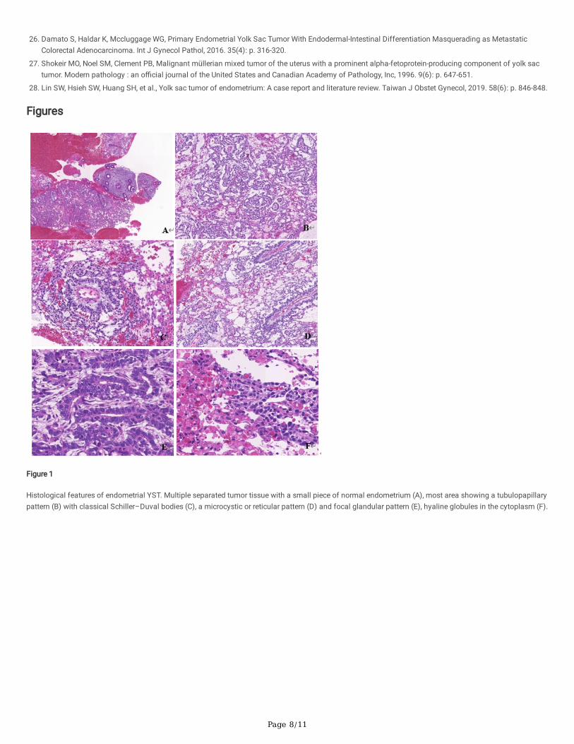
Microscopically, pure endometrial YST without any other type of germ cell tumor or somatic carcinoma components was found (Fig. 1). A reticular patterncoexisted with papillary growth. The reticulum was a labyrinth of channels lined by primitive cells expanding to form microcysts with �attened, clear atypicalepithelial cells. Papillary growth displayed papillary �brovascular structures in which a central blood vessel with tumor cells projects into the surroundingspace (endodermal sinuses, Schiller–Duval bodies (S-D bodies)). Hyaline globules were observed in the cells. The stroma was hypocellular and myxoid.
The specimen was subjected to immunochemical staining on the Ventana Benchmark XT platform. AFP, SALL4, GPC-3 and AE1/AE3 were diffuse positive.PAX8 was focal positive. ER, PR, CD30, OCT4, HNF-1β, Napsin A, and CD117 were all negative expressed (Fig. 2).
ResultsThe description of primary YST of the endometrium in the literature is limited to case reports and small series. After systematically reviewing and screeningthe literature, 19 citations were found. The clinicopathological features, therapies and prognosis of 30 cases (the present case and 29 cases from theliterature) are summarized in Table 1.

Page 4/11
Table 1summary of clinicopathologic features of primary endometrial YSTs.
case Age AFP level Symptoms Tumoursize(cm)
Surgery AssociatedComponent
Metastasissite
Chemotherapy Radiotherapy
1[3 28 380 Metrorrhagia,Pelvic pain
unknow TAH BSO None None VAC NO
2[20] 27 1580 Metrorrhagia 2.4 TAH BSO OMT None None VAC NO
3[21] 24 3600 Abdominal pain 10 SH BSO None ovary VAC YSE
4[2] 49 NA Metrorrhagia 1.3 TAH BSO ILD None None No YES
5[22] 59 25385 Postmenopausal
bleeding
Unknow TAB BSO PLDPALD
EC Liver BEP EP YES
6[10] 65 2306 Waterydischarge
7 MRH BSO PLDPALD
carcinosarcoma LN TP NO
7[6] 42 18530 AVB 6 TAH BSO None None PVB NO
8[18] 30 1762 AVB 6.5 TAH None None BEP NO
9[16] 29 3593.4 AVB 6.7 MRH LSO PLDPALD
None None BEP NO
10[23] 28 1522 AVB 6 TAH BSO PLDOMTappendectomy,partialsigmoidectomy
EC Peritoneumomental
PTX, ADM, DDP,CBDCA, MTX,
Act-D, VP16,BLM,
pingyangmycin,VCR, FUDR,
oxaliplatin, CPA
NO
11[24] 31 242.3 Menorrhagia 4 None None None BEP NO
12[25] 57 31844 Abdominal pain,weight loss
10.5 TAH BSO OMTPLD PALD
None Liver ovarylungvertebrae
BEP NO
13[25] 44 30000 AVB, vaginalmass prolapsing
19 TAH BSO OMTPLD PALD
None None BEP NO
14[26] 63 244.6(6wkpostoperatively)
Postmenopausal
bleeding
12 None None Ovaryomentum
BEP NO
15[4] 71 NA AVB NA YES SC EC NA NA NA
16[4] 55 NA AVB NA Yes Complexhyperplasia
NA Yes YES
17[4] 59 NA AVB, uterinemass
NA NA EC NA YES NO
18[4] 68 NA AVB, uterinemass
NA YES None NA YES NO
19[4] 77 NA AVB, uterinemass
NA NA EC UDC NA NA NA
20[4] 64 NA AVB NA YES AC NA YES YES
21[4] 87 NA AVB NA YES AC NA YES NO
22[4] 61 NA AVB NA YES None NA YES NO
23[4] 63 NA AVB NA YES MMMT NA YES YES
24[4] 62 NA AVB NA YES SC NA YES NO
AVB: abnormal vaginal bleeding; TAH: total abdominal hysterectomy; BSO: bilateral salpingo-oophorectomy; OMT: omentectomy; SH: supracervical hysterectlymphadenectomy; PLD: pelvic lymphadenectomy PALD: paraaortic lymphadenectomy; MRH: modi�ed radical hysterectomy; LSO: left salpingo-oophorectom
EC:endometrioid adenocarcinoma; SC: serous carcinoma; UDC: undifferentiated carcinoma; AC: Adenocarcinoma; MMMT: malignant mixed Mullerian tumor;
VAC: vincristine, actinomycin D and Cyclophosphamide; BEP: bleomycin, etoposide and cisplatin; EP: etoposide and cisplatin; TP: taxol and cisplatin; PVB: blecisplatin; PTX: paclitaxel; ADM: adriamycin; DDP: cisplatin; CBDCA: carboplatin; MTX: methotrexate; Act-D: actinomycin D; VP16:etoposide; BLM: bleomycin; V�uoro-2-deoxy-β-uridine; CPA: cyclophosphamide;
REC: recurrence; DOD: Dead from the disease; NED: No evidence of disease; AWD: Alive with disease; LFT: Lost to follow-up; NA: Not available; AFP: α-fetopro

Page 5/11
case Age AFP level Symptoms Tumoursize(cm)
Surgery AssociatedComponent
Metastasissite
Chemotherapy Radiotherapy
25[4] 77 NA AVB NA YES SC,CCC,UDC NA YES NO
26[27] 64 15918 Abdominaldistension
NA NA None NA NA NA
27[17] 27 >800 AVB 5.4 TAH OMT PLDPALD
None None TC NO
28[12] 38 normal AVB, prolongedmenstruation
NA TAH BSO OMTPLD PALD
CCC None BEP NO
29[28] 68 133.4(6dayspostoperatively)
AVB 3.3 TAH BSO OMTPLD PALD
None NO BEP NO
30present
43 1465 AVB, epigastricpain
4 TAH BSO PLD None NO BEP NO
AVB: abnormal vaginal bleeding; TAH: total abdominal hysterectomy; BSO: bilateral salpingo-oophorectomy; OMT: omentectomy; SH: supracervical hysterectlymphadenectomy; PLD: pelvic lymphadenectomy PALD: paraaortic lymphadenectomy; MRH: modi�ed radical hysterectomy; LSO: left salpingo-oophorectom
EC:endometrioid adenocarcinoma; SC: serous carcinoma; UDC: undifferentiated carcinoma; AC: Adenocarcinoma; MMMT: malignant mixed Mullerian tumor;
VAC: vincristine, actinomycin D and Cyclophosphamide; BEP: bleomycin, etoposide and cisplatin; EP: etoposide and cisplatin; TP: taxol and cisplatin; PVB: blecisplatin; PTX: paclitaxel; ADM: adriamycin; DDP: cisplatin; CBDCA: carboplatin; MTX: methotrexate; Act-D: actinomycin D; VP16:etoposide; BLM: bleomycin; V�uoro-2-deoxy-β-uridine; CPA: cyclophosphamide;
REC: recurrence; DOD: Dead from the disease; NED: No evidence of disease; AWD: Alive with disease; LFT: Lost to follow-up; NA: Not available; AFP: α-fetopro
Among the 30 primary endometrial YSTs, the average patient age was 52 years (range, 24–87 y). The mean tumor size was 6.94 cm (range 1.3–19.0 cm). Themain clinical symptom was abnormal vaginal bleeding. Increasing serum levels of AFP were reported in 18 of 19 patients, whereas normal AFP levels werereported in only 1 patient; the serum level of AFP was not described for the other 11 patients.
Among all 16 of 25 patients who had detailed surgical resection ranges, 3/16 had bilateral adnexal resection. Unilateral and bilateral ovaries were reversed in1 and 2 patients, respectively. The FIGO stages were as follows: I (n = 11), II (n = 5), III (n = 6), and IV (n = 7). In total, 26/30 (87%) patients underwentchemotherapy after the operation. BEP was the most common chemotherapy regimen in 11/26 patients (42%). Only 6 patients (6/30, 20%) who enduredradiotherapy were not sensitive to radiotherapy.
Of all 30 patients, 17 (57%) had pure endometrial YST, and 13 (43%) had a concomitant somatic neoplasm representing < 10–90% of the tumor. The somaticneoplasms followed the histological types endometrial adenocarcinoma (n = 4), carcinosarcoma (n = 2), clear cell carcinoma (n = 1), adenocarcinoma (n = 1),serous carcinoma (n = 1), serous carcinoma and endometrial adenocarcinoma (n = 1), serous carcinoma and clear cell carcinoma (1), and endometrialcomplex hyperplasia (n = 1). Patients with pure YST were younger than those with a concomitant somatic tumor (ranging from 24–68 years, mean 44.5 yearsvs. range 28–87 years, mean 61.92 years, P = 0.008).
Follow-up information was available in 90% (27/30) of patients, for which the mean follow-up time was 17.25 months (range 2–72 months), 48.1% (13/27) ofpatients had no evidence of disease during the follow-up time (the longest was more than 72 months), 8 patients died of disease (range 2.5–24 months), and6 patients were alive with disease (range 7–30 months). The overall survival (OS) and disease-free survival (DFS) rates were 70.4% (19/27) and48.1% (13/27),respectively.
DiscussionThe histogenesis of extragonadal YST remains speculative and controversial. There are four potential mechanisms by which a germ cell neoplasm can arisein the endometrium[6]. The �rst is the aberrant migration of primordial germ cells in a lateral direction during embryogenesis, which can remain in the basallayer of the endometrium for many years. The second potential mechanism is metastasis from occult ovarian YST. Residual fetal tissues remaining in theuterus because of an incomplete abortion and somatic cells that have undergone aberrant differentiation by which YST originates in unusual sites are theother two potential mechanisms.
We summarized 30 primary endometrial YSTs: the present case and 29 cases from the literature. Patients with pure YSTs were younger than those with aconcomitant somatic tumor. Therefore, pure endometrial YST and endometrial YST with somatic tumors may have had different histogeneses. Pureendometrial YSTs may originate from pluripotent germ cells, while endometrial YSTs with somatic tumors may arise from malignant pluripotent somatic stemcells or possibly via “retrodifferentiation”, by which a differentiated cell transforms into a more primitive form[7]. Reports of ovarian YST arising from anendometrioid carcinoma support this hypothesis[8–10]. YSTs of the female genital tract in older adults are commonly derived from somatic epithelialneoplasms[11].
AFP, a special tumor marker elevated in the vast majority of patients with tumors containing a YST component, is essential for the diagnosis and monitoringof progressiveness. In the overwhelming most endometrial YSTs, AFP is used as a signi�cant follow-up indicator, but only a few patients had normal serumAFP levels [12].
Page 6/11
Since primary endometrial YST is rare, pathologists who lack experience and are not familiar with the morphology of YST may make an incorrect diagnosis,especially in biopsy specimens. Moreover, its immunohistochemical pro�le overlaps between that of YST and carcinoma. AE1/AE3 is positive in bothcarcinoma and YST, as observed in the current case, and is not a good marker for differential diagnosis[13]. Both HNF-1β and PAX8 can be positive in YST andthus result in an incorrect diagnosis of clear cell carcinoma[14]. However, both marker commonly are patchy positive in YSTs rather than diffuse positive inclear cell carcinoma. SALL4, as a sensitive marker for germ cell tumors, is a useful marker for diagnosis when combined with GPC-3 and AFP[15]. Hence, apanel of markers is necessary for the diagnosis of YST at rare sites.
Given the rarity of primary endometrial YSTs, there is no consensus on the treatment of this extremely rare tumor. Surgery combined with adjuvantchemotherapy is the main treatment. However, the speci�c resection range remains controversial, and whether ovaries are preserved still needs further study.Among the 30 patients described herein, unilateral ovary reservation was reported in only one patient[16], and bilateral ovary reservation was reported in 2patients[17],[18]. Rossi described a 30-year-old woman with stage II primary endometrial YST treated with simple total hysterectomy[18], preserving thebilateral ovary. The patient remained free of disease for more than 6 years after completion of the therapy. Tao reported a similar case, a 27-year-old womanwith stage IA disease[17] who remained free of disease for 14 months after surgery. Wang described a 29-year-old woman with stage II disease who preservedher right adnexa to maintain endocrine function[16] and was alive without recurrence for 39 months. This fact revealed the possibility of treating a youngpatient with early-stage primary YST of the endometrium with surgery to conserve the ovary. The surgeons effectively retained the patient’s ovarian endocrinefunction and improved her quality of life.
For germ cell tumors, including ovarian YST, the BEP regimen results in high cure rates, even for advanced-stage tumors[19]. However, the outcome for EGYST,especially in endometrial tissue, is less certain. In the literature series, 5/16 (31.3%) patients with pure YST died of disease (range 2 to 24 mo; median, 8 mo),in which 4 of 5 patients were FIGO stage IV, 1 received a regimen of vincristine, vinblastine and cyclophosphamide, 1 (6.2%) was alive with disease (follow-up,8 mo), and 10/16 (62.5%) had no evidence of disease (range 6 to 72 mo; median follow-up 19 mo). Of the patients in whom an associated somatic tumor wasreported, 3/10 (30%) died of their disease, 5/10 (50%) were alive with disease, and only 2/10 (20%) had no evidence of disease. There are too few cases todetermine whether the poor prognosis is related to a somatic origin or to the fact that 6/8 (75%) of those who died of disease had FIGO stage III or IV atpresentation. These �ndings suggest that tumor staging and somatic components are 2 important factors that in�uence the prognosis of patients withendometrial YST, and patients with YST of somatic origin may have a poor prognosis.
ConclusionPrimary YST of the endometrium, a highly malignant germ cell tumor, is extremely rare. Surgery combined with postoperative chemotherapy is consideredeffective for the treatment of primary endometrial YST. YST combined with somatic neoplasms may have a worse prognosis. However, additional cases needto be reported to further understand these rare tumors.
AbbreviationsYST: yolk sac tumor; EGYST: experience extragonadal YST; CT:computerized tomography ; BEP:bleomycin, etoposide, and cisplatin; S-D bodies:Schiller–Duvalbodies; AVB: abnormal vaginal bleeding; TAH: total abdominal hysterectomy; BSO: bilateral salpingo-oophorectomy; OMT: omentectomy; SH: supracervicalhysterectomy; ILD: iliac lymphadenectomy; PLD: pelvic lymphadenectomy PALD: paraaortic lymphadenectomy; MRH: modi�ed radical hysterectomy; LSO: leftsalpingo-oophorectomy;EC:endometrioid adenocarcinoma; SC: serous carcinoma; UDC: undifferentiated carcinoma; AC: Adenocarcinoma; MMMT: malignantmixed Mullerian tumor; CCC: clear cell carcinoma;VAC: vincristine, actinomycin D and Cyclophosphamide; BEP: bleomycin, etoposide and cisplatin; EP:etoposide and cisplatin; TP: taxol and cisplatin; PVB: bleomycin, vincristine and cisplatin; PTX: paclitaxel; ADM: adriamycin; DDP: cisplatin; CBDCA:carboplatin; MTX: methotrexate; Act-D: actinomycin D; VP16:etoposide; BLM: bleomycin; VCR: vincristine; FUDR:5-�uoro-2-deoxy-β-uridine; CPA:cyclophosphamide; REC: recurrence; DOD: Dead from the disease; NED: No evidence of disease; AWD: Alive with disease; LFT: Lost to follow-up; NA: Notavailable; AFP: α-fetoprotein.
DeclarationsAcknowledgements
We gratefully acknowledge technical assistance from members of the Department of Pathology, shanghai cancer center
Authors’ contributions
Rui Bi provided the case and critically revised the manuscript.Huijuan Ge was responsible for the acquisition and interpretation of patient data and manuscriptpreparation. Bi and Ge performed pathological examination. All authors approved the �nal manuscript.
Funding
This research was funded by National Natural Foundation Science of China, grant number NSFC 81802597”
Availability of data and materials
Scan slides of the case can be provided if required.
Ethics approval and consent to participate

Page 7/11
All procedures followed were in accordance with the ethical standards of the Iwate Medical University and with the Helsinki Declaration. Substitute forinformed consent (approval of the institutional review board of Iwate Medical University) was obtained from all patients for being included in the study, asbelow: This study was approved by the institutional review board Facility Ethical Committee (Fudan University Cancer Center Ethics Committee, China;approval No. 050432-4-121B, 13 December 2012).
Consent for publication
Not applicable.
Competing interests
The authors declare that they have no competing interests.
References1. Ulbright Tm, Germ cell tumors of the gonads: a selective review emphasizing problems in differential diagnosis, newly appreciated, and controversial
issues. Modern pathology : an o�cial journal of the United States and Canadian Academy of Pathology, Inc, 2005. null(unde�ned): p. S61-79.
2. Spatz A, Bouron D, Pautier P, et al.Primary yolk sac tumor of the endometrium_ a case report and review of the literature. Gynecol Oncol, 1998. 31(2): p.285-288.
3. Pileri S, Martinelli G, Serra L, Endodermal sinus tumor arising in the endometrium. Obstet Gynecol, 1980. 56(3): p. 391-396.
4. Ravishankar S, Malpica A, Ramalingam P,et al, Yolk Sac Tumor in Extragonadal Pelvic Sites_ Still a Diagnostic Challenge. Am J Surg Pathol, 2017. 41(1):p. 1-11.
5. Mutch DG and Prat J, 2014 FIGO staging for ovarian, fallopian tube and peritoneal cancer. Gynecologic oncology, 2014. 133(3): p. 401-404.
�. Joseph MG, Fellows FG, Hearn SA, Primary endodermal sinus tumor of the endometrium. A clinicopathologic, immunocytochemical, and ultrastructuralstudy. cancer, 1990. 65(2): p. 297-302.
7. Nogales FF, Preda O, Nicolae A, Yolk sac tumours revisited. A review of their many faces and names. Histopathology, 2012. 60(7): p. 1023-1033.
�. Rutgers JL, Young RH, Scully RE, Ovarian yolk sac tumor arising from an endometrioid carcinoma. Human pathology, 1987. 18(12): p. 1296-1299.
9. 9.Nogales FF, Bergeron C, Carvia RE, et al., Ovarian endometrioid tumors with yolk sac tumor component, an unusual form of ovarian neoplasm. Analysisof six cases. The American journal of surgical pathology, 1996. 20(9): p. 1056-1066.
10. Oguri H, Sumitomo R, Maeda N, et al., Primary yolk sac tumor concomitant with carcinosarcoma originating from the endometrium: case report. GynecolOncol, 2006. 103(1): p. 368-371.
11. Mcnamee T, Damato S, Mccluggage WG, Yolk sac tumours of the female genital tract in older adults derive commonly from somatic epithelial neoplasms:somatically derived yolk sac tumours. Histopathology, 2016. 69(5): p. 739-751.
12. Song L,Wei XX,Wang DQ, Primary yolk sac tumor originating from the endometrium- A case report and literature review. Medicin, 2019. 98(15).
13. Chia-Sui K,MuhammadT,IdreesY et al, Solid pattern yolk sac tumor: a morphologic and immunohistochemical study of 52 cases. The American journal ofsurgical pathology, 2012. 36(3): p. 360-367.
14. Rougemont AL, Tille JC, Role of HNF1beta in the differential diagnosis from other germ cell tumors. Human pathology, 2018.
15. Cao D, Humphrey PA, Allan RW, SALL4 is a novel sensitive and speci�c marker of ovarian primitive germ cell tumors and is particularly useful indistinguishing yolk sac tumor from clear cell carcinoma. The American journal of surgical pathology, 2009. 33(6): p. 894-904.
1�. Wang C, Li G. Xi L, et al., Primary yolk sac tumor of the endometrium. Int J Gynaecol Obstet, 2011. 114(3): p. 291-293.
17. Lu T, Qi L,Ma Y, et al., Primary yolk sac tumor of the endometrium: a case report and review of the literatures. Archives of gynecology and obstetrics, 2019.300(5): p. 1177-1187.
1�. Rossi R, Stacchiotti D, Bernardini MG, et al., Primary yolk sac tumor of the endometrium: a case report and review of the literature. Am J Obstet Gynecol,2011. 204(4): p. e3-4.
19. Gershenson DM, Management of ovarian germ cell tumors. Journal of clinical oncology: o�cial journal of the American Society of Clinical Oncology,2007. 25(20): p. 2938-2943.
20. Ohta M, Sakakibara K, Mizuno K,et al, Successful treatment of primary endodermal sinus tumor of the endometrium. Gynecol Oncol, 1988. 31(2): p. 357-364.
21. Magers MJ, Kao CS, Cole CD, et al, "Somatic-type" malignancies arising from testicular germ cell tumors: a clinicopathologic study of 124 cases withemphasis on glandular tumors supporting frequent yolk sac tumor origin. The American journal of surgical pathology, 2014. 38(10): p. 1396-1409.
22. Patsner B, Primary endodermal sinus tumor of the endometrium presenting as "recurrent" endometrial adenocarcinoma. Gynecol Oncol, 2001. 80(1): p. 93-95.
23. Ji M, Lu Y, Guo L, et al., Endometrial carcinoma with yolk sac tumor-like differentiation and elevated serum beta-hCG: a case report and literature review.Onco Targets Ther, 2013. 6: p. 1515-1522.
24. Abhilasha N, Bafna UD, Pallavi VR, et al., Primary yolk sac tumor of the endometrium: A rare entity. Indian J Cancer, 2014. 51(4): p. 446.
25. Qzler A, Dogan S, Mamedbeyli G, et al, Primary yolk sac tumor of endometrium: report of two cases and review of literature. J Exp Ther Oncol, 2015. 11(1):p. 5-9.
Page 8/11
2�. Damato S, Haldar K, Mccluggage WG, Primary Endometrial Yolk Sac Tumor With Endodermal-Intestinal Differentiation Masquerading as MetastaticColorectal Adenocarcinoma. Int J Gynecol Pathol, 2016. 35(4): p. 316-320.
27. Shokeir MO, Noel SM, Clement PB, Malignant müllerian mixed tumor of the uterus with a prominent alpha-fetoprotein-producing component of yolk sactumor. Modern pathology : an o�cial journal of the United States and Canadian Academy of Pathology, Inc, 1996. 9(6): p. 647-651.
2�. Lin SW, Hsieh SW, Huang SH, et al., Yolk sac tumor of endometrium: A case report and literature review. Taiwan J Obstet Gynecol, 2019. 58(6): p. 846-848.
Figures
Figure 1
Histological features of endometrial YST. Multiple separated tumor tissue with a small piece of normal endometrium (A), most area showing a tubulopapillarypattern (B) with classical Schiller–Duval bodies (C), a microcystic or reticular pattern (D) and focal glandular pattern (E), hyaline globules in the cytoplasm (F).

Page 9/11
Figure 1
Histological features of endometrial YST. Multiple separated tumor tissue with a small piece of normal endometrium (A), most area showing a tubulopapillarypattern (B) with classical Schiller–Duval bodies (C), a microcystic or reticular pattern (D) and focal glandular pattern (E), hyaline globules in the cytoplasm (F).

Page 10/11
Figure 2
Immunocytochemical pro�le of endometrial YST. Diffuse positive for AFP (A), GPC-3 (B), SALL-4 (C) and AE1/AE3 (D). Focal positive for PAX8 (E) andnegative for EMA (F).

Page 11/11
Figure 2
Immunocytochemical pro�le of endometrial YST. Diffuse positive for AFP (A), GPC-3 (B), SALL-4 (C) and AE1/AE3 (D). Focal positive for PAX8 (E) andnegative for EMA (F).
Supplementary Files
This is a list of supplementary �les associated with this preprint. Click to download.
checklist.pdf
checklist.pdf