Embed Size (px)
Citation preview

1vyREVIEW OF OMPHALOCELES (EXOMPHALOS)
KENYATTA NATIONAL HOSPITAL 1976-1985
MAJOR (DR) JOSEPH MUNENE MBUI NJOKAH M.B.Ch.B. (Makerere)
Member of Under Sea Medical Society (USA)
A THESIS SUBMITTED IN PART FULFILMENT FOR THE DEGREE OF MASTER OF MEDICINE (SURGERY) IN THE UNIVERSITY OF NAIROBI
IN
1985
j" w£»s,t y

DECLARATIONS:
(a) ’THIS THESIS IS MY ORIGINAL
WORK AND HAS NOT BEEN PRESENTED FOR
A DEGREE IN ANY OTHER UNIVERSITY’.
Major (Dr) J.H.NJOKAH
THIS THESIS HAS BEEN SUBMITTED
FOR EXAMINATION WITH MY APPROVAL
AS UNIVERSITY* SUPERVISOR.
PROF. J.M,KYAMBI

3
CONTENT. Pages
Chapter 1. 1 Summary.................... 4-5
2. Review of Literature...... 6-8
3. Aim.of the study.......... 9-10
4. Materials and Methods..... 11-12
Chapter 2. 0. Results:
1. Tables 1 - 10.............. 13-22
2. Explanation of results.... 23-27
Chapter 3. 0 Discussion
1. Defination................. 28-29
»• 2. Embryology of omphalocele.. 30-31
3. Pathogenesis of omphalocele. 32-33
4. Incidence of omphalocele....34
5. Classification of omphalocele 35-36
6. Natural History of omphaloceleand non-surgical treatment.... 37-41
7. Surgical management ofomphalocele............... 42-52
Chapter 4 4. 1. Conclusion ................... 53-54
2. Recommendations ............. 55-57
Chapter 5. 1. Annexes : ..................... 58-59
2. Acknowledgement.............. 60
3. Photographs.................. 61
4 . References .62 -68

4
SUMMARY:
A retrospective study of the omphlocele has been done.
The study was carried out at Kenyatta National Hospital which
is the referral hospital for the Republic of Kenya and the
teaching hospital for the Faculty of Medicine of the
University of Nairobi.
This study covers a ten year period from 19?6 - 1%.
A total of 3*+ cases have been studied. The yearly incidence
has been static notwithstanding our high population growth.
Among the 3^ cases there were l8 males and l6
females. Of the 3*+ cases there were 19 survivals and 15 deaths
12 deaths (80%) among the 15 followed early surgery.
Only 3 patients had surgery among 19 survivals. The major
cause of death (6 out of 1 2) among the surgical cases was
respiratory complications. Surgery had been performed within
1 -3 days of birth.
Surgical management consisted of excision and repair
of the omphalocele. Non-surgical regime entailed paintingi ‘
the omphalocele with mercurochrome (1 patient) or simply
daily clen6ing with antiseptics followed by sofratule%
dressing (18 patients).

5
Complication of the omphaloceles occured in k cases
only (Table 4). Malformations co-existing with omphalocele
occured in 12 cases of which 7 died. In our series non-surgical
regime produced more survival (8^,2%) 16 out of 19
cases.

6
REVIEW OF LITERATURE:
Literature on omphalocele has been reviewed with a
view to bring out views as regards embryology, Pathogenesis
incidence and various regimes of management. It is also the
aim of the review to evaluate various Authors* results on
various regimes of management and see how they compare
with our results.
Earlier review of the literature was done by Jarcho
(15) in 1937 which goes back to 163*+. Prior to 1937,
omphalocele as an entity has not appeared in literature.
Jarcho (15) described omphalocele and discussed presentation,
incidence, embryology, pathogenesis and modes of treatment
bo oh surgical and non surgical at the time.
Discussion of the embryology and pathogenesis has been
done by de Vries (13) Ackerman (3*0 ♦ Williams (32), Soper
(17), Holcomb (28) and Lamura (37)* On treatment Grob (30)
Soave (29) Ochola and Kyambi (22) describe non-surgical management
while Gross (16) Eckstein (18), Moazam (23) and Schuster
(2*+) discuss the various surgical techniques available forf
management of omphalocele. Othersen (27) discusses a new
approach to surgical management where a pneumatic device
is used to reduce the omphalocele followed by surgical repair.

7
Stringel (21) and Eckstein (18) discuss factors which
influence the management of omphalocele both surgical and
non surgical.
V<elsebey (2 5) discusses a device for measuring the
intra-abdominal pressure during the repair of the
omphalocele.
King (19) and Touloukian (20) discuss the investigations
which can be done to make intra-uterine diagnosis of
omphalocele.
Total parental Nutrition in the omphalocele management,
es ecially where gut function has been compromised by early
rupture and subsequent infection is the subject of the paper
by Filber (26).
Incidence of the omphalocele has been discussed by
Jarcho (15)» Beiley and Love (1), Soper (17) and Ackerman
(3*0 • The incidence varies from author to author-ranging
from 1:6,600 births in Jarcho (15) and 1; 1,860 births in
Ackerman (3*0*
Most authors prefer early surgery. Early surgery in
our series resulted in very high death rate. 12 deaths
out of a total of 15 deaths had early surgery.

8
In contrast to world literature non-surgical
management of omphalocele ha6 been our regime of .cft'oice,»• ■*
Only those cases which must be subjected to surgery are
managed surgically. This regime of non-surgical management
has produced very encouraging results, there were only 3
deaths among the 19 patients on this regime.

9
AIM OF THE STUDY:
The aim of this study is to review the management
of omphalocele in Kenyatta National Hospital during the
period of 1976 - 1985* It is also the aim of the study to
try and establish facts about
1, Presentation and incidence of the omphalocele in
K.N.H.
2. The results of both surgical and non-surgical regimes
o of management,
3* Criterion for advocating one form of management in
preference to the other.
When all these factors have been studied, a
clear policy on management of the omphalocele should be
made taking into account the following:-
(i) Location of Paediatric Surgery Service in Kenyatta
National Hospital.
(ii) Transportation of patients from the periphery into
K.N.H.
(iii) The supportive care of the patients postoperatively
with special reference to the intensive care unit.
(iv) Materials available in this country for the
managements of omphalocele.

10
It is felt that when all these aims are satisfied
recommendation should be able to be made to enhance
survival of babies with imphaloceles in this country.

11
1.3 MATERIAL AND METHODS:
This is a retrospective study covering a 10 years
period from 1976 to 1985*
MATERIALS:
Material for the study came from study of case records
held at Kenyatta National Hospital records department.
Retrieval of the case notes has not been as uneasy matter
because before 1978 the paediatric surgical unit had not
been established. All the omphaloceles were operated on by
any surgeon on duty at the time of admission. The other
problem was the classification of the omphalocele for records
keeping purposes. Omphalocele has been moved from
classification to classification even by the international
classification index books held by our records department. It
has been classified as umbilical hernia, as congenital umbilical
hernia, gastroschisis, umbilical defect and as either omphalocele
or exomphalos.
The most helpful retrieval of the case notes has been
going through theatre records books, Theatre recovery room
record notes, ward admission record books and by trial and
error by various classification for notes retrieval held by
the records department.

_ 12
METHODS:
After the case notes were found, data was extracted
as per data collecting form Annex A. Such data as name,
race, location in Kenya (District) inpatient number, sex,
birth weight, Birth defects both complications and malformation!
were extracted. Data partaining to treatments given
before surgery, age at surgery, surgical treatment done and
the results were also extracted.
The data was then analysed and put into tables as
laid out in the chapter on results. All the case notes
were read thoroughly and all the factors contributing to
death or the survival of the case recorded.
A total of patients recorda were retrieved and
analysed
13
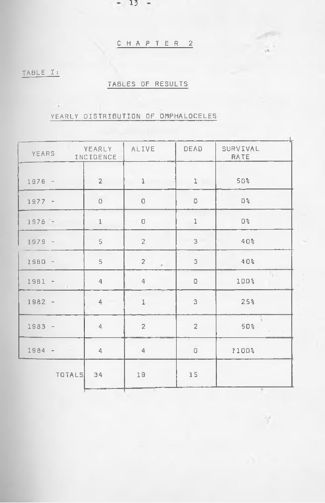
C H A P T E R 2
TABLE I:TABLES OF RESULTS
YEARLY DISTRIBUTION OF OMPHALOCELES
YEARS YEARLY INCIDENCEALIVE DEAD
----------------------------------------- ■ -
SURVIVALRATE
1976 - 2 1 1 50%
1977 - 0 0 0 0%
1978 1 0 1 0%
1979 - 5 2 3 4 0%
1980 - 5 2 3 4 0%
1981 - 4 4 0 100%
1982 - 4 1 3 25%
1983 - 4 2 2V
50%
1984 - 4 4 0 ?100%
TOTALS 34 19 15

TABLE 2:
SEX DISTRIBUTION
MALES 18
FEMALES 16
TOTAL 34

15
TABLE 3: ETHNIC (TRIBAL) DISTRIBUTION
NUMBER OF CASES
%OF TOTAL (347 )
ADMIK.N.HQ
K I KU YU 18 52.94 3
KANBA 6 17.65 1
LUHYA 5 14.71
LUO 1 2.94 11EMBU 1 2.94
MFRU 1 2.94I
SOMALI 1 2.941?
KALENJIN 1 2.94 1
TOTAL 34 100%
9
2

TABLE 3: ETHNIC (TRIBAL) DISTRIBUTION
NUMBER OF CASES
%OF TOTAL (347 )
ADMISSIONS K.N.HOSPITAL %
KI K U Y U 18 52.94 3 3.9 \
KANBA 6 17.65 19.8
LUHYA 5 14.71 9.2
LUO 1 2.94 17.2
EMBU 1 2.94
MFRU 1 2.94
SOMALI 1 2.9419.9
KALENJIN 1 2.94
TOTAL 34 100% 100%

16
TABLE 4:
COMPLICATIONS
No. Of Cases
Intestinal Obstruction 1
Gangrenous gut 1
Intussusception 1
Ruptured Sac 1
Total 4

- 1? -
CO-EXCISTENT MALFORMATIONS IN INDIVIDUAL CASES WITH MALFORMATIONS
TABLE 5 :
Tetralogy of Fallot
Stenosis Ascending Colon
Beckwith Wideman Syndrome
Entero-Vita 11 ine Fistula and Intussusception
Ano-Rectal Malformation and Epispadias
Ano-Rectal Malformation
Ectopia Vesicae and Gangrenousgut
Cleft Palate
Ano Rectal Malformation and Ectopia Vesicae
Downes S.yndrome
TOTAL
No. Of Patients
- 1?
CD-EXCISTENT MALFORMATIONS IN INDIVIDUAL CASES WITH MALFORMATIONS
TABLE 5 :
iNo. Of Patients
Tetralogy of Falloti
1
Stenosis Ascending Colon 1
B>eckwith Wideman Syndrome 1
Entero-Vitalline Fistulaand Intussusception 1
Ano-Rectal Malformation andEpispadias „ 2
Ano-Rectal Malformation 1
Ectopia Vesicae and Gangrenousgut 1
Cleft Palate 1
Ano Rectal Malformation andEti topia Vesicae 1
Down's S.yndrome 1
TOTAL 11

18
TABLE 6 :
INDIVIDUAL MALFORMATIONS
NUMBER OF CASES
% OF TOTAL CASES (34)
Ano-Rectal Malformations 3 Q.8%
Efc topia Vesicae 2 5.9%
Down's Syndrome 1 2.94%
Entero Vitelline Fistula 1 2.94%
Beckwith Wideman syndrome 1 2.94%
Cleft Palate 1 2.94%
Fallot's Tetralogy 1 2.94%
Stenosis of Ascending colon 1 2.94%
TOTAL 11

19 -
DEATHS
(A) MANAGEMENT REGIMES:
TABLE 7:
NUMBER OF CASES
Surgical 12
Non Surgical 3
Total 15
(B) ASSOCIATED MALFORMATIONS; -------- . ---- - - — - ■ • --- -■■■
NUMBER OF CASES
With Malformation 8
Without Malformatior 7
Total 15

20
CAUSES OF DEATH
(A) SURGICAL CASES
TABLE 8:
NUMBER OF CASES
r e s p i r a t o r y 6
CARDIAC 4
BURST ABDOMEN 1
FAILURE TO THRIVE 1
TOTAL 12
r----------------------------
(B) NON SURGICAL CASES
NUMBER OF CASES
CARDIAC FAILURE.••••.• \ i. ... i. . . . 3
TOTAL 3--------------------- -------- -

21
(A) LIVE CASES
TABLE 9:
NUMBER OF CASES
SURGICAL 3
NON SURGICAL 16
TOTAL19
CB) LIVE CASES WITH ASSOCIATED MALFORMATIONS
.!-- !
NUMBER OF CASES
WITH m a l f o r m a t i o n s 5
- WITHOUT MALFORMATIONS 14-
TOTAL 19

22
TABLE 10
METHODS OF TREATMENT
SURGERY 15 '
NON SURGICAL 19
TOTAL 34

- 23 -
RESULTS:
The results of the 3^ cases studied are analysed
graphically in the tables 1 - 10, The following can be
inferred frpm these results.r
1. Presenting Complaints:
The presenting complaints are from the parents. The
infant is normally very comfortable. The parents
complain about the hernia at the umbilical region at
birth. The neonate may also have other complaints.
With improved diagnostic methods (19) and (20),
the diagnosis: should be made before birth.
2* Family History:
History of omphalocele appearing as a familial disease
nas not been elicited. v This is in keeping with
world literature.
3. Past Medical History:
Except in the four cases seen in 1985 no medical
history pertaining to omphalocele had been recorded.
In the k cases no history of previous malformations
in the family was recorded. No history of hydramnio3
was recorded. History of drugs, radiation and food
eaten would be interesting but was not recorded either.:
Incidence: (Table I)
Inspite of fast population growth in Kenya the
yearly incidence of omphalocele appears to be constant.
1985 shows an early start with k cases in the 1st quarter.
- 2k -
5 * Ethnic distribution - (Table 3)
The results are a reflection of the normal
hospital admissions except that Luos though being
one of the major population groups in the hospital (17.2#)
have very few case, 1 in those 10 years. A better
method of displaying this data would be by locality
of origin. This would be important when considering
teratogens both industrial and resident to a place.
£'• Sex “ (Table 2)
Both sexes are represented equally
male:female, l8:l6.
7 • Complications encountered (Table 4 )
Only cases had complications of their
omphalocele. Complication is that medical
problem superimposed on the omphalocele#
The complications’were
1. Intussusception -1
2. Gangrenous gut -1
3# Intestinal obstruction-1
*f. Ruptured Sac -1
There was only 1 death (33%)• The patient is
one with the gangrenous gut discovered at operation
of the omphalocele# The neonate had to have
excision of the sac, resection and anastomosis
of the gut and primary repair of the abdomen. Thi6
patient also had ectopia vesicae. He died 3 days

- 25 -
poet operatively. The other three survived
The second patient developed obstruction in the
ward on the 3rd day of non surgical management.
At operation viable gut was found and reduced.
The omphalocele was excised and repaired primarily.
The patient was discharged on the 13th postoperative day.
The third patient with intussusception
developed it on the l8th day while on conservative
treatment. This was reduced and the omphalocele
was excised and repaired intone stage. 'A concomitant ’
enterovitelline fistula, discovered at operation,
was also excised. The neonate was discharged on the
2*+th post operative day.
8. Congenital malformation encountered Tables 6,7»9
The commonest malformations were found in the
ihtestines. Out of the 31 individual malformation only
3 were not from the gastrointestinal tract. These were
the Tetrology of Fallot one case, Cleft palate one
case and one case of Down's Syndrome,
Table 3 shows that there were 11 patients with
malformation and in k cases there were more than
one malformation at the same time in the same patient.

- 26 -
9 * Effect of Congenital Malformation on the outcome
of treatment:
Table 7* 8, 9
Out of the J>k cases there were 15 deaths and
19 survivals. 7 of the patients who died and 5 of the
living ones had other congenital malformations. It is
observed that among those who died,of the 12 who died after
surgery, 5 had malformations. Apparently malformations
increased mortality among the surgical patients.
Xo • Treatment (Table 7» 8, 9, 10).
Out of the 3k patients 19 had non-surgical regime of1 ■%treatment producing 16 survivals. The three deaths died suddenly
of cardiac arrest. Of the 15 who had surgery, 12 died, there
were only 5 survivals. It appears the early surgery is
associated with higher mortality. Among the 12 dead, k had
intact omphalocele membranes with no complications. These
sacs were excised and the defect covered with dura graft. These
grafts got infected causing severe peritonitis. One of the
patients had 5 burst abdomen, in weekly intervals. This
frustrated the mother so much that she tore the patients
notes before deserting the patient who eventually died.

- 27 -
The major causes of death was cardio pulmonary in.
10 patients. Comparing the two regimes of management non-
surgical regime produced higher rate of survivals.
11. Type of Surgery:
This was normally excision of the omphalocele sac and
closure. In k cases this wa6 difficult and dura graft had to
be used to cover the defect. The four died postoperatively.
12. Type of Non-surgical Treatment:
The neonate was put in an incubator for warmth and
the sac painted with mercurochrome in one case while the other.
l8 had simple dressing with sofratuUe which is a light weight
dressing containing Framycetin sulphate 1%. This is changed
daily and the sac cleaned with antiseptic solution like Eusol.
Only 3 patients died under this regime. 1st case was under
2500 gms and had failure to thrive. This would have
benefitted from Total parenteral Nutrition if it was available
here. The 2nd patient died suddenly of cardiac causes. In
addition the patient had Ectopia vesicae and epispadias, rj»he
3rd patient had Down*s Syndrome and also died suddenly. The
inference we can draw from the various regimes of management is
that non-surgical treatment in our series yields better
results

- 28 -
CHAPTER 3
DISCUSSION:
3.1 DEFINATION: The name omphalocele according to
"Dorland's illustrated medical dictionary" (6)
is a synthesis from two Greek words "omphalus" Greek
for umbilicus and "Kele" = Greek for 'hernia'. Hence
omphalocele means "Umbilical hernia".
A true umbilical hernia contains abdominal
visceral herniating through a large-Umbilical ring.
This hernia is covered by skin and peritoneum.
Omphalocele on the other hand is still herniation
„ of ubdominal viscera through a large umbilical ring.
The herniating viscera is covered by peritoneum, parti,
of the original intra embryonic coelomic Epithelium
(de Vries (13) ) and a membrane derived from the
distended cord. This membrane consists of Amniotic
membrane externally (13) and Wharton's Jelly in between.
The whole sac is a clear membrane which allows visibility
of the viscera enclosed within. Cruising on it are
found umbilical vein cranially and two umbilical arteries
caudally.
According to Fasana (5), Gross (l6), William (32),
Boyd (33) and de Vries (13) the membrane contains in
the Wharton Jelly, lots of fibroblasts and a mesh of
collagen fibres. The interstasis are filled with
mucopolysaccharides ground substance.

- 29 -
This composition is very important when considering
non-surgical regime of management of the omphalocele as
when denatured by drying, the collagen fibres Shrink
(Gross 16) reducing the content of the omphalocele into the
adominal cavity at the same time giving a gentle traction to
the abdominal wall to encourage growth. Grob (30) Joy (31).
So omphalocele is essentially a herniation of abdominal
viscera into the umbilical cord. de Vries (13), Fasana (5)
Jarcho (15)» Sober (17) and Gross (16) do not agree this is
a hernia. They describe it us an arrest of embryonic stage
of development when the gut developed outside the abdomen -
an eventration - as this gut has never been inside the abdominal
cavity and then herniated subsequently nor does it reduce and then
herniate like most hernias do before they become irreducable
due to*obstruction. This is a more acceptable explanation
considering the embryology of the omphalocele which is the
subject of the next few pages.

\
Most of the embryologists (Fasana (5) de Vries (13)»
Jarcho (15)» Gross (16) and Soper (1?) agreed that
omphalocele (exomphalos) is an arrested embryonic developmental
stage. Though resembling a hernia omphalocele content has
never been inside the abdominal cavity and then later herniated
out. They further agreed that between k - 12 weeks of
intrauterine life mid gut of the embryo grows very fast. The
growth is faster than the elongation of the embryo
(Fasana (5) Soper (17). This is also the stage of fast
growth of the liver which pushes the gut out of the abdomen.
This is made worse by the curviture of the embryo ventrally.
The mid and the hind gut then herniate into the umbilical
cord base forming an umbilical cord sac where the guts
continues to grow. At about 12th week the embryo when about
cmm, the gut returns into the abdominal cavity. This
is encouraged by the rapid growth of the caudal part of the
embryo creating a large abdominal cavity in the pelvis
and lower abdomen. The embryo also tends to straighten
at this stage. The return of the gut is associated with
270° rotation of the small gut anticlockwise. Failure
of the gut to return is normally associated with
malformation of the gut and persistence of the mesocolon
due to failure of fusion of the dorsal mesocolon to the
posterior abdominal wall r. v peritoneum producing
attachment of the colon to the posterior abdominal wall.
- 30 -
3.2 EMBRYOLOGY OF OMPHALOCELE:

- 3 1 -
Whe.n' this process of reduction of the gut from the
umbilical cord sac. to the abdominal cavity fails then
an omphalocele is formed from about 12 weeks of intrauterine
life.
It is this finding of gut in a sac outside the
abdomen through a large umbilical ring which made some
authors call this an umbilical hernia,
When the membrane gets ruptured and lost in the
intra uterine life the condition is mistaken with
another condition called Gastroschisis. In this
congenital condition the gut herniates out of the
abdominal cavity through a hole, not covered by a membrane,
mostly at the left side of the umbilical ring leaving
the umbilical cord attached at its normal position.
When the omphalocele membrane is ruptured during the
intrauterine life, umbilical vessels must be preserved
or else the foetus will die.
These vessels will be attached to the umbilical ring
which is a distinct entity. It consists of a
fibrous ring covered internally by peritoneum and
externally by embryonic ectoderm, towards the abdominal
wall, and Amniotic ectoderm towards the umbilical cord
(Boyd J, 33)* There is an area of transitional epithelium
between the two.

- 32 -
There is a general concensus that omphalocele is due
to failure of the gut to return to the abdominal
cavity after 12 weeks of intrauterine life or
when the embryo is about kb mm#
The area of controversy is how this happens#
Some authors (Fasana (5) and Gross (16) Soper (17)
agree that it is not known how this happens# However
they agree it could be associated withs-
Failure of abdominal cavity to enlarge or
- May be due to failure of the abdominal wall
to grow#
There are authors who offer other explanations. As
quoted by Gross (16) Alfield (1899) suggested that
it i6 the persistence of the omphalomesenteric duct
tithered to the umbilical cord sac which prevents
the gut from reduction# This exclusion of the gut from
the abdominal cavity may cause failure of the abdominal
wall to grow due to lack of stimulus as the gut stays♦out of it, a form of disuse atrophy# This reasoning
was based on the fact that Meckel's diverticulum in it6\various formsfibrous band, Fistula, Sinus or a Cyst
commonly associated with omphalocele 9% in Herbert T (18),
and if.5% Gross R. (16), Enbom 03) who studied human
and opossum embryos suggest that contruction of the
longitudinal muscles in the omphalomesenteric artery
3.3 PATHOGENESIS OF OMPHALOCELE

- 33 -
may help to withdraw the loops of gut into the
abdominal cavity, failure of which could result
in an omphalocele, de Vries (13) implies that
omphalocele is caused by failure of the abdominal wall
spmites to grow and then meet at the midline. Another
explanation is persistence of a large embryos body stalk,1
which keeps the rectus abdominis muscle from fussing at
the midline.
The arrest of the abdominal wall growth and failure
to come together at about *+*f mm size embryo can be explained
as a generalised failure of fusion: of the ventral side of the
eiibryo as it folds ventrally. This could explain the
associated anomalies of midl-ine defficiency like in (i) ectopia
cordis due to abnormality of sternum, stenal edges of ribs
and pericardium. (ii) Ectopia vesicae due to failure of
anterior or ventral wall of the urinary bladder. This ventral
midline failure with the associ-ated anomalies in thorax
and caudal part of the abdomen is called by Cautrel (36) and
Towne (1*0 midline failure syndrome. In the midline failure
syndrome the amphalocele may contain herniating organs like
heart, liver, stomach, spleen, pancreas and at times gall
bladder. These organs are usually normal as they were never
involved in the failure affecting the abdominal wall,
Towne (1*0 has observed that in very large amphalocelee
the r ecti muscles are inserted more laterally at the costal
margin. This is in keeping with large Embryonic connecting
stalk explanation where the stalk is supposed to keep the
rectus abdominis muscles from uniting at the midline

3 A INCIDENCE;
Incidence of omphalocele has been reported
by Jarcho (15) 1937 as 1
Beiley and Love (1) as 1
Soper (17) as 1
Ackerman p (3*0 as 1
1
6,600 deliveries,
6,000 deliveries
*f,000 births and
3,200 in England
1,860 in U.S.A.
It has not been possible to work out incidence in
Kenyatta National Hospital* The neonates who were treated
in Kenyatta National Hospital were referrals from other
hospitals* Small omphaloceles from these centres are treated in these
centres conservatively. The deliveries in Kenyatta National
Hospital are normally of ladies with complications referred
from the other hospitals so they do not form a good
sample of deliveries from where incidence can be calculated.
Notwithstanding it is important that incidence be worked
out in forward planning especially where government has
put a lot of effort in having accurate census and recording of
deaths and births

35
Various methods have been used to classify omphalocele.
Some authors Bailey and Love- (1) classify them into major and
minor. This is in relation to insertion of the cord and size.
Minor have cord at the apex of the sac and are small. They
contain intestines and at times Meckel's diverticulum.
Major are large mostly contain liver and the cord incerted
caudally. Other authors classify them according to size.
These fall into two classes large - when more than 5 cm in
diameter and small - when less than this as sighted by Towne
(1^), Stringel (21), Ochola and Kyambi (22) and Schuster (29).
Others classify them according to the viscera present Liver
having a bud prognosis. Towne (1^) calls them ’'Hepatomphalocele"
when they contain the liver. In our set up all these have
been tried initially depending on surgeon who saw the patient.
In the last three years our imphaloceles were classified
only as large or small; 5 cm diameter still being the cut off.
The largest omphaloceles are about 15 cm. This is practically
about the anatomically possible diameter or else beyond this,
it will cease to be imphalocele because it will take up
all the abdominal walls and include bladder and part of the
3.5 CLASSIFICATION OF OMPHALOCELE;
chest in it

Cl assification of the omphalocele has not been found to
influence prognosis. Any classification should take into
account that a very large omphalocele may have very small
neck. In these cases the viscera which has come out through
the small umbilical ring will still have problem being
returned into abdominal cavity. If the neonate has been fed
the gaseous distension of the intestines may prodispose it
to intestinal abstruction, as in one case in our series.V
Eckstein (l8) sites cases of empty omphalocele sac. This
should be a joy to the surgeon there being no gut to reduce.
Eckstein (18) noted that when umbilical cord has cranial/ inr artion to the sac (10%) omphalocele was usually associate
with agenesis of the hindgut* ectopia vesicae, and in 2%
had abnormal lower limbs.
Most authors agree, Eckstein (l8), Stringel (21),
Towne (1^) and Ochola and Kyambi (22) that the size of the
omphalocele sac does not influence mortality significantly.
The factors which influence mortality and should be included
in classification are birth weight, associated congenital
defects and rupture of the omphalocele sac before surgery.
It is a comfort for those bent on non-surgical management of
omphalocele that repair of the omphaloce sac which is torn
is done and Stringel (21) reports survival after the repair
and subsequent non-surgical management.

3.6 NATURAL HISTORY OF OMPHALOCELE AND NON-SURGICAL
TREATMENT:
Omphalocele starts its life between 6 and 12th week of
intrauterine life. At birth it is found as a transparent
avascular sac through which the enclosed viscera can be seen.
Cruising on it are one umbilical vein cranially and two
umbilical arteries caudally. Gross (16, Soper (1?),
Halocomb (28) and Soave (29).
Only rarely does omphalocele rupture before birth 2.9^
in our series. If rupture has not occured at birth it could
then happen after birth due to rough handling of the
omphalocele, A few days after birth (3 - ^ days) the oraphalocel
membrane starts to change and becomes opaque. Later on
dries and cracks as it shrivels up. At times especially
when wet it becomes infected, necrotic, smelly and may rupture
causing eventration of the gut and severe peritonitis with
cellulitis of the abdominal wall. When this process is going
on at the surface of theomP^a\oce^e at the peritoneal side the
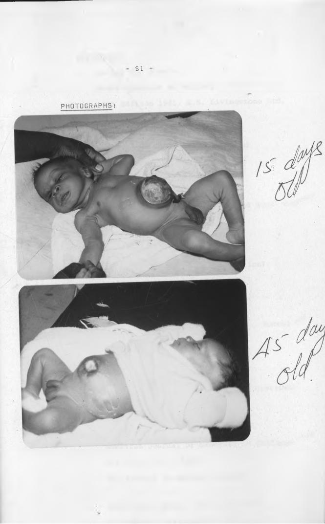
membrane is invaded by granulation tissue starting from the .
edge of the sac. In small sacs this would take a few days
and even if the original membrane crusts off the granulation
tissue wl\ich has now turned into epithelial tissue holds on
(see photos).
38
In our set up a 15 days old granulation tissue
has been found strong enough to contain the gut even when
the neonates cries.' (see pictures).»
Later 6n the granulation tissue is invaded externally
by skin epithelial cells from the edge of the omphalocele
and the whole sac is covered by skin converting the original
omphalocele to a ventral hernia.
Understanding of this natural history is important%when designing a model for non-surgical treatment of the
unruptured omphalocele.I
It~is imperative that when non-surgical omphalocele is
undertaken one has to find a way of*,-
(1) Preventing rupture of the omphalocele
(ii) Prevent infection of the omphalocele sac.
(iii) Keep the membrane long enough for
epithelialization to take place.
(iv) Plan to repair the subsequent Ventral hernia
(v) A criteria for selection of patients to be
included in the non-surgical management is also
essential.
Before 19^0 when Penicillin came into general use
infection was very important and conservative management of
the omphalocele was disastrous. The best treatment «as surgery
and the earlier it was the better. Soper (17) reports that

39 . >;>*1
mortality rate was 12% when operation was within 12 .hours and
31% when within 2b hours and 62% when operated within **8 hours.
After this it was even higher.
At this age of good antibiotics, infection can be controlled.
To prevent rupture of the membrane omphalocele should be
anticipated even before birth. Robert J. (20) had found
maternal utrasonography accurate in diagnosing congenital
malformations especially when combined with good history and
measurement of Alfa-Foetal Proteins in the amniotic fluid
Charles (19)* The best time is at 33 weeks gestation. With
untrasonography Omphalocele was one of the malformations easily/diagnosed. The decision should be made this early on how to
protect the sac from rupture. Most centres have had only one or
two rupture before birth. In our series only one had ruptured
in 3*+ cases 2.9*+%. After the child is born this exposed sac
should be dressed with wet saline gauze6 to keep it wet and
to prevent rupture. It should be emphasized that this thin
membrane especially in large omphalocele where loops of gut
liver and even stomach may be included, forms a very poor heat
insulation for the body core. The infant as a rule suffers
from hypothermia very fast. To prevent this most authors
suggest covering the imphalocele sac with warm gauze soaked
in saline and covering the rest of the body especially lower
part in a plastic bag. Akcerman (3*0» Towne (1*0 and Ochola
and Kyarabi (22).

- ko -
In early literature where conservative management
was advocated Jaricho (15) Holcomb (28), Soave (29)
Grob (3U)» the omphalocele sac was cleaned with spirit
then painted with 2% aqueous mercurochrome. The mercurochrome
denatures the membrane and hardened it. It also made it
difficult to be infected by bacteria. This unfortunately
dried up and craked exposing the granulation tissue below
it. With lack by the time it dropped off granulation tissue
and strong epithelia had formed and then covered with skin
taking one to three months Soave (29). The only
contraindication to mercurochrome method is the risk of
mercury toxicity as reported by Fagan et al(39)£tringel (21). ' *
* H
In our set up only one case had mercurochrome painting
afterwards a very simple dressing of sofratulle has been very
successful. This has the advantage of keeping the sac stefile
and also moist. When it is moist it stays longer, over four
weeks and by the time it sloughs off the underlaying granulation
tissue and epithelialization is strong. As for the repair of
tthe ventral hernia, this posses very little problem. In
the authors series six patients have had their ventral
hernias repaired. They range in age between 1 month and 2
years. All had well formed abdominal wall and the hernia
was easy to repair in one stage.

Their hospital stay during the repair ranged betweent
5 days and 15 days. As regards criteria most authors Gross
(16), Holcomb (28), Soave (29) and Grob (50), advocate that
patients for conservative management should, be thoseiwith
complications and other congenital malformations which make
surgery risky. Patients with very large omphaloceles even if
they have no complications are also recommended for conservative
treatment.
This was iklso the principle, "womb to theatre" practiced
before 1978 in this hospital. Most of the operated patient
died miserably following the surgery. One had multiple burst/ ' • V "abdomen. The other three had intact omphalocele membrane
which was excised and then the abdomen repaired with dura
graft. All these patients had infection of the dura graft
and all died with severe peritonitis.
As from 1979 only those patients who needed very
urgent surgery were operated. Those were the cases which needed
reduction of intussusception one case, colostomy for ano
rectal malformation two cases, release of abstruction - one case
colostomy for stenosis of ascending colon - one case. All
others who did not need urgent surgery were treated conservativelyowith sofratulle dressing. Most of these patients healed .f\
well to had their central hernia repaired later.

3.7 SURGICAL MANAGEMENT OF OMPHALOCELE;
Surgical repair of the omphalocele is as old as the
surgery. Surgeons of all the ages have been plagued by this
congenital condition whose mortality to date has stayed
high. What has changed over the aged has been the ability
to diagnose omphalocele before birth, Robert (20) and
Charles (19).
Another field which has changed is in improvement of
management of sepsis especially the introduction of penicillin
in 19^0 and availability of prosthetic surgical materials
for covering of the omphalocele* With all these improvements
omphalocele still has high mortality in most centres,
Jarcho (15) and Schuster (2*f) have reviewed history
of omphalocele surgery over the ages, Celsus and his followerr
Paulu6 all recognised umbilical hernia and suggested ReductionV.
of the content and ligation of the sac in older children.
The same treatment was not advocated for infants with the
same condition. Francos* treatise on hernia in 19th century
does not deal with umbilical type,/
Ambroise Pare described congenital umbilical hernia as early
as 163^1 but inspite of all these literature very little was
written on omphaloceles before 19th century,
Aribat reviewed older literature in 1901 and found only
9 cases reported before 1800, The earliest mention is by
Scultetus in 163^ though no surgical treatment was attempted.

It is William Hey who in l80J described 3 cases of "hernia
congenita umbilicalis". Before that, records of umbilical
hernia never mentioned it as congenital. Pott Richer and
Kauriceau in 17th century described umbilical hernia and
treatment was advised as bandaging only though no success was
guaranteed as "the success of it is very rare".
In 1807 Sir Asthey Cooper in his treatise on umbilical
hernia mentions that some infants are born with umbilical
hernia and described the first recorded surgical management
on congental umbilical hernia. He reports one case where
James Hamilton reduced the hernia tied the sac at the base
a*id then "cautiously opened it". The edges of the abdonun&l
wall was then brought together with two silver pins and some
adhesive straps".
The sac was allowed to drop off and the cure was said
to have been complete in a few days. The child was still
well 8 months later.
Before this operation Hamiltons had several other
neonates with congenital umbilical hernia but had not attempted
operation as he considered them as "strictly desparate".
Samuel Bard in America in 1819 mentions children born with
congenital umbilical hernia. He recommends treatment
by "reduction, straping with adhesive plaster and moderate

Some treatment was advicedpressure bya proper bandage”.
by Scanzoni in 1955* It appears then that early treatment
of the omphalocele (congenital umbilical hernia) was
non-surgical and reduction and compression was the method of
choice. It is only Hamilton who attempted surgery of the
omphalocele. Thi3 apathy on treatment was mainly due to the
poor record of survival of these infants.
Pybus 1922, Jarcho 1937* Fresher, 1926 and Cullen 1916
all advocate early surgery in omphaloceles.
They advocate excision of the sac and closure of the
abdominal wall. The operation should be undertaken as
early as possible after birth. When the omphalocele was
simple containing & only radical operation gave very good
results. Aribat in 1901 operated 68 cases with **7 (69#)
recoveries. Altpeter in 1900 operated 91 cases with
69 (75*8#) recoveries.
When the sac contained liver or other viscera results
were not as good. Out of ^5 patients operated only 11(2**.3#)
recovered. N.M. Dott in Edinburgh Medical Journal in 1932 put
the thinking of the time very well "The child should pass
straight from the womb onto the operating table.”
This one stage repair of omphalocele posses its own
problems. It is no wonder that those neonates with
compound omphaloceles fared so badly with only 2^.5#
survival rate. Most of the omphalocele surgery has as it
evolved from those early days of 19th century, had to
find solutions to improvement on survival on the/compund omphaloceles.

One stage repair technique solves only one problem
the closure of the abdominal wall defect when possible
The closure brinjf in new problems which were not there
before surgery. All the problems stem from the effort tot
place the herniated viscera into the abdominal vavity, a
place it has never ever been and which does not have the
space to receive them as it has never developed it. In
one stage repair except in few cases this entails forcing
the viscera into an underdeveloped abdominal cavity. A
series of problems then e'minates from this increased
abdominal pressure./ . V
Gross (16), Soper (17), Towne (1*0* and Stringel (21)
list complications emanating from increased abdominal pressure
as: -
(1) Displacing the diapragm into the chest causing respiratory
distress.
(2) Pressure on the inferior vcna cava causing reduction of c^rdia
return, circulatory collapse, multiple haemorrhages in the
gut and oedema of the legs.
(3) Intestinal Kinking causing obstruction and paralytic ileus.
(4) Herniation of the stomach into the thorax causing
respiratory distress, hiatus hernia and gastroesophageal
reflux.
(5) Budd-Chiari phenomenon with ascities due to Kinking of the
inferior venacava at the diaphram level as the liver is
manipulated back into the crowded abdominal cavity.

Any surgeon should try and overcome these problem
as death from respiratory and cardiac collapse is the major
cause of mortality within the first 6-**8 hours after
omphalocele surgery. This was 29*7% in Sckstein's (18)
series. In our series of the 15 patients who died, 6 (**0%)
patients had respiratory associated deaths, and (26.6%) had
sudden death which could be circulatory collapse. Combined
respiratory and cardiac related deaths accounted for 86.6%
of the deaths in the surgical cases.
Respiratory embarrassments seem to be a very common cause
of death. This is not strange as a neonate up to 10 days old
breaths with the diaphragm according to Baur (7)» Gross (})»
Swenson (*♦), Bren TCK (9)» Richardson (10), Respiration vol.
1 (11), Respiration vol. II (12). Any surgery which interferes
with the diaphragmatic respiration makes it very difficult for
the neonate to breath. It has been found hence very necessary
to have a very good set up for respiratory support where
omphaloceles are operated. Sckstein (l8). Towne (1*0
suggest that the respiratory support can be a lengthy problem
lasting for months at times. There were 12 respiratory problems
out of 53 cases (22.6*+%) Towne (1*0 • One of the 12 (8.3%) needed
a tracheostomy. *+ cases out of 9 (*+**•**%) treated for congestive
cardiac failure had pulmonary hypertension as the primary cause.

k?
Towne (l*f) also mentions a condition when liver has to
be pushed back into the abdominal cavity. The manipulation
of the liver causes angulation of the inferior vena cava at the
diaphragmatic level causing demonstrable Kinking when
cardiac catheteriz*tion is done. This causes ascites secondary
to Budd-Chi&ri phenomenon. This has been supported by Waldinar
(1977) and Carlton as quoted by Towne (1*0.
To solve these problems surgeons have developed various
methods of surgery to make omphalocele surgery safe. The sole
aim has been to device a method which does not raise the
intra abdominal pressure after the abdomen is closed. Other
lines have been to find an equipment which can be used
firstly for research purposes to define what pressure is
acceptable without causing the complications mentioned earlier.
Welsley et al (2 5) came out with a safe pressure of 20 cms
of water, Othersen et al (27) in their pneumatic device for
gradual reduction of the omphalocele using air pressure found
that 50 cms of water pressure was safe.
One none prosthetic solution to the increased abdominal
pressure after closure was offered by Buchaman (56) who
advocated splenectomy and partial hepatectomy up to 20 gms
to allow closure. This method should be condeiniiedas splemectomy
before 2 years of life has been associated with severe
infections later, in life. La Mura (57) and Morgensten (58)*

As regards surgical technique staged closure has been found
to be the safest. The staged closure refers to the abdominal
wall. To achieve this the earliest method perfected by Gross
(1 6) in 19^0 involved excision of the omphalocele, dissection
of the skin from the abdominal wall as far posteriorly as
possible, reduction of the omphalocele content and then
closure of the skin, Subcutenous fat may be closed also if
intrabdominal pressure allows.• Abdominal muscles were not
approximated.
The infant would then grow up with a ventral hernia which
c< uld be repaired later in life. There are basically two
problems with this method of- surgery. Firstly since the
abdominal wall is left free, one is essentially substituting
omphalocele sac with skin and so the gut continues to grow
under the skin cover while the abdominal wall receives no
stimulus to .grow. When one goes to repair the ventral hernia»
problem of closure would still surface again. Release incisions
at the flanks were used to try and archieve closure of the
ventral hernia Denison (2). Second problem was adhesion of
the intestines to the raw skin surface needing sometimes
excision of the gut to release the adhesions* Towne (1*0.

^9Gross,(16) solved his problem of adhesions by leaving
the omphalocele sac intact cutting into the skin just near
its margin then dissecting the akin as before and then
closing it over the sac thus leaving a peritoneum covered
membrane still covering the intestines. Since the gut was not
handled, incidences of ileus were rare. Hilcomb (28) 1961\ 9
still practise Gross's technique. Burying of infected membrane
was a source of problems with infection at times. ./
More current surgical techniques make use of artificial
membranes to cover the omphalocele contents after -the sac has
been excised. The membranes are sutured to the abdominal wall
and the growth of the abdominal viscera produces enough
gentle tension to stimulate growth of the abdominal wall making
later closure of the ventral hernia possible and easy.
The common artificial membranes used are Teflon Mesh
Moazom (23) Schuster (2^) and Filter (26), and Silastic
Sheet Othersen (17).
The original use of the prosthesis by Moazam (23) was to
bridge the abdominal muscles with the Teflon (Polytetrafluoethylene)1
Mesh and then with the prosthesis in place to approximate the
mobilised skin over it. This was sort of improving of the quality
of theventral hernia over Gross (16) whose skin cover only did
not stimulate the growth of the abdominal muscles for easy
closure of the ventral hernia later on. The prosthesis could
be removed later when the abdominal wall has grown and repair
of the ventral hernia done or the prosthesis could be
left in place for a long time.

50
The Mesh is then invaded by fibroblasto and then
incorporated into a psendomembrane. Some of the meshes
have been left in indefinately. Towne (1*0 reviewed this
method of treatment and found that infection, fistula
formation and severe adhesion of the gut requiring gut
resection complicated the surgery. In one patient 8 surgical
procesures were required with a 27** days stay in hospital and
$122,000 bill.
Schuster (2*0 improved on this technique. The improvement
was on prevention of the adhesions and shortening of the
hospital stay. The Teflon Mesh was covered by silastic to
make it less liable to produce adhesions. To reduce the
chances of adhesions formation even further the Taflon Mesh
was then lined by a non reactive and impermeable sheet of
polyethylene or silicone rubber. This kept the abdominal
viscera away from the Teflon Mesh. Like the Teflon this was
in two pieces each for each side overlapping at the midline.\ *
The Teflon without tension was then sutured at the midline and
then where possible skin was sutured over it. When this was
not possible an Island of Mesh without skin was left ‘to be
dressed wi£.h sterile dressing. On the *+ - 6th day the patient
was taken back to theatre and the Mesh which was now laxf
due to the stretching of the abdominal wall and growth of theVs
muscles primmed and ftgain without tension the procedure was
repeated. This was repeated again and again till’the abdominal
wall could be aproximated at the midline without unduo
tension.

51
The average stay in the hospital was 2^-63 days.
Mortality was reduced to 27*2# and a remarkable servival
of 5 patients with antenatal ruptured omphalocele membrane
compared with only 1 case before.
Othersen (27) bas come up with a device like a
belljar which is made of Dacron and lined with silastic
sheet. When it is sutured to the omphalocele edge the
area between the bell and the silastic sheep is filed with
air at 50 mm water which acts as a pneumatic reduction
device of the omphalocele facilitating closure of the
hernia later. The procedure can be done under local
anaesthtic and the reduction procedure takes about
7 - 1 0 days after which the repair of the omphalocele can
be undertaken.
Most of the surgeons agree that supportive
procedures are very important for success of good
omphalocele surgery. The prenatal diagnosis (19) and (20)
is important if preparations are to be made to receive
the neonate born with omphalocele. After birth presence
or abscence of the omphalocele sac is important. When
abscent the quality of the gut is 'compromised as they
are meconium stained, oederaatous and matted together.
Soak in 50*000 units of fibriolysm in 50 mis of water
helps to lyse the fibrin exudate , Othersen (27)*
Intack membrane should be covered with wet soaks and
neonate protected from the inevitable hypoctermia.

52
Total parental nutrition is important especially
where gut is compromised usually in ruptured omphaloceles,
Filber (26), This is important since the neonate has to
be starved for surgery# For easy surgery the gut needs
to be decompressed by gastrostomy or Nasal gastric suction.
In our series Nasal gastric suction has been sufficient.
Most surgeono prefer emraediate surgery, some in
consideration of cost Towne (1*0. The preference is
for primary closure and where this is difficult
staged repair. This could become obsolete if devices• >
like the pneumatic reduction are perfected since the gentle
traction causes the abdominal wall to grow as the
omphalocele is reduced for easy one stage repair.
Advocates of early surgery argue that when
the abdomen is opened one is able to inspect the
whole gut and acertain whether other operable abnormalities
do exist. The commonest abnormalities arising from the
gut are atresia, obstruction or agenesis. If there is a
method which can lower the mortality rate without necessarily
having to open the abdomen like the pneumatic reduction
device its^ould be encouraged as, the anomalies can be
investigated by none-'-invasive methods as in other neonates
with no omphalocele.
Anaesthesia is important in neonate surgery. The best
anaesthesia has been found to be non paralysing anaesthesia
as toneless abdominal muscle can not be used to judge the
intra-abdominal pressure reached at closure of the abdomen.

53
CHAPTER 4
4.1. CONCLUSION:
There is still controversy on pathogenesis
of omphalocele. Though review of the literature
suggests that most surgeons prefer surgical
management of the omphalocele and the same
excuted as early as possible our results seem
to suggest that our patients survive better uhder
non-surgical treatment.
Neonatal surgery has its own problems and
supportive facilities like early diagnosis,
transport, theatre and intensive care unit with
good respiratory support are important. We have
encountered problems in postoperative period because
supportive equipments such as respirators and
incubators may not be operative and Parental nutrition
is not always available. Since respiratory support*«•is vital availability of functioning ventilators
for long periods is mandatory* Temperature control
is also important and working incubatorsar® essential.
Knowing our limitations non-surgical treatment
where practicable should be the treatment of choice.
Infact all the omphalocele patients in our set up
should be programed for conservative management
until other conditions dictate, early surgery.

5*+
This entails a good incubator, and simple
moist dressing like sofratulle. Early surgery
where essential may be performed but simpler
methods like pneumatic reduction before surgery
could be advocated since they do away with the need
for prosthetic sheets and multiple operations.
In surgical management primary repair
should be the aim. If this is not possible
then Gross's method of mobilising skin cover
over intact sac would be a simple straight forward
procedure leaving the ventral hernia for later
repair.
- 55 -
-'f.2. 3SCOMM2NDAT10NS
This study has been very stimulating in
that it has unearthened lots of controversy on
management of omphalocele. The controversy
ranges from pathogenesis, classification,
nomenclature and even management. Without
getting involved in the controversy over
Embryology and pathogenesis one could concentrate
on management which I think is beneficial to the
patiento
Suffice it to say that in present state
of our medical services non-surgical management
appears to yield the best results. Surgery
should be restricted to those cases which must
be operated and simple technique is what we need.
To achieve this end I would recommend that:-
1. It is important to have good obstetric
service where good history and management
of pregnancy can be done so that those
cases who by behaviour of the pregnancy,
like hydramnios are likely to have congenital
malformations, should be delivered in
a hospital where the malformations can
be adquately treated.

56
2. As regards omphalocele when discovered
at birth the sac or the exposed intestines,
when sac is ruptured, should be covered
by wet sterile swabs and a firm bandage
put on.
3. The neonate should then be kept warm
possibly in an incubator. If no incubator
is available cover the baby with warm cotton
wool and a blanket.
*+. A nasal gastric tube should be passed to
decompress the abdomen and when necessary / •
I-V fluids should be started and administered
at the appropriate rate.
5. Thorough medical examination of all the
systems should be done to rule out any
complication or associated anormally.
6. The omphalocele should be classified and then
a decision made whether surgery is needed
or non-surgical management is the treatment
of choice. All ruptured omphaloceles
would benefit from immediate surgery.
7o The neonates on conservative management should
receive clear fluid for a few days to keep the'lgut undistended then later start on dilute
milk feed

- 57
8, The neonate should be warmed in an incubator or
wrapped in a warm blanket*
9. The neonate should be observed very closely
for infection of the omphalocele membrane
and respiratory system as any cough may
contribute to the rupture of the sac*
10. Plan for repair of the ventral hernia can
be done when the child is big enough but
preferably before school age*
11. As regards surgery the simplest procedure
should be done. Gross procedure of covering
the omphalocele with skin should be tried
as a method of choice.

58
\■1
5.1 ANNEXA
FORM USED FOR COLLECTING DATA
FROM CASE NOTES.
/
>»

59
NAME Race Location IP.No. Sex Birthweight
BirthDefects
Treatmentbeforesurgery
Ace a t surgery
Surgicaltreatment
Results
--
*

60
ACKNOWLEDGEMENTS
My sincere thanks to many people who gave me moral
support as I worked through this thesis. Special
thanks to Professor Ac Wasunna and Professor N.O.Bwibo
without whose personnal help I would not have been
able to finish this course*
I am very much indebted to Professor J,M,Kyambi
who tirelessly supervised this thesis and sacrificed
a lot of his spare time to proof read this thesis at
every stage,
I cannot forget the staff of the records office
who worked so hard to provide the case notes.
Miss Rachel Wanjiru takes a special place in that
she typed and retyped the manuscript to the accepted
standard.
To members of my family patience with my odd
working hours has been imense.
61

t
62
REFERENCE
1. Bailey and Love's
Short Practice of Surgery
18th Edition 1981. E.S. Livingstone Ltd.
2. W.M. Dennison
Surgery in infancy and childhood \
Seven Edition 1967.
3. Gross
The Surgery of Infancy and Child Hood- Saunders
June 1955.
4. Oruar Swenson 1962.
Paediatric Surgery (second Edition)
Appleton Century 7 Crofts MC.
5; F. Fasana
Text book of Medical Embryology. Normal and
Abnormal Development.
Kenya Literature Bureau 1980.
6 . Dovland's. Illustrated Medical Dictionary
24th Edition 1965.
7. A. Robert Bauer M.D.
American Journal of Diseases in Children 1940
vol. 60: 1342 - 1350
Respiration in Newborn Infants.
8 . James Mason Brown 1962 802 - 803
"Surgery of Childhood

6*3
9. Brown T.C.K. 1979
"Anaesthesia for Children" pg. 6.
10. Richardson P.P. and John J.H.
"Neonatal Surgery"<
1970 pg. 35 - 36. Butterwoths London.
11. "Respiration" vol I 1965
American Physiological Society pg. 213.
12. "Respiration" vol. II 1965
American Physiological Society Pg. 1334.
/ ' '/13. Pieter A de Vries
"The Pathogenesis*of Gastroschisis and Omphalocele".Journal of Paediatric Surgery vol. 15 No. 3:
245 - June 1980.
14. Barbara H. Towne and George Peters
"The Problem of the Giant Omphalocele".
Journal of Paediatric Surgery 1980 vol. 15:
543 - 548.
15. Julius Jarcho, M.D. F.R.C.S.
Surgery Gynaecology and Obstetrics vol. 65
Nov. 1939 No. 5: 593 - 600.
"Congenital Umbilical Hernia." *
.
_____________

- 6i+
16. Robert Gross M.D. and James B. Blodget M.D.
Omphalocele (Umbilical Hernia) in thei
Newly Born.
Surgery, Gynaecology and Obstetrics 1940
Vo1. 71: 520 - 527.
17. Robert T. Soper M.D. and Edarward W. Green.
"Omphalocle".
Journal of Surgery, Gynaecology and Obstetrics
Oct. 1961. 501 - 508.
'18. Herbert and Echstein
"Exophalos". A review of 100 cases.
British Journal of Surgery 1963 vol. 50:.
• 405 - 410.
19. Chairles R. King M.D. and Gerald H. Prescott D.M.O
"Amniotic Fluid Alpha-Foetoprotein Elevation
with Fetal Omphalocele and a Possible mechanism. . /
for its occurence."
American Journal of Obstetrics and Gynaecology
vol. 130 (3) Feb. 1978.
20. Robert J. Touloukian and John C.
"Maternal Utrasonography in the Antenatal
Diagnosis of Surgically Correctable Fetal
Abnormalities" .
Paediatric Surgery vol. 15: No. 4 August
1980 373 - 377.

65
21. G. Stringel and R.M. Miller
"Prognostic Factors in Omphalocele and
Gastroschisis".
, Journal of Paediatric Surgery vol. 14 No. 5:
Octo. 1979.
22. Ochola-Abila P.M. Med. J.M. Kyambi M.D.,
F.A. and S.M. Barrack M.D.
"The management of Giant Omphaloceles"
The Proceedings of the Association of
Surgeons of E.A. 82.
23. Farhat Moazam, Bradley M., Rodgers and J.N. Tabert
"Use of Teflon Mesh for Repair of Abdominal
wall Defects in Neonates".
Journal of Paediatric Surgery vol. 14 No. 3
(June) 1977.
24. Samuel R. Schuster M.D. F.R.C.S." Anew Method for the Staged Repair of Large
Omphaloceles".
Surgery, Gynaecology and Obstetrics Oct. 1967
vol. 125: 837 - 850.
f
25. John R. Welsley, Robert Drougowski and Annold G.
C. Coran.
"Intragastric Pressure Measurements:
A guide for Reduction and Closure of the
Silastic Chimney in Imphaloce and Gastroschisis".
Journal of Paediatric Surgery 1981 vol. 16 No.3
(June). 264 - 270.
67
31. Jay L. Grosfield, M.D. Lilian Dawes and Thomas'
R. Weber.
"Congenital Abdominal Wall Defects: Current
Management and Survival."
Surgical Clinical of North America (Oct.) 1981
vol. 61 No. 5: 1037 - 1049.
32. P.L.- William C.P. Wnedell-Smith
Basic Human Embroyology
Second Edition
Pitman Medical and Scientific Publication
1969 Pg. 66 - 67.
33. Boyd J.D.
Human Placenta
Cambridge W. Heffer and Sons Ltd. 1970.
34. Ackerman P.
Congenital Malformation
Chapter XV (215 - 231) Excepta Medica.
35. Ebbon G.Der depositions me-chanismus des Physiologischen
Nabelbruches bei Saugetieren und beim Menschen.
"Morphol." Johnb 1938, 82:271.
Buchanan R.W. and Cain W.L. 1956
"A case of a complete Omphalocele".
36.

68
37. Joseph La Mura et al.
"Splenorrhaphy for the Treatment of
Splenic Rupture in Infant and Children".
Surgery (May) 1977 vol. 8 No. 5: 497 - 501.
38. Loen Morgensten
"Techniques of Splenic Conservation".Arch. Surg. 1979 vol. 114 449 - 453.
39. Fagan D.G. Pritchard J.S. Clarkson T.W.
"Organ Mercury Levels in Infants with
Omphalocele Treated with Organic Mercurial
antiseptics.2
1977 Arch. Dis. Child 52: 962 - 964.