Embed Size (px)
Citation preview

Review of Mechanical Properties of Human Body Soft Tissues in the
Head, neck and spine
Dr. S. Mukherjee, Non-member
Dr. A. Chawla1, Member
B. Karthikeyan Non-Member
ABSTRACT
In this paper we review the mechanical properties of soft tissues available in literature. Human body regions are
split into different parts to pursue this study. This review paper focuses on the soft tissues in the head, neck and
spine. The tissues studied include brain tissues, scalp tissues, ligaments in cervical spine, neck muscles and spinal
soft tissues. Material properties, which are directly extracted from the experimental methods, and the constitutive
properties that have been used in finite element models are looked at. Isolated tissue tests, sub-segmental tests and
full-scale tests used for validating the respective finite element models are investigated. Static and dynamic
properties are sorted according to the tissue type. Variations in the data from different sources has been studied
and summarized. Scatter in the static properties and less frequently available dynamic properties indicate the need
for further testing and alternate material models.
Keywords: Material properties, Human soft tissues, Head, Neck, Spine
INTRODUCTION
Human body finite element (FE) models, if based on a realistic geometry and bio-fidelic material properties, can
be useful in designing safer vehicles in order to reduce incidences of injuries and fatalities in road crashes1,2. To
identify the reliability and variations within the material properties reported in literature, a review of the properties
of soft tissue in the human body has been conducted. Human body regions are divided into three parts a) Lower
extremities b) Head, neck and spine c) Upper extremity, chest and abdomen. The present study reviews the
properties of the soft tissues in head, neck and spine region. Constitutive properties of soft tissues used in the finite
element models and the validating experimental procedures are also reviewed. Mechanical properties are
categorized in the following sections according to major tissue type in the respective body regions. Variations in
reported properties have been used to identify issues which still need to be addressed.
HEAD
Head injuries are the most common injuries with Abbreviated Injury Scale (AIS) >=2 for belted occupants in
automotive frontal impacts3,4 and the second leading cause of injuries (after lower extremities) having AIS
between 2 and 6 in pedestrian accidents5. Head injuries may cause either a temporary or a permanent damage to
parts of the head and can be life threatening. These injuriescan be grouped as those causing scalp damage, skull
fracture, brain injury, or a combination of these6. Anatomy of the head is mainly divided into two parts a) face,
which represents the front part of the head and b) head which comprises the center and rear part of the head7. Soft
tissues in the face include skin, muscles, tongue, cartilage and ligaments. Studies related to facial soft tissues are
scarce due to its low load sharing capability and mostly have a low severity. They therefore have been excluded 1
Corresponding Author
Dr. A Chawla, Associate Professor,
Department of Mechanical Engineering,
IIT Delhi, Hauz Khas, New Delhi 110016.

from this study. Soft tissues in the head are mainly present in the scalp, meninges and brain regions. Scalp consists
of skin, connective tissue, aponerosis, loose connective tissue, periosteum. Meninges region which separate the
brain and the spinal cord from the surrounding bones consists of dura mater, arachnoid and the pia mater. Brain
region is subdivided as cerebrum, cerebellum, medulla oblongata, midbrain and pons. Readers are encouraged to
refer to a text on anatomy8, for a detailed anatomical description.
HEAD INJURY – LOAD CASES
Dynamic load causing injuries are divided into contact and non contact type7. Injuries due to contact type loads
mainly occur due to impact on the head. They are further subdivided as injuries arising due to direct contact loads
(which result in skull deformation and cause local brain deformation) and injuries arising due to propagation of
stress waves from the impact region (causing negative pressure in the opposite side of the impact). Non contact
type loads causes injuries due to inertial loads which arise due to linear and or angular acceleration / deceleration
of the head.
HEAD INJURY ASSESSMENT FUNCTIONS
Several Injury criterions or injury assessment functions are developed to establish the degree of human tolerance
to head impact. Head Injury Criterion (HIC) based on Wayne state tolerance curves (WSTC) is the most widely
referred injury assessment function9. Other reported injury assessment functions include maximum linear
acceleration, maximum linear acceleration with dwell times, Severity Index (SI), Angular acceleration combined
with angular velocity change, Generalized Acceleration Model for Brain Injury Threshold (GAMBIT) – angular
and linear acceleration10. These injury assessment functions predict injury risk from the external mechanical load
and do not account for the internal mechanical response11. Hence the injuries risk at tissue level cannot be
predicted in detail. Computational model with detailed geometry and biofiedilic material properties will overcome
this difficulty and provide a better insight to injuries6. With substantial improvement in the geometry of the
models using MRI and CT Scans, a review revealing the gaps in the tissue properties would help develop
biofidelic finite element models. Hence a review of tissue properties extracted from experiments and those
obtained by inverse mapping in finite element modeling is presented. We also discuss experiments conducted for
developing the injury assessment functions.
EVOLUTION OF EXPERIMENTAL METHODS AND FINITE ELEMENT MODELS ON
HEAD
Since the seventies, FE models have been used to study the behavior of head under impact loads. Khalil12
predicted the impact loads causing brain damage by cavitation using three axisymmetric head models. Khalil13
reviewed the issues in human head finite element models with respect to the experimental observations.
Insufficient modeling accuracy, unrealistic boundary conditions related to neck attachments, scope for
improvements in the brain tolerance criteria and required material properties were highlighted. Later, Sauren14
reviewed the second generation finite element models published in the period 1982-1992. Large deformation
models with nonlinear viscoelasticity were sought to overcome the limitations of linear elastic models. Visco-
elastic models with incompressible theories were evolved for constituting the large strain and strain rate dependant
behavior of brain tissue15. Zhou16 developed a three-dimensional finite element model of human head and
compared the responses of the homogenous and inhomogeneous human brain. The inhomogeneous brain model
basically represents the gray and white matter with different material properties. This study conducted for frontal
impacts reported variations in the shear responses due to the assumption of improper shear and volumetric
properties showing the scope for experimental studies to measure shear strain in the brain due to impact.
Claessens17 collated the Young’s Modulus data reported in literature and found its variation to be significant to
influence the pressure and stresses in coup and counter coup regions. Kang18 modeled the brain with linear,
isotropic, viscoelastic material properties and validated the human head model against cadaver experiments.
Newman19 proposed a methodology to develop biomechanical criteria for mild traumatic brain injury using the

data collected from soccer injuries. Updated Wayne state brain injury finite element model was used to reconstruct
the incidents recorded during game. This study constituted the brain tissue as viscoelastic material under shear
loading and elastic behavior under compressive loading. The deviatoric stress in shear loading was constituted as
rate dependent and represented using shear relaxation modulus. Grey and white matter were subjected to different
shear modulii. Followed by this study a new injury criterion, Head Impact Power (HIP) has been proposed to
access the mild traumatic brain injury10.
TISSUE LEVEL FINITE ELEMENT MODELS AND CONSTITUTIVE MODELS OF BRAIN
TISSUE
A nonlinear viscoelastic constitutive model for brain tissue capable of predicting its response for 30%
compression level was demonstrated by Miller20 for very low strain (< 0.64s-1). A single-phase, linear viscoelastic
model based on the strain energy function for loading velocities varying over five orders up to 0.64s-1 has been
implemented in ABAQUS21 for modeling the brain tissue. This linear model21 requires fewer input material
parameters than the earlier model20. Sarron22 proposed a multi domain modeling technique to characterize the
brain tissue and to identify the constitutive law parameters of each domain. Miller23 tested the isolated brain soft
tissues in uniaxial tension. The theoretical solution obtained from this study was valid only for isotropic,
incompressible materials for moderate deformations (<30%) and cannot be used for load bearing tissues having
directional properties. Miller24 performed in vitro uniaxial tension experiments on swine brain tissue in finite
deformation and developed a non-linear, viscoelastic model based on the generalization of the Ogden strain energy
hyperelastic constitutive equation. This study has been extended for in vivo conditions and reports that the
hyperelastic model predicts better response than the standard linear viscoelastic model25. Similarly, Kyriacou26
compared the behavior of elastic, viscoelastic and poroelastic constitutive models and proposed compressible
viscoelastic solid model suitable for low strain rate studies. Recently Miller27 studied the behavior of brain tissue
in unconfined condition with top and bottom surfaces of the tissue were glued with platens for compression
loading. Arbogast28 demonstrated the transversely isotropic behavior of brain stem under shear loading by
analyzing the regional differences in the overall material stiffness and anisotropic mechanical properties of brain
stem over a range of frequencies from 20-200 Hz, for 2.5-7.5 % engineering strain. Using this oscillatory shear
loading experimental study, a fiber reinforced composite model composed of viscoelastic fibers surrounded by a
viscoelastic matrix was developed to predict the response of anisotropic mechanical behavior29. Darvish30 tested
the quasilinear viscoelastic model with single hereditary integral and nonlinear viscoelastic model with multiple
hereditary integrals to replicate the experimentally obtained nonlinear behavior of bovine brain tissue under shear
loading. The experiments were reported for the forced vibrations from 0.5-200 Hz with finite amplitudes up to 20
% Lagrangian shear strain. The nonlinear model with multiple hereditary were found to be superior especially at
frequencies above 44 Hz. Under finite strains in this study the linear complex modulus demonstrated
nonrecoverable asymptotic strain behavior indicating the discrepancies with the assumed material properties of
brain tissue. Brands31 developed a three dimensional nonlinear viscoelastic model for predicting the brain tissue
behavior under impact. The model predicts the strain dependent behavior up to 20 % strain and up to 8 s-1 strain
rate. With devaitoric stress modeled as non-linear viscoelastic and volumetric stress as linear elastic, the brain
tissue in this study was considered as nearly incompressible. Aida32, proposed the influence of short term shear
modulus and bulk modulus of brain tissue in shear response. The material properties of soft tissues in the head are
categorized into tissue specific and indicated in the tables listed in appendix-A (Refer Table A1-Table A6). Figure
1 to Figure 4 show the properties graphically and indicate the mean or range values of the respective material
properties.

1
10
100
1000
10000
100000
Bra
in (
32)
Bra
in (
32)
Bra
in (
32)
Bra
in (
32)
Bra
in (
32)
Bra
in (
79)
Bra
in (
17)
Bra
in (
17)
Bra
in (
17)
Bra
in (
17)
Bra
in (
17)
Bra
in (
26)
Bra
in (
26)
Bra
in (
26)
Bra
in (
80)
Bra
in (
22)
Bra
in (
22)
Bra
in (
22)
Bra
in (
22)
Bra
in (
22)
Bra
in (
22)
Bra
in (
22)
Bra
in (
22)
Bra
in (
22)
Bra
in (
22)
Bra
in (
22)
Bra
in (
22)
Bra
in (
22)
Bra
in (
14)
Bra
in (
14)
Bra
in (
14)
Bra
in (
14)
Bra
in (
81)
Bra
in (
82)
Bra
in (
14)
Tissue (Reference)
Log
arit
hmic
Ela
stic
Mod
ulus
(kP
a)
(a)
1
10
100
1000
10000
100000
1000000
10000000
Cer
ebel
lum
(17
)
Cer
ebru
m (
17)
CSF
(83
)
CSF
(80
)
CSF
(14
)
CSF
(82
)
Dur
a (1
9)
Dur
a (8
0)
Dur
a (1
4)
Dur
a (1
4)
Dur
a (1
6)
Fac
e (1
7)
Fac
e (8
3)
Falx
(83
)
Falx
(17
)
Falx
(18
)
Falx
(19
)
Falx
(80
)
Falx
(14
)
Falx
(81
)
Falx
(16
)
Falx
(82
)
Tissue (Reference)
Log
arit
hmic
Ela
stic
Mod
ulus
(kP
a)
(b)

1
10
100
1000
10000
100000
GM
(17
)
GM
(17
)
GM
(84
)
Pia
(19
)
Pia
(16
)
Sca
lp (
83)
Sca
lp (
18)
Sca
lp (
80)
Sca
lp (
16)
Sca
lp (
6)
Ski
n (1
9)
Ten
tori
um (
83)
Ten
tori
um (
79)
Ten
tori
um (
17)
Ten
tori
um (
18)
Ten
tori
um (
19)
Ten
tori
um (
14)
Ten
tori
um (
81)
Ten
tori
um (
16)
Ten
toti
um (
82)
WM
(17
)
WM
(17
)
WM
(84
)
Tissue (Reference)
Log
arit
hm
ic E
last
ic M
odu
lus
(kP
a)
(c)
Figure 1 Elastic Modulus of different tissues a) Brain Tissue b) Head tissues (Cerebellum, CSF, Dura, Face
tissue and Falx,) c) Head tissues continued (Gray Matter, Pia, Scalp Layer, Skin, Tentorium and White
Matter)
0.3
0.35
0.4
0.45
0.5
0.55
Bra
in (
32)
Bra
in (
32)
Bra
in (
32)
Bra
in (
32)
Bra
in (
32)
Bra
in (
79)
Bra
in (
17)
Bra
in (
17)
Bra
in (
17)
Bra
in (
17)
Bra
in (
17)
Bra
in (
26)
Bra
in (
26)
Bra
in (
80)
Bra
in (
22)
Bra
in (
22)
Bra
in (
22)
Bra
in (
22)
Bra
in (
22)
Bra
in (
22)
Bra
in (
22)
Bra
in (
22)
Bra
in (
22)
Bra
in (
22)
Bra
in (
22)
Bra
in (
14)
Bra
in (
14)
Bra
in (
14)
Bra
in (
14)
Bra
in (
14)
Bra
in (
14)
Bra
in (
81)
Bra
in (
82)
Bra
in (
14)
Bra
in (
14)
Bra
in (
14)
Bra
in s
tem
(16
)B
rain
stem
(17
)
Tissue (Reference)
Poi
sson
's R
atio
(a)

0
0.1
0.2
0.3
0.4
0.5
0.6
Cer
ebel
lum
(17
)
Cer
ebel
lum
(16
)
Cer
ebru
m (
17)
CS
F (
83)
CS
F (
80)
CS
F (
14)
CS
F (
14)
CS
F (
14)
CS
F (
85)
CS
F (
16)
CS
F (
82)
Dur
a (1
9)
Dur
a (8
0)
Dur
a (1
4)
Dur
a (1
6)
Fac
e (1
7)
Fac
e (8
3)
Fal
x (8
3)
Fal
x (1
7)
Fal
x (1
8)
Fal
x (1
9)
Fal
x (8
0)
Fal
x (1
4)
Fal
x (8
1)
Fal
x (1
6)
Fal
x (8
2)
Tissue (Reference)
Poi
sson
's R
atio
(b)
0
0.1
0.2
0.3
0.4
0.5
0.6
G M
(85
)
GM
(17
)
GM
(84
)
GM
(16
)
Men
inge
s (8
5)
Pia
(19
)
Pia
(16
)
Sca
lp (
83)
Sca
lp (
18)
Sca
lp (
80)
Sca
lp (
16)
Sca
lp (
6)
Ten
tori
um (
79)
Ten
tori
um (
83)
Ten
tori
um (
17)
Ten
tori
um (
18)
Ten
tori
um (
19)
Ten
tori
um (
14)
Ten
tori
um (
81)
Ten
tori
um (
16)
Ten
toti
um (
82)
WM
(17
)
WM
(84
)
WM
(85
)
WM
(16
)
Tissue (Reference)
Poi
sson
's R
atio
(c)
Figure 2 Poisson’s Ratio of (a) brain tissues (b) Soft tissues in the head (Cerebellum, CSF, Dura, Face and
Falx) (c) Soft tissues in the head (contd) (Gray Matter, Meninges, Pia, Scalp, Tentorium and White Matter)

0
200
400
600
800
1000
1200
1400
Bra
in (
32)
Bra
in (
32)
Bra
in (
32)
Bra
in (
32)
Bra
in (
32)
Bra
in (
32)
Bra
in (
32)
Bra
in (
79)
Bra
in (
11)
Bra
in (
17)
Bra
in (
17)
Bra
in (
17)
Bra
in (
17)
Bra
in (
17)
Bra
in (
86)
Bra
in (
80)
Bra
in (
22)
Bra
in (
22)
Bra
in (
22)
Bra
in (
22)
Bra
in (
22)
Bra
in (
22)
Bra
in (
22)
Bra
in (
14)
Bra
in (
14)
Bra
in (
14)
Bra
in (
14)
Bra
in (
14)
Bra
in (
81)
Bra
in (
83)
Bra
in (
82)
Bra
in (
14)
Bra
in s
tem
(19
)B
rain
ste
m (
16)
Bra
inst
em (
17)
Tissue (Reference)
Den
sity
(k
g/m
3)
(a)
0
1000
2000
3000
4000
5000
6000
Cer
ebel
lum
(17
)C
ereb
ellu
m (
16)
Cer
ebru
m (
17)
CS
F (
11)
CS
F (
19)
CS
F (
86)
CS
F (
80)
CS
F (
14)
CS
F (
14)
CS
F (
83)
CS
F (
85)
CS
F (
85)
CS
F (
16)
CS
F (
82)
Dur
a (1
9)D
ura
(80)
Dur
a (1
4)D
ura
(16)
Fac
e (1
7)F
ace
(83)
Fal
x (1
7)F
alx
(18)
Fal
x (1
9)F
alx
(80)
Fal
x (1
4)F
alx
(81)
Fal
x (8
3)F
alx
(16)
Fal
x (8
2)
G M
(85
)G
M (
17)
GM
(16
)G
M (
19)
Tissue (Reference)
Den
sity
(k
g/m
3)
(b)

900
950
1000
1050
1100
1150
1200
1250
Men
inge
s (1
1)
Men
inge
s (8
5)
Pia
(19
)
Pia
(16
)
Sca
lp (
83)
Sca
lp (
18)
Sca
lp (
80)
Sca
lp (
16)
Sca
lp (
6)
Ski
n (1
9)
Ten
tori
um (
79)
Ten
tori
um (
17)
Ten
tori
um (
18)
Ten
tori
um (
19)
Ten
tori
um (
14)
Ten
tori
um (
81)
Ten
tori
um (
83)
Ten
tori
um (
16)
Ten
toti
um (
82)
Ven
tric
le (
19)
WM
(17
)
WM
(19
)
WM
(85
)
WM
(16
)
Tissue (Reference)
Den
sity
(k
g/m
3)
(c)
Figure 3 Density of (a) brain tissue (b) Brain soft tissues (Cerebellum, CSF, Dura, Face, Falx andGM) and
(c) Brain soft tissues (contd) (Meninges, Pia, Scalp, Skin, Tentorium, Ventricle and White Matter)
0.01
0.1
1
10
100
1000
10000
Bra
in (
32)
Bra
in (
32)
Bra
in (
32)
Bra
in (
11)
Bra
in (
18)
Bra
in (
86)
Bra
in (
22)
Bra
in (
22)
Bra
in (
22)
Bra
in (
22)
Bra
in (
22)
Bra
in (
14)
Bra
in (
14)
Bra
in (
81)
Bra
in (
87)
Bra
in (
87)
Bra
in (
87)
Bra
in (
83)
Bra
in (
14)
Bra
in s
tem
(19
)B
rain
ste
m (
16)
Cer
ebel
lum
(16
)
CS
F (
11)
CS
F (
19)
CS
F (
86)
CS
F (
14)
CS
F (
14)
CS
F (
85)
CS
F (
85)
CS
F (
16)
Dur
a (1
4)
G M
(85
)G
M (
16)
GM
(19
)
Men
inge
s (1
1)
Ven
tric
le (
19)
Whi
te M
atte
rW
M (
85)
WM
(85
)W
M (
16)
Tissue (Reference)
Log
arit
hm
ic B
ulk
Mod
ulu
s (M
Pa)
(a)
0.1
1
10
100
1000
10000
100000
Bra
in (
26)
Bra
in (
86)
Bra
in (
14)
Bra
in (
14)
Bra
in s
tem
(16
)
Cer
ebel
lum
(16
)
CS
F (
11)
CS
F (
19)
CS
F (
86)
CS
F (
14)
CS
F (
85)
CS
F (
85)
CS
F (
16)
G M
(85
)
GM
(16
)
Men
inge
s (1
1)
Men
inge
s (8
5)
Ven
tric
le (
19)
WM
(85
)
WM
(85
)
WM
(16
)
Tissue (Reference)
Log
arit
hm
ic S
hea
r M
odu
lus
(kP
a)
(b)
1
10
100
1000
Bra
in (
32)
Bra
in (
18)
Bra
in (
14)
Bra
in (
81)
Bra
in (
83)
Bra
in (
87)
Bra
in (
87)
Bra
in (
87)
Bra
in (
87)
Bra
in (
87)
Bra
in (
87)
Bra
in (
14)
Bra
in (
14)
Bra
in (
14)
Bra
in s
tem
(19
)
CS
F (
14)
Gra
y M
atte
r (1
9)
Whi
te M
atte
r (1
9)
Tissue (Reference)
Log
arit
hm
ic S
hor
t te
rm s
hea
r M
odu
lus
(kP
a)
1
10
100
1000
Log
arit
hm
ic L
ong
term
sh
ear
Mod
ulu
s (k
Pa)
Short term shear modulus Long term shear modulus
(c)
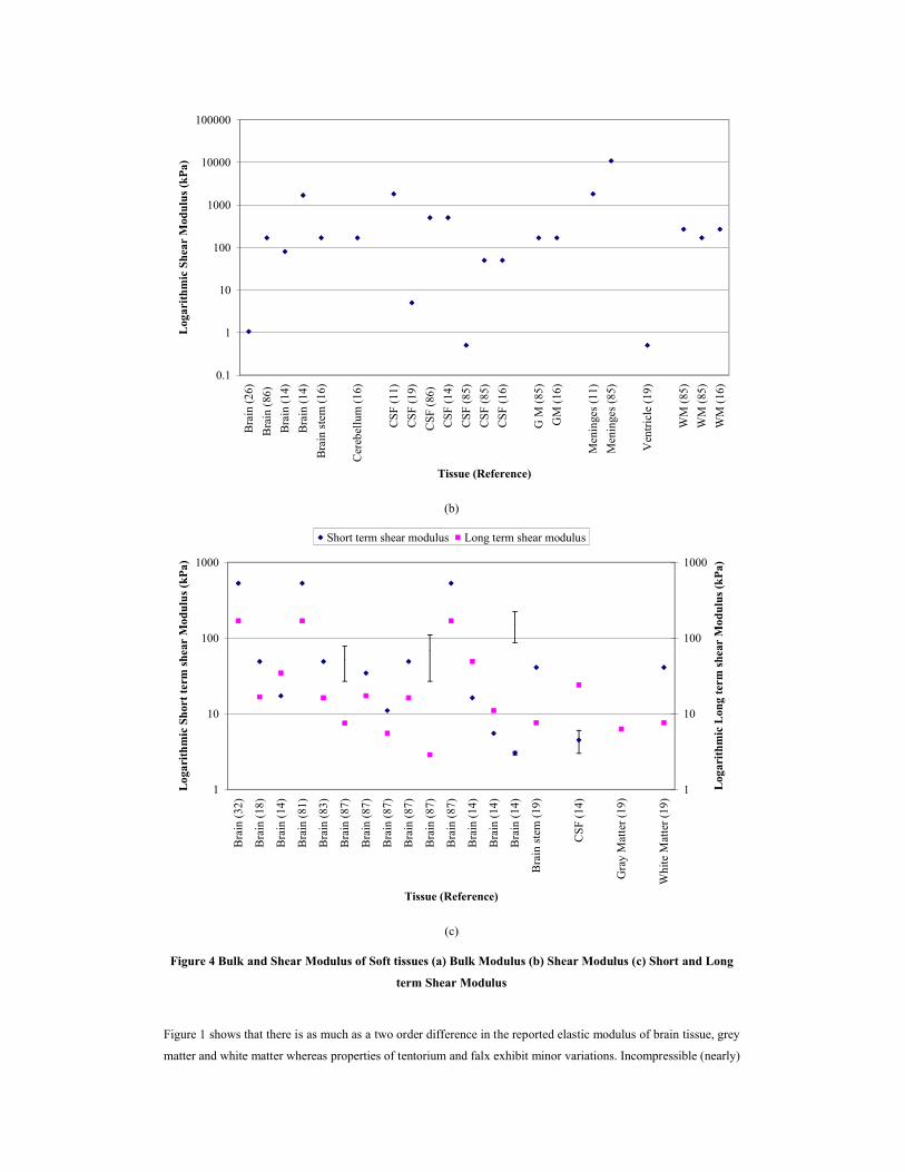
Figure 4 Bulk and Shear Modulus of Soft tissues (a) Bulk Modulus (b) Shear Modulus (c) Short and Long
term Shear Modulus
Figure 1 shows that there is as much as a two order difference in the reported elastic modulus of brain tissue, grey
matter and white matter whereas properties of tentorium and falx exhibit minor variations. Incompressible (nearly)

modeling is found to be widely used to model the soft tissues in head as the Poisson’s ratio indicated in Figure 2
ranges from 0.4 to 0.5. Estimation of density is comparatively easier than the other properties and as shown in
Figure 3, only minor variations are seen. Bulk modulus (Figure 4) varies in three orders of magnitude whereas
only very few studies on shear modulus (Figure 4 (b)) are found. Dynamic shear moduli, short term and long term,
properties (Figure 4 (c)) are available only for brain tissue but show a lot of variations.
NECK-SPINE
Neck injuries associated with excessive flexion-extension constitute the most prevalent trauma to occupants
involved in motor vehicle accidents33. Severe neck injuries are extremely devastating because of possible damage
to the cervical spinal cord as the cervical spine is responsible for the motion of the head as well as for protecting
the spinal chord from injuries34.
NECK-SPINE – ANATOMY OF SOFT TISSUES
Ligaments, intervertebral discs, cartilage, synovial membrane and muscles are the soft tissues in the neck-spine
region. Ligaments stabilize the joints in the spine and restrict its motion. The cervical spinal ligaments are divided
into lower cervical spinal ligaments and upper cervical spinal ligaments. The lower cervical spinal ligaments are
anterior longitudinal ligament (ALL), posterior longitudinal ligament (PLL), the capsular ligaments (CL) and
ligamentum flavum (LF) whereas the upper cervical spine include apical ligament, the alar ligament and
transverse ligament (TL). Articular cartilage reduces the friction in the zygapophysial joint during motion. The
main function of intervertebral discs is to resist the compressive loading. Detailed anatomy of the neck muscles
and its functions are well described by Mertz34 and the soft tissues in cervical part have been reviewed by
Yoganandan35.
NECK-SPINE INJURY – LOAD CASES
Whiplash injuries are the most common injuries in occupants in automobile collisions. In addition the neck and the
cervical spine are subjected to flexion (frontal collision), extension or hyperextension (rear end collision), lateral
bending (Side impacts) and axial loads (tensile during airbag deployment and compressive during roof contact).
EVOLUTION OF EXPERIMENTAL METHODS AND NECK INJURY TOLERANCE
IN this section we briefly review volunteer as well as cadaver studies used to obtain neck injury tolerance values
in various loading conditions. Mertz’s investigation on whiplash injuries in late sixties and early seventies is a
pioneer work in this area. A severity index (?? Name) for unsupported heads based on voluntary human tolerance
limits has been proposed based on his investigations of the kinematics and kinetics of whiplash injuries36. Further,
equivalent moment at the occipital condyle was proposed as the injury parameter for flexion and extension, and
for hyperextension and hyperflexion34. These studies indicate that the neck muscles significantly influence the
dynamic response of the spine by reducing the possibility of neck injury. Gadd37 presented an injury criterion
based on moment of the resistance offered by the neck in hyperextension and lateral flexion. Other criterion
proposed for injury assessment are based on moment-angle response of the neck in low severity direct head impact
loading38, cervical damage as a function of applied force39, dynamic tolerance for compressive loading40, shear
force and magnitude of eccentricity41. Passive responses of the cervical spine under torsion are time dependent42.
Cervical motion segments in bending and axial torsion exhibit lower stiffness than lumbar motion segments43.
Yoganandan44 conducted rear sled impact tests to determine soft tissue related injuries on the head-neck complex.
Injuries in soft tissues were reported on facet joints of the lower cervical spine. Svensson45 presented an injury
criterion using the pressure changes measured in the cervical spinal canal in swift extension-flexion using
anaesthetized pigs. Later, Bostrom46 developed a mathematical model to predict this pressure change as a function
of volume change inside the spinal canal during neck bending in the saggital plane. Subsequently neck injury
criterion (NIC), based on the relative acceleration and velocity between the top and the bottom of the cervical
spine and the muscle influence on NIC were presented46,47. Although the NIC is widely followed among other

injury assessment function for neck its inabilities towards representing the hyperextension injury mechanism and,
flexion motion after rebound is to be noted48. Also NIC was primarily developed using nonhuman subjects for low
velocity impacts, hence its validation for human injuries and for higher rates are still sought.
EVOLUTION OF FINITE ELEMENT MODELS
Though the above mentioned experimental studies were conducted with the soft tissues included, injuries can yet
not be related to tissue behaviour. Over the years many finite element models have evolved. These FE models aim
to help in injury assessment and overcome difficulties in cadaver testing such as the need of advanced
experimental facilities, tissue availability, tissue measurements and time intensive preparation and repeatability.
Kleinberger49 developed a 3D finite element model of human cervical spine for axial compression and frontal
flexion to study the gross vertebral kinematics and deformation using reported experimental data50. This FE model
included ALL, PLL, LF, CL and supraspinous ligament (SLL) as soft tissues but could not be validated for frontal
flexion. This suggested that advanced material models, like viscoelastic and hyperelastic could be better than
linear elastic model for modeling soft tissues. Lizee51 developed a total human body model in which the disks are
represented along with intervertebral joints. He has considered dynamic properties for thoracic and lumbar discs.
Nitsche52 developed a FE model of the human cervical spine and simulated volunteer tests53,54 for frontal, lateral
flexion and compression experiments40,50,55. Material properties of all components were assumed as homogenous
and linear elastic and were taken from Yamada56. Vertebrae and the intervertebral discs are considered as isotropic
whereas anisotropic material model was chosen for the articular cartilage and the ligaments. The fibers of the
ligaments are in the direction of the applied tensile force. The articular cartilages between Cl and C2 are modeled
with Young’s modulus for compression. The maximum displacement in the simulation was lower than the test
data because on use of linear elastic material properties. The variation in the material properties of the
intervertebral discs and ligament structures representing the soft tissues alters the angular motion and the stresses
in the inferior and the superior intervertebral discs of the cervical spine during flexion, extension, lateral bending
and axial torsion57. Young’s modulus of ligaments was found to have larger influence on the response whereas the
Poisson’s ratio of the spinal elements has little effect. Intervertebral discs transfer higher axial forces than shear
forces through different regions of the disc under axial and eccentric loads in the ventral region and vice-versa in
the dorsal region58. Facet joint anatomy idealized using a fluid model predict better response than hyperelastic
solid model for compression, flexion, extension and lateral bending59. Other reported finite element models
include a heck neck model60 for rear impact velocities up to 2.6 m/s and a cervical spine model61 to study the spine
motion and analysis of C4-C6 unit62.
MECHANICAL PROPERTIES OF SOFT TISSUE
A series of review on mechanical behavior of the cervical vertebrae and the soft-tissues of the cervical spine have
been reported63,64,65,66. Structural properties measured in tensile failure load on isolated ligaments reveals that the
ALL is the strongest and the PLL the weakest among the spinal ligaments studied67. Other ligaments included in
this study were interspinous ligament (ISL), LF and joint capsule, which were tested for displacement rates
ranging from 1 to 100 cm/s. Significant differences were reported between the animal and human ligaments in
terms of structural properties as human ligaments are two to five times stronger than those of monkeys67. Posterior
ligament tear under flexion and anterior longitudinal ligament tear under extension under dynamic loads40. The
ligamentous upper cervical spine was significantly stronger in extension than in flexion where as upper cervical
spine was stronger than the lower cervical spine in extension68. Segmental motions are statistically greater for
females than for males at C2–C3, C4–C5, C5– C6, and C6–C7 levels, indicating that female soft tissues sustain
greater magnitudes of stretch in rear impact69. Shear stiffness plays a major role in the stabilization of cadaver
lumbar motion exhibits rate and directional dependency70.

0.01
0.1
1
10
100
AL
L (
57)
AL
L (
58)
AL
L (
78)
AL
L (
78)
AL
L (
78)
Art
icul
ar C
artil
age
(52)
Art
icul
ar c
artil
age
(59)
Cap
sula
r L
igam
ent (
57)
Cap
sula
r L
igam
ent (
58)
Cap
sula
r L
igam
ent (
78)
Cap
sula
r L
igam
ent (
78)
Cap
sula
r L
igam
ent (
78)
Dis
c an
nulu
s (5
8)D
isc
annu
lus
(59)
Dis
c an
nulu
s (6
1)D
isc
annu
lus
(62)
Dis
c an
nulu
s (7
8)D
isc
annu
lus
(78)
Dis
c an
nulu
s (7
8)D
isc
annu
lus
(78)
Dis
c nu
cleu
s (5
8)D
isc
nucl
eus
(61)
Dis
c nu
cleu
s (6
2)D
isc
nucl
eus
(78)
ISL
(57
)IS
L (
58)
ISL
(78
)IS
L (
78)
ISL
(78
)L
F (5
7)L
F (5
8)L
F (7
8)L
F (7
8)L
F (7
8)PL
L (
57)
PLL
(58
)PL
L (
78)
PLL
(78
)PL
L (
78)
SSL
(78
)SS
L (
78)
SSL
(78
)
Tissue (Reference)
Log
arit
hm
ic E
last
ic M
odu
lus
(MP
a)
Figure 5 Elastic Modulus of the soft tissue in neck and spine region
0
0.1
0.2
0.3
0.4
0.5
0.6
AL
L (
49)
AL
L (
57)
AL
L (
58)
Art
icul
ar C
artil
age
(52)
Art
icul
ar c
artil
age
(59)
Cap
sula
r L
igam
ent (
57)
Cap
sula
r L
igam
ent (
58)
Dis
c an
nulu
s (5
8)
Dis
c an
nulu
s (5
9)
Dis
c an
nulu
s (5
9)
Dis
c an
nulu
s (6
1)
Dis
c an
nulu
s (6
2)
Dis
c an
nulu
s (7
8)
Dis
c an
nulu
s (7
8)
Dis
c an
nulu
s (7
8)
Dis
c an
nulu
s (7
8)
Dis
c nu
cleu
s (5
8)
Dis
c nu
cleu
s (6
1)
Dis
c nu
cleu
s (6
2)
Dis
c nu
cleu
s (7
8)
ISL
(57
)
ISL
(58
)
LF
(49)
LF
(57)
LF
(58)
PLL
(49
)
PLL
(57
)
PLL
(58
)
SSL
(49
)
TL
(49
)
Tissue (Reference)
Poi
sson
's R
atio
(M
Pa)
Figure 6 Poisson’s Ratio of the soft tissue in neck and spine region
Failure loads of endplate and vertebral body of human lumbar vertebrae show rate dependency in compressive
impact loads71. A kinematic analysis of head and neck unit conducted on cadavers indicates that the intervertebral
disc is the most frequently injured tissue in frontal and lateral collisions, followed by LF in C1 to T4 region72.
Several other studies have been performed on intervertebral disc to study its response to compressive73,74 and
dynamic loading75. Annulus fibrosus exhibits anisotropic shear properties through separate contributions from the
matrix, the collagen fibers, and collagen fiber interactions76. Significant variations have been found in the shear

modulus between the outer and inner annulus and influence of pre-strain in shear modulus. A linear material
model with fiber-induced anisotropic behavior of annulus fibrosus has been proposed by Elliot77 under tensile
loading. Variation in the material properties of disc annulus has a significant influence on both the external
biomechanical response and internal stress of the disc annulus and its neighboring hard bones78.
The reported elastic modulus in the ALL and CL varies by a factor exceeding 2 while in the PLL, ISL and SLL the
variation is less (Figure 5). Elastic modulus of disc annulus found to have little variation in most studies. Few data
on Poisson’s ratio of individual ligament tissues suggests the need for more tests (Figure 6). Dynamic properties
are rarely reported in terms of short term and long term shear modulus for constituting the behavior in viscoelastic
model.
Studies reported in the above section indicate the following observations in both head and neck-spine regions,
1. Most of the studies are performed at lower strain rates and methods to characterize the tissue at higher
strain rates and related properties are needed.
2. Nonlinear viscoelasticity, anisotropy and rate dependency are not well characterized and more tissue
level experiments are needed.
3. Muscles predominate the response in the neck spine region and are capable of altering the kinetics and
kinematics of head. But studies including muscle behavior and its active tones are very few.
4. Advanced material models capable of predicting the above behavior have to be developed.
CONCLUSIONS
A body of knowledge about mechanical properties of soft tissues in head, neck and spine regions, assembled in
recent years, is collated. Reported experimental methods, injury assessment functions and finite element models
are investigated. Scatter and uncertainty among the reported material properties of soft tissues are observed on
both static and dynamic properties. It is felt that isolated specimen tests aimed at developing the material models
needed in finite element analysis should be prioritized. This will help understand the complex behavior of these
tissues and subsequently aid in injury prediction using finite elements.
REFERENCES
1. I Watanabe, K Furusu, C Kato, K Miki, J Hasegawa. ‘Development of practical and simplified human whole
body FEM model’. JSAE Review, vol. 22, 2001. pp. 189 – 194.
2. M Iwamoto, Y Kisanuki, I Watanabe, K Furusu, K Miki, J Hasegawa. ‘Development of a finite element
model of the total human model for Safety THUMS and application to injury reconstruction’. Proceedings of
the 2002 International IRCOBI Conference on the Biomechanics of Impact, Munich, Germany, 2002, pp. 31
– 42.
3. R M Morgan, R H Eppinger, B C Hennessey. ‘Ankle joint injury mechanism for adults in frontal automotive
impact’. Proceedings of the 35th Stapp Car Conference, SAE Paper number 912902, 1991.
4. M Beaugonin, E Haug, D Cesari. ‘A numerical model of the human ankle/foot under impact loading in
inversion and eversion’. Proceedings of the 40th Stapp car conference, SAE Paper Number 962428, 1996.
5. Y Mizuno. ‘Summary of IHRA Pedestrian safety WG activites (2005) - proposed test methods to evaluate
pedestrian protection afforded by passenger cars’. Proceedings of the 19th International Technical
Conference on the Enhanced Safety of Vehicles. 2005.
6. T B Khalil, R P Hubbard. ‘Parametric study of head response by finite element modeling’. Journal of
Biomechanics. vol. 10, 1977. pp. 119 – 132.
7. J S H M Wismans, E G Janssen, M Beusenberg, W P Koppens, H A Lupker. ‘Injury Biomechanics - Course
Notes’. Faculty of Mechanical Engineering - Division of computational and experimental mechanics -
Eindhoven University of Technology, 1994.

8. H Gray. ‘Anatomy of the Human Body’. Philadelphia: Lea & Febiger, 1918; Bartleby.com, 2000.
www.bartleby.com/107/
9. J Versace. ‘A review of the severity index’. Proceedings of the 15th Stapp Car Crash Conference, SAE Paper
710881, 1971.
10. J Newman. C Barr, M Beusenberg, E Fournier, N Shewchenko, E Welbourne, C Withnall. ‘A New
Biomechanical Assessment of Mild Traumatic Brain Injury Part 2: Results and Conclusions’. Proceedings of
the 2000 IRCOBI International Conference on the Biomechanics of Impact, Montpellier, France, 2000, pp.
223-233.
11. D W A Brands. 'Predicting brain mechanics during closed head impact: numerical and constitutive aspects'.
Ph.D. Dissertation, Eindhoven University of Technology, Eindhoven, The Netherlands, 2002.
12. T B Khalil, R P Hubbard. ‘Parametric study of head response by finite element modeling’, Journal of
Biomechanics, vol 10, 1977, pp. 119 – 132.
13. T B Khalil , Viano D C. ‘Critical issues in finite element modeling of head impact’. Proceedings of the 26th
Stapp Car Conference, SAE Paper number 821150, 1982.
14. A A H J Sauren, M H A Claessens. ‘Finite element modeling of head impact: The second decade’.
Proceedings of the 2000 IRCOBI International Conference on the Biomechanics of Impact, Eindhoven, The
Netherlands, 1993, pp. 241-254.
15. K K Mendis, R L Stalnaker, S H Advani. ‘A constitutive relationship for large deformation finite element
modeling of brain tissue’. Journal of biomechanical engineering, vol 117, 1995. pp. 279-285.
16. C Zhou, T B Khalil, A I King. ‘A new model comparing impact responses of the homogeneous and
inhomogeneous human brain’. Proceedings of the 39th Stapp Car Conference, SAE Paper number 952714,
1995.pp. 122-137.
17. M Claessens, F Sauren, J Wismans. ‘Modelling of the human head under impact conditions: A parametric
study’. Transactions of SAE, SAE Paper number 973338, 1997, pp. 3829 – 3848.
18. H S Kang, R Willinger, B M Diaw, B Chinn. ‘Validation of a 3D anatomic human head model and
replication of head impact in motorcycle accident by finite element modelling’. Transactions of SAE, SAE
Paper number 973339, 1997, pp. 3849 – 3858.
19. J Newman, M Beusenberg, E Fournier, N Shewchenko, C Withnall, L Thibault, G McGinnis. ‘A new
biomechanical assessment of mild traumatic brain injury Part I – Methodology’. Proceedings of the 1999
IRCOBI International Conference on the Biomechanics of Impact, Sitges, Spain, 1999, pp. 17 – 36
20. K Miller, K Chinzei. ‘Constitutive modelling of brain tissue experiments and theory’. Journal of
Biomechanics, vol 30, 1997, pp. 1115-1121.
21. K Miller. ‘Constitutive model of brain tissue suitable for finite element analysis of surgical procedures’.
Journal of Biomechanics, vol 32, 1999, pp. 531-537.
22. J-C Sarron, C Blondeau, A Guillaume, D Osmont. ‘Identification of linear viscoelastic constitutive models’.
Journal of Biomechanics, vol 33, 2000, pp. 685 – 693.
23. K Miller. ‘How to test very soft biological tissues in extension’, Journal of Biomechanics, vol 34, 2001, pp.
651 – 657.
24. K Miller, K Chinzei. ‘Mechanical properties of brain tissue in tension’. Journal of Biomechanics, vol 35,
2002, pp. 483 – 490.
25. K Miller, K Chinzei, G Orssengo, P Bednarz. ‘Mechanical properties of brain tissue in-vivo: experiment and
computer simulation’. Journal of Biomechanics, vol 33, 2000, pp. 1369-1376.
26. S S Kkyriacou, A Mohamed, K Miller, S Neff. ‘Brain Mechanics for Neurosurgery: modeling issues’.
Biomechanical Modeling and Mechanobiology vol 1, 2002, pp. 151 – 164.

27. K Miller. ‘Method of testing very soft biological tissues in compression’. Journal of Biomechanics, vol 38,
2005, pp. 153-158.
28. K B Arbogast, S S Margulies. ‘Mechanical characterization of the brainstem from oscillatory shear tests’.
Journal of Biomechanics, vol 31, 1998, pp. 801-807.
29. K B Arbogast, S S Margulies. ‘A fiber-reinforced composite model of the viscoelastic behavior of the
brainstem in shear’. Journal of Biomechanics, vol 32, 1999, pp. 865-870.
30. K K Darvish, J R Crandall. ‘Nonlinear viscoelastic effects in osciallatory shear deformation of brain tissue’.
Medical Engineering & Physics, vol 23, 2001, pp. 633-645.
31. D W A Brands, G W M Peters, P H M Bovendeerd. ‘Design and numerical implementation of a 3-D non-
linear constitutive model for brain tissue during impact’. Journal of Biomechanics, vol 37, 2004. pp. 127-134.
32. T Aida. ‘Study of human head impact: brain tissue constitutive models’, Ph.D. Dissertation, West Virginia
University, Morgan Town, United States of America, 2000.
33. K Langweider, S H Backaitis, F William, S Partyka, A Ommaya. ‘Comparative studies of neck injuries of car
occupants in frontal collisions in the United States and in the Federal Republic of Germany’. Proceedings of
the 25th Stapp Car Conference, SAE Paper number 811030, 1981, pp. 71-127.
34. H J Mertz, L M Patrick. ‘Strength and response of the human neck’. Proceedings of the 14th Stapp Car
Conference, SAE Paper number 710855, 1971, pp. 207 – 255.
35. N Yoganandan, S Kumaresan, F A Pintar. ‘Biomechanics of the cervical spine Part 2. Cervical spine soft
tissue responses and biomechanical modeling’. Clinical Biomechanics, vol 16, 2001, pp. 1-27.
36. H J Mertz, L M Partick. Investigations of the kinematics and kinetics of whiplash during vehicle rear-end
collisions, Proceedings of the Eleventh Stapp Car Conference, SAE Paper number 670919, 1967, pp. 267-
317.
37. C W Gadd, C C Culver, A M Nahum. ‘Study of responses and tolerances of the neck’. Proceedings of the
15th Stapp Car Conference, SAE Paper number 710856, pp. 256-268.
38. K Ono, K Kaneoka, E A Sun, E G Takhounts, R H Eppinger. ‘Biomechanical response of human cervical
spine to direct loading of the head’. Proceedings of the 2001 IRCOBI International Conference on the
Biomechanics of Impact, Isle of Man, United Kingdom, 2001, pp. 189 – 199.
39. F A Pintar, A Jr Sances, N Yoganandan, J Reinartz, D J Maiman, J K Suh, G Unger, J F Cusick, S J Larson.
‘Biodynamics of the total human cadaver cervical spine’. Proceedings of the 34th Stapp Car Conference,
SAE Paper number 902309, 1990, pp. 55-72.
40. F A Pintar, N Yoganandan, L Voo, J F Cusick, D J Maiman, A Jr Sances. ‘Dynamic Characteristics of
Human Cervical Spine’. Proceedings of the 39th Stapp Car Conference, SAE Paper number 952722, 1995,
pp. 195-202.
41. J H McElhaney, J G Paver, B S Myers, L Gray. ‘Combined bending and axial loading responses of the
human cervical spine’. Proceedings of the 32nd Stapp Car Conference, SAE Paper number 8817709, 1988,
pp.21-28.
42. J H McElhaney, G P Jecquellne, B M Mayers, G Linda. ‘Responses of Human Cervical Spine to Torsion’.
Proceedings of the 33rd Stapp Car Conference, SAE Paper number 892437. 1989, pp. 215-222.
43. S P Moroney, A B Schultz, J A A Miller, G B J Andersson. ‘Load-displacement properties of lower cervical
spine motion segments’. Journal of Biomechanics, 21, 1998, pp. 769 – 779.
44. N Yoganandan, F A Pintar, B D Stemper, J F Cusick, R D Rao, T A Gennarelli. ‘Single rear impact produces
lower cervical soft tissue injuries’. Proceedings of the 2001 IRCOBI International Conference on the
Biomechanics of Impact, Isle of Man, United Kingdom, 2001, pp. 201-211.
45. M Y Svensson, B Aldman, H A Hansson. ‘Pressure effects in the spinal canal during whiplash extension
motion: a possible cause of injury to the cervical spinal ganglia’. Proceedings of the 1993 International
IRCOBI Conference on the Biomechanics of Impact, Eindhoven, The Netherlands, 1993, pp. 189–200.
46. O Bostrom, M Y Svennson, B Aldman, H A Hansson, Y Haland, P Lovsund, T Seeman, A Suneson, A Saijo,
T Ortengren. ‘A new neck injury criterion candidate-based on injury findings in the cervical spine ganglia
after experimental neck extension trauma’. Proceedings of the 1996 International (IRCOBI) Conference on
the Biomechanics of Impact, Dublin, Ireland. 1996, pp. 123-136.
47. O Bostrom, , M Krafft, B Aldman, A Eichberger, R Fredriksson, Y Haland, P Lovsund, P Lovsund, H
Steffan, M Y Svennson, C Tingvall. ‘Predictions of neck injuries in rear impacts based on accident data and
simulations’. Proceedings of the 1997 International IRCOBI Conference on the Biomechanics of Impact,
Hannover, Germany, 1997, pp. 251-264.
48. A C Croft, P Herring, M D Freeman, M T Haneline. ‘The neck injury criterion: future considerations’.
Accident Analysis and Prevention, vol 34, 2002, pp. 247-255.
49. M Kleinberger, Application of finite element techniques to the study of cervical spine mechanics.
Proceedings of the 37th Stapp Car Conference, SAE Paper number 933131, 1993, pp. 261 – 272.
50. F A Pintar, N Yoganandan, A Jr Sancee, J Reinartz, G Harris, S J Laroan. ‘Kinematic and anatomical
analysis of human cervical spinal column under axial loading’. Proceedings of the 33rd Stapp Car
Conference, SAE Paper number 892436, 1989, pp. 191 – 214.
51. E Lizee, S Robin, E Song, N Bertholan, J Y Lecoz, B Besnault, F Lavaste. ‘Development of 3D Finite
Element Model of the Human Body’. SAE Transactions, SAE Paper number 983152, 1998. pp. 2760 – 2782.
52. S Nitsche, G Krabbel, H Appel, E Haug. ‘Validation of a finite-element-model of the human neck’.
Proceedings of the 1996 International IRCOBI Conference on the Biomechanics of Impact, Dublin, Ireland,
1996. pp.107 – 122.
53. J Wismans, E V Oorschot, H J Woltring. ‘Omni-directional human head-neck response’. Proceedings of the
30th Stapp Car Conference, SAE Paper number 861893, 1986.
54. J Wismans, M Philippens, E V Oorschot, D Kallieris, R Mattern. 1987. Comparison of human volunteer and
cadaver head-neck response in frontal flexion’. Proceedings of the 31st Stapp Car Conference, SAE Paper
number 872194, 1987.
55. F A Pintar, J A Sances, N Yoganandan, J Reinhartz, S J Larson, C Kurakami, W Rauschning. ‘Injury
Biomechanics of the Head-Neck Complex’. Proceedings of the 12th conference on enhanced safety of
vehicles (ESV), 1989.
56. H Yamada. ‘Strength of biological materials’. F G Evans. The Williams & Wilkins Company, Baltimore.
1970.
57. S Kumaresan, N Yoganandan, F A Pintar. ‘Finite element analysis of the cervical spine: a material property
sensitivity study’. Clinical Biomechanics, vol 14, 1999, pp. 41 – 53.
58. S Kumaresan, N Yoganandan, F A Pintar, D J Maiman. ‘Finite element modeling of the cervical spine: role
of intervertebral disc under axial and eccentric loads’. Medical Engineering & Physics, vol 21, 1999, pp. 689-
700.
59. S Kumaresan, N Yoganandan, F A Pintar. ‘Finite element modeling approaches of human cervical spine facet
joint capsule’. Journal of Biomechanics, vol 31, 1998, pp. 371-376.
60. B D Stemper, N Yoganandan, F A Pintar. ‘Validation of a head-neck computer model for whiplash
simulation’. Medical Biological Engineering & Computings, vol 42, 2004. pp. 333-338.
61. J S S Wu, J H Chen. ‘Clarification of the mechanical behaviour of spinal motion segments through a three-
dimensional poroelastic mixed finite element model’. Medical Engineering Physics, vol 18, 1996. pp. 215-
224.
62. N Yoganandan, S C Kumaresan, L Voo, F A Pintar, S J Larson. ‘Finite element modeling of the C4-C6
cervical spine unit’. Medical Engineering Physics, vol 18, 1996, pp. 569-574.
63. N Bogduk, S Mercer. ‘Biomechanics of the cervical spine. I: Normal kinematics’. Clinical Biomechanics, vol
15, 2000, pp. 633-648.
64. N Yoganandan, S Kumaresan, F A Pintar. ‘Biomechanics of the cervical spine Part2. Cervical spine soft
tissue responses and biomechanical modeling’. Clinical Biomechanics, vol 16, 2001, pp. 1-27.
65. N Bogduk, N Yoganandan. ‘Biomechanics of the cervical spine Part3: minor injuries’. Clinical
Biomechanics, vol 16, 2001, pp. 267-275.
66. J F Cusick, N Yoganandan. ‘Biomechanics of the cervical spine 4: major injuries’, Clinical Biomechanics,
vol 17, 2002, pp. 1-20.
67. J Myklebust, A Jr Sances, D Maiman, F Pintar, M Chilbert, W Rausching, S Larson, J Cusick, C Ewing, D
Thomas, B Saltzberg. ‘Experimental spinal trauma studies in the human and monkey cadaver’. Proceedings
of the 27th Stapp Car Conference, SAE Paper number 83164, 1983, pp. 149-161.
68. R W Nightingale, B A Winkelstein, K E Knaub, W J Richardson, J F Luck, B S Myers. ‘Comparative
strengths and structural properties of the upper and lower cervical spine in flexion and extension’. Journal of
Biomechanics, vol 35, 2002, pp. 725-732.
69. B D Stemper, N Yoganandan, F A Pintar. ‘Gender dependent cervical spine segmental kinematics during
whiplash’. Journal of Biomechanics, vol 36, 2003, pp. 1281-1289.
70. P C Begeman, H Visarlus, L P Nolte, P Prasad. ‘Viscoelastic shear responses of the cadaver and Hybrid III
lumbar spine, Proceedings of the 38th Stapp Car Conference, SAE Paper number 942205, 1994, pp. 1-14.
71. R S Ochia A F Tencer, R P Ching. ‘Effect of loading rate on endplate and vertebral body strength in human
lumbar vertebrae’, Journal of Biomechanics, vol 36, 2003, pp. 1875-1881.
72. D Kallieris, R Mattern, E Miltner, G Schmiddt, K Stein. ‘Considerations for a neck injury criterion.
Proceedings of the 35th Stapp Car Conference, SAE Paper number 912916, pp.401-417.
73. J B Martinez, V O A Oloyede, N D Broom. ‘Biomechanics of load-bearing of the intervertebral disc: an
experimental and finite element model’. Medical Engineering Physics, vol 19, 1997, pp. 145-156.
74. M Kasra, V Goel, J Martin, S-T Wang, W Choi, J Buckwalter. ‘Effect of dynamic hydrostatic pressure on
rabbit intervertebral disc cells’. Journal of Orthopaedic Research, vol 21, 2003, pp. 597-603.
75. A J L Walsh, J C Lotz. ‘Biological response of the intervertebral disc to dynamic loading’. Journal of
Biomechanics, vol 37, 2004, pp. 329-337.
76. D M Elliott, L A Setton. ‘Anisotropic and inhomogeneous tensile behavior of the human anulus fibrosus:
Experimental measurements and material model predictions’. Journal of Biomechanical Engineering, vol
123, 2001, pp. 256-263.
77. D M Elliott, L A Setton. ‘A linear material model for fiber-induced anisotropy of the annulus fibrosus’.
Journal of Biomechanical Engineering, vol 122, 2000, pp. 173-179.
78. H W Ng, E C Teo, V S Lee. ‘Statistical factorial analysis on the material property sensitivity of the
mechanical responses if the C4-C6 under compression, anterior and posterior shear’. Journal of
Biomechanics, vol 37, 2004, pp. 771-777.
79. R W G Anderson, C J Brown, P C Blumbergs, G Scott, J W Finney, N R Jones, A J McLean. ‘Mechanisms
of axonal injury: an experimental and numerical study of a sheep model of head impact’. Proceedings of
1999 International IRCOBI Conference on the Biomechanics of Impact, Sites, Spain, 1999, pp. 107-120.
80. J S Ruan, P Prasad. ‘Study of the biodynamic characteristics of the human head’. Proceedings of 1996
International IRCOBI Conference on the Biomechanics of Impact, Dublin, Ireland, 1996, pp. 63-74.
81. F Turquler, Ho Sung, Kang, X Trossellie, R Willinger, F Lavaste, C Tarriere, A Domont. ‘Validation of a 3D
finite element head model against experimental data’. Proceedings of the 40th Stapp Car Crash Conference,
SAE Paper 962431.
82. R Willinger, L Taleb, P Pradoura. ‘Head biomechanics: From the finite element model to the physical
model’. Proceedings of the 1995 International IRCOBI Conference on the Biomechanics of Impact, Brunnen,
Switzerland, 1995, pp. 245-259.
83. R Willinger, D Baumgartner, B Chinn, M Neale. ‘Head tolerance limits derived from numerical replication of
real world accidents’ Proceedings of the 2000 International IRCOBI Conference on the Biomechanics of
Impact, Montpellier, France, 2000, pp. 209-221.
84. M I Miga, K D Paulsen, P J Hoopes, F E Kennedy, A Hartov, D W Roberts. ‘In vivo modeling of interstitial
pressure in the brain under surgical load using finite element’. Journal of Biomechanical Engineering, vol
122, 2000, pp. 354-363.
85. C Zhou, T B Khalil, A I King. ‘Shear stress distribution in the porcine brain due to rotational impact’
Proceedings of the 38th Stapp Car Crash Conference, SAE Paper 942214, 1994, pp. 133-143.
86. T Nishimoto, S Murakamai. ‘Direct impact simulations of diffuse axonal injury by axial head model’. JSAE
Review, vol 21, 2000, pp. 117-123.
87. C Zhou, T B Khalil, A I King. ‘Viscoelastic response of the human brain to sagittal and lateral rotational
acceleration by finite element analysis’. Proceedings of the 1996 International IRCOBI Conference on the
Biomechanics of Impact, Dublin, Ireland, 1996, pp. 35-48.

APPENDIX –A- MECHANICAL PROPERTIES OF SOFT TISSUE RELATED TO HEAD
Table A1. Elastic Modulus of soft tissues
References (Other sources cited therein) Soft tissue
Elastic modulus (MPa)
Brain
32 (Chu 1994) Brain 2.50E-01
32 (Hosey 1982) Brain 6.67E-0232 (Ruan 1991) Brain 6.67E-02
32 (Ruan 1993) Brain 5.06-5.26
32 (Ward 1980) Brain 6.67E-02
79 (Claessens 1997) Brain 1.00E-01
17 (Average) Brain 1.00E+00
17 (Bandak 1995) Brain 6.80E+01
17 (Kumaresan 1996) Brain 6.67E-02
17 (Ruan 1991) Brain 5.04E+00
17 (Ruan 1996) Brain 5.58E-01
26 (Kaczmarek) Brain 1.00E-02
26 (Miga 2000) Brain 2.10E-03
26 (Miller 1997) Brain 3.16E-03
80 (Ruan 1993) Brain 5.58E-0122 (Dimasi 1991) Brain 5.10E-02
22 (Galford 1970) Brain 1.72-2.20E-02
22 (Galford 1970) Brain 1.51-1.65E-02
22 (Hirakawa 1981) Brain 2.04-9E-02
22 (Hosey 1980 Brain 6.67E-02
22 (Koeneman 1966) Brain 1.50E-02
22 (Kumaresan 1996) Brain 6.67E-02
22 (Mendis 1995) Brain 8.24E-03
22 (Miller 1997) Brain 1.00E-03
22 (Ruan 1991) Brain 6.67E-02
22 (Sahay 1992) Brain 3.40E-02
22 (Ueno 1995) Brain 2.40E-01
22 (Ward 1978) Brain 6.67E-02
14 (Chu 1991) Brain 2.50E-0114 (Ruan 1991a) Brain 6.67E-02
14 (Trosseille 1992) Brain 2.40E-01
14 (Willinger 1992) Brain 6.75E-01
81 (NA) Brain 6.75E-01
82 (Ward 1975, Ruan 1992) Brain 6.75E-01
14 (Lee 1990) Brain (gel) 80-121.2E-03
17 (Average) Brainstem 1.00E+00
Cerebellum17 (Average) Cerebellum 1.00E+00
17 (Average) Cerebrum 1.00E+00
CSF83 (Zhou 1996) CSF 1.20E-02
80 (Ruan 1993) CSF 1.49E-01
14 (Ruan 1991a) CSF 6.67E-02
82 (Ward 1975, Ruan 1992) CSF 10-100E-03
References (Other sources cited therein) Soft tissue
Elastic modulus (MPa)
Dura19 (Shuk 1970) Dura 3.15E+01
80 (Ruan 1993) Dura 3.15E+01
14 (DiMasi 1991a, 1991b) Dura 6.89E+00
14 (Ruan 1991a) Dura 3.15E+01
16 (Ruan 1994) Dura 3.15E+01
Face17 (Average) Face 6.50E+03
83 (Zhou 1996) Face 5.00E+03
Falx83 (Zhou 1996) Falx 3.15E+0117 (Ruan 1991) Falx 3.15E+01
18 (Shuck 1972) Falx 3.15E+01
19 (Shuk 1970) Falx 3.15E+01
80 (Ruan 1993) Falx 3.15E+01
14 (Ruan 1991a) Falx 3.15E+01
81 (NA) Falx 3.15E+01
16 (Ruan 1994) Falx 3.15E+01
82 (Ward 1975, Ruan 1992) Falx 3.15E+01
Gray Matter
17 (Zhou 1995) Gray Matter 5.00E-01
17 (Zhou 1996) Gray Matter 1.88-10.1E-02
84 (Nagashima 1990, Baser 1992, Kalyanasundaram 1997) Gray Matter 2.10E-03
Pia19 (Shuk 1970) Pia 1.15E+01
16 (Ruan 1994) Pia 1.15E+01
Scalp83 (Khalil 1977) Scalp 1.67E+01
18 (Shuck 1972) Scalp 1.67E+01
80 (Ruan 1993) Scalp 1.67E+01
16 (Ruan 1994) Scalp 1.67E+01
6 Scalp Layer 3.45E+01
Skin19 (Shuk 1970) Skin 1.67E+01
Tentorium83 (Zhou 1996) Tentorium 3.15E+01
79 (Ruan 1997) Tentorium 3.15E+01
17 (Ruan 1991) Tentorium 3.15E+01
18 (Shuck 1972) Tentorium 3.15E+01
19 (Shuk 1970) Tentorium 3.15E+01
14 (Ruan 1991a) Tentorium 3.15E+01
81 (NA) Tentorium 3.15E+01
16 (Ruan 1994) Tentorium 3.15E+01
82 (Ward 1975, Ruan 1992) Tentorium 3.15E+01

References (Other sources cited therein) Soft tissue
Elastic modulus (MPa)
White Matter17 (Zhou 1995) White Matter 8.00E-01
17 (Zhou 1996) White Matter 2.27-12.2E-02
84 (Nagashima 1990, Baser 1992, Kalyanasundaram 1997) White Matter 2.10E-03
Table A2. Poisson’s ratio of soft tissues
References (Other sources cited therein) Soft tissue
Poisson's ratio
Brain32 (Chu 1994) Brain 0.49
32 (Hosey 1982) Brain 0.48
32 (Ruan 1991) Brain 0.48
32 (Ruan 1993) Brain 0.4996
32 (Ward 1980) Brain 0.4879 (Claessens 1997) Brain 0.46
17 (Average) Brain 0.48
17 (Bandak 1995) Brain 0.48
17 (Kumaresan 1996) Brain 0.48
17 (Ruan 1991) Brain 0.499
17 (Ruan 1996) Brain 0.499
26 (Miga 2000) Brain 0.45
26 (Miller 1997) Brain 0.499
80 (Ruan 1993) Brain 0.499
22 (Dimasi 1991) Brain 0.4998
22 (Khalil 1982) Brain 0.4996
22 (Kumaresan 1996) Brain 0.48
22 (Mendis 1995) Brain 0.522 (Miller 1997) Brain 0.5
22 (Ruan 1991) Brain 0.45-0.49999
22 (Ruan 1994) Brain 0.4996
22 (Sahay 1992) Brain 0.5
22 (Ueno 1995) Brain 0.49
22 (Wang 1972) Brain 0.5
22 (Ward 1978) Brain 0.48
14 (Chu 1991) Brain 0.49
14 (Lee 1987, Lighthall 1989, Ueno 1989, Ueno 1991) Brain 0.475 &0.49
14 (Ruan 1991b) Brain 0.4996
14 (Ruan 1991a) Brain0.48-0.49999492
14 (Trosseille 1992) Brain 0.49-0.499
14 (Willinger 1992) Brain 0.48
81 (NA) Brain 0.48
82 (Ward 1975, Ruan 1992) Brain 0.48
14 (Cheng 1990) Brain (gel) 0.5
References (Other sources cited therein) Soft tissue
Poisson's ratio
14 (Galbraith 1988, Tong 1989) Brain (gel) 0.4995
14 (Lee 1990) Brain (gel) 0.49
16 (Ruan 1994) Brain stem 0.4996
17 (Average) Brainstem 0.4
Cerebellum17 (Average) Cerebellum 0.48
16 (Ruan 1994) Cerebellum 0.4996
Cerebrum17 (Average) Cerebrum 0.48
CSF83 (Zhou 1996) CSF 0.49
80 (Ruan 1993) CSF 0.485
14 (Ruan 1991b) CSF 0.486
14 (Ruan 1991a) CSF 0.499
14 (Trosseille 1992) CSF 0.49999
85 (Ruan 1991) CSF 0.49999
16 (Ruan 1994) CSF 0.4996
82 (Ward 1975, Ruan 1992) CSF 0.499
Dura19 (Shuk 1970) Dura 0.4580 (Ruan 1993) Dura 0.45
14 (Ruan 1991a) Dura 0.45
16 (Ruan 1994) Dura 0.45
Face17 (Average) Face 0.22
83 (Zhou 1996) Face 0.23
Falx83 (Zhou 1996) Falx 0.45
17 (Ruan 1991) Falx 0.45
18 (Shuck 1972) Falx 0.23
19 (Shuk 1970) Falx 0.45
80 (Ruan 1993) Falx 0.45
14 (Ruan 1991a) Falx 0.45
81 (NA) Falx 0.4516 (Ruan 1994) Falx 0.45
82 (Ward 1975, Ruan 1992) Falx 0.49
Gray Matter85 (Ruan 1991) Gray Matter 0.4996
17 (Zhou 1995) Gray Matter 0.499
84 (Nagashima 1990, Baser 1992, Kalyanasundaram 1997) Gray Matter 0.45
16 (Ruan 1994) Gray Matter 0.4996
Meninges85 (Ruan 1991) Meninges 0.45
Pia19 (Shuk 1970) Pia 0.42
16 (Ruan 1994) Pia 0.45

References (Other sources cited therein) Soft tissue
Poisson's ratio
Scalp Layer83 (Khalil 1977) Scalp 0.42
18 (Shuck 1972) Scalp 0.4280 (Ruan 1993) Scalp 0.42
16 (Ruan 1994) Scalp 0.42
6 Scalp layer 0.4
Tentorium79 (Ruan 1997) Tentorium 0.45
83 (Zhou 1996) Tentorium 0.45
17 (Ruan 1991) Tentorium 0.45
18 (Shuck 1972) Tentorium 0.23
19 (Shuk 1970) Tentorium 0.22
14 (Ruan 1991a) Tentorium 0.45
81 (NA) Tentorium 0.45
16 (Ruan 1994) Tentorium 0.45
82 (Ward 1975, Ruan 1992) Tentorium 0.45
White Matter
17 (Zhou 1995) White Matter 0.499
84 (Nagashima 1990, Baser 1992, Kalyanasundaram 1997) White Matter 0.45
85 (Ruan 1991) White Matter 0.4996
16 (Ruan 1994) White Matter 0.4996
Table A3. Density of soft tissues
References (Other sources cited therein) Soft tissue
Density (kg/mm3)
Brain32 (Chu 1994) Brain 1000
32 (Hosey 1982) Brain 1040
32 (Khalil 1974) Brain 1050
32 (Khalil 1977) Brain 1010
32 (Ruan 1991) Brain 1040
32 (Ruan 1993) Brain 1040
32 (Ward 1980) Brain 1040
79 (Claessens 1997) Brain 1040
11 (Ruan 1991) Brain 1040
17 (Average) Brain 1040
17 (Bandak 1995) Brain 122017 (Kumaresan 1996) Brain 1040
17 (Ruan 1991) Brain 1040
17 (Ruan 1996) Brain 1040
86 (Galford 1970, Sauren 1993) Brain 1040
80 (Ruan 1993) Brain 1040
22 (Hosey 1980 Brain 1040
22 (Khalil 1982) Brain 1040
22 (Kumaresan 1996) Brain 1040
References (Other sources cited therein) Soft tissue
Density (kg/mm3)
22 (Mendis 1995) Brain 1000
22 (Ruan 1991) Brain 1040
22 (Ruan 1994) Brain 104022 (Ward 1978) Brain 1040
14 (Chu 1991) Brain 1000
14 (Lee 1987, Lighthall 1989, Ueno 1989, Ueno 1991) Brain 1000
14 (Ruan 1991b) Brain 104014 (Ruan 1991a) Brain 1040
14 (Trosseille 1992) Brain 1000
81 (NA) Brain 1140
83 (Zhou 1996) Brain 1040
82 (Ward 1975, Ruan 1992) Brain 1140
14 (NA) Brain (gel) 950
19 (Shuk 1970) Brain stem 1040
16 (Ruan 1994) Brain stem 1040
17 (Average) Brainstem 1040
Cerebellum
17 (Average) Cerebellum 1040
16 (Ruan 1994) Cerebellum 1040
Cerebrum17 (Average) Cerebrum 1040
CSF11 (Ruan 1991) CSF 1130
19 (Shuk 1970) CSF 1040
86 (Galford 1970, Sauren 1993) CSF 104080 (Ruan 1993) CSF 1040
14 (Ruan 1991b) CSF 1040
14 (Ruan 1991a) CSF 1040
83 (Zhou 1996) CSF 1040
Zhou 1994 CSF 1000
85 (Ruan 1991) CSF 1040
16 (Ruan 1994) CSF 1040
82 (Ward 1975, Ruan 1992) CSF 1040
Dura19 (Shuk 1970) Dura 1133
80 (Ruan 1993) Dura 1130
14 (Ruan 1991a) Dura 1133
16 (Ruan 1994) Dura 1133
Face17 (Average) Face 500083 (Zhou 1996) Face 2500
Falx17 (Ruan 1991) Falx 1130
18 (Shuck 1972) Falx 1140
19 (Shuk 1970) Falx 1133
80 (Ruan 1993) Falx 1130
14 (Ruan 1991a) Falx 1133
81 (NA) Falx 1140

References (Other sources cited therein) Soft tissue
Density (kg/mm3)
83 (Zhou 1996) Falx 1040
16 (Ruan 1994) Falx 1133
82 (Ward 1975, Ruan 1992) Falx 1040
Gray Matter85 (Ruan 1991) Gray Matter 1040
17 (Zhou 1995) Gray Matter 1040
16 (Ruan 1994) Gray Matter 1040
19 (Shuk 1970) Gray Matter 1040
Meninges11 (Ruan 1991) Meninges 1130
85 (Ruan 1991) Meninges 1130
Pia19 (Shuk 1970) Pia 1133
16 (Ruan 1994) Pia 1133
Scalp83 (Khalil 1977) Scalp 1000
18 (Shuck 1972) Scalp 1200
80 (Ruan 1993) Scalp 113016 (Ruan 1994) Scalp 1200
6 Scalp layer 1180
Skin19 (Shuk 1970) Skin 1200
Tentorium
79 (Ruan 1997) Tentorium 1130
17 (Ruan 1991) Tentorium 1130
18 (Shuck 1972) Tentorium 1140
19 (Shuk 1970) Tentorium 1133
14 (Ruan 1991a) Tentorium 1133
81 (NA) Tentorium 1140
83 (Zhou 1996) Tentorium 1140
16 (Ruan 1994) Tentorium 1133
82 (Ward 1975, Ruan 1992) Tentorium 1140
Ventricle19 (Shuk 1970) Ventricle 1040
White Matter
17 (Zhou 1995) White Matter 1040
19 (Shuk 1970) White Matter 1040
85 (Ruan 1991) White Matter 1040
16 (Ruan 1994) White Matter 1040
Table A4. Shear Modulus of soft tissues
References (Other sources cited therein) Soft tissue
Shear Modulus (MPa)
26 (Miller) Brain 1.05E-03
86 (Galford 1970, Sauren 1993) Brain 1.68E-0114 (Ruan 1991b) Brain 1.68E+00
References (Other sources cited therein) Soft tissue
Shear Modulus (MPa)
14 (Lee 1987, Lighthall 1989, Ueno 1989, Ueno 1991) Brain 8.00E-02
16 (Ruan 1994) Brain stem 1.68E-01
16 (Ruan 1994) Cerebellum 1.68E-01
11 (Ruan 1991) CSF 1.81E+00
19 (Shuk 1970) CSF 5.00E-03
86 (Galford 1970, Sauren 1993) CSF 5.00E-01
14 (Ruan 1991b) CSF 5.00E-01
85 CSF 5.00E-04
85 (Ruan 1991) CSF 5.00E-02
16 (Ruan 1994) CSF 5.00E-0285 (Ruan 1991) Gray Matter 1.68E-01
16 (Ruan 1994) Gray Matter 1.68E-01
11 (Ruan 1991) Meninges 1.81E+00
85 (Ruan 1991) Meninges 1.09E+01
19 (Shuk 1970) Ventricle 5.00E-04
85 White Matter 2.68E-01
85 (Ruan 1991) White Matter 1.68E-01
16 (Ruan 1994) White Matter 2.68E-01
Table A5. Bulk Modulus of soft tissues
Reported Author (Taken from other references) Soft tissue
Bulk modulus (MPa)
Brain
32 (Khalil 1974) Brain 2.07E+03
32 (Khalil 1977) Brain 2.19E+03
32 (Ruan 1993) Brain 1.28E+02
11 (Ruan 1991) Brain 2.50E+03
18 (Shuck 1972) Brain 1.13E+03
86 (Galford 1970, Sauren 1993) Brain 2.19E+03
22 (Dimasi 1991) Brain 6.89E+03
22 (Khalil 1982) Brain 2.19E+03
22 (Ruan 1994) Brain 2.19E+03
22 (Ueno 1995) Brain 4.00E+00
22 (Wang 1972) Brain 2.07E+03
14 (DiMasi 1991a, 1991b) Brain 6.89E-02
14 (Ruan 1991b) Brain 2.19E+00
81 (Willinger 1995) Brain 5.63E+00
87 (DiMasi 1991) Brain 6.89E+01
87 (Lee 1990) Brain 1.25-5.44
87 (Ruan 1994) Brain 1.28E+0283 (Zhou 1996) Brain 1.13E+03
14 (NA) Brain (gel) 1.25-5.44
19 (Shuk 1970) Brain stem 2.19E+03
16 (Ruan 1994) Brain stem 2.19E+02
16 (Ruan 1994) Cerebellum 2.19E+02

Reported Author (Taken from other references) Soft tissue
Bulk modulus (MPa)
CSF11 (Ruan 1991) CSF 1.05E+02
19 (Shuk 1970) CSF 2.19E+03
86 (Galford 1970, Sauren 1993) CSF 2.19E+02
14 (NA) CSF 4.45E-01
14 (Ruan 1991b) CSF 2.19E+01
85 CSF 2.19E+03
85 (Ruan 1991) CSF 2.19E+0116 (Ruan 1994) CSF 2.19E+01
Dura
14 (Ruan 1991a) Dura0.219-2.19E+03
Reported Author (Taken from other references) Soft tissue
Bulk modulus (MPa)
Gray Matter85 (Ruan 1991) Gray Matter 2.19E+02
16 (Ruan 1994) Gray Matter 2.19E+02
19 (Shuk 1970) Gray Matter 2.19E+03
Meninges11 (Ruan 1991) Meninges 1.05E+02
Ventricle19 (Shuk 1970) Ventricle 2.19E+03
White Matter19 (Shuk 1970) White Matter 2.19E+03
Zhou 1994 White Matter 4.39E+02
85 (Ruan 1991) White Matter 2.19E+02
16 (Ruan 1994) White Matter 3.49E+02
Table A6. Shear Modulus of soft tissues (Dynamic Properties)
Reported Author (Taken from other references) Soft tissue
Short term shear modulus (MPa)
Long Term shear modulus (MPa)
Decay Constant (1/s)
32 (Ruan 1993 Brain 5.28E-01 1.68E-01 35
18 (Shuck 1972) Brain 4.90E-02 1.67E-02 145
14 (DiMasi 1991a, 1991b) Brain 1.72E-02 3.45E-02 100
81 (Galford 1970) Brain 5.28E-01 1.68E-01 3583 (Zhou 1996) Brain 4.90E-02 1.62E-02 145
87 (Cheng 1990) Brain 35-70E-03 7.51E-03 50-300
87 (DiMasi 1991) Brain 3.45E-02 1.72E-02 100
87 (Galbraith 1988) Brain 1.10E-02 5.51E-03 200
87 (Khalil 1977) Brain 4.90E-02 1.62E-02 145
87 (Lee 1990) Brain 26.9-110E-03 2.87E-03 50
87 (Ruan 1994) Brain 5.28E-01 1.68E-01 35
14 (Cheng 1990) Brain (gel) 1.62E-02 4.90E-02 145
14 (Galbraith 1988, Tong 1989) Brain (gel) 5.51E-03 1.10E-02 200
14 (NA) Brain (gel) 2.87E-03-18E-03 26.9-110E-03 50
19 (Shuk 1970) Brain stem 4.10E-02 7.60E-03 700
14 (NA) CSF 3-6E-03 2.40E-02 50
19 (Shuk 1970) Gray Matter 3.40E-02 6.30E-03 700
19 (Shuk 1970) White Matter 4.10E-02 7.60E-03 700
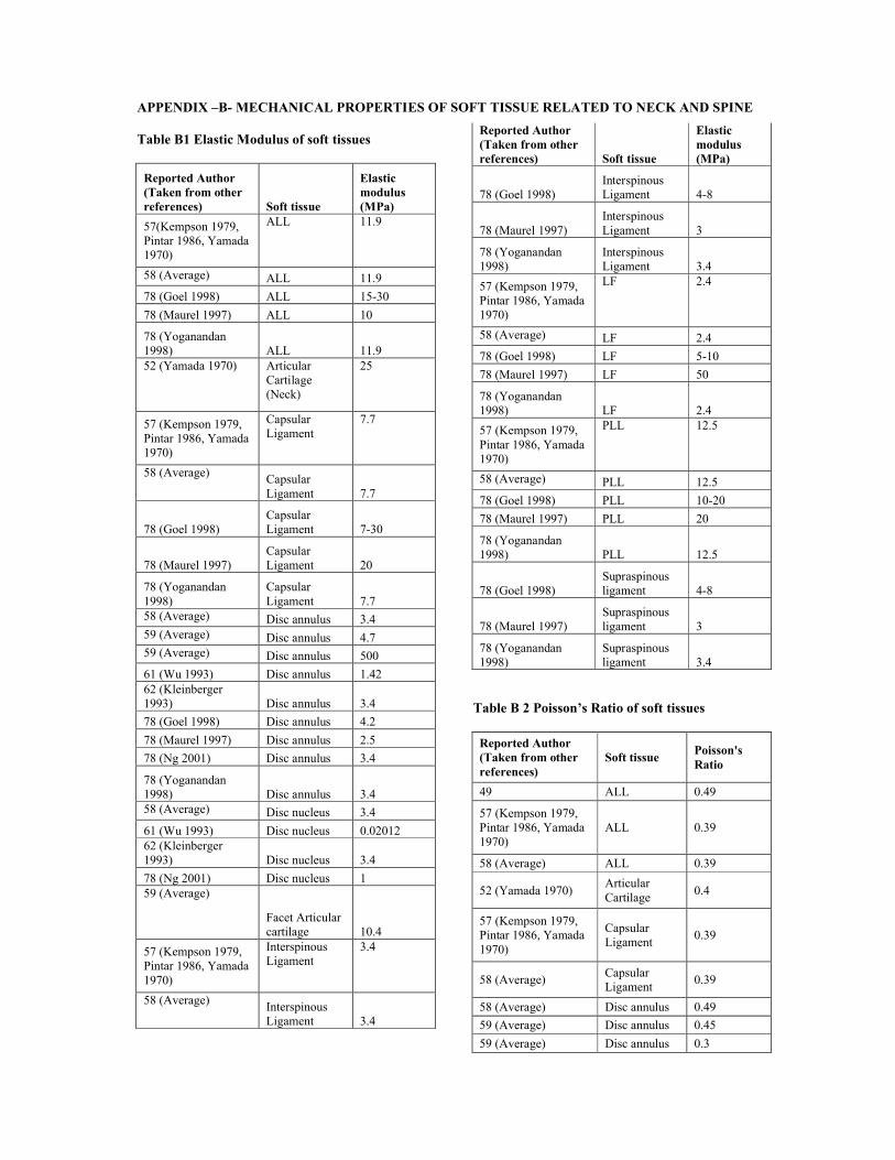
APPENDIX –B- MECHANICAL PROPERTIES OF SOFT TISSUE RELATED TO NECK AND SPINE
Table B1 Elastic Modulus of soft tissues
Reported Author (Taken from other references) Soft tissue
Elastic modulus (MPa)
57(Kempson 1979, Pintar 1986, Yamada 1970)
ALL 11.9
58 (Average) ALL 11.9
78 (Goel 1998) ALL 15-30
78 (Maurel 1997) ALL 10
78 (Yoganandan 1998) ALL 11.952 (Yamada 1970) Articular
Cartilage (Neck)
25
57 (Kempson 1979, Pintar 1986, Yamada 1970)
Capsular Ligament
7.7
58 (Average) Capsular Ligament 7.7
78 (Goel 1998)Capsular Ligament 7-30
78 (Maurel 1997)Capsular Ligament 20
78 (Yoganandan 1998)
Capsular Ligament 7.7
58 (Average) Disc annulus 3.459 (Average) Disc annulus 4.759 (Average) Disc annulus 500
61 (Wu 1993) Disc annulus 1.4262 (Kleinberger 1993) Disc annulus 3.4
78 (Goel 1998) Disc annulus 4.2
78 (Maurel 1997) Disc annulus 2.5
78 (Ng 2001) Disc annulus 3.4
78 (Yoganandan 1998) Disc annulus 3.458 (Average) Disc nucleus 3.4
61 (Wu 1993) Disc nucleus 0.0201262 (Kleinberger 1993) Disc nucleus 3.4
78 (Ng 2001) Disc nucleus 159 (Average)
Facet Articular cartilage 10.4
57 (Kempson 1979, Pintar 1986, Yamada 1970)
Interspinous Ligament
3.4
58 (Average) Interspinous Ligament 3.4
Reported Author (Taken from other references) Soft tissue
Elastic modulus (MPa)
78 (Goel 1998)Interspinous Ligament 4-8
78 (Maurel 1997)Interspinous Ligament 3
78 (Yoganandan 1998)
Interspinous Ligament 3.4
57 (Kempson 1979, Pintar 1986, Yamada 1970)
LF 2.4
58 (Average) LF 2.4
78 (Goel 1998) LF 5-10
78 (Maurel 1997) LF 50
78 (Yoganandan 1998) LF 2.4
57 (Kempson 1979, Pintar 1986, Yamada 1970)
PLL 12.5
58 (Average) PLL 12.5
78 (Goel 1998) PLL 10-20
78 (Maurel 1997) PLL 20
78 (Yoganandan 1998) PLL 12.5
78 (Goel 1998)Supraspinous ligament 4-8
78 (Maurel 1997)Supraspinous ligament 3
78 (Yoganandan 1998)
Supraspinous ligament 3.4
Table B 2 Poisson’s Ratio of soft tissues
Reported Author (Taken from other references)
Soft tissuePoisson's Ratio
49 ALL 0.49
57 (Kempson 1979, Pintar 1986, Yamada 1970)
ALL 0.39
58 (Average) ALL 0.39
52 (Yamada 1970)Articular Cartilage
0.4
57 (Kempson 1979, Pintar 1986, Yamada 1970)
Capsular Ligament
0.39
58 (Average)Capsular Ligament
0.39
58 (Average) Disc annulus 0.49
59 (Average) Disc annulus 0.45
59 (Average) Disc annulus 0.3
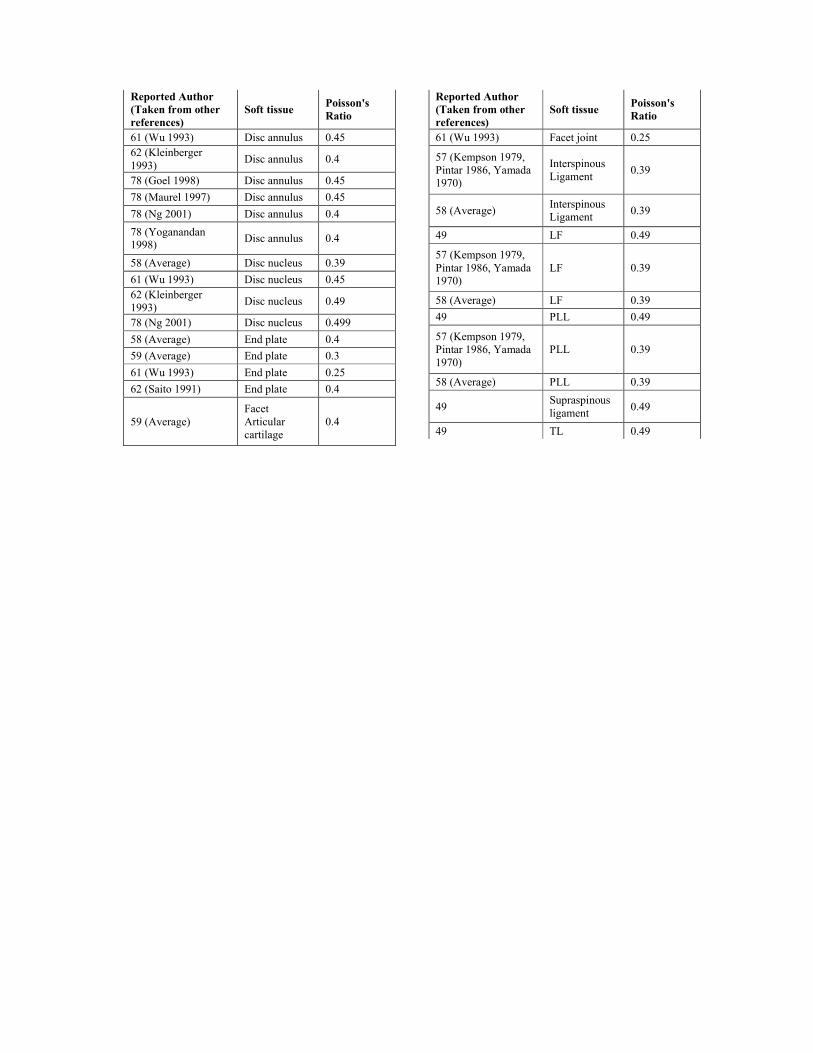
Reported Author (Taken from other references)
Soft tissuePoisson's Ratio
61 (Wu 1993) Disc annulus 0.4562 (Kleinberger 1993)
Disc annulus 0.4
78 (Goel 1998) Disc annulus 0.45
78 (Maurel 1997) Disc annulus 0.45
78 (Ng 2001) Disc annulus 0.4
78 (Yoganandan 1998)
Disc annulus 0.4
58 (Average) Disc nucleus 0.39
61 (Wu 1993) Disc nucleus 0.4562 (Kleinberger 1993)
Disc nucleus 0.49
78 (Ng 2001) Disc nucleus 0.499
58 (Average) End plate 0.4
59 (Average) End plate 0.3
61 (Wu 1993) End plate 0.25
62 (Saito 1991) End plate 0.4
59 (Average)Facet Articular cartilage
0.4
Reported Author (Taken from other references)
Soft tissuePoisson's Ratio
61 (Wu 1993) Facet joint 0.25
57 (Kempson 1979, Pintar 1986, Yamada 1970)
Interspinous Ligament
0.39
58 (Average)Interspinous Ligament
0.39
49 LF 0.49
57 (Kempson 1979, Pintar 1986, Yamada 1970)
LF 0.39
58 (Average) LF 0.39
49 PLL 0.49
57 (Kempson 1979, Pintar 1986, Yamada 1970)
PLL 0.39
58 (Average) PLL 0.39
49Supraspinous ligament
0.49
49 TL 0.49

April 22, 2006
The Director (Technical)
The Institution of Engineers (India)
8 Gokhale Road
Kolkata 70020
Dear Sir,
Kindly find enclosed four copies of a manuscript of our paper, titled, “Review of Mechanical Properties of Human Body
Soft Tissues in the head neck and spine”, co-authored by Dr. S Mukherjee, myself and B Karthikeyan, along with a CD
containing the soft version for consideration for publication in the Institution of Engineers, Journal of Mechanical
Engineering. I request you to kindly consider for the same. The author form and the paper submission forms are also
enclosed. Being a review paper the word count (not including figures, tables and appendices) has reached 3657 and we have
had to include 6 figures instead of your limit of 5. I hope you will be able to consider the same as in a review paper of this
kind, it was very difficult to stick to these limits and we believe that this paper will have a very strong archival value and in
the interest of quality of Indian Journals, it should appear in Indian Journals.
Yours truly,
(A Chawla)


![A free boundary mechanobiological model of epithelial tissues · 2020/7/2 · Epithelial tissues consist of tightly packed monolayers of cells [1{3]. Mechanical cell properties,](https://img.dokumen.tips/doc/110x75/5f84cbcfaa47d969b17c8e73/a-free-boundary-mechanobiological-model-of-epithelial-tissues-202072-epithelial.jpg)