Embed Size (px)
Citation preview

Page 1/7
Epithelioid Angiosarcoma of the Tibia: A Case Report and LiteratureReviewQianqian Yao
The Second A�liated Hospital of Shandong First Medical UniversityXubo Ge
the Fourth People's Hospital of TaianYinghua Gao
The Second A�liated Hospital of Shandong First Medical UniversityChangqin Li
The Second A�liated Hospital of Shandong First Medical Universityjian qin ( [email protected] )
The Second A�liated Hospital of Shandong First Medical University
Research Article
Keywords: Epithelioid angiosarcoma, Tibia, Computed tomography, Magnetic resonance imaging
Posted Date: September 13th, 2021
DOI: https://doi.org/10.21203/rs.3.rs-827733/v1
License: This work is licensed under a Creative Commons Attribution 4.0 International License. Read Full License

Page 2/7
Discussion And ConclusionsEA is relatively rare and only accounts for < 1% of primary bone
AbstractBackground Epithelioid angiosarcoma (EA) is characterized by epithelioid-like neoplastic cells. In fact, there is little literature regarding EA ofbone. Accordingly, this study presents an imaging analysis of a 66-year-old man who suffered from EA in his right tibia.
Case presentation: A 66-year-old man developed right ankle pain four months prior to initial evaluation at our institute, which was progressivelyworsening for six days. All laboratory data were within the reference ranges. Plain radiographs and computed tomography (CT) revealedosteolytic lesions with multiple separations in the distal tibia, measuring 7.9 cm × 4.6 cm × 4.4 cm in size. The lytic lesions were ill-circumscribed and lacked marginal sclerosis. Punctate irregular calci�cations and low-density areas were observed within lesion's areas. Thecortical bone was irregularly thinned and was discontinuous. On magnetic resonance imaging (MRI), the lesions were heterogeneous.Immunohistochemically, the tumor cells expressed the vascular markers CD31, CD34, and factor VIII and revealed 5% positivity for Ki67. Finally,the patient was diagnosed with EA of bone.
Conclusions: We reported a case of EA that occurred in the right tibia and summarized the imaging features of EA by reviewing the literature.Although pathological examination remains the gold standard for diagnosing EA, speci�c imaging features may assist in diagnosis.
BackgroundEpithelioid angiosarcoma (EA) is a rare variant of angiosarcoma that is characterized by epithelioid-like neoplastic cells. These tumors areusually poorly differentiated and biologically aggressive [1]. In small series, EA has been reported to occur in various parts such as thyroid, skin,adrenal gland, and deep soft tissue, whereas bone-originated EA is relatively rare [2, 3, 4]. EA has a male predilection and is more prevalent inolder individuals [1, 5], a population with a higher rate of metastatic carcinoma. The current study presents the imaging analysis of a 66-year-old man who suffered from EA in his right tibia, and its imaging features were summarized by reviewing the literature. The patient providedwritten informed consent. To our knowledge, this is the �rst report to analyze and summarize the imaging features of EA.
Case PresentationA 66-year-old man was admitted at our institute with right ankle pain four months before initial evaluation, which was progressively worseningfor six days. At rest, symptoms were alleviated, while exercise aggravated them. On physical examination, his right ankle was markedly swollenand �rm, he suffered from local pressing pain, and a surgical scar was visible on the inside aspect of his right tibia. All laboratory data werewithin the reference ranges. The patient had a prior medical history of right lower limb trauma and underwent surgical treatment in 2011 at theage of 57. He smoked and consumed alcohol for about 30 years. He denied that there was no family or personal history of malignancy orautoimmune disease.
In the distal tibia, plain radiographs revealed osteolytic lesions with multiple separations (Fig. 1). The lytic lesions were ill-circumscribed andlacked marginal sclerosis. Some were accompanied by thinning and ballooning of bone cortexes. Besides, cement was observed in theproximal tibia following a trauma nine years ago. As a result, a CT (computed tomography) scan of the right ankle was performed and revealedosteolytic and expansive destruction of bone in the distal tibia (Fig. 2), measuring 7.9 cm × 4.6 cm × 4.4 cm in size. The normal trabecular bonestructure disappeared and was replaced by heterogeneous density soft tissue mass. Punctate irregular calci�cations and low-density areaswere also observed within lesion areas. The cortical bone was irregularly thinned and was discontinuous. The mass extended into adjacent softtissue through the discontinuous cortical bone, and punctate calci�cations were also observed. On ankle MRI (magnetic resonance imaging),the lesions appeared to be isointense or slightly hyperintense to the muscles on T1-weighted images, and fat-suppressed T2 images, centrallyscattered occurring patchy, with a slightly low signal (Fig. 3). Edema signal could be observed in the surrounding soft tissue.
Based on the �ndings from imaging studies and the patient’s age, the differential diagnoses of osteolytic bone lesions combined withcalci�cation included chondrosarcoma, angiosarcoma, and bone metastasis.
During operation, brown-red tumor tissue was observed in medullary cavity, which was sandy, soft, and partly calci�ed. For pain relief andhistological diagnosis, curettage of the lesion in the distal tibia and arti�cial bone grafting were performed. Microscopically, the resected tumorcells exhibited nestlike epithelioid features between trabeculae with prominent nucleoli and abundant cytoplasm. Numerous red blood cellswere also discovered between the epithelial nests. Immunohistochemically, the tumor cells expressed the vascular markers CD31, CD34, andfactor VIII and revealed 5% positivity for Ki67 (Fig. 4). Finally, the patient was diagnosed with EA of the bone. The patient was fully recoveredand was discharged.

Page 3/7
tumors [6]. Since EA did not exhibit characteristic performance in imaging, the de�nitive diagnosis is done using pathological examination [7].The immunohistochemical examination relies on applying vascular markers for EA diagnosis, including CD31, CD34, and factor VIII [8, 9]. Themost important vascular marker is CD31, which is considered to provide a relatively high index of sensitivity and speci�city [10]. Factor VIII andCD34 markers are useful for diagnostic studies. Desphande et al. examined ten patients and discovered that 75% of them tested positive forfactor VIII, and 40% tested positive for CD34 markers [1].
Although pathological examination remains the gold standard for diagnosing EA, speci�c imaging features may assist in diagnosis. To ourknowledge, few reports have discussed EA imaging characteristics.
We searched the Pubmed and found 6 previously reported cases of primary EA of bone, the inclusion criteria were that they underwent CT andMRI at the same time. Including our case, clinical information and imaging features of the 7 cases are summarized in Table 1.There were fourmen and three women, who ranged in age from 31 to 76 years (mean, 59 years). The maximum diameter of lesions was greater than or equalto 5cm. More than half of the cases presented the disease in the lower limb(5/7), two were located in the femur. In the present case, theimaging examination mainly revealed osteolytic and expansive destruction of bone in the distal tibia, which were similar to that reported in the6 cases. Besides, the cortical bone was thinning and discontinuous(7/7). The normal trabecular bone structure disappeared and was replacedby heterogeneous soft tissue. Phlebolithlike calci�cations or hemorrhaging support this diagnosis of EA. However, only half of the cases(4/8)showed signs of calci�cations and hemorrhaging at the same time. In CT and MRI scans, cortical breakthrough(6/7), no marginalsclerosis(5/7) and soft tissue extension(6/7) indicated the presence of an aggressive, high-grade bone tumor.
Malignant tumors of cartilage origin, such as chondrosarcoma, must be distinguished. While it is di�cult to distinguish from metastases thatcause osteolytic changes on imaging, it is easy to identify based on clinical symptoms and primary lesions.
This study aimed to help radiologists improve their EA understanding. Together, X-ray, CT, and MRI scans may effectively diagnose epithelioidangiosarcomas by eliminating a differential diagnosis of other osteolytic lesions masses of the bone.
AbbreviationsEAEpithelioid AngiosarcomaCTComputed TomographyMRIMagnetic Resonance Imaging
DeclarationsEthics Approval and Consent to Participate
As it is a case report, ethics approval is unnecessary after consulting the Ethics Committee of The Second A�liated Hospital of Shandong FirstMedical University.
Consent to publish
Written informed consent was obtained from the patient and his wife for publishing this case report and any accompanying images.
Availability of data and materials
The datasets used and/or analyzed during the current study are available from the corresponding author upon reasonable request.
Competing interests
The authors declare no competing interests.
Funding
This study was funded by Academic Promotion Programme of Shandong First Medical University (No.2019QL017).
Authors' Contributions

Page 4/7
All authors read and approved the �nal manuscript. QQY reviewed the medical record and wrote the manuscript. XBG and YHG performed thehistopathological diagnosis and immunohistochemistry. CQL performed the imaging diagnosis. JQ designed the case report and revised themanuscript for important intellectual content.
Acknowledgments
The authors would like to thank the patient's family for giving consent.
Authors’ Information
a Department of Radiology, The Second A�liated Hospital of Shandong First Medical University,Taian,Shandong, 271000,China.
b Department of Radiology, The Fourth People's Hospital of Taian, Taian, Shandong, 271000,China.
c Department of Pathology, The Second A�liated Hospital of Shandong First Medical University, Taian, Shandong, 271000, China.
References1. Deshpande V. Rosenberg AE, O'Connell JX,et al. Epithelioid angiosarcoma of the bone: a series of 10 cases. Am J Surg
Pathol.2003;27(6):709–16.
2. Li Y, Zou X, Chang X, et al. Right femoral pathological fracture caused by primary bone epithelioid angiosarcoma: Case report.Medicine.(Baltimore).2017;96:e6951.
3. Wang J, Zhao M, Huang J,et al. Primary epithelioid angiosarcoma of right hip joint: A case report and literature review. Medicine.2018;97(15):e0307.
4. Lv L. Xu P, Shi Y, et al. Imaging features of soft tissue epithelioid angiosarcoma in the lower extremity: A case report. OncolLett.2016;11(5):3457–60.
5. Fletcher CDM, Beham A, Bekir S, et al. Epithelioid angiosarcoma of deep soft tissue: a distinctive tumor readily mistaken for an epithelialneoplasm. Am J Surg Pathol. 1991;15(10):915–24.
�. Mendeszoon MJ. Mendeszoon ER Jr, Rasmussen S, et al. Epithelioid angiosarcoma of the talus. J Foot Ankle Surg.2011;50(1):87–92.
7. Markaki S, Kokka H, Kyparidou E, et al. Primary vascular bone sarcomas. A clinicopathological and immunohistochemical study of twocases. Arch Anat Cytol Pathol. 1990;38(4):163–7.
�. Oc Y. Kilinc BE, Ertugrul R, et al. Epithelioid Angiosarcoma in Femur: A Case Presentation. World J Oncol.2017;8(6):196–8.
9. Chen M. Zhang W, Qu J, et al. Epithelioid angiosarcoma of the ilium: a case report. Int J Clin Exp Pathol.2014;7(12):9099–103.
10. Lang J. Chen L, Chen B, et al Epithelioid angiosarcoma of the spine: A case report of a rare bone tumor. Oncol Lett.2014;7(6):2170 – 217.
11. Sakamoto A. Takahashi Y, Oda Y, et al Aggressive clinical course of epithelioid angiosarcoma in the femur: a case report. World J SurgOncol. 2014;12:281.
12. Bürk J. Gerlach U, Baumann T, et al Epithelioid angiosarcoma of the scapula. In Vivo. 2010;24(5):783–6.
Tables

Page 5/7
Table 1clinical information and imaging features of bone-originated EA in literature
Reference Age/
Gender
Location Size Expansion/osteolyticbone destruction
Corticalbreakthrough/soft tissueextension
marginalsclerosis
Calci�cations/hemorrhage
[3] F/61 hip 11cm×2cm×7.5cm Yes/Yes corticalthinning/No
slightly Yes/Yes
[8] M/31 femur 7cm in diameter Yes/Yes Yes/Yes Yes No/No
[9] F/62 ilium 10.2cm×6.2cm×5.3cm
Yes/Yes Yes/Yes No Yes/Yes
[10] M/76 L4 5cm×4cm Yes/Yes Yes/Yes No Yes/Yes
[11] F/69 femur 7 cm inlongitudinal
diameter
Yes/Yes Yes/Yes No No/No
[12] M/48 scapula 5.8 cm×3.9 cm Yes/Yes Yes/Yes No No/No
Presentstudy
M/66 tibia 7.9cm×4.6cm×4.4cm
Yes/Yes Yes/Yes No Yes/Yes
M: male; F: female; L4: the fourth lumbar
Figures
Figure 1
anteroposterior (A) and lateral (B). Radiographs showing the osteolytic lesion with multiple separations in the distal tibia.

Page 6/7
Figure 2
axial (A), coronal (B), sagittal (C). CT showing osteolytic and expansive destruction of bone in the distal tibia. Punctate irregular calci�cationswere observed within lesion's areas. The cortical bone was irregularly thinned and was discontinuous.
Figure 3
MRI, sagittal fs PD (A), sagittal T1 FSE (B), coronal fs PD (C). On ankle MRI, the lesions showed isointense or slightly hyperintense to themuscles on T1-weighted images and fat-suppressed T2 images, centrally scattered occurring patchy and slightly low signal. Edema signalcould be observed in the surrounding soft tissue.
Page 7/7
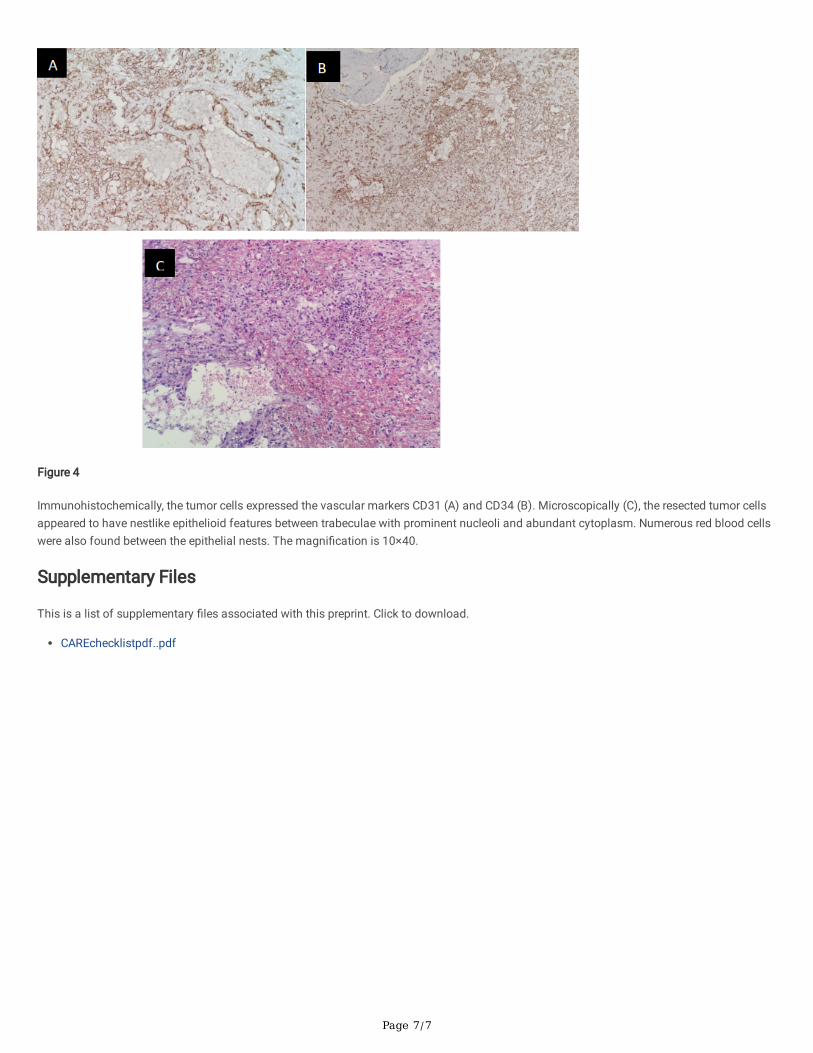
Figure 4
Immunohistochemically, the tumor cells expressed the vascular markers CD31 (A) and CD34 (B). Microscopically (C), the resected tumor cellsappeared to have nestlike epithelioid features between trabeculae with prominent nucleoli and abundant cytoplasm. Numerous red blood cellswere also found between the epithelial nests. The magni�cation is 10×40.
Supplementary Files
This is a list of supplementary �les associated with this preprint. Click to download.
CAREchecklistpdf..pdf