Embed Size (px)
Citation preview

N
R
Cb
J
a
b
RA
I
Ss
vT
CT
s
1h
ano Today (2013) 8, 98—113
Available online at www.sciencedirect.com
journa l homepage: www.e lsev ier .com/ locate /nanotoday
EVIEW
omposite magnetic—plasmonic nanoparticles foriomedicine: Manipulation and imaging
itKang Lima,b,∗, Sara A. Majetichb,∗∗
School of Chemical Engineering, Universiti Sains Malaysia, Nibong Tebal 14213, Penang, MalaysiaPhysics Department, Carnegie Mellon University, Pittsburgh, PA 15213, USA
eceived 19 September 2012; received in revised form 13 November 2012; accepted 27 December 2012vailable online 12 February 2013
KEYWORDSIron oxide—goldnanoparticles;Surface plasmonresonance;Magnetophoresis;Colloidal stability;Core—shell;
Summary Iron oxide—gold nanoparticles that exhibit both magnetic and plasmonic behaviorshave great potential for biomedical applications. The ability to remotely control the spatialposition of a nanoparticle in real time while tracking its motion provides an exciting new toolfor nanoscale sensing. In this review we summarize the major efforts in the design and synthesisof iron oxide—gold nanoparticles. The underlying magnetophoretic and plasmonic characteris-tics of gold and iron oxide nanoparticles that enable their use for biomedical applications aredescribed. We discuss the challenges associated with the integration of iron oxide and gold inone unified nanostructure, including the chemical techniques involved in making such compositematerial. We emphasize on the importance of colloidal stability, and explain how this property
Biomedicaldetermines the functionality of iron oxide—gold nanoparticles in physiological environment.Afterwards, we examine both the magnetophoresis and localized surface plasmon resonance ofiron oxide—core gold—shell structure and provide theoretical explanations for these properties.Finally we suggest potential opportunities for use of iron oxide—gold nanoparticles.ts re
b
© 2013 Elsevier Ltd. All righ
ntroduction
ingle particle sensing offers the possibility of probing smallamples in precisely defined regions near, on, or within
∗ Corresponding author at: School of Chemical Engineering, Uni-ersiti Sains Malaysia, Nibong Tebal 14300, Penang, Malaysia.el.: +60 4 599 6423; fax: +60 4 594 1013.∗∗ Corresponding author at: 5000 Forbes Ave., Physics Department,arnegie Mellon University, Pittsburgh, PA 15213, USA.el.: +1 412 268 3105; fax: +1 412 681 0648.
E-mail addresses: [email protected] (J. Lim),[email protected] (S.A. Majetich).
utHp[zmocattt
748-0132/$ — see front matter © 2013 Elsevier Ltd. All rights reserved.ttp://dx.doi.org/10.1016/j.nantod.2012.12.010
served.
iological cells. The ability to magnetically guide and manip-late the motion of individual particles is crucially importanto modulate cellular behavior in a non-contact mode [1].owever, the techniques most commonly used to manipulatearticles, including electrophoresis [2,3], dielectrophoresis4,5], traveling-wave dielectrophoresis [6], magnetic twee-ers [7] and acoustic traps [8], are focused mainly on theanipulation of micron size objects. Successful application
f these schemes in manipulating the motion of nanoparti-les (NPs) is quite challenging because nano-sized particles
re extremely susceptible to Brownian motion [9] and iner-ial forces associated with their motion are negligible (i.e.,hey move with low Reynolds number) [10]. While opticalweezers offer high-resolution control of colloidal particles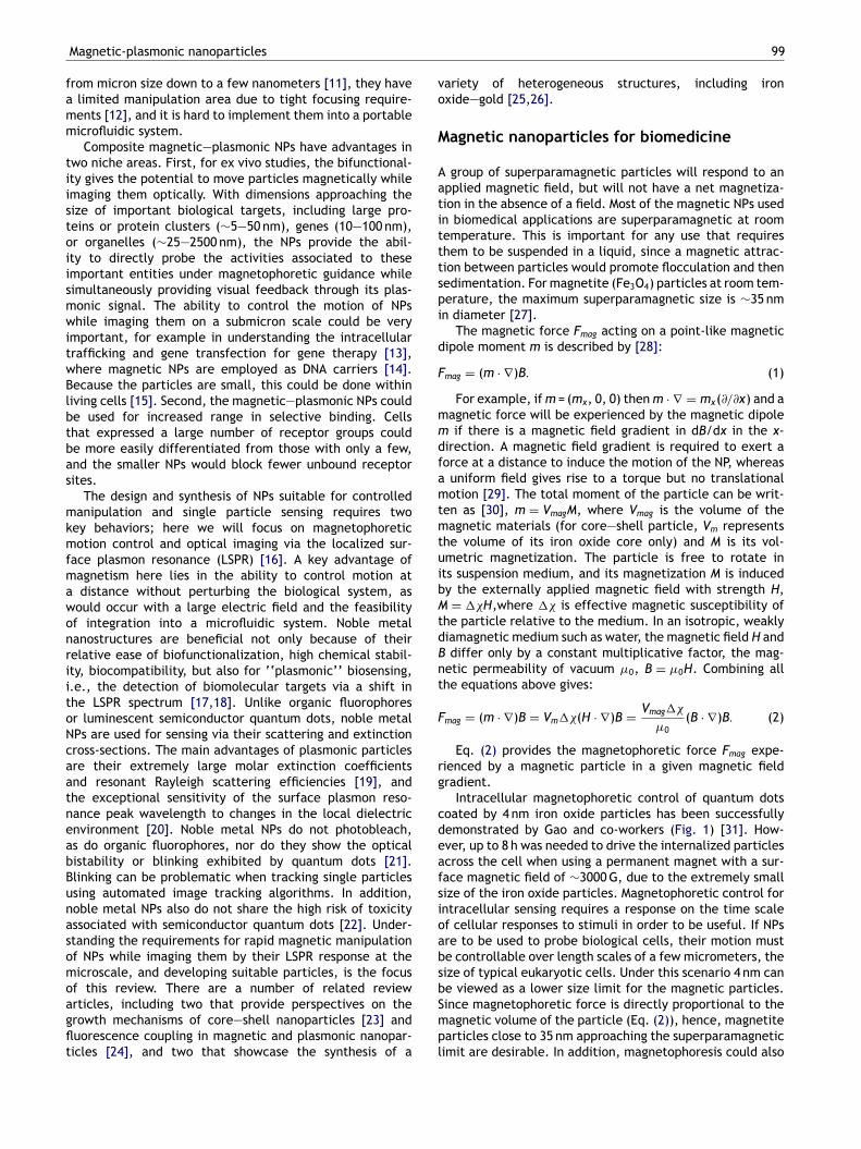
vo
M
Aatitttspi
d
F
mmdfamtmtuibM
tdBnt
F
rg
cdeafsioabsb
Magnetic-plasmonic nanoparticles
from micron size down to a few nanometers [11], they havea limited manipulation area due to tight focusing require-ments [12], and it is hard to implement them into a portablemicrofluidic system.
Composite magnetic—plasmonic NPs have advantages intwo niche areas. First, for ex vivo studies, the bifunctional-ity gives the potential to move particles magnetically whileimaging them optically. With dimensions approaching thesize of important biological targets, including large pro-teins or protein clusters (∼5—50 nm), genes (10—100 nm),or organelles (∼25—2500 nm), the NPs provide the abil-ity to directly probe the activities associated to theseimportant entities under magnetophoretic guidance whilesimultaneously providing visual feedback through its plas-monic signal. The ability to control the motion of NPswhile imaging them on a submicron scale could be veryimportant, for example in understanding the intracellulartrafficking and gene transfection for gene therapy [13],where magnetic NPs are employed as DNA carriers [14].Because the particles are small, this could be done withinliving cells [15]. Second, the magnetic—plasmonic NPs couldbe used for increased range in selective binding. Cellsthat expressed a large number of receptor groups couldbe more easily differentiated from those with only a few,and the smaller NPs would block fewer unbound receptorsites.
The design and synthesis of NPs suitable for controlledmanipulation and single particle sensing requires twokey behaviors; here we will focus on magnetophoreticmotion control and optical imaging via the localized sur-face plasmon resonance (LSPR) [16]. A key advantage ofmagnetism here lies in the ability to control motion ata distance without perturbing the biological system, aswould occur with a large electric field and the feasibilityof integration into a microfluidic system. Noble metalnanostructures are beneficial not only because of theirrelative ease of biofunctionalization, high chemical stabil-ity, biocompatibility, but also for ‘‘plasmonic’’ biosensing,i.e., the detection of biomolecular targets via a shift inthe LSPR spectrum [17,18]. Unlike organic fluorophoresor luminescent semiconductor quantum dots, noble metalNPs are used for sensing via their scattering and extinctioncross-sections. The main advantages of plasmonic particlesare their extremely large molar extinction coefficientsand resonant Rayleigh scattering efficiencies [19], andthe exceptional sensitivity of the surface plasmon reso-nance peak wavelength to changes in the local dielectricenvironment [20]. Noble metal NPs do not photobleach,as do organic fluorophores, nor do they show the opticalbistability or blinking exhibited by quantum dots [21].Blinking can be problematic when tracking single particlesusing automated image tracking algorithms. In addition,noble metal NPs also do not share the high risk of toxicityassociated with semiconductor quantum dots [22]. Under-standing the requirements for rapid magnetic manipulationof NPs while imaging them by their LSPR response at themicroscale, and developing suitable particles, is the focusof this review. There are a number of related review
articles, including two that provide perspectives on thegrowth mechanisms of core—shell nanoparticles [23] andfluorescence coupling in magnetic and plasmonic nanopar-ticles [24], and two that showcase the synthesis of aSmpl
99
ariety of heterogeneous structures, including ironxide—gold [25,26].
agnetic nanoparticles for biomedicine
group of superparamagnetic particles will respond to anpplied magnetic field, but will not have a net magnetiza-ion in the absence of a field. Most of the magnetic NPs usedn biomedical applications are superparamagnetic at roomemperature. This is important for any use that requireshem to be suspended in a liquid, since a magnetic attrac-ion between particles would promote flocculation and thenedimentation. For magnetite (Fe3O4) particles at room tem-erature, the maximum superparamagnetic size is ∼35 nmn diameter [27].
The magnetic force Fmag acting on a point-like magneticipole moment m is described by [28]:
mag = (m · ∇)B. (1)
For example, if m = (mx, 0, 0) then m · ∇ = mx(∂/∂x) and aagnetic force will be experienced by the magnetic dipoleif there is a magnetic field gradient in dB/dx in the x-
irection. A magnetic field gradient is required to exert aorce at a distance to induce the motion of the NP, whereas
uniform field gives rise to a torque but no translationalotion [29]. The total moment of the particle can be writ-
en as [30], m = VmagM, where Vmag is the volume of theagnetic materials (for core—shell particle, Vm represents
he volume of its iron oxide core only) and M is its vol-metric magnetization. The particle is free to rotate ints suspension medium, and its magnetization M is inducedy the externally applied magnetic field with strength H,= ��H,where �� is effective magnetic susceptibility of
he particle relative to the medium. In an isotropic, weaklyiamagnetic medium such as water, the magnetic field H anddiffer only by a constant multiplicative factor, the mag-
etic permeability of vacuum �0, B = �0H. Combining allhe equations above gives:
mag = (m · ∇)B = Vm��(H · ∇)B = Vmag��
�0(B · ∇)B. (2)
Eq. (2) provides the magnetophoretic force Fmag expe-ienced by a magnetic particle in a given magnetic fieldradient.
Intracellular magnetophoretic control of quantum dotsoated by 4 nm iron oxide particles has been successfullyemonstrated by Gao and co-workers (Fig. 1) [31]. How-ver, up to 8 h was needed to drive the internalized particlescross the cell when using a permanent magnet with a sur-ace magnetic field of ∼3000 G, due to the extremely smallize of the iron oxide particles. Magnetophoretic control forntracellular sensing requires a response on the time scalef cellular responses to stimuli in order to be useful. If NPsre to be used to probe biological cells, their motion muste controllable over length scales of a few micrometers, theize of typical eukaryotic cells. Under this scenario 4 nm cane viewed as a lower size limit for the magnetic particles.
ince magnetophoretic force is directly proportional to theagnetic volume of the particle (Eq. (2)), hence, magnetitearticles close to 35 nm approaching the superparamagneticimit are desirable. In addition, magnetophoresis could also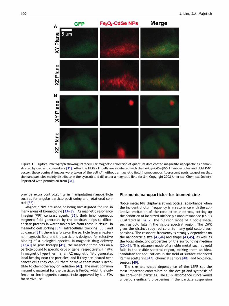
100 J. Lim, S.A. Majetich
Figure 1 Optical micrograph showing intracellular magnetic collection of quantum dots coated magnetite nanoparticles demon-strated by Gao and co-workers [31]. After the HEK293T cells are incubated with the Fe3O4—CdSe@GSH nanoparticles and pEGFP-N1vector, these confocal images were taken of the cell (A) without a magnetic field (homogeneous fluorescent spots suggesting thatthe nanoparticles mainly distribute in the cytosol) and (B) under a magnetic field for 8 h. Copyright 2008 American Chemical Society.R
pst
mimemgnb[pilctmff
P
Ntltisgptt[fcRs
eprinted with permission from [31].
rovide extra controllability in manipulating nanoparticleuch as for angular particle positioning and rotational con-rol [32].
Magnetic NPs are used or being investigated for use inany areas of biomedicine [33—35]. As magnetic resonance
maging (MRI) contrast agents [36], their inhomogeneousagnetic field generated by the particles helps to differ-
ntiate protons in water molecules from those in tissue. Inagnetic cell sorting [37], intracellular tracking [38], and
uidance [31], there is a force on the particle from an exter-al magnetic field and the particle is designed for selectiveinding of a biological species. In magnetic drug delivery39,40] or gene therapy [41], the magnetic force acts on aarticle bound to specific drug or gene, respectively. Finally,n magnetic hyperthermia, an AC magnetic field generatesocal heating near the particles, and if they are located nearancer cells they can kill them or make them more suscep-
ible to chemotherapy or radiation [42]. The most commonagnetic material for the particles is Fe3O4, which the onlyerro- or ferrimagnetic nanoparticle approved by the FDAor in vivo use.
mtu
lasmonic nanoparticles for biomedicine
oble metal NPs display a strong optical absorbance whenhe incident photon frequency is in resonance with the col-ective excitation of the conduction electrons, setting uphe condition of localized surface plasmon resonance (LSPR)llustrated in Fig. 2. The plasmon mode of a noble metaluch as gold falls in the visible spectral region. The LSPRives the distinct ruby red color to many gold colloid sus-ensions. The resonant frequency is strongly dependent onhe nanoparticle size [43,44] and shape [43,45], as well ashe local dielectric properties of the surrounding medium20,46]. This plasmon mode of a noble metal such as goldalls in the visible spectral region, making them an idealandidate for applications in the field of surface enhancedaman scattering [47], chemical sensors [48], and biologicalensors [49].
The size and shape dependence of the LSPR set theost important constraints on the design and synthesis of
he core—shell particles. The LSPR absorbance curve wouldndergo significant broadening if the particle suspension
Magnetic-plasmonic nanoparticles 101
Figure 2 A scheme illustrating the excitation of the dipole surface plasmon oscillation [43]. The electric field of the incominggrays not
lpsDpmacdd[gbe
light induces a polarization of the free conduction electrons (nanoparticle (dark sphere). The extension of the charge cloud i
were composed of a mixture of particles with differentsizes (high polydispersity) [43]. The same scenario appliesto the shape uniformity as well. A mixture of particleswith different shapes is also going to exhibit broadeningof the LSPR absorbance curve [45]. For any sensing pur-pose the signal employed for detection should be welldefined. Hence, monodisperse particles with uniform shapeare greatly desired to produce as sharp an LSPR spectrumas possible. The presence of LSPR makes direct imag-ing/tracking of noble metal NPs with sizes much smallerthan diffraction limit possible by using darkfield opticalmicroscopy. For an example, Van Duyne and co-workers[20] have successfully imaged 35 nm silver particles immo-bilized on top of a microscope coverslip. In addition,El-Sayed and co-workers have also demonstrated the poten-
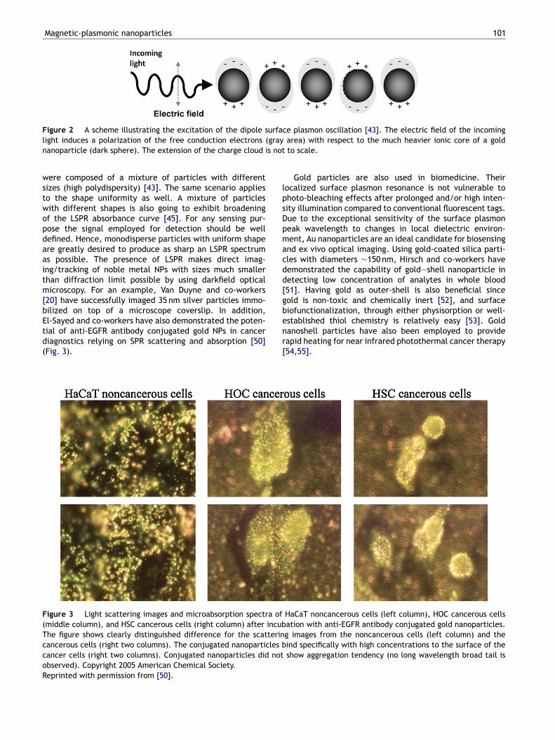
tial of anti-EGFR antibody conjugated gold NPs in cancerdiagnostics relying on SPR scattering and absorption [50](Fig. 3).nr[
Figure 3 Light scattering images and microabsorption spectra of(middle column), and HSC cancerous cells (right column) after incubThe figure shows clearly distinguished difference for the scatterincancerous cells (right two columns). The conjugated nanoparticles bcancer cells (right two columns). Conjugated nanoparticles did notobserved). Copyright 2005 American Chemical Society.Reprinted with permission from [50].
area) with respect to the much heavier ionic core of a goldto scale.
Gold particles are also used in biomedicine. Theirocalized surface plasmon resonance is not vulnerable tohoto-bleaching effects after prolonged and/or high inten-ity illumination compared to conventional fluorescent tags.ue to the exceptional sensitivity of the surface plasmoneak wavelength to changes in local dielectric environ-ent, Au nanoparticles are an ideal candidate for biosensing
nd ex vivo optical imaging. Using gold-coated silica parti-les with diameters ∼150 nm, Hirsch and co-workers haveemonstrated the capability of gold—shell nanoparticle inetecting low concentration of analytes in whole blood51]. Having gold as outer-shell is also beneficial sinceold is non-toxic and chemically inert [52], and surfaceiofunctionalization, through either physisorption or well-stablished thiol chemistry is relatively easy [53]. Gold
anoshell particles have also been employed to provideapid heating for near infrared photothermal cancer therapy54,55].HaCaT noncancerous cells (left column), HOC cancerous cellsation with anti-EGFR antibody conjugated gold nanoparticles.g images from the noncancerous cells (left column) and theind specifically with high concentrations to the surface of theshow aggregation tendency (no long wavelength broad tail is
1
Cp
TmNmtnmensictblt
otpoc∼
iacntattT[l
Dn
TdoobaCspetaisol
N
ocofidmsdttsbtrsc
rcahsshfntpHa
Apusa
otimrpdf
opicIfHraii
02
hallenges in combining magnetic andlasmonic particles for biomedicine
here are numerous challenges in the development ofagnetic—plasmonic NPs. Successfully applying magneticPs for in vitro biosensing, requires the particles to (1) beagnetically responsive within the times scale appropriate
o the phenomenon of interest, (2) emit a well defined sig-al that can be used for characterization purposes, and (3)aintain good colloidal stability in biological media of mod-
rate to high ionic strength. The magnetic manipulation ofanoscale objects is challenging because magnetic NPs areusceptible to the thermal displacements [10]. This scenarios further complicated with the fact that the magnetic, vis-ous drag and random Brownian forces scale differently withhe particle size [9,56]. To overcome contributions fromoth viscous drag forces and Brownian diffusion, extremelyarge magnetic field gradients are needed to induce magne-ophoretic motion in deterministic pattern.
Another critical challenge in controlling the transportf NPs is the need for a visualization scheme with whicho track the motion [57]. Having gold as part of the com-osite NP enables the particle location to be monitoredptically, since gold NPs have a very large molar extinctionoefficient, greater than 105 M−1 cm−1 at wavelengths in the500—600 nm range [58].
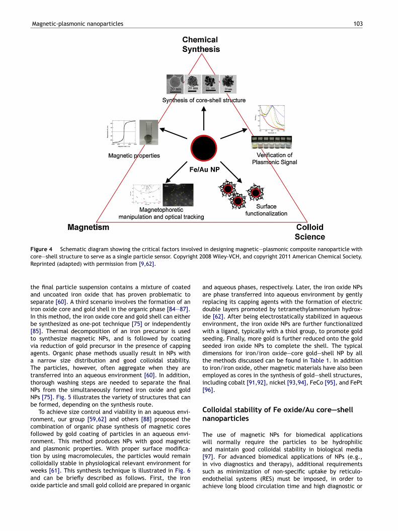
Both the magnetic manipulation and plasmonic imag-ng require reliable techniques to synthesize particles thatre uniform in size and shape. The size and shape of theonstituent particles can have a dramatic effect on the mag-etic properties (iron oxide volume and superparamagnetichreshold) and plasmonic behavior (gold—shell thickness)nd a high degree of uniformity is desired [59]. Furthermore,he particles must have a surface coating that enables themo form stable dispersions in biological media (0.15 M NaCl).his requires steric, rather than charge-based, stabilization60,61]. As illustrated in Fig. 4, these factors are interre-ated, and hence a holistic approach should be adopted.
esign and synthesis of magnetic plasmonicanoparticles
he section ‘Magnetic nanoparticles for biomedicine’escribed the constraints on the magnetic particle size inrder to maximize the magnetic force on the particle with-ut causing agglomeration. There are other constraints foriocompatibility and chemical stability that favor Fe3O4
s a material rather than the higher magnetization Fe oro. The section ‘Plasmonic nanoparticles for biomedicine’howed that there were fewer constraints on the plasmonicroperties, except that uniformity in the local dielectricnvironment was important if the SPR wavelength was usedo detect binding of biomolecules. For selective bindingpplications the use of NPs with a non-continuous gold coat-ng for plasmonic sensing is limited as its SPR absorbancepectrum is strongly retarded [62]. If the plasmon signal is
nly used as a location marker, this requirement becomesess critical.Introduction of a gold layer onto the surface of iron oxidePs is extremely challenging due to the large surface energy
omnb
J. Lim, S.A. Majetich
f the gold [63], which tends to form separate gold parti-les. During the gold deposition process the iron oxide NPsften aggregate irreversibly as the reductive gold materialsorming complexes with the particle surface and suppressts stabilizing agent (either a capping agents or electricouble layers). In numerous papers describing combinationagnetic—plasmonic NPs, the electron microscopy clearly
hows that there are separate magnetic and Au particles,ue to the large difference in the atomic number Z, andherefore the electron transparency. Special care is neededo promote attachment of Au to iron oxide. For plasmonicensing, there is often a requirement for the LSPR signal toe tuned to a specific wavelength; for a core—shell structurehis is achieved by controlling the gold shell thickness withespect to core diameter [64]. All these factors make theynthesis of magnetic—plasmonic NP for biomedical appli-ations extremely challenging.
There are three particle morphologies that meet theseequirements: (i) iron oxide NPs decorated with Au seedslusters [65,66], (ii) iron oxide—gold dumbell particles [67],nd (iii) iron oxide—core gold—shell particles [68—71]. Eachas the potential for a uniform magnetic and plasmonictructure, and each could be coated with polymers to form atable dispersion in biological media. Other iron oxide/goldybrid nanomaterials have also been developed, and haveound niche uses, including gold—core iron oxide—shellanoparticles with spherical [72] and rod-like [73] struc-ures. Such hybrid materials were proven to be useful forrotein separation and optical imaging or catalysis [72,74].owever, for biofunctionalization, there are no obviousdvantages of having iron oxide on the outside.
Large quantities of iron oxide particles decorated withu seeds, up to a few liters at a time, can be pre-ared by gamma ray irradiation [65,74]. NPs prepared bysing this method were used to selectively separate theulfur-containing amino acids, cystine and methionine, bypermanent magnet [74].Binary Fe oxide/Au particles with a dumbbell morphol-
gy are formed through epitaxial growth of iron oxide onhe Au seeds, and the growth can be affected by the polar-ty of the solvent [67]. The advantage of this structure is itsagnetic and plasmonic properties can be fined tuned by
egulating the size, structure and chemical nature of eachart of the dumbbell through adjusting the synthetic con-itions. Structures like this possesses tremendous potentialor applications involving catalytic reactions [75].
According to Amal and co-workers [60] the synthesisf plasmonic—magnetic core—shell NPs involves two mainrocesses:iron oxide core synthesis followed by gold coat-ng. Methods of obtaining gold-coated magnetite can beategorized according to the variations in each process [60].n some cases reverse micelles are used as confined reactorsor both particle synthesis and gold coating [76—81].owever, this technique suffers from low yield and pooreproducibility. Another approach involves both synthesisnd coating of NPs in the aqueous phase [82,83], with theron oxide core synthesized via coprecipitation of iron saltsn an alkaline environment followed by the direct reduction
f chloroauric acid to form a gold outer—shell. This aqueousethod produces well-dispersed NPs in water and there iso need for phase transfer. The gold shell thickness formedy using this technique, however, is difficult to control and
Magnetic-plasmonic nanoparticles 103
Figure 4 Schematic diagram showing the critical factors involved in designing magnetic—plasmonic composite nanoparticle withcore—shell structure to serve as a single particle sensor. Copyright 2008 Wiley-VCH, and copyright 2011 American Chemical Society.
aardiewssdttei[
Cn
Twa[
Reprinted (adapted) with permission from [9,62].
the final particle suspension contains a mixture of coatedand uncoated iron oxide that has proven problematic toseparate [60]. A third scenario involves the formation of aniron oxide core and gold shell in the organic phase [84—87].In this method, the iron oxide core and gold shell can eitherbe synthesized as one-pot technique [75] or independently[85]. Thermal decomposition of an iron precursor is usedto synthesize magnetic NPs, and is followed by coatingvia reduction of gold precursor in the presence of cappingagents. Organic phase methods usually result in NPs witha narrow size distribution and good colloidal stability.The particles, however, often aggregate when they aretransferred into an aqueous environment [60]. In addition,thorough washing steps are needed to separate the finalNPs from the simultaneously formed iron oxide and goldNPs [75]. Fig. 5 illustrates the variety of structures that canbe formed, depending on the synthesis route.
To achieve size control and viability in an aqueous envi-ronment, our group [59,62] and others [88] proposed thecombination of organic phase synthesis of magnetic coresfollowed by gold coating of particles in an aqueous envi-ronment. This method produces NPs with good magneticand plasmonic properties. With proper surface modifica-tion by using macromolecules, the particles would remain
colloidally stable in physiological relevant environment forweeks [61]. This synthesis technique is illustrated in Fig. 6and can be briefly described as follows. First, the ironoxide particle and small gold colloid are prepared in organicisea
nd aqueous phases, respectively. Later, the iron oxide NPsre phase transferred into aqueous environment by gentlyeplacing its capping agents with the formation of electricouble layers promoted by tetramethylammonium hydrox-de [62]. After being electrostatically stabilized in aqueousnvironment, the iron oxide NPs are further functionalizedith a ligand, typically with a thiol group, to promote gold
eeding. Finally, more gold is further reduced onto the goldeeded iron oxide NPs to complete the shell. The typicalimensions for iron/iron oxide—core gold—shell NP by allhe methods discussed can be found in Table 1. In additiono iron/iron oxide, other magnetic materials have also beenmployed as cores in the synthesis of gold—shell structures,ncluding cobalt [91,92], nickel [93,94], FeCo [95], and FePt96].
olloidal stability of Fe oxide/Au core—shellanoparticles
he use of magnetic NPs for biomedical applicationsill normally require the particles to be hydrophilicnd maintain good colloidal stability in biological media97]. For advanced biomedical applications of NPs (e.g.,
n vivo diagnostics and therapy), additional requirementsuch as minimization of non-specific uptake by reticulo-ndothelial systems (RES) must be imposed, in order tochieve long blood circulation time and high diagnostic or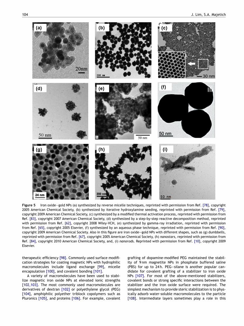
104 J. Lim, S.A. Majetich
Figure 5 Iron oxide—gold NPs (a) synthesized by reverse micelle techniques, reprinted with permission from Ref. [78], copyright2005 American Chemical Society, (b) synthesized by iterative hydroxylamine seeding, reprinted with permission from Ref. [79],copyright 2009 American Chemical Society, (c) synthesized by a modified thermal activation process, reprinted with permission fromRef. [83], copyright 2007 American Chemical Society, (d) synthesized by a step-by-step reactive decomposition method, reprintedwith permission from Ref. [62], copyright 2008 Wiley-VCH, (e) synthesized by gamma-ray irradiation, reprinted with permissionfrom Ref. [65], copyright 2005 Elsevier, (f) synthesized by an aqueous phase technique, reprinted with permission from Ref. [90],copyright 2009 American Chemical Society. Also in this figure are iron oxide—gold NPs with different shapes, such as (g) dumbbells,reprinted with permission from Ref. [67], copyright 2005 American Chemical Society, (h) nanostars, reprinted with permission fromR anorE
tcme
l[d[P
gi(dNc
ef. [84], copyright 2010 American Chemical Society, and, (i) nlsevier.
herapeutic efficiency [98]. Commonly used surface modifi-ation strategies for coating magnetic NPs with hydrophilicacromolecules include ligand exchange [99], micelle
ncapsulation [100], and covalent bonding [101].A variety of macromolecules have been used to stabi-
ize magnetic iron oxide NPs at elevated ionic strengths
102,103]. The most commonly used macromolecules areerivatives of dextran [102] or polyethylene glycol (PEG)104], amphiphilic polyether triblock copolymers such asluronics [105], and proteins [106]. For example, covalentssi[
ods. Reprinted with permission from Ref. [10], copyright 2009
rafting of dopamine-modified PEG maintained the stabil-ty of 9 nm magnetite NPs in phosphate buffered salinePBS) for up to 24 h. PEG—silane is another popular can-idate for covalent grafting of a stabilizer to iron oxidePs [107]. For most of the above-mentioned stabilizers,ovalent bonds or strong specific interactions between the
tabilizer and the iron oxide surface were required. Theimplest mechanism to provide steric stabilization is to phys-cally adsorb water-soluble macromolecules to the particle108]. Intermediate layers sometimes play a role in this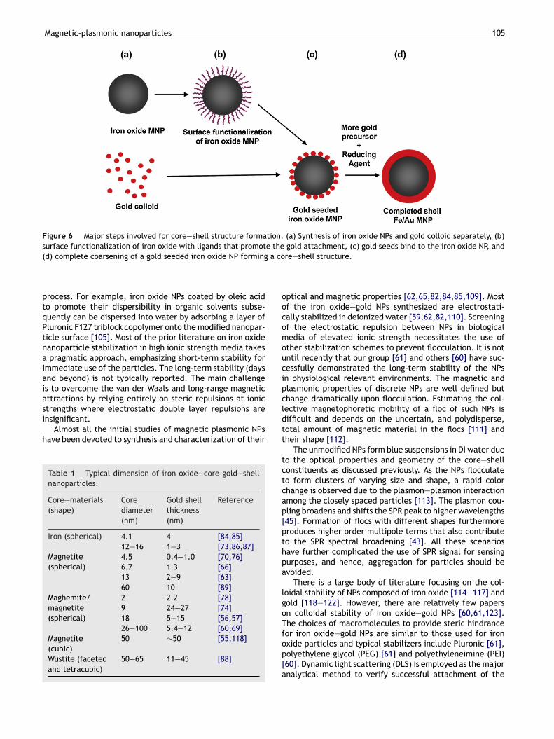
Magnetic-plasmonic nanoparticles 105
Figure 6 Major steps involved for core—shell structure formation. (a) Synthesis of iron oxide NPs and gold colloid separately, (b)surface functionalization of iron oxide with ligands that promote the gold attachment, (c) gold seeds bind to the iron oxide NP, and
a co
oocomoucipcl
(d) complete coarsening of a gold seeded iron oxide NP forming
process. For example, iron oxide NPs coated by oleic acidto promote their dispersibility in organic solvents subse-quently can be dispersed into water by adsorbing a layer ofPluronic F127 triblock copolymer onto the modified nanopar-ticle surface [105]. Most of the prior literature on iron oxidenanoparticle stabilization in high ionic strength media takesa pragmatic approach, emphasizing short-term stability forimmediate use of the particles. The long-term stability (daysand beyond) is not typically reported. The main challengeis to overcome the van der Waals and long-range magneticattractions by relying entirely on steric repulsions at ionicstrengths where electrostatic double layer repulsions are
insignificant.Almost all the initial studies of magnetic plasmonic NPshave been devoted to synthesis and characterization of their
Table 1 Typical dimension of iron oxide—core gold—shellnanoparticles.
Core—materials(shape)
Corediameter(nm)
Gold shellthickness(nm)
Reference
Iron (spherical) 4.1 4 [84,85]12—16 1—3 [73,86,87]
Magnetite(spherical)
4.5 0.4—1.0 [70,76]6.7 1.3 [66]13 2—9 [63]60 10 [89]
Maghemite/magnetite(spherical)
2 2.2 [78]9 24—27 [74]18 5—15 [56,57]26—100 5.4—12 [60,69]
Magnetite(cubic)
50 ∼50 [55,118]
Wustite (facetedand tetracubic)
50—65 11—45 [88]
dtt
tctcap[pthpa
lgoTfop[a
re—shell structure.
ptical and magnetic properties [62,65,82,84,85,109]. Mostf the iron oxide—gold NPs synthesized are electrostati-ally stabilized in deionized water [59,62,82,110]. Screeningf the electrostatic repulsion between NPs in biologicaledia of elevated ionic strength necessitates the use of
ther stabilization schemes to prevent flocculation. It is notntil recently that our group [61] and others [60] have suc-essfully demonstrated the long-term stability of the NPsn physiological relevant environments. The magnetic andlasmonic properties of discrete NPs are well defined buthange dramatically upon flocculation. Estimating the col-ective magnetophoretic mobility of a floc of such NPs isifficult and depends on the uncertain, and polydisperse,otal amount of magnetic material in the flocs [111] andheir shape [112].
The unmodified NPs form blue suspensions in DI water dueo the optical properties and geometry of the core—shellonstituents as discussed previously. As the NPs flocculateo form clusters of varying size and shape, a rapid colorhange is observed due to the plasmon—plasmon interactionmong the closely spaced particles [113]. The plasmon cou-ling broadens and shifts the SPR peak to higher wavelengths45]. Formation of flocs with different shapes furthermoreroduces higher order multipole terms that also contributeo the SPR spectral broadening [43]. All these scenariosave further complicated the use of SPR signal for sensingurposes, and hence, aggregation for particles should bevoided.
There is a large body of literature focusing on the col-oidal stability of NPs composed of iron oxide [114—117] andold [118—122]. However, there are relatively few papersn colloidal stability of iron oxide—gold NPs [60,61,123].he choices of macromolecules to provide steric hindranceor iron oxide—gold NPs are similar to those used for iron
xide particles and typical stabilizers include Pluronic [61],olyethylene glycol (PEG) [61] and polyethyleneimine (PEI)60]. Dynamic light scattering (DLS) is employed as the majornalytical method to verify successful attachment of the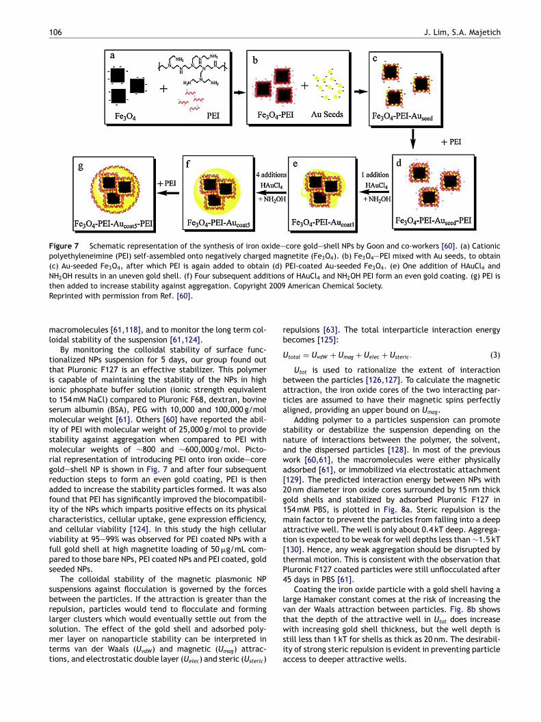
106 J. Lim, S.A. Majetich
Figure 7 Schematic representation of the synthesis of iron oxide—core gold—shell NPs by Goon and co-workers [60]. (a) Cationicpolyethyleneimine (PEI) self-assembled onto negatively charged magnetite (Fe3O4). (b) Fe3O4—PEI mixed with Au seeds, to obtain(c) Au-seeded Fe3O4, after which PEI is again added to obtain (d) PEI-coated Au-seeded Fe3O4. (e) One addition of HAuCl4 andNH2OH results in an uneven gold shell. (f) Four subsequent additions of HAuCl4 and NH2OH PEI form an even gold coating. (g) PEI isthen added to increase stability against aggregation. Copyright 2009 American Chemical Society.R
ml
ttiitsmismrgraficavfps
sbrlsmtt
rb
U
bata
snawa[2g1mat[tP4
lvt
eprinted with permission from Ref. [60].
acromolecules [61,118], and to monitor the long term col-oidal stability of the suspension [61,124].
By monitoring the colloidal stability of surface func-ionalized NPs suspension for 5 days, our group found outhat Pluronic F127 is an effective stabilizer. This polymers capable of maintaining the stability of the NPs in highonic phosphate buffer solution (ionic strength equivalento 154 mM NaCl) compared to Pluronic F68, dextran, bovineerum albumin (BSA), PEG with 10,000 and 100,000 g/mololecular weight [61]. Others [60] have reported the abil-
ty of PEI with molecular weight of 25,000 g/mol to providetability against aggregation when compared to PEI witholecular weights of ∼800 and ∼600,000 g/mol. Picto-
ial representation of introducing PEI onto iron oxide—coreold—shell NP is shown in Fig. 7 and after four subsequenteduction steps to form an even gold coating, PEI is thendded to increase the stability particles formed. It was alsoound that PEI has significantly improved the biocompatibil-ty of the NPs which imparts positive effects on its physicalharacteristics, cellular uptake, gene expression efficiency,nd cellular viability [124]. In this study the high cellulariability at 95—99% was observed for PEI coated NPs with aull gold shell at high magnetite loading of 50 �g/mL com-ared to those bare NPs, PEI coated NPs and PEI coated, goldeeded NPs.
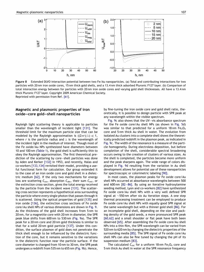
The colloidal stability of the magnetic plasmonic NPuspensions against flocculation is governed by the forcesetween the particles. If the attraction is greater than theepulsion, particles would tend to flocculate and formingarger clusters which would eventually settle out from the
olution. The effect of the gold shell and adsorbed poly-er layer on nanoparticle stability can be interpreted inerms van der Waals (UvdW) and magnetic (Umag) attrac-ions, and electrostatic double layer (Uelec) and steric (Usteric)
wsia
epulsions [63]. The total interparticle interaction energyecomes [125]:
total = UvdW + Umag + Uelec + Usteric. (3)
Utot is used to rationalize the extent of interactionetween the particles [126,127]. To calculate the magneticttraction, the iron oxide cores of the two interacting par-icles are assumed to have their magnetic spins perfectlyligned, providing an upper bound on Umag.
Adding polymer to a particles suspension can promotetability or destabilize the suspension depending on theature of interactions between the polymer, the solvent,nd the dispersed particles [128]. In most of the previousork [60,61], the macromolecules were either physicallydsorbed [61], or immobilized via electrostatic attachment129]. The predicted interaction energy between NPs with0 nm diameter iron oxide cores surrounded by 15 nm thickold shells and stabilized by adsorbed Pluronic F127 in54 mM PBS, is plotted in Fig. 8a. Steric repulsion is theain factor to prevent the particles from falling into a deep
ttractive well. The well is only about 0.4 kT deep. Aggrega-ion is expected to be weak for well depths less than ∼1.5 kT130]. Hence, any weak aggregation should be disrupted byhermal motion. This is consistent with the observation thatluronic F127 coated particles were still unflocculated after5 days in PBS [61].
Coating the iron oxide particle with a gold shell having aarge Hamaker constant comes at the risk of increasing thean der Waals attraction between particles. Fig. 8b showshat the depth of the attractive well in Utot does increase
ith increasing gold shell thickness, but the well depth istill less than 1 kT for shells as thick as 20 nm. The desirabil-ty of strong steric repulsion is evident in preventing particleccess to deeper attractive wells.
Magnetic-plasmonic nanoparticles 107
Figure 8 Extended DLVO interaction potential between two Fe/Au nanoparticles. (a) Total and contributing interactions for twoparticles with 20 nm iron oxide cores, 15 nm thick gold shells, and a 13.4 nm thick adsorbed Pluronic F127 layer. (b) Comparison oftotal interaction energy between for particles with 20 nm iron oxide cores and varying gold shell thicknesses. All have a 13.4 nmthick Pluronic F127 layer. Copyright 2009 American Chemical Society.
boa
fwciiFccotapdf
sasFstFtai[oN5s
Reprinted with permission from Ref. [61].
Magnetic and plasmonic properties of ironoxide—core gold—shell nanoparticles
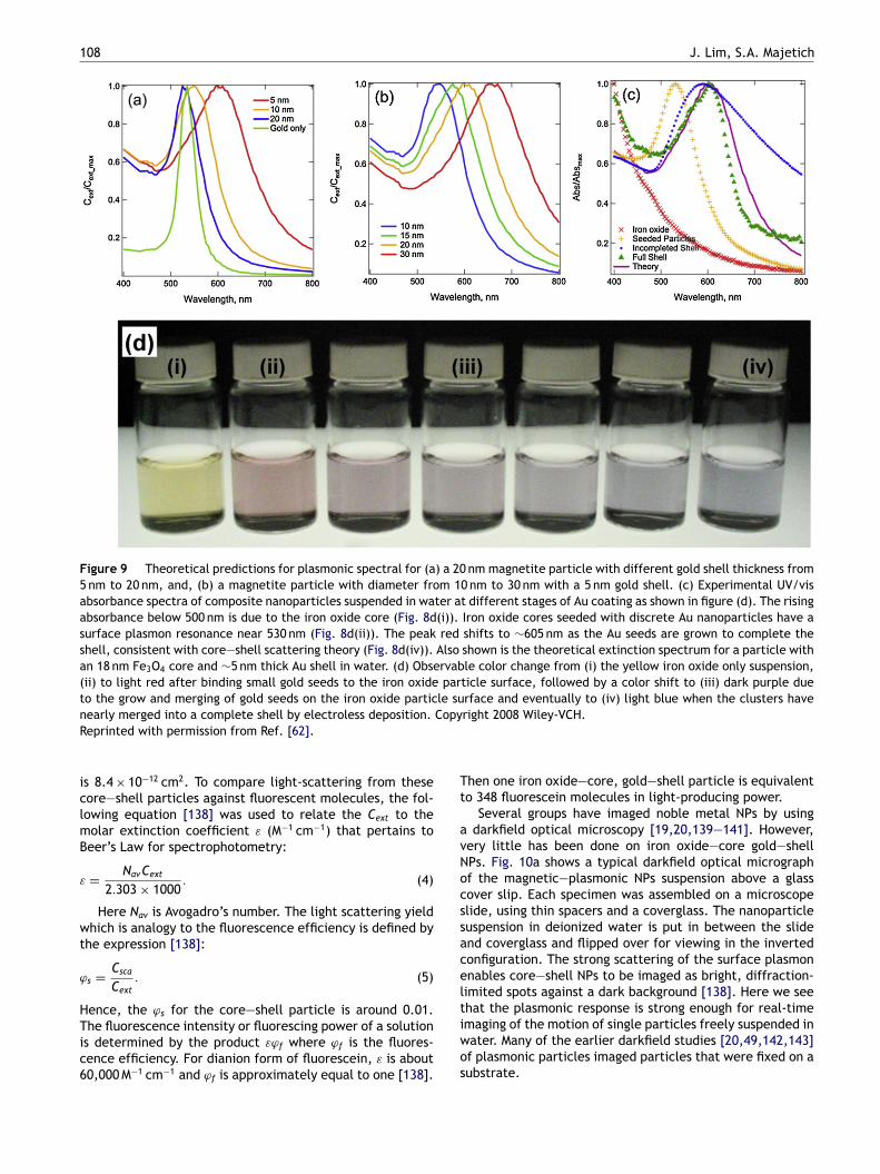
Rayleigh light scattering theory is applicable to particlessmaller than the wavelength of incident light [131]. Thethreshold limit for the maximum particle size that can bemodeled by the Rayleigh approximation is ((2�r)/�) � 1,where r is the particle radius and � is the wavelength ofthe incident light in the medium of interest. Though most ofthe Fe oxide/Au NPs synthesized have diameters between10 and 100 nm (Table 1), the gold shell is sufficiently thin toobey the Rayleigh approximation. The first theoretical pre-diction of the scattering by core—shell particles was doneby Aden and Kerker [132] in 1953, and recently, Halas andco-workers [133,134] revisited their model, providing a use-ful functional form for calculation. Our group extended itto the case of an iron oxide core and gold shell in a dielec-tric medium [62]. If the only two mechanisms for energyloss are scattering Csca, absorption Cabs, their sum Cext, orthe extinction cross-section, gives the total energy receivedby the particle from the incident wave [131]. The scatter-ing cross section represents a hypothetical area surroundingthe particle where every single photon that passes through itis scattered. Using the optical properties of gold [135] andiron oxide [136], the extinction cross sections of Fe oxidecore/Au shell NPs of various dimensions are shown in Fig. 9.As the thickness of the gold shell increases from 5 nm to20 nm, for a magnetite core with 20 nm in diameter, the SPRpeak blue shifts from 600 nm to 530 nm (Fig. 9a). The SPRpeak for a 20 nm core and 20 nm gold thickness is similar tothat for a solid 20 nm gold particle [137]. Under this con-dition, the surface plasmon of gold does not penetrate thethick shell enough to be influenced by the dielectric func-
tion of the core, but it remains sensitive to the variationsin the dielectric function near the particle surface. If thecore diameter is changed from 10 nm to 30 nm, the SPR peakundergoes a redshift from 535 nm to 655 nm (Fig. 9b). Hence,ss
5
y fine-tuning the iron oxide core and gold shell ratio, the-retically, it is possible to design particle with SPR peak atny wavelength within the visible spectrum.
Fig. 9c also shows that the UV—vis absorbance spectrumor the Fe oxide core/Au shell NPs (as shown in Fig. 5d)as similar to that predicted for a uniform 18 nm Fe3O4
ore and 5 nm thick Au shell in water. The evolution fromsolated Au clusters into a complete shell shows the theoret-cally predicted redshift in the plasmon peak, as indicated inig. 9c. The width of the resonance is a measure of the parti-le homogeneity. During electroless deposition, but beforeompletion of the shell, considerable spectral broadeningccurs owing to the creation of clusters of various sizes. Ashe shell is completed, the particles become more uniformnd the peak sharpens again. The wide range of colors dis-layed in Fig. 9d resulting from the variation in Au shellevelopment allows for potential use of these nanoparticlesor spectroscopic or colorimetric labeling [90].
In most cases, the plasmon peaks for Fe oxide core/Auhell NPs occurred at absorbance wavelengths between 500nd 600 nm [82—86]. By using an iterative hydroxylamineeeding method, Lyon and co-workers [82] have synthesizede oxide core/Au shell NPs with a very well defined SPRignal at ∼550 nm after six Au iteration steps. A one stephermal processing treatment can be employed to producee oxide core/Au shell NPs with equally good SPR signal athe same wavelength but with a thinner gold shell [86]. Forn incomplete gold shell, depending on the size and pack-ng density of the gold seeds, a more pronounced SPR peak60,62] and a small shoulder or flat peak have both beenbserved [65]. After assembling the Fe oxide core/Au shellPs into a thin film, the SPR wavelength can be varied from20 nm to 620 nm by changing the dielectric properties of theurrounding media [85]. The SPR signal of Fe oxide core/Au
hell NPs can also be fine tuned by changing the pH of itsuspension medium [83].The calculated Cext for a uniform 18 nm Fe3O4 core andnm thick Au shell in water at the SPR resonance frequency
108 J. Lim, S.A. Majetich
Figure 9 Theoretical predictions for plasmonic spectral for (a) a 20 nm magnetite particle with different gold shell thickness from5 nm to 20 nm, and, (b) a magnetite particle with diameter from 10 nm to 30 nm with a 5 nm gold shell. (c) Experimental UV/visabsorbance spectra of composite nanoparticles suspended in water at different stages of Au coating as shown in figure (d). The risingabsorbance below 500 nm is due to the iron oxide core (Fig. 8d(i)). Iron oxide cores seeded with discrete Au nanoparticles have asurface plasmon resonance near 530 nm (Fig. 8d(ii)). The peak red shifts to ∼605 nm as the Au seeds are grown to complete theshell, consistent with core—shell scattering theory (Fig. 8d(iv)). Also shown is the theoretical extinction spectrum for a particle withan 18 nm Fe3O4 core and ∼5 nm thick Au shell in water. (d) Observable color change from (i) the yellow iron oxide only suspension,(ii) to light red after binding small gold seeds to the iron oxide particle surface, followed by a color shift to (iii) dark purple dueto the grow and merging of gold seeds on the iron oxide particle surface and eventually to (iv) light blue when the clusters haven CopyR
iclmB
ε
wt
ϕ
HTic6
Tt
avNocssacelt
early merged into a complete shell by electroless deposition.eprinted with permission from Ref. [62].
s 8.4 × 10⁻¹² cm2. To compare light-scattering from theseore—shell particles against fluorescent molecules, the fol-owing equation [138] was used to relate the Cext to theolar extinction coefficient ε (M−1 cm−1) that pertains toeer’s Law for spectrophotometry:
= NavCext
2.303 × 1000. (4)
Here Nav is Avogadro’s number. The light scattering yieldhich is analogy to the fluorescence efficiency is defined by
he expression [138]:
s = Csca
Cext
. (5)
ence, the ϕs for the core—shell particle is around 0.01.
he fluorescence intensity or fluorescing power of a solutions determined by the product εϕf where ϕf is the fluores-ence efficiency. For dianion form of fluorescein, ε is about0,000 M−1 cm−1 and ϕf is approximately equal to one [138].
iwos
right 2008 Wiley-VCH.
hen one iron oxide—core, gold—shell particle is equivalento 348 fluorescein molecules in light-producing power.
Several groups have imaged noble metal NPs by usingdarkfield optical microscopy [19,20,139—141]. However,
ery little has been done on iron oxide—core gold—shellPs. Fig. 10a shows a typical darkfield optical micrographf the magnetic—plasmonic NPs suspension above a glassover slip. Each specimen was assembled on a microscopelide, using thin spacers and a coverglass. The nanoparticleuspension in deionized water is put in between the slidend coverglass and flipped over for viewing in the invertedonfiguration. The strong scattering of the surface plasmonnables core—shell NPs to be imaged as bright, diffraction-imited spots against a dark background [138]. Here we seehat the plasmonic response is strong enough for real-time
maging of the motion of single particles freely suspended inater. Many of the earlier darkfield studies [20,49,142,143]f plasmonic particles imaged particles that were fixed on aubstrate.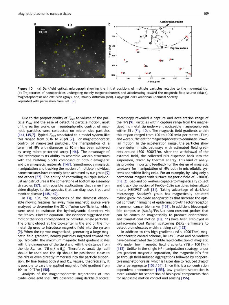
Magnetic-plasmonic nanoparticles 109
Figure 10 (a) Darkfield optical micrograph showing the initial positions of multiple particles relative to the mu-metal tip.(b) Trajectories of nanoparticles undergoing mainly magnetophoresis and accelerating toward the magnetic field source (black),magnetophoresis and diffusion (gray), and, mainly diffusion (red). Copyright 2011 American Chemical Society.
mttwtaimeessttp(aimhcaicasd
nhN[lgt
Reprinted with permission from Ref. [9].
Due to the proportionality of Fmag to volume of the par-ticle Vmag and the ease of detecting particle motion, mostof the earlier works on magnetophoretic control of mag-netic particles were conducted on micron size particles[144,145,7]. Typical Fmag associated to a model system likethis ranged from 50 fN to 20 pN [7]. For magnetophoreticcontrol of nano-sized particles, the manipulation of aswarm of NPs with diameter at 10 nm has been achievedby using micro-patterned array [146]. The advantage ofthis technique is its ability to assemble various structureswith the building blocks composed of both diamagneticand paramagnetic materials [147]. Simultaneous magneticmanipulation and tracking the motion of multiple individualnanostructures have recently been achieved by our group [9]and others [57]. The ability of controlling multiple individ-ual nanostructures is the cornerstone of bottom up assemblystrategies [57], with possible applications that range fromvideo displays to therapeutics that can diagnose, treat andmonitor disease [148,149].
In Fig. 10a, the trajectories of the dimmest observ-able moving features far away from magnetic source wereanalyzed to determine the 2D diffusion coefficients, whichwere used to estimate the hydrodynamic diameters viathe Stokes—Einstein equation. The evidence suggested thatmost of the spots corresponded to individual single particles.The bright object at the top-center is the end of the mu-metal tip used to introduce magnetic field into the system[9]. When the tip was magnetized, generating a large mag-netic field gradient, nearby particles migrated toward thetip. Typically, the maximum magnetic field gradient scaleswith the dimensions of the tip ˇ and with the distance fromthe tip Rdis as ∇B ∝ ˇ ∝ R−1
dis. Therefore, small tip radiishould be used and the tip should be positioned close tothe NPs or even directly immersed into the particle suspen-sion. By fine tuning both ˇ and Rdis values, theoretically, it
is possible to vary the applied magnetic field gradient from102 to 107 T/m [150].Analysis of the magnetophoretic trajectories of ironoxide—core gold shell NPs observed using darkfield optical
tdmf
icroscopy revealed a capture and acceleration range ofhe NPs [9]. Particles within capture range from the magne-ized mu metal tip underwent noticeable magnetophoresisithin 25 s (Fig. 10b). The magnetic field gradients within
his region ranged from 100 to 1000 tesla per meter (T/m)nd were sufficient for magnetophoresis to dominate Brown-an motion. In the acceleration range, the particles showore deterministic pathways with estimated field gradi-
nts around 1300—3000 T/m. After the withdrawal of thexternal field, the collected NPs dispersed back into theuspension, driven by thermal energy. This kind of analy-is provides important feedback for the design of magneticweezers for manipulation of NPs both in microfluidic sys-ems and within living cells. For an example, by using only aermanent magnet with surface magnetic field of ∼3000 GFig. 2), Gao and co-workers capable to magnetically collectnd track the motion of Fe3O4—CdSe particles internalizednto a HEK293T cell [31]. Taking advantage of darkfieldicroscopy, Sokolov’s group has magnetically actuated
ybrid gold/iron oxide nanoparticles that increase the opti-al contrast in imaging of epidermal growth factor receptor,common cancer biomarker [151]. In addition, biocompat-
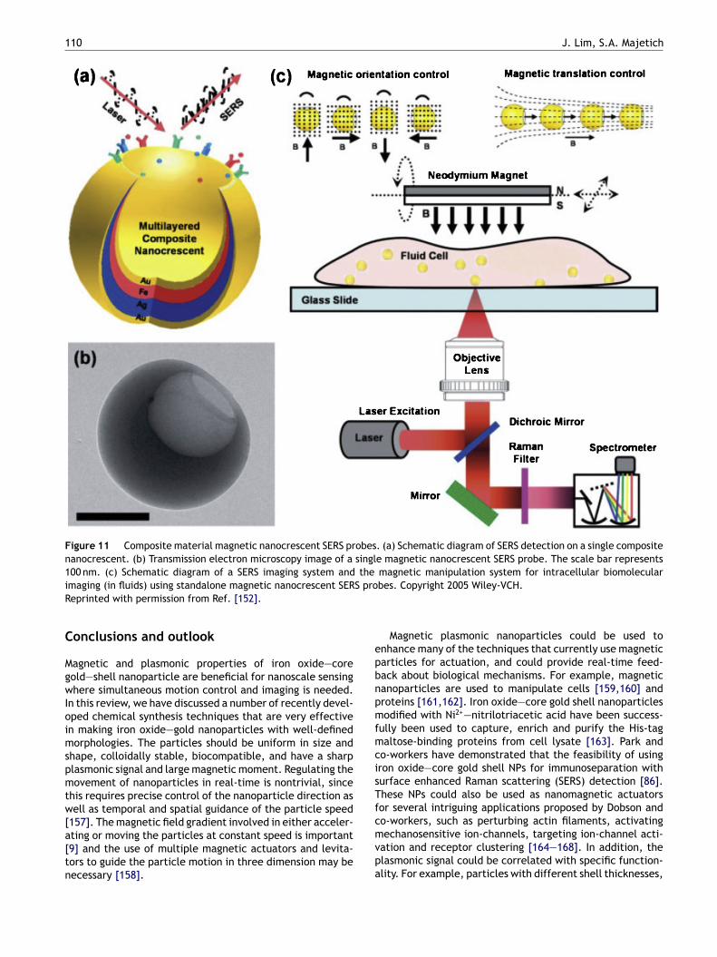
ble composite (Au/Ag/Fe/Au) nano-crescent probes thatan be controlled magnetically to produce orientationalnd translational motion (Fig. 11) have been employed asurface-enhanced Raman scattering (SERS) enhancers toetect biomolecules within a living cell [152].
In addition to this high gradient (∇B > 1000 T/m) mag-etophoretic control scheme, De Las Cuevas and co-workersave demonstrated the possible rapid collection of magneticPs under low magnetic field gradients (∇B > 100 T/m)112]. Unlike in the single NP manipulation strategy, underow gradient magnetic separation, the magnetic NPs firsto through field-induced aggregations followed by coopera-ive magnetophoresis, which is faster due to reduced drag of
he large aggregate [153,154]. Since this is a concentrationependent phenomenon [155], low gradient separation isore suitable for separation of biological components thanor nanoscale motion control and sensing [156].
110 J. Lim, S.A. Majetich
Figure 11 Composite material magnetic nanocrescent SERS probes. (a) Schematic diagram of SERS detection on a single compositenanocrescent. (b) Transmission electron microscopy image of a single magnetic nanocrescent SERS probe. The scale bar represents100 nm. (c) Schematic diagram of a SERS imaging system and the magnetic manipulation system for intracellular biomoleculari S proR
C
MgwIoimspmtw[a[tn
epbnpmfmcisTfc
maging (in fluids) using standalone magnetic nanocrescent SEReprinted with permission from Ref. [152].
onclusions and outlook
agnetic and plasmonic properties of iron oxide—coreold—shell nanoparticle are beneficial for nanoscale sensinghere simultaneous motion control and imaging is needed.
n this review, we have discussed a number of recently devel-ped chemical synthesis techniques that are very effectiven making iron oxide—gold nanoparticles with well-definedorphologies. The particles should be uniform in size and
hape, colloidally stable, biocompatible, and have a sharplasmonic signal and large magnetic moment. Regulating theovement of nanoparticles in real-time is nontrivial, since
his requires precise control of the nanoparticle direction asell as temporal and spatial guidance of the particle speed
157]. The magnetic field gradient involved in either acceler-
ting or moving the particles at constant speed is important9] and the use of multiple magnetic actuators and levita-ors to guide the particle motion in three dimension may beecessary [158].mvpa
bes. Copyright 2005 Wiley-VCH.
Magnetic plasmonic nanoparticles could be used tonhance many of the techniques that currently use magneticarticles for actuation, and could provide real-time feed-ack about biological mechanisms. For example, magneticanoparticles are used to manipulate cells [159,160] androteins [161,162]. Iron oxide—core gold shell nanoparticlesodified with Ni2+—nitrilotriacetic acid have been success-
ully been used to capture, enrich and purify the His-tagaltose-binding proteins from cell lysate [163]. Park and
o-workers have demonstrated that the feasibility of usingron oxide—core gold shell NPs for immunoseparation withurface enhanced Raman scattering (SERS) detection [86].hese NPs could also be used as nanomagnetic actuatorsor several intriguing applications proposed by Dobson ando-workers, such as perturbing actin filaments, activating
echanosensitive ion-channels, targeting ion-channel acti-ation and receptor clustering [164—168]. In addition, thelasmonic signal could be correlated with specific function-lity. For example, particles with different shell thicknesses,
Magnetic-plasmonic nanoparticles
or Au and Ag shells of the same thickness, would have dif-ferent plasmon wavelengths. If the two types of particleshad surface coatings with different specific binding affini-ties, the binding kinetics would be observed in parallel. Thecurrent limitation is in the restricted availability of uniformmagnetic plasmonic NPs that are stable in biological media,and we hope that the material in this review and its refer-ences helps more researchers to investigate and expand thepossible uses.
Acknowledgments
This material is based on work supported by NationalScience Foundation grant #CBET 0853963. J.K. Lim grate-fully acknowledges the financial support from ExploratoryResearch Grant Scheme (ERGS/203/PJKIMIA/6730012) fromMOHE Malaysia. J.K. Lim is affiliated to the Membrane Sci-ence and Technology cluster USM.
References
[1] Y. Pan, X. Du, F. Zhao, B. Xu, Chem. Soc. Rev. 41 (2012) 2912.[2] R.C. Hayward, D.A. Saville, I.A. Aksay, Nature 404 (2000) 56.[3] S. Gangwal, O.J. Cayre, M.Z. Bazant, O.D. Velev, Phys. Rev.
Lett. 100 (2008) 0058302.[4] H. Zhou, L.R. White, R.D. Tilton, J. Colloid Interface Sci. 285
(2005) 179.[5] J.K. Lim, H. Zhou, R.D. Tilton, J. Colloid Interface Sci. 332
(2009) 113.[6] R. Hagedorn, G. Fuhr, T. Muller, J. Gimsa, Electrophoresis 13
(1992) 49.[7] C. Gosse, V. Croquette, Biophys. J. 82 (2002) 3314.[8] H.M. Hertz, J. Appl. Phys. 78 (1995) 4845.[9] J.K. Lim, C. Lanni, E.R. Evarts, F. Lanni, R.D. Tilton, S.A.
Majetich, ACS Nano 5 (2011) 217.[10] J.K. Lim, D.X. Tan, F. Lanni, R.D. Tilton, S.A. Majetich, J.
Magn. Magn. Mater. 321 (2009) 1557.[11] D.G. Grier, Nature 424 (2003) 810.[12] P.Y. Chiou, A.T. Ohta, M.C. Wu, Nature 436 (2005) 370.[13] I. Roy, T.Y. Ohulchanskyy, D.J. Bharali, H.E. Pudavar, R.A. Mis-
tretta, N. Kaur, et al., Proc. Natl. Acad. Sci. U.S.A. 102 (2005)279.
[14] B. Steitz, H. Hofmann, S.W. Kamau, P.O. Hassa, M.O. Hottiger,B. Rechenberg, et al., J. Magn. Magn. Mater. 311 (2007) 300.
[15] L. Rodriguez-Lorenzo, Z. Krpetie, S. Barbosa, R.A. Alvarez-Puebla, L.M. Liz-Marzan, I.A. Prior, et al., Integr. Biol. 3 (2011)922.
[16] P.K. Jain, X. Huang, I.H. El-sayed, M.A. El-Sayed, Acc. Chem.Res. 41 (2008) 1578.
[17] J.J. Storhoff, R. Elghanian, R.C. Mucic, C.A. Mirkin, R.L.Letsinger, J. Am. Chem. Soc. 120 (1998) 1959.
[18] C.R. Yonzon, E. Jeoung, S. Zou, G.C. Schatz, M. Mrksich, R.P.Van Duyne, J. Am. Chem. Soc. 126 (2004) 12669.
[19] J. Yguerabide, E.E. Yguerabide, Anal. Biochem. 262 (1998)157.
[20] A.D. McFarland, R.P. Van Duyne, Nano Lett. 3 (2003) 1057.[21] W.G.J.H.M. van Sark, P.L.T.M. Frederix, D.J. Van den Heuvel,
H.C. Gerritsen, A.A. Bol, J.N.J. van Lingen, et al., J. Phys.Chem. B 105 (2001) 8281.
[22] R. Hardman, Environ. Health Perspect. 114 (2005) 165.
[23] L. Carbone, P. Davide Cozzoli, Nano Today 5 (2010) 449.[24] N.C. Bigall, W.J. Parak, D. Dorfs, Nano Today 7 (2012) 282.[25] M.R. Jones, K.D. Osberg, R.J. Macfarlane, M.R. Langille, C.A.Mirkin, Chem. Rev. 111 (2011) 3736.
111
[26] V. Salgueirino-Maceira, M.A. Correa-Duarte, Adv. Mater. 19(2007) 4131.
[27] D.J. Dunlop, Science 176 (1972) 41.[28] R. Berker, Electromagnetic Fields and Interactions, Dover,
New York, 1982.[29] Q.A. Pankhurst, J. Connolly, S.K. Jones, J. Dobson, J. Phys.
D: Appl. Phys. 36 (2003) R167.[30] M. Zborowski, L. Sun, L.R. Moore, P.S. Williams, J.J.
Chalmers, J. Magn. Magn. Mater. 194 (1999) 224.[31] J. Gao, W. Zhang, P. Huang, B. Zhang, X. Zhang, B. Xu, J. Am.
Chem. Soc. 130 (2008) 3710.[32] A.A. Kayani, K. Khoshmanesh, S.A. Ward, A. Mitchell, K.
Kalantarzadeh, Biomicrofluidic 6 (2012) 031501.[33] Q.A. Pankhurst, N.T.K. Thanh, S.K. Jones, J. Dobson, J. Phys.
D: Appl. Phys. 42 (2009) 224001.[34] N.L. Adolphi, D.L. Huber, H.C. Bryant, T.C. Monson, D.L.
Fegan, J.K. Lim, et al., Phys. Med. Biol. 55 (2010) 5985.[35] C.C. Berry, A.S.G. Curtis, J. Phys. D: Appl. Phys. 36 (2003)
R198.[36] D. Pouliquen, J.J. Le Jeune, R. Perdrisot, A. Ermias, P. Jallet,
Magn. Reson. Imaging 9 (1993) 275.[37] N. Pamme, C. Wilhelm, Lab Chip 6 (2006) 974.[38] L. Jesephson, C.H. Tung, A. Moore, R. Weissleder, Bioconju-
gate Chem. 10 (1999) 186.[39] S.C. McBain, H.H.P. Yiu, J. Dobson, Int. J. Nanomed. 3 (2008)
169.[40] J. Dobson, Drug Dev. Res. 67 (2006) 55.[41] J. Dobson, Gene Ther. 13 (2006) 283.[42] F. Sonvico, S. Mornet, S. Vasseur, C. Dubernet, D. Jaillard, J.
Degrouard, et al., Bioconjugate Chem. 16 (2005) 1181.[43] S. Link, M.A. El-Sayed, Int. Rev. Phys. Chem. 19 (2000) 409.[44] S. Link, M.A. El-Sayed, J. Phys. Chem. B (1999) 4212.[45] A.J. Haes, C.L. Haynes, A.D. McFarland, G.C. Schatz, R.P. Van
Duyne, S. Zou, MRS Bull. 30 (2005) 368.[46] M.A. Mahmoud, M. Chamanzar, A. Adibi, M.A. El-Sayed, J. Am.
Chem. Soc. 134 (2012) 6434.[47] K. Kneipp, H. Kneipp, I. Itzkan, R.R. Dasari, M.S. Feld, J.
Phys.: Condens. Matter 14 (2002) R597.[48] J.N. Anker, W.P. Hall, O. Lyandres, N.C. Shah, J. Zhao, R.P.
Van Duyne, Nat. Mater. 7 (2008) 442.[49] S. Schultz, D.R. Smith, J.J. Mock, D.A. Schultz, Proc. Natl.
Acad. Sci. U.S.A. 97 (2000) 996.[50] I.H. El-Sayed, X. Huang, M.A. El-Sayed, Nano Lett. 5 (2005)
829.[51] L.R. Hirsch, J.B. Jackson, A. Lee, N.J. Halas, J.L. West, Anal.
Chem. 75 (2003) 2377.[52] E.E. Connor, J. Mwamuka, A. Gole, C.J. Murphy, M.D. Wyatt,
Small 1 (2005) 325.[53] D.J. Lavrich, S.M. Wetterer, S.L. Bernasek, G. Scoles, J. Phys.
Chem. B 102 (1998) 3456.[54] S. Lal, S.E. Clare, N.J. Halas, Acc. Chem. Res. 41 (2008) 1842.[55] A.M. Gobin, M.H. Lee, N.J. Halas, W.D. James, R.A. Drezek,
J.L. West, Nano Lett. 7 (2007) 1929.[56] E.M. Purcell, Am. J. Phys. 45 (1977) 3.[57] G. Ruan, G. Vieira, T. Henighan, A. Chen, D. Thakur, R.
Sooryakumar, et al., Nano Lett. 10 (2010) 2220.[58] T.R. Jensen, M.D. Malinsky, C.L. Haynes, R.P. Van Duyne, J.
Phys. Chem. B 104 (2000) 10549.[59] J.K. Lim, R.D. Tilton, A. Eggeman, S.A. Majetich, J. Magn.
Magn. Mater. 311 (2007) 78.[60] I.Y. Goon, M.H. Leo, M. Lim, P. Munroe, J.J. Gooding, R. Amal,
Chem. Mater. 21 (2009) 673.[61] J.K. Lim, S.A. Majetich, R.D. Tilton, Langmuir 25 (2009)
13384.
[62] J.K. Lim, R.D. Tilton, A. Eggeman, F. Lanni, S.A. Majetich,Adv. Mater. 20 (2008) 1721.[63] S. Wu, Polymer Interface and Adhesion, Marcel Dekker, Inc.,
New York, 1982.
1
[
12
[64] F. Tam, G.P. Goodrich, B.R. Johnson, N.J. Halas, Nano Lett. 7(2007) 496.
[65] S. Seino, T. Kinoshita, Y. Otome, T. Nakagawa, K. Okitsu, Y.Mizukoshi, et al., J. Magn. Magn. Mater. 293 (2005) 144.
[66] J. Bao, W. Chen, T. Liu, Y. Zhu, P. Jin, L. Wang, et al., ACSNano 1 (2007) 293.
[67] H. Yu, M. Chen, P.M. Rice, S.X. Wang, R.L. White, S. Sun, NanoLett. 5 (2005) 379.
[68] M. Mandal, S. Kundu, S.K. Ghosh, S. Panigrahi, T.K. Sau, S.M.Yusuf, et al., J. Colloid Interface Sci. 286 (2008) 187.
[69] L. Wang, H.Y. Park, S.L.L. Lim, M.J. Schadt, D. Mott, J. Luo,et al., J. Mater. Chem. 18 (2008) 2692.
[70] W. Dong, Y. Li, D. Niu, Z. Ma, J. Gu, Y. Chen, et al., Adv. Mater.23 (2011) 5392.
[71] I. Robinson, L.D. Tung, S. Maenosono, C. Walti, N.T.K. Thanh,Nanoscale 2 (2010) 2624.
[72] E.V. Shevchenko, M.I. Bodnarchuk, M.V. Kovalenko, D.V.Talapin, R.K. Smith, S. Aloni, et al., Adv. Mater. 20 (2008)4323.
[73] A. Gole, J.W. Stone, W.R. Gemmill, H.C. Loye, C.J. Murphy,Langmuir 24 (2008) 6232.
[74] T. Kinoshita, S. Seino, Y. Mizukoshi, T. Nakagawa, T.A.Yamamoto, J. Magn. Magn. Mater. 311 (2007) 255.
[75] C. Wang, H. Yin, S. Dai, S. Sun, Chem. Mater. 22 (2010) 3277.[76] W.L. Zhou, E.E. Carpenter, J. Lin, A. Kumbhar, J. Sims, C.J.
O’Connor, Eur. Phys. J. D 16 (2001) 289.[77] M. Mikhaylova, D.K. Kim, N. Bobrysheva, M. Osmolowsky, V.
Semenov, T. Tsakalakos, et al., Langmuir 20 (2004) 2472.[78] S.J. Cho, J.C. Idrobo, J. Olamit, K. Liu, N.D. Browning, S.M.
Kauzlarich, Chem. Mater. 17 (2005) 3181.[79] J. Lin, W. Zhou, A. Kumbhar, J. Wiemann, J. Fang, E.E. Car-
penter, et al., J. Solid State Chem. 159 (2001) 26.[80] S.J. Cho, S.M. Kauzlarich, J. Olamit, K. Liu, F. Grandjean, L.
Rebbouh, et al., J. Appl. Phys. 95 (2004) 6804.[81] S.J. Cho, A.M. Shahin, G.J. Long, J.E. Davies, K. Liu, F. Grand-
jean, et al., Chem. Mater. 18 (2006) 960.[82] J.L. Lyon, D.A. Fleming, M.B. Stone, P. Schiffer, M.E. Williams,
Nano Lett. 4 (2009) 719.[83] C.K. Lo, D. Xiao, M.M.F. Choi, J. Mater. Chem. 17 (2007)
2418.[84] L. Wang, J. Luo, Q. Fan, M. Suzuki, I.S. Suzuki, M.H. Engel-
hard, et al., J. Phys. Chem. B 109 (2005) 21593.[85] L. Wang, J. Luo, M.M. Maye, Q. Fan, R. Qiang, M.H. Engelhard,
et al., J. Mater. Chem. 15 (2005) 1821.[86] H.Y. Park, M.J. Schadt, L. Wang, I.S. Lim, P.N. Njoki, S.H. Kim,
et al., Langmuir 23 (2007) 9050.[87] H.M. Song, Q. Wei, Q.K. Ong, A. Wei, ACS Nano 4 (2010) 5163.[88] Z. Xu, Y. Hou, S. Sun, J. Am. Chem. Soc. 129 (2007) 8698.[89] E.E. Carpenter, J. Magn. Magn. Mater. 225 (2001) 17.[90] C.S. Levin, C. Hofmann, T.A. Ali, A.T. Kelly, E. Morosan, P.
Nordlander, et al., ACS Nano 3 (2009) 1379.[91] Y. Bao, K.M. Krishnan, J. Magn. Magn. Mater. 293 (2005) 15.[92] Y. Bao, H. Calderon, K.M. Krishnan, J. Phys. Chem. C 111
(2007) 1941.[93] D. Chen, J. Li, C. Shi, X. Du, N. Zhao, J. Sheng, S. Liu, Chem.
Mater. 19 (2007) 3399.[94] D. Chen, S. Liu, J. Li, N. Zhao, C. Shi, X. Du, J. Sheng, J.
Alloys Compd. 475 (2009) 494.[95] J. Bai, J.P. Wang, Appl. Phys. Lett. 87 (2005) 152502.[96] T. Hartling, T. Uhlig, A. Seidenstucker, N.C. Bigall, P. Olk, U.
Wiedwald, et al., Appl. Phys. Lett. 96 (2010) 183111.[97] C. Fang, N. Bhattarai, C. Sun, M. Zhang, Small 5 (2009) 1637.[98] S.M. Moqhimi, A.C. Hunter, J.C. Murray, FASEB J. 19 (2005)
311.
[99] Y.W. Jun, Y.M. Huh, J.S. Choi, J.H. Lee, H.T. Song, S. Kim,et al., J. Am. Chem. Soc. 127 (2005) 5732.100] W.W. Yu, E. Chang, J.C. Falkner, J. Zhang, A.M. Al-Somali,
C.M. Sayes, et al., J. Am. Chem. Soc. 129 (2007) 2871.
J. Lim, S.A. Majetich
[101] O. Veiseh, C. Sun, J. Gunn, N. Kohler, P. Gabikian, D. Lee,et al., Nano Lett. 5 (2005) 1003.
[102] S. Laurent, D. Forge, M. Port, A. Roch, C. Robic, L. VanderElst, et al., Chem. Rev. 108 (2008) 2064.
[103] A.K. Gupta, M. Gupta, Biomaterials 26 (2005) 3995.[104] Y. Zhang, N. Kohler, M. Zhang, Biomaterials 23 (2002) 1553.[105] T.K. Jain, M.A. Morales, S.K. Sahoo, D.L. Leslie-Pelecky, V.
Labhasetwar, Mol. Pharmacol. 2 (2005) 194.[106] S.J.H. Soenen, H. Hodenius, T. Schmitz-Rode, M. De Cuyper,
J. Magn. Magn. Mater. 320 (2008) 634.[107] N. Kohler, G.E. Fryxell, M. Zhang, J. Am. Chem. Soc. 126
(2004) 7206.[108] M. Gonzales, K.M. Krishnan, J. Magn. Magn. Mater. 311 (2007)
59.[109] M. Chen, S. Yamamuro, D. Farrell, S.A. Majetich, J. Appl.
Phys. 93 (2003) 7551.[110] Q.H. Lu, K.L. Yao, D. Xi, Z.L. Liu, X.P. Luo, Q. Ning, J. Magn.
Magn. Mater. 301 (2006) 44.[111] A. Ditsch, S. Lindenmann, P.E. Laibinis, D.I.C. Wang, T.A. Hat-
ton, Ind. Eng. Chem. Res. 44 (2005) 6824.[112] G. De Las Cuevas, J. Faraudo, J. Camacho, J. Phys. Chem. C
112 (2008) 945.[113] S.J. Oldenburg, R.D. Averitt, S.L. Westcott, N.J. Halas, Chem.
Phys. Lett. 288 (1998) 243.[114] H.T.R. Wiogo, M. Lim, V. Bulmus, J. Yun, R. Amal, Langmuir
27 (2011) 843.[115] I.Y. Goon, C. Zhang, M. Lim, J. Gooding, R. Amal, Langmuir
26 (2010) 12247.[116] Y. Park, R.D. Whitaker, R.J. Naps, J.L. Paulsen, V. Mathiyazha-
gan, L.H. Doerrer, et al., Langmuir 28 (2012) 6246.[117] P.L. Golas, S. Louie, G.V. Lowry, K. Matyjaszewski, R.D. Tilton,
Langmuir 26 (2012) 16890.[118] J. Zhou, J. Ralston, R. Sedev, D.A. Beattie, J. Colloid Interface
Sci. 331 (2009) 251.[119] R. Levy, N.T.K. Thanh, R.C. Doty, I. Hussain, R.J. Nichols, D.J.
Schiffrin, et al., J. Am. Chem. Soc. 126 (2004) 10076.[120] F. Zhang, M.W.A. Skoda, R.M.J. Jacobs, S. Zorn, R.A. Martin,
C.M. Martin, et al., J. Phys. Chem. A 111 (2007) 12229.[121] K.S. Mayya, V. Patil, M. Sastry, Langmuir 13 (1997) 3944.[122] B.C. Mei, E. Oh, K. Susumu, D. Farrell, T.J. Mountziaris, H.
Mattoussi, Langmuir 25 (2009) 10604.[123] C.J. Meledandri, J.K. Stolarczyk, D.F. Brougham, ACS Nano 5
(2011) 1747.[124] M. Arsianti, M. Lim, S.N. Lou, I.Y. Goon, C.P. Marquis, R. Amal,
J. Colloid Interface Sci. 354 (2011) 536.[125] O.T. Mefford, M.L. Vadala, J.D. Goff, M.R.J. Carroll, R. Mejia-
Ariza, B.L. Caba, et al., Langmuir 24 (2008) 5060.[126] T. Phenrat, N. Saleh, K. Sirk, R.D. Tilton, G.V. Lowry, Environ.
Sci. Technol. 41 (2007) 284.[127] W.C. Miles, J.D. Goff, P.P. Huffstetler, C.M. Reinholz, N.
Pothayee, B.L. Caba, et al., Langmuir 25 (2009) 803.[128] P.C. Hiemenz, R. Rajagopalan, Principles of Colloid and Sur-
face Chemistry, third ed., Marcel Dekker, Inc., New York,1997.
[129] J.F. Berret, N. Schonbeck, F. Gazeau, D.E. Kharrat, O. Sandre,A. Vacher, et al., J. Am. Chem. Soc. 128 (2006) 1755.
[130] D.H. Napper, Polymeric Stabilization of Colloidal Dispersion,Academic Press, New York, 1983.
[131] H.C. van de Hulst, Light Scattering by Small Particles, firsted., Dover, New York, 1981.
[132] A. Aden, M. Kerker, J. Appl. Phys. 22 (1951) 1242.[133] R.D. Averitt, D. Sarkar, N.J. Halas, Phys. Rev. Lett. 78 (1997)
4217.[134] R.D. Averitt, S.L. Westcott, N.J. Halas, J. Opt. Soc. Am. B 16
(1999) 1824.[135] J.B. Johnson, R.W. Christy, Phys. Rev. B 6 (1972) 4370.[136] U. Buchenau, I. Muller, Solid State Commun. 11 (1972) 1291.[137] P. Mulvaney, Langmuir 12 (1996) 788.
[
[
[
[[
DiTUgnin
baiaa
Magnetic-plasmonic nanoparticles
[138] J. Yguerabide, E.E. Yguerabide, Anal. Biochem. 262 (1998)137.
[139] Y. Huang, D.H. Kim, Nanoscale 3 (2011) 3228.[140] M. Hu, C. Novo, A. Funston, H. Wang, H. Staleva, S. Zou,
et al., J. Mater. Chem. 18 (2008) 1949.[141] W. Cao, T. Huang, X.H.N. Xu, H.E. Elsayed-Ali, J. Appl. Phys.
109 (2011) 034310.[142] C. Sonnichsen, A.P. Alivisatos, Nano Lett. 5 (2005) 301.[143] C. Sonnichsen, T. Franzl, T. Wilk, G. Plessen, J. Feldmann, O.
Wilson, P. Mulvaney, Phys. Rev. Lett. 88 (2002) 077402.[144] F. Amblard, B. Yurke, A. Pargellis, S. Leibler, Rev. Sci. Instrum.
67 (1996) 1.[145] C. Carr, M. Espy, P. Nath, S.L. Martin, M.D. Ward, J. Martin, J.
Magn. Magn. Mater. 321 (2009) 1440.[146] B.B. Yellen, O. Hovorka, G. Friedman, Proc. Natl. Acad. Sci.
U.S.A. 102 (2005) 8860.[147] R.M. Erb, H.S. Son, B. Samanta, V.M. Rotello, B.B. Yellen,
Nature 457 (2009) 999.[148] G.M. Whitesides, B. Grzybowski, Science 295 (2002) 2418.[149] P.S. Weiss, Nature 413 (2001) 585.[150] A.H.B. de Vries, B.E. Krenn, R. van Driel, J.S. Kanger, Biophys.
J. 88 (2005) 2137.[151] J.S. Aaron, J. Oh, T.A. Larson, S. Kumar, T.E. Milner, K.V.
Sokolov, Opt. Express 14 (2006) 12930.[152] G.L. Liu, Y. Lu, J. Kim, J.C. Doll, L.P. Lee, Adv. Mater. 17
(2005) 2683.[153] J.S. Andreu, J. Camacho, J. Faraudo, M. Benelmekki, C.
Rebollo, L.l.M. Martinez, Phys. Rev. E 84 (2011) 021402.[154] M. Benelmekki, C. Caparros, A. Montras, R. Goncalves,
S. Lanceros-Mendez, L.l.M. Martinez, J. Nanopart. Res. 13(2011) 3199.
[155] J.S. Andreu, J. Camacho, J. Faraudo, Soft Matter 7 (2011)2336.
[156] J.K. Lim, C.J.C. Derek, S.A. Jalak, P.Y. Toh, N.H.M. Yasin, B.W.Ng, A.L. Ahmad, Small 8 (2012) 1683.
[157] J. Wang, K.M. Manesh, Small 6 (2010) 338.[158] J. Gu, W.J. Kim, S. Verma, J. Dyn. Syst. Meas. Control 127
(2005) 433.[159] M.D. Krebs, R.M. Erb, B.B. Yellen, B. Samanta, A. Bajai, V.M.
Rotello, et al., Nano Lett. 9 (2009) 1812.[160] A. Hofmann, D. Wenzel, U.M. Becher, D.F. Freitag, A.M. Klein,
D. Eberbeck, et al., Proc. Natl. Acad. Sci. U.S.A. 106 (2009)44.
[161] M.J.C. Long, Y. Pang, H.C. Lin, L. Hedstrom, B. Xu, J. Am.Chem. Soc. 133 (2011) 10006.
[162] J.H. Chang, J. Lee, Y. Jeong, J. Hyung Lee, I.J. Kim, S.E. Park,Anal. Biochem. 405 (2010) 135.
[163] H.Y. Xie, R. Zhen, B. Wang, Y.J. Feng, P. Chen, J. Hao, J. Phys.Chem. C 114 (2010) 4825.
FooA
113
164] S. Hughes, S. McBain, J. Dobson, A.J. El Haj, J. R. Soc. Inter-face 5 (2008) 855.
165] A.J. El Haj, S. Hughes, J.D. Dobson, Comp. Biochem. Physiol.A: Physiol. 134 (2003) S110.
166] R.J. Mannix, S. Kumar, F. Cassiola, M. Montoya-Zavala, E. Fein-stein, M. Prentiss, et al., Nat. Nanotechnol. 3 (2008) 36.
167] J. Dobson, Nat. Nanotechnol. 3 (2008) 139.168] S.F. Chin, K.S. Iyer, C.L. Raston, Cryst. Growth Des. 9 (2009)
2685.
JitKang Lim was born in Muar, Malaysia.He received his Ph.D. degree in ChemicalEngineering from Carnegie Mellon Universitywith Prof. Robert D. Tilton and Prof. Sara A.Majetich (Physics) in 2009, and is currently asenior lecturer in the School of Chemical Engi-neering, Universiti Sains Malaysia (USM). SinceOctober 2009, he has also held a courtesyappointment as Visiting Research Professor atDepartment of Physics, Carnegie Mellon Uni-versity (CMU) in United States. He was named
owd Fellow of Institute for Complex Engineered Systems (ICES)n 2006 and has received Mark Dennis Karl Outstanding Graduateeaching Award (2007) at CMU and a Summer School Travel Award ofniversity California at Santa Barbara (UCSB). He is the recipient ofrants from Nippon Sheet Glass Foundation of Japan and the Inter-ational Science Foundation of Sweden (IFS). His current researchnterests focus on the fundamental and engineering aspects of mag-etic nanoparticles.
Sara Majetich is a Professor in the PhysicsDepartment at Carnegie Mellon University.She received her A.B. degree in Chemistryat Princeton University, a Masters degreein Physical Chemistry from Columbia Uni-versity, and a Ph.D. in Solid State Physicsfrom the University of Georgia. Followingpostdoctoral work at Cornell University, shejoined the faculty at Carnegie Mellon in1990. Her research interests are in magneticnanoparticles and nanostructures, including
oth materials preparation and characterization, for potentialpplications in data storage media, biomedicine, high frequencynductors, permanent magnets, and magnetic refrigeration. She hasuthored over 100 papers and has three patents. She has receivedNational Young Investigator Award from the US National Science
oundation for her work, and in 2007 was a Distinguished Lecturerf the IEEE Magnetics Society. In 2007 she was also elected a Fellowf the American Physical Society. In 2010 she received the Carnegieward for Emerging Female Scientist.