Embed Size (px)
Citation preview

UNCO
RREC
TED
PROO
F
A R T I C L E I N F O
Article history:Received 7 March 2016Received in revised form 20 June 2016Accepted 16 August 2016Available online xxx
Keywords:Broca’s areaWernicke’s areaArcuate fasciculusLanguage neurobiologyLanguage connectome
A B S T R A C T
With the advancement of cognitive neuroscience and neuropsychological research, the field of language neurobiology isat a cross-roads with respect to its framing theories. The central thesis of this article is that the major historical framingmodel, the Classic “Wernicke-Lichtheim-Geschwind” model, and associated terminology, is no longer adequate for con-temporary investigations into the neurobiology of language. We argue that the Classic model (1) is based on an outdatedbrain anatomy; (2) does not adequately represent the distributed connectivity relevant for language, (3) offers a modu-lar and “language centric” perspective, and (4) focuses on cortical structures, for the most part leaving out subcorticalregions and relevant connections. To make our case, we discuss the issue of anatomical specificity with a focus on thecontemporary usage of the terms “Broca’s and Wernicke’s area”, including results of a survey that was conducted withinthe language neurobiology community. We demonstrate that there is no consistent anatomical definition of “Broca’s andWernicke’s Areas”, and propose to replace these terms with more precise anatomical definitions. We illustrate the distrib-uted nature of the language connectome, which extends far beyond the single-pathway notion of arcuate fasciculus con-nectivity established in Geschwind’s version of the Classic Model. By illustrating the definitional confusion surrounding“Broca’s and Wernicke’s areas”, and by illustrating the difficulty integrating the emerging literature on perisylvian whitematter connectivity into this model, we hope to expose the limits of the model, argue for its obsolescence, and suggest apath forward in defining a replacement.
© 2016 Published by Elsevier Ltd.
Brain and Language xxx (2016) xxx-xxx
Contents lists available at ScienceDirect
Brain and Languagejournal homepage: www.elsevier.com
Review
Broca and Wernicke are dead, or moving past the classic model of languageneurobiologyPascale Tremblay a, b, Anthony Steven Dick c, ⁎
a Département de Réadaptation, Faculté de Médecine, Université Laval, Québec City, QC, Canadab Centre de Recherche de l’Institut Universitaire en Santé Mentale de Québec, Québec City, QC, Canadac Florida International University, Miami, FL, USA
1. Introduction
“We are tied down to a language which makes up in obscurity whatit lacks in style”
-Tom Stoppard, Rosencrantz and Guildenstern are Dead, p. 61
A major theme of Stoppard’s play Rosencrantz and Guildensternare Dead deals with the protagonists’ tendency to move through lifewithout direction, unable to make meaningful progress. Thus, Act IIof the play opens with a disagreement between the title characters.Guildenstern confidently declares “I think we can say we made someheadway”, to which Rosencrantz, disagreeing, responds “You thinkso”? Such an argument often plays out in the field of the neurobi-ology of language. In this paper we sympathize with Rosencrantz’sskepticism and suggest that progress in the field of language neu-robiology, though initially bolstered by the development of the firstneurobiology of language models, in particular the “Wer-nicke-Lichtheim-Geschwind model” (i.e., the “Classic model”), isnow
⁎⁎ Corresponding author at: Florida International University, Modesto A. MaidiqueCampus AHC-4 454, 11200 S. W. 8th Street, Miami, FL 33199, USA.Email address: [email protected] (A.S. Dick)
ready to move beyond this model and its terminology to adopt a moremodern and integrative perspective.
To make our case, we first provide a brief overview of the Clas-sic Model and show that its terminology is still in wide use. Next wediscuss the issue of anatomical specificity with a focus on the contem-porary usage of the terms “Broca’s and Wernicke’s area”, includingresults of a survey that was conducted within the language neurobiol-ogy community. We will argue that there is no consistent definition of“Broca’s and Wernicke’s areas”, and propose that the terms’ usage incontemporary models should be replaced by more precise anatomicaldefinitions. Following this, we undertake a brief review of the majoradvances in understanding the fiber pathway connectivity of the lan-guage network. Here, we argue that the notion that a single fiber path-way, the arcuate fasciculus, supports language functions in the humanbrain is obsolete. Without the two major pillars of the theory—i.e., theregions and their connections—we consider the model to be obsolete,and suggest a path forward in defining a replacement.
In making these arguments, we refer throughout the paper to ashort survey that was conducted online during November and De-cember of 2015 (the survey was approved by the Florida Interna-tional University Institutional Review Board, IRB-15-0259). The sur-vey was posted online and distributed through the Neurobiology ofLanguage Society newsletter, and through targeted emails to language
http://dx.doi.org/10.1016/j.bandl.2016.08.0040093-934/© 2016 Published by Elsevier Ltd.
UNCO
RREC
TED
PROO
F
2 Brain and Language xxx (2016) xxx-xxx
neurobiology researchers. A total of 159 responses were collected,most of them from PhD-level academics. The majority of the respon-dents (87%) reported working in an academic setting, and 11% re-ported working in a clinical or hospital setting (3% reported “Other”).Most (73%) reported holding a PhD; 13% a master’s degree; 9% amedical degree, and 4% a baccalaureate. Respondents worked in a va-riety of disciplines (speech and language pathology, psychology, bio-medical engineering, neurology, linguistics/neurolinguistics), with anaverage of 9 years of experience (SD = 8.63 years; Max = 40 years).For this survey, respondents reported familiarity with the Classicmodel (94%), and good expertise in neuroanatomy (47% reported“A lot [extensive training and frequent use of neuroanatomy knowl-edge]”; 29% “Some [a single course in neuroanatomy]”; 20% “A little[some exposure in coursework]”; 4% “None”).
Importantly, only 2% of the respondents endorsed the idea that theClassic Model (in a generic sense, not referring to any particular iter-ation of the model) is the best available theory of language neurobi-ology. But it is notable that while 90% of respondents endorsed thenotion that the Classic Model is outdated, they were split on whetherthere is a good replacement for the model. Only 24% endorsed theidea that the model should be replaced by another available modelfrom the literature, but 19% suggested that there is not a good replace-ment. A large number of the respondents (47%) suggested that, whilethey thought the Classic Model was outdated, they considered that itstill served a heuristic function. Thus, the survey reflects a significantrange of opinions about the Classic Model. Some support its use. Forexample, one respondent wrote “The classical model is conceptuallycorrect in many (perhaps most) ways. Certain details are wrong…[but]the classical model is still a wonderful teaching tool.” In contrast, an-other respondent wrote “The ‘classic’ model is not a model of lan-guage neurobiology. It simply associates poorly defined functions topoorly defined anatomical regions. It doesn’t try to explain how anylanguage-related processes actually happen in the brain.” Several re-searchers over the last decade have already endorsed the spirit of thelatter opinion (see for e.g. Guenther, 1994, 2006; Hickok & Poeppel,2004; Poeppel, Emmorey, Hickok, & Pylkkanen, 2012). Yet a lit-erature search conducted in Pubmed and PsycINFO shows that re-searchers still regularly use the model’s terminology to frame their re-search questions. Table 1 shows that the use of the terms “Broca” and“Wernicke” is still commonplace in the field.
2. The Classic model: History, architecture and functions
“I like to know where I am. Even if I don’t know where I am, I liketo know that.”
-Tom Stoppard, Rosencrantz and Guildenstern are Dead, p. 74
The Classic Model, often referred to as the “Broca–Wer-nicke–Lichtheim- Geschwind model” (e.g. Geranmayeh, Brownsett,& Wise, 2014; Poeppel & Hickok, 2004), the “Wer-nicke–Lichtheim–Geschwind model” (Hagoort, 2013, 2014, 2016;Schwartz, 1984), or simply the “Wernicke-Lichtheim model” (e.g.Graves, 1997), originates from the pioneer work of researchers inthe late 19th century. It should be stated at the outset that the Clas-sic Model is really a family of models—there are important differ-ences in each of the historical instantiations of the Model, and someseem better supported by contemporary evidence than others (Weiller,Bormann, Saur, Musso, & Rijntjes, 2011). However, the ClassicModel as most people present and understand it is the revived andreinterpreted version proposed by the neurologist Geschwind (1965a,1965b, 1970). It is essentially Geshwind’s version that is depicted inintroductory neuropsychology and medical textbooks, webpages andblogs, despite the fact that it obscures several important aspects ofthe original theories en vogue at the end of the 19th century (Weilleret al., 2011). This “Geschwind flavored” Classic Model is composedof an anterior inferior frontal area (referred to as “Broca’s area”)and a posterior temporal area (referred to as “Wernicke’s area”). InGeschwind’s version, these regions are connected through a singlewhite-matter pathway, the arcuate fasciculus, in contrast to the workof Wernicke who explicitly mentioned a second pathway in his laterwritings (Wernicke, 1908).
The foundation of the Classic Model can be found in the pioneerclinical work of the French surgeon Paul Broca, who in 1861 firstdescribed the posterior two thirds of the inferior frontal gyrus (IFG)as the seat of the ability to articulate language based on observationsof brain lesions and associated behavioral consequences. However,Broca did not propose a model of language function. Instead it was theGerman neurologist Carl Wernicke who, in 1874, described two pa-tients who had difficulty understanding spoken language, even thoughtheir articulation was fluent (Wernicke, 1874/1969). An autopsy con-ducted by Wernicke on these patients revealed lesions in the supe-rior temporal gyrus, which led Wernicke to conclude that this regionwas crucial to language comprehension. In the same publication, Wer-nicke provided one of the first descriptions of a language model basedon brain anatomy: “[…] around the Sylvian fissure (S) extends thefirst primitive convolution. Within this convolution, a1 is the centralend of the acoustic nerve, a its site of entry into the medulla oblon-gata; b designates the representation of movements governing soundproduction, and is connected with the preceding through the associ-ation fibers a1b running in the cortex of the insula. From b the effer-ent pathways of the sound-producing motor nerves run to the oblon-gata and exit there for the most part […]” (Wernicke, 1874/1969).Wernicke’s model was later clarified and illustrated by Lichtheim(1885). The “Wernicke-Lichtheim model” was revolutionary in its ap-proach to brain-behavior relationships, as it argued for functional spe-cialization of brain regions (which was at the time a controversialtopic) and it included as part of the model proposed neuroanatomical
Table 1Results of a literature search conducted on November 26th 2015 in two databases (PubMed and PsycINFO) using “Broca’s area” and “Wernicke’s area” as keywords in the Title andAbstract fields.
Search PubMed results PsycINFO results
Publication date Publication date
2000–2005 2005–2010 2010–2015 Total 2000–2005 2010–2015 2005–2010 Total
Wernicke’s area 84 86 116 286 65 72 102 239Broca’s area 247 317 374 938 186 264 291 741Total 331 403 490 1224 251 336 393 980

UNCO
RREC
TED
PROO
F
Brain and Language xxx (2016) xxx-xxx 3
pathways that might support communication among brain regions.The model was extremely useful for organizing the early explorationsin language neurobiology as well as for understanding clinical syn-dromes, in particular the different types of aphasia and agnosia. Themodel was further elaborated by Lissauer (1890), and remained a coreneuropsychological model for the following century.
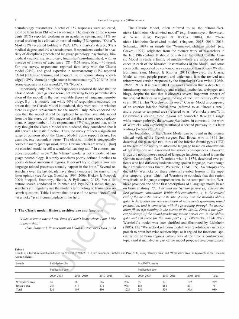
However, applied to contemporary research questions, with currentknowledge of brain structure and function, the Classic Model instan-tiations offer a spatial accuracy that is too limited to test modern hy-pothesis about brain/behavior relationships. For example, in Fig. 1 itis unclear if “a” is located within the primary (core) auditory area (i.e.,the transverse temporal gyrus, which is the main output of the ascend-ing auditory projections from the medial geniculate body of the thala-mus), or in the surrounding secondary (belt) auditory areas. Moreover,“b” is not precisely localized within the inferior frontal area, and isassumed to be directly connected to the motor nerves (which we nowknow to be false).
In addition to a limited spatial precision, another problem with theClassic model is the notion that it has been interpreted as focusingon two interconnected “language epicenters” (Papathanassiou et al.,2000), “Broca’s and “Wernicke’s areas”, which implies a high degreeof functional modularity. This notion was not endorsed by everyone inthe early development of the language neurobiology models. For ex-ample, writing about his language model, Lichtheim (1885) stated “Ido not consider the function to be localized in one spot of the brain,but rather to result from the combined action of the whole sensorialsphere” (p. 477). Wernicke is in agreement here. For him “only themost elementary psychic functions can be assigned to defined areas ofthe cortex” and “everything which goes beyond these simplest func-tions, the association of different impressions into a concept, thinking,consciousness, is an achievement of the fiber tracts which connect thedifferent regions of the cortex to each other”. Thus, Wernicke does ap-peal to the notion of “language epicenters”, but in his conception lan-guage emerges out of their interactions (Weiller et al., 2011). Despitethis, the notion of language centers is central to the Classic Model as itis commonly presented (Hagoort, 2016). While the importance of in-ferior frontal and posterior temporal regions for expressive and recep-tive language functions is not disputed here, evidence that the networksupporting language functions is vastly distributed across the brain isnow overwhelming. Indeed, speech and language functions engage avery large number of brain regions that extend far beyond “Broca’sand “Wernicke’s areas”, in the frontal, parietal, and temporal lobes, inthe medial hemispheres of the brain, as well as in the basal ganglia,thalamus and cerebellum (for reviews, see for example Crosson, 2013;Hebb & Ojemann, 2013; Marien et al., 2014; Price, 2010). Hence,despite a tendency in some early writings toward an encompassingrather than a strictly modular approach, language neurobiology framedwithin the Classic Model has focused almost exclusively on under-standing the functions of “Broca’s area” and “Wernicke’s area”.
Hence, the Classic model suffers from at least four major issues:(1) the spatial precision of the model is too limited to test specific hy-pothesis about brain/behavior relationships; (2) it is centered on two“language regions”, (3) it focuses on cortical structures, and for themost part leaves out subcortical structure and relevant connections1,and (4) because of its limited spatial extent and cortical focus, it is dif-ficult to reconcile the model with modern knowledge about the whitematter connectivity supporting speech and language function.
1 It should be noted, however, that Wernicke alluded to subcortical structuresincluding the claustrum and cerebellum, in his writings.
Despite consensus among many language scientists that the Classicmodel is outdated (Poeppel et al., 2012), the model survives, both interms of the terminology it uses (“Wernicke’s and Broca’s areas”), andin its prevalence. While the model was, and remains, an extremely im-portant milestone in the history of neurosciences, it is often the domi-nant model presented in undergraduate, graduate, and medical schoolpresentations. In these cases, it is not treated as a historic model, butrather as a model on equal footing with contemporary models of lan-guage neurobiology. Furthermore, presentations of this historic model,or variations of it, are often not followed by a presentation of moremodern accounts of language neurobiology, leaving the pupil with aninaccurate understanding of modern knowledge about brain and lan-guage relationships.
Different versions of the Classic Model are also routinely used inthe evaluation and treatment of acquired language disorders, wherethey fail to account for symptoms resulting from damage to regions ortracts not included in the model, such as the cerebellum and the thal-amus. Moreover, because modern accounts on language neurobiologyuse a variable terminology, sometimes linked to the Model but oftennot, this makes the integration of research into clinical practice diffi-cult. Arguably clinical practice in speech-language pathology wouldbe facilitated if we all used the same terms to refer to the same brainregions and connections.
3. There is no consistent definition of Broca’’s and Wernicke’’sareas, and the terms should no longer be used
“Words, words. They’re all we have to go on.”“Consistency is all I ask!”
-Tom Stoppard, Rosencrantz and Guildenstern are Dead, pp. 32;35
Many contemporary researchers continue to state their aims interms of localizing language function to “Broca’s” and “Wernicke’sareas” (e.g. Ardila, Bernal, & Rosselli, 2016; Binder, 2015; DeWitt& Rauschecker, 2013; Grodzinsky & Santi, 2008; Hagoort, 2014;Hagoort & van Berkum, 2007; Heim, Opitz, & Friederici, 2002;Kunert, Willems, Casasanto, Patel, & Hagoort, 2015; Matchin &Hickok, 2016; Mesulam, Thompson, Weintraub, & Rogalski, 2015;Meyer, Obleser, Anwander, & Friederici, 2012; Santi, Friederici,Makuuchi, & Grodzinsky, 2015; Schnur et al., 2009; Thothathiri,Kim, Trueswell, & Thompson-Schill, 2012; Wang et al., 2015; Wise,Greene, Büchel, & Scott, 1999). Yet the field still lacks consistentdefinition of either region, over 150 years after their initial introduc-tion. This is in keeping with the field of cognitive neuroscience morebroadly, which can be ambivalent about anatomical specificity, some-times advocating precise anatomy, but at other times adopting anatom-ically ambiguous terminology like “temporo-parietal junction”, “in-ferior frontal junction”, and “dorsolateral prefrontal cortex”, labelingsulcal locations as the nearest gyrus (Lancaster et al., 2000), or pre-ferring functional labels without specifying underlying anatomy suchas the “visual word form area” or the “premotor cortex”. This en-courages researchers in the field to conflate functional definitions withanatomical definitions. This approach is, in the long-term, unsustain-able if microsurgical, electrostimulation, or genetic interventions fornervous system diseases are to become a reality. Even contempo-rary neurosurgical interventions that might affect eloquent cortex re-quire precise targets and a precise neurobiological model of language(Fujii et al., 2016). An approach focusing on precise anatomy is, ofcourse, not without its own shortcomings—the highly varied struc-tural patterns of the cortical surface across individuals are well-es-tablished. However, anatomists have shown that reliable iden

UNCO
RREC
TED
PROO
F
4 Brain and Language xxx (2016) xxx-xxx
Fig. 1. Left: The original model from Wernicke, 1874. For unknown reasons, the model is represented on the right hemisphere. Right: An update of the Classic model fromGeschwind, 1972. In this figure, according to most anatomical definitions, the superior temporal gyrus is inadvertently mislabeled as the angular gyrus.
tification is possible despite the variability (Ono, Kubik, & Abernathy,1990; Tomaiuolo et al., 1999). Moreover, as we hope to show, the de-finitional problems surrounding Broca’s and Wernicke’s areas are sig-nificant, and the continued use of these terms is counterproductive.
The definitional problem is most acute for Wernicke’s area. Therehas never been a consistent anatomical definition for Wernicke’s area.Indeed, this was a topic of discussion during Professor Mesulam’skeynote address to the Society for the Neurobiology of Language,2015, in Chicago. Forty years prior, this same problem promptedBogen and Bogen (1976) to ask “Wernicke’s region—Where is it?”.No consensus was reached then, and none has been reached since.The confusion can be traced to the very beginning, when Wernicke(Wernicke, 1874/1969 ) placed a small dot on the superior temporalgyrus (see Fig. 1). The text is clear that Wernicke did not intend thesmall focused area to represent the “speech center”, and in 1881, heis more specific, drawing a hatched area covering much of the left su-perior temporal gyrus (Wernicke, 1881). Despite this, almost immedi-ately, the definition undergoes significant revision by Wernicke’s con-temporaries. In some cases, the region is simply on and around the su-perior temporal gyrus. In other cases, it extends widely to include theinferior parietal lobe, and middle temporal gyrus.
This lack of specificity is, at least partly, attributable to the fact thatpatients presenting with a “posterior lesion” and presenting with lan-guage comprehension deficits were historically referred to as “Wer-nicke’s aphasics”, even when the lesion was incongruent with Wer-nicke’s writings. This issue can be alleviated by using a symp-tom-based classification of the aphasias rather than a lesion-based ap-proach (for instance, referring to fluent rather than Wernicke’s apha-sia), an approach that is gaining support in speech-language pathol-ogy. Lesions can be described in more precise anatomical terms, suchas “posterior third of the superior temporal gyrus” or “anterior thirdof the supramarginal gyrus”. But the fact remains that, throughout thetwentieth century, almost every patch of perisylvian temporal and in-ferior parietal cortex has been presented to fall under the definition ofWernicke’s area (Bogen & Bogen, 1976).
Our survey suggests that, despite various historical attempts to de-fine the area, there is still no consensus. Respondents seemed to pre-fer two anatomical definitions of “Wernicke’s area”, but neither gar-nered more than 30% of the votes. The most popular anatomical de-finition is one that we provided ourselves (marked “Authors’ defin-ition” in Fig. 2, covering the posterior part of the superior temporal
gyrus and including part of the supramarginal gyrus), and which is notfound in any published paper (to our knowledge), nor based on anyempirical study (although it, like Lewandowsky’s definition, closelyresembles Geschwind’s, 1972 definition) (see Fig. 3).
The second most popular anatomical definition of “Wernicke’sarea” is based on Geschwind (1970; marked “Geschwind, 1970” inFig. 2, covering only the posterior part of the superior temporal gyrus),and is also the most recent published definition we included. 12%of respondents did not provide a vote, and instead provided variouscomments such as “the term is meaningless”, or “it seems that theseare all possibilities, depending on what you read”. Definitions that in-cluded the posterior middle temporal gyrus and inferior parietal lobulewere unpopular, despite evidence for the importance of these regionsin language comprehension (Dronkers, Wilkins, Van Valin, Redfern,& Jaeger, 2004; Mesulam et al., 2015). Only 8% of respondents fa-vored Wernicke’s (1881) original definition. Thus, even among ex-perts within the field of language neurobiology, there is still no con-sensus definition of Wernicke’s area.
Broca’s area is a smaller piece of cortex than Wernicke’s area,and so might be expected that a consensus could be reached. Indeed,Bogen and Bogen (1976)) suggest that there has always been agree-ment about the location of Broca’s area in the posterior third of theIFG. Wernicke is equally confident: “As is well-known, [Broca] lo-calized the faculty of speech to the posterior portion of the so-calledthird frontal gyrus… That is that portion of the most inferior and ex-ternal part of the operculum, located in the frontal part of the centralgyrus, just anterior to its juncture”(Wernicke, 1874/1977, pp. 70–71).But this is not the case—from the earliest definition, there was debate(Lorch, 2008). The report of the meeting of the Norwich British Asso-ciation for the Advancement of Science shows that while Broca pro-posed that the region for articulated language was confined to the pos-terior part of the IFG (The Lancet, 92, 1868: 293, reported in Lorch(2008), other meeting attendees suggested that the evidence, presentedas diagrams and plaster casts, showed a larger extent (Dickson, 1868).A re-evaluation using magnetic resonance imaging (MRI) of the orig-inal brain of Broca’s patient Leborgne suggests that the lesion indeedincluded the posterior part of the IFG but also extended beyond it(Dronkers, Plaisant, Iba-Zizen, & Cabanis, 2007).
However, the field has not restricted the definition to that definedon Leborgne. For example, a popular definition is that Broca’s area issynonymous with the left IFG, which Mesulam et al. (2015; p. 2424)argues is a designation so widely accepted “that its location is nolonger a subject of scientific debate.” Yet, on the contrary, our survey

UNCO
RREC
TED
PROO
F
Brain and Language xxx (2016) xxx-xxx 5
Fig. 2. Anatomical definitions of Wernicke’s area, and the percentage of respondents to the survey endorsing each definition. Associated citations are provided in the referencessection.
of language researchers suggests that a lack of consensus remains.Only 23% of survey respondents agreed with Mesulam’s definition(Mesulam et al., 2015), and only 8% agreed with Bogen and Bogen(1976; this is also Dejerine’s (1914) definition). A larger percentage(50%) chose the region comprised of the pars triangularis and parsopercularis, which, though a high percentage, still reflects the lack ofa strong consensus (it does not reach a simple majority).
The survey suggests that search for a consistent anatomical defini-tion for “Broca’s and Wernicke’s areas” is a Sisyphean task. First andforemost, anatomically, the regions typically referred to as Broca’sand Wernicke’s areas are large enough such that they do not havecytoarchitectonic and myeloarchitectonic homogeneity. Although thishas been known since the early twentieth century (Brodmann, 2006;Campbell, 1905; Smith, 1907; Vogt & Vogt, 1919; Von Economo& Koskinas, 1925) and has been shown repeatedly in recent years(Amunts & Zilles, 2012; Amunts et al., 2010; Annese, Pitiot, Dinov,& Toga, 2004; Goucha & Friederici, 2015; Zilles & Amunts, 2012;Zilles et al., 1997), the initial definitions of Broca’s and Wernicke’sareas precede the major findings in this area of investigation. This iseven the case for Broca’s area, which by most definitions is a rela-tively circumscribed patch of cortex (Petrides, Cadoret, & Mackey,2005). Anatomical heterogeneity is naturally coupled with functionalheterogeneity. For example, a number of studies have suggested a dis-sociation between semantic and phonological processing within ante-rior and posterior regions of the IFG (e.g., Katzev, Tuscher, Hennig,Weiller, & Kaller, 2013; Price, 2010). Because the specific functionsor sets of functions with which each patch of cortex is involved arestill under investigation, it is even more important to be careful andprecise about which parts of the brain we are referring.
Binder (2015) recently argued for a similar conclusion for Wernicke’sarea, noting that “speech comprehension is a highly distributed func-tion, involving a bi-hemispheric phoneme perception system and awidely distributed semantic network. To refer to all of these regionsas the Wernicke area seems to sacrifice any utility that the term mighthave…” (p. 5). However, instead of rejecting the label, he suggeststhat we retain the Wernicke label and re-define the function of the re-gion. Rather than continue to search for the functions of Broca’s andWernicke’s areas, we argue for the opposite, namely that we shouldsimply retire the labels.
Why should the labels be retired? The reason is that the vocab-ulary in use in any scientific endeavor matters, and continued con-ceptual work and elaboration and revision of the standard vocabu-lary of the field is a necessary feature of science. We can and do be-come “captives of a … set of verbal categories” (Searle, 1992; p. 31).We inherit this vocabulary from the giants of previous generationsand “with the vocabulary a certain set of categories, within which weare historically conditioned to think about [the] problems. The vocab-ulary is not innocent, because implicit in the vocabulary are a sur-prising number of theoretical claims” (Searle, 1992, p. 14). Paradig-matic changes, which are occurring in the field of language neurobi-ology, cause scientists to see the world of their research engagementdifferently, and these changes cause “old terms, concepts and exper-iments fall into new relationships one with the other” (Kuhn, 1970,p. 149). The terms Broca’s and Wernicke’s Areas are not innocuousterms—they carry with them a notion of functional relevance to lan-guage, but not everyone agrees on their anatomical definition, and noteveryone agrees on their function. This contributes to significant con

UNCO
RREC
TED
PROO
F
6 Brain and Language xxx (2016) xxx-xxx
Fig. 3. Anatomical definitions of Broca’s area, and the percentage of respondents to the survey endorsing each definition. Associated citations are provided in the references section.
ceptual confusion, and, outside of a historical review context, there issimply no reason to continue to use them for contemporary theories.
To illustrate the issue with respect to Broca’s area, we encour-age the reader to examine Hagoort’s recent Memory, Unification, andControl model (Hagoort, 2016). It is notable that Hagoort begins thisexamination with a brief review of the Classic Model, states that itis a historical model, and then initially continues to work within thatframework, using the terminology of Broca’s and Wernicke’s areas.For example, in the first figure of the paper (Fig. 28.1), the functionof “Unification requires the contribution of Broca’s area (Brodmannareas 44 and 45) and adjacent cortex (Brodmann areas 47 and 6) inthe frontal lobe” (p. 340). It is difficult, though, for Hagoort to workwithin this framework for long, because if he does so, he does notgo very far toward advancing a new theory that is different in ma-jor respects from the Classic Model. His pivot is to take seriouslythe notion that “language is subserved by dynamic networks of brainregions.” (p. 340). With this perspective in hand, Hagoort is carefulto break up Broca’s and Wernicke’s regions into smaller anatomicalparts, as we advocate. For example, his model parses the sub-regionsof the IFG into pars opercularis, pars triangularis, and pars orbitalis,which he notes have different associated connectivity and functions.He does the same with the temporal and parietal lobes—in this wayhis model evolves to having nine nodes supporting language function,anchored by a network of fiber pathways (which we review in thenext section). Thus, in order to present his new model, he makes asignificant break with the Classic Model, even closing his expositionwith a section titled “Beyond the Classical Model”. Hagoort showsthat the Classic Model terminology is too constraining, for him, to
develop a serious model of language neurobiology in the face of newthinking about network architectures supporting cognition, which re-quires the specification of multiple interacting nodes within the net-work, and serious reflection on their connectivity. The new modelspecification is also more amenable to empirical assessment usingmore modern analytic techniques, such as network analysis (Sporns,2011). The Classic Model cannot be tested with such techniques be-cause, as a two-node, one connection model, it is too simple.
To illustrate the definitional issue with respect to Wernicke’s area,we point to DeWitt and Rauschecker’s recent paper (DeWitt &Rauschecker, 2013). A central focus of their paper is to re-locate Wer-nicke’s area. Thus, they write:
“Where is Wernicke’s area? Answering this question today—withthe benefit of far greater understanding of neuroanatomy and corti-cal processing than either Wernicke or Geschwind had access to—wemight conclude that the functions Wernicke subsumes within a singlearea are actually performed by multiple cortical areas… The hypoth-esis most strongly supported by available empirical data for the loca-tion of Wernicke’s AWFA [auditory word form area] is anterior STG[superior temporal gyrus]… This region, however, is neither a strongcandidate site for encoding representations that resemble Wernicke’sword-concepts (i.e., inner speech) nor for performing the correctivefunction Wernicke ascribes to them.” (p. 186).
But the question we are trying to address as a field is not “Whereis Wernicke’s area?” A more interesting question, we believe, mightbe: How does the brain accomplish and integrate the various sub-func-tions that comprise human language, can we parse the network im-plementing these sub-functions into its constituent compo

UNCO
RREC
TED
PROO
F
Brain and Language xxx (2016) xxx-xxx 7
nents, and can we identify the role specific patches of cortex (or sub-cortical nuclei or regions) play in the context of the broader systemimplementing language? For DeWitt and Rauschecker, the question ismore specifically “where are the patches of cortex associated with au-ditory word form recognition?” But instead of addressing this ques-tion, DeWitt and Rauschecker (2013) continue to try to localize Wer-nicke’s area. At the end of their investigation they write: “Wernicke’sarea, functionally defined, therefore appears to consist of two areas:an AWFA in anterior STG and an ‘‘inner-speech area’’ in posterior[superior temporal gyrus/inferior parietal lobule] STG/IPL” (p. 187).It would be more productive, in our opinion, to simply try to definethe network for auditory word recognition, rather than come up withyet another definition of Wernicke’s area.
Hagoort and DeWitt and Rauschecker show us that the use of theterms “Broca’s and Wernicke’s areas” are still in wide use, still framea lot of the models and model-building steps in language neurobi-ology, and carry historical conceptual baggage that slows theoreticaladvance. the Broca and Wernicke terminology, we suggest follow-ing the lead advocated by others before us (Devlin & Poldrack, 2007;Toga & Thompson, 2007)—anatomical definitions with reference toa published atlas are preferred over poorly defined functional labels.Our thesis is also an endorsement of precise neuroanatomy for anymodel of language neurobiology. Brodmann, over a hundred yearsago, wrote: “functional localization of the cerebral cortex without thelead of anatomy is utterly impossible… In all domains, physiologyhas its firmest foundations in anatomy.” (Brodmann, 1909; Brodmann,2006, p. 262). Wernicke was himself a precise anatomist in the schoolof Meynert (Gage & Hickok, 2005), and the field could benefit by em-ulating that precision.
Obviously, the Classic Model developed in the 19th and 20th cen-turies is not based on modern macroscopical neuroanatomy. Becauseour knowledge of brain anatomy and function has evolved, it seemsmore productive to build new models based modern terminologies andclear anatomical definitions. Thus we advocate a clean break from theClassic Model and its associated terminology. Reliable anatomical de-finitions and reporting of findings in more specific anatomical land-marks will also facilitate the definition of the broader language net-work, including its connectivity. Understanding of this “language con-nectome”—the white matter connectivity of the perisylvian regionsassociated with speech and language—has expanded rapidly in the lastdecade, and further supports our argument that the classic model is notsustainable as a useful model of language neurobiology. The recentevolution of this literature is briefly summarized in the next section.
4. Fiber pathways supporting speech and language: beyond thearcuate fasciculus
“What a fine persecution—to be kept intrigued without ever quitebeing enlightened.”
-Tom Stoppard, Rosencrantz and Guildenstern are Dead, p. 32
.The fiber pathway connectivity that support speech and language
functions has come under intense scrutiny in the last decade, largelydue to the advent of advanced diffusion-weighted imaging techniquesthat can map fiber pathways in vivo, even though, as Saur (2015) con-cisely states, “precise long-distant region-to-region structural connec-tivity between lobes is still difficult to obtain and represents one ofthe greatest challenges in systems neuroscience.” In short, a compre-hensive mapping of the “language connectome” remains elusive. It iswithin this context that we consider the role of the arcuate fasciculus(AF) as the “language pathway” of the Classic model.
In blunt fiber dissection, the core fibers of the AF (historicallyalso the superior longitudinal fasciculus; SLF) are easily identifiable,and definition of the pathway appeared in Burdach’s early anatomi-cal treatments of the 19th century (Burdach, 1819–1826). In his orig-inal treatise Wernicke (1874/1969) refers to this pathway as “associa-tion fibers”, the “path a1b” or “fibra propria”, which connects inferiorfrontal and temporal regions to support speech and language. Thereare also a few mentions of a “fibrae arcuatae” throughout the article.It is von Monakow (1897), though, who more explicitly names thesefibers, and the AF is established as the “language pathway” over the20th century, notably by Geschwind (1970); Wernicke later agreedthat the arcuate fasciculus was a language pathway (Wernicke, 1908).
Contemporary research suggests that the notion that a single fiberpathway supports language function in the human brain should be con-sidered obsolete (even the two pathways, uncinate fasciculus and ar-cuate fasciculus, that Wernicke (Wernicke, 1908) advocated are in-sufficientsee Weiller et al., 2011 for an account of the history ofthe “lost” ventral tract. Modern perspectives on language connectiv-ity should consider several sets of association pathways: fronto-tem-poral, parieto-temporal, occipito-temporal, and fronto-frontal connec-tions (see Fig. 4), as well as thalamic radiations, and cortico-subcorti-cal loops connecting the cortex to the basal ganglia, cerebellum, mid-brain and pontine nuclei. A brief review of the different pathwaysthat may support language functions is presented below (and morecomprehensively elsewhere; see Axer, Klingner, & Prescher, 2013;
Fig. 4. An emerging picture of perisylvian long association fiber pathways supporting language. The image on the left shows the “classic” arcuate fasciculus. In the image on theright, the arcuate fasciculus is split into three components (Catani, Jones, & Ffytche, 2005). Additional fiber pathways discussed in the text are shown. SLF III = Superior longitudinalfasciculus, third subcomponent.

UNCO
RREC
TED
PROO
F
8 Brain and Language xxx (2016) xxx-xxx
Dick, Bernal, & Tremblay, 2014; Dick & Tremblay, 2012; Gierhan,2013; Saur et al., 2008; Weiller et al., 2011).
Fronto-temporal connections supporting language functions in-clude, in addition to the AF, the uncinate fasciculus (UF), extremecapsule/extreme capsule fiber system (EmC), and the inferiorfronto-occipital fasciculus (IFOF). The UF connects the orbital andlateral frontal cortex with the temporal pole, anterior temporal cor-tex, parahippocampal gyrus, and amygdala (Von Der Heide, Skipper,Klobusicky, & Olson, 2013). Some investigators believe the UF tobe associated with semantic processing, given its strong connectivitywith the anterior temporal cortex and temporal pole, a proposed “hub”for semantic processing (Holland & Lambon Ralph, 2010). Evidencefor loss of semantic function (e.g., picture naming deficits) followingresection of the UF supports this notion (Papagno et al., 2011), but thisis not without controversy (Kho et al., 2008; Moritz-Gasser, Herbet, &Duffau, 2013). Another fronto-temporal connection, the EmC or “ex-treme capsule fiber system” is a collection of axons located betweenthe claustrum (medially) and the insula (laterally). Some evidence inthe human suggests that EmC connects the ventral and lateral frontallobe with the most of the superior and middle temporal cortex, extend-ing anterior-to-posterior (Makris & Pandya, 2009; Saur et al., 2008).Such a pathway could provide an alternative route between the an-terior inferior frontal and temporal lobes, which may support syntac-tic and semantic processing (Griffiths, Marslen-Wilson, Stamatakis,& Tyler, 2013; Rolheiser, Stamatakis, & Tyler, 2011). Finally, theIFOF originates in the inferior and medial occipital lobe (and possi-bly the medial parietal lobe), sends projections to the ventral temporallobe, and travels through the temporal stem to project to the IFG, themedial and orbital frontal cortex, and the frontal pole (Catani, Jones,Donato, & Ffytche, 2003; Sarubbo, De Benedictis, Maldonado, Basso,& Duffau, 2013). Duffau and colleagues (Martino et al., 2013) havesuggested that the IFOF is a “direct” pathway anchoring the ventralsemantic system for language, but additional research is needed to un-derstand whether these different pathways operate as part of non-lan-guage networks, which may offer alternative interpretations of theirfunctions.
Parieto-temporal and occipito-temporal connections include themiddle longitudinal fasciculus (MdLF) and inferior longitudinal fas-ciculus (ILF). The MdLF is well-established in the macaque(Schmahmann & Pandya, 2006; Seltzer & Pandya, 1994), but less soin the human (Makris & Pandya, 2009; Makris et al., 2013, 2013;Maldonado et al., 2013; Saur et al., 2008; Turken & Dronkers, 2011;Wang et al., 2013). The available evidence suggests that the MdLForiginates in the posterior superior temporal, inferior and superiorparietal lobe, and possibly occipital lobe, producing terminationsalong the course of the temporal cortex to the temporal pole. It maythus be important for language comprehension (Turken & Dronkers,2011) or semantic processing (Saur et al., 2008), although some ques-tion its role in language (Wang et al., 2013). The ILF connects theoccipital lobe with the temporal lobe, originating in secondary visualareas and connecting to the middle and inferior temporal gyri, thetemporal pole, parahippocampal gyrus, hippocampus, and amygdala(Catani et al., 2003). Several authors have suggested that the ILF is amajor component of a ventral system supporting semantic processes(Agosta et al., 2013; Saur et al., 2008; Turken & Dronkers, 2011).If we expand the focus to paralinguistic functions such as literacy, arecently re-discovered fiber pathway (Yeatman et al., 2014), the ver-tical occipital fasciculus (VOF), becomes yet another potentially im
portant pathway2. This pathway appears to connect the lateral occip-itotemporal sulcus and gyrus (associated with the processing of vi-sual word forms) with inferior, and possibly superior, parietal regionsthat are important for literacy and numeracy (Bouhali et al., 2014;Greenblatt, 1973, 1976; Yeatman, Rauschecker, & Wandell, 2013).
To date, much of the research on perisylvian long association fiberpathways has focused on speech perception and language comprehen-sion, and has largely neglected the contribution of cortical and sub-cortical networks for speech production. But speech production was,of course, a major component of the Classic Model from the ear-liest description of Broca’s patient Leborgne and other case-studieswith Broca’s aphasia. Additional fiber pathways of the cortico-bulbar,cortico-cerebellar, and cortico-striatal systems are known to supportspeech production. Even fronto-frontal fiber pathways only recentlyidentified in children and adults using diffusion-weighted MRI, suchas the frontal aslant tract (FAT; (Broce, Bernal, Altman, Tremblay, &Dick, 2015; Catani et al., 2013) (Fig. 5), which connects the inferiorfrontal regions with the pre-supplementary motor area, may play a rolein spoken language production (see Dick et al., 2014 for a more de-tailed review).
The Classic Model as it is most commonly presented in contempo-rary textbooks, with a single connection between two central nodes, isthus insufficient to account for the overwhelming evidence that mul-tiple fiber pathways support language function in the human brain.Since all these pathways may make important contributions to a vari-ety of linguistic functions, there is no reason to continue to focus ona single pathway. Moreover, returning to the issue of language-cen-tricity, it will be important to examine the contributions of each ofthe pathways to other cognitive and sensorimotor functions in orderto better understand the computations that they may be involved withduring the processing and production of language.
5. Conclusions, or where to go from here
“…look on every exit being an entrance somewhere else.”-Tom Stoppard, Rosencrantz and Guildenstern are Dead, p. 21
The central thesis of this article is that the Classic Model, in itsmost common iteration, is neither an anatomically precise nor a com-prehensive model of language neurobiology (cf. Poeppel, 2014), andthat the maintenance of the terminology of this model artificiallymaintains it as a legitimate model. Although the field as a whole hasmade tremendous progress in the past few decades, due in part tosignificant advances in the neuroimaging and neurostimulation meth-ods, we believe abandoning the Classic Model and the terminology ofBroca’s and Wernicke’s areas would provide a catalyst for additionaltheoretical advancement.
Focusing on the Classic Model has, we believe, limited our atten-tion to a rich theoretical and empirical literature that tries to bring tothe forefront important notions about the neurobiology of language: adistributed architecture which includes cortical and subcortical com-ponents, a distributed anatomical connectivity, and, perhaps most im-portantly, a heavy reliance on domain-general neural resources (e.g.Bornkessel-Schlesewsky, Schlesewsky, Small, & Rauschecker, 2015;Rijntjes, Weiller, Bormann, & Musso, 2012). Understanding how lan-guage functions are organised in the brain and how they relate to otherfunctions is, no doubt, a critical issue: “The most fundamental ques-tion in the study of the human language faculty is its place in the nat-ural world: what kind of biological system it is, and how it relates
2 It was, incidentally, Wernicke who first named and defined this pathway, whichhe called the perpendicular occipital fasciculus, in his 1881–1883 Lehrbuch derGehirnkrankheiten für Aerzte und Studirende. 3 Volumes. Kassel: Fischer.

UNCO
RREC
TED
PROO
F
Brain and Language xxx (2016) xxx-xxx 9
Fig. 5. Connections of the frontal aslant tract (FAT) in coronal section, with outline of the inferior frontal and superior frontal origins and terminations in the medial and lateral sagittalviews. IFGOp = inferior frontal gyrus, pars opercularis; SFG = superior frontal gyrus; SMA = supplementary motor area; Pre-SMA = pre-supplementary motor area. Reprinted withpermission from Dick et al. (2014). The language connectome: New pathways, new concepts. The Neuroscientist, 20, 453–467.
to other systems in our own species and others” (Pinker & Jackendoff,2005). Because the simple architecture of the Classic Model suggestsa language-centric perspective, the resilience of the model has per-petuated different flavors of the longstanding idea that the neural ma-chinery for language is “special”, that is, the notion that there ex-ists neural tissue dedicated to the specific task of processing and pro-ducing language. An alternative view is that language is, at least inpart, an overlaid functional system that “gets what service it can outof nervous tissues that have come into being and are maintained forvery different ends than its own” (adapted from Sapir, 1921). Al-though some language-specific mechanisms may exist, our emergingunderstanding of brain function is of mutual interactions and com-mon control mechanisms. Wernicke, over 140 years ago, was alreadyon the right track—“a priori reasoning would view restriction ofthe speech center to a single area, namely, Broca’s gyrus, as highlyimprobable” (Wernicke, 1874/1994; p. 74). As a field, we need tostudy the interactions between language and other functional systemsin order to fully understand the neurobiological underpinning of hu-man language and language disorders, and the degree to which itis dependent upon various other cognitive, sensorimotor and emo-tional processes, all of which must come together to put languageinto action. Consistent with these notions, most contemporary mod-els of the neurobiology of language propose a much more complexarchitecture encompassing regions that had never before been con-sidered to support language functions. Though we agree with thosewho have completed our survey that there is not one clear, compre-hensive alternative, we do think there are a number of promising de-velopments (Ballard, Robin, & Folkins, 2003; Binder & Desai, 2011;Binder, Desai, Graves, & Conant, 2009; Bornkessel-Schlesewsky etal., 2015; Duffau, Moritz-Gasser, & Mandonnet, 2014; Friederici &Singer, 2015; Guenther, 2006; Hagoort, 2013, 2014, 2016; Hickok,2009, 2014, Love-Geffen, & Klima, 2002; Hickok & Poeppel, 2000,2004, 2007; Mesulam et al., 2015; Price, 2010; Rauschecker & Scott,2009;
Scott & Johnsrude, 2003; Skeide & Friederici, 2016), each present-ing a more comprehensive architecture for language than the Clas-sic Model. In fact, to many researchers we may be “preaching to thechoir”. However, our analysis of the literature clearly reveals that theClassic Model, or at the very least its terminology, is still robust. Wewould urge the field of language neurobiology as a whole to considerthese other promising avenues on which to establish a new, compre-hensive alternative to the Classic Model.
Acknowledgments
P. Tremblay holds a Career Award from the “Fonds de Recherchedu Québec – Santé” (FRQS). We thank Michael Andric and Uri Has-son for their comments on previous versions of this manuscript. Wealso thank everyone who answered our online survey.
References
Amunts, K., Lenzen, M., Friederici, A.D., Schleicher, A., Morosan, P., Palomero-Gal-lagher, N., Zilles, K., 2010. Broca’s region: Novel organizational principles andmultiple receptor mapping. PLoS Biology 8 (9)http://dx.doi.org/10.1371/journal.pbio.1000489.
Amunts, K., Zilles, K., 2012. Architecture and organizational principles of Broca’s re-gion. Trends in Cognitive Sciences 16 (8), 418–426. http://dx.doi.org/10.1016/j.tics.2012.06.005.
Annese, J., Pitiot, A., Dinov, I.D., Toga, A.W., 2004. A myelo-architectonic methodfor the structural classification of cortical areas. Neuroimage 21 (1), 15–26.
Ardila, A., Bernal, B., Rosselli, M., 2016. The language area of the brain: A functionalreassessment. Revista de neurologia 62 (3), 97–106.
Axer, H., Klingner, C.M., Prescher, A., 2013. Fiber anatomy of dorsal and ventral lan-guage streams. Brain and Language 127 (2), 192–204. http://dx.doi.org/10.1016/j.bandl.2012.04.015.
Ballard, K.J., Robin, D.A., Folkins, J.W., 2003. An integrative model of speech motorcontrol: A response to Ziegler. Aphasiology 17 (1), 37–48.
Binder, J.R., 2015. The Wernicke area: Modern evidence and a reinterpretation. Neu-rology http://dx.doi.org/10.1212/WNL.0000000000002219.

UNCO
RREC
TED
PROO
F
10 Brain and Language xxx (2016) xxx-xxx
Binder, J.R., Desai, R.H., 2011. The neurobiology of semantic memory. Trends inCognitive Sciences 15 (11), 527–536. http://dx.doi.org/10.1016/j.tics.2011.10.001.
Binder, J.R., Desai, R.H., Graves, W.W., Conant, L.L., 2009. Where is the semanticsystem? A critical review and meta-analysis of 120 functional neuroimaging stud-ies. Cerebral Cortex 19 (12), 2767–2796. http://dx.doi.org/10.1093/cercor/bhp055.
Bogen, J.E., Bogen, G.M., 1976. Wernicke’s region–Where is it?. Annals of the NewYork Academy of Sciences 280, 834–843.
Bornkessel-Schlesewsky, I., Schlesewsky, M., Small, S.L., Rauschecker, J.P., 2015.Neurobiological roots of language in primate audition: Common computationalproperties. Trends in Cognitive Sciences 19 (3), 142–150. http://dx.doi.org/10.1016/j.tics.2014.12.008.
Bouhali, F., Thiebaut de Schotten, M., Pinel, P., Poupon, C., Mangin, J.F., Dehaene,S., Cohen, L., 2014. Anatomical connections of the visual word form area. Journalof Neuroscience 34 (46), 15402–15414. http://dx.doi.org/10.1523/JNEUROSCI.4918-13.2014.
Broce, I., Bernal, B., Altman, N., Tremblay, P., Dick, A.S., 2015. Fiber tracking of thefrontal aslant tract and subcomponents of the arcuate fasciculus in 5–8-year-olds:Relation to speech and language function. Brain and Language 149, 66–76. http://dx.doi.org/10.1016/j.bandl.2015.06.006.
Brodmann, K., 1909. Vergleichende lokalisationslehre der gro hirnrinde. Verlag vonJohann Ambrosius Barth, Leipzig.
Brodmann, K., 2006. Brodmann’s localisation in the cerebral cortex (L. J. Garey,trans.). Springer.
Burdach, K.F., 1819. Vom bau und leben des gehirns und rückenmarks (3 vols). 1826.In der dyk’schen buchandlung, Leipzig.
Campbell, A.W., 1905. Histological studies on the localisation of cerebral function.University Press, Cambridge, UK.
Catani, M., Jones, D.K., Donato, R., Ffytche, D.H., 2003. Occipito-temporal connec-tions in the human brain. Brain 126 (Pt 9), 2093–2107. http://dx.doi.org/10.1093/brain/awg203.
Catani, M., Jones, D.K., Ffytche, D.H., 2005. Perisylvian language networks of the hu-man brain. Annals of Neurology 57, 8–16.
Catani, M., Mesulam, M.M., Jakobsen, E., Malik, F., Martersteck, A., Wieneke, C.,… Rogalski, E., 2013. A novel frontal pathway underlies verbal fluency in primaryprogressive aphasia. Brain 136 (Pt 8), 2619–2628. http://dx.doi.org/10.1093/brain/awt163.
Crosson, B., 2013. Thalamic mechanisms in language: A reconsideration based on re-cent findings and concepts. Brain and Language 126 (1), 73–88. http://dx.doi.org/10.1016/j.bandl.2012.06.011.
Devlin, J.T., Poldrack, R.A., 2007. In praise of tedious anatomy. Neuroimage 37 (4),1033–1041. http://dx.doi.org/10.1016/j.neuroimage.2006.09.055 (discussion1050–1038).
DeWitt, I., Rauschecker, J.P., 2013. Wernicke’s area revisited: Parallel streams andword processing. Brain and Language 127 (2), 181–191.
Dick, A., Bernal, B., Tremblay, P., 2014. The language connectome: New pathways,new concepts. The Neuroscientist 20, 453–467.
Dick, A.S., Tremblay, P., 2012. Beyond the arcuate fasciculus: Consensus and contro-versy in the connectional anatomy of language. Brain 135 (Pt 12), 3529–3550.http://dx.doi.org/10.1093/brain/aws222.
Dickson, J.T., 1868. Reports of society. British Medical Journal http://dx.doi.org/10.1136/bmj.2.401.259.
Dronkers, N.F., Plaisant, O., Iba-Zizen, M.T., Cabanis, E.A., 2007. Paul Broca’s his-toric cases: High resolution MR imaging of the brains of Leborgne and Lelong.Brain 130 (Pt 5), 1432–1441. doi: awm042 [pii] 0.1093/brain/awm042.
Dronkers, Nina F., Wilkins, David P., Van Valin Jr., Robert D., Redfern, Brenda B.,Jaeger, Jeri J., 2004. Lesion analysis of the brain areas involved in language com-prehension. Cognition 92 (1–2), 145–177.
Duffau, H., Moritz-Gasser, S., Mandonnet, E., 2014. A re-examination of neural basisof language processing: Proposal of a dynamic hodotopical model from data pro-vided by brain stimulation mapping during picture naming. Brain and Lan-guage 131, 1–10. http://dx.doi.org/10.1016/j.bandl.2013.05.011.
Friederici, A.D., Singer, W., 2015. Grounding language processing on basic neuro-physiological principles. Trends in Cognitive Sciences 19 (6), 329–338. http://dx.doi.org/10.1016/j.tics.2015.03.012.
Fujii, M., Maesawa, S., Ishiai, S., Iwami, K., Futamura, M., Saito, K., 2016. Neural ba-sis of language: An overview of an evolving model. Neurologia medico-chirurgica(Tokyo) http://dx.doi.org/10.2176/nmc.ra.2016-0014.
Gage, N., Hickok, G., 2005. Multiregional cell assemblies, temporal binding and therepresentation of conceptual knowledge in cortex: A modern theory by a “classi-cal” neurologist, Carl Wernicke. Cortex: A Journal Devoted to the Study of theNervous System and Behavior 41 (6), 823–832.
Geranmayeh, F., Brownsett, S.L., Wise, R.J., 2014. Task-induced brain activity inaphasic stroke patients: What is driving recovery?. Brain 137 (Pt 10), 2632–2648.http://dx.doi.org/10.1093/brain/awu163.
Geschwind, N, 1965. Disconnexion syndromes in animals and man: Part I.Brain 88 (2), 237–294. http://dx.doi.org/10.1093/brain/88.2.237.
Geschwind, , 1965. Disconnexion syndromes in animals and man: Part II. Brain 88 (3),585–644. http://dx.doi.org/10.1093/brain/88.3.585.
Geschwind, N., 1970. The organization of language and the brain. Sci-ence 170, 940–944.
Gierhan, S.M., 2013. Connections for auditory language in the human brain. Brain andLanguage 127 (2), 205–221. http://dx.doi.org/10.1016/j.bandl.2012.11.002.
Goucha, T., Friederici, A.D., 2015. The language skeleton after dissecting meaning: Afunctional segregation within Broca’s Area. Neuroimage 114, 294–302. http://dx.doi.org/10.1016/j.neuroimage.2015.04.011.
Graves, R.E., 1997. The legacy of the Wernicke-Lichtheim model. Journal of the His-tory of the Neurosciences 6 (1), 3–20. http://dx.doi.org/10.1080/09647049709525682.
Greenblatt, S.H., 1973. Alexia without agraphia or hemianopsia anatomical analysis ofan autopsied case. Brain: A Journal of Neurology 96 (2), 307–316.
Greenblatt, S.H., 1976. Subangular alexia without agraphia or hemianopsia. Brain andLanguage 3 (2), 229–245.
Griffiths, J.D., Marslen-Wilson, W.D., Stamatakis, E.A., Tyler, L.K., 2013. Functionalorganization of the neural language system: Dorsal and ventral pathways are criti-cal for syntax. Cerebral Cortex 23 (1), 139–147. http://dx.doi.org/10.1093/cercor/bhr386.
Grodzinsky, Y., Santi, A., 2008. The battle for Broca’s region. Trends in CognitiveSciences 12 (12), 474–480. http://dx.doi.org/10.1016/j.tics.2008.09.001.
Guenther, F.H., 1994. A neural network model of speech acquisition and motor equiv-alent speech production. Biological Cybernetics 72 (1), 43–53.
Guenther, F.H., 2006. Cortical interactions underlying the production of speechsounds. Journal of Communication Disorders 39 (5), 350–365. http://dx.doi.org/10.1016/j.jcomdis.2006.06.013.
Hagoort, P., 2013. MUC (Memory, Unification, Control) and beyond. Frontiers in Psy-chology 4, 416. http://dx.doi.org/10.3389/fpsyg.2013.00416.
Hagoort, P., 2014. Nodes and networks in the neural architecture for language: Broca’sregion and beyond. Current Opinion in Neurobiology 28, 136–141. http://dx.doi.org/10.1016/j.conb.2014.07.013.
Hagoort, P., 2016. Chapter 28 – MUC (Memory, Unification, Control): A model on theneurobiology of language beyond single word processing. In: Hickok, G., Small,S.L. (Eds.), Neurobiology of language. Elsevier.
Hagoort, P., van Berkum, J., 2007. Beyond the sentence given. Philosophical Transac-tions of the Royal Society of London. Series B, Biological sciences 362 (1481),801–811. http://dx.doi.org/10.1098/rstb.2007.2089.
Hebb, A.O., Ojemann, G.A., 2013. The thalamus and language revisited. Brain andLanguage 126 (1), 99–108. http://dx.doi.org/10.1016/j.bandl.2012.06.010.
Heim, S., Opitz, B., Friederici, A.D., 2002. Broca’s area in the human brain is involvedin the selection of grammatical gender for language production: Evidence fromevent-related functional magnetic resonance imaging. Neuroscience Let-ters 328 (2), 101–104.
Hickok, G., 2009. The functional neuroanatomy of language. Physics of Life Re-views 6 (3), 121–143. http://dx.doi.org/10.1016/j.plrev.2009.06.001.
Hickok, G., 2014. The architecture of speech production and the role of the phonemein speech processing. Language, Cognition and Neuroscience 29 (1), 2–20. http://dx.doi.org/10.1080/01690965.2013.834370.
Hickok, G., Love-Geffen, T., Klima, E.S., 2002. Role of the left hemisphere in signlanguage comprehension. Brain and Language 82 (2), 167–178.
Hickok, G., Poeppel, D., 2000. Towards a functional neuroanatomy of speech percep-tion. Trends in Cognitive Sciences 4 (4), 131–138.
Hickok, G., Poeppel, D., 2004. Dorsal and ventral streams: A framework for under-standing aspects of the functional anatomy of language. Cognition 92 (1–2),67–99. http://dx.doi.org/10.1016/j.cognition.2003.10.011.
Hickok, G., Poeppel, D., 2007. The cortical organization of speech processing. NatureReviews Neuroscience 8 (5), 393–402.
Holland, R., Lambon Ralph, M.A., 2010. The anterior temporal lobe semantic hub is apart of the language neural network: Selective disruption of irregular past tenseverbs by rTMS. Cerebral Cortex 20 (12), 2771–2775. http://dx.doi.org/10.1093/cercor/bhq020.
Katzev, M., Tuscher, O., Hennig, J., Weiller, C., Kaller, C.P., 2013. Revisiting thefunctional specialization of left inferior frontal gyrus in phonological and semanticfluency: The crucial role of task demands and individual ability. Journal of Neuro-science 33 (18), 7837–7845. http://dx.doi.org/10.1523/JNEUROSCI.3147-12.2013.
Kho, K.H., Indefrey, P., Hagoort, P., van Veelen, C.W., van Rijen, P.C., Ramsey, N.F.,2008. Unimpaired sentence comprehension after anterior temporal cortex resec-tion. Neuropsychologia 46 (4), 1170–1178. http://dx.doi.org/10.1016/j.neuropsychologia.2007.10.014.
Kuhn, T.S., 1970. The structure of scientific revolutions Chicago. The University ofChicago Press.
Kunert, R., Willems, R.M., Casasanto, D., Patel, A.D., Hagoort, P., 2015. Music andlanguage syntax interact in Broca’s area: An fMRI study. PLoS One 10 (11),e0141069. http://dx.doi.org/10.1371/journal.pone.0141069.

UNCO
RREC
TED
PROO
F
Brain and Language xxx (2016) xxx-xxx 11
Lancaster, J.L., Woldorff, M.G., Parsons, L.M., Liotti, M., Freitas, C.S., Rainey, L.,… Fox, P.T., 2000. Automated Talairach atlas labels for functional brain mapping.Human Brain Mapping 10 (3), 120–131.
Lichtheim, L., 1885. On aphasia. Brain 433–484.Lissauer, H., 1890. Ein Fall von Seelenblindheit nebst einem Beitrag zür Theorie der-
selben. [A case of visual agnosia with a contribution to theory]. Archiv für Psychi-atrie 21, 222–270.
Lorch, M.P., 2008. The merest Logomachy: The 1868 Norwich discussion of aphasiaby Hughlings Jackson and Broca. Brain 131 (Pt 6), 1658–1670. http://dx.doi.org/10.1093/brain/awn058.
Makris, N., Pandya, D.N., 2009. The extreme capsule in humans and rethinking of thelanguage circuitry. Brain Structure and Function 213, 343–358.
Makris, N., Preti, M.G., Asami, T., Pelavin, P., Campbell, B., Papadimitriou, G.M.,… Kubicki, M., 2013. Human middle longitudinal fascicle: Variations in patternsof anatomical connections. Brain Structure and Function 218 (4), 951–968. http://dx.doi.org/10.1007/s00429-012-0441-2.
Makris, N., Preti, M.G., Wassermann, D., Rathi, Y., Papadimitriou, G.M., Yergatian,C., … Kubicki, M., 2013. Human middle longitudinal fascicle: Segregation andbehavioral-clinical implications of two distinct fiber connections linking temporalpole and superior temporal gyrus with the angular gyrus or superior parietal lobuleusing multi-tensor tractography. Brain Imaging and Behavior 7 (3), 335–352. http://dx.doi.org/10.1007/s11682-013-9235-2.
Maldonado, I.L., de Champfleur, N.M., Velut, S., Destrieux, C., Zemmoura, I., Duffau,H., 2013. Evidence of a middle longitudinal fasciculus in the human brain fromfiber dissection. Journal of Anatomy 223 (1), 38–45. http://dx.doi.org/10.1111/joa.12055.
Marien, P., Ackermann, H., Adamaszek, M., Barwood, C.H., Beaton, A., Desmond, J.,… Ziegler, W., 2014. Consensus paper: Language and the cerebellum: An ongoingenigma. Cerebellum 13 (3), 386–410. http://dx.doi.org/10.1007/s12311-013-0540-5.
Martino, J., De Witt Hamer, Philip C., Berger, Mitchel S., Lawton, Michael T., Arnold,Christine M., de Lucas, E.M., Duffau, H., 2013. Analysis of the subcomponentsand cortical terminations of the perisylvian superior longitudinal fasciculus: Afiber dissection and DTI tractography study. Brain Structure and Function 218 (1),105–121. http://dx.doi.org/10.1007/s00429-012-0386-5.
Matchin, W., Hickok, G., 2016. ’Syntactic perturbation’ during production activatesthe right IFG, but not Broca’s area or the ATL. Frontiers in Psychology 7, 241.http://dx.doi.org/10.3389/fpsyg.2016.00241.
Mesulam, M.M., Thompson, C.K., Weintraub, S., Rogalski, E.J., 2015. The Wernickeconundrum and the anatomy of language comprehension in primary progressiveaphasia. Brain 138 (Pt 8), 2423–2437. http://dx.doi.org/10.1093/brain/awv154.
Meyer, L., Obleser, J., Anwander, A., Friederici, A.D., 2012. Linking ordering inBroca’s area to storage in left temporo-parietal regions: The case of sentence pro-cessing. Neuroimage 62 (3), 1987–1998. http://dx.doi.org/10.1016/j.neuroimage.2012.05.052.
Moritz-Gasser, S., Herbet, G., Duffau, H., 2013. Mapping the connectivity underlyingmultimodal (verbal and non-verbal) semantic processing: A brain electrostimula-tion study. Neuropsychologia 51 (10), 1814–1822. http://dx.doi.org/10.1016/j.neuropsychologia.2013.06.007.
Ono, M., Kubik, S., Abernathy, C.D., 1990. Atlas of the cerebral sulci. Thieme.Agosta, F., Galantucci, S., Canu, E., Cappa, S.F., Magnani, G., Franceschi, M., … Fil-
ippi, M., 2013. Disruption of structural connectivity along the dorsal and ventrallanguage pathways in patients with nonfluent and semantic variant primary pro-gressive aphasia: A DT MRI study and a literature review. Brain and Lan-guage 127 (2), 157–166. http://dx.doi.org/10.1016/j.bandl.2013.06.003.
Papagno, C., Miracapillo, C., Casarotti, A., Lauro, R., Leonor, J., Castellano, A.,Falini, A., … Bello, L., 2011. What is the role of the uncinate fasciculus? Surgicalremoval and proper name retrieval. Brain 134 (Pt 2), 405–414. http://dx.doi.org/10.1093/brain/awq283.
Papathanassiou, D., Etard, O., Mellet, E., Zago, L., Mazoyer, B., Tzourio-Mazoyer, N.,2000. A common language network for comprehension and production: A contri-bution to the definition of language epicenters with PET. Neuroimage 11 (4),347–357. http://dx.doi.org/10.1006/nimg.2000.0546 (S1053-8119(00)905469[pii]).
Petrides, M., Cadoret, G., Mackey, S., 2005. Orofacial somatomotor responses in themacaque monkey homologue of Broca’s area. Nature 435 (7046), 1235–1238. doi:nature03628 [pii] 10.1038/nature03628.
Pinker, S., Jackendoff, R., 2005. The faculty of language: What’s special about it?.Cognition 95 (2), 201–236. http://dx.doi.org/10.1016/j.cognition.2004.08.004.
Poeppel, D., 2014. The neuroanatomic and neurophysiological infrastructure forspeech and language. Current Opinion in Neurobiology 28, 142–149. http://dx.doi.org/10.1016/j.conb.2014.07.005.
Poeppel, D., Emmorey, K., Hickok, G., Pylkkanen, L., 2012. Towards a new neurobi-ology of language. Journal of Neuroscience 32 (41), 14125–14131. http://dx.doi.org/10.1523/JNEUROSCI.3244-12.2012.
Poeppel, D., Hickok, G., 2004. Towards a new functional anatomy of language. Cogni-tion 92 (1–2), 1–12. http://dx.doi.org/10.1016/j.cognition.2003.11.001 (S0010027703002257 [pii]).
Price, C.J., 2010. The anatomy of language: A review of 100 fMRI studies publishedin 2009. Annals of the New York Academy of Sciences 1191, 62–88. doi:NYAS5444 [pii]10.1111/j.1749-6632.2010.05444.x.
Rauschecker, J.P., Scott, S.K., 2009. Maps and streams in the auditory cortex: Nonhu-man primates illuminate human speech processing. Nature Neuroscience 12 (6),718–724. doi: nn.2331 [pii] 10.1038/nn.2331.
Rijntjes, M., Weiller, C., Bormann, T., Musso, M., 2012. The dual loop model: Its rela-tion to language and other modalities. Frontiers in Evolutionary Neuroscience 4, 9.http://dx.doi.org/10.3389/fnevo.2012.00009.
Rolheiser, T., Stamatakis, E.A., Tyler, L.K., 2011. Dynamic processing in the humanlanguage system: Synergy between the arcuate fascicle and extreme capsule. Jour-nal of Neuroscience 31 (47), 16949–16957. http://dx.doi.org/10.1523/JNEUROSCI.2725-11.2011.
Santi, A., Friederici, A.D., Makuuchi, M., Grodzinsky, Y., 2015. An fMRI study disso-ciating distance measures computed by Broca’s area in movement processing:Clause boundary vs. identity. Frontiers in Psychology 6, 654. http://dx.doi.org/10.3389/fpsyg.2015.00654.
Sapir, E., 1921. Language: An introduction to the study of speech. Harcourt, Brace andcompany, New York.
Sarubbo, S., De Benedictis, A., Maldonado, I.L., Basso, G., Duffau, H., 2013. Frontalterminations for the inferior fronto-occipital fascicle: Anatomical dissection, DTIstudy and functional considerations on a multi-component bundle. Brain Structureand Function 218 (1), 21–37. http://dx.doi.org/10.1007/s00429-011-0372-3.
Saur, D., 2015. Commentary on Bajada et al., Cortex, in press: Transport for languagesouth of the sylvian fissure: The routes and history of the main tracts and stationsin the ventral language network. Cortex: A Journal Devoted to the Study of theNervous System and Behavior http://dx.doi.org/10.1016/j.cortex.2015.06.004.
Saur, D., Kreher, B.W., Schnell, S., Kümmerer, D., Kellmeyer, P., Vry, M.-S.,… Weiller, C., 2008. Ventral and dorsal pathways for language. Proceedings of theNational academy of Sciences of the United States of America 105 (46),18035–18040.
Schmahmann, J.D., Pandya, D.N., 2006. Fiber pathways of the brain. Oxford Univer-sity Press, Oxford, England.
Schnur, T.T., Schwartz, M.F., Kimberg, D.Y., Hirshorn, E., Coslett, H.B., Thomp-son-Schill, S.L., 2009. Localizing interference during naming: Convergent neu-roimaging and neuropsychological evidence for the function of Broca’s area. Pro-ceedings of the National academy of Sciences of the United States of Amer-ica 106 (1), 322–327. http://dx.doi.org/10.1073/pnas.0805874106.
Schwartz, M.F., 1984. What the classical aphasia categories can’t do for us, and why.Brain and Language 21 (1), 3–8.
Scott, S.K., Johnsrude, I.S., 2003. The neuroanatomical and functional organization ofspeech perception. Trends in Neurosciences 26 (2), 100–107. http://dx.doi.org/10.1016/S0166-2236(02)00037-1.
Searle, J.R., 1992. The rediscovery of the mind. The MIT Press.Seltzer, B., Pandya, D.N., 1994. Parietal, temporal, and occipital projections to cortex
of the superior temporal sulcus in the rhesus monkey: A retrograde tracer study.Journal of Computational Neurology 343, 445–463.
Skeide, M.A., Friederici, A.D., 2016. The ontogeny of the cortical language network.Nature Reviews Neuroscience 17 (5), 323–332. http://dx.doi.org/10.1038/nrn.2016.23.
Smith, G.E., 1907. A new topographical survey of the human cerebral cortex, being anaccount of the distribution of the anatomically distinct cortical areas and their rela-tionship to the cerebral sulci. Journal of Anatomy and Physiology 41 (4), 237–254.
Sporns, O., 2011. From simple graphs to the connectome: Networks in neuroimaging.Neuroimage http://dx.doi.org/10.1016/j.neuroimage.2011.08.085.
Thothathiri, M., Kim, A., Trueswell, J.C., Thompson-Schill, S.L., 2012. Parametric ef-fects of syntactic-semantic conflict in Broca’s area during sentence processing.Brain and Language 120 (3), 259–264. http://dx.doi.org/10.1016/j.bandl.2011.12.004.
Toga, A.W., Thompson, P.M., 2007. What is where and why it is important. Neuroim-age 37 (4), 1045–1049. http://dx.doi.org/10.1016/j.neuroimage.2007.02.018 (dis-cussion 1066–1048).
Tomaiuolo, F., MacDonald, J.D., Caramanos, Z., Posner, G., Chiavaras, M., Evans,A.C., Petrides, M., 1999. Morphology, morphometry and probability mapping ofthe pars opercularis of the inferior frontal gyrus: An in vivo MRI analysis. Euro-pean Journal of Neuroscience 11 (9), 3033–3046.
Turken, A.U., Dronkers, N.F., 2011. The neural architecture of the language compre-hension network: Converging evidence from lesion and connectivity analyses.Frontiers in Systems Neuroscience 5, 1. http://dx.doi.org/10.3389/fnsys.2011.00001.
Vogt, C., Vogt, O., 1919. Ergebnisse unserer hirnforschung 1.-4. Mitteilung. Journal ofPsychology and Neurology 25, 279–461.

UNCO
RREC
TED
PROO
F
12 Brain and Language xxx (2016) xxx-xxx
Von Der Heide, R.J., Skipper, L.M., Klobusicky, E., Olson, I.R., 2013. Dissecting theuncinate fasciculus: Disorders, controversies and a hypothesis. Brain 136 (Pt 6),1692–1707. http://dx.doi.org/10.1093/brain/awt094.
Von Economo, C.F., Koskinas, G.N., 1925. Die cytoarchitektonik der hirnrinde deserwachsenen menschen. Springer, Berlin.
von Monakow, C., 1897. Gehirnpathologie. I. Allgemeine einleitung. II. Localisation.III. Gehirnblutungen. IV. Verstopfung der hirnarterien, Vol. 9(1). Alfred Hölder,Wien.
Wang, J., Fan, L., Wang, Y., Xu, W., Jiang, T., Fox, P.T., … Jiang, T., 2015. Determi-nation of the posterior boundary of Wernicke’s area based on multimodal connec-tivity profiles. Human Brain Mapping 36 (5), 1908–1924. http://dx.doi.org/10.1002/hbm.22745.
Wang, Y., Fernandez-Miranda, J.C., Verstynen, T., Pathak, S., Schneider, W., Yeh,F.C., 2013. Rethinking the role of the middle longitudinal fascicle in language andauditory pathways. Cerebral Cortex 23 (10), 2347–2356. http://dx.doi.org/10.1093/cercor/bhs225.
Weiller, C., Bormann, T., Saur, D., Musso, M., Rijntjes, M., 2011. How the ventralpathway got lost: And what its recovery might mean. Brain and Lan-guage 118 (1–2), 29–39. http://dx.doi.org/10.1016/j.bandl.2011.01.005.
Wernicke, C., 1874/1969. The symptom complex of aphasia: A psychological study onan anatomical basis. Boston studies in the philosophy of science, D. Reidel Pub-lishing Company, Dordrecht, 34–97.
Wernicke, C., 1874/1977. The aphasic symptom complex (G. E. Eggert, trans.).Reprinted in wernicke’s works on aphasia: A source book and review, Mouton,The Hague, Netherlands, 91–144.
C., 1874. Der aphasiche symptomenkomplex: Eine psychologische studie aufanatomischer basis. Cohn & Weigert, Breslau.
Wernicke, C., 1881. Lehrbuch der gehirnkrankheiten fur aerzte und studirende. KasselTheodor Fischer 2, 229–242.
Wernicke, C., 1908. The symptom-complex of aphasia. (J. L. Salinger, Trans.). In:Church, A. (Ed.), Diseases of the nervous system. Appleton, New York, pp.265–324.
Wise, R.J., Greene, J., Büchel, C., Scott, S.K., 1999. Brain regions involved in articula-tion. Lancet 353, 1057–1061.
Yeatman, J.D., Rauschecker, A.M., Wandell, B.A., 2013. Anatomy of the visual wordform area: Adjacent cortical circuits and long-range white matter connections.Brain and Language 125 (2), 146–155. http://dx.doi.org/10.1016/j.bandl.2012.04.010.
Yeatman, J.D., Weiner, K.S., Pestilli, F., Rokem, A., Mezer, A., Wandell, B.A., 2014.The vertical occipital fasciculus: A century of controversy resolved by in vivomeasurements. Proceedings of the National academy of Sciences of the UnitedStates of America 111 (48), E5214–E5223. http://dx.doi.org/10.1073/pnas.1418503111.
Zilles, K., Amunts, K., 2012. Neuroscience. Segregation and wiring in the brain. Sci-ence 335 (6076), 1582–1584. http://dx.doi.org/10.1126/science.1221366.
Zilles, K., Schleicher, A., Langemann, C., Amunts, K., Morosan, P., Palomero-Gal-lagher, N., … Roland, P.E., 1997. Quantitative analysis of sulci in the human cere-bral cortex: Development, regional heterogeneity, gender difference, asymmetry,intersubject variability and cortical architecture. Human Brain Mapping 5 (4),218–221. http://dx.doi.org/10.1002/(SICI)1097-0193(1997) 5:4<218::AID-HBM2>3.0.CO;2-6.