Embed Size (px)
Citation preview

Review ArticleVascular Tissue Engineering: Recent Advances inSmall Diameter Blood Vessel Regeneration
Valentina Catto,1,2 Silvia Farè,1,2 Giuliano Freddi,3 and Maria Cristina Tanzi1,2
1 Laboratorio Biomateriali, Dipartimento di Chimica, Materiali e Ingegneria Chimica “G. Natta”, Politecnico di Milano,Piazza L. da Vinci 32, 20133 Milano, Italy
2 Local Unit Politecnico di Milano, INSTM, Via G. Giusti 9, 50121 Firenze, Italy3 INNOVHUB-SSI, Divisione Stazione Sperimentale per la Seta, Via G. Colombo 83, 20133 Milano, Italy
Correspondence should be addressed to Silvia Fare; [email protected]
Received 29 October 2013; Accepted 21 November 2013; Published 27 January 2014
Academic Editors: B. Hambly and M. Zhang
Copyright © 2014 Valentina Catto et al.This is an open access article distributed under the Creative Commons Attribution License,which permits unrestricted use, distribution, and reproduction in any medium, provided the original work is properly cited.
Cardiovascular diseases are the leading cause of mortality around the globe. The development of a functional and appropriatesubstitute for small diameter blood vessel replacement is still a challenge to overcome the main drawbacks of autograftsand the inadequate performances of synthetic prostheses made of polyethylene terephthalate (PET, Dacron) and expandedpolytetrafluoroethylene (ePTFE, Goretex). Therefore, vascular tissue engineering has become a promising approach for smalldiameter blood vessel regeneration as demonstrated by the increasing interest dedicated to this field. This review is focused on themost relevant and recent studies concerning vascular tissue engineering for small diameter blood vessel applications. Specifically,the present work reviews research on the development of tissue-engineered vascular grafts made of decellularized matrices andnatural and/or biodegradable synthetic polymers and their realization without scaffold.
1. Introduction
Cardiovascular diseases are the leading cause of death aroundthe world. In 2008, 17.3 million people died from cardio-vascular related reasons; specifically 7.3 million were due tocoronary heart disease [1]. Currently, in the United States17% of overall national health expenditures is linked withcardiovascular diseases [2].
Bypass surgeries are commonly performed to allow theperipheral or coronary revascularization. To date, autograftsremain the standard clinical approach for the replacementof small diameter blood vessels (inner diameter (ID) <6mm). Nevertheless, autografts (such as saphenous vein,arm vein, mammalian artery, or radial artery [3, 4]) showsome drawbacks: considerable morbidity associated withautologous harvest and scarce availability due to diseasesor previous organ harvesting [4, 5]. Arterial autografts aremore indicated for coronary by-pass surgeries due to theirhigher mechanical properties [6]; internal mammary artery
showed a higher patency rate than saphenous vein (85%versus 61%, after 10 years) [7]. Synthetic materials, forexample, polyethylene terephthalate (PET) and expandedpolytetrafluoroethylene (ePTFE), are successfully used for thereplacement of medium-large diameter blood vessels (ID >6mm), when high blood flow and low resistance conditionsprevail [3, 4, 8]. However, synthetic grafts used for below-the-knee vascular by-pass and coronary by-pass (ID < 6mm)fail for unacceptable patency rates in the long term. Patencyof ePTFE prostheses is 40–50% when used to bypass theproximal popliteal artery at 5 years and 20% when usedfor infrapopliteal bypass at 3 years [3]. The use of PETor ePTFE for small diameter blood vessels leads to severalcomplications like aneurysm, intimal hyperplasia, calcifica-tion, thrombosis, infection, and lack of growth potential forpediatric applications [3, 4, 9]. These drawbacks are mainlycorrelated to the regeneration of a nonfunctional endothe-lium and a mismatch between the mechanical propertiesof grafts and native blood vessels [3, 5, 6]. A comparison
Hindawi Publishing CorporationISRN Vascular MedicineVolume 2014, Article ID 923030, 27 pageshttp://dx.doi.org/10.1155/2014/923030

2 ISRN Vascular Medicine
between the compliance and the elastic modulus of syntheticprostheses and those of native vessels is reported in Table 1.
Causes of graft failure may be classified into early,midterm, and late [10, 11]. Early failures (within 30 days afterthe implantation) are related to technical complications, flowdisturbances, or acute thrombosis [10, 11]. Midterm failures(3 months to 2 years after the implantation) consist of lumenocclusion due to intimal hyperplasia, while late failures (>2years) are related to atherosclerotic disease [10, 11].
With the aim to improve the scarce patency of syntheticgrafts, Deutsch et al. [12] developed a procedure for the autol-ogous in vitro endothelialization of ePTFE prostheses (ID =6-7mm). Specifically, the inner side of ePTFE prostheses wascoated with fibrin to allow for seeding of autologous ECs in invitro rotating conditions; after about 9 days of in vitro culture,341 grafts were implanted as infrainguinal bypass in 310patients [12]. During 15 years of clinical use, endothelializedePTFE prostheses showed the presence of endothelium 2–4years after the surgery and a patency rate similar to vein grafts[12]. The patency rate of 7mm prostheses was significantlyhigher than that of 6mm grafts (78% versus 62% at 5 years,71% versus 55% at 10 years) [12]. Nevertheless, vascular tissueengineering has become a promising approach to overcomethe limits of autografts (e.g.,morbidity and scarce availability)and the inappropriate properties of synthetic grafts.
This review summarizes the most relevant and recentstudies on vascular tissue engineering for small diameterblood vessel regeneration, focusing on the development ofscaffolds made of decellularized matrices and natural and/orbiodegradable synthetic polymers and on the realization oftissue-engineered vascular grafts (TEVGs) without scaffold(Figure 1).
2. Vascular Tissue Engineering
As reported by Couet et al. [6], vascular tissue engineering“aims to apply the principles of engineering and life sciencestowards the development of a vascular construction thatdemonstrates biological and mechanical properties as closeas possible to those of a native vessel”.
The requirements of an ideal tissue-engineered vasculargraft (TEVG), with both large or small diameter, are summa-rized in Table 2 [3, 5, 13, 14]. Among all requirements for anideal TEVG (Table 2), the strictest requisites are correlatedto the regeneration of a functional endothelium and thesimilarity between the mechanical proprieties of TEVG andnatural blood vessels. These two requirements are related tothe failure of PET and ePTFE prostheses for small calibervessels.
The basic strategy for vascular tissue engineering consistsof the design and the production of appropriate scaffolds forvascular cell adhesion, proliferation, and differentiation andthe choice of cell type. For human applications, the idealcells should be nonimmunogenic, functional, and easy toisolate and expand [10]. Two different approaches are mainlydeveloped; the first method consists of the bioreactor uses togenerate physiological-like stimuli onto cell seeded scaffoldsfor in vitroTEVGmaturation, before the in vivo implantation.The latter approach regards the direct implantation of cell
seeded scaffolds in the body that acts as a bioreactor forTEVG maturation. Recently, some studies are focused onthe need for off-of-the-shelf grafts for the regeneration ofsmall diameter blood vessels, analyzing the possibility todirectly implant acellular scaffolds in the body. The aim ofthis approach is to develop readily available grafts for urgentvascular surgery.
In the last years increasing interest has been paid todevelop an appropriate substitute for small diameter bloodvessel replacement. The next paragraphs are focused onthe most relevant and recent approaches for vascular tissueregeneration.
2.1. Scaffolds from Decellularized Matrices. Decellularizationprocess aims to remove all cellular and nuclear matter min-imizing any adverse effects on the composition, biologicalactivity, andmechanical integrity of the remaining extracellu-larmatrix (ECM) for the development of a new tissue [15–17].The process usually consists of mechanical shaking, chemicalsurfactant treatment, and enzymatic digestion [4]. As poten-tial sources of ECM, many organs and tissues (such as skin,ureter, and liver) from humans and animals (such as bovine,sheep, monkeys, pigs, and rabbits) have been decellularizedfor different applications, such as skin, bone, and valvularheart regeneration [15, 16]. Decellularized matrix advantagesare correlated to its natural three-dimensional ultrastructureand its structural and functional proteins, essential for celladhesion, migration, proliferation, and differentiation [10,17, 18]. However, the specific composition of the ECM isrelated to the tissue source. ECM matrices demonstrated tobemainly affected by the age and health status of the animal atharvest and by the manufacturing process, influencing theirquality, mechanical and biochemical properties, biocompati-bility, and clinical performance [15].The presence of potentialantigenic compound traces (e.g., lipids, DNA, and glycosy-lation products) may cause an inflammatory response [15,17]. Furthermore, decellularization procedures may removedesirable ECMcomponents, such as collagen, thus decreasingmechanical properties [15–17].Decellularizedmatrices can bestored in hydrated state or in dehydrated lyophilized form.Hydrated ECM matrices demonstrate excellent biomechan-ical characteristics and improved cellular ingrowth rates; incontrast lyophilized ECM matrices show long shelf life andeasy transportability [15, 17].
From a historical context, decellularizedmatrices derivedfrom many animal organs and tissues (such as porcinesmall intestinal submucosa, porcine aortas) were widelyinvestigated. In 1999, Sullivan’s group [19] developed TEVGsmade of porcine small intestinal submucosa and type I bovinecollagen. Specifically, porcine small intestinal submucosa waschemically decellularized, wrapped around a 4mmmandrel,and impregnated with bovine collagen in the TEVG lumen[19].Then, the collagen layer was crosslinked and coated withheparin-benzalkonium chloride complex [19]. TEVGs wereinterpositionally implanted in the common carotid artery ofrabbits for 90 days [19].These TEVGs demonstrated excellentpatency without hyperplasia and aneurysm formations andallowed the SMC cellularization and the endothelialization,showing physiological vasoreactivity to agonists [19]. In 2000,

ISRN Vascular Medicine 3
Table 1: Compliance of natural vessels and synthetic prostheses.
Graft type Compliance Elastic modulus Reference
Artery 5.9 ± 0.5%/100mmHg — [41]677%/MPa 0.455MPa [42]
Saphenous vein 4.4 ± 0.8%/100mmHg — [41]
PET 1.9 ± 0.3%/100mmHg — [41]145%/MPa 1.9MPa [42]
ePTFE 1.6 ± 0.2%/100mmHg — [41]124%/MPa 2.2MPa [42]
Vascular tissue engineering
Decellularizedmatrices
Synthetic polymers
PGA, PLA, PCL, and PGS
Hybrid scaffoldsCompletely
Fibrin, elastin,hyaluronan, silk In vivo bioreactor,
cell sheet-based,and cell ring-based
approaches
Naturalpolymers biological
TEVGs
fibroin, andcollagen
Figure 1: Sketch of the review content.
Table 2: Requirements of an ideal TEVG, in particular small diam-eter vessels.
Biocompatibility
NontoxicityNonimmunogenicityNonthrombogenicityNonsusceptibility to infectionAbility to grow for pediatric patientsMaintenance of a functional endothelium
Mechanical properties
Compliance similar to native vesselBurst pressure similar to native vesselKink and compression resistanceGood suture retention
Processability
Low manufacturing costsReadily available with a large variety oflengths and diametersSterilizableEasy storage
Haverich’s group [20] developed a trypsin-based decellular-ization procedure to remove cells from porcine aortas and,then, a recellularization method using a bioreactor, humanperipheral venous ECs, and myofibroblasts. To study thein vivo immune response, decellularized aortas were sub-cutaneously implanted in the rat model showing a reducedpresence of t-lymphocytes and leukocytes in comparisonwith the control (not decellularized porcine aortas) [20].They reported for the first time the complete in vitro
endothelialization and the in vitro intramural myofibroblastrepopulation into decellularized matrices using a bioreactorand human cells [20]. In 2001, Mayer’s group [21] chemicallydecellularized porcine iliac vessels and noninvasively isolatedendothelial progenitor cells (EPCs) from peripheral blood ofsheep. EPCs were in vitro cultured and rotationally seededinto the decellularized matrices. After 4 days in a laminarflow bioreactor, decellularized vessels were in vivo implantedinto the common carotid arteries of sheep by an end-to-endanastomosis [21]. Decellularized vessels remained patent for130 days due to the presence of EPCs; in fact not seededdecellularized vessels occluded within 15 days [21]. Further-more, EPCs-decellularized matrices demonstrated contrac-tile activity and nitric oxidemediated vascular relaxation thatwas similar to native carotid arteries [21]. In 2003, for thefirst time, Niklason’s group [22] developed a decellularizationstrategy for TEVGs obtained by culturing bovine aortic orporcine carotid SMCs onto polyglycolic acid (PGA) meshes.This approach avoided the use of allogeneic or xenogeneictissue, eliminating the risk of viral disease transmission dueto the possible utilization of highly screened cells [22]. Thegroup improved this approach in the next years as reportedin the following (Table 3).
Table 3 reports the most relevant and recent (2008–2013)studies on decellularized matrices for vascular tissue engi-neering, focusing on the material source, decellularizationmethod, and mechanical and biological performances.
Among all the approaches reported in Table 3, particularattention is paid to an interesting strategy developed byNiklason’s group [23] (Figure 2).This approach consisted of a

4 ISRN Vascular Medicine
Table3:Stud
ieso
fthe
scientificliterature
(2008–2013)o
ndecellu
lariz
edmatric
esforv
asculartiss
ueengineering.
Organ/m
aterial
Decellulariz
ation
metho
dDecellulariz
ation
results
IDMechanicalresults
Biologicalin
vitro
results
Biologicalin
vivo
results
Reference
Bovine
jugu
lar
vein
Multistepdetergent
enzymaticprocess+
photoo
xidativ
elycrosslink
ing
Integrity
ofcollagen
fibrilsa
ndelastic
fibers
Patch
UTS∼6∗
MPa
significant
high
erthan
nativ
ebo
vine
veins(∼5∗
MPa)
Coatin
gwith
fibronectin,
gelatin
,and
collagenIV;
SEM
imagesandhisto
logical
analysis(7
days):
confl
uent
layer
ofHUVEC
s
Mod
el:rat(12weeks);
implantedsubcutaneously;
graft
stability;
chronicinfl
ammatory
respon
se
[59]
Rataortic
cond
uitg
raft
Detergent-based
protocol
——
——
Mod
el:rat(8weeks);
fibronectin-coatedgraft
s(on
both
graft
surfa
ces);
fibronectin
surfa
cecoating
persistentfor
atleast8
weeks;
luminalendo
thelialization
acceleratedby
fibronectin;
localm
yofib
roblast
hyperplasia
increasedby
fibronectin;
cellinvasio
nfro
mthe
adventitiallayerintothe
mediaincreasedby
fibronectin;
enhanced
matrix
metalloproteinase
activ
ityby
fibronectin;
noinflammatorycellmarkers;
nosig
nsof
thrombo
sis
[60]
Urin
arybladder
Soakingin
salin
e+mechanical
delamination+soaking
inph
osph
ate-bu
ffered
salin
e[61]
Intactbasement
mem
brane;
bimod
alsurfa
cePatch
1layer:
UTS
=1.9
–2.3MPa,
𝜀𝑏=38–4
0%;
4layers:
UTS
=21–30M
Pa,
𝜀𝑏=38–4
0%
SEM
andconfocalmicroscope
images(5
days):
excellent
adhesio
n,spread,
andproliferatio
nwith
phenotypep
reservationof
HAo
ECsa
ndHAo
SMCs
—[62]
Porcine
abdo
minalaorta
andcarotid
Detergent-enzym
atic
process
PreservedEC
Mstructurew
ithno
resid
ualcells;
removalof
the
majority
ofDNA
content
2–11mm
Circum
ferentia
ltensile
tests
𝐸=0.22
aMPa,
UTS
=2.01
aMPa,
𝜀𝑏=1.3
5a,
𝐶=2.27×10⁻³a
1/mmHg,
BP=2560
ammHg,
SRS=732ag;
——
[18]

ISRN Vascular Medicine 5Ta
ble3:Con
tinued.
Organ/m
aterial
Decellulariz
ation
metho
dDecellulariz
ation
results
IDMechanicalresults
Biologicalin
vitro
results
Biologicalin
vivo
results
Reference
stress-r
elaxatio
ntests
𝜎resid
ual=0.78%;
similartona
tivep
orcine
arteries
(except𝜀𝑏,
𝜎resid
ual):
𝐸=0.19
aMPa,
UTS
=1.5
5aMPa,
𝜀𝑏=1.8
3a,
𝐶=2.61×10⁻³a
1/mmHg,
BP=2331
ammHg,
SRS=882akP
a,𝜎resid
ual=0.58%
Porcine
abdo
minalaorta
Non
ionicd
etergent
process+
lyop
hilization
Intactcollagenand
elastin
7-8m
m—
Smoo
thmuscle
-like
and
endo
thelial-likec
ells
differentiatedin
vitro
from
autologous
BMMNCs
(3weeks)
Mod
el:pig(18weeks);
before
implant:seedingof
SMCs
andEC
songraft
s(1
weekof
cultu
re);
nosig
nof
thrombu
sform
ation,
dilatation,
orste
nosis
;regeneratio
nof
endo
thelium,
tunica
media,and
adventitia;
presence
ofcollagenand
elastin
;grow
thpo
tential
[63]
Hum
anum
bilicalartery
Detergent
process
Integrity
ofextracellular
collageno
usmatrix
;maybe
partial
redu
ctionof
elastin
1.5mm
BP=840.37±
114.67m
mHg,
UTS
=1618.21±
691.2
6kPa,
𝐸=7.4
1±3.85
MPa,
𝐶=4.26±
2.96%/10
0mmHg;
similartonativeh
uman
umbilicalarterie
s:BP
=969.6
6±
154.42
mmHg,
UTS
=1372.23±
809.3
0kPa,
𝐸=13.33±6.85
MPa,
𝐶=5.84±
3.10%/10
0mmHg
—
Mod
el:rat(8weeks);
acellularg
rafts
implanted;
5ratsdied
with
infewho
urs
after
implantatio
ndu
eto
thrombo
sis;
other6
TEVG
srem
ained
patent;
thrombo
sisatthep
roximal
anastomosis;
noruptureo
raneurysm
form
ation
[64]

6 ISRN Vascular Medicine
Table3:Con
tinued.
Organ/m
aterial
Decellulariz
ation
metho
dDecellulariz
ation
results
IDMechanicalresults
Biologicalin
vitro
results
Biologicalin
vivo
results
Reference
PGAtubu
lar
scaffoldcultu
red
with
human
allogeneicor
canine
SMCs
usinga
bioreactor
(7–10
weeks)
Detergent
process
—
3,4m
mcanine
TEVG
6mm
human
TEVG
Canine
TEVG
sBP
=1618±67
bmmHg;
human
TEVG
sSR
S=178±11
bg;
BP=3337±343b
mmHg;
C=3.3±
0.8b%/10
0mmHg;
similartonativ
ehum
anbloo
dvessels
Canine
TEVG
sbefore
implantatio
n:cultu
rewith
autologous
ECs;
∼21
days
forE
Cexpansion
and2days
forseeding
and
precon
ditio
ning
ina
bioreactor
Canine
TEVG
sfor
coronary
artery
bypass
mod
el:dog
(12mon
ths);
excellent
long
-term
patency,
andno
steno
sisor
dilatatio
n,andno
intim
alhyperplasia
;1animaldied
with
apatent
graft
;1TE
VGocclu
dedat1
week;
canine
TEVG
sfor
perip
heral
artery
bypassmod
el:dog
(1mon
th);
only1animaldied
with
apatent
TEVG
(1day);
human
TEVG
sfor
arterio
venous
accessfor
hemodialys
is;mod
el:babo
on(6
mon
ths);
patency:88%(7
of8);1
TEVG
with
thrombo
sisat3mon
ths;
noaneurysm
aldilatatio
n,and
nocalcificatio
n,andno
substantialintim
alhyperplasia
[23]
UTS
:ultimatetensilestre
ngth;𝜀𝑏:strainatbreak;𝐸:elasticm
odulus;B
P:bu
rstp
ressure;𝐶:com
pliance;SR
S:sutureretentionstr
ength;∗
values
wereg
raph
icallyread;amedian;
b mean±sta
ndarderroro
fthe
mean;
𝜎:stre
ss.
HUVEC
s:hu
man
umbilicalvein
endo
thelialcells;
HAo
SMCs
:hum
anaorticsm
ooth
muscle
cells;H
AoEC
s:hu
man
aorticendo
thelialcells;
BMMNCs
:bon
emarrowmon
onuclear
cells.

ISRN Vascular Medicine 7
first step for growing allogeneic SMCs onto a PGA tubularscaffold in a bioreactor (Figure 2(a)). The PGA scaffoldrapidly degrades, while cells secrete ECM proteins for tissueregeneration (Figure 2(b)). After the maturation period (7–10weeks), the construct was decellularized and stored in a buffersolution at 4∘C until the in vivo implantation (Figure 2(c)).TEVGs with ID ≥ 6mmwere readily available for the implant(Figure 2(d)); in contrast TEVGs with ID = 3-4mm wereseededwith autologous ECs isolated by a biopsy (Figure 2(e)),before implantation, to provide an antithrombogenic luminalsurface,minimizing the risk of graft occlusion [23].Niklason’sgroup used PGA scaffolds only as a support for the ECMdeposition; therefore, the final decellularized constructs didnot include the synthetic polymer. This approach may allowthe production of many TEVGs using only one humandonor [23]. In addition, TEVGs may be produced withthe appropriate diameter and ready available due to use ofallogeneic cells and decellularization [23].
2.2. Scaffolds from Natural Polymers. Natural polymers gen-erally show excellent biological performances; specifically,they do not activate chronic inflammation or toxicity [24].
Among the natural polymers studied for application invascular tissue engineering, this paragraph is focused onfibrin, elastin, hyaluronan, silk fibroin, and collagen thatshow interesting properties for vascular tissue engineeringapplications and are now the main studied natural polymers.
Fibrin is an insoluble body protein entailed in woundhealing and tissue repair [6, 25]. Fibrin clot, obtained byfibrinogen polymerization due to thrombin, is a fibrillarnetwork gel that provides a structural support for adhesion,proliferation, and migration of cells involved in the healing[6, 25]. Finally, fibrin clot is resorbed through the fibrinolysis,a fibrinolytic process that breaks down fibrin fibrils [6, 25].Fibrinogen may be purified from autologous blood and usedfor scaffold fabrication avoiding immunological problems[6, 25].
Elastin is one of the major ECM proteins in the arterialwall [6, 25–27] that confers elastic recoil, resilience, anddurability [26–28]. It is an important autocrine regulator toSMC and EC activity, inhibiting migration and proliferationof SMCs and enhancing attachment and proliferation of ECs[26, 28]. Elastin, as a coating of vascular devices (made ofePTFE [29], PET, a copolymer of ePTFE and polyethylene,and a polycarbonate polyurethane [30]), demonstrated lowthrombogenicity with reduced platelet adhesion and activa-tion [26, 27].
Hyaluronan is an anionic nonsulfated glycosaminogly-can (GAG) that consists of glucuronic acid and N-acetylglucosamine [6]. It can be produced in large amount bymicrobial fermentation [25]. Furthermore, hyaluronic acid ishydrophilic, nonadhesive, biocompatible, and biodegradable[6, 25].
Silk fibroin is a protein produced by silkworms andspiders [31]. The amino acid structure of silk fibroin fromBombyx mori is composed mainly of glycine (43%), alanine(30%), and serine (12%) [31]. It shows excellent mechanicalproperties and biocompatibility [31]. Silk degrades slowly and
(a)
(b)
(c)
(d)
(e)
Figure 2: Sketch of the innovative strategy developed by Niklason’sgroup, from [23] (reprinted with permission from AAAS).
proteolytically in vivo, maintaining more than 50% of itsmechanical properties after 2 months [32].
Collagen is the major ECM protein in the body thatsupplies mechanical support to many tissues [6]. Collagendemonstrates low antigenicity, low inflammatory response,biocompatibility, biodegradability, and excellent biologicalproperties [6, 25, 33]. Collagen type I is one of the maincomponents of the vascular wall, whereas it is widely used asscaffold for vascular tissue engineering applications [6].
Seminal studies on the use of natural polymers for TEVGare concerned with the development of scaffolds made ofcollagen or fibrin. In 1986, Weinberg and Bell reported thedesign and the fabrication of the first TEVGmade of collagengel and cells [34]. Specifically, they developed a tubular graftby casting and gelling bovine aortic SMCs and collagen inan annular mold at 37∘C, to mimic the tunica media [34].Then, a mixture of collagen and bovine aortic adventitialfibroblasts was cast around the first tube to recreate thetunica adventitia and, finally, bovine aortic ECs were seededinto the lumen [34]. Unfortunately, the burst pressure of theobtained TEVG was very low and less than 10mmHg [34].Therefore, a PET mesh was added allowing for an increaseof the burst pressure (40–70mmHg) [34]. Furthermore, aTEVG composed of three layers of collagen gel and two PETmeshes reached a burst strength of 120–180mmHg [34]. In2000, Nerem’s group [35] designed a dynamic mechanicalconditioning method to improve the mechanical propertiesof adult rat aortic SMCs entrapped in a collagen-gel scaffold.The TEVG was cultured onto an inflatable silicone tubeallowing for the transmission of the mechanical stimuli (i.e.,

8 ISRN Vascular Medicine
cyclic strain) [35]. Due to the mechanical conditioning,SMCs increased their circumferential orientation, leadingto an improvement of mechanical properties (higher yieldstress, ultimate stress, and elastic modulus) [35]. In 2002,Tranquillo’s group [36] demonstrated that neonatal aortic ratSMCs embedded in fibrin gel were stimulated to increasethe collagen production in comparison with SMCs entrappedin collagen gel. Furthermore, they inhibited the rapid fibrindegradation by SMCs, due to the addition of 𝜀-aminocaproicacid to the culture medium that avoids the binding ofplasmin or plasminogen to fibrin [36]. After 6 weeks of invitro incubation in TGF-𝛽 and insulin, tubular fibrin gelwith entrapped SMCs exhibited ultimate tensile strength andelastic modulus similar to those of rat abdominal aorta [37].In 2005, Andreadis’s group [38] developed scaffolds basedon ovine SMCs embedded in fibrin gels, using aprotinin asfibrinolysis inhibitor. After 2 weeks in culture, ovine ECswereseeded on the outer surface of the TEVG and cultured for3 or 10 days [38]. After the reversion of TEVGs (i.e., ECsin the TEVG lumen), TEVGs were interpositionally in vivoimplanted in jugular veins of 12-week-old lambs for 15 weeks[38]. TEVGs remained patent and showed blood flow ratessimilar to those of the native jugular vein [38]. Furthermore,TEVGs allowed the production of elastin and collagen fibers,the SMC circumferential alignment, and the presence of auniform endothelium [38].
In the last years (2008–2013, Table 4), many researchgroups studied the possible use of natural polymers forTEVGs, following the first results previously obtained [34–38].
Among the number of recent studies developed to fab-ricate TEVGs using natural polymers, particular attention ispaid to structural proteins, such as fibrin [39, 40], elastin[27], and collagen [33], and to the methods to improve theirmechanical properties [39, 40] (Table 4). Simultaneously, silkfibroin is widely investigated for vascular application dueto its higher mechanical properties in comparison to othernatural polymers, such as fibrin [39] (Table 4). However, it isdifficult to compare mechanical properties among differentstudies because of the different conditions used for mechan-ical characterization, such as crosshead speed, sample shape(rectangular, tubular), and applied load direction.
2.3. Scaffolds from Biodegradable Synthetic Polymers. Biodeg-radable synthetic polymers generally demonstrate tailorablemechanical properties and high reproducibility and, com-pared to natural polymers, can be produced in large amounts[27, 43].
Among the biodegradable synthetic polymers understudy for application in vascular tissue engineering, this par-agraph is focused on polyglycolic acid (PGA), polylacticacid (PLA), poly-𝜀-caprolactone (PCL), and polyglycerol-sebacate (PGS).
PGA is a semicrystalline, thermoplastic aliphatic polyes-ter synthesized by the ring-opening polymerization of gly-colide [6, 25]. It degrades rapidly in vivo by hydrolysis toglycolic acid, metabolized and eliminated as carbon dioxideand water, and completely degrades in vivo within 6 months[6]. PGA is a Food andDrugAdministration (FDA) approvedpolymer [10] for human clinical use.
PLA is a thermoplastic aliphatic polyester synthesized byring-opening polymerization of lactic acid [6, 25]. It demon-strates good biocompatibility and mechanical properties andthe ability to be dissolved in common solvents for processing[44]. PLA ismore hydrophobic than PGA, leading to a slowerdegradation rate [6, 25]. It is a FDA approved polymer [10] forhuman clinical use. PLA is a chiral molecule: poly-D-lactide(PDLA) and poly-L-lactide (PLLA) are the enantiomericsemicrystalline forms; in contrast the racemate, poly-D,L-lactide (PLA), presents an amorphous structure [6, 44]. PLLAtakes months or even years to lose its mechanical integrity[25].
PCL is a semicrystalline, aliphatic polyester synthesizedby the ring-opening polymerization of 𝜀-caprolactone [6, 25,45]. It shows good mechanical properties, specifically highelongation and strength, and good biocompatibility [45, 46].Furthermore, PCL degrades very slowly in vivo by enzymaticaction and by hydrolysis to caproic acid and its oligomers[6, 45]. It takesmore than 1 year to completely degrade in vivo[45, 46]. PCL is a FDA approved polymer [6].
PGS is an elastomer synthesized by polycondensation ofglycerol and sebacic acid [47]. It demonstrates good biocom-patibility and good mechanical properties, specifically highelongation and low modulus, indicating an elastomeric andtough behavior [47]. PGS degrades in vivo by hydrolysis in2 months [47]. The FDA approved glycerol and polymerscontaining sebacic acid for medical applications [47].
Seminal studies are mainly concerned with the devel-opment of scaffolds made of PGA. In 1997, Langer’s group[49] designed PGAmesh tubular scaffolds coated with PLLAor 50/50 PLGA copolymer to stabilize PGA meshes. TEVGswere implanted into the omentum of rats demonstrating thein vivomaintenance of their structure. Furthermore, TEVGswere in vitro seeded and cultured with bovine aortic SMCsand ECs, demonstrating good adhesion and cell prolifera-tion up to 14 days. In 1999, they developed PGA scaffoldschemically modified with sodium hydroxide to increasehydrophilicity [50]. Bovine aortic SMCs were in vitro seededand cultured onto PGA scaffolds under pulsatile radial stressusing a bioreactor [50]. After 8 weeks, SMCs migrated in theTEVG wall thickness and bovine aortic ECs were seeded andcultured into the TEVG lumen under continuous perfusion[50]. The TEVG demonstrated good mechanical strength[50]. TEVG was interpositionally implanted into the rightsaphenous artery of miniature swine and remained patentfor 4 weeks, even though there was a decrease of the bloodflow [50]. Furthermore, histological analysis showed highlyorganized structurewithminimal inflammation [50]. In 1999,Mayer’s group [51] designed scaffolds made of a copolymerof PGA and polyhydroxyalkanoate (PHA). Specifically, ovinecarotid arteries were harvested and cultured in vitro [51].After 6–8 weeks, autologous mixed cell population of ECs,SMCs, and fibroblasts was seeded and cultured onto TEVGs[51].Then, TEVGs were implanted in the abdominal aortas oflambs for 5 months [51]. All TEVGs remained patent withoutaneurysm formations and histological analysis showed thepresence of elastic fibers in the tunica media and ECs inthe tunica intima [51]. During the implantation period, theTEVG mechanical properties changed and became similar

ISRN Vascular Medicine 9
Table4:Stud
ieso
fthe
scientificliterature
(2008–2013)o
nTE
VGsfabric
ated
with
naturalp
olym
ers.
Material
Fabricationmetho
dID
Mechanicalproperties
Biologicalin
vitro
results
Biologicalin
vivo
results
Reference
Fibrin
Two-layerg
rafts:SMCs
embedd
edin
fibrin
hydrogel+fib
rinlay
ercompo
sedof
high
concentrationfib
rinogen
4mm
After2we
eks
twolayers
UTS∼90∗
kPa,
BP=177±5.3m
mHg;
singlelayer
UTS∼65∗
kPa,
BP=18±7.7
mmHg;
UTS
calculated
from
circum
ferentialtensiletests
SMCs
isolatedfro
m3-day-old
lambs;
histo
logicalanalys
isaft
er2weeks
ofcultu
re:
SMCs
distrib
uted
unifo
rmlyin
theinn
erlay
er,SMCs
didno
tmigrateinto
theo
uter
fibrin
layer,andSM
Csmaintainedtheir
contractile
prop
ertie
sinsideg
els
—[39]
Fibrin
nDFentrapmentin
fibrin
gel+
static
cultu
refor2
weeks
+dynamicbioreactor
cultu
refor5
–7weeks
(stim
uli:cyclic
stretching
+luminal,
ablumenal,transmural
flows)
2.4m
m
After7–9we
eks
circumfer
entia
ltensiletests
𝐸∼2500∗
kPa,
UTS∼1800∗
kPa;
axialtensiletests
𝐸∼900∗
kPa,
UTS∼1200∗
kPa;
ID=2m
m,
BP=1366±177m
mHg,
SRS=0.19±0.05
N,
𝐶∼4.5∗%/10
0mmHg;
ID=4m
mBP
=1542±188m
mHg,
SRS=1.3
2±0.58
N(w
ithentrappedPL
Acuffs),
𝐶∼2.5∗%/10
0mmHg;
ovinefem
oralartery
BP=2297±207m
mHg,
𝐶∼3.25∗
%/10
0mmHg
Histologica
lanalys
isaft
er7–9
weeks
ofsta
tic+dynamic
cultu
re:circ
umferentialcollagen
fiber
alignm
ent,
minor
resid
ualfi
brin
in2m
mgraft
s,no
neevidentin4m
mgraft
s,andnh
DFs
evenly
distrib
uted
acrossthethickness
—[40]
Silkfib
roin
Electro
spinning
usinga
rotatin
gtransla
ting
mandrel
6mm
BP=575.67±17.47
mmHg,
𝐶=3.51±0.42%/10
0mmHg;
circumfer
entia
ltensiletests
DR=5m
m/m
in:
UTS∼2.4∗
N,
𝜀𝑏∼57∗
%;
DR=25
mm/m
in:
UTS∼1.8∗
N,
𝜀𝑏∼37∗
%;
DR=50
mm/m
in:
UTS∼1.2∗
N,
𝜀𝑏∼34∗
%
MTT
assay(
7days):
linearincreaseo
fNIH
/3T3
viabilityover
time;
SEM
images(7
days):
NIH
/3T3
attached,spread,and
grew
;cellm
otilitythroug
hmatrix
—[65]

10 ISRN Vascular MedicineTa
ble4:Con
tinued.
Material
Fabricationmetho
dID
Mechanicalproperties
Biologicalin
vitro
results
Biologicalin
vivo
results
Reference
Silkfib
roin
Gelspinning
1,1.5
mm
Silk
𝐸=2.20±0.90
MPa,
UTS
=0.273±0.11MPa;
nativerat
aorta
𝐸=2.44±0.76
MPa,
UTS
=0.519±0.11MPa;
PTFE
𝐸=918±52.9MPa,
UTS
=43.4±4.6M
Pa
Protein
adsorptio
n:lower
onsilkthan
PTFE
;platele
tadh
esionandspreading:
silkandPT
FErelativ
elyno
nthrom
bogenic;
confocalim
ages(overnight
incubatio
n):
high
erHCA
SMCandHUVEC
attachmento
nsilkthan
PTFE
;Pico
GreenDNA
assay(
for14
days):
high
errateof
HCA
SMCand
HUVEC
proliferatio
non
silk
than
PTFE
Mod
el:rat(4weeks);
noocclu
sion,
clotting
,or
ischemia;
vascular
cellremod
eling
:confl
uent
endo
thelium,
SMCs
migratio
nand
proliferatio
n
[66]
Silkfib
roin
+collagentype
I
Electro
spinning
usinga
rotatio
nalm
andrel+
dynamicdipp
ing
6mm
BP=894.00±24.91m
mHg,
𝐶=3.24±0.58%/10
0mmHg;
circumfer
entia
ltensiletests
DR=5m
m/m
inUTS
=3.17±0.32
N,
𝜀𝑏=58.96±4.07%;
DR=25
mm/m
inUTS
=2.76±0.39
N,
𝜀𝑏=35.6±2.13%;
DR=50
mm/m
inUTS
=2.5±0.12N,
𝜀𝑏=39.04±5.31%
MTT
assay(
7days):
linearincreaseo
fNIH
/3T3
viabilityover
time,
collagenincrease
metabolic
activ
ityatthee
arlytim
epoints;
SEM
images(7
days):
NIH
/3T3
attached,spread,and
grew
—[33]
Hyaff-11
(hyaluronan-
based
material)
Coatin
g+coagulation
bath
(ethanol)
4mm
——
Mod
el:porcine
(5mon
ths);
fibrin
glue:sealed
anastomoticsites;
anim
alsurvival:100%,n
osig
nsof
vascular
insufficiency;
2graft
scom
pletely
occlu
ded
after
2and3mon
ths,1g
raft
partially
occlu
dedaft
er4
mon
ths,and7graft
ssho
wed
noocclu
sionor
intim
alhyperplasia
;graft
salm
ostcom
pletely
degraded;
form
ationof
neotissue
(SMCs
,collagen,
elastin
fiberso
rganized
inlay
ers,
completee
ndotheliu
m)
[28]

ISRN Vascular Medicine 11
Table4:Con
tinued.
Material
Fabricationmetho
dID
Mechanicalproperties
Biologicalin
vitro
results
Biologicalin
vivo
results
Reference
Recombinant
human
tropo
elastin
#
Electro
spinning
usinga
rotatin
gtransla
ting
mandrel
4mm
1cm
𝐶=20.2±2.6%
/100m
mHg,
BP=485±25
mmHg;
circumfer
entia
ltensiletests
UTS
=0.34±0.14MPa,
𝜀𝑏=79±6%
,𝐸=0.15±0.05
MPa;
axialtensiletests
UTS
=0.38±0.05
MPa,
𝜀𝑏=75±5%
,𝐸=0.15±0.03
MPa;
nativ
eporcine
carotid
arteries
𝐶=3.4±0.5%
/100m
mHg;
circumfer
entia
ltensiletests
UTS
=2.59±0.31MPa,
𝜀𝑏=125±15%,
𝐸=0.41±0.09
MPa;
axialtensiletests
UTS
=0.95±0.13MPa,
𝜀𝑏=105±11%,
𝐸=0.20±0.06
MPa
Confocalim
agesaft
er48
hof
cultu
re:
BMEO
Csattached,spread,and
grew
;cellsform
edac
onflu
ent
mon
olayer
—[27]
# mon
omer
unitof
elastin
,cross-link
edtro
poelastin
mim
icnativ
eelastinfib
ers.
UTS
:ultimatetensiles
treng
th;𝜀𝑏:strainatbreak;𝐸:elasticm
odulus;𝐶
:com
pliance;BP
:burstpressure;SRS
:suturer
etentio
nstr
ength;DR:
deform
ationrate;∗values
wereg
raph
icallyread.
nDFs:neonatalhum
anderm
alfib
roblasts;
BMEO
Cs:porcine
bone
marrow-derived
endo
thelialoutgrow
thcells;N
IH/3T3
:murinefi
brob
lastcelllin
e;HUVEC
s:hu
man
umbilicalvein
endo
thelialcells;
HCA
SMCs
:hu
man
coronary
artery
smoo
thmuscle
cells.

12 ISRN Vascular Medicine
to those of native vessels [51]. In 2001, Shin’oka’s group [52]performed the first transplantation of a TEVG in four-year-old girl. The TEVG consisted of PGA mesh coated with a50 : 50 copolymer of L-lactide and 𝜀-caprolactone, previouslyin vitro seeded with autologous vascular cells isolated fromperipheral vein biopsies [52]. After 7 months, there was noevidence of aneurysmor stenosis formations [52].The clinicaltrial performed by Shin’oka’s group is widely described inSection 4.
Among the several recent studies (2008–2013, Table 5)developed to fabricate TEVGs using biodegradable syntheticpolymers, particular attention is paid to PCL due to itsdeformability and mechanical strength (Table 5). PCL wasstudied alone [46, 53, 54], as multilayers (PGA-PCL-PGA[55]), electrospun coating on PGS [56], or copolymer withL-lactide [57, 58] (Table 5). For example, the use of PCLas PGS tube coating increased the mechanical properties ofthe substrate, specifically suture retention strength, elasticmodulus, and ultimate strength stress [56].
2.4. Synthetic versus Natural Scaffolds. TEVGs should haveappropriate biological behavior andmechanical properties toreach the clinical use. TEVG biological performance shouldsupport the complete integration of the grafts in the humanbody, avoiding the induction of a chronic inflammatoryresponse during the material degradation process. Further-more, TEVG mechanical properties should match those ofthe native blood vessels, specifically in terms of deformability,compliance, and strength.
One of the essential principles of tissue engineeringis the choice of a scaffold with appropriate degradationrate during the in vivo remodeling process. The speed ofthe scaffold degradation has to match the tissue-dependentregeneration rate to develop a functional substitute. Forvascular tissue engineering, the key point is the maintenanceof the structural and mechanical integrity of the neovasculartissue over all the regeneration process.
Generally, natural polymers lack mechanical perfor-mance, but they exhibit excellent biological behavior. Asreported in Table 4, tropoelastin electrospun scaffoldsshowed low mechanical strength compared to native bloodvessels, specifically, in terms of ultimate tensile strength(0.34 ± 0.14MPa) and burst pressure (485 ± 25mmHg)[27]. However, in vitro cell culture demonstrated a goodinteraction with porcine bone marrow-derived endothelialoutgrowth cells, forming a confluent monolayer after 2 days[27].
Normally, natural scaffolds are seeded and cultured invitrowith cells prior to in vivo implantation to obtain a partialremodeling of the structure due to ECM deposition. Thisin vitro remodeling usually leads to an increase of the graftmechanical properties. As reported in Table 4, fibrin gels withentrapped SMCs were dynamically cultured in a bioreactorfor several weeks to achieve a circumferential alignmentsimilar to native arteries [40]. This alignment improved themechanical properties of the grafts, reaching values similarto native blood vessels [40]. To date, studies related tonatural materials for vascular applications are focused on theimprovement of graft mechanical properties. To reach this
goal, some approaches consist of tissue in vitro maturation[40]. Other methods combine the use of natural materialswith synthetic polymers (such as tropoelastin with PCL [26]),reaching sometimes promising results in terms ofmechanicalproperties and cell/material interaction.
Synthetic polymers show controllable physical andmechanical properties but lack in biological performances.The products of PGA degradation could lead to a SMCdedifferentiation and to a pH decrease, potentially inducingan undesired inflammatory response [10, 39, 68]. Forexample, as reported in Table 5, PCL electrospun scaffoldsdemonstrated high elongation, similar to that of nativearteries, and slow degradation, allowing for cell infiltrationand tissue regeneration [46, 53, 54]. A long-term in vivostudy of the PCL electrospun tubes was performed in therat aorta for 18 months [46]. During the first 6 months, PCLelectrospun scaffolds induced a promising tissue regenerationwith a rapid colonization and a wide neovascularization[46]. In fact, fibroblasts and macrophages infiltrated andproliferated in the PCL graftmainly from the adventitial side;the fibroblasts become myofibroblasts that deposed a naturalECM in the PCL graft wall and the macrophages secretedangiogenic factors to induce neovascularization [46]. After6 months, tissue regeneration showed a clear regressionwith a reduction of capillaries and fibroblasts, an increaseof calcification, and a cell regression in intimal hyperplasia[46]. The tissue regression was caused by macrophagedisappearance, inducing the neovascularization decreaseand the consequent myofibroblast reduction, due to thedecrease of oxygen and nutrient presence [46]. Furthermore,calcifications were related to the process of chondroidmetaplasia that initially regards the transdifferentiationof smooth muscle cells into chondrocytes in the intimalhyperplasia layers [46]. Therefore, PCL electrospuntubes developed by de Valence et al. [46] demonstratedinsufficient regeneration of the blood vessels on the longterm (18 months); this result was unexpected because ofthe promising conclusions obtained in a previous study[79] regarding the implantation of PCL electrospun graft inrat model for 6 months. Specifically, the PCL electrospungrafts demonstrated patency, structural integrity, fasterendothelialization, ECM formation, and fiber degradationduring 6 months [79].
2.5. Hybrid Scaffolds from Synthetic and Natural Polymers.Recent studies have proposed hybrid scaffolds, made ofsynthetic and natural polymers, to combine the adequatemechanical properties of synthetic materials with the excel-lent biological behavior of natural polymers.
Table 6 reports the most relevant and recent (2008–2013)studies on TEVGs fabricated using hybrid scaffolds madeof natural and biodegradable synthetic materials, focusingon the fabrication method and mechanical and biologicalperformances.
Among the several recent studies aimed to fabricateTEVGs using hybrid scaffolds, particular attention is paid tothe use of PCL with natural polymers such as elastin [26, 68]and collagen [45, 68, 70] (Table 6). As an example, the addi-tion of PCL to tropoelastin electrospun scaffolds improved

ISRN Vascular Medicine 13
Table5:Stud
ieso
fthe
scientificliterature
(2008–2013)o
nTE
VGsfabric
ated
with
biod
egradables
yntheticpo
lymers.
Material
Fabricationmetho
dID
Mechanicalproperties
Biologicalin
vitro
results
Biologicalin
vivo
results
Reference
PCL
Electro
spinning
of2
layersw
ithdifferent
porosityusinga
cylin
dricalrotatin
gtransla
tingmandrel;
ILLP
=innerlayer
with
lower
porosity;
OLL
P=ou
terlayer
with
lower
porosity
2mm
ILLP
UTS
=3.67±0.76
MPa,
𝜀𝑏=900±229%
,𝐸=8.8±0.9M
Pa,
BP=2845±241m
mHg,
SRS=556±47
gf,
WL=33.2±4.6m
Lcm−2min−1 ,
BL=0.25±0.06
mLcm−2min−1 ;
OLL
PUTS
=3.66±0.30
MPa,
𝜀𝑏=862±142%
,𝐸=8.5±0.8M
Pa,
BP=2688±192m
mHg,SR
S=490±41
gf,
WL=39.3±2.8m
Lcm−2min−1 ,
BL=0.89±0.39
mLcm−2min−1 ;
UTS
,𝐸,and𝜀𝑏calculated
from
axialtensiletests
—
Mod
el:rat(12weeks);
endo
thelializationnearly
complete;
ILLP
:allo
wed
good
cell
invasio
nfro
mthea
dventitia;
OLL
P:inhibitsgraft
cellu
lariz
ation;
perfe
ctpatencywith
nothrombo
sis
[53]
PCL
Electro
spinning
usinga
spinning
coun
ter
electrode
0.7m
m—
—
Mod
el:rat(18
mon
ths);
patency=72.5%;
noaneurysm
form
ation;
nocalcificatio
n;scaffolds
didno
tdisa
ppear
completely
;endo
thelium:com
plete
regeneratio
n;media:partia
lregeneration
[54]
PCL
Electro
spinning
usinga
cylin
dricalrotatin
gtransla
tingmandrel
2mm
BP=3280±280m
mHg,
SRS=936±32
g,WL=32.1±1.3
mLcm−2min−1 ,
BL=0.87±0.08
mLcm−2min−1 ;
axialtensiletests:
UTS
=4.1±
0.5M
Pa,
𝜀𝑏=1092±28%
—
Mod
el:rat(18
mon
ths);
graft
invivo𝐶=7.8±
0.9%
/mmHg,
proxim
alnativ
earteryin
vivo
𝐶=21.0±2.8%
/mmHg;
excellent
structuralintegrity:
noaneurysm
aldilation,
perfe
ctpatencywith
nothrombo
sis,and
limited
intim
alhyperplasia
(maxim
um5%
lumen
loss);
prom
ising
tissuer
egeneration
until
6mon
ths,aft
er6mon
ths
aclear
regressio
n;calcificatio
ns:spread
transm
urallyat18
mon
ths;
limitedvascularizationat18
mon
ths
[46]

14 ISRN Vascular MedicineTa
ble5:Con
tinued.
Material
Fabricationmetho
dID
Mechanicalproperties
Biologicalin
vitro
results
Biologicalin
vivo
results
Reference
PGAor
PLLA
nonw
oven
felts
+PC
L/PL
Asealant
solutio
n
Dualcylinderc
hamber
molding
syste
m
PGA-
PCL/PL
A=0.9m
m;
PLLA
-PC
L/PL
A=0.7m
m
PGA-PC
L/PL
A:BP
=2710±282m
mHg,
SRS=3.13±0.72
N,
𝐸=33.0±7.0
MPa,
UTS
=5.3±0.72
MPa;
PLLA
-PCL
/PLA
:BP
=2790±180m
mHg,
SRS=4.37±0.67
N,
𝐸=24.0±5.9M
Pa,
UTS
=3.7±0.53
MPa
MTT
after
24h:no
differencein
HAo
SMCviabilitybetween
PGA-
PCL/PL
A,
PLLA
-PCL
/PLA
,and
TCP;
histo
logicalanalys
isaft
er72
h:HAo
SMCs
penetrated
and
adheredto
both
scaffolds
Mod
el:m
ice(6weeks);
acellularg
rafts
implanted;
foreignbo
dyreactio
nwith
giantcellformation(at3
weeks);
encapsulationarou
ndthe
externalperip
hery
ofPL
LA-PCL
/PLA
graft
s;no
thrombo
embo
liccomplications
oraneurysm
form
ation;
organizedvascular
neotiss
ue(end
otheliu
m,m
edia,and
collagen)
[57]
PGAno
nwoven
mesh+PC
Lpo
rous
sheet+
PGAno
nwoven
mesh
PGAandPC
Lseeded
with
SMCs
+aft
er4days
SMC-
PGAwou
ndarou
ndas
ilicone
tube
+aft
er3days
SMC-
PCL
wou
ndarou
ndSM
C-PG
A+aft
er3
weeks
PGAseeded
with
fibroblasts+aft
er2days
fibroblast-P
GAwou
ndarou
ndSM
C-PC
L(2
days)+
silicon
etub
eremoved
+seedingof
ECsinthelum
en+static
cultu
re(2
days)+
pulsa
tileb
ioreactor(14
days)
6mm
Circum
ferentia
ltensiletests
after
14da
ysof
cultu
re:
UTS
=827±155k
Pa,
𝐸of
collagenregion
=3.75±0.78
MPa,
𝐸of
elastin
region∼0.1∗MPa,
𝜀𝑏∼0.42∗
;on
lypo
lymericscaff
olds:
UTS
=91±21kP
a,𝜀𝑏∼0.6∗
;bo
vine
nativ
earteries:
UTS
=882±133k
Pa,
𝐸in
collagenregion
=3.31±0.56
MPa,
𝐸in
elastin
region∼0.075∗
MPa,
𝜀𝑏∼0.6∗
SMCs
,fibrob
lasts
,and
ECs
isolatedfro
mcalf;
histo
logicalanalys
is(afte
r14days
intheb
ioreactor):
form
ationof
3layers,
luminalarea:presenceo
fECs
,middlelayer:presenceo
fabun
dant
elastin
,well-organized
densec
ollagenEC
M,SMCs
,and
PCL,
outerregion:
presence
ofloosec
ollagenEC
M
—[55]
Heparin-coated
PGSpo
rous
tube
wrapp
edwith
aPC
Lthin
electro
spun
layer
Saltfusio
nmetho
d+
electro
spinning
720𝜇
m
PGS+PC
LSR
S=0.45±0.031N
,𝐸=536±119
kPa,
UTS
=3790±1450
kPa;
PGS
SRS=0.11±0.0087
N,
𝐸=243±71.8kP
a,UTS
=76.6±15.7kP
a;𝐸,U
TScalculated
from
circum
ferentialtensile
tests
;neoartery
BP=2360±673ammHg,
𝐶=11±2.2a%/10
0mmHg;
nativea
orta
—
Mod
el:rat(3
mon
ths);
noaneurysm
andste
nosis;
regu
lar,str
ong,and
synchron
ousp
ulsatio
nof
graft
s;confl
uent
endo
thelium,
contractile
smoo
thmuscle
layers,andcoexpressio
nof
elastin
,collagen,
andGAG
;
[56]

ISRN Vascular Medicine 15
Table5:Con
tinued.
Material
Fabricationmetho
dID
Mechanicalproperties
Biologicalin
vitro
results
Biologicalin
vivo
results
Reference
BP=3415±529a
mmHg,
𝐶=6.7±2.3a%/10
0mmHg
graft
extensived
egradatio
n+
ECM
synthesiz
ationwith
in14
days;
remod
eling
stillactiv
eat3
mon
ths
PCL/PL
A
Patie
nt-specific
scaffolds:
rapidprototyping(usin
gradiograph
icX-
ray
slices)+lostwax
+dip
coating+selective
dissolution+salt
leaching
>1m
m𝐸=0.6to
5.2M
Pa;higher𝐸
forlow
erpo
rosity
andhigh
erconcentration
PCL/PL
Ascaffolds
coated
with
collagen;
fluorescent
images(4
days):
HUVEC
attachment;
materialbiocompatib
ility
confi
rmed
—[58]
PGAno
nwoven
mesh
Seedingof
SMCs
+cultu
refor7
days
instaticcond
ition
+mesh
wou
ndarou
ndas
ilicone
tube
+pu
lsatile
bioreactor
(8we
eks)
4mm
After8we
ekso
fculture
UTS
=0.62×10
6Pa,
𝐸=10.5±1.2
5MPa,
SRS=1.2
6±0.16N,
BP∼1.2∗
MPa;
∼50–60%
ofthoseo
fhum
ansaphenousv
eins:
UTS∼0.95×10
6∗Pa,
𝐸∼14.5∗
MPa,
SRS∼2.25∗
N,
BP∼1.2∗
MPa;
UTS
,𝐸calculated
from
circum
ferentialtensile
tests
SMCs
differentiatedfro
mhA
SC(7
days);
histo
logicalanalys
is(afte
r8we
eksintheb
ioreactor):
noremnant
ofun
degraded
PGA
fibers,
denses
tructure
with
well-p
opulated
SMCs
,SM
Csorientated
inlay
ers,and
noelastic
fibers
—[67]
PCL/PL
A:50:
50copo
lymer
of𝜀-caprolacton
eand
L-lactide.
UTS
:ultimatetensile
strength;𝐸:elasticmod
ulus;𝜀𝑏:strainatbreak;SR
S:suture
retentionstr
ength;
BP:burstpressure;𝐶
:com
pliance;WL:
water
leakageat120m
mHg;BL
:blood
leakageat120m
mHg;∗
values
wereg
raph
icallyread;amean±sta
ndarderroro
fthe
mean.
hASC
s:hu
man
adipose-deriv
edste
mcells;H
AoSM
Cs:hum
anaorticsm
ooth
musclec
ells;
HUVEC
s:hu
man
umbilicalvein
endo
thelialcells;
GAG
:glycosaminoglycan;
TCP:
tissuec
ulture
plastic
.

16 ISRN Vascular Medicine
Table6:Stud
ieso
fthe
scientificliterature
(2008–2013)o
nTE
VGsfabric
ated
with
naturaland
synthetic
polymers.
Material
Fabricationmetho
dID
Mechanicalproperties
Biologicalin
vitro
results
Biologicalin
vivo
results
Reference
Blendof
PCL+
elastin
+collagentype
I
Electro
spinning
of3layersatdifferent
polymer
ratio
susin
ga
cylin
dricalrotatin
gtransla
tingmandrel
2mm
SRS=91–189∘
gf,
BP=2387–>
3000∘
mmHg;
𝐶=2.83–0
.77∘%/10
0mmHg,
increase
ofPC
L+decrease
ofelastin
:increase
ofSR
SandBP
,decreaseo
f𝐶
——
[68]
Recombinant
human
tropo
elastin
(innerlayer)+
PCL(outer
layer)
Electro
spinning
usinga
rotatin
gmandrel;
cross-lin
king
:glutaraldehyde
vapo
rs
2.8m
m
𝐸∼300∗
kPa,
BP∼1900∗
mmHg,
𝐶∼0.065∗
kPa−
1 ,HP=minim
al;
similartointernalmam
marya
rtery:
𝐸∼267±46
kPa,
BP∼2267±215m
mHg,𝐶∼0.09∗
kPa−
1
SEM
andflu
orescencem
icroscope
(3days):
HUVEC
satta
ched
and
proliferated;
platele
tatta
chment:
redu
ced,comparedto
ePTF
Eand
PCL
Mod
el:rabbit(1m
onth);
maintenance
ofph
ysical
integrity
;no
evidence
ofdilatation,
anastomoticdehiscence,or
seroma;
mechanicalpropertieso
fim
plantedgraft
srem
ained
similartocontrols
[26]
PGA+PL
LA+
collagentype
I
Collagenmicrospon
ge+
woven
tube
madeo
fcore-sheathyarns(PL
LAandPG
A,resp.)
fabricated
byair-jet
spinning
4mm
Axialtensiletests
before
implan
tatio
n:UTS∼30∗
MPa
versus,
𝐸∼210∗
MPa;
after
implan
tatio
n:UTS∼5–9∗
MPa,
𝐸∼70–100∗
MPa;
nativ
ecarotid
artery:
UTS∼7.5∗
MPa,
𝐸∼60∗
MPa
Cellculture
with
NIH
/3T3
orHUVEC
(3days);
SEM
analysis:
high
ercellnu
mber
andbette
radh
esionforg
rafts
with
collagen
Mod
el:dog
(12mon
ths);
acellularg
rafts;
100%
patency,no
thrombo
sisor
aneurysm
;excellent
tissuer
egeneration:
completee
ndothelialization,
SMCs
,elastin,and
collagen
fibers;
PGAfib
ersc
ompletely
absorbed
at2mon
ths,PL
LAfib
ersu
nabsorbedat12
mon
ths
[69]
Blendof
PCL+
collagentype
IElectro
spinning
usinga
rotatin
gmandrel
4.75
mm
SRS=3.0±1.1
N,
BP=4915±155m
mHg,𝐶=5.6±
1.6%/10
0mmHg;
axialtensiletests:
UTS
=4.0±0.4M
Pa,
𝐸=2.7±1.2
MPa,
𝜀𝑏=140±13%;
maintenance
oftensile
prop
ertie
sinap
erfusio
nbioreactor
syste
mun
dero
verphysio
logical
cond
ition
sfor
4weeks;
nativ
eporcine
corona
ryartery:
UTS∼2.5∗
MPa,
𝐸∼1∗
MPa,
𝜀𝑏∼100∗
%
ECsa
ndSM
Csfro
mbo
vine
carotid
artery;
cytotoxicityv
iadirectcontact(7
days—MTS
assay):
nodifferences
incellviability
comparedto
TCPs;
cytocompatib
ility(7
days—MTS
assay):
ECandSM
Cadhesio
nand
proliferatio
n;cellcultu
reon
tubularg
rafts
(48h
—SE
Mandhisto
logical
analysis):SMCs
form
edmultip
lecelllayerson
theo
uter
region
,EC
sformed
amon
olayer
onthe
innersurface
—[45]

ISRN Vascular Medicine 17
Table6:Con
tinued.
Material
Fabricationmetho
dID
Mechanicalproperties
Biologicalin
vitro
results
Biologicalin
vivo
results
Reference
Blendof
PCL+
collagentype
IElectro
spinning
usinga
rotatin
gmandrel
4.75
mm
Circum
ferentia
ltensiletests
before
implan
tatio
n:UTS∼1.8∗
MPa;
after
implan
tatio
n:UTS∼0.8∗
MPa;
compa
rabletona
tivea
orta:
UTS∼1.2∗
MPa
ECsd
erived
from
endo
thelial
progenito
rcellsiso
latedfro
mlive
sheep;SM
Csiso
latedfro
msheep
artery
explants;
dynamiccultu
reusingap
ulsatile
bioreactor
underp
hysio
logic
cond
ition
s(9days);
SEM
+histo
logicalana
lysis:
ECsformed
acon
fluentlayer
ontheinn
erlumen,SMCs
assumed
amultilayered
confi
guratio
non
thee
xterior
Platele
tadh
esionin
alive
sheepmod
elfor15m
in:n
oadhesio
nforseededgraft
s,abun
dant
adhesio
nfor
unseeded
graft
s;model:
rabbit(1mon
th);
acellularg
rafts;
noaneurysm
aldegeneratio
n;majority
oftheg
rafts
remainedpatent
at1m
onth;
absenceo
finfl
ammatory
infiltrate
[70]
PCL/PL
A+
collagentype
I
PCL/PL
Aelectro
spinning
usinga
rotatin
gmandrel+
collagencoatingaft
erair
plasmatreatment
1,3m
m
Axialtensiletests:
UTS
=7.0±0.4M
Pa,
𝐸=16.0±7.1
MPa,
𝜀𝑏=289±55%;
circumfer
entia
ltensiletests:
UTS
=3.9±0.3M
Pa,
𝐸=16.6±4.4M
Pa,
𝜀𝑏=292±87%
HCA
ECsrotationally
seeded
intheb
ioreactorfor
4h+sta
ticcultu
re(10days—SE
Mand
histo
logicalanalysis
):HCA
ECse
venlydistrib
uted
and
wellspreadon
thelum
en
Mod
el:rabbit(7weeks);
acellularg
rafts
with
out
collagencoating;
nobloo
dleaking;
graft
structure
integrity
and
patency;
foreignbo
dyrespon
se:nocell
infiltration
[71]
PLDLA
+fib
rin
PLDLA
tubu
larw
arp
knitstructure+
coating
offib
ringelw
ithSM
Csandfib
roblasts
∼5m
m—
ECs,SM
Cs,and
fibroblasts
isolatedfro
movinec
arotid
arterie
s;EC
sseeding
intheinn
erlay
erusingar
otatingcham
ber;
dynamiccultu
rein
abioreactor
under
physiologicalpressurec
onditio
nsfor2
1–28
days;
histo
logicalanalys
is:ad
ense,hom
ogenou
sdistrib
utionof
cells
throug
hout
thes
caffo
ldthickn
ess,extensive
collagen
Mod
el:sheep
(6mon
ths);
ID∼6m
m;
cellu
larg
rafts
(dyn
amic
cultu
refor2
1–28
days);
at3mon
ths1
graft
with
asig
nificantsteno
sis;
otherg
rafts:com
pletely
patent,
nothrombu
sformationand
aneurysm
,infectio
n,or
calcificatio
n;remod
eling:confl
uent
endo
thelium,collagenand
otherE
CMcompo
nents,and
elastin
(not
assembled
incohesiv
esheets);
[43]

18 ISRN Vascular Medicine
Table6:Con
tinued.
Material
Fabricationmetho
dID
Mechanicalproperties
Biologicalin
vitro
results
Biologicalin
vivo
results
Reference
form
ation,
andno
evidence
ofcalcificatio
n
fibrin
completely
degraded
after
1mon
th,sparseP
LDLA
materialrem
ainedat6mon
ths
PCL/PL
A:70:
30copo
lymer
of𝜀-caprolacton
eand
L-lactide;PL
DLA
:96:
4copo
lymer
ofL-lactidea
ndD-la
ctide.
SRS:sutureretentionstr
ength;BP
:burstpressure;𝐶
:com
pliance;∘
rangeo
faverage
valuesofgraft
swith
3differentm
ediacompo
sitions;H
P:hydraulic
perm
eability;UTS
:ultimatetensilestre
ngth;𝐸
:elasticm
odulus;
𝜀𝑏:strainatbreak;∗
values
wereg
raph
icallyread.
HUVEC
s:hu
man
umbilicalvein
endo
thelialcells;
HCA
ECs:hu
man
coronary
artery
endo
thelialcells;
NIH
/3T3
:murinefi
brob
lastcelllin
e;TC
Ps:tissue
cultu
replates.

ISRN Vascular Medicine 19
Silastic tubing
Myofibroblast layer
Mesothelium
(a) (b)
Figure 3: Sketch of the in vivo bioreactor approach; (a) fibrous capsule formed around a Silastic tubing in the peritoneal cavity; (b) afterremoval of Silastic tubing, reversion of fibrous capsule, the mesothelium was the inner layer.
SMC
Fibroblasts
TEVM
TEVA
SMC
Fibroblasts
TEVTTEVTEVEV
TEV
(a)
stdTEVMA
SMC
Fibroblasts
(b)
ssTEVMASMC Fibroblasts
(c)
Figure 4: Sketch of different cell sheet-based approaches; (a) tissue-engineered vascular media (TEVM) and tissue-engineered vascularadventitia (TEVA) were assembled by rolling a single cell sheetof SMCs or fibroblasts; (b) the standard approach, to produce aTEVMA, required rolling a fibroblast sheet around a SMC sheet(stdTEVMA); (c) the single-step TEVMA was produced due to thepresence of SMCs and fibroblasts in the same sheet (ssTEVMA),from [48]. The publisher for this copyrighted material is Mary AnnLiebert, Inc. publishers.
the mechanical properties, making them more similar tonative blood vessels [26, 27] (Tables 4 and 6). Specifically,compliance and burst pressure of PCL/tropoelastin scaffoldsdecreased and increased, respectively, when compared toscaffolds made of tropoelastin alone [26, 27] (Tables 4 and6).
2.6. TEVGs without Scaffolds. Some studies were focused onfabrication of completely biological TEVGswithout the use ofscaffolds. This strategy avoids problems that may be relatedto synthetic materials, such as inflammation, stenosis, andinfection, allowing for the complete graft integration andincrease of the patency rate [8, 25]. Three main approacheshave been developed: the in vivo bioreactor, the cell sheet-based, and the cell ring-based methods.
The in vivo bioreactor approach consists in the use of thebody as a bioreactor, as a consequence of the foreign bodyreaction due to the implantation of a “nonself ” material [6].A Silastic (silicone compound) tubing was implanted in theautologous peritoneal cavity inducing a foreign body reactionwith the formation of a fibrous capsule around the mandrel[8, 25, 80]. The fibrous capsule was composed of myofibrob-lasts, collagen matrix, and a single layer of mesothelial cells(Figure 3(a)) [8, 25, 80]. After 2-3 weeks, the explanted tubeof tissue was removed from the Silasticmandrel and reversed,so the mesothelium represented the inner layer (Figure 3(b))[25].Themain limitations of thismethod are the requirementof a double surgery and the possible TEVG adhesion to theperitoneal wall during maturation [8, 80] and, hence, thedamage to the mesothelium (the external layer).
The cell sheet-based approach consists of the in vitrogrowing of cells in the presence of ascorbic acid in the culturemedium to generate a large production of ECM [8]. Aftera maturation period, cell sheets were detached from theculture flasks and rolled onto amandrel (Figure 4) [8]. TEVGswere cultured for about 8 weeks until the layers merged ina uniform structure (Figure 4) [8]. The major limitation ofthis approach is the long time required for the in vitro TEVGgrowth prior to implantation (6 to 9 months [81]).
The cell ring-based approach consists of the preparationof TEVGs from aggregated cells and cell-derived ECM, dueto the use of annular agarose wells [73]. Immortalized rataortic smooth muscle cells seeded into annular agarose wellswith different ID (2, 4, or 6mm) aggregated and formedthick tissue rings within 2 weeks of in vitro static culture(Figure 5) [73]. Tissue rings could be fused to generate tubularconstructs. Specifically, tissue rings were placed onto siliconetube mandrels and cultured in close contact for 7 days toprepare cohesive tubular constructs [73].

20 ISRN Vascular Medicine
Side
vie
wTo
p vi
ew
Ring-shaped agarose wells
Cell suspension in agarose wells
Cells aggregate to form rings on posts
(a)
1 cm
(b)
Side
vie
w
Polycarbonate mold PDMS template Agarose well
w
p
h
(c)
Figure 5: Sketch of cell ring-based approach; (a) schematic of cell seeding and ring self-assembly process; monodisperse cells (black dots) arepipetted into ring-shaped agarose wells; within 24 hours, cells form ring-shaped aggregates (black circles) contracted around center posts; (b)photograph of 2mm diameter rat smooth muscle cell rings (arrowhead) in agarose wells; (c) schematic of agarose well fabrication process;silicone elastomer (PDMS) is cured on machined polycarbonate molds to form a template; molten agarose is poured into the PDMS templateto form agarose wells; polycarbonate mold dimensions (post diameter, 𝑝; well width,𝑤; and well height, ℎ) can be modified to make cell ringsof different sizes (courtesy of Dr. Marsha W. Rolle, Worcester Polytechnic Institute).
The most relevant and recent (2008–2013) studies onTEVG development without the use of scaffolds are reportedin Table 7, focusing on the fabrication method and mechan-ical and biological performances. Cell sheet-based TEVGs,developed by L’Heureux’s group and involved in a clinicaltrial, are widely described in Section 4.
3. Clinical Trials of VascularTissue Engineering
Clinical trials were conducted by Shin’oka’s et al. [52] andL’Heureux’s [82] groups using two different types of TEVGs.Although Shin’oka’s group did not develop TEVGs for smalldiameter blood vessel regeneration (ID = 12–24mm), theirstudies are described in this paragraph because they carriedout the first successful clinical trial of TEVGs.
In 2001, Shin’oka et al. [52] performed the first successfulclinical trial of a TEVG in humans (25 patients), specificallyin the high-flow low-pressure pulmonary venous systemof pediatric and young patients. The patients were nottreated with long-term antiplatelet and anticoagulation ther-apy [83]. The investigated TEVGs consisted of a polymericscaffold made of PGA or PLLA fiber-based meshes coatedwith a 50 : 50 copolymer of L-lactide and 𝜀-caprolactone(PCLA/PGA or PCLA/PLLA) [83]. Specifically, TEVGs werefabricated by pouring and freeze-drying a PCLA solutiononto the PGA or PLLA woven mesh (ID = 12–24mm) [14].Shin’oka’s group designed these scaffolds to improve the poorcompliance match with blood vessels and the poor surgicalhandling properties of PGA fibers [83]. For the first surgeries,grafts were in vitro seeded with autologous vascular cellsisolated from peripheral vein biopsies [52, 83, 84]. This pro-cedure was time-consuming, limiting its clinical utility: cellexpansion took approximately 60 days and cell cultured onto
scaffolds approximately 10 days, increasing the contaminationrisks. In addition, this methodology demanded an invasivetechnique for cell harvesting and showed the difficulty tocollect healthy cells fromdiseased patients [83, 84].Therefore,another cell type was found to avoid the limitations related tothe use of autologous vascular cells. The improved procedureconsisted of in vitro scaffold seeding with autologous bonemarrow-derived mononuclear cells, isolated from the iliaccrest, instead of autologous vascular cells [84]. Bone marrowmononuclear cells could be harvested the same day of thevascular surgery and required a short scaffold incubationtime before the implant (2 hours) [9, 84]. Approximately2 years after implantation, TEVGs were functional, withoutany sign of thrombosis, stenosis, obstruction, aneurysm, orcalcification [4, 9]. Approximately 7 years after implantation,the long-term followup showed no mortality related toTEVGs. Nevertheless, 16% of patients showed TEVG stenosisthat was successfully treated with angioplasty and stenting[4, 9, 14, 83, 84]. This complication was developed in TEVGswith smaller diameter (ID < 18mm) [83]. The clinical trialdemonstrated the TEVG growth potential and also someTEVG limitations: long-term graft stenosis [4, 9, 14, 83, 84]and lack ofmechanical properties necessary for a use as high-pressure arterial replacements [5].
L’Heureux’s group [82] developed a cell sheet-basedtechnology to generate completely autologous TEVGs andperformed a clinical trial for hemodialysis in 10 patients withend-stage renal disease and previous hemodialysis failure.TEVGs were implanted as arteriovenous shunts in the limbthat was contralateral to the existing graft, specifically placedin the upper arm, typically between the humeral artery andaxillary vein [81]. To fabricate completely autologous TEVGs,dermal fibroblasts were isolated from a skin biopsy and in

ISRN Vascular Medicine 21
Table7:Stud
ieso
fthe
scientificliterature
(2008–2013)o
nTE
VGdevelopm
entw
ithou
tthe
useo
fscaffo
lds.
Fabricationmetho
dCelltype
IDMechanicalproperties
Biologicalin
vivo
results
Reference
Invivo
bioreactor
approach:
device
implantedin
the
periton
ealcavity
toattract
cells
arou
ndatub
ular
scaffold;
scaffold=str
etchableurethane
+silicon
ewith
acoatin
gof
nonadh
esivem
aterial
Autologous
tissue
6mm
outer
diam
eter
Axialtensiletests
UTS
=0.2[0.04,0.45]bMPa,
𝜀𝑏=0.75
[0.37
,0.86]
b ;circumfer
entia
ltensiletests
UTS
=0.07
[0.02,0.72]bMPa,
𝜀𝑏=0.69
[0.39
,1.09]
b ;sim
ilartona
tiveo
vine
bloo
dvessels:
axialtensiletests
UTS
=5.7[3.39
,7.12
]bMPa,
𝜀𝑏=0.9[0.57,1.2
5]b ;
circumfer
entia
ltensiletests
UTS
=4.43
[2.76
,5.54]
bMPa,
𝜀𝑏=0.72
[0.65,1.15]
b
Mod
el:sheep;
device
implantatio
nfor10days:
manypo
rous
spaces
inneotissue;
somec
ontractilep
rotein
expressio
n;no
prod
uctio
nof
collagenbu
ndles
andela
stin;
carotid
artery
interposition:
quickrupture;
patch
esin
carotid
artery
(2we
eks):1
graft
failedaft
er1w
eek;
otherg
raftremainedpatent;
aneurysm
form
ationin
both
cases
[72]
Cellrin
g-basedapproach:
seedingof
cells
into
annu
lar
agarosew
ells+cell
aggregation+form
ationof
thicktissuer
ings
(static
cultu
re);
tissuer
ings
couldbe
fusedto
generatetubu
larc
onstr
ucts
Rataortic
SMCs
2,4,and
6mm
Circum
ferentia
ltensiletests
ID2m
mday8
:UTS
=169±45
kPa,
𝐸=0.81±0.28
MPa,𝜀𝑏∼0.46∗
;day14:UTS
=97±30
kPa,
𝐸=0.50±0.09
MPa,𝜀𝑏∼0.5∗
;ID
4mm
day8
:UTS
=339±131k
Pa,
𝐸=1.2
1±0.46
MPa,𝜀𝑏∼0.49∗
;day14:UTS
=201±
63kP
a,𝐸=0.71±0.20
MPa,𝜀𝑏∼0.52∗
;ID
6mm
day8
:UTS
=503±76
kPa,
𝐸=1.9
8±0.4M
Pa,𝜀𝑏∼0.46∗
;day14:UTS
=302±42
kPa,
𝐸=1.0
8±0.14MPa,𝜀𝑏∼0.47∗
—[73]
Cellsheet-b
ased
approach:
prod
uctio
nof
acom
pletely
autologous
TEVG
(7mon
ths)
+air-drieddevitalization+
freezing+sto
ringfor5
mon
thsa
t−80∘
C+seeding
with
autologous
ECs(cultu
refor4
days)
Autologous
fibroblastsand
ECsisolatedfro
mskin
and
vein
biop
sies
4.8m
mBP
=64
07±633m
mHg,
SRS=261±
20gf,
𝐶=3.1±
0.7%
Clinicaltrial(1p
atient);
redu
ctionof
thep
rodu
ctiontim
e(∼2weeks
versus∼6–
9mon
ths);
implantedwith
outcom
plication;
noevidence
ofleakagea
tanastomoses;
at8weeks:
nocomplication;
graft
patency;
stablediam
eter;
noadverser
eactions
[74]

22 ISRN Vascular Medicine
Table7:Con
tinued.
Fabricationmetho
dCelltype
IDMechanicalproperties
Biologicalin
vivo
results
Reference
Cellsheet-b
ased
approach
(singles
tepmetho
d):tun
ica
mediaandadventitia
prod
uced
byrolling
atissue
sheetcon
tainingbo
thcell
typesc
ontig
uously(SMCs
grew
besid
efibrob
lasts
inthe
samec
ulture
dish)
SMCs
isolatedfro
mhu
man
umbilicalcord;
TEVMAprod
uced
bySM
Csandeither
DFs
(TEV
MA-
DF)
orSV
Fs(TEV
MA-
SVF)
4.5m
m
TEVMA-
DF
BP∼1000∗
mmHg;
circumfer
entia
ltensiletests:
UTS∼2.1∗MPa,𝐸∼12∗
MPa,
𝜀𝑏∼30∗
%;
stress-r
elaxatio
ntests:
𝐸𝑖∼8∗
N,𝐸𝑒∼6∗
N;
TEVMA-
SVF
BP∼250∗
mmHg;
circumfer
entia
ltensiletests:
UTS∼0.8∗
MPa,𝐸∼2∗
MPa,
𝜀𝑏∼35∗
%;
stress-r
elaxatio
ntests:
𝐸𝑖∼5∗
N,𝐸𝑒∼2∗
N
—[48]
Cellsheet-b
ased
approach
(singles
tepmetho
d):tun
ica
mediaandadventitia
prod
uced
byrolling
atissue
sheetcon
tainingbo
thcell
typesc
ontig
uously(SMCs
grew
besid
efibrob
lasts
inthe
samec
ulture
dish)
SMCs
andfib
roblastsiso
lated
from
3distincthu
man
umbilicalcords;
aTEV
MA-
arteria
lTEV
MA;
vTEV
MA-
veno
usTE
VMA
4.5m
m
aTEV
MA
BPe∼
250∗
mmHg;
circumfer
entia
ltensiletests:
UTS∼3∗
MPa,𝐸∼2∗
MPa,
𝜀𝑏∼30∗
%;
stress-r
elaxatio
ntests:
𝐸𝑖∼2∗
N,𝐸𝑒∼1.5∗
N;
vTEV
MA
BPe∼
100∗
mmHg;
circumfer
entia
ltensiletests:
UTS∼1∗MPa,𝐸∼0.5∗
MPa,
𝜀𝑏∼30∗
%;
stress-r
elaxatio
ntests:
𝐸𝑖∼1∗
N,𝐸𝑒∼0.5∗
N
—[75]
Cellsheet-b
ased
approach
+decellu
lariz
ation(dTE
VM):
sheetfrom
fibroblasts(21
days)+
decellu
lariz
ationby
osmoticshock+seedingof
SMCs
+cultu
refor7
days
+cellu
larsheetrolledon
amandrel+cultu
refor2
1days
Hum
anarteria
lSMCs
isolated
from
anum
bilicalcord;
DFs
obtained
from
areductiv
ebreastsurgery;
dTEV
M-D
Fob
tained
from
DFs;
dTEV
M-SVF
obtained
from
SVFs;
TEVM
standard
cell-sheet
graft
s(no
decellu
lariz
ation),
prod
uced
bySM
Cs
4.5m
m
dTEV
M-D
FUTS∼3.1∗MPa,𝐸∼22∗
MPa,
𝜀𝑏∼23∗
%;
BPe∼
2600∗
mmHg;
dTEV
M-SVF
UTS∼2.2∗
MPa,𝐸∼20∗
MPa,
𝜀𝑏∼20∗
%;
BPe∼
1800∗
mmHg;
TEVM
UTS∼0.8∗
MPa,𝐸∼5∗
MPa,
𝜀𝑏∼3∗
%;
BPe∼
400∗
mmHg;
UTS
,𝐸,and𝜀𝑏calculated
from
circum
ferential
tensile
tests
—[76]

ISRN Vascular Medicine 23
Table7:Con
tinued.
Fabricationmetho
dCelltype
IDMechanicalproperties
Biologicalin
vivo
results
Reference
Microtissue
self-assembly
approach:m
icrotissues
prod
uced
high
eram
ountso
fEC
M+bioreactor
toassemble
microtissues
inatub
ular
shape+
dynamiccultu
refor14
days
(pulsatilefl
owand
circum
ferentialm
echanical
stim
ulation)
Microtissues
compo
sedof
myofib
roblastsandEC
s;HAFs
andHUVEC
susedfor
microtissueg
eneration
3mm
——
[77]
Scaffold-fre
erapid
prototyping
bioprin
tinga
pproach:cells
aggregated
into
cylin
ders+
biop
rintin
gof
cellcylin
ders
usingagaroser
odsa
smolding
template+
2–4days
inthe
mold(fu
sionperio
d)+
maturationin
aperfusio
nbioreactor
HUVS
MCandHSF
aggregated
into
cylin
ders
0.9to
2.5m
mou
ter
diam
eter
——
[78]
TEVMA:tiss
ue-eng
ineeredvascular
mediaandadventitia;TE
VM:tiss
ue-eng
ineeredvascular
media.
UTS
:ultimatetensiles
treng
th;𝜀𝑏:strainatbreak;𝐸:elasticm
odulus;𝐸𝑖:initia
lmod
ulus;𝐸𝑒:equ
ilibrium
mod
ulus;B
P:bu
rstp
ressure;BP
e:estim
ated
burstp
ressure;∗
values
wereg
raph
icallyread;bdataexpressed
asmedians
(min.,max.),
SRS:suture
retentionstr
ength,C:
compliance.
SVFs:h
uman
saph
enou
sveinfib
roblasts;
DFs:h
uman
derm
alfib
roblasts;
HAFs:p
rimaryhu
man
artery-derived
fibroblasts;
HUVEC
s:hu
man
umbilicalvein
endo
thelialcells;
HUVS
MCs
:hum
anum
bilicalvein
smoo
thmuscle
cells;H
SFs:hu
man
skin
fibroblasts.
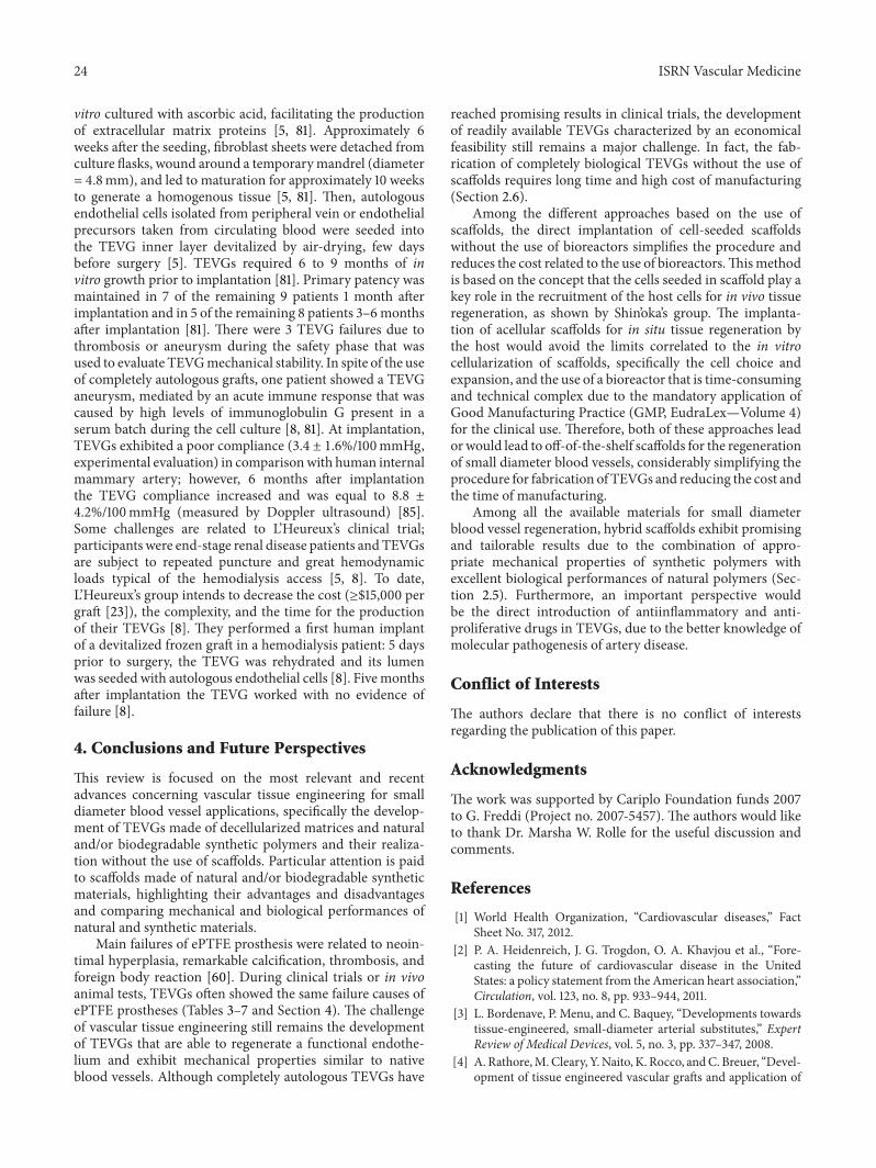
24 ISRN Vascular Medicine
vitro cultured with ascorbic acid, facilitating the productionof extracellular matrix proteins [5, 81]. Approximately 6weeks after the seeding, fibroblast sheets were detached fromculture flasks, wound around a temporarymandrel (diameter= 4.8mm), and led to maturation for approximately 10 weeksto generate a homogenous tissue [5, 81]. Then, autologousendothelial cells isolated from peripheral vein or endothelialprecursors taken from circulating blood were seeded intothe TEVG inner layer devitalized by air-drying, few daysbefore surgery [5]. TEVGs required 6 to 9 months of invitro growth prior to implantation [81]. Primary patency wasmaintained in 7 of the remaining 9 patients 1 month afterimplantation and in 5 of the remaining 8 patients 3–6monthsafter implantation [81]. There were 3 TEVG failures due tothrombosis or aneurysm during the safety phase that wasused to evaluate TEVGmechanical stability. In spite of the useof completely autologous grafts, one patient showed a TEVGaneurysm, mediated by an acute immune response that wascaused by high levels of immunoglobulin G present in aserum batch during the cell culture [8, 81]. At implantation,TEVGs exhibited a poor compliance (3.4 ± 1.6%/100mmHg,experimental evaluation) in comparisonwith human internalmammary artery; however, 6 months after implantationthe TEVG compliance increased and was equal to 8.8 ±4.2%/100mmHg (measured by Doppler ultrasound) [85].Some challenges are related to L’Heureux’s clinical trial;participants were end-stage renal disease patients andTEVGsare subject to repeated puncture and great hemodynamicloads typical of the hemodialysis access [5, 8]. To date,L’Heureux’s group intends to decrease the cost (≥$15,000 pergraft [23]), the complexity, and the time for the productionof their TEVGs [8]. They performed a first human implantof a devitalized frozen graft in a hemodialysis patient: 5 daysprior to surgery, the TEVG was rehydrated and its lumenwas seeded with autologous endothelial cells [8]. Fivemonthsafter implantation the TEVG worked with no evidence offailure [8].
4. Conclusions and Future Perspectives
This review is focused on the most relevant and recentadvances concerning vascular tissue engineering for smalldiameter blood vessel applications, specifically the develop-ment of TEVGs made of decellularized matrices and naturaland/or biodegradable synthetic polymers and their realiza-tion without the use of scaffolds. Particular attention is paidto scaffolds made of natural and/or biodegradable syntheticmaterials, highlighting their advantages and disadvantagesand comparing mechanical and biological performances ofnatural and synthetic materials.
Main failures of ePTFE prosthesis were related to neoin-timal hyperplasia, remarkable calcification, thrombosis, andforeign body reaction [60]. During clinical trials or in vivoanimal tests, TEVGs often showed the same failure causes ofePTFE prostheses (Tables 3–7 and Section 4). The challengeof vascular tissue engineering still remains the developmentof TEVGs that are able to regenerate a functional endothe-lium and exhibit mechanical properties similar to nativeblood vessels. Although completely autologous TEVGs have
reached promising results in clinical trials, the developmentof readily available TEVGs characterized by an economicalfeasibility still remains a major challenge. In fact, the fab-rication of completely biological TEVGs without the use ofscaffolds requires long time and high cost of manufacturing(Section 2.6).
Among the different approaches based on the use ofscaffolds, the direct implantation of cell-seeded scaffoldswithout the use of bioreactors simplifies the procedure andreduces the cost related to the use of bioreactors.Thismethodis based on the concept that the cells seeded in scaffold play akey role in the recruitment of the host cells for in vivo tissueregeneration, as shown by Shin’oka’s group. The implanta-tion of acellular scaffolds for in situ tissue regeneration bythe host would avoid the limits correlated to the in vitrocellularization of scaffolds, specifically the cell choice andexpansion, and the use of a bioreactor that is time-consumingand technical complex due to the mandatory application ofGood Manufacturing Practice (GMP, EudraLex—Volume 4)for the clinical use. Therefore, both of these approaches leadorwould lead to off-of-the-shelf scaffolds for the regenerationof small diameter blood vessels, considerably simplifying theprocedure for fabrication of TEVGs and reducing the cost andthe time of manufacturing.
Among all the available materials for small diameterblood vessel regeneration, hybrid scaffolds exhibit promisingand tailorable results due to the combination of appro-priate mechanical properties of synthetic polymers withexcellent biological performances of natural polymers (Sec-tion 2.5). Furthermore, an important perspective wouldbe the direct introduction of antiinflammatory and anti-proliferative drugs in TEVGs, due to the better knowledge ofmolecular pathogenesis of artery disease.
Conflict of Interests
The authors declare that there is no conflict of interestsregarding the publication of this paper.
Acknowledgments
The work was supported by Cariplo Foundation funds 2007to G. Freddi (Project no. 2007-5457). The authors would liketo thank Dr. Marsha W. Rolle for the useful discussion andcomments.
References
[1] World Health Organization, “Cardiovascular diseases,” FactSheet No. 317, 2012.
[2] P. A. Heidenreich, J. G. Trogdon, O. A. Khavjou et al., “Fore-casting the future of cardiovascular disease in the UnitedStates: a policy statement from the American heart association,”Circulation, vol. 123, no. 8, pp. 933–944, 2011.
[3] L. Bordenave, P. Menu, and C. Baquey, “Developments towardstissue-engineered, small-diameter arterial substitutes,” ExpertReview of Medical Devices, vol. 5, no. 3, pp. 337–347, 2008.
[4] A. Rathore,M.Cleary, Y.Naito, K. Rocco, andC. Breuer, “Devel-opment of tissue engineered vascular grafts and application of

ISRN Vascular Medicine 25
nanomedicine,” Wiley Interdisciplinary Reviews: Nanomedicineand Nanobiotechnology, vol. 4, no. 3, pp. 257–272, 2012.
[5] N. L’Heureux, N. Dusserre, A. Marini, S. Garrido, L. de laFuente, and T. McAllister, “Technology Insight: the evolutionof tissue-engineered vascular grafts—from research to clinicalpractice,” Nature Clinical Practice Cardiovascular Medicine, vol.4, no. 7, pp. 389–395, 2007.
[6] F. Couet, N. Rajan, and D. Mantovani, “Macromolecularbiomaterials for scaffold-based vascular tissue engineering,”Macromolecular Bioscience, vol. 7, no. 5, pp. 701–718, 2007.
[7] S. Goldman, K. Zadina, T. Moritz et al., “Long-term patencyof saphenous vein and left internal mammary artery graftsafter coronary artery bypass surgery: results from a departmentof veterans affairs cooperative study,” Journal of the AmericanCollege of Cardiology, vol. 44, no. 11, pp. 2149–2156, 2004.
[8] M. Peck, D. Gebhart, N. Dusserre, T. N. McAllister, and N.L’Heureux, “The evolution of vascular tissue engineering andcurrent state of the art,” Cells Tissues Organs, vol. 195, no. 1-2,pp. 144–158, 2011.
[9] E. W. Dean, B. Udelsman, and C. K. Breuer, “Current advancesin the translation of vascular tissue engineering to the treatmentof pediatric congenital heart disease,”Yale Journal of Biology andMedicine, vol. 85, no. 2, pp. 229–238, 2012.
[10] L. V. Thomas, V. Lekshmi, and P. D. Nair, “Tissue engineeredvascular grafts—preclinical aspects,” International Journal ofCardiology, vol. 167, no. 4, pp. 1091–1100, 2013.
[11] M. S. Conte,M. J.Mann, H. F. Simosa, K. K. Rhynhart, and R. C.Mulligan, “Genetic interventions for vein bypass graft disease: areview,” Journal of Vascular Surgery, vol. 36, no. 5, pp. 1040–1052,2002.
[12] M. Deutsch, J. Meinhart, P. Zilla et al., “Long-term experiencein autologous in vitro endothelialization of infrainguinal ePTFEgrafts,” Journal of Vascular Surgery, vol. 49, no. 2, pp. 352–362,2009.
[13] M. S. Baguneid, A. M. Seifalian, H. J. Salacinski, D. Murray,G. Hamilton, and M. G. Walker, “Tissue engineering of bloodvessels,” The British Journal of Surgery, vol. 93, no. 3, pp. 282–290, 2006.
[14] Y. Naito, T. Shinoka, D. Duncan et al., “Vascular tissue engi-neering: towards the next generation vascular grafts,” AdvancedDrug Delivery Reviews, vol. 63, no. 4, pp. 312–323, 2011.
[15] A. V. Piterina, A. J. Cloonan, C. L. Meaney et al., “ECM-basedmaterials in cardiovascular applications: inherent healingpotential and augmentation of native regenerative processes,”International Journal of Molecular Sciences, vol. 10, no. 10, pp.4375–4417, 2009.
[16] J. J. Song and H. C. Ott, “Organ engineering based on decellu-larized matrix scaffolds,” Trends in Molecular Medicine, vol. 17,no. 8, pp. 424–432, 2011.
[17] S. F. Badylak, D. O. Freytes, and T. W. Gilbert, “Extracellularmatrix as a biological scaffold material: structure and function,”Acta Biomaterialia, vol. 5, no. 1, pp. 1–13, 2009.
[18] A. F. Pellegata, M. A. Asnaghi, I. Stefani et al., “Detergent-enzymatic decellularization of swine blood vessels: insight onmechanical properties for vascular tissue engineering,” BioMedResearch International, vol. 2013, Article ID 918753, 8 pages,2013.
[19] T. Huynh, G. Abraham, J. Murray, K. Brockbank, P. Hagen, andS. Sullivan, “Remodeling of an acellular collagen graff into aphysiologically responsive neovessel,”Nature Biotechnology, vol.17, no. 11, pp. 1083–1086, 1999.
[20] A. Bader, G. Steinhoff, K. Strobl et al., “Engineering of humanvascular aortic tissue based on a xenogeneic starter matrix,”Transplantation, vol. 70, no. 1, pp. 7–14, 2000.
[21] S. Kaushal, G. E. Amiel, K. J. Guleserian et al., “Functionalsmall-diameter neovessels created using endothelial progenitorcells expanded ex vivo,” Nature Medicine, vol. 7, no. 9, pp. 1035–1040, 2001.
[22] S. L. M. Dahl, J. Koh, V. Prabhakar, and L. E. Niklason,“Decellularized native and engineered arterial scaffolds fortransplantation,” Cell Transplantation, vol. 12, no. 6, pp. 659–666, 2003.
[23] S. L. Dahl, A. P. Kypson, J. H. Lawson et al., “Readily avail-able tissue-engineered vascular grafts,” Science TranslationalMedicine, vol. 3, no. 68, Article ID 68ra9, 2011.
[24] J. F. Mano, G. A. Silva, H. S. Azevedo et al., “Natural originbiodegradable systems in tissue engineering and regenerativemedicine: present status and some moving trends,” Journal ofthe Royal Society Interface, vol. 4, no. 17, pp. 999–1030, 2007.
[25] D. Pankajakshan and D. K. Agrawal, “Scaffolds in tissue engi-neering of blood vessels,” Canadian Journal of Physiology andPharmacology, vol. 88, no. 9, pp. 855–873, 2010.
[26] S. G. Wise, M. J. Byrom, A. Waterhouse, P. G. Bannon, M.K. C. Ng, and A. S. Weiss, “A multilayered synthetic humanelastin/polycaprolactone hybrid vascular graft with tailoredmechanical properties,”Acta Biomaterialia, vol. 7, no. 1, pp. 295–303, 2011.
[27] K. A. McKenna, M. T. Hinds, R. C. Sarao et al., “Mechanicalproperty characterization of electrospun recombinant humantropoelastin for vascular graft biomaterials,”Acta Biomaterialia,vol. 8, no. 1, pp. 225–233, 2012.
[28] B. Zavan,V.Vindigni, S. Lepidi et al., “Neoarteries grown in vivousing a tissue-engineered hyaluronan-based scaffold,” FASEBJournal, vol. 22, no. 8, pp. 2853–2861, 2008.
[29] S. W. Jordan, C. A. Haller, R. E. Sallach, R. P. Apkarian, S. R.Hanson, and E. L. Chaikof, “The effect of a recombinant elastin-mimetic coating of an ePTFE prosthesis on acute thrombo-genicity in a baboon arteriovenous shunt,” Biomaterials, vol. 28,no. 6, pp. 1191–1197, 2007.
[30] K. A. Woodhouse, P. Klement, V. Chen et al., “Investigation ofrecombinant human elastin polypeptides as non-thrombogeniccoatings,” Biomaterials, vol. 25, no. 19, pp. 4543–4553, 2004.
[31] C. Vepari and D. L. Kaplan, “Silk as a biomaterial,” Progress inPolymer Science, vol. 32, no. 8-9, pp. 991–1007, 2007.
[32] R. L. Horan, K. Antle, A. L. Collette et al., “In vitro degradationof silk fibroin,” Biomaterials, vol. 26, no. 17, pp. 3385–3393, 2005.
[33] B. Marelli, M. Achilli, A. Alessandrino et al., “Collagen-reinforced electrospun silk fibroin tubular construct as smallcalibre vascular graft,” Macromolecular Bioscience, vol. 12, no.11, pp. 1566–1574, 2012.
[34] C. B. Weinberg and E. Bell, “A blood vessel model constructedfrom collagen and cultured vascular cells,” Science, vol. 231, no.4736, pp. 397–400, 1986.
[35] D. Seliktar, R. A. Black, R. P. Vito, and R. M. Nerem, “Dynamicmechanical conditioning of collagen-gel blood vessel constructsinduces remodeling in vitro,” Annals of Biomedical Engineering,vol. 28, no. 4, pp. 351–362, 2000.
[36] E. D. Grassl, T. R. Oegema, and R. T. Tranquillo, “Fibrin as analternative biopolymer to type-I collagen for the fabrication ofa media equivalent,” Journal of Biomedical Materials Research,vol. 60, no. 4, pp. 607–612, 2002.

26 ISRN Vascular Medicine
[37] E. D. Grassl, T. R. Oegema, and R. T. Tranquillo, “A fibrin-basedarterial media equivalent,” Journal of Biomedical MaterialsResearch A, vol. 66, no. 3, pp. 550–561, 2003.
[38] D. D. Swartz, J. A. Russell, and S. T. Andreadis, “Engineering offibrin-based functional and implantable small-diameter bloodvessels,”The American Journal of Physiology—Heart and Circu-latory Physiology, vol. 288, no. 3, pp. H1451–H1460, 2005.
[39] L. Yao, J. Liu, and S. T. Andreadis, “Composite fibrin scaffoldsincreasemechanical strength andpreserve contractility of tissueengineered blood vessels,” Pharmaceutical Research, vol. 25, no.5, pp. 1212–1221, 2008.
[40] Z. H. Syedain, L. A. Meier, J. W. Bjork, A. Lee, and R. T.Tranquillo, “Implantable arterial grafts from human fibroblastsand fibrin using a multi-graft pulsed flow-stretch bioreactorwith noninvasive strength monitoring,” Biomaterials, vol. 32,no. 3, pp. 714–722, 2011.
[41] H. J. Salacinski, S. Goldner, A. Giudiceandrea et al., “Themechanical behavior of vascular grafts: a review,” Journal ofBiomaterials Applications, vol. 15, no. 3, pp. 241–278, 2001.
[42] M. Zidi and M. Cheref, “Mechanical analysis of a prototypeof small diameter vascular prosthesis: numerical simulations,”Computers in Biology and Medicine, vol. 33, no. 1, pp. 65–75,2003.
[43] S. Koch, T. C. Flanagan, J. S. Sachweh et al., “Fibrin-polylactide-based tissue-engineered vascular graft in the arterial circula-tion,” Biomaterials, vol. 31, no. 17, pp. 4731–4739, 2010.
[44] K. Kim, M. Yu, X. Zong et al., “Control of degradation rateand hydrophilicity in electrospun non-woven poly(D,L-lactide)nanofiber scaffolds for biomedical applications,” Biomaterials,vol. 24, no. 27, pp. 4977–4985, 2003.
[45] S. J. Lee, J. Liu, S. H. Oh, S. Soker, A. Atala, and J. J. Yoo,“Development of a composite vascular scaffolding system thatwithstands physiological vascular conditions,”Biomaterials, vol.29, no. 19, pp. 2891–2898, 2008.
[46] S. de Valence, J. Tille, D.Mugnai et al., “Long term performanceof polycaprolactone vascular grafts in a rat abdominal aortareplacement model,” Biomaterials, vol. 33, no. 1, pp. 38–47, 2012.
[47] Y. Wang, G. A. Ameer, B. J. Sheppard, and R. Langer, “A toughbiodegradable elastomer,” Nature Biotechnology, vol. 20, no. 6,pp. 602–606, 2002.
[48] R. Gauvin, T. Ahsan, D. Larouche et al., “A novel single-stepself-assembly approach for the fabrication of tissue-engineeredvascular constructs,” Tissue Engineering A, vol. 16, no. 5, pp.1737–1747, 2010.
[49] D. J. Mooney, C. L. Mazzoni, C. Breuer et al., “Stabilizedpolyglycolic acid fibre-based tubes for tissue engineering,”Biomaterials, vol. 17, no. 2, pp. 115–124, 1996.
[50] L. E. Niklason, J. Gao, W. M. Abbott et al., “Functional arteriesgrown in vitro,” Science, vol. 284, no. 5413, pp. 489–493, 1999.
[51] D. Shum-Tim, U. Stock, J. Hrkach et al., “Tissue engineering ofautologous aorta using a new biodegradable polymer,” Annalsof Thoracic Surgery, vol. 68, no. 6, pp. 2298–2305, 1999.
[52] T. Shin’oka, Y. Imai, and Y. Ikada, “Transplantation of a tissue-engineered pulmonary artery,” The New England Journal ofMedicine, vol. 344, no. 7, pp. 532–533, 2001.
[53] S. de Valence, J. C. Tille, J. P. Giliberto et al., “Advantages ofbilayered vascular grafts for surgical applicability and tissueregeneration,” Acta Biomaterialia, vol. 8, no. 11, pp. 3914–3920,2012.
[54] F. Kuwabara, Y. Narita, A. Yamawaki-Ogata et al., “Long-termresults of tissue-engineered small-caliber vascular grafts in a rat
carotid arterial replacementmodel,” Journal of Artificial Organs,vol. 15, no. 4, pp. 399–405, 2012.
[55] K. Iwasaki, K. Kojima, S. Kodama et al., “Bioengineeredthree-layered robust and elastic artery using hemodynamically-equivalent pulsatile bioreactor,” Circulation, vol. 118, no. 14,supplement, pp. S52–S57, 2008.
[56] W. Wu, R. A. Allen, and Y. Wang, “Fast-degrading elastomerenables rapid remodeling of a cell-free synthetic graft into aneoartery,” Nature Medicine, vol. 18, no. 7, pp. 1148–1153, 2012.
[57] J. D. Roh, G. N. Nelson, M. P. Brennan et al., “Small-diameterbiodegradable scaffolds for functional vascular tissue engineer-ing in the mouse model,” Biomaterials, vol. 29, no. 10, pp. 1454–1463, 2008.
[58] T. Uchida, S. Ikeda, H. Oura et al., “Development of biodegrad-able scaffolds based on patient-specific arterial configuration,”Journal of Biotechnology, vol. 133, no. 2, pp. 213–218, 2008.
[59] W. D. Lu, M. Zhang, Z. S. Wu, and T. H. Hu, “Work inprogress report—congenital decellularized and photooxida-tively crosslinked bovine jugular veins as potential tissueengineering scaffolds,” Interactive Cardiovascular and ThoracicSurgery, vol. 8, no. 3, pp. 301–305, 2009.
[60] A. Assmann, C. Delfs, H. Munakata et al., “Acceleration ofautologous in vivo recellularization of decellularized aorticconduits by fibronectin surface coating,” Biomaterials, vol. 34,no. 25, pp. 6015–6026, 2013.
[61] B. Brown, K. Lindberg, J. Reing, D. B. Stolz, and S. F. Badylak,“The basement membrane component of biologic scaffoldsderived from extracellular matrix,” Tissue Engineering, vol. 12,no. 3, pp. 519–526, 2006.
[62] A. V. Piterina, A. Callanan, L. Davis, C. Meaney, M. Walsh,and T. M. McGloughlin, “Extracellular matrices as advancedscaffolds for vascular tissue engineering,” Bio-Medical Materialsand Engineering, vol. 19, no. 4-5, pp. 333–348, 2009.
[63] S. W. Cho, I. K. Kim, J. M. Kang et al., “Evidence for in vivogrowth potential and vascular remodeling of tissue-engineeredartery,” Tissue Engineering A, vol. 15, no. 4, pp. 901–912, 2009.
[64] L. Gui, A. Muto, S. A. Chan, C. K. Breuer, and L. E. Niklason,“Development of decellularized human umbilical arteries assmall-diameter vascular grafts,”Tissue EngineeringA, vol. 15, no.9, pp. 2665–2676, 2009.
[65] B. Marelli, A. Alessandrino, S. Fare, G. Freddi, D. Mantovani,and M. C. Tanzi, “Compliant electrospun silk fibroin tubes forsmall vessel bypass grafting,” Acta Biomaterialia, vol. 6, no. 10,pp. 4019–4026, 2010.
[66] M. Lovett, G. Eng, J. A. Kluge, C. Cannizzaro, G. Vunjak-Novakovic, and D. L. Kaplan, “Tubular silk scaffolds for smalldiameter vascular grafts,” Organogenesis, vol. 6, no. 4, pp. 217–224, 2010.
[67] C. Wang, L. Cen, S. Yin et al., “A small diameter elasticblood vessel wall prepared under pulsatile conditions frompolyglycolic acid mesh and smooth muscle cells differentiatedfrom adipose-derived stem cells,” Biomaterials, vol. 31, no. 4, pp.621–630, 2010.
[68] M. J. McClure, S. A. Sell, D. G. Simpson, B. H. Walpoth, andG. L. Bowlin, “A three-layered electrospun matrix to mimicnative arterial architecture using polycaprolactone, elastin, andcollagen: a preliminary study,” Acta Biomaterialia, vol. 6, no. 7,pp. 2422–2433, 2010.
[69] T. Yokota, H. Ichikawa, G. Matsumiya et al., “In situ tissueregeneration using a novel tissue-engineered, small-calibervascular graft without cell seeding,” Journal of Thoracic andCardiovascular Surgery, vol. 136, no. 4, pp. 900–907, 2008.

ISRN Vascular Medicine 27
[70] B. W. Tillman, S. K. Yazdani, S. J. Lee, R. L. Geary, A. Atala, andJ. J. Yoo, “The in vivo stability of electrospun polycaprolactone-collagen scaffolds in vascular reconstruction,” Biomaterials, vol.30, no. 4, pp. 583–588, 2009.
[71] W. He, Z. Ma, W. E. Teo et al., “Tubular nanofiber scaffoldsfor tissue engineered small-diameter vascular grafts,” Journalof Biomedical Materials Research A, vol. 90, no. 1, pp. 205–216,2009.
[72] P. Stickler, G. de Visscher, L. Mesure et al., “Cyclically stretchingdeveloping tissue in vivo enhances mechanical strength andorganization of vascular grafts,” Acta Biomaterialia, vol. 6, no.7, pp. 2448–2456, 2010.
[73] T. A. Gwyther, J. Z. Hu, A. G. Christakis et al., “Engineeredvascular tissue fabricated from aggregated smoothmuscle cells,”Cells Tissues Organs, vol. 194, no. 1, pp. 13–24, 2011.
[74] W. Wystrychowski, L. Cierpka, K. Zagalski et al., “Case study:first implantation of a frozen, devitalized tissueengineered vas-cular graft for urgent hemodialysis access,” Journal of VascularAccess, vol. 12, no. 1, pp. 67–70, 2011.
[75] R. Gauvin, M. Guillemette, T. Galbraith et al., “Mechanicalproperties of tissue-engineered vascular constructs producedusing arterial or venous cells,” Tissue Engineering A, vol. 17, no.15-16, pp. 2049–2059, 2011.
[76] J. M. Bourget, R. Gauvin, D. Larouche et al., “Human fibroblast-derived ECM as a scaffold for vascular tissue engineering,”Biomaterials, vol. 33, no. 36, pp. 9205–9213, 2012.
[77] J. M. Kelm, V. Lorber, J. G. Snedeker et al., “A novel conceptfor scaffold-free vessel tissue engineering: self-assembly ofmicrotissue building blocks,” Journal of Biotechnology, vol. 148,no. 1, pp. 46–55, 2010.
[78] C. Norotte, F. S. Marga, L. E. Niklason, and G. Forgacs,“Scaffold-free vascular tissue engineering using bioprinting,”Biomaterials, vol. 30, no. 30, pp. 5910–5917, 2009.
[79] E. Pektok, B. Nottelet, J. Tille et al., “Degradation and healingcharacteristics of small-diameter poly(𝜀-caprolactone) vasculargrafts in the rat systemic arterial circulation,” Circulation, vol.118, no. 24, pp. 2563–2570, 2008.
[80] A. de Mel, C. Bolvin, M. Edirisinghe, G. Hamilton, and A.M. Seifalian, “Development of cardiovascular bypass grafts:endothelialization and applications of nanotechnology,” ExpertReview of Cardiovascular Therapy, vol. 6, no. 9, pp. 1259–1277,2008.
[81] T. N. McAllister, M. Maruszewski, S. A. Garrido et al., “Effec-tiveness of haemodialysis access with an autologous tissue-engineered vascular graft: a multicentre cohort study,” TheLancet, vol. 373, no. 9673, pp. 1440–1446, 2009.
[82] N. L’Heureux, T. N. McAllister, and L. M. de la Fuente, “Tissue-engineered blood vessel for adult arterial revascularization,”TheNew England Journal of Medicine, vol. 357, no. 14, pp. 1451–1453,2007.
[83] T. Shinoka and C. Breuer, “Tissue-engineered blood vessels inpediatric cardiac surgery,” Yale Journal of Biology and Medicine,vol. 81, no. 4, pp. 161–166, 2008.
[84] M. A. Cleary, E. Geiger, C. Grady, C. Best, Y. Naito, andC. Breuer, “Vascular tissue engineering: the next generation,”Trends in Molecular Medicine, vol. 18, no. 7, pp. 394–404, 2012.
[85] G. Konig, T. N. McAllister, N. Dusserre et al., “Mechanicalproperties of completely autologous human tissue engineeredblood vessels compared to human saphenous vein and mam-mary artery,” Biomaterials, vol. 30, no. 8, pp. 1542–1550, 2009.

Submit your manuscripts athttp://www.hindawi.com
Stem CellsInternational
Hindawi Publishing Corporationhttp://www.hindawi.com Volume 2014
Hindawi Publishing Corporationhttp://www.hindawi.com Volume 2014
MEDIATORSINFLAMMATION
of
Hindawi Publishing Corporationhttp://www.hindawi.com Volume 2014
Behavioural Neurology
EndocrinologyInternational Journal of
Hindawi Publishing Corporationhttp://www.hindawi.com Volume 2014
Hindawi Publishing Corporationhttp://www.hindawi.com Volume 2014
Disease Markers
Hindawi Publishing Corporationhttp://www.hindawi.com Volume 2014
BioMed Research International
OncologyJournal of
Hindawi Publishing Corporationhttp://www.hindawi.com Volume 2014
Hindawi Publishing Corporationhttp://www.hindawi.com Volume 2014
Oxidative Medicine and Cellular Longevity
Hindawi Publishing Corporationhttp://www.hindawi.com Volume 2014
PPAR Research
The Scientific World JournalHindawi Publishing Corporation http://www.hindawi.com Volume 2014
Immunology ResearchHindawi Publishing Corporationhttp://www.hindawi.com Volume 2014
Journal of
ObesityJournal of
Hindawi Publishing Corporationhttp://www.hindawi.com Volume 2014
Hindawi Publishing Corporationhttp://www.hindawi.com Volume 2014
Computational and Mathematical Methods in Medicine
OphthalmologyJournal of
Hindawi Publishing Corporationhttp://www.hindawi.com Volume 2014
Diabetes ResearchJournal of
Hindawi Publishing Corporationhttp://www.hindawi.com Volume 2014
Hindawi Publishing Corporationhttp://www.hindawi.com Volume 2014
Research and TreatmentAIDS
Hindawi Publishing Corporationhttp://www.hindawi.com Volume 2014
Gastroenterology Research and Practice
Hindawi Publishing Corporationhttp://www.hindawi.com Volume 2014
Parkinson’s Disease
Evidence-Based Complementary and Alternative Medicine
Volume 2014Hindawi Publishing Corporationhttp://www.hindawi.com


![Stem cells in vascular graft tissue engineering for ... · 649 Stem cells in vascular graft tissue engineering for congenital heart surgery REVIEW Darcon grafts [13].Other studies](https://img.dokumen.tips/doc/110x75/5e7a747788383848980b07bd/stem-cells-in-vascular-graft-tissue-engineering-for-649-stem-cells-in-vascular.jpg)