Embed Size (px)
Citation preview

1 Advances in Polymer Science and Technology: An International Journal 2012; 2 (1): 1-15
Review Article
DRUG DELIVERY: SPECIAL EMPHASIS GIVEN ON BIODEGRADABLE POLYMERS.
Swati Aggarwal,* Achhrish Goel, Sandeep Singla
Department of pharmaceutics, M.M. College of pharmacy, Maharishi Markandeshwar University, Mullana, Ambala. (H.R.)
Correspondace author:- Swati Aggarwal* Department of pharmaceutics, M.M. College of Pharmacy, Maharishi
Markandeshwar University, Mullana, Ambala(H.R.)
Ph no- 09729905526
E mail: - [email protected]
Received 29 January 2011; accepted 03 February 2011
Abstract
System of administering drug through controlled delivery so that an optimum amount reaches the target site. Drug delivery
system encompass the carrier, route, and target. Drug dosage forms contain many components in addition to the active
pharmaceutical ingredient(s) to assist in the manufacturing process as well as to optimize drug delivery. Due to advances in
drug delivery technology, excipients are currently included in novel dosage forms to fulfil specific functions and in some cases
they directly or indirectly influence the extent and/or rate of drug release and absorption. Since plant polysaccharides comply
with many requirements expected of pharmaceutical excipients such as non-toxicity, stability, availability and renewability
they are extensively investigated for use in the development of solid oral dosage forms. Polymers have been used as a main
tool to control the drug release rate from the formulations. Extensive applications of polymers in drug delivery have been
realized because polymers offer unique properties which so far have not been attained by any other materials. Advances in
polymer science have led to the development of several novel drug-delivery systems. These newer technological development
include drug modification by chemical means, career based drug delivery and drug entrapment in polymeric matrices or within
pumps that are placed in desired bodily compartments. These technical development in drug delivery/targeting approaches
improve the efficacy of drug therapy thereby improve human health. Biodegradable polymers have been widely used in
biomedical applications because of their known biocompatibility and biodegradability. In the biomedical area, polymers are
generally used as implants and are expected to perform long term service. These improvements contribute to make medical
treatment more efficient and to minimize side effects and other types of inconveniences for patients.
© 2011 Universal Research Publications. All rights reserved
Key words: Drug delivery, Biodegradable polymer, Factor Affecting Polymer.
Introduction:- Drug delivery is the method or process of
administering pharmaceutical compound to achieve a
therapeutic effect in humans or animals.[1,2]. Drug delivery
technologies modify drug release profile, absorption,
distribution and elimination for the benefit of improving
product efficacy, safety, as well as patient compliance and
convenience.
Novel drug delivery:- A novel drug delivery system is a
system that offer multiple drug delivery solutions such as:
Oral Drug Delivery Systems and Materials
Parenteral and Implant Drug Delivery Systems
Pulmonary and Nasal Drug Delivery
Transmucosal Drug Delivery
Transdermal and Topical Drug Delivery
Delivery of Proteins and Peptides
Drug Delivery Pipelines
Drug Delivery Deals
Polymers:- polymers are macromolecules having very large
chains, contain a variety of functional groups, can be blended
with low and high molecular weight materials. Polymers are
becoming increasingly important in the field of drug delivery.
Advances in polymer science have led to the development of
several novel drug delivery systems. A proper consideration
of surface and bulk properties can aid in the designing of
polymers for various drug delivery applications.[3].These
newer technological development include drug modification
Available online at http://www.urpjournals.com
Advances in Polymer Science and Technology: An International Journal
Universal Research Publications. All rights reserved

2 Advances in Polymer Science and Technology: An International Journal 2012; 2 (1): 1-15
Table1. List of polymers used in drug delivery, [6-10]
by chemical means, career based drug delivery and drug
entrapment in polymeric matrices or within pumps that are
placed in desired bodily compartments. These technical
development in drug delivery approaches improve human
health. Use of polymeric material in novel drug delivery
approaches has attracted the scientists.[4]
Pharmaceutical application of polymers:- Used to achieve
taste masking, Use as a binder in tablets to viscosity and flow
controlling agent in liquids, suspension and emulsions.
Polymers can be used as film coating to disguise the
unpleasant taste of drug and to enhance drug stability and to
modify drug release characteristics.[5]
Natural polymers:- are described as below:-
Collagen:- Collagen is a major natural protein component in
mammals that is fabricated from glycine-proline-(hydroxy)
proline repeats to form a triple helix molecular structure [11].
So far, nineteen types of collagen molecules have been
isolated, characterized, and reported in both medical and
pharmaceutical applications [12-14]. Collagen has been
widely used in pharmaceutical applications due to the
fulfillment of many requirements of a drug delivery system
such as good biocompatibility, low antigenicity, and
degradability upon implantation [15]. Furthermore, collagen
gels are one of the first natural polymers to be used as a
promising matrix for drug delivery and tissue engineering
[16]. Biodegradable collagen-based systems have served as
3D scaffold for cell culture, survival of transfected
fibroblasts, and gene therapy [17, 18]. In this case, collagen
scaffolds were fabricated through introducing various
chemical cross-linking agents (i.e., glutaraldehyde,
formaldehyde, carbodiimide) or by physical treatments
(i.e., UV irradiation, freeze-drying, and heating) [19, 20–23].
Figure 1:- A schematic representation of collagen-based
liposome.(source; kang et al. [27] ).
Example:-The combination of liposomes and collagen-based
technologies has been long achieved since the early 80s [16].
In this case, drugs and other bioactive agents were firstly
encapsulated in the liposomes and then embedded inside a
depot composed of collagen-based systems, including
scaffolds and gels. The combination of these two
technologies (i.e., liposomes and collagen-based system) has
improved storage stability, prolonged the drug release rate,
and increased the therapeutic efficacy [11, 24, 25]. In
addition, a study that was conducted by Marston et al. [26],
demonstrated that temperature sensitive liposomes and

3 Advances in Polymer Science and Technology: An International Journal 2012; 2 (1): 1-15
collagen may thermally trigger the release of calcium and
phosphate salts. Multiple collagen-based system for
pharmaceutical carriers or medicinal applications are
currently available for clinical purposes [27]. Figure 1 depicts
a schematic representation of collagen-based liposome.
Gelatin:- Gelatin is a common natural polymer ( water
soluble polymer) or protein which is normally produced by
denaturing collagen [28]. It has been used in pharmaceutical
and medical applications due to its outstanding properties
such as biodegradability, biocompatibility, and low
antigenicity [29]. In addition, gelatin can be easy to
manipulate due to its isoelectric point that allows it to change
from negative to positive charge in an appropriate
physiological environment or during the fabrication, a
property that has found it being very attractive to many
pharmaceutical researchers [30]. Gelatin is one of the natural
polymers used as support material for gene delivery, cell
culture, and more recently tissue engineering. Gelatin-based
systems have the ability to control release of bioactive agents
such as drugs, protein, and dual growth factors [31, 29, 32]. It
has been reported that it is possible to incorporate liposome-
loaded bioactive compounds into PEG-gelatin gel which
function as porous scaffold gelatin-based temporary depots
with controlled drug release over prolonged periods of time
[33, 34]. However, some setbacks have been identified, and
they are said be associated with the use of gelatin-based
systems in pharmaceutical applications. These setbacks
include poor mechanical strength and ineffectiveness in the
management of infected sites [12].
Albumin:- Serum albumin was conjugated to poly-(ethylene
glycol) (PEG) and cross-linked to form mono-PEGylated
albumin hydrogels. These hydrogels were used as a basis for
drug carrying tissue engineering scaffold materials, based on
the natural affinity of various drugs and compounds for the
tethered albumin in the polymer network.
Alginate:- Alginate also serves as an example of a naturally
occurring linear polysaccharide. It is extracted from seaweed,
algae, and bacteria [35-37].The fundamental chemical
structure of alginate is composed of (1–4)-b-D-mannuronic
acid (M) and (1–4)-a-L-guluronic acid (G) units in the form
of homo polymeric (MM- or GG-blocks) and hetero
polymeric sequences (MG or GM-blocks) [38]. Alginate and
their derivates are widely used by many pharmaceutical
scientists for drug delivery and tissue engineering
applications due to its many unique properties such as
biocompatibility, biodegradability, low toxicity, non-
immunogenicity, water solubility, relatively low cost, gelling
ability, stabilizing properties, and high viscosity in aqueous
solutions [39,40]. Since alginate is anionic, fabrication of
alginate hydrogels has successively been achieved through a
reaction with cross-linking agents such as divalent or trivalent
cations mainly calcium ions, water-soluble carbodiimide,
and/or glutaraldehyde [41]. The cross-linking methodology
was conducted at room temperature and physiological pH
[42]. The success in fabricating highly porous 3D alginate
scaffolds has been through lyophilization [43]. Thus far,
alginate-based systems have been successfully used as a
matrix for the encapsulation of stem cells and for controlled
release of proteins, genes, and drugs [44-48]. In addition,
alginate-based systems have been used as depots for bioactive
agent-loaded liposomes, for slow drug release [49, 50].
Highly increased efficacy has been reported from these
integrated delivery systems when compared to polymeric-
based systems or liposome-based systems alone [51,52].
Dextran:- Dextran is a natural linear polymer of glucose
linked by a 1–6 linked-glucoyranoside, and some branching
of 1,3 linked side-chains [53]. Dextran is synthesized from
sucrose by certain lactic-acid bacteria, the best-known being
Leuconostoc mesenteroides and Streptococcus mutans. There
are two commercial preparations available, namely dextran
40 kilodaltons (kDa) (Rheomacrodex) and dextran 70
Kilodaltons (kDa) (Macrodex) [54,55]. In pharmaceutics,
dextran has been used as model of drug delivery due to its
unique characteristics that differentiate it from other
types of polysaccharide. This include water solubility,
biocompatibility, and biodegradability [56]. In recent studies,
dextran has been regarded as a potential polysaccharide
polymer that can sustain the delivery of both proteins,
vaccines, and drugs [57-60]. Interleukin-2, which is a highly
effective anticancer drug, is among the success obtained in
delivering a combination of drug-loaded liposome and
injectable dextran hydrogel [61]. Injectable and degradable
dextran-based systems for drug delivery were generated by a
cross-linking reaction with photo-polymerization or free
radical polymerization [62].
A study by Stenekes and coworkers [63] demonstrated the
successive encapsulation of a drug-loaded liposome depot
into a dextran polymer-based material. The polymeric-based
materials were fabricated using a two phase system, the first
phase was water and poly(ethylene glycol) and the second
one water methacyrlated dextran. The slower degradation of
dextran polymeric material resulted in sustained liposome
release over a period of 100 days [63]. Liposomes released
from depot were reported to be intact, and there was no
significant change in liposomal size. In a gene therapy study
by Liptay and coworkers [64], it was reported that
recombinant DNA (which contains chloramphenicol
acetyltransferase) was successively encapsulated in cationic
liposomes and then integrated within dextran. This system
was reported to be a suitable delivery system since it could
stop transfection efficiency within the colon epithelium wall
in vivo [64].
4 Advances in Polymer Science and Technology: An International Journal 2012; 2 (1): 1-15
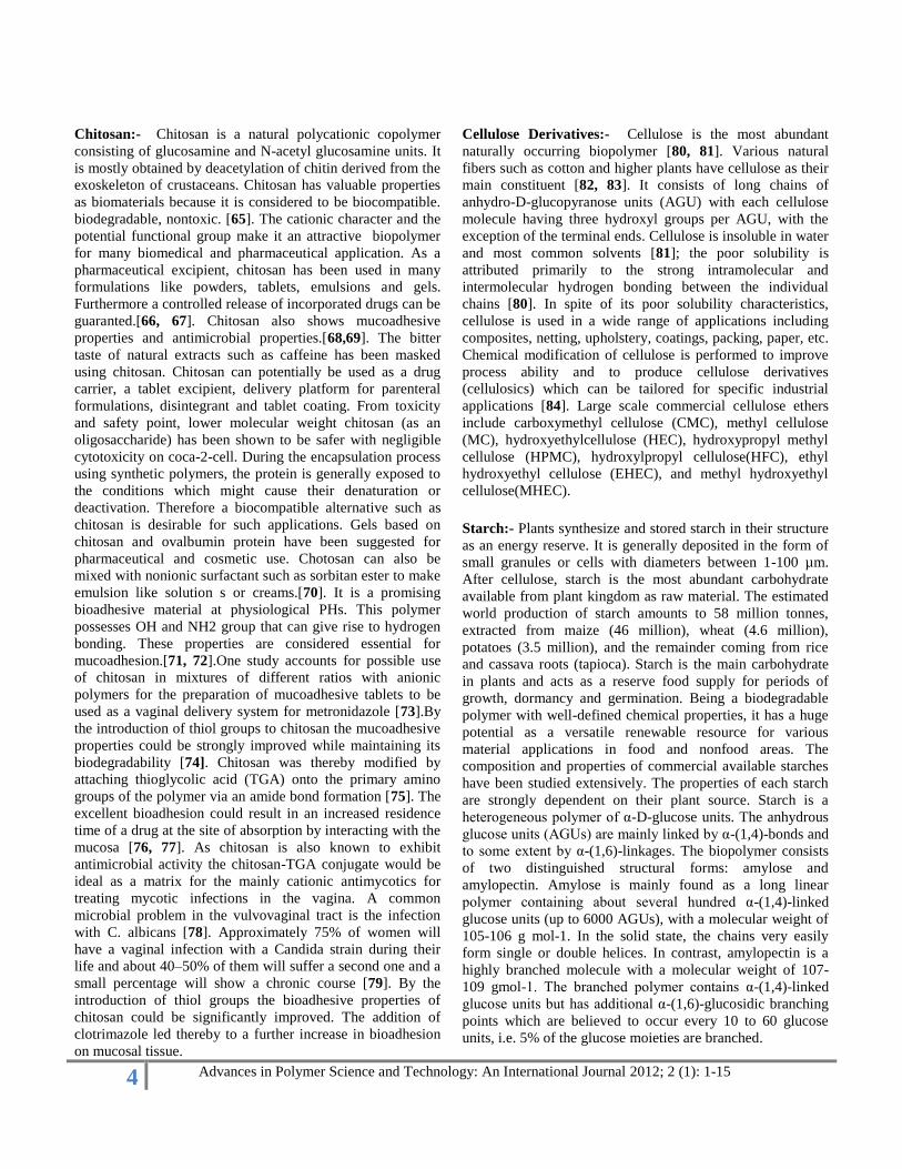
Chitosan:- Chitosan is a natural polycationic copolymer
consisting of glucosamine and N-acetyl glucosamine units. It
is mostly obtained by deacetylation of chitin derived from the
exoskeleton of crustaceans. Chitosan has valuable properties
as biomaterials because it is considered to be biocompatible.
biodegradable, nontoxic. [65]. The cationic character and the
potential functional group make it an attractive biopolymer
for many biomedical and pharmaceutical application. As a
pharmaceutical excipient, chitosan has been used in many
formulations like powders, tablets, emulsions and gels.
Furthermore a controlled release of incorporated drugs can be
guaranted.[66, 67]. Chitosan also shows mucoadhesive
properties and antimicrobial properties.[68,69]. The bitter
taste of natural extracts such as caffeine has been masked
using chitosan. Chitosan can potentially be used as a drug
carrier, a tablet excipient, delivery platform for parenteral
formulations, disintegrant and tablet coating. From toxicity
and safety point, lower molecular weight chitosan (as an
oligosaccharide) has been shown to be safer with negligible
cytotoxicity on coca-2-cell. During the encapsulation process
using synthetic polymers, the protein is generally exposed to
the conditions which might cause their denaturation or
deactivation. Therefore a biocompatible alternative such as
chitosan is desirable for such applications. Gels based on
chitosan and ovalbumin protein have been suggested for
pharmaceutical and cosmetic use. Chotosan can also be
mixed with nonionic surfactant such as sorbitan ester to make
emulsion like solution s or creams.[70]. It is a promising
bioadhesive material at physiological PHs. This polymer
possesses OH and NH2 group that can give rise to hydrogen
bonding. These properties are considered essential for
mucoadhesion.[71, 72].One study accounts for possible use
of chitosan in mixtures of different ratios with anionic
polymers for the preparation of mucoadhesive tablets to be
used as a vaginal delivery system for metronidazole [73].By
the introduction of thiol groups to chitosan the mucoadhesive
properties could be strongly improved while maintaining its
biodegradability [74]. Chitosan was thereby modified by
attaching thioglycolic acid (TGA) onto the primary amino
groups of the polymer via an amide bond formation [75]. The
excellent bioadhesion could result in an increased residence
time of a drug at the site of absorption by interacting with the
mucosa [76, 77]. As chitosan is also known to exhibit
antimicrobial activity the chitosan-TGA conjugate would be
ideal as a matrix for the mainly cationic antimycotics for
treating mycotic infections in the vagina. A common
microbial problem in the vulvovaginal tract is the infection
with C. albicans [78]. Approximately 75% of women will
have a vaginal infection with a Candida strain during their
life and about 40–50% of them will suffer a second one and a
small percentage will show a chronic course [79]. By the
introduction of thiol groups the bioadhesive properties of
chitosan could be significantly improved. The addition of
clotrimazole led thereby to a further increase in bioadhesion
on mucosal tissue.
Cellulose Derivatives:- Cellulose is the most abundant
naturally occurring biopolymer [80, 81]. Various natural
fibers such as cotton and higher plants have cellulose as their
main constituent [82, 83]. It consists of long chains of
anhydro-D-glucopyranose units (AGU) with each cellulose
molecule having three hydroxyl groups per AGU, with the
exception of the terminal ends. Cellulose is insoluble in water
and most common solvents [81]; the poor solubility is
attributed primarily to the strong intramolecular and
intermolecular hydrogen bonding between the individual
chains [80]. In spite of its poor solubility characteristics,
cellulose is used in a wide range of applications including
composites, netting, upholstery, coatings, packing, paper, etc.
Chemical modification of cellulose is performed to improve
process ability and to produce cellulose derivatives
(cellulosics) which can be tailored for specific industrial
applications [84]. Large scale commercial cellulose ethers
include carboxymethyl cellulose (CMC), methyl cellulose
(MC), hydroxyethylcellulose (HEC), hydroxypropyl methyl
cellulose (HPMC), hydroxylpropyl cellulose(HFC), ethyl
hydroxyethyl cellulose (EHEC), and methyl hydroxyethyl
cellulose(MHEC).
Starch:- Plants synthesize and stored starch in their structure
as an energy reserve. It is generally deposited in the form of
small granules or cells with diameters between 1-100 µm.
After cellulose, starch is the most abundant carbohydrate
available from plant kingdom as raw material. The estimated
world production of starch amounts to 58 million tonnes,
extracted from maize (46 million), wheat (4.6 million),
potatoes (3.5 million), and the remainder coming from rice
and cassava roots (tapioca). Starch is the main carbohydrate
in plants and acts as a reserve food supply for periods of
growth, dormancy and germination. Being a biodegradable
polymer with well-defined chemical properties, it has a huge
potential as a versatile renewable resource for various
material applications in food and nonfood areas. The
composition and properties of commercial available starches
have been studied extensively. The properties of each starch
are strongly dependent on their plant source. Starch is a
heterogeneous polymer of α-D-glucose units. The anhydrous
glucose units (AGUs) are mainly linked by α-(1,4)-bonds and
to some extent by α-(1,6)-linkages. The biopolymer consists
of two distinguished structural forms: amylose and
amylopectin. Amylose is mainly found as a long linear
polymer containing about several hundred α-(1,4)-linked
glucose units (up to 6000 AGUs), with a molecular weight of
105-106 g mol-1. In the solid state, the chains very easily
form single or double helices. In contrast, amylopectin is a
highly branched molecule with a molecular weight of 107-
109 gmol-1. The branched polymer contains α-(1,4)-linked
glucose units but has additional α-(1,6)-glucosidic branching
points which are believed to occur every 10 to 60 glucose
units, i.e. 5% of the glucose moieties are branched.

5 Advances in Polymer Science and Technology: An International Journal 2012; 2 (1): 1-15
Fig 2:- Structures of (A) amylopectin or α- amylase and (B) β-amylose.
Modified starch was tested for general applicability of a new
pregelatinized starch product in directly compressible
controlled-release matrix systems. It was prepared by
enzymatic degradation of potato starch followed by
precipitation (retrogradation), filtration and washing with
ethanol. The advantages of the material include ease of tablet
preparation, the potential of a constant release rate (zero-
order) for an extended period of time and its ability to
incorporate high percentages of drugs with different
physicochemical properties. Release rates from retrograded
pre gelatinized starch tablets can be enhanced or decreased to
the desired profile by different parameters like geometries of
the tablet, compaction force and the incorporation of
additional excipients. [85].
Hyaluronic acid:- Hyaluronic acid (also called as
Hyaluronan and Hyaluronate (HA) and Sodium
Hyaluronate(SA) is sodium salt form of hyaluronic acid) is a
biodegradable, biocompatible, and viscoelastic linear
polysaccharide of a wide molecular weight range (1000 to
10,000,000 Da). It is a naturally occurring biopolymer, which
serves important biological functions in bacteria and higher
animals including humans. Naturally occurring hyaluronic
acid may be found in the tissue of higher animals, particularly
as intercellular space filler. It is found in greatest
concentrations in the vitreous humor of the eye and in the
synovial fluid of articular joints. [86]. Hyaluronic acid
comprises linear, unbranching, polyanionic disaccharide units
consisting of glucuronic acid (GlcUA) an N-acetyl
glucosamine (GlcNAc) joined alternately by β-1-3 and β-1-4
glycosidic bonds. Hyaluronic acid solutions are
characteristically viscoelastic and pseudoplastic. The
viscoelastic property of hyaluronic acid solutions that is
important in its use as a biomaterial is controlled by the
concentration and molecular weight of the hyaluronic acid
chains. As a microcapsule, it can be used for targeted drug
delivery. [87].
Cyclodextrin:- They are cyclic oligosaccharides consisting of
six to eight glucose units joined through α-1, 4 glucosidic
bonds. Cyclodextrins remains intact during their passage
throughout the stomach and small intestine of the GI tract.
However, in colon, they undergo fermentation in the presence
of vast colonic microfloras into small monosaccharide and
thus absorbed from these regions. [88, 90] β-cyclodextrins are
degraded to a very small extent in the small intestine but are
completely digested in the large intestine. Most bacterial
strains found abundantly in human beings are capable of
degrading cyclodextrins polysaccharide.
Biodegradable Polymer:- Biodegradation is a natural
process by which organic chemicals in the environment are
converted to simpler compounds, mineralized and
redistributed through elemental cycles such as carbon,
nitrogen and sulphur cycles. Biodegradable polymers have
been widely used in biomedical applications because of their
known biocompatibility and biodegradability. Biodegradable
polymers are intended for temporary aids, such as sutures,
tissue-supporting scaffolds, and drug delivery devices [90].
Polymers within this group retain their properties for a

6 Advances in Polymer Science and Technology: An International Journal 2012; 2 (1): 1-15
limited period of time and then gradually degrade into soluble
molecules that can be excreted from the body [91].
Biodegradable polymers are preferred for drug delivery
applications, since the need for surgical removal of the
depleted device is eliminated. Although the number of
biodegradable polymers is large, only a limited number of
polymers is suitable for drug delivery applications. Suitable
candidates must not only be biodegradable but also fit the
high prerequisites of biocompatibility. In addition, a polymer
should ideally offer processability, sterilizability, and storage
stability if it is to be useful for biomedical applications [92].
The greatest advantage of degradable polymers is that they
are broken down into biologically acceptable molecules that
are metabolized and removed from the body via normal
metabolic pathways. However, biodegradable materials do
produce degradation by-products that must be tolerated with
little or no adverse reactions within the biological
environment. These degradation products—both desirable
and potentially non desirable—must be tested thoroughly,
since there are a number of factors that will affect the
biodegradation of the original materials. The most important
of these factors are shown in the box below—a list that is by
no means complete, but does provide an indication of the
breadth of structural, chemical, and processing properties that
can affect biodegradable drug delivery systems.
Factor affecting biodegradation of polymers:-
Chemical structure.
Chemical composition.
Distribution of repeat units in multimers.
Presents of ionic groups.
Presence of unexpected units or chain defects.
Configuration structure.
Molecular weight.
Molecular-weight distribution.
Morphology (amorphous/semicrystalline,
microstructures, residual stresses).
Presence of low-molecular-weight compounds.
Processing conditions.
Annealing.
Sterilization process.
Storage history.
Shape.
Site of implantation.
Adsorbed and absorbed compounds (water, lipids,
ions, etc.).
Physicochemical factors (ion exchange, ionic
strength, pH).
Physical factors (shape and size changes, variations
of diffusion coefficients, mechanical stresses, stress-
and solvent-induced cracking, etc.).
Mechanism of hydrolysis (enzymes versus water).
Biodegradable polymers mainly investigated for drug
delivery applications are of either natural or synthetic origin.
Natural polymers, already we have discussed above.
Synthetic Polymers:-
Polyester:-
I). Polylactic acid (PLA):- PLA is thermoplastic
biodegradable polymer produced synthetically by
polymerization of lactic acid monomers or cyclic lactide
dimmers. Lactic acid is produced by fermentation of natural
carbohydrates for example, maize or wheat or waste products
from the agricultural or food industry. Commercial quantities
of PLA for packaging applications are produced through ring
opening polymerization of lactide, a reaction favoring the
formation of high molecular polymers. The final crystallioity
and mechanical properties of the polymer depends on the
stereochemistry of polymer backbone. PLA is degraded by
hydrolysis (the breaking of a chemical bond by adding water
to it) of the backbone esters of the polymer. The esters are
broken at random, so that the PLA chains in the material get
shorter and shorter until monomers of lactic acid start to
come loose and the plastic essentially dissolves. This process
is called ‗bulk degradation‘. PLA does not degrade by
microbial attack.
PLA is currently used in loose fill packaging, food packaging
films, thermoformed containers and short shelf life bottles.
PLA can be laminated to paper and paperboard by extrusion
coating for further use as packaging material. Drinking cups
and food containers for short shelf life products are other
application areas.
Fabrics produced from PLA provide a silky feel, durability,
and moisture-management properties. PEA is useful for
producing compost bags and disposable tableware also. PLA
has number of biomedical applications, such as sutures,
stents, dialysis media and drug delivery devices. General
structure of PLA as shown in fig. 3
Figure:-3

7 Advances in Polymer Science and Technology: An International Journal 2012; 2 (1): 1-15
II). Polyglycolic acid (PGA):- PGA is commonly obtained by
ring-opening polymerization of the cyclic diester of glycolic
acid, glycolide [93,94]. PGA is a hard, tough, crystalline
polymer with a melting temperature of ª225 °C and a glass
transition temperature, Tg, of 36 °C [93]. Unlike closely
related polyesters such as PLA, PGA is insoluble in most
common polymer solvents [93]. PGA has excellent fiber-
forming properties and was commercially introduced in 1970
as the first synthetic absorbable suture under the trade name
Dexon™ [93]. The low solubility and high melting point of
PGA limits its use for drug delivery applications, since it
cannot be made into films, rods, capsules, or microspheres
using solvent or melt techniques. General structure of PGA is
shown in fig. 4
Figure:- 4
III). Polyhydroxybutyrate (PHB):- PHB is a biopolymer,
which is present in all living organisms. Many bacteria
produce PHB in large quantities as storage material. It is not
toxic and is totally biodegradable. The polymer is primarily a
product of carbon assimilation (from glucose or starch) and is
employed by microorganisms as a form of ‗energy storage
molecule‘ to be metabolized when other common energy
sources are not available. PHB and its copolymers have
attracted much attention because they are produced
biosynthetically from renewable resources. Microcapsules
from PHB has been prepared by various techniques and
investigated for the release of bovine serum albumin [95].
PHB has also been suggested as a suitable matrix for drug
delivery in veterinary medicine, for instance in the rumen of
cattle [96].
IV). Poly(lactide-co-glycolide), PLGA:- Among the co-
polyesters investigated, extensive research has been
performed in developing a full range of PLGA polymers.
Both L- and DL-lactides have been used for co-
polymerization. The ratio of glycolide to lactide at different
compositions allows control of the degree of crystallinity of
the polymers.
[97]. When the crystalline PGA is co-
polymerized with PLA, the degree of crystallinity is reduced
and as a result this leads to increases in rates of hydration and
hydrolysis. It can therefore be concluded that the degradation
time of the copolymer is related to the ratio of monomers
used in synthesis. In general, the higher the content of
glycolide, the quicker the rate of degradation. However, an
exception to this rule is the 50:50 ratio of PGA: PLA, which
exhibits the fastest degradation. [98, 99]. PLGA is used in
drug delivery applications. Non-steroidal anti-inflammatory
drugs, e.g., diflunisal [100] and diclofenac sodium [101, 102],
have been incorporated into PLGA microspheres and
investigated for the treatment of rheumatoid arthritis,
osteoarthritis, and related diseases. The encapsulation of bio
macromolecules, e.g., proteins and vaccines, into polymeric
microspheres presents a formidable problem because of the
delicacy of these agents; bioactivity might be lost during
preparation, and the release may be poor due to adsorption
and/or aggregation. For instance, the release of recombinant
human interferon-g from PLGA microspheres was
incomplete and the instability of the system limited its use to
7 days or less [103]. Similarly, incomplete release of
lysozyme, recombinant human growth hormone, and a nerve
growth factor from PLGA microspheres was reported [104,
105, 106]. Hence, much effort has been spent in evaluating
PLGA delivery systems, with special regard to microsphere
preparation, protein stability, and release characteristics.
Model proteins studied include bovine serum albumin,
lysozyme, transferrin, and trypsin [107, 104, 108]. Several
peptides, including vapreotide and rismorelin porcine, have
been successfully incorporated and released from PLGA
microspheres [109–111]. Systems for the controlled release
of antigens have a great potential as vaccine adjuvants [112,
113, 108]. Recently, several studies of controlled release
systems for DNA have been presented. DNA of different
sizes has successfully been incorporated into PLGA
microspheres but the loss of DNA integrity and activity still
remains an important issue to be solved for these systems
[114, 115-117]. General structure of PLGA is shown in fig. 5
Figure:-5
V). Poly(e-caprolactone), PCL:- PCL is obtained by ring-
opening polymerization of the 6-membered lactone,e-
caprolactone (e-CL). Anionic, cationic, coordination, or
radical polymerization routes are all applicable [118].
Recently, enzymatic catalyzed polymerization of e-CL has
been reported [119, 120]. PCL crystallizes readily due to the
regular structure and has a melting temperature of 61 °C. It is
tough and flexible [118]. The Tg of PCL is low (–60 °C).
Thus, PCL is in the rubbery state and exhibits high
permeability to low molecular species at body temperature.
These properties, combined with documented
biocompatibility, make PCL a promising candidate for
controlled release applications [121]. PCL degradation
proceeds through hydrolysis of backbone ester bonds as well

8 Advances in Polymer Science and Technology: An International Journal 2012; 2 (1): 1-15
as by enzymatic attack. Hence, PCL degrades under a range
of conditions, biotically in soil, lake waters, sewage sludge,
in vivo, and in compost, and abiotically in phosphate buffer
solutions [122-125]. Hydrolysis of PCL yields 6-
hydroxycaproic acid, an intermediate of the w-oxidation,
which enters the citric acid cycle and is completely
metabolized. Hydrolysis, however, proceeds by homogeneous
erosion at a much slower rate than PLA and PLGA [125].
Hydrolysis of PCL is faster at basic pH and higher
temperatures [118]. PCL hydrolyzes slowly compared to PLA
and PLGA, it is most suitable for long-term drug delivery.
Capronor®, a 1-year contraceptive represents such a system
[118].
VI). Polydioxanone(PDS):- Although biodegradable
polylactides and glycolides have been used to develop
versatile resorbable multi-filament structures, there is
growing research involved in the development of materials
that form monofilament sutures. Multifilament sutures have a
higher risk of infection associated with their use and cause a
greater amount of friction when penetrating tissues. [126,
127]. Polydioxanone (referred to as PDS) is made by a ring-
opening polymerization of thep-dioxanone monomer. It is
characterized by a glass transition temperature in the range of
–10 to 0°C and a degree of crystallinity of about 55%.
Materials prepared with PDS show enhanced flexibility due
to the presence of an ether oxygen within the backbone of the
polymer chain. When usedin vivo, it degrades into monomers
with low toxicity and also has a lower modulus than PLA or
PGA. For the production of sutures, PDS is generally
extruded into fibers at the lowest possible temperature, in
order to avoid its spontaneous depolymerization back to the
monomer. General Structure of Polydioxanone is shown in
fig. 6
Figure:-6
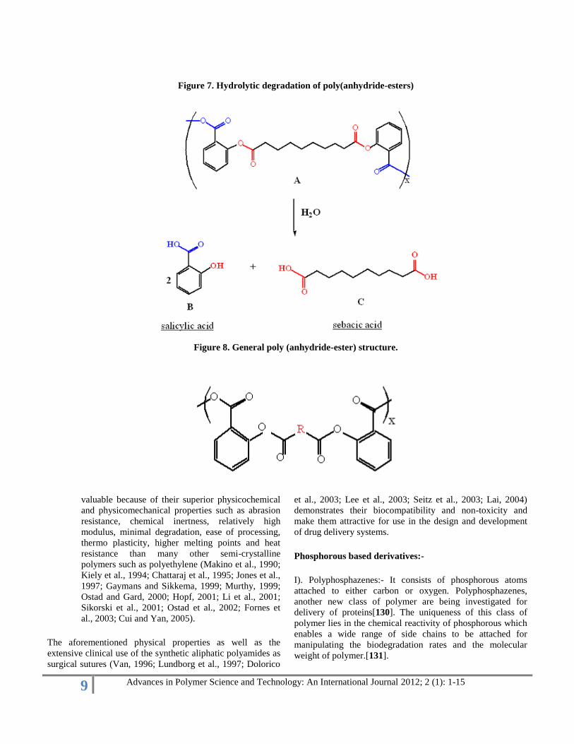
Polyanhydrides:- Poly anhydride are class of
biodegradable polymer characterized by anhydride
bonds that connect repeat unit of polymers backbone
chain. Poly(anhydride-esters) are polymeric
compounds consisting of salicylic acid moieties
bridged by linker structures. An example of the
polymer structure is shown in Figure 7. These
compounds contain two types of important bonds -
anhydride and ester bonds shown in Figure 7 in blue
and red respectively. In the presence of water, both
bonds may degrade hydrolytically, releasing
salicylic acid (B) and sebacic acid (C). Salicylic acid
(B) is the active form of aspirin, an anti-
inflammatory agent, and sebacic acid is currently
used in drug delivery systems (Brem et al., 1995). In
vivo mice studies have indicated that this polymer
assists in wound healing and that it promotes bone
growth (Macedo, 1999).
The release of salicylic acid (B) via bond hydrolysis opens
up a variety of possibilities for creating drug delivery
systems. Potential applications include treatment of
inflammatory bowel disease, dental implants, and tissue
scaffolding. The studies on the degradation rate, the rate of
release of salicylic acid as a function of pH, have shown that
this polymer takes three months to degrade in acidic and
neutral environment, but at basic pH it degrades in 19-40
hours (Erdmann and Uhrich, 1999). Since the upper
gastrointestinal tract is acidic or neutral, this polymeric drug
can reach the intestines undamaged, and release salicylic acid
directly to the lower intestinal tract to treat the disease.
Aspirin is also used in dentistry, for example, in cases of
tooth breakage when there cannot be an immediate operation.
In this procedure, the fast influx of salicylic acid irritates the
surrounding tissue. Slower release of the medication,
provided by the poly(anhydride-esters) would be a gentler
solution.
A drawback to the poly(anhydride-ester) structure shown
in Figure 7 is its low Tg (glass transition temperature). This is
a temperature at which a polymer goes from a solid into a
rubbery state. The Tg of this polymer is several degrees below
body temperature (37 C), which means that it becomes a
soft, sticky material when placed in the body. Because the
polymer is being developed for use as a suture material, it is
undesirable for the polymer to become soft during the
suturing process. Therefore, poly(anhydride-esters) with Tg's
higher than body temperature need to be developed.[128,
129]
Polyamide:- The synthetic aliphatic polyamides are
polymeric compounds frequently referred to as
Nylons which form an important group of poly
condensation polymers. They are linear molecules
(i.e. aliphatic) that are semi-crystalline and
thermoplastic in nature (Kiely et al.,1994; Gaymans
and Sikkema, 1999; Hopf, 2001). A typical
polyamide chain consists of amide groups separated
by alkane segments and the number of carbon atoms
separating the nitrogen atoms which defines the
particular polyamide type. The aliphatic polyamides
are very useful and versatile material that are
9 Advances in Polymer Science and Technology: An International Journal 2012; 2 (1): 1-15
Figure 7. Hydrolytic degradation of poly(anhydride-esters)
Figure 8. General poly (anhydride-ester) structure.
valuable because of their superior physicochemical
and physicomechanical properties such as abrasion
resistance, chemical inertness, relatively high
modulus, minimal degradation, ease of processing,
thermo plasticity, higher melting points and heat
resistance than many other semi-crystalline
polymers such as polyethylene (Makino et al., 1990;
Kiely et al., 1994; Chattaraj et al., 1995; Jones et al.,
1997; Gaymans and Sikkema, 1999; Murthy, 1999;
Ostad and Gard, 2000; Hopf, 2001; Li et al., 2001;
Sikorski et al., 2001; Ostad et al., 2002; Fornes et
al., 2003; Cui and Yan, 2005).
The aforementioned physical properties as well as the
extensive clinical use of the synthetic aliphatic polyamides as
surgical sutures (Van, 1996; Lundborg et al., 1997; Dolorico
et al., 2003; Lee et al., 2003; Seitz et al., 2003; Lai, 2004)
demonstrates their biocompatibility and non-toxicity and
make them attractive for use in the design and development
of drug delivery systems.
Phosphorous based derivatives:-
I). Polyphosphazenes:- It consists of phosphorous atoms
attached to either carbon or oxygen. Polyphosphazenes,
another new class of polymer are being investigated for
delivery of proteins[130]. The uniqueness of this class of
polymer lies in the chemical reactivity of phosphorous which
enables a wide range of side chains to be attached for
manipulating the biodegradation rates and the molecular
weight of polymer.[131].

10 Advances in Polymer Science and Technology: An International Journal 2012; 2 (1): 1-15
Others:-
Polyorthoesters:- Poly(orthoester)s (POE) are another
family of polymers identified as degradable polymers suitable
for orthopaedic applications. Heller and coworkers reported
on the synthesis of a family of polyorthoesters (Figure 6) that
degrades by surface erosion (Ng et al., 1997). With the
addition of lactide segments as part of the polymer structure,
tunable degradation times ranging from 15 to hundreds of
days can be achieved. The degradation of the lactide
segments produces carboxylic acids, which catalyze the
degradation of the orthoester (Ng et al., 1997). Preliminary
in-vivo studies have shown that POE (Figure 6) to increase
bone growth in comparison with poly(dilactide-co-glycolide)
(Andriano et al., 1999).
Conclusion: - Biodegradable polymers have proven their
potential for the development of new, advanced and efficient
drug delivery system. They are capable of delivering a wide
range of bioactive materials. Today the stress is on patient
compliance and to achieve this odjective there is spurt in
development of NDDS. As the herbal excipients are
promising biodegradable materials, these can be chemically
compatible with the excipients in drug delivery systems. In
addition herbal excipients are non-toxic, freely available, and
less expensive compared to their synthetic counterparts. They
have a major role to play in pharmaceutical industry.
Therefore, in the years to come, there is going to be
continued interest in the natural excipients to have better
materials for drug.
References:-
M. N. V. Ravi Kumar (2008), Handbook of
Particulate Drug Delivery (2-Volume Set),
American Scientific Publishers. ISBN 1-58883-123-
X
1. ^"Definition".http://www.building
biotechnology.com/glossary2.php. Retrieved 2008-
05-01.
2. ^"Definition".http://www.biostrate
gy .gc.ca/english/View.asp?mid=413& x=696.
Retrieved 2008-05-01.
3. Omanathanu Pillai, Rasmesh,
Polymers in drug delivery, Current Opinion in
chemical biology, Vol5, issue 4,2001, 447-451.
4. Clochard M, Dinand E, Rankin S,
Simic S, Brocchini S, New strategies for polymer
development in pharmaceutical science- a short
review, J Pharm Pharmacol, 2001, 53(9), 1175-
1184.
5. Kathryn E. Uhrich, Scott M.
Cannizzaro, Robert S. Langer, Polymeric System for
Controlled Drug Release, Chem. Rev, 1999, 99,
3181-3198.
6. Reis, R.L., Cunha, A.M.., Allan,
P.S., Bevis, M.J. Mechanical behavior of injection-
molded starch based polymers. Polym. Adv.
Technol., 1996, 7, 784-90.
7. Seal, B.L., Otero, T.C., A.
Polymeric biomaterials for tissue and organ
regeneration. Mater. Sci. Eng. Rep., 2001, 34, 147-
230.
8. Di Martino, A., Sittinger, M.;
Risbud, M.V. Chitosan: a versatile biopolymer for
orthopaedic tissue-engineering. Biomaterials, 2005,
26, 5983-90.
9. Lee, S.B.; Kim, Y.H.; Chong,
M.S.; Hong, S.H.; Lee, Y.M. Study of gelatin
containing artificial skin V: Fabrication of gelatin
scaf-folds using a salt leaching method.
Biomaterials2005, 26, 1961-8.
10. Mohanty, A.K.; Misra, M.;
Hinrichsen, G. Biodegradable polymers and
biocomposites: An overview. Macromol. Mater.
Eng., 2000, 276-277.
11. C. Kojima, S. Tsumura, A.
Harada, and K. Kono, ―A collagen-mimic dendrimer
capable of controlled release,‖ Journal of the
American Chemical Society, vol. 131, no. 17, pp.
6052–6053, 2009.
12. R. Parenteau-Bareil, R. Gauvin,
and F. Berthod, ―Collagen-based biomaterials for
tissue engineering applications,‖ Materials, vol. 3,
no. 3, pp. 1863–1887, 2010.
13. H. Chen and Z. H. Shana,
―Stabilization of collagen by cross-linking with
oxazolidine E-resorcinol,‖ International Journal of
Biological Macromolecules, vol. 46, no. 5, pp. 535–
539, 2010.
14. C. Holladay, M. Keeney, U.
Greiser, M. Murphy, T. O'Brien, and A. Pandit, ―A
matrix reservoir for improved control of non-viral
gene delivery,‖ Journal of Controlled Release, vol.
136, no. 3, pp. 220–225, 2009.
15. C. Yang, P. J. Hillas, J. A. Báez et
al., ―The application of recombinant human collagen
in tissue engineering,‖ BioDrugs, vol. 18, no. 2, pp.
103–119, 2004.
16. A. L. Weiner, S. S. Carpenter-
Green, and E. C. Soehngen, ―Liposome-collagen gel
matrix: a novel sustained drug delivery system,‖
Journal of Pharmaceutical Sciences, vol. 74, no. 9,
pp. 922–925, 1985.
17. K. Wolf, S. Alexander, V. Schacht
et al., ―Collagen-based cell migration models in
vitro and in vivo,‖ Seminars in Cell &

11 Advances in Polymer Science and Technology: An International Journal 2012; 2 (1): 1-15
Developmental Biology, vol. 20, no. 8, pp. 931–941,
2009.
18. C. Holladay, M. Keeney, U.
Greiser, M. Murphy, T. O'Brien, and A. Pandit, ―A
matrix reservoir for improved control of non-viral
gene delivery,‖ Journal of Controlled Release, vol.
136, no. 3, pp. 220–225, 2009.
19. H. Chen and Z. H. Shana,
―Stabilization of collagen by cross-linking with
oxazolidine E-resorcinol,‖ International Journal of
Biological Macromolecules, vol. 46, no. 5, pp. 535–
539, 2010.
20. Q. Lu, K. Hu, Q. Feng, and F. Cui,
―Growth of fibroblast and vascular smooth muscle
cells in fibroin/collagen scaffold,‖ Materials Science
and Engineering C, vol. 29, no. 7, pp. 2239–2245,
2009.
21. N. Davidenko, J. J. Campbell, E.
S. Thian, C. J. Watson, and R. E. Cameron,
―Collagen-hyaluronic acid scaffolds for adipose
tissue engineering,‖ Acta Biomaterialia, vol. 6, pp.
3957–3968, 2010.
22. M. Kikuchi, H. N. Matsumoto, T.
Yamada, Y. Koyama, K. Takakuda, and J. Tanaka,
―Glutaraldehyde cross-linked
hydroxyapatite/collagen self-organized
nanocomposites,‖ Biomaterials, vol. 25, no. 1, pp.
63–69, 2004.
23. C. M. Tierney, M. J. Jaasma, and
F. J. O'Brien, ―Osteoblast activity on collagen-GAG
scaffolds is affected by collagen and GAG
concentrations,‖ Journal of Biomedical Materials
Research Part A, vol. 91, no. 1, pp. 92–101, 2009.
24. H. Tabandeh, R. Aboufazelia, et
al., ―Liposomes dispersed in two soluble types of
collagens and the effect of collagens on the release
rate of entrapped sodium shromate,‖ Iranian Journal
of Pharmaceutical Research, vol. 2, pp. 161–165,
2003.
25. A. W. Pederson, J. W. Ruberti,
and P. B. Messersmith, ―Thermal assembly of a
biomimetic mineral/collagen composite,‖
Biomaterials, vol. 24, no. 26, pp. 4881–4890, 2003.
26. W. A. Marston, A. Isala, R. S.
Hill, R. Mendes, and M.-A. Minsley, ―Initial report
of the use of an injectable porcine collagen-derived
matrix to stimulate healing of diabetic foot wounds
in humans,‖ Wound Repair and Regeneration, vol.
13, no. 3, pp. 243–247, 2005.
27. H. Kang, M. B. O'Donoghue, H.
Liu, and W. Tan, ―A liposome-based nanostructure
for aptamer directed delivery,‖ Chemical
Communications, vol. 46, no. 2, pp. 249–251, 2010.
28. S. Hao, L. Li, X. Yang et al., ―The
characteristics of gelatin extracted from sturgeon
(Acipenser baeri) skin using various pretreatments,‖
Food Chemistry, vol. 115, no. 1, pp. 124–128, 2009.
29. A. Narita, M. Takahara, T. Ogino,
S. Fukushima, Y. Kimura, and Y. Tabata, ―Effect of
gelatin hydrogel incorporating fibroblast growth
factor 2 on human meniscal cells in an organ culture
model,‖ The Knee, vol. 16, no. 4, pp. 285–289,
2009.
30. S. Young, M. Wong, Y. Tabata,
and A. G. Mikos, ―Gelatin as a delivery vehicle for
the controlled release of bioactive molecules,‖
Journal of Controlled Release, vol. 109, no. 1–3, pp.
256–274, 2005.
31. T. A. Holland, Y. Tabata, and A.
G. Mikos, ―Dual growth factor delivery from
degradable oligo(poly(ethylene glycol) fumarate)
hydrogel scaffolds for cartilage tissue engineering,‖
Journal of Controlled Release, vol. 101, no. 1–3, pp.
111–125, 2005.
32. K. Ofokansi, G. Winter, G.
Fricker, and C. Coester, ―Matrix-loaded
biodegradable gelatin nanoparticles as new approach
to improve drug loading and delivery,‖ European
Journal of Pharmaceutics and Biopharmaceutics,
vol. 76, pp. 1–9, 2010.
33. V. DiTizio, G. W. Ferguson, M.
W. Mittelman, A. E. Khoury, A. W. Bruce, and F.
DiCosmo, ―A liposomal hydrogel for the prevention
of bacterial adhesion to catheters,‖ Biomaterials,
vol. 19, no. 20, pp. 1877–1884, 1998.
34. S. A. Burke, M. Ritter-Jones, B. P.
Lee, and P. B. Messersmith, ―Thermal gelation and
tissue adhesion of biomimetic hydrogels,‖
Biomedical Materials, vol. 2, no. 4, pp. 203–210,
2007.
35. W. R. Gombotz and S. F. Wee,
―Protein release from alginate matrices,‖ Advanced
Drug Delivery Reviews, vol. 31, no. 3, pp. 267–285,
1998.
36. N. Saude, H. Chèze-Lange, D.
Beunard et al., ―Alginate production by Azotobacter
vinelandii in a membrane bioreactor,‖ Process
Biochemistry, vol. 38, no. 2, pp. 273–278, 2002.
37. S. M. Willerth and S. E.
Sakiyama-Elbert, ―Approaches to neural tissue
engineering using scaffolds for drug delivery,‖
Advanced Drug Delivery Reviews, vol. 59, no. 4-5,
pp. 325–338, 2007.
38. C. Gao, M. Liu, S. Chen, S. Jin,
and J. Chen, ―Preparation of oxidized sodium
alginate-graft-poly((2-dimethylamino) ethyl
methacrylate) gel beads and in vitro controlled
release behavior of BSA,‖ International Journal of
Pharmaceutics, vol. 371, no. 1-2, pp. 16–24, 2009.

12 Advances in Polymer Science and Technology: An International Journal 2012; 2 (1): 1-15
39. M. George and T. E. Abraham,
―Polyionic hydrocolloids for the intestinal delivery
of protein drugs: alginate and chitosan—a review,‖
Journal of Controlled Release, vol. 114, no. 1, pp. 1–
14, 2006.
40. T. Li, X. W. Shi, Y. M. Du, and Y.
F. Tang, ―Quaternized chitosan/alginate
nanoparticles for protein delivery,‖ Journal of
Biomedical Materials Research Part A, vol. 83, no.
2, pp. 383–390, 2007.
41. J. B. Xu, J. P. Bartley, and R. A.
Johnson, ―Preparation and characterization of
alginate hydrogel membranes crosslinked using a
water-soluble carbodiimide,‖ Journal of Applied
Polymer Science, vol. 90, no. 3, pp. 747–753, 2003.
42. O. Jeon, K. H. Bouhadir, J. M.
Mansour, and E. Alsberg, ―Photocrosslinked
alginate hydrogels with tunable biodegradation rates
and mechanical properties,‖ Biomaterials, vol. 30,
no. 14, pp. 2724–2734, 2009.
43. N. Mohan and P. D. Nair, ―Novel
porous, polysaccharide scaffolds for tissue
engineering applications,‖ Trends in Biomaterials
and Artificial Organs, vol. 18, no. 2, pp. 219–224,
2005.
44. O. Khanna, M. L. Moya, E. C.
Opara, and E. M. Brey, ―Synthesis of multilayered
alginate microcapsules for the sustained release of
fibroblast growth factor-1,‖ Journal of Biomedical
Materials Research Part A, vol. 95 A, no. 2, pp.
632–640, 2010.
45. N. Wang, G. Adams, L. Buttery,
F. H. Falcone, and S. Stolnik, ―Alginate
encapsulation technology supports embryonic stem
cells differentiation into insulin-producing cells,‖
Journal of Biotechnology, vol. 144, no. 4, pp. 304–
312, 2009.
46. J. R. Nixon and V. W. Yeung,
―Preparation of microencapsulated liposomes, II.
Systems containing nylon-gelatin and nylon-gelatin-
acacia walling material,‖ Journal of
Microencapsulation, vol. 6, no. 1, pp. 43–52, 1989.
47. H. K. Tilakaratne, S. K. Hunter,
M. E. Andracki, J. A. Benda, and V. G. J. Rodgers,
―Characterizing short-term release and
neovascularization potential of multi-protein growth
supplement delivered via alginate hollow fiber
devices,‖ Biomaterials, vol. 28, no. 1, pp. 89–98,
2007.
48. H. J. Kong, E. S. Kim, Y. C.
Huang, and D. J. Mooney, ―Design of biodegradable
hydrogel for the local and sustained delivery of
angiogenic plasmid DNA,‖ Pharmaceutical
Research, vol. 25, no. 5, pp. 1230–1238, 2008.
49. M. Hara and J. Miyake, ―Calcium
alginate gel-entrapped liposomes,‖ Materials
Science and Engineering C, vol. 17, no. 1-2, pp.
101–105, 2001.
50. M. Monshipouri and A. S.
Rudolph, ―Liposome-encapsulated alginate:
controlled hydrogel particle formation and release,‖
Journal of Microencapsulation, vol. 12, no. 2, pp.
117–127, 1995.
51. N. O. Dhoot and M. A. Wheatley,
―Microencapsulated liposomes in controlled drug
delivery: strategies to modulate drug release and
eliminate the burst effect,‖ Journal of
Pharmaceutical Sciences, vol. 92, no. 3, pp. 679–
689, 2003.
52. C. Dai, B. Wang, H. Zhao, B. Li,
and J. Wang, ―Preparation and characterization of
liposomes-in-alginate (LIA) for protein delivery
system,‖ Colloids and Surfaces B, vol. 47, no. 2, pp.
205–210, 2006.
53. R. Mehvar, ―Dextrans for targeted
and sustained delivery of therapeutic and imaging
agents,‖ Journal of Controlled Release, vol. 69, no.
1, pp. 1–25, 2000.
54. G. Sun, Y. I. Shen, C. C. Ho, S.
Kusuma, and S. Gerecht, ―Functional groups affect
physical and biological properties of dextran-based
hydrogels,‖ Journal of Biomedical Materials
Research Part A, vol. 93, no. 3, pp. 1080–1090,
2010.
55. S. Hornig, H. Bunjes, and T.
Heinze, ―Preparation and characterization of
nanoparticles based on dextran-drug conjugates,‖
Journal of Colloid and Interface Science, vol. 338,
no. 1, pp. 56–62, 2009.
56. P. K. Shrivastava and S. K.
Shrivastava, ―Dextran carrier macromolecule for
colon specific delivery of celecoxib,‖ Current Drug
Delivery, vol. 7, no. 2, pp. 144–151, 2010.
57. J. Qi, P. Yao, F. He, C. Yu, and C.
Huang, ―Nanoparticles with dextran/chitosan shell
and BSA/chitosan core-Doxorubicin loading and
delivery,‖ International Journal of Pharmaceutics,
vol. 393, no. 1-2, pp. 177–185, 2010.
58. # S. R. van Tomme and W. E.
Hennink, ―Biodegradable dextran hydrogels for
protein delivery applications,‖ Expert Review of
Medical Devices, vol. 4, no. 2, pp. 147–164, 2007.
59. T. Jin, J. Zhu, F. Wu, W. Yuan, L.
L. Geng, and H. Zhu, ―Preparing polymer-based
sustained-release systems without exposing proteins
to water-oil or water-air interfaces and cross-linking
reagents,‖ Journal of Controlled Release, vol. 128,
no. 1, pp. 50–59, 2008.

13 Advances in Polymer Science and Technology: An International Journal 2012; 2 (1): 1-15
60. # E. M. Bachelder, T. T.
Beaudette, K. E. Broaders et al., ―In vitro analysis of
acetalated dextran microparticles as a potent
delivery platform for vaccine adjuvants,‖ Molecular
Pharmaceutics, vol. 7, no. 3, pp. 826–835, 2010.
61. C. J. De Groot, J. A. Cadée, J. W.
Koten, W. E. Hennink, and W. Den Otter,
―Therapeutic efficacy of IL-2-loaded hydrogels in a
mouse tumor model,‖ International Journal of
Cancer, vol. 98, no. 1, pp. 134–140, 2002.
62. J. Maia, L. Ferreira, R. Carvalho,
M. A. Ramos, and M. H. Gil, ―Synthesis and
characterization of new injectable and degradable
dextran-based hydrogels,‖ Polymer, vol. 46, no. 23,
pp. 9604–9614, 2005.
63. R. J. H. Stenekes, A. E. Loebis, C.
M. Fernandes, D. J. A. Crommelin, and W. E.
Hennink, ―Controlled release of liposomes from
biodegradable dextran microspheres: a novel
delivery concept,‖ Pharmaceutical Research, vol. 17,
no. 6, pp. 690–695, 2000.
64. S. Liptay, H. Weidenbach, G.
Adler, and R. M. Schmid, ―Colon epithelium can be
transiently transfected with liposomes, calcium
phosphate precipitation and DEAE dextran in vivo,‖
Digestion, vol. 59, no. 2, pp. 142–147, 1998.
65. A. K. Singla, M Chawala,
Chitosan:some pharmaceutical and biological
aspects- an update, J. Pharm. Pharmacol. 53 (2001)
1047-1167.
66. L. Illum, Chitosan and its use as a
pharmaceutical excipient, Pharm. Res.15 (1998).
67. V. Dodane, V.D. Vilivalam,
Pharmaceutical application of chitosan, Pharm Sci.
Technol Today 1 (1998) 246-253.
68. K.W. Kim, R. L. Thomas, C. Lee,
H. J. Park, Antimicrobial activity of native chitosan
degraded chitosan and o- carboxy methlated
chitosan, J. Food prot. 66 (2003) 1495-1498.
69. H. l. Lussen, B. J. de Leeuw, M.
W. Langemeyer, A. B. de Boer, J. C. Verhoef, H.E.
Junginger, mucoadhesive polymer in peroral peptide
drug delivery: VI. Carbomer and chitosan improve
the intestinal absorption of peptide drud buserelin in
vivo, Pharm. Res.13 (1996) 1668-1672.
70. Mani Prabaharan, Chitosan
Derivatives as Promising Materials for Controlled
Drug Delivery, Journal of Biomaterials
Applications,2008, 23, 1, 5-36.
71. N.A. Peppas, P.A. Bury, Surface
interfacial and molecular aspects of polymer
bioadhesion on soft tissues, J. Control. Release 2
(1985) 257 – 275.
72. [100] J.R. Robinson, G.M.
Mlynek, Bioadhesive and phase-change polymers
for ocular drug delivery, Adv. Drug Deliv. Rev. 16
(1995) 45 – 50.
73. A. El-Kamel, M. Sokar, V.
Naggar, S. Al Gamal, Chitosan and sodium alginate-
based bioadhesive vaginal tablets, AAPS PharmSci
44 (2002) (Artcle).
74. A. Bernkop-Schnurch, M. Hornof,
D. Guggi, Thiolated chitosans, Eur. J. Pharm.
Biopharm. 57 (2004) 9 – 17.
75. C.E. Kast, C. Valenta, M.
Leopold, A. Bernkop-Schnurch, Design and in vitro
evaluation of a novel bioadhesive vaginal drug
delivery system for clotrimazole, J. Control. Release
81 (2002) 347 – 354.
76. D. Duchene, G. Ponchel, Principle
and investigation of the bioadhesion mechanism of
solid dosage forms, Biomaterials 13 (1992) 709 –
714.
77. K. Khanvilkar, M.D. Donovan,
D.R. Flanagan, Drug transfer through mucus, Adv.
Drug Deliv. Rev. 48 (2001) 173 – 193.
78. J.L. Lanchares, M.L. Hernandez,
Recurrent vaginal candidiasis changes in
etiopathogenical patterns, Int. J. Gynaecol. Obstet.
71 (2000) S29 – S35.
79. J. Ferrer, Vaginal candidosis:
epidemiological and etiological factors, Int. J.
Gynaecol. Obstet. 71 (2000) S21 – S27.
80. Hinterstoisser B., Salmen L.:
Application of dynamic 2D FTIR to cellulose.
Vibrational Spectroscopy, 22, 111–118 (2000).
81. Bochek A. M.: Effect of hydrogen
bonding on cellulose solubility in aqueous and
nonaqueous solvents. Russian Journal of Applied
Chemistry, 76, 1711–1719 (2003).
82. Myasoedova V. V.: Physical
chemistry of non-aqueous solutions of cellulose and
its derivatives. John Wiley and Sons, Chirchester
(2000).
83. Gross R. A., Scholz C.:
Biopolymers from polysaccharides and agroproteins.
American Chemical Society, Washington (2000).
84. Akira I.: Chemical modification of
cellulose. In ‗Wood and Cellulosic Chemistry‘ (eds.:
Hon D. N-S., Shiraishi N.) Marcel Dekker, New
York, 599–626 (2001).
85. Te-Wierik GH, Eissens AC,
Bergsma J, Arends-Scholte AW, Bolhuis GK. A
new generation starch product as excipient in
pharmaceutical tablets, III: Parameters affecting
controlled drug release from tablets based on high
surface area retrograded pregelatinized potato starch.
Int J Pharm. 1997;157:181–7. [PubMed]
86. O'Regan M, Martini I, Crescenzi
F, De Luca C, Lansing M. Molecular mechanisms

14 Advances in Polymer Science and Technology: An International Journal 2012; 2 (1): 1-15
and genetics of hyaluronan biosynthesis. Int J Biol
Macromol 1994;16:283-6.
87. Fraser JR, Brown TJ, Laurent TC.
Catabolism of hyaluronan. In: Lauret TC, editor.
The chemistry, biology, and medical applications of
hyaluronan and its derivatives. Miami, FL: Portland
Press; 1998. p. 85-92.
88. Gerloczy A, Fonagy A, Keresztes
P, Perlaky L, Szejtli J. Absorption, distribution,
excretion and metabolism of orally administered 14
C- Â -cyclodextrin in rat. Arzneim Forsch/Drug Res
1985;35:1042-7.
89. Flourie B, Molis C, Achour L,
Dupas H, Hatat C, Rambaud JC. Fate of Â-
cyclodextrinin the human intestine. J Nutr
1993;123:676-80.
90. Vert M (1989) Angew Makromol
Chem 166/167:155.
91. Vainionpää S, Rokkanen P,
Törmälä P (1989) Prog Polym Sci 14:679.
92. Kronenthal RL (1975)
Biodegradable polymers in medicine and surgery.
In: Kronenthal RL, Oser Z, Martin E (eds),
Polymers in medicine and surgery. Plenum Press,
New York, pg no- 119.
93. Frazza EJ, Schmitt EE (1971) J
Biomed Mater Res Symp 1:43.
94. Benicewicz BC, Hopper PK
(1990) J Bioact Compat Polym 5:453
95. Atkins TW, Peacock SJ (1996) J
Microencapsulation 13:709.
96. Holmes PA (1985) Phys Technol
16:32
97. Cohn, D.; Younes, H.; Marom,
G.Polymer1987,28, 2018.
98. Park, T.G.; Lu, W.Q.; Crotts, G.J.
Controlled Release1995,33, 211.
99. Miller, R.A.; Brady, J.M.;
Cutright, D.E.J. Biomed. Mater. Res.1977,11, 711.
100. Castelli F, Giunchedi P, LaCamera
O, Conte U (2000) Drug Delivery 7:1
101. Chandrashekar G, Udupa N (1996)
J Pharm Pharmacol 48:669
102. Tuncay M, Calis S, Kas HS, Ercan
MT, Peksoy I, Hincal AA (2000) Int J Pharm
195:179
103. Yang J, Cleland JL (1997) J
Pharm Sci 86:908
104. Park TG, Lee HY, Nam YS (1998)
J Control Rel 55:181
105. Kim HK, Park TG (1999) Biotech
Bioeng 65:659
106. Péan J-M, Venier-Julienne M-C,
Boury F, Menei P, Denizot B, Benoit J-P (1998) J
Control Rel 56:175
107. Crotts G, Park TG (1998) J
Microencapsulation 15:699.
108. Sah H, Toddywala R, Chien YW
(1995) J Control Rel 35:137
109. Blanco-Prieto MJ, Besseghir K,
Zerbe O, Andris D, Orsolino P, Heimgartner F,
Merkle HP, Gander B (2000) J Control Rel 67:19
110. Metha RC, Thanoo BC, DeLuca
PP (1996) J Control Rel 41. 249
111. Thompson WW, Anderson DB,
Heiman ML (1997) J Control Rel 43:9
112. O‘Hagan DT (1998) Adv Drug
Del Rev 34:305
113. Sturesson C, Artursson P, Ghaderi
R, Johansen K, Mirazimi A, Uhnoo I, Svensson L,
Albertsson A-C, Carlfors J (1999) J Control Rel
59:377
114. Luo D, Woodrow-Mumford K,
Belcheva N, Saltzman WM (1999) Pharm Res
16:1300
115. Walter E, Moelling K, Pavlovic J,
Merkle HP (1999) J Control Rel 61:361
116. Tinsley-Bown AM, Fretwell R,
Dowsett AB, Davis SL, Farrar GH (2000) J Control
Rel 66:229
117. Wang D, Robinson DR, Kwon
GS, Samuel J (1999) J Control Rel 57:9
118. Pitt CG (1990) Poly-e-
caprolactone and its copolymers. In: Chasin M,
Langer R (eds), Biodegradable polymers as drug
delivery systems. Marcel Dekker, New York, chap
3, pg no-71.
119. Dong H, Cao S-G, Li Z-Q, Han S-
P, You D-L, Shen J-C (1999) J Polym Sci A Polym
Chem 37:1265
120. Henderson LA, Svirkin YY, Gross
RA, Kaplan DL, Swift G (1996) Macromolecules
29:7759
121. Dubernet C, Benoit JP, Couarraze
G, Duchêne D (1987) Int J Pharm 35:145.
122. Benedict CV, Cameron JA, Huang
SJ (1983) J Appl Polym Sci 28:335
123. Eldsäter C, Erlandsson B, Renstad
R, Albertsson A-C, Karlsson S (2000) Poylmer
41:1297
124. Rutkowska M, Dereszewska A,
Jastrzebska M, Janik H (1998) Macromol Symp
130:199
125. Chen DR, Bei JZ, Wang SG
(2000) Polym Degr Stab 67:455
126. Krukowski, Z.H.; Cusick, E.L.;
Engeset, J.; Matheson, N.A.Br. J. Surg.1987,74,
828.
127. Schoetz, D.J.; Coller,
J.A.;Veidenheimer, M.C.Arch. Surg.1988,123, 72.

15 Advances in Polymer Science and Technology: An International Journal 2012; 2 (1): 1-15
128. Erdmann, L., Campo, C., Bedell,
C. and Uhrich, K. (1999) Polymeric prodrugs: Novel
polymers with bioactive components. In: Tailored
Polymeric Materials for Controlled Delivery
Systems (Editors: S. Shalaby and I. McColluch),
American Chemical Society, 709, 83-91.
129. Macedo, B., Uhrich, K. and
Erdmann, L. (1999) The in vivo response to a
bioactive biodegradable polymer. Journal of Dental
Research, 78, 459.
130. Andrianov, A. K.; Payne, L. G.
Protein release from polyphoshazene matrices Adv.
Drug Deliv. Rev., 1998, 31, 185-196.
131. Mao, H.Q.;Kdaiyala, I.; Leong,
K.W.; Zhao, Z.; Dang, W. Biodegradable polymer:
poly (phosphoester). In Encyclopaedia of Controlled
Drug Delivery. Mathowitz E, Ed.; John Wiley and
Sons: New York, 1999, vol. 1, 45-60.
Source of support: Nil; Conflict of interest: None declared


![Original Article Acrylic and methacrylic hompolymers …urpjournals.com/tocjnls/1_1.pdf · Original Article Acrylic and methacrylic hompolymers based on pyramido [4, 5-d] pyrimidine](https://img.dokumen.tips/doc/110x75/5aa762187f8b9ac5648c0a41/original-article-acrylic-and-methacrylic-hompolymers-article-acrylic-and-methacrylic.jpg)