Embed Size (px)
Citation preview
Review ArticleTreatment Options for Class III Malocclusion inGrowing Patients with Emphasis on Maxillary Protraction
Zeinab Azamian and Farinaz Shirban
Torabinejad Dental Research Center, Department of Orthodontics, School of Dentistry,Isfahan University of Medical Sciences, Isfahan 81746-73461, Iran
Correspondence should be addressed to Farinaz Shirban; [email protected]
Received 29 December 2015; Revised 14 March 2016; Accepted 27 March 2016
Academic Editor: Kazuhisa Bessho
Copyright © 2016 Z. Azamian and F. Shirban. This is an open access article distributed under the Creative Commons AttributionLicense, which permits unrestricted use, distribution, and reproduction in any medium, provided the original work is properlycited.
It is very difficult to diagnose and treat Class III malocclusion. This type of malocclusion involves a number of cranial baseand maxillary and mandibular skeletal and dental compensation components. In Class III malocclusion originating frommandibular prognathism, orthodontic treatment in growing patients is not a good choice and in most cases orthognathic surgeryis recommended after the end of growth. Approximately 30–40% of Class III patients exhibit some degree of maxillary deficiency;therefore, devices can be used formaxillary protraction for orthodontic treatment in early mixed dentition. In cases in which dentalcomponents are primarily responsible for Class III malocclusion, early therapeutic intervention is recommended. An electronicsearch was conducted using the Medline database (Entrez PubMed), the Cochrane Collaboration Oral Health Group Database ofClinical Trials, Science Direct, and Scopus. In this review article, we described the treatment options for Class III malocclusionin growing patient with an emphasis on maxillary protraction. It seems that the most important factor for treatment of Class IIImalocclusion in growing patient is case selection.
1. Introduction
Etiologic factors for Class III malocclusions include a widespectrum of skeletal and dental compensation components[1]. The condition might be characterized by mandibularprognathism, maxillary retrognathism, retrusive mandibulardentition, protrusive maxillary dentition, and a combinationof the above [2].
Clinically, Class III malocclusion is in two forms: (a)“pseudo or functional Class III,” due to an early interferencewith the muscular reflex of mandibular closure and (b) the“true Class III” [3].
The etiology of Class III malocclusion is multifactorial,with genetic, ethnic, environmental, and habitual compo-nents [4]. It was believed until 1970 that only the mandibleis responsible for Class III malocclusion [5]; however, almost30–40% of patients exhibit some degree of maxillary defi-ciency [6].
Different ethnic groups exhibit different prevalence ratesof Class III, with different methods of classification being
used. The prevalence rate was reported to be around 1–3%in the Caucasians and around 13-14% among the Chineseand Japanese [7–11]. In the Asian population the majority ofpatients exhibit midface deficiency [12]. It has been reportedthat more than 60% of Class III malocclusion cases are due toskeletal discrepancies [13].
Final and definitive diagnosis of skeletal Class III maloc-clusion is based on the following:
(a) Verification of the normal centric position with thehabitual position.
(b) Presence or absence of a familial predisposition.(c) Cephalometric parameters, including a decrease in
SNA, negative ANB, mandibular protrusion, obtusegonial angle, and great LAFH.
(d) Incisor relationship [14].
This study was undertaken to evaluate different types ofdevices used to correct Class III malocclusion in growing
Hindawi Publishing CorporationScientificaVolume 2016, Article ID 8105163, 9 pageshttp://dx.doi.org/10.1155/2016/8105163
2 Scientifica
Table 1: Appliances for correction of class III malocclusion in growing patients.
Intraoral appliances
Fixed Class III elastic with skeletal anchorage [1] Skeletal effect
Removable
Modified Balters’ Bionator III [15] Dental effectFrankel III [17] Skeletal/dental effectReverse twin block [40] Dental effectEschler/progenic appliance (removable mandible retractor) [18] Dental effectDouble-piece corrector [19] Dental effectTandem appliance [20] Skeletal/dental
Extraoral appliancesChin cap [21] Skeletal
Face mask [24, 25] SkeletalHeadgear for mandibular arch [22, 23] Skeletal/dental
patients with an emphasis on devices used for maxillaryprotraction.
2. Search Strategy
A computerized search was carried out using the Medlinedatabase (Entrez PubMed, http://www.ncbi.nlm.nih.gov/),the Cochrane Collaboration Oral Health Group Databaseof Clinical Trials (http://www.cochrane.org/), Science Direct(http://www.sciencedirect.com/), and Scopus (http://www.scopus.com/) from 1956 to 2015 and from 20th February to4th April 2015.The terms used included maxillary deficiency,Class III malocclusion, maxillary protraction, and boneanchors. During the preliminary search, 250 articles wereselected based on the article titles and then, based on the aimsof this study, 96 articles were evaluated.
3. Appliances
The options for correction of Class III malocclusion ingrowing patients consist of two principal categories: intraoralappliances and extraoral appliances (Table 1).
3.1. Intraoral Appliances
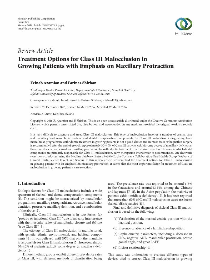
3.1.1. Class III Elastics with Skeletal Anchorage. Four mini-plates are inserted in the left and right infrazygomatic crestof the maxillary buttress and between the lower left and rightlateral incisors and canines (Figure 1). A mucoperiosteal flapis elevated and the miniplates are placed in the underlyingbone byminiscrews.The extension of the plates perforates theattached gingiva and they are loaded three weeks later withClass III elastics [1].
3.1.2. Bionator III. The reverse Bionator or Bionator III, amodified form of the traditional Bionator, is used in thetreatment of Class III malocclusion cases. The modifiedBalters’ Bionator III [15] exhibits differences from the orig-inal version, with deeper and wider lingual wings, acrylicvestibular lateral shields extending deep into the upper fornix,upper labial buttons, and upper incisor inclined plane. Theconstruction bite is normally taken by gentle repositioning ofthe mandible in the centric relation. Patients are expected towear such an appliance for a minimum of 22 hours a day [16].
Figure 1: Class III elastic with skeletal anchorage.
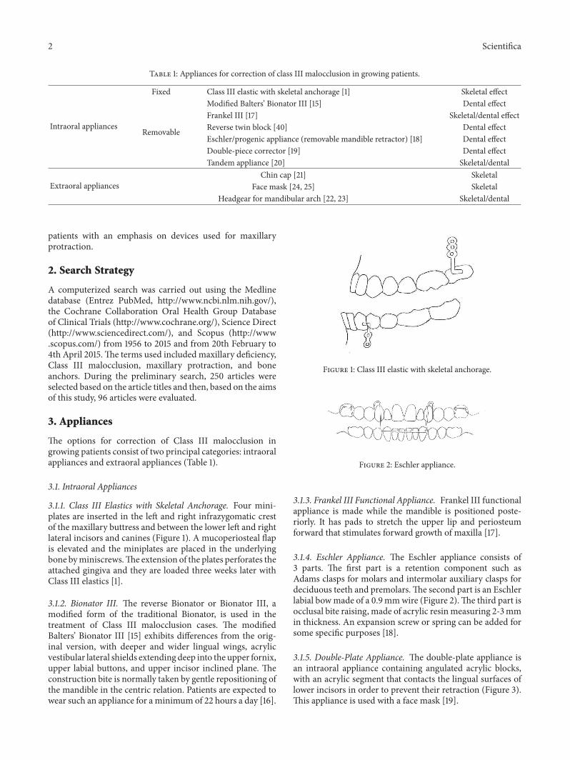
Figure 2: Eschler appliance.
3.1.3. Frankel III Functional Appliance. Frankel III functionalappliance is made while the mandible is positioned poste-riorly. It has pads to stretch the upper lip and periosteumforward that stimulates forward growth of maxilla [17].
3.1.4. Eschler Appliance. The Eschler appliance consists of3 parts. The first part is a retention component such asAdams clasps for molars and intermolar auxiliary clasps fordeciduous teeth and premolars.The second part is an Eschlerlabial bowmade of a 0.9mmwire (Figure 2).The third part isocclusal bite raising, made of acrylic resin measuring 2-3mmin thickness. An expansion screw or spring can be added forsome specific purposes [18].
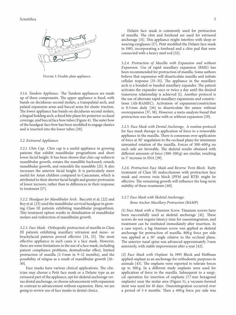
3.1.5. Double-Plate Appliance. The double-plate appliance isan intraoral appliance containing angulated acrylic blocks,with an acrylic segment that contacts the lingual surfaces oflower incisors in order to prevent their retraction (Figure 3).This appliance is used with a face mask [19].
Scientifica 3
Figure 3: Double-plate appliance.
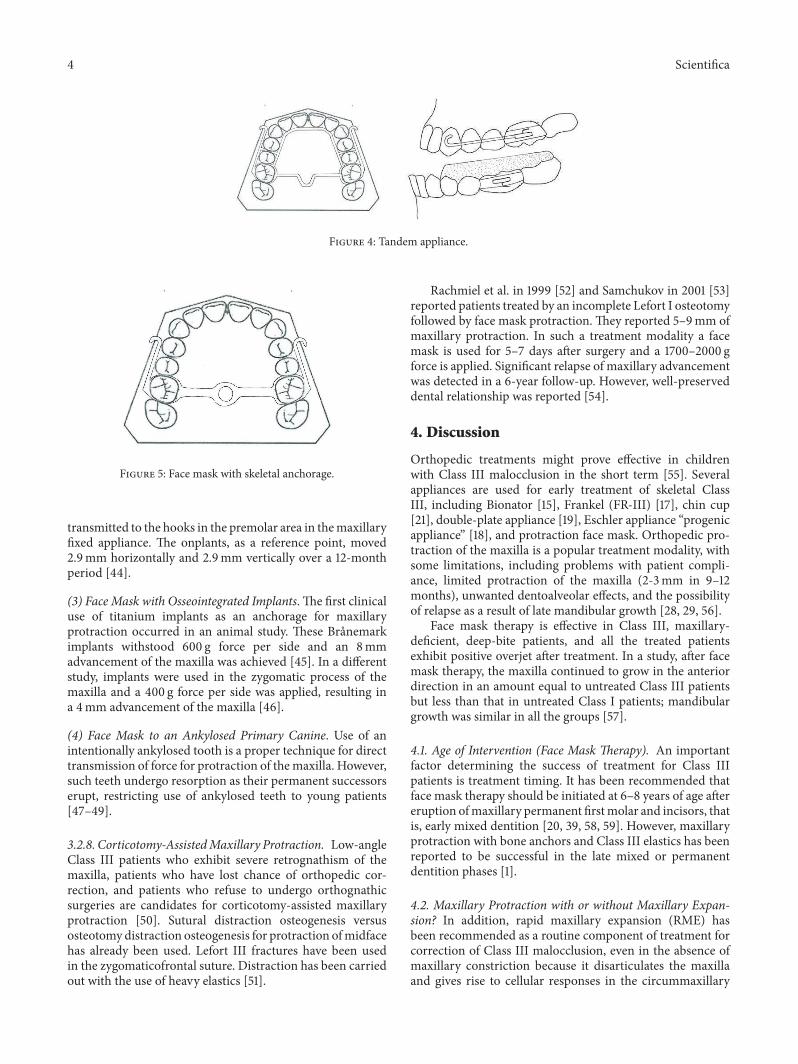
3.1.6. Tandem Appliance. The Tandem appliances are madeup of three components. The upper appliance is fixed, withbands on deciduous second molars, a transpalatal arch, andpalatal expansion arms and buccal arms for elastic traction.The lower appliance has bands on deciduous second molars,a lingual holding arch, a fixed bite plane for posterior occlusalcoverage, and buccal face bow tubes (Figure 4).The outer bowof the headgear face bow has beenmodified to engage elasticsand is inserted into the lower tubes [20].
3.2. Extraoral Appliances
3.2.1. Chin Cap. Chin cap is a useful appliance in growingpatients that exhibit mandibular prognathism and shortlower facial height. It has been shown that chin cap redirectsmandibular growth, rotates the mandible backward, retardsmandibular growth, and remodels the mandible [21]. It alsoincreases the anterior facial height. It is particularly moreuseful for Asian children compared to Caucasians, which isattributed to their shorter facial height and greater protrusionof lower incisors, rather than to differences in their responseto treatment [17].
3.2.2. Headgear for Mandibular Arch. Baccetti et al. [22] andRey et al. [23] used themandibular cervical headgear in grow-ing Class III patients exhibiting mandibular prognathism.This treatment option results in distalization of mandibularmolars and redirection of mandibular growth.
3.2.3. Face Mask. Orthopedic protraction of maxilla in ClassIII patients exhibiting maxillary retrusion and meso- orbrachyfacial patterns proved effective [24, 25]. The mosteffective appliance in such cases is a face mask. However,there are some limitations in the use of a face mask, includingpatient compliance problems, dentoalveolar effect, limitedprotraction of maxilla (2-3mm in 9–12 months), and thepossibility of relapse as a result of mandibular growth [26–30].
Face masks have various clinical applications. The clin-ician may choose a Petit face mask or a Delaire type as anextraoral part of the appliance, opt for skeletal anchorage ver-sus dental anchorage, or choose advancement with expansionin contrast to advancement without expansion. Here, we aregoing to review use of face masks in dental clinics.
Delaire face mask is commonly used for protractionof maxilla. The chin and forehead are used for extraoralanchorage [31]. This appliance might interfere with sleep orwearing eyeglasses [17]. Petit modified the Delaire face maskin 1983, incorporating a forehead and a chin pad that wereconnected with a heavy steel rod [32].
3.2.4. Protraction of Maxilla with Expansion and withoutExpansion. Use of rapid maxillary expansion (RME) hasbeen recommended for protraction of maxilla. Some authorsbelieve that expansion will disarticulate maxilla and initiatecellular response [33–35]. The appliance in the maxillaryarch is a bonded or banded maxillary expander. The patientactivates the expander once or twice a day until the desiredtransverse relationship is achieved [1]. Another protocol isthe use of alternate rapid maxillary expansions and constric-tions (Alt-RAMEC). Activation of expansion/constrictionis 0.5mm daily [36] to disarticulate the suture withoutoverexpansion [37, 38]. However, a meta-analysis found thatprotraction was the same with or without expansion [29].
3.2.5. Face Mask with Dental Anchorage. A routine protocolfor face mask therapy is application of force to a removableappliance in the maxilla. There is consensus over applicationof force at 30∘ angulation to the occlusal plane for minimumunwanted rotation of the maxilla. Forces of 300–600 g oneach side are favorable. The skeletal results obtained withdifferent amounts of force (300–500 g) are similar, resultingin 3∘ increase in SNA [39].
3.2.6. Protraction Face Mask and Reverse Twin Block. Earlytreatment of Class III malocclusions with protraction facemask and reverse twin block (PFM and RTB) might beeffective. The remaining growth will influence the long-termstability of these treatments [40].
3.2.7. Face Mask with Skeletal Anchorage:Bone Anchor Maxillary Protraction (BAMP)
(1) Face Mask with a Titanium Screw. Titanium screws havebeen successfully used as skeletal anchorage [41]. Thesescrews do not require latency time for osseointegration, andtreatment can be instituted immediately after insertion. Ina case report, a lag titanium screw was applied as skeletalanchorage for protraction of maxilla. 800 g force per sidewas applied at a 30∘ angle relative to the occlusal plane.The anterior nasal spine was advanced approximately 3mmanteriorly, with stable improvement after a year [42].
(2) Face Mask with Onplant. In 1995 Block and Hoffmanapplied onplant as an anchorage for orthodontic purposes inanimals [43]. The onplants were reported to tolerate forcesup to 300 g. In a different study onplants were used forapplication of force to the maxilla. Subsequent to a surgi-cal operation for insertion of onplants (7.7mm hexagonalonplants) near the molar area (Figure 5), a vacuum-formedstent was used for 10 days. Osseointegration occurred overa period of 3-4 months. Then a 400 g force per side was
4 Scientifica
Figure 4: Tandem appliance.
Figure 5: Face mask with skeletal anchorage.
transmitted to the hooks in the premolar area in themaxillaryfixed appliance. The onplants, as a reference point, moved2.9mm horizontally and 2.9mm vertically over a 12-monthperiod [44].
(3) Face Mask with Osseointegrated Implants. The first clinicaluse of titanium implants as an anchorage for maxillaryprotraction occurred in an animal study. These Branemarkimplants withstood 600 g force per side and an 8mmadvancement of the maxilla was achieved [45]. In a differentstudy, implants were used in the zygomatic process of themaxilla and a 400 g force per side was applied, resulting ina 4mm advancement of the maxilla [46].
(4) Face Mask to an Ankylosed Primary Canine. Use of anintentionally ankylosed tooth is a proper technique for directtransmission of force for protraction of themaxilla. However,such teeth undergo resorption as their permanent successorserupt, restricting use of ankylosed teeth to young patients[47–49].
3.2.8. Corticotomy-AssistedMaxillary Protraction. Low-angleClass III patients who exhibit severe retrognathism of themaxilla, patients who have lost chance of orthopedic cor-rection, and patients who refuse to undergo orthognathicsurgeries are candidates for corticotomy-assisted maxillaryprotraction [50]. Sutural distraction osteogenesis versusosteotomydistraction osteogenesis for protraction ofmidfacehas already been used. Lefort III fractures have been usedin the zygomaticofrontal suture. Distraction has been carriedout with the use of heavy elastics [51].
Rachmiel et al. in 1999 [52] and Samchukov in 2001 [53]reported patients treated by an incomplete Lefort I osteotomyfollowed by face mask protraction. They reported 5–9mm ofmaxillary protraction. In such a treatment modality a facemask is used for 5–7 days after surgery and a 1700–2000 gforce is applied. Significant relapse of maxillary advancementwas detected in a 6-year follow-up. However, well-preserveddental relationship was reported [54].
4. Discussion
Orthopedic treatments might prove effective in childrenwith Class III malocclusion in the short term [55]. Severalappliances are used for early treatment of skeletal ClassIII, including Bionator [15], Frankel (FR-III) [17], chin cup[21], double-plate appliance [19], Eschler appliance “progenicappliance” [18], and protraction face mask. Orthopedic pro-traction of the maxilla is a popular treatment modality, withsome limitations, including problems with patient compli-ance, limited protraction of the maxilla (2-3mm in 9–12months), unwanted dentoalveolar effects, and the possibilityof relapse as a result of late mandibular growth [28, 29, 56].
Face mask therapy is effective in Class III, maxillary-deficient, deep-bite patients, and all the treated patientsexhibit positive overjet after treatment. In a study, after facemask therapy, the maxilla continued to grow in the anteriordirection in an amount equal to untreated Class III patientsbut less than that in untreated Class I patients; mandibulargrowth was similar in all the groups [57].
4.1. Age of Intervention (Face Mask Therapy). An importantfactor determining the success of treatment for Class IIIpatients is treatment timing. It has been recommended thatface mask therapy should be initiated at 6–8 years of age aftereruption ofmaxillary permanent firstmolar and incisors, thatis, early mixed dentition [20, 39, 58, 59]. However, maxillaryprotraction with bone anchors and Class III elastics has beenreported to be successful in the late mixed or permanentdentition phases [1].
4.2. Maxillary Protraction with or without Maxillary Expan-sion? In addition, rapid maxillary expansion (RME) hasbeen recommended as a routine component of treatment forcorrection of Class III malocclusion, even in the absence ofmaxillary constriction because it disarticulates the maxillaand gives rise to cellular responses in the circummaxillary
Scientifica 5
sutures, bringing about a more positive reaction to protrac-tion forces [34, 35, 60]. Nevertheless, when used to enhanceanterior movement of the maxilla during face mask therapy,preliminary RME does not appear to exert any effect onthe efficacy of orthopedic treatment [61]. There are reportsthat use of RME alone might not properly disarticulatecircummaxillary sutures and it might be better dealt withby Alt-RAMEC [36, 62, 63]. A meta-analysis showed similarresults for protraction with or without expansion [29].
4.3. Treatment Outcome for Face Mask Therapy with Dentalor Skeletal Anchorage. Cephalometric analyses have shownskeletal and dentoalveolar changes in face mask therapy.Skeletal changes include maxillary protrusive movement(SNA,N-perpA) and downward and backward rotation of themandible (SNGoGN, SNGn, and LAFH), with a decrease inprognathism severity (SNB). Such changes induce favorablechanges in the facial profile. Dentoalveolar changes mainlyconsist of linguoversion of mandibular incisors and labialinclination of maxillary incisors [64]. The forces used formaxillary protraction are usually applied to maxillary teeth.Therefore, there might be a significant mesial migration ofmaxillary teeth, possibly resulting in severe anterior crowdingand in a decrease in the orthopedic effects of treatment,which might cause problems [65, 66]. It has been demon-strated that themandibular plane angle increases significantlyduring protraction face mask therapy [67–69]. Some of thecomplications associated with dental anchorage are resolvedby skeletal anchorage. Recently, osseointegrated implants,titanium screws, onplants, and miniplates have been used asstable anchorage for maxillary protraction [41, 46, 56, 70].
The greatest disadvantage of palatal osseointegratedimplants is that they can only be placed on the palate and areindicated for moving maxillary teeth. A palatal implant costsmuch higher than a miniscrew andminiplate and requires anosseointegration period [71].
Temporary anchorage devices for maxillary protractionhave become very popular in recent years [1, 42, 44, 46, 72].
A study showed an improvement inmolar relation, whichwas not influenced by dental inclination and there was nosignificant amount of lingual inclination of the lower incisorsin the BAMP group [1].
The proclination of mandibular incisors might beexplained by the new posture of the tongue that acts on theseincisors after correction of anterior crossbite [56, 73].
In Class III cases that are treated with skeletal anchor-age, the amount of maxillary protraction might vary from3.0 to 5.6mm [36, 73–75]. The BAMP protocol can causesignificantly larger maxillary advancement compared to theRME/FM therapy. BAMP results in 2.3–3mm more max-illary protraction compared to face mask or rapid maxil-lary expansion [1]. BAMP protocol results in fewer verticalchanges. Furthermore, these patients do not exhibit clockwiserotation of the mandible or dental compensation [1, 76]. A3.82mm forward movement of the nose tip was reported asa result of the BAMP protocol [77]. The upper lip and lipsulcus also moved forward, and the soft tissue B point andpogonion moved backward during the protraction period,indicating improvements in the soft tissue profile in line with
the underlying skeletal components during the protractionprocedure [56, 78, 79].
It has been reported that it is difficult to control thevertical growth and lower anterior facial height increases tosome extent during maxillary protraction, with both dentaland bone anchorage [1, 26, 34, 80]. The low mandibularplane angle group exhibited a greater maxillary forwarddisplacement and a larger increase in the maxillary bodycompared to the high mandibular plane angle group [81].The craniomaxillary complex in the dental anchorage modelis displaced forward along with rotation, and the amount ofthis rotation decreases gradually with an increase in the anglebetween the force vector and occlusal plane from 0 to 30degrees. However, the craniomaxillary complex in the boneanchorage model is displaced forward along with rotation,and the rotation degree decreases gradually with an increasein the angle from 0 to 20 [82].
A cephalometric analysis of pharyngeal airway showednosignificant changes in the oro- and nasopharyngeal sagittalairway dimensions in the face mask + bite block therapygroup compared to the untreated Class III subjects [83].
Ghiz et al. [84] reported that four variables were sig-nificant in predicting successful treatment outcomes: (1)the position of the condyle relative to the cranial base; (2)ramus length; (3) mandibular length; and (4) gonial angle[85]. Three pretreatment cephalometric variables exhibitedthe highest predictive power in terms of discriminatinggonial angle, nasion-A-pogonion angle, and ramus plane-to-sella-nasion angle. Patients exhibiting larger values forthese three measurements before treatment were catego-rized as the unstable group at the end of the observationperiod.Orthopedic treatment ofClass IIImalocclusionmightgive rise to more favorable craniofacial adaptations when apatient’s pretreatment cephalometric analyses reveal a shortmandibular ramus (i.e., decreased posterior facial height) anda low mandibular plane angle [30].
4.4. Skeletal Analysis of Treatment Outcomes. An approach isto make use of a 3D skeletal color map of the superimposi-tion on the anterior cranial base. The superimposition andsemitransparent overlays show that bone-anchoredmaxillaryprotraction growth and treatment response lead to boneapposition at the anterior eminence of the TMJ, whichcorrelates well with the posterior displacement of the anteriorsurface of the condyle, and the bone resorption of theposterior wall of the articular eminence correlates well withthe posterior displacement of the posterior surface of thecondyle. Mandibular shape, rather than the mandibular size,is under the influence of continuous intermaxillary traction[14]. Forward movement of the maxilla might be registeredat the posterior nasal spine and at pterygomaxillary fissurepoints [72].
Another method is to use the patient’s lateral cephalo-grams, which can be extracted from CBCT, in order to carryout TPS analysis [86]. TPS analysis deforms one landmarkconfiguration into another, indicating that this change inshape is the deformation of a grid, at the same time makingstatistical comparisons possible. TPS has specific cephalo-metric indications to demonstrate differences in shape as a
6 Scientifica
result of orthodontic treatment techniques or growth-relatedchanges. In fact, TPS analysis has been used to study growthchanges in treated and untreated subjects with different typesof malocclusion [87, 88].
4.5. Retention and Follow-Up. In previous studies retentionhas been recommended after overjet and overbite correctionfrom three months to two years during the night [24, 89,90]. Long-term follow-ups of maxillary protraction indicatea 25–33% chance of relapse to negative overjet after thecompletion of mandibular growth [30, 67, 91, 92]. It wasconcluded that relapse to a Class III pattern primarily resultsfrom mandibular growth rather than a relapse in the maxilla[50, 66]. In another study, patients with larger values for theinclination of the mandibular ramus to the mandibular body(gonial angle) before treatment exhibited a higher probabilityof relapse at the end of the observation period [30, 93, 94].
5. Conclusion
An important factor for treatment ofClass IIImalocclusion ingrowing patient is the origin of malocclusion. The skeletal ordental origin of themalocclusion and in skeletal Class IIImal-occlusions mandibular prognathism or maxillary deficiencyare important for choosing early intervention and selection ofthe appliance for treatment. All appliances described in thispaper can be useful when the clinicians use them in correctmanner.
Competing Interests
The authors declare that there are no competing interestsregarding the publication of this paper.
References
[1] L. Cevidanes, T. Baccetti, L. Franchi, J. A. McNamara Jr., andH. De Clerck, “Comparison of two protocols for maxillary pro-traction: bone anchors versus face mask with rapid maxillaryexpansion,”The Angle Orthodontist, vol. 80, no. 5, pp. 799–806,2010.
[2] M. Celikoglu andH. Oktay, “Effects of maxillary protraction forearly correction of class III malocclusion,” European Journal ofOrthodontics, vol. 36, no. 1, pp. 86–92, 2014.
[3] R. E. Moyers, Handbook of Orthodontics, Year Book MedicalPublishers, 1988.
[4] M. B. Khan and A. Karra, “Early treatment of class III maloc-clusion: a boon or a burden?” International Journal of ClinicalPediatric Dentistry, vol. 7, pp. 130–136, 2014.
[5] J. Mermigos, C. A. Full, and G. Andreasen, “Protraction of themaxillofacial complex,” American Journal of Orthodontics andDentofacial Orthopedics, vol. 98, no. 1, pp. 47–55, 1990.
[6] A. Arman, T. U. Toygar, and E. Abuhijleh, “Profile changesassociated with different orthopedic treatment approaches inclass III malocclusions,” Angle Orthodontist, vol. 74, no. 6, pp.733–740, 2004.
[7] M. Fu, D. Zhang, B. Wang, Y. Deng, F. Wang, and X. Ye,“The prevalence of malocclusion in China—an investigation of25,392 children,” Chinese Journal of Stomatology, vol. 37, no. 5,pp. 371–373, 2002.
[8] M. Irie and S. Nakamura, “Orthopedic approach to severe skele-tal Class III malocclusion,” American Journal of Orthodontics,vol. 67, no. 4, pp. 377–392, 1975.
[9] M. T. Bukhary, “Comparative cephalometric study of Class IIImalocclusion in Saudi and Japanese adult females,” Journal ofOral Science, vol. 47, no. 2, pp. 83–90, 2005.
[10] M.Mouakeh, “Cephalometric evaluation of craniofacial patternof Syrian children with Class III malocclusion,” AmericanJournal of Orthodontics andDentofacial Orthopedics, vol. 119, no.6, pp. 640–649, 2001.
[11] M. Celikoglu, S. Akpinar, and I. Yavuz, “The pattern of mal-occlusion in a sample of orthodontic patients from Turkey,”Medicina Oral, Patologia Oral y Cirugia Bucal, vol. 15, no. 5, pp.e791–e796, 2010.
[12] G. V. Newman, “Prevalence of malocclusion in children sixto fourteen years of age and treatment in preventable cases,”Journal of the American Dental Association, vol. 52, no. 5, pp.566–575, 1956.
[13] C. B. Staudt and S. Kiliaridis, “Different skeletal types underly-ing Class III malocclusion in a random population,” AmericanJournal of Orthodontics and Dentofacial Orthopedics, vol. 136,no. 5, pp. 715–721, 2009.
[14] H. De Clerck, T. Nguyen, L. K. De Paula, and L. Cevidanes,“Three-dimensional assessment of mandibular and glenoidfossa changes after bone-anchored Class III intermaxillarytraction,” American Journal of Orthodontics and DentofacialOrthopedics, vol. 142, no. 1, pp. 25–31, 2012.
[15] A. Levrini and L. Levrini, “Il Bionator concetti classici e nuoveacquisizioni,” Rivista Italiana di Stomatologia, vol. 10, pp. 499–506, 1993.
[16] G.Garattini, L. Levrini, P. Crozzoli, andA. Levrini, “Skeletal anddental modifications produced by the Bionator III appliance,”American Journal of Orthodontics and Dentofacial Orthopedics,vol. 114, no. 1, pp. 40–44, 1998.
[17] W. R. Proffit, H. W. Fields Jr., and D. M. Sarver, ContemporaryOrthodontics, Elsevier Health Sciences, Philadelphia, Pa, USA,2014.
[18] M. R. de Almeida, R. R. de Almeida, P. V. P. Oltramari-Navarro,A. C. D. C. F. Conti, R. D. L. Navarro, and J. G. D. D. Camacho,“Early treatment of Class III malocclusion: 10-year clinicalfollow-up,” Journal of AppliedOral Science, vol. 19, no. 4, pp. 431–439, 2011.
[19] D. Gencer, E. Kaygisiz, S. Yuksel, and T. Tortop, “Comparisonof double-plate appliance/facemask combination and facemasktherapy in treating Class III malocclusions,”Angle Orthodontist,vol. 85, no. 2, pp. 278–283, 2015.
[20] R. Sukh, G. P. Singh, and P. Tandon, “A new modified tandemappliance for management of developing Class III malocclu-sion,”Contemporary Clinical Dentistry, vol. 4, no. 4, pp. 515–519,2013.
[21] J. Sugawara, T. Asano, N. Endo, and H. Mitani, “Long-termeffects of chincap therapy on skeletal profile in mandibularprognathism,” American Journal of Orthodontics and Dentofa-cial Orthopedics, vol. 98, no. 2, pp. 127–133, 1990.
[22] T. Baccetti, D. Rey, D. Angel, G. Oberti, and J. A. McNamara Jr.,“Mandibular cervical headgear vs rapidmaxillary expander andfacemask for orthopedic treatment of class III malocclusion,”The Angle Orthodontist, vol. 77, no. 4, pp. 619–624, 2007.
[23] D. Rey, J. F. Aristizabal, G. Oberti, and D. Angel, “Mandibularcervical headgear in orthopedic and orthodontic treatment ofClass III cases,”World Journal of Orthodontics, vol. 7, no. 2, pp.165–176, 2006.
Scientifica 7
[24] A. L. Ramos, “Class III treatment using facial mask: stabilityafter 10 years,” Dental Press Journal of Orthodontics, vol. 19, no.5, pp. 123–135, 2014.
[25] R. S. Tindlund and P. Rygh, “Maxillary protraction: differenteffects on facial morphology in unilateral and bilateral cleft lipand palate patients,” Cleft Palate-Craniofacial Journal, vol. 30,no. 2, pp. 208–221, 1993.
[26] T. Baccetti, J. S. McGill, L. Franchi, J. A. McNamara Jr., andI. Tollaro, “Skeletal effects of early treatment of Class IIImalocclusionwithmaxillary expansion and face-mask therapy,”American Journal of Orthodontics and Dentofacial Orthopedics,vol. 113, no. 3, pp. 333–343, 1998.
[27] P. K. Turley, “Managing the developing class III malocclusionwith palatal expansion and facemask therapy,”American Journalof Orthodontics and Dentofacial Orthopedics, vol. 122, no. 4, pp.349–352, 2002.
[28] K.-S. Cha, “Skeletal changes of maxillary protraction in patientsexhibiting skeletal class III malocclusion: a comparison of threeskeletal maturation groups,”TheAngle Orthodontist, vol. 73, no.1, pp. 26–35, 2003.
[29] J.-H. Kim, M. A. Viana, T. M. Graber, F. F. Omerza, andE. A. BeGole, “The effectiveness of protraction face masktherapy: ameta-analysis,”American Journal of Orthodontics andDentofacial Orthopedics, vol. 115, no. 6, pp. 675–685, 1999.
[30] T. Baccetti, L. Franchi, and J. A. McNamara Jr., “Cephalo-metric variables predicting the long-term success or failure ofcombined rapid maxillary expansion and facial mask therapy,”American Journal of Orthodontics and Dentofacial Orthopedics,vol. 126, no. 1, pp. 16–22, 2004.
[31] J. D. Elai, “La croissancemaxillaire: deductions therapeutiques,”1971.
[32] H. Petit, “Adaptation following accelerated facial mask therapy,”in Clinical Alteration of the Growing Face, J. A. McNamaraJr., K. A. Ribbens, and R. P. Howe, Eds., Monograph no. 14,Craniofacial Growth Series, pp. 253–289, Center for HumanGrowth and Development, University of Michigan, Ann Arbor,Mich, USA, 1983.
[33] P. K. Turley, “Orthopedic correction of Class III malocclusionwith palatal expansion and custom protraction headgear,”Journal of Clinical Orthodontics, vol. 22, no. 5, pp. 314–325, 1988.
[34] Z. Altug and A. D. Arslan, “Skeletal and dental effects of a minimaxillary protraction appliance,” The Angle Orthodontist, vol.76, no. 3, pp. 360–368, 2006.
[35] B. H. Canturk and M. Celikoglu, “Comparison of the effectsof face mask treatment started simultaneously and after thecompletion of the alternate rapid maxillary expansion andconstriction procedure,” Angle Orthodontist, vol. 85, no. 2, pp.284–291, 2015.
[36] D. Kaya, I. Kocadereli, B. Kan, and F. Tasar, “Effects of face-mask treatment anchored with miniplates after alternate rapidmaxillary expansions and constrictions; a pilot study,” AngleOrthodontist, vol. 81, no. 4, pp. 639–646, 2011.
[37] E. J.-W. Liou, “Effective maxillary orthopedic protraction forgrowing Class III patients: a clinical application simulatesdistraction osteogenesis,” Progress in Orthodontics, vol. 6, no. 2,pp. 154–171, 2005.
[38] E. J.-W. Liou and W.-C. Tsai, “A new protocol for maxillaryprotraction in cleft patients: repetitive weekly protocol ofalternate rapid maxillary expansions and constrictions,” CleftPalate-Craniofacial Journal, vol. 42, no. 2, pp. 121–127, 2005.
[39] E. Yepes, P. Quintero, Z. V. Rueda, and A. Pedroza, “Opti-mal force for maxillary protraction facemask therapy in the
early treatment of class III malocclusion,” European Journal ofOrthodontics, vol. 36, no. 5, pp. 586–594, 2014.
[40] J. Seehra, P. S. Fleming, N. Mandall, and A. T. Dibiase, “Acomparison of two different techniques for early correction ofClass III malocclusion,” Angle Orthodontist, vol. 82, no. 1, pp.96–101, 2012.
[41] T. D. Creekmore and M. K. Eklund, “The possibility of skeletalanchorage,” Journal of Clinical Orthodontics, vol. 17, no. 4, pp.266–269, 1983.
[42] A. Enacar, B. Giray, M. Pehlivanoglu, and H. Iplikcioglu,“Facemask therapy with rigid anchorage in a patient withmaxillary hypoplasia and severe oligodontia,”American Journalof Orthodontics and Dentofacial Orthopedics, vol. 123, no. 5, pp.571–577, 2003.
[43] M. S. Block and D. R. Hoffman, “A new device for absoluteanchorage for orthodontics,” American Journal of Orthodonticsand Dentofacial Orthopedics, vol. 107, no. 3, pp. 251–258, 1995.
[44] H. Hong, P. Ngan, H. G. Li, L. G. Qi, and S. H. Y. Wei, “Useof onplants as stable anchorage for facemask treatment: a casereport,”TheAngleOrthodontist, vol. 75, no. 3, pp. 453–460, 2005.
[45] W. M. Smalley, P. A. Shapiro, T. H. Hohl, V. G. Kokich,and P.-I. Branemark, “Osseointegrated titanium implants formaxillofacial protraction in monkeys,” American Journal ofOrthodontics and Dentofacial Orthopedics, vol. 94, no. 4, pp.285–295, 1988.
[46] S. L. Singer, P. J. Henry, and I. Rosenberg, “Osseointegratedimplants as an adjunct to facemask therapy: a case report,”TheAngle Orthodontist, vol. 70, no. 3, pp. 253–262, 2000.
[47] B. Sheller and L. Omnell, “Therapeutic ankylosis of primaryteeth,” Journal of Clinical Orthodontics, vol. 25, no. 8, pp. 499–502, 1991.
[48] M. L. Omnell and B. Sheller, “Maxillary protraction to inten-tionally ankylosed deciduous canines in a patient with cleftpalate,” American Journal of Orthodontics and DentofacialOrthopedics, vol. 106, no. 2, pp. 201–205, 1994.
[49] V. G. Kokich, P. A. Shapiro, R. Oswald, L. Koskinen-Moffett,and S. K. Clarren, “Ankylosed teeth as abutments for maxillaryprotraction: a case report,” American Journal of Orthodontics,vol. 88, no. 4, pp. 303–307, 1985.
[50] S. Nevzatoglu and N. Kucukkeles, “Long-term results of sur-gically assisted maxillary protraction vs regular facemask,”TheAngle Orthodontist, vol. 84, no. 6, pp. 1002–1009, 2014.
[51] C. Liu, M. Hou, L. Liang et al., “Sutural distraction osteogenesis(SDO) versus osteotomy distraction osteogenesis (ODO) formidfacial advancement: a new technique and primary clinicalreport,” Journal of Craniofacial Surgery, vol. 16, no. 4, pp. 537–548, 2005.
[52] A. Rachmiel, D. Aizenbud, L. Ardekian,M. Peled, andD. Laufer,“Surgically-assisted orthopedic protraction of the maxilla incleft lip and palate patients,” International Journal of Oral andMaxillofacial Surgery, vol. 28, no. 1, pp. 9–14, 1999.
[53] M. L. Samchukov, Craniofacial Distraction Osteogenesis, Mosby,2001.
[54] S. Nevzatoglu and N. Kucukkeles, “Long-term results ofsurgically-assisted maxillary protraction,” Australian Ortho-dontic Journal, vol. 30, no. 1, pp. 19–31, 2014.
[55] H. Long, F. Jian, and W. Lai, “Weak evidence supports theshort-term benefits of orthopaedic treatment for Class IIImalocclusion in children,” Evidence-Based Dentistry, vol. 15, no.1, pp. 21–22, 2014.

8 Scientifica
[56] H. N. Yilmaz, H. Garip, T. Satilmis, and N. Kucukke-les, “Corticotomy-assisted maxillary protraction with skeletalanchorage and Class III elastics,”Angle Orthodontist, vol. 85, no.1, pp. 48–57, 2015.
[57] K. E. Macdonald, A. J. Kapust, and P. K. Turley, “Cephalometricchanges after the correction of class III malocclusion withmaxillary expansion/facemask therapy,” American Journal ofOrthodontics and Dentofacial Orthopedics, vol. 116, no. 1, pp. 13–24, 1999.
[58] C. T. Hino, L. H. S. Cevidanes, T. T. Nguyen, H. J. De Clerck,L. Franchi, and J. A. McNamara Jr., “Three-dimensional anal-ysis of maxillary changes associated with facemask and rapidmaxillary expansion compared with bone anchored maxillaryprotraction,” American Journal of Orthodontics and DentofacialOrthopedics, vol. 144, no. 5, pp. 705–714, 2013.
[59] A. Jager, B. Braumann, C. Kim, and S. Wahner, “Skeletal anddental effects of maxillary protraction in patients with angleclass III malocclusion: a meta-analysis,” Journal of OrofacialOrthopedics, vol. 62, no. 4, pp. 275–284, 2001.
[60] M. Celikoglu andH. Oktay, “Effects of maxillary protraction forearly correction of class IIImalocclusion,”TheEuropean Journalof Orthodontics, vol. 36, no. 1, pp. 86–92, 2014.
[61] G. Cordasco, G. Matarese, L. Rustico et al., “Efficacy oforthopedic treatment with protraction facemask on skeletalClass III malocclusion: a systematic review and meta-analysis,”Orthodontics & Craniofacial Research, vol. 17, no. 3, pp. 133–143,2014.
[62] E.-W. Liou and W.-C. Tsai, “A new protocol for maxillary pro-traction in cleft patients: repetitive weekly protocol of alternaterapid maxillary expansions and constrictions,” Informationenaus Orthodontie & Kieferorthopadie, vol. 39, no. 4, pp. 267–274,2007.
[63] Y.-C. Wang, P. M. S. Chang, and E. J.-W. Liou, “Opening ofcircumaxillary sutures by alternate rapid maxillary expansionsand constrictions,” The Angle Orthodontist, vol. 79, no. 2, pp.230–234, 2009.
[64] O. G. da Silva Filho, A. C.Magro, and L. Capelozza Filho, “Earlytreatment of the class III malocclusion with rapid maxillaryexpansion and maxillary protraction,” American Journal ofOrthodontics and Dentofacial Orthopedics, vol. 113, no. 2, pp.196–203, 1998.
[65] M. Nienkemper, B. Wilmes, A. Pauls, and D. Drescher, “Max-illary protraction using a hybrid hyrax-facemask combination,”Progress in Orthodontics, vol. 14, no. 1, pp. 1–8, 2013.
[66] M. D. Williams, D. M. Sarver, P. L. Sadowsky, and E. Bradley,“Combined rapid maxillary expansion and protraction face-mask in the treatment of Class III malocclusions in grow-ing children: a prospective long-term study.,” Seminars inOrthodontics, vol. 3, no. 4, pp. 265–274, 1997.
[67] G. C. Heymann, L. Cevidanes, M. Cornelis, H. J. De Clerck, andJ. F. C. Tulloch, “Three-dimensional analysis of maxillary pro-traction with intermaxillary elastics to miniplates,” AmericanJournal ofOrthodontics andDentofacial Orthopedics, vol. 137, no.2, pp. 274–284, 2010.
[68] K. Takada, S. Petdachai, andM. Sakuda, “Changes in dentofacialmorphology in skeletal Class III children treated by a modifiedmaxillary protraction headgear and a chin cup: A longitudinalcephalometric appraisal,” European Journal of Orthodontics, vol.15, no. 3, pp. 211–221, 1993.
[69] J. Lim and Y. Park, “A study on profile change of skeletal ClassIII malocclusion patients after wearing protraction headgear,”Korean Journal of Orthodontics, vol. 25, pp. 375–401, 1995.
[70] F. Janssens, G. Swennen, T. Dujardin, R. Glineur, and C.Malevez, “Use of an onplant as orthodontic anchorage,” Amer-ican Journal of Orthodontics and Dentofacial Orthopedics, vol.122, no. 5, pp. 566–570, 2002.
[71] W. K. Tsui, H. D. P. Chua, and L. K. Cheung, “Bone anchorsystems for orthodontic application: a systematic review,” Inter-national Journal of Oral and Maxillofacial Surgery, vol. 41, no.11, pp. 1427–1438, 2012.
[72] T. Baccetti, H. J. De Clerck, L. H. Cevidanes, and L. Franchi,“Morphometric analysis of treatment effects of bone-anchoredmaxillary protraction in growing Class III patients,” The Euro-pean Journal of Orthodontics, vol. 33, no. 2, pp. 121–125, 2011.
[73] B. H. Kircelli and Z. O. Pektas, “Midfacial protraction withskeletally anchored face mask therapy: a novel approach andpreliminary results,” American Journal of Orthodontics andDentofacial Orthopedics, vol. 133, no. 3, pp. 440–449, 2008.
[74] H. J. De Clerck, M. A. Cornelis, L. H. Cevidanes, G. C.Heymann, and C. J. F. Tulloch, “Orthopedic traction of themaxilla with miniplates: a new perspective for treatment ofmidface deficiency,” Journal of Oral and Maxillofacial Surgery,vol. 67, no. 10, pp. 2123–2129, 2009.
[75] S.-H. Baek, K.-W. Kim, and J.-Y. Choi, “New treatmentmodalityfor maxillary hypoplasia in cleft patients: protraction facemaskwith miniplate anchorage,” The Angle Orthodontist, vol. 80, no.4, pp. 783–791, 2010.
[76] M. Morales-Fernandez, A. Iglesias-Linares, R. M. Yanez-Vico, A. Mendoza-Mendoza, and E. Solano-Reina, “Bone- anddentoalveolar-anchored dentofacial orthopedics for Class IIImalocclusion: new approaches, similar objectives? A systematicreview,” Angle Orthodontist, vol. 83, no. 3, pp. 540–552, 2013.
[77] T. Nguyen, L. Cevidanes, M. A. Cornelis, G. Heymann, L. K.de Paula, and H. de Clerck, “Three-dimensional assessmentof maxillary changes associated with bone anchored maxillaryprotraction,” American Journal of Orthodontics and DentofacialOrthopedics, vol. 140, no. 6, pp. 790–798, 2011.
[78] H. De Clerck, L. Cevidanes, and T. Baccetti, “Dentofacial effectsof bone-anchored maxillary protraction: a controlled study ofconsecutively treated class III patients,” American Journal ofOrthodontics and Dentofacial Orthopedics, vol. 138, no. 5, pp.577–581, 2010.
[79] C. Sar, A. Arman-Ozcırpıcı, S. Uckan, and A. C. Yazıcı, “Com-parative evaluation of maxillary protraction with or withoutskeletal anchorage,” American Journal of Orthodontics andDentofacial Orthopedics, vol. 139, no. 5, pp. 636–649, 2011.
[80] P. Cozza, T. Baccetti, M. Mucedero, C. Pavoni, and L. Franchi,“Treatment and posttreatment effects of a facial mask combinedwith a bite-block appliance in Class IIImalocclusion,”AmericanJournal of Orthodontics and Dentofacial Orthopedics, vol. 138,no. 3, pp. 300–310, 2010.
[81] I. Yoshida, T. Shoji, and I. Mizoguchi, “Effects of treatmentwith a combined maxillary protraction and chincap appliancein skeletal class III patients with different vertical skeletalmorphologies,” The European Journal of Orthodontics, vol. 29,no. 2, pp. 126–133, 2007.
[82] X. Yan, W. He, T. Lin et al., “Three-dimensional finite ele-ment analysis of the craniomaxillary complex during maxil-lary protraction with bone anchorage vs conventional dentalanchorage,” American Journal of Orthodontics and DentofacialOrthopedics, vol. 143, no. 2, pp. 197–205, 2013.
[83] T. Baccetti, L. Franchi, M. Mucedero, and P. Cozza, “Treatmentand post-treatment effects of facemask therapy on the sagittal
Scientifica 9
pharyngeal dimensions in Class III subjects,” The EuropeanJournal of Orthodontics, vol. 32, no. 3, pp. 346–350, 2010.
[84] M. A. Ghiz, P. Ngan, and E. Gunel, “Cephalometric variables topredict future success of early orthopedic class III treatment,”American Journal of Orthodontics and Dentofacial Orthopedics,vol. 127, no. 3, pp. 301–306, 2005.
[85] K. Hirata, S. Haraguchi, W. Tome, and T. Yamashiro, “Two-phase treatment in a patient with a skeletal Class III maloc-clusion: early orthopedic treatment and subsequent camouflagetreatment with temporary anchorage devices,” OrthodonticWaves, vol. 73, no. 3, pp. 102–109, 2014.
[86] G. T. McIntyre and P. A. Mossey, “Size and shape measure-ment in contemporary cephalometrics,” European Journal ofOrthodontics, vol. 25, no. 3, pp. 231–242, 2003.
[87] T. Baccetti, L. Franchi, and J. A. McNamara Jr., “Thin-platespline analysis of treatment effects of rapid maxillary expansionand face mask therapy in early class III malocclusions,” Euro-pean Journal of Orthodontics, vol. 21, no. 3, pp. 275–281, 1999.
[88] C. J. Lux, J. Rubel, J. Starke, C. Conradt, A. Stellzig, and G.Komposch, “Effects of early activator treatment in patients withclass II malocclusion evaluated by thin-plate spline analysis,”Angle Orthodontist, vol. 71, no. 2, pp. 120–126, 2001.
[89] N. Kucukkeles, S. Nevzatoglu, and T. Koldas, “Rapid maxillaryexpansion compared to surgery for assistance in maxillary facemask protraction,” Angle Orthodontist, vol. 81, no. 1, pp. 44–51,2011.
[90] J. C. Palma, N. Tejedor-Sanz, M. D. Oteo, and J. A. Alarcon,“Long-term stability of rapid maxillary expansion combinedwith chincup protraction followed by fixed appliances,” AngleOrthodontist, vol. 85, no. 2, pp. 270–277, 2015.
[91] P. V. Westwood, J. A. McNamara Jr., T. Baccetti, L. Franchi,and D. M. Sarver, “Long-term effects of Class III treatmentwith rapid maxillary expansion and facemask therapy followedby fixed appliances,” American Journal of Orthodontics andDentofacial Orthopedics, vol. 123, no. 3, pp. 306–320, 2003.
[92] U. Hagg, A. Tse, M. Bendeus, and A. B. M. Rabie, “Long-termfollow-up of early treatment with reverse headgear,” EuropeanJournal of Orthodontics, vol. 25, no. 1, pp. 95–102, 2003.
[93] K. Tahmina, E. Tanaka, and K. Tanne, “Craniofacial morphol-ogy in orthodontically treated patients of Class IIImalocclusionwith stable and unstable treatment outcomes,”American Journalof Orthodontics and Dentofacial Orthopedics, vol. 117, no. 6, pp.681–690, 2000.
[94] A. Zentner, G. M. Doll, and S. M. Peylo, “Morphologicalparameters as predictors of successful correction of Class IIImalocclusion,” The European Journal of Orthodontics, vol. 23,no. 4, pp. 383–392, 2001.

Submit your manuscripts athttp://www.hindawi.com
Hindawi Publishing Corporationhttp://www.hindawi.com Volume 2014
Oral OncologyJournal of
DentistryInternational Journal of
Hindawi Publishing Corporationhttp://www.hindawi.com Volume 2014
Hindawi Publishing Corporationhttp://www.hindawi.com Volume 2014
International Journal of
Biomaterials
Hindawi Publishing Corporationhttp://www.hindawi.com Volume 2014
BioMed Research International
Hindawi Publishing Corporationhttp://www.hindawi.com Volume 2014
Case Reports in Dentistry
Hindawi Publishing Corporationhttp://www.hindawi.com Volume 2014
Oral ImplantsJournal of
Hindawi Publishing Corporationhttp://www.hindawi.com Volume 2014
Anesthesiology Research and Practice
Hindawi Publishing Corporationhttp://www.hindawi.com Volume 2014
Radiology Research and Practice
Environmental and Public Health
Journal of
Hindawi Publishing Corporationhttp://www.hindawi.com Volume 2014
The Scientific World JournalHindawi Publishing Corporation http://www.hindawi.com Volume 2014
Hindawi Publishing Corporationhttp://www.hindawi.com Volume 2014
Dental SurgeryJournal of
Drug DeliveryJournal of
Hindawi Publishing Corporationhttp://www.hindawi.com Volume 2014
Hindawi Publishing Corporationhttp://www.hindawi.com Volume 2014
Oral DiseasesJournal of
Hindawi Publishing Corporationhttp://www.hindawi.com Volume 2014
Computational and Mathematical Methods in Medicine
ScientificaHindawi Publishing Corporationhttp://www.hindawi.com Volume 2014
PainResearch and TreatmentHindawi Publishing Corporationhttp://www.hindawi.com Volume 2014
Preventive MedicineAdvances in
Hindawi Publishing Corporationhttp://www.hindawi.com Volume 2014
EndocrinologyInternational Journal of
Hindawi Publishing Corporationhttp://www.hindawi.com Volume 2014
Hindawi Publishing Corporationhttp://www.hindawi.com Volume 2014
OrthopedicsAdvances in