Embed Size (px)
Citation preview
Review ArticlePeroxisome Proliferator-Activated Receptors and the Heart:Lessons from the Past and Future Directions
Wang-Soo Lee1 and Jaetaek Kim2
1Division of Cardiology, Department of Internal Medicine, College of Medicine, Chung-Ang University, Seoul 06973, Republic of Korea2Division of Endocrinology and Metabolism, Department of Internal Medicine, College of Medicine, Chung-Ang University,Seoul 06973, Republic of Korea
Correspondence should be addressed to Jaetaek Kim; [email protected]
Received 4 August 2015; Accepted 5 October 2015
Academic Editor: Alexander N. Orekhov
Copyright © 2015 W.-S. Lee and J. Kim.This is an open access article distributed under the Creative CommonsAttribution License,which permits unrestricted use, distribution, and reproduction in any medium, provided the original work is properly cited.
Peroxisome proliferator-activated receptors (PPARs) belong to the nuclear family of ligand activated transcriptional factors andcomprise three different isoforms, PPAR-𝛼, PPAR-𝛽/𝛿, and PPAR-𝛾. The main role of PPARs is to regulate the expression ofgenes involved in lipid and glucose metabolism. Several studies have demonstrated that PPAR agonists improve dyslipidemiaand glucose control in animals, supporting their potential as a promising therapeutic option to treat diabetes and dyslipidemia.However, substantial differences exist in the therapeutic or adverse effects of specific drug candidates, and clinical studies haveyielded inconsistent data on their cardioprotective effects. This review summarizes the current knowledge regarding the molecularfunction of PPARs and the mechanisms of the PPAR regulation by posttranslational modification in the heart. We also describe theresults and lessons learned from important clinical trials on PPAR agonists and discuss the potential future directions for this classof drugs.
1. Introduction
Peroxisome proliferator-activated receptors (PPARs) belongto the nuclear receptor superfamily of ligand-activated tran-scription factors and include three member isoforms—𝛼,𝛽/𝛿, and 𝛾—encoded by distinct genes located on differentchromosomes with a high degree of interspecies sequenceconservation [1–5]. Interestingly, while significant homologyexists between PPAR proteins, they play distinct, functionalroles in energy metabolism [5].
PPARs are subjected to transactivation or transrepressionthrough distinct mechanisms, which lead to the inductionor repression of target gene expression [1]. For this, PPARsdimerize with the retinoid receptor and subsequently bindsequence-specific promoter elements in their target genesto control several facets of normal cellular physiology aswell as pathology. Disruption of this pathway contributesto disease progression in obesity, diabetes, and cancers.This occurs through regulation of growth and migration,
apoptosis, fatty acid (FA) metabolism pathways, and oxida-tive stress responses. Moreover, PPARs are also known toregulate inflammatory processes that are linked to metabolichomeostasis in tissues, such as liver, adipose tissue, intestine,skeletal muscle, and cardiovascular system [1–9]. Impor-tantly, each PPAR family member has distinct metabolicfunctions determined by their ligand affinity, expression, andactivity, which are both tissue- and pathway-dependent [6].
All three PPAR isoforms are expressed in the heart;however, their roles in cardiac function and the outcomes ofrespective agonists in preclinical animal models and clinicaltrials vary immensely. Furthermore, studies of PPARs onmyocardial fatty acid metabolism and cardiac function arecurrently being conducted. Thus, it is necessary to under-stand current PPAR research, as well as PPAR biology in theheart. In this review, we focus on the functions of PPARs inmyocardial biology in addition to their regulatory effects onglucose and lipidmetabolism, and we describe their potentialclinical implications and future directions.
Hindawi Publishing CorporationPPAR ResearchVolume 2015, Article ID 271983, 18 pageshttp://dx.doi.org/10.1155/2015/271983

2 PPAR Research
PPAR-RXR heterodimer and
conformational change
N
A/B C D E/F (AF-2)
AF-1 DBD HD LBD RXR
C
9-cis-retinoic acid Coactivator
Corepressor(NCoR, SMART)
N AF-1 DBD HD LBD RXR
C
DNAPPRE
No transcription
N DBD HD LBD RXR
C
DNAPPRE
Transcription
AF-1
(i) Phosphorylation(ii) Ubiquitination(iii) SUMOylation
(PGC-1𝛼)
Corepressor + no ligandCoactivator + ligand
PPARactivator or ligand
Figure 1: Structure of PPAR and its transactivation or transrepression process. In the absence of ligand, the PPAR-RXR heterodimer recruitscorepressors (left process). When ligand binds, conformational changes in PPAR-RXR induce dissociation of corepressor complex. Activetranscriptional complex assembles with coactivator proteins. PPAR binds to PPRE and assembles coactivator complexes (right process).PGC-1𝛼: PPAR-𝛾 coactivator 1𝛼, NCoR: nuclear receptor corepressor, SMART: silencing mediator of retinoid and thyroid hormone receptor,AF: activation function, DBD: DNA-binding domain, HD: hinge domain, LBD: ligand-binding domain, RXR: retinoid X receptor, and PPRE:peroxisome proliferator response element.
2. Molecular Structure of PPARs
PPARs are orphan nuclear receptors that belong to thethyroid, steroid, and retinoid hormone receptor superfamiliesof ligand-activated nuclear hormone receptors [4, 5, 10–12].After bindingwith their respective ligands, PPARs translocateto the nucleus, where they undergo a conformational change,interact with transcriptional cofactors, and regulate genetranscription [13–15]. PPAR isoforms possess five or sixstructural regions within four functional domains, termedA/B, C, D, and E/F (Figure 1) [6, 12].The variable N-terminal,ligand-independent transactivation domain (A/B domain)contains an activation function- (AF-) 1 motif, which is atarget of kinase phosphorylation [6, 12]. The 70-amino-acidPPARDNA-binding domain (C domain) contains two highlyconserved zinc finger motifs that facilitate binding to theperoxisome proliferator response element (PPRE) [6, 12].Thehinge region (D domain) acts as a docking site for cofactors.The C-terminal or ligand-binding domain (the E/F domain)is responsible for ligand specificity and the activation of PPARbinding to the PPRE, which increases target gene expression.The E/F domain uses cofactors for the transactivation viathe ligand-dependent trans-AF-2 [6, 12]. When activatedby endogenous or synthetic ligands, PPARs heterodimerizewith the 9-cis-retinoic acid receptor (retinoid X receptor;RXR), triggering a conformational change and their nucleartranslocation [6, 12].The PPAR-RXR heterodimer then bindsthe PPRE in the target gene promoter region, subsequentlyaltering coactivator/corepressor dynamics to modulate thetranscription machinery controlling gene expression [6, 16–20]. In the past 20 years, many PPAR cofactors have beenidentified; however, the complete physiological functions
of these molecules in receptor-, gene-, and/or cell-specifictranscription remain to be elucidated [21].
3. Extracardiac Function of PPARs andTheir Ligands
The first PPAR isoform to be cloned, PPAR-𝛼, was identifiedin 1990 and its name of PPAR originated from its activationby peroxisome proliferator chemicals [22, 23]. The PPAR-𝛼 gene is located on human chromosome 22q12.2-13.1 [24],and its expression is highest in tissues with elevated FAoxidation rates—such as liver, heart, and skeletal muscle—where it functions as a major regulator of FA homeostasis[23–27]. PPAR-𝛼 is also highly expressed in brown adiposetissue, kidney, adrenal gland, and the majority of cell types,includingmacrophages, smoothmuscle cells, and endothelialcells [6, 26–28]. Unsaturated/saturated FAs, leukotriene (LT)derivatives, and very low-density lipoprotein (VLDL) hydrol-ysis products are endogenous ligands that bind PPAR-𝛼 withthe greatest affinity. Moreover, PPAR-𝛼 is a major regulatorof the mitochondrial and peroxisomal 𝛽-oxidation pathways,which are reported to be involved in the pathogenesis ofvarious liver complications—such as hepatocarcinogenesisin rodent model and drug-induced liver injury [29]. PPAR-𝛼 activation inhibits proinflammatory gene expression invascular smooth muscle cells (VSMCs) and attenuates devel-opment of atherosclerosis [30, 31].
The PPAR-𝛽/𝛿 gene is located on human chromosome6p21.1-21.2 [24] and is expressed at relatively high levels in adi-pose tissue, liver, cardiac and skeletal muscle, brain, kidney,colon, and vasculature [28, 32, 33]. Unlike PPAR-𝛾 and PPAR-𝛼, PPAR-𝛽/𝛿 is not easily targeted by currently available drugs
PPAR Research 3
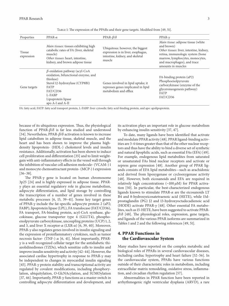
Table 1: The expression of the PPARs and their gene targets. Modified from [49, 51].
Properties PPAR-𝛼 PPAR-𝛽/𝛿 PPAR-𝛾
Tissueexpression
Main tissues: tissues exhibiting highcatabolic rates of FA (liver, skeletalmuscle)Other tissues: heart, intestine,kidney, and brown adipose tissue
Ubiquitous: however, the biggestexpression is in liver, esophagus,intestine, kidney, and skeletalmuscle
Main tissue: adipose tissue (whiteand brown)Other tissues: liver, intestine, kidney,retina, immunologic system (bonemarrow, lymphocytes, monocytes,and macrophages), and traceamounts in muscles
Gene targets
𝛽-oxidation pathway (acyl-CoAoxidation, bifunctional enzyme, andthiolase)Sterol 12-hydroxylase (CYP8B1)FATPFAT/CD36L-FABPLipoprotein lipaseapo A-I and A-II
Genes involved in lipid uptake; itrepresses genes implicated in lipidmetabolism and efflux
FA-binding protein (aP2)Phosphoenolpyruvatecarboxykinase (enzyme of theglyceroneogenesis pathway)FATPFAT/CD36
FA: fatty acid, FATP: fatty acid transport protein, L-FABP: liver cytosolic fatty acid-binding protein, and apo: apolipoprotein.
because of its ubiquitous expression. Thus, the physiologicalfunction of PPAR-𝛽/𝛿 is far less studied and understood[34]. Nevertheless, PPAR-𝛽/𝛿 activation is known to increaselipid catabolism in adipose tissue, skeletal muscle, and theheart and has been shown to improve the plasma high-density lipoprotein- (HDL-) cholesterol levels and insulinresistance. Additionally, activation has been shown to inducecell proliferation and differentiation [35] and to limit weight-gain with anti-inflammatory effects in the vessel wall throughthe inhibition of vascular cell adhesionmolecule- (VCAM-) 1andmonocyte chemoattractant protein- (MCP-) 1 expression[36–38].
The PPAR-𝛾 gene is located on human chromosome3p25 [24] and is highly expressed in adipose tissue. PPAR-𝛾 plays an essential regulatory role in glucose metabolism,adipocyte differentiation, and lipid storage by controllingthe transcription of a number of genes involved in thesemetabolic processes [6, 15, 39–41]. Some key target genesof PPAR-𝛾 include the fat-specific adipocyte protein 2 (aP2;FABP), lipoprotein lipase (LPL), FA translocase (FAT/CD36),FA transport, FA-binding protein, acyl-CoA synthase, glu-cokinase, glucose transporter type 4 (GLUT4), phospho-enolpyruvate carboxykinase, uncoupling proteins (UCP) 1, 2,and 3, and liver X receptor-𝛼 (LXR-𝛼) [6, 39, 40]. Moreover,PPAR-𝛾 also regulates genes involved in insulin signaling andthe expression of proinflammatory cytokines, such as tumornecrosis factor- (TNF-) 𝛼 [6, 41]. Most importantly, PPAR-𝛾 is a well-recognized cellular target for the antidiabetic thi-azolidinediones (TZDs), which sensitize cells to insulin andimprove insulin sensitivity and activity [42–44].However, theassociated cardiac hypertrophy in response to PPAR-𝛾 maybe independent to changes in myocardial insulin signaling[45]. PPAR-𝛾 protein stability and transcriptional activity areregulated by covalent modifications, including phosphory-lation, ubiquitylation, O-GlcNAcylation, and SUMOylation[37, 46]. Importantly, PPAR-𝛾 functions as a master switch incontrolling adipocyte differentiation and development, and
its activation plays an important role in glucose metabolismby enhancing insulin sensitivity [37, 47].
To date, many ligands have been identified that activateandmodulate PPARactivity [48]. PPAR ligand-binding activ-ities are 3-4 times greater than that of the other nuclear recep-tors and thus have the ability to bind a diverse set of syntheticand natural lipophilic acids, such as essential FAs (EFA) [49].For example, endogenous lipid metabolites from saturatedor unsaturated FAs bind nuclear receptors and activate orrepress gene expression [48]. Another group of PPAR lig-ands consists of EFA lipid metabolites—such as arachidonicacid derived from lipoxygenase or cyclooxygenase activity[48]. However, both eicosanoids and EFA are required inrelatively high concentrations (∼100 𝜇M) for PPAR activa-tion [50]. In particular, the best-characterized endogenousligands known to stimulate PPAR-𝛼 are the eicosanoids LTB4 and 8-hydroxyeicosatetraenoic acid (HETE), while 15d-prostaglandin (PG) J2 and 13-hydroxyoctadecadienoic acid(HODE) activate PPAR-𝛾 [48]. Other essential FA metabo-lites, such as 15-HETE, have been suggested to activate PPAR-𝛽/𝛿 [48]. The physiological roles, expression, gene targets,and ligands of the various PPAR isoforms are summarized inTables 1 and 2 and the following references [49, 51].
4. PPAR Functions inthe Cardiovascular System
Many studies have reported on the complex metabolic andbiological roles of PPARs in several cardiovascular diseases,including cardiac hypertrophy and heart failure [52–56]. Inthe cardiovascular system, PPARs have various functionsoutside of their characteristic roles in metabolism, includingextracellular matrix remodeling, oxidative stress, inflamma-tion, and circadian rhythm regulation [57].
Abnormalities in PPAR function have been reported inarrhythmogenic right ventricular dysplasia (ARVD), a rare

4 PPAR Research
Table 2: The natural and synthetic ligands of the PPARs and their physiological roles. Modified from [49, 51].
Properties PPAR-𝛼 PPAR-𝛽/𝛿 PPAR-𝛾
Natural ligands Unsaturated FA, PG, and LT B48-Hydroxyeicosatetraenoic acid
Unsaturated FACarbaprostacyclinComponents of VLDL
Unsaturated FA15-Hydroxyeicosatetraenoic acid9- and13-hydroxyoctadecadienoic acid15-Hydroxy delta 12,14-PG J2PG J2
Syntheticligands
Clofibrate and fenofibrateGemfibrozil GW501516
Rosiglitazone and pioglitazoneTroglitazone and ciglitazoneFarglitazar, S26948, and INT131
Physiologicalroles
Lipid catabolism and homeostasis(stimulating 𝛽-oxidation of fatty acids),increased breakdown of TG and FA,increased cellular FA uptake, reduced TGand FA synyheis, control of inflammatoryprocesses, and vascular integrity mediate thehypolipidemic function of fibratesLiver: increasing FA oxidation and uptakeand increasing apoA-I, apoA-II, and HDLVessel: increasing TG, HDL, ABCA1, andapoE and decreasing FFA, VLDL, cytokines,and NF-𝜅B
Dyslipidemia?Wound healing?Increasing fat oxidation inskeletal and cardiac muscleresponsible for insulin sensitivityand glucosehomeostasis and vascularintegrityAdipocentric action: decreasingcytokines, resistin, fFFA, andNF-𝜅B and increasing ABCA1and GLUT4Skeletal muscle: increasingglucose uptake and glycogensynthesis
Glucose homeostasis and lipidstorage:differentiation and maturation ofadipocytesIncreasing IS and glucosehomeostasis(it prevents hyperglycemia) andvascular integritySkeletal muscle/liver/adipocyte:increasing FA oxidation, UCP,and HDL and decreasing TG
FA: fatty acid; apo: apolipoprotein, PG: prostaglandin, LT: leukotriene, TG: triglyceride,HDL: high-density lipoprotein,ABCA1:ATP-binding cassette subfamilyA member 1, FFA: free fatty acid, VLDL: very low-density lipoprotein, NF-𝜅B: nuclear factor kappa-light-chain-enhancer of activated B cells, GLUT4: glucosetransporter type 4, and UCP: uncoupling protein.
genetic disease characterized by a progressive fibrofatty infil-tration, decreased PPAR-𝛼, and increased PPAR-𝛾 expressionin the right ventricle.The link betweenPPARdysfunction anddesmosomal geneticmutations is beginning to be understoodvia Wnt/𝛽-catenin pathway analyses [58–61]. PPAR-𝛾 is aprime inducer of adipogenesis in ARVD, and the Wnt-𝛽-catenin pathway appears to act though a similar mechanismfor desmosomal abnormalities [58].
The biological functions of PPAR-𝛼 in the myocardiumhave been extensively investigated using PPAR-𝛼 knockout(KO) mice [62–64]. Despite a normal life span, PPAR-𝛼KO mice exhibit progressive cardiac fibrosis with abnormalmitochondria and myofibrils [63]. Histological studies alsorevealed significant cardiomyocyte hypertrophy [65]. In addi-tion, ex vivo left ventricular papillarymuscle exhibits reducedshortening velocity and isometric tension, suggesting that theloss of PPAR-𝛼 is closely involved in the cardiac dysfunctioninduced by affecting the impairment of myosin moleculeitself, targeting for oxidative stress [65–68]. This is alsoapparent in echocardiography studies [65]. Interestingly, thedevelopment of physiological cardiac hypertrophy, such asis seen after birth and in response to exercise, showed theincreased PPAR-𝛼 expression that parallels an induction ofFA utilization [69, 70]. In contrast, PPAR-𝛼 gene expression isdownregulated in the heart of some pathological conditions,especially pressure overload-induced cardiac hypertrophy,that lead to the cardiac lipotoxicity as an accumulation oftriglyceride and diacylglycerol [70–73].
The redox system in PPAR-𝛼 KO mice is subjected todramatic and/or long-lasting perturbations as well as cardiacdysfunction that appear to result from the direct impairmentof myosin II [65]. There is strong evidence that PPAR-𝛼 acti-vation is necessary to prevent cellular oxidative damage thatmay occur during physiological cellular metabolism or underconditions of inflammation and oxidative stress, likely causedby repressing NF-𝜅B signaling and limiting inflammatorycytokine production [74, 75].Therefore, chronic deactivationof the PPAR-𝛼 signaling pathway may upset the normalequilibrium between oxidant production and antioxidantdefenses, which can contribute to cardiac damage [58]. Arecent study in PPAR-𝛾 KO mice revealed that PPAR-𝛾 playsa crucial protective role in cardiomyocytes and may preventmyocardial ischemia-reperfusion injury by modulating NF-𝜅B-associated inflammatory mechanisms in the infarctedmyocardium [76].
The heart responds to FA variations by activating PPARs[77]. PPAR-𝛼 can mediate diurnal variations in the respon-siveness of the heart to both FAs and specific PPAR-𝛼agonism (WY-14 643) [78]. In the normal heart, however,PPAR-𝛼 mRNA exhibits only weak circadian oscillations,although the circadian clock within the cardiomyocyte medi-ates diurnal variations in the responsiveness of the heart toincreased workload, according to contractile function andmetabolic flux levels [79, 80].
PPAR-𝛼 overexpression in the mouse myocardium atten-uates glucose transporter gene expression and glucose uptake

PPAR Research 5
[81]. Inmyocardium perfused with FA and ketone bodies, theglycolytic rate is decreased and additionally cardiomyocyte-specific PPAR-𝛼 overexpression leads to an augmentation oftriglyceride-derived FAs [82]. PPAR-𝛼 interferes in pyruvatedehydrogenase kinase (PDK), phosphofructokinase, pyru-vate dehydrogenase complex (PDC), and phosphofructok-inase (PFK) activities, and the cellular regulation of theseproteins is influenced by circadian rhythms [83, 84]. Recentlyit has been demonstrated that vascular PPAR-𝛾 is a peripheralregulator of cardiovascular rhythms that controls circadianvariations in blood pressure and heart rate through brain andmuscle Arnt-like protein- (BMAL-) 1 [85]. PPAR-𝛾 appearsto be a main component of the vascular clock. Pioglita-zone, a PPAR-𝛾 activator, readjusts the circadian rhythmof blood pressure from nondipper to dipper in patientswith type 2 diabetes [86]. Accordingly, the impairment ofcardiovascular rhythmicity parallels the diurnal variations inurinary excretion of epinephrine and norepinephrine, whichare suppressed in PPAR-𝛾 mutant mice [85], similar to thatobserved in BMAL1 KO mice [87].
5. PPAR Regulation by PosttranslationalModification in the Myocardium
Energy utilization in heart is transcriptionally controlled inpart by the PPAR family and their coreceptors/coactivators,including PPAR-𝛼, PPAR-𝛽/𝛿, PPAR-𝛾, RXR-𝛼, and PPAR-𝛾 coactivator- (PGC-) 1𝛼. Mechanistically, PPAR-𝛼, PPAR-𝛽/𝛿, and PPAR-𝛾 heterodimerize with the RXR-𝛼 and coac-tivators (e.g., PGC-1𝛼) and repressors (e.g., nuclear receptorcorepressor (NCoR)) to regulate the transcription of genesinvolved in energy regulation and lipid metabolism [4, 88–90]. Both PPAR and RXR-𝛼 interact with their respectiveligands to enhance PPAR-DNA binding [88, 91]. In theabsence of ligand binding, the unbound PPAR-RXR het-erodimer remains bound to NCoR and silencing mediator ofretinoid and thyroid hormone receptor (SMRT), two maincorepressors within the corepressor complex [92, 93]. BothNCoR and SMRT directly interact with the Sin3 complexto form a multisubunit repressor complex [92, 94]. SMRTfunctions as a protein platform to promote the mobilizationof histone deacetylases (HDACs) to the DNA promotersbound by specific interacting transcription factors [92, 94].Receptor interacting protein- (RIP-) 140, also known asnuclear receptor interacting protein- (NRIP-) 1, is anothercorepressor that directly recruits HDAC and represses theactivity of numerous nuclear receptors including PPARs bycompeting with their coactivators [95–97]. In the absence ofligand activation of nuclear receptor, the corepressor proteincomplex is known to suppress target gene transcription bycausing the deacetylation of histones [92, 93].
Alterations in the cardiac expression of all three PPARscause disturbances in glucose and FA metabolism that resultin an increased susceptibility to insults or significant dys-function [91]. While PPAR regulation is known to play arole in cardiovascular disease pathogenesis, the mechanismsregulating their expression and function at the cardiomy-ocyte level have not been clearly depicted. However, some
progress has been made. For example, PPARs may be con-trolled through posttranslational modifications (PTM), suchas SUMOylation and ubiquitination [88]. The conjugationof small ubiquitin-like modifier (SUMO) or ubiquitin isdistinctive among PTMs in that it induces the attachment ofanother polypeptide, rather than the addition of a functionalgroup [88, 98–101]. To date, SUMOylation- or ubiquitination-mediated PPAR regulation in the heart has not been reported;however, PPARs are controlled by these PTMs in other closelyrelated muscle cell types. Other studies have establishedthat SUMOylation of PPAR-𝛾1 promotes VMSC migrationand proliferation. This has been demonstrated by usingVSMCs transfected with a SUMOylation-defective lysine(K107R) PPAR-𝛾1 mutant, which results in a more potenttranscriptional inhibition of inducible nitric oxide synthasewhen compared to cells transfected with a wild-type con-struct [88, 102]. These findings regarding the role of PPAR-𝛾 SUMOylation in regulating the FA oxidation response andapoptosis in striated muscle and vascular smooth muscle,respectively, provide support for the concept that PPARscould be subjected to posttranslational regulation in theheart. Moreover, PPAR-𝛼 phosphorylation by theMAPK p38decreases PPAR-𝛼 transcriptional activity [88, 103]. Since thep38 pathway is activated in response to cardiac stress—asfound in diabetes, heart failure, and cardiac hypertrophy—this study implicates PPAR-𝛼 activationmechanism bywhichthe heart responds to unfavorable stimuli. The broaderimplications of these studies indicate that the FA and glucoseshifts seen in these diseases may be due to these regulatorymechanisms [88, 104].
6. Cardiac Pathophysiology in Genetic AnimalModels of PPARs and Their Regulators
6.1. PPAR-𝛼. The functions of cardiac PPAR-𝛼 have beenevaluated in PPAR-𝛼 KO mice. While viable and outwardlynormal, these mice exhibit mild aging-associated cardiacfibrosis [63]. The basal expression of several PPAR-𝛼 targetgenes and rates of FA oxidation are also diminished in heartsof PPAR-𝛼 KO mice [63, 105, 106] and fail to be induced inresponse to fasting or diabetes [105]. Moreover, PPAR-𝛼 KOmice exhibit increased glucose uptake, GLUT4 expression,and reliance on glucose for cardiac ATP production [64, 107].Notwithstanding the age-associated fibrosis, cardiac functionis relatively normal in young PPAR-𝛼 KOmice; however, theresponse to several physiological stressors is perturbed. Forexample, hearts isolated from PPAR-𝛼 KO mice are unableto compensate when challenged with an increased workload[64, 108]. Furthermore, transgenic animal models overex-pression of PPAR-𝛼 results in a cardiomyopathy that mimicsthat seen in diabetes mellitus (DM) [109] that is dependenton dietary fat. This implies that serum-free FA is an essentialmediator during cardiac maladaptation [110]. Paradoxically,although chronic exposure to excess FA represses PPAR-𝛼expression in cardiomyocytes, this downregulation of PPAR-𝛼 may result in further myocardial damage by suppressingcellular free FA oxidation on a background of excess free FAswithin cells and in the circulation [111]. The PPAR-𝛼 agonistBM 17.0744 (Roche Pharmaceuticals) normalized cardiac

6 PPAR Research
metabolismbutwas unable to improve cardiac functionwhengiven orally to type 2 DM db/db mice for 8 weeks [112].Apoptosis plays a role in the pathophysiology of diabeticcardiomyopathy and the PPAR-𝛼 ligand, fenofibrate, wasshown to suppress apoptosis. These findings support thepotential role of PPAR-𝛼 ligands in diabetic cardiomyopathy[109, 113].
Cardiovascular PPAR-𝛼 expressionhas anti-inflammatoryand antioxidative effects, and activation of inflammatorysignaling pathways is important in cardiomyocyte hyper-trophy [65, 114]. Accordingly, PPAR-𝛼 agonists have beenuseful in repressing the inflammation caused by cardiovas-cular disease. Pretreatment of neonatal cardiomyocytes withPPAR-𝛼 agonist significantly decreases lipopolysaccharide-(LPS-) stimulated TNF-𝛼 release, interleukin- (IL-) 1-inducedIL-6 secretion, and PG and cyclooxygenase-2 expression[115, 116]. The nuclear translocation of NF-𝜅B and apoptosiswere also demonstrated to be reduced after treatment withthe PPAR-𝛼 agonists in the reperfused myocardium. Thesefindings suggest an important role of PPAR-𝛼 agonists ininhibiting inflammation in many cell types in cardiovasculardisease [117, 118].Moreover, potent PPAR-𝛼 agonistWY14643has cardioprotective and cardiodepressive effects when usedto treat encephalomyocarditis virus-induced myocarditis indiabetic mice, which may be due to its anti-inflammatoryproperties and its ability to increase cardiac adiponectinexpression, whereas the reduced cardiac efficiency may bedue to its enhancement of cardiac UCP3 mRNA expression[6, 119].
6.2. PPAR-𝛽/𝛿. A decrease in cardiac expression of PPAR-𝛽/𝛿 was found in rats with diabetic cardiomyopathy [120,121], and reduction in PPAR-𝛽/𝛿 expression during hyper-glycemia is associated with increased reactive oxygen speciesproduction [121], TNF-𝛼, IL-6, and nicotinamide-adeninedinucleotide phosphate (NADPH) activity. Further studiesare needed to evaluate the precise role of PPAR-𝛽/𝛿 lig-ands in regulating diabetic cardiomyocytes [109, 120]. Theselective PPAR-𝛽/𝛿 ligand GW501516 was evaluated for itseffect on FA-induced inflammation in cardiomyocytes [122].GW501516 was also found to reduce expression of the NF-𝜅Btarget genes, MCP-1 and TNF-𝛼 in both human cardiac AC16cells stimulated by palmitate, as well as in the hearts of micefed with a high-fat diet. This data implies that PPAR-𝛽/𝛿maycounteract NF-𝜅B activity; thus, PPAR-𝛽/𝛿 activation mightbe therapeutically useful as an anti-inflammatory agent indiabetic cardiomyopathies [122].
6.3. PPAR-𝛾. In contrast to the induction of the other PPARfamily members, there are several studies that revealedthat PPAR-𝛾 expression is elevated in diabetic rat models[109, 120, 123, 124]. PPAR-𝛾 leads to elevations in lipogenicenzymes, which subsequently increase triglyceride produc-tion [123]. In addition, recent evidence from animal modelsshowed that cardiomyocyte PPAR-𝛾 activation is associatedwith compromised cardiac function through its lipogeniceffects, which may contribute to intracellular triglycerideaccumulation and cardiac lipotoxicity [125]. The PPAR-𝛾ligand rosiglitazone may also have a protective role against
apoptosis in diabetic cardiomyopathy, similar to the PPAR-𝛼 ligand [113]. Rosiglitazone has also been demonstratedto decrease cardiac fibrosis and improve left ventriculardiastolic dysfunction through the inhibition of receptors foradvanced glycated endproducts and connective tissue growthfactor in diabetic myocardium [126]. Moreover, pioglitazoneattenuated the deterioration of ischemic preconditioningagainst reperfusion arrhythmias in type 2 DM rats [127].Although PPAR-𝛾 levels are relatively low inmyocardial cells,activation during inflammation might have important effectson cardiomyocytes.
The therapeutic effects of PPAR-𝛾 ligands have beenattributed primarily to their anti-inflammatory properties.Previous studies showed that both natural and syntheticPPAR-𝛾 ligands have anti-inflammatory potentials [128].The pretreatment of neonatal cardiomyocytes with PPAR-𝛾agonists significantly decreased the LPS-stimulated TNF-𝛼release by cardiac myocytes [115]. Moreover, PPAR-𝛾 ligandssuppressed myocardial mRNA expressions of inflammatorycytokines and IL-1𝛽 in an autoimmune myocarditis model[129]. Interestingly, treatment with rosiglitazone or pioglita-zone decreased the expression of proinflammatory markersand reduced accumulation of neutrophils and macrophagesin reperfused myocardium [130, 131]. Nevertheless, highdoses of PPAR-𝛾 agonists were shown to induce cardiacdysfunction with marked changes in the utilization of freeFA and glucose.Thus, the pathophysiological mechanisms onthe cardiac effects of PPAR-𝛾 agonists causing an increasedincidence of myocardial dysfunction are yet to be elucidated[109, 132]. The model of constitutive, whole-body disruptionof PPAR-𝛾 results in embryonic lethality due to cardiacand placental defects [133], preventing the evaluation of thecardiac phenotype of these mice. However, cardiac-specificPPAR-𝛾 (csPPAR-𝛾) KO mice revealed that csPPAR-𝛾 defi-ciency only caused modest ventricular hypertrophy and didnot impair systolic function in the unstressed condition [134].Increased PPAR-𝛾 expressionwas found in the spontaneouslyhypertensive rat that may have resulted from increased lipiduptake or as a compensatory response to cardiac hypertrophyand failure, thereby compromising cardiac function [124,125].
7. Therapeutic Outcomes of PPAR Ligands inHeart Disease
7.1. PPAR-𝛼 Agonists. Synthetic PPAR-𝛼 ligands—such asclofibrate, fenofibrate, and bezafibrate—decrease triglycer-ide-rich lipoproteins through an increase in the gene expres-sion of FA-𝛽-oxidation and decrease in the expression ofapolipoprotein (Apo) C-III [135, 136].The above-noted drugsare extensively used in the treatment of hypertriglyceridemia.Such fibrates not only have a triglyceride-lowering effect,but also increase HDL-cholesterol levels resulting from theincrease in the expressions of ApoA-I and ApoA-II [135–137].
Human trials with PPAR-𝛼 agonists have largely, butnot uniformly, supported possible atherosclerotic benefits. Inthe Bezafibrate Coronary Atherosclerosis Intervention Trial

PPAR Research 7
(BECAIT), bezafibrate treatment decreased angiographic evi-dence of coronary atherosclerosis [138, 139]. In the HelsinkiHeart Study (HHT), gemfibrozil decreased cardiovascularevents, especially among patients with diabetes, but anincreased rate of noncoronary death was also noted [140].In the Bezafibrate Infarction Prevention (BIP) trial, onlythe subgroup with the highest triglyceride levels showed adecrease in adverse cardiovascular events with fibrate therapy[141]. In the Veteran’s Administration-HDL InterventionTrial (VA-HIT), gemfibrozil treatment showed a statisticallysignificant decrease in cardiovascular events in the cohortwith average LDL-cholesterol levels, history of cardiovasculardisease, and modestly decreased HDL-cholesterol/elevatedtriglycerides [142–144]. Of note, VA-HIT subjects were noton any 3-hydroxy-3-methylglutaryl coenzyme A reductaseinhibitors (statins); therefore, the outcomes of this trial mayhave been driven largely by the effect of gemfibrozil inpatients with insulin resistance and/or diabetes [143, 144].The Fenofibrate Intervention and Event Lowering inDiabetes(FIELD) study—a large, randomized, placebo controlledtrial—investigated the effects of fenofibrate on first or recur-rent cardiovascular events in patients with type 2 diabetesand found that the primary end point did not achieve astatistically significant difference between treatment groups.Several secondary end points were significantly reduced,including total cardiovascular events and nonfatal myocar-dial infarction. Somewhat surprising was the finding thatdecreases in small-vessel diseases; namely, nephropathy andretinopathy were also found. An increase in cardiovascu-lar mortality also was noted with fenofibrate but did notreach statistical significance [145]. Comparing the positiveoutcomes of VA-HIT with gemfibrozil, a less potent PPAR-𝛼agonist, to the negative results seen in FIELD achieved withfenofibrate, a more potent PPAR-𝛼 agonist might supportPPAR modulation, as opposed to more powerful activation,as being clinically effective. More potent PPAR bindingmay not necessarily correlate with greater clinical advantage,particularly because PPAR agonists have been definedmainlyin vitro [138, 146, 147].
Importantly, FIELDdoes not establish the impact of statinplus fibrate combination therapy on cardiovascular disease.Thus, the hypothesis that combination of a statin plus afibrate might offer greater cardiovascular risk reduction thana statin alone implies the requirement of another clinicalstudy such as the Action to Control Cardiometabolic Risk inDiabetes (ACCORD) trial. However, ACCORD-lipid arm inpatients with DM did not demonstrate any reduction in fatalcardiovascular incidences or nonfatal myocardial infarctionand stroke compared with simvastatin alone [148]. From thedisappointing cardiovascular outcomes in these studies, wemight expect VA-HIT and FIELD to specify advantages offibrates in patients who are statin intolerant or for possiblefibrate benefits tomicrovessel disease, which is amajor sourceof morbidity in diabetes [138]. Furthermore, prespecifiedsubgroup analysis of the ACCORD data suggested a possiblebenefit of fenofibrate in patients with high triglyceride andlow HDL-cholesterol baseline levels. Therefore, fibrates mayprove to be beneficial in treating atherogenic dyslipidemia indiabetes patients [51, 148].
7.2. PPAR-𝛾 Agonists. PPAR-𝛾 is a regulator of glucose andlipid metabolism; therefore, its synthetic PPAR-𝛾 ligands—such as glitazones and TZD derivatives (such as trogli-tazone, rosiglitazone, and pioglitazone)—improve glucoseand insulin parameters and increase whole body insulinsensitivity. Therefore, they are called insulin-sensitizers andare used in the treatment of diabetes [149]. In early humantrials, PPAR-𝛾 agonists showed decreased in-stent restenosisafter coronary stent implantation [150, 151]. Furthermore,in the Carotid Intima-Media Thickness in AtherosclerosisUsing Pioglitazone (CHICAGO) study, significant effectsof pioglitazone on the slow progression of carotid intima-media thickness were reported in patients nearly matched forglycemic control with glimepiride [152].
Rosiglitazone and pioglitazone are used in the treatmentof patients with type 2 diabetes; however, the effects ofthese TZDs on cardiovascular outcomes in patients withDM are different. The Prospective Pioglitazone Clinical Trialin Macrovascular Events (PROactive) trial investigated theeffects of pioglitazone combined with standard contem-porary antidiabetic treatment versus active, but non-TZD,antidiabetic treatment on a combined vascular end pointin individuals with known macrovascular disease [153]. Thepurpose of the PROactive study was to achieve similar,matched hemoglobin A1c (HbA1c) levels in the TZD andnon-TZD groups in order to provide more definitive insightsinto glucose-independent vascular effects of TZDs. In spite ofthe extensive in vivo and in vitro data supporting TZD effectson atherosclerosis, no statistically significant difference wasnoted in the primary end point between study groups. Incontrast, the main secondary end point was revealed with astatistically significant 16% decrease in clinical events [153,154]. Contrary to pioglitazone, rosiglitazone was associatedwith significant increases in death from cardiovascular causesand myocardial infarction after a relatively short-term ofexposure [155]. Thus, the European Medicines Agency with-drew approval of rosiglitazone in 2010 due to these cardio-vascular safety concerns [156]. Importantly, these divergentoutcomes may result from their diverse effects on lipid sub-fractions [157]. Pioglitazone increases HDL-cholesterol anddecreases fasting plasma free FAs and triglycerides withoutany influence on total cholesterol and LDL-cholesterol; how-ever, rosiglitazone significantly augments HDL-cholesterollevels, as well as total cholesterol and the LDL-cholesterolfraction levels [156, 158, 159].
In the Diabetes Reduction Approaches with Ramipriland Rosiglitazone Medications (DREAM) study, the effectsof the angiotensin-converting enzyme inhibitor ramipril androsiglitazone on the prevention of diabetes were studiedusing a two-by-two placebo-controlled design [160]. Inter-estingly, rosiglitazone significantly reduced the progressionto diabetes in a cohort with impaired fasting glucose and/orimpaired glucose tolerance, whereas ramipril had no effecton this measure [161]. The Actos Now for the Prevention ofDiabetes (ACT NOW) trial analyzed with a similar questionto DREAM in patients with impaired glucose tolerancerandomized to receive either pioglitazone (45mg) or placebo.After a mean follow-up of 2.2 years, progression to diabetesoccurred in 5% of the pioglitazone group, compared with

8 PPAR Research
16.7% of the placebo group, but too few cardiovascular eventsoccurred (pioglitazone 26, placebo 23) to draw any inferencesregarding effect of treatment on cardiovascular outcomes[162, 163]. This decrease in diabetes progression with piogli-tazone was consistent with previous studies, including thetroglitazone arm of the Diabetes Prevention Program andwomen with a history of gestational diabetes [164, 165].Additionally, the pioglitazone arm of the Pioneer studyrevealed significantly greater improvements in inflamma-tory markers—including high-sensitivity CRP, MMP-9, andMCP-1—than the glimepiride-treated group despite equiva-lent reductions in fasting glucose and HbA1c levels. In anadditional subgroup analysis, patients with no significantglucose responses to pioglitazone still had improved surro-gate markers for atherosclerosis. Despite limitation by thesmall numbers of patients in these subgroups, such findingscontinue to raise possible disassociations between TZD-mediated effects on the vasculature and inflammation versusits glycemic advantages [138, 166].
In the Cardiovascular Outcomes in Oral Agent Combi-nation Therapy for Type 2 Diabetes (RECORD) trial, 4,447subjects with type 2 DM poorly controlled on monotherapywith metformin or sulfonylurea, a noninferiority hypothesiswas explored for rosiglitazone as second-line therapy in type2 diabetes [163, 167]. The primary end point of RECORDwas time to cardiovascular hospitalization or cardiovasculardeath. After a mean follow-up of 5.5 years, primary endpointevents occurred in 321 patients in the rosiglitazone groupand 323 patients in the metformin/sulfonylurea group, thusmeeting the requirement for noninferiority of rosiglitazone.Fatal or nonfatal HF occurred more frequently in the rosigli-tazone group than in the active control group (61 versus 29patients). Limitations of RECORD include an event rate thatwas substantially lower than that projected in trial designwith consequent reduction of statistical power, and potentialcomplications resulting from the differential use of statinsand diuretics, and an open-labeled study design [163, 167].
Despite many beneficial features of glitazones, they alsoexhibit adverse effects, such as edema, heart failure, weightgain, bone fractures, and increased risk of myocardial infarc-tions, which have limited the use of TZDs in diabetic patientswith high lipid levels [168]. In the PROactive study, anincreased incidence of congestive HF was reported in thepioglitazone group, although these events were not welljudged. Previous work has clearly demonstrated that TZDscan cause fluid retention, as evident from themodest decreasein hematocrit and volume expansion documented with TZDexposure [169]. The incidence of pedal edema observedwith TZD monotherapy is about 3% to 5% compared with1.2% in placebo arms [170]. The incidence of pedal edemawith TZDs approaches 7.5% when combined with eithermetformin or sulfonylurea, compared with 2.5% and 2.1%with sulfonylurea or metformin alone, respectively [171]. Therisk of pedal edema appears similar with both rosiglitazoneand pioglitazone in clinical use [172]. Concomitant insulinand TZD use has been associated with a 2- to 3-fold higherrate of edema compared to insulin alone, with rates increasingfrom 5% to 7% with insulin alone to 13% to 15% withTZD and insulin [171]. Recent data suggest that upregulation
of a specific sodium channel—sodium channel, nonvoltagegated 1 gamma subunit (SCNN1G)—in the distal nephronis a PPAR-𝛾-mediated mechanism for TZD-induced edema[173, 174]. Other mechanisms involved for TZD-mediatededema include altered interstitial ion transport, increasedsympathetic nervous system activity, and altered endothelialpermeability [175–177]. This edema is reversible and shouldnot necessarily be equated with myocardial toxicity althoughsome patients with DM, even absent class III or IV HF, maynot tolerate this volume expansion [138].
Another clinically significant side effect of TZDs is bodyweight gain. This change, which likely involves both fluidretention and increases in adiposity, is typically in the rangeof 2 to 5 kg [178]. Some of theweight induced by TZDsmay beadvantageous, involving a shift from visceral to subcutaneousareas, and also track the increase in adiponectin, anti-inflammatory protein, induced by TZDs [179]. The changein fat distribution with TZDs includes a change in energybalance and possible effects on other factors and pathwaysinfluencing body weight, because a simple rearrangement infat location would not explain an overall net increase in bodymass [138, 180]. Nevertheless, the weight increase seen withPPAR-𝛾 activation has clearly contributed to the hesitation ofTZDs usage as antidiabetic drug, which may be more seriouswhen combined with insulin [181].
7.3. PPAR-𝛼/𝛾 Dual Agonists. A new class of dual PPAR-𝛼/𝛾 agonists has been shown to have a positive influence onboth glucose and lipid metabolism and are currently underdevelopment as a response to the treatment challenge of coex-isting type 2 diabetes with dyslipidemia. These dual agonistsnot only reduce arteriosclerosis development, but also havean antidiabetic capacity. They also exhibit improvement ofendothelial function, anti-inflammatory, and anticoagulantaction, decrease plasma free FAs, and lower blood pressure,indicative of advantageous effects on the vasculature [49].
Until now, several attempts to develop a dual agonistfor diabetes have failed due to various safety concerns:ragaglitazar, MK-0767, and naveglitazar were all found tobe associated with an increased incidence of bladder cancerand hyperplasia in rodent studies [51, 182], and tesaglitazardevelopment was discontinued due to indications that it maycause renal dysfunction [183]. The most-studied dual agonistmuraglitazar was found to be effective in reducingHbA1c andtriglyceride levels while increasing HDL-cholesterol levels[51, 184–188]. One randomized, double-blind trial of 1,477drug-naive patients with type 2 diabetes found a −0.25% to−1.76% (3–17mmol/mol) reduction in HbA1c from baselineafter 24 weeks of muraglitazar treatment, compared witha reduction of −0.57% (5mmol/mol) with pioglitazone [51,186]. At 12 weeks, triglycerides had decreased by −4 to−41% with muraglitazar and 9% with pioglitazone and HDL-cholesterol had increased by 6–23% with muraglitazar and10% with pioglitazone. Nevertheless, Bristol-Myers Squibbdiscontinued further development of this dual agonist in2006 after Nissen and colleagues published an analysis ofthe available material from the clinical trial program, whichrevealed that muraglitazar was associated with an increasedincidence of the composite end point of death, major adverse

PPAR Research 9
cardiovascular events, congestive HF (relative risk: 2.62; 𝑃 =0.04), and excessive morbidity for all individual componentsof the composite endpoint when compared to placebo orpioglitazone [51, 188].
Aleglitazar (Hoffmann-La Roche) is the most recent dualPPAR-𝛼/𝛾 agonist that has completed in phase III trialsand has a balanced affinity for both PPAR-𝛼 and PPAR-𝛾receptor subtypes. Preclinical and clinical trial results havebeen promising [51, 189–192]. Phase II study SYNCHRONYhas shown a significant dose-dependent reduction in HbA1cof −0.36% (4mmol/mol, 50 𝜇g; 𝑃 = 0.048) to −1.35%(15mmol/mol, 600 𝜇g; 𝑃 < 0.0001) after 16 weeks of treat-ment with aleglitazar once daily when compared with pla-cebo. Importantly, statistically significant beneficial effects onlipid subfractions were also found. Significant decreases intriglyceride (𝑃 < 0.001 for percentage changes) and increasesin HDL-cholesterol (𝑃 < 0.05 for percentage changes) werefound with all doses of aleglitazar (−43 and +21%, resp.,with the 150 𝜇g dose). In addition, significant reductions inLDL-cholesterol were found at doses of 150 𝜇g or higher,compared with placebo (𝑃 < 0.05 for percentage changes):placebo-adjusted reduction in LDL-cholesterol with the 150-𝜇g dose of aleglitazar was −15.5%. Indeed, aleglitazar, at the150-𝜇g dose, was associated with a greater effect on triglyc-erides, HDL-cholesterol, and LDL-cholesterol than pioglita-zone 45mg. Further analysis of this study data suggests thataleglitazar produces a shift from the atherogenic small denseLDL particles associated with type 2 diabetes to larger LDLparticles [51, 193]. Phase III study ALECARDIO, randomizeddouble-blind placebo-controlled clinical trial, had evaluatedthe hypothesis that aleglitazar (150 𝜇g daily dose) can reducecardiovascular mortality and morbidity in patients with type2 DM who have suffered from a recent acute coronarysyndrome (ACS) event. However, use of aleglitazar in patientswith type 2 diabetes and recent ACS did not significantlyreduce the incidence of cardiovascular death, myocardialinfarction, or stroke. Unfortunately, aleglitazar increased therisks ofHF, renal dysfunction, bone fractures, gastrointestinalhemorrhage, and hypoglycemia [194].
There are several potential explanations for why alegli-tazar did not reduce cardiovascular mortality and morbid-ity in ALECARDIO trial. First, the magnitude of changesin HDL-cholesterol and triglyceride levels achieved withaleglitazar may not be sufficient to impart additional car-diovascular benefits when administered concurrently withstatins. Second, some therapies may be unable to exert acardioprotective effect in patients with extensive atheroscle-rosis and long-standing diabetes or may require a very longduration of exposure to achieve such effects. Third, favorablelipid and metabolic effects of aleglitazar may have beennegated by adverse effects of the drug, including heart failure,reduced renal function, hypoglycemia, and increased LDL-cholesterol, resulting in no net cardiovascular benefit. Thesefindings do not support the use of aleglitazar in this settingwith a goal of reducing cardiovascular risk [51, 194].
8. New Modalities and Future Directions ofPPAR-Directed Therapeutics
The impact of fibrates andTZDs ondyslipidemia anddiabetesis linked primarily to PPAR-𝛼 and PPAR-𝛾 activation, respec-tively [195, 196]. However, substantial clinical and preclinicalexperience has shown that individual drugs differ from oneanother in therapeutic and side effect properties [42, 197].Furthermore, PPAR expression in multiple tissues raises thepossible value of targeting PPAR agents in therapeutic indi-cations of a number of other diseases (e.g., cancer and colitis)[122, 198–201]. Althoughmany clinical studies of PPARs havedemonstrated inconsistent results for cardioprotective effects[139–141, 145, 153, 167, 193, 194], the evidence reviewed abovesuggests that this is still a lucrative area of study. Therefore,the needs of new PPAR-directed therapeutic modalities mustinclude pan-PPAR agonists, selective PPARmodulators, dualPPAR agonists, PPAR-𝛾 antagonists, and nutraceuticals, all ofwhich are being considered as possible approaches to reducethe adverse events seen with current TZDs [138, 181, 202].
8.1. Pan-PRAR Agonists. The significant structural similarityof PPAR-𝛼, PPAR-𝛽/𝛿, and PPAR-𝛾—particularly withintheir ligand-binding domains—has allowed the identificationof several synthetic dual- or pan-PPAR agonists [203]. Activemetabolites of fibrates, such as fenofibric acid and clofibricacid, are dual activators of PPAR-𝛼 and PPAR-𝛾, with about a10-fold selectivity for PPAR-𝛼. Another compound from thisgroup, bezafibrate, is a broader activator because it activatesall three PPAR subtypes at comparable doses to other fibrates.Therefore, bezafibrate is regarded as a pan-agonist with thepotential to directly improve insulin sensitization via PPAR-𝛾 activation [10, 15].
8.2. Selective Modulators and Partial Agonists. The intensivesearch for safer PPAR agonists led to the development ofselective partial PPAR modulators. Currently, new selectivePPAR-𝛾 modulators are in development—including S26948[204] and INT131 [205], which should stimulate glucosemetabolism and minimize the adverse effects of full PPAR-𝛾agonists [49]. INT131 recruits vitaminD3 receptor interactingprotein- (DRIP-) 205 and promotes its binding to a levelof approximately 30% of that conferred by the full PPAR-𝛾 agonist rosiglitazone [206]. In animal models of diabetes,INT131 caused less weight gain compared to pioglitazone orrosiglitazone while retaining efficacy to reduce plasma glu-cose [206, 207]. Importantly, toxicity of INT131 in cynomol-gus monkeys and rats was not associated with fluid retention,changes in hematocrit, or weight gain over 6 months [207,208]. In a phase II study, however, INT131 was associatedwith an increase in the incidence of edema, weight gain,and decreased hematocrit at the 10mg dose versus placebo,highlighting the difficulty in translating promising preclinicalprofiles into patients [209]. While the cardiac adverse effectprofile of rosiglitazone-like PPAR-𝛾 full agonists is unfor-tunate, the therapeutic potential of novel pharmacologicalagents targeting PPAR-𝛾 submaximal cannot be excluded.Interestingly, newly synthesized partial PPAR-𝛾 agonists,
10 PPAR Research
such as balaglitazone, MBX-102, MK-0533, PAR-1622, PAM-1616, KR-62776, and SPPAR-𝛾M5, have a reduced tendency tocause the adverse effects associated with full PPAR-𝛾 agonistsor may be entirely devoid of such effects [6, 47].
8.3. Phosphorylation and Posttranslational Control. As notedabove several compelling new mechanisms of posttransla-tional control of PPAR action have recently been described,including phosphorylation, SUMOylation, ubiquitination,and nitration [210]. In addition to enhancing the tran-scriptional activity of PPAR-𝛾, rosiglitazone was found toinhibit the PPAR-𝛾 phosphorylation at Ser273 by cyclin-dependent kinase 5 (CDK5) in adipose tissue, preserving thetranscription of insulin-response genes and correlating withantidiabetic activity. A second PPAR-𝛾 agent, MRL24, wasas effective as rosiglitazone at blocking phosphorylation andimproving diabetes in animal models, despite being only apartial PPAR-𝛾 agonist. Taken together, these results suggestthat the insulin-sensitizing benefits of PPAR-𝛾 agonists aredue in part to their ability to block phosphorylation and notsolely to their agonist activity [211].
8.4. Nongenomic Regulation. Recent evidence also suggeststhe potential role of nongenomic regulation of PPAR-𝛾and PPAR-𝛼, mediated by interaction with cytosolic secondmessengers, including kinases and phosphatases [210]. TheMAP/ERK kinase, MAPK kinase- (MEK-) 1, was reportedto bind directly to the AF-2 domain of PPAR-𝛾 in responseto mitogenic stimulation, leading to the sequestration ofPPAR-𝛾 in the cytoplasm [212]. Selective inhibition of MEK-1/PPAR-𝛾 interactions has recently been proposed as a con-cept for treatment of cancer, inflammation, and metabolicdisorders but has yet to gain significant acceptance [212].
8.5. New Dual PPAR-𝛼/𝛾 Agonists. Saroglitazar, a PPARagonist with predominant PPAR-𝛼 and moderate PPAR-𝛾activity, was launched exclusively in India for the control ofdyslipidemia [213, 214]. However, limited data is available onits molecular profile, and the treatment duration and lowpatient number in its phase III programmake it impossible todraw conclusions regarding its cardiovascular and long-termsafety profiles [203].
8.6. Nutraceuticals and Life-Style Modification. As endoge-nous nuclear receptor ligands, dietary n-3 and n-6 polyun-saturated FAs (PUFAs) and their derivatives can upregulatePPAR-𝛾 expression in vitro and in vivo and reduce aninflammatory response [215]. Furthermore, it has been shownthat any type of regular exercise and crataegus species wouldimprove cardiovascular function and minimizes several riskfactors via stimulating lipidmetabolismby acting on enzymesand genes expression such as ATP-binding cassette trans-porter A1 (ABCA1) and PPAR-𝛼 which are involved in thisprocess [216]. However, though dietary PUFAs similar tosynthetic ligands were able to bind to the ligand-bindingdomain and cause conformational changes to activate thereceptor, they are considered as weak PPAR-𝛾 ligands becauseof their low physiological concentrations. Another cautionof nutraceuticals is that some of the flavonoids have been
associated with tumor and altering pharmacodynamics andpharmacokinetics of various drugs via interacting withcytochrome P450 enzymes [202].
9. Conclusions
PPARs are critical gene regulators in cardiomyocytes, yettheir functions are not fully established. PPAR agonists con-vey beneficial effects as therapeutic agents for diabetes andatherosclerosis by lowering blood glucose, improving insulinresistance, inflammation, and lipid metabolism; however,adverse side effects limit their clinical use. As such, thefuture of PPAR-directed agents in cardiometabolic therapyremains uncertain, although several late-stagemoleculesmaystill hold promise [203]. Future directions in PPAR agonistdevelopment are likely to focus on optimizing the PPARsubtype interaction profile, maximizing the inhibition ofPPAR-𝛾 phosphorylation, and screening against off-targetactivity. At the present time, clinicians should keep in mindthe risk/benefit ratio of PPAR activators. Intensive researchon this therapeutic target will likely lead to the developmentof safer and more effective PPAR agonists in the near future.
Abbreviations
ACS: Acute coronary syndromeAF: Activation functionApo: ApolipoproteinBMAL-1: Brain and muscle Arnt-like protein-1CDK5: Cyclin-dependent kinase 5csPPAR-𝛾: Cardiac-specific PPAR-𝛾DM: Diabetes mellitusDRIP: Vitamin D3 receptor interacting proteinEFA: Essential FAsFA: Fatty acidGLUT4: Glucose transporter type 4HDAC: Histone deacetylaseHDL: High-density lipoproteinHETE: Hydroxyeicosatetraenoic acidHF: Heart failureHODE: Hydroxyoctadecadienoic acidIL: InterleukinKO: KnockoutLPS: LipopolysaccharideLT: LeukotrieneLXR-𝛼: Liver X receptor-𝛼MCP: Monocyte chemoattractant proteinMEK: MAPK kinasemRNA: Messenger RNANCoR: Nuclear receptor corepressorNRIP: Nuclear receptor interacting proteinPG: ProstaglandinPGC: PPAR-𝛾 coactivatorPPAR: Peroxisome proliferator-activated
receptorPPRE: Peroxisome proliferator response
elementPTM: Posttranslational modificationPUFA: Polyunsaturated fatty acid

PPAR Research 11
RIP: Receptor interacting proteinRXR: Retinoid X receptorSMRT: Silencing mediator of retinoid and
thyroid hormone receptorSUMO: Small ubiquitin-like modifierTNF: Tumor necrosis factorTZD: ThiazolidinedioneVLDL: Very low-density lipoproteinVSMC: Vascular smooth muscle cell.
Conflict of Interests
The authors declare no conflict of interests.
References
[1] A. Oyekan, “PPARs and their effects on the cardiovascularsystem,” Clinical and Experimental Hypertension, vol. 33, no. 5,pp. 287–293, 2011.
[2] R. Chen, F. Liang, J. Moriya et al., “Peroxisome proliferator-activated receptors (PPARs) and their agonists for hypertensionand heart failure: are the reagents beneficial or harmful?”International Journal of Cardiology, vol. 130, no. 2, pp. 131–139,2008.
[3] B. Desvergne and W. Wahli, “Peroxisome proliferator activatedreceptors: nuclear control of metabolism,” Endocrine Reviews,vol. 20, no. 5, pp. 649–688, 1999.
[4] L. Michalik, J. Auwerx, J. P. Berger et al., “International unionof pharmacology. LXI. Peroxisome proliferator-activated recep-tors,” Pharmacological Reviews, vol. 58, no. 4, pp. 726–741, 2006.
[5] A. L. Bookout, Y. Jeong, M. Downes, R. T. Yu, R. M. Evans, andD. J. Mangelsdorf, “Anatomical profiling of nuclear receptorexpression reveals a hierarchical transcriptional network,” Cell,vol. 126, no. 4, pp. 789–799, 2006.
[6] D. Usuda and T. Kanda, “Peroxisome proliferator-activatedreceptors for hypertension,”World Journal of Cardiology, vol. 6,no. 8, pp. 744–754, 2014.
[7] H. A. Elrod and S.-Y. Sun, “PPAR𝛾 and apoptosis in cancer,”PPAR Research, vol. 2008, Article ID 704165, 12 pages, 2008.
[8] K. Tachibana, D. Yamasaki, K. Ishimoto, and T. Doi, “The role ofPPARs in cancer,” PPAR Research, vol. 2008, Article ID 102737,15 pages, 2008.
[9] J. M. Peters, Y. M. Shah, and F. J. Gonzalez, “The role of per-oxisome proliferator-activated receptors in carcinogenesis andchemoprevention,”Nature ReviewsCancer, vol. 12, no. 3, pp. 181–195, 2012.
[10] J. Berger and D. E. Moller, “The mechanisms of action ofPPARs,” Annual Review of Medicine, vol. 53, pp. 409–435, 2002.
[11] E. Boitier, J.-C. Gautier, and R. Roberts, “Advances in under-standing the regulation of apoptosis andmitosis by peroxisome-proliferator activated receptors in pre-clinicalmodels: relevancefor human health and disease,” Comparative Hepatology, vol. 2,no. 1, pp. 3–18, 2003.
[12] L. Guo and R. Tabrizchi, “Peroxisome proliferator-activatedreceptor gamma as a drug target in the pathogenesis of insulinresistance,” Pharmacology and Therapeutics, vol. 111, no. 1, pp.145–173, 2006.
[13] A. Rogue, C. Lambert, R. Josse et al., “Comparative geneexpression profiles induced by PPAR𝛾 and PPAR𝛼/𝛾 agonists inhuman hepatocytes,” PLoS ONE, vol. 6, no. 4, Article ID e18816,15 pages, 2011.
[14] A. Rogue, C. Spire,M. Brun,N.Claude, andA.Guillouzo, “Geneexpression changes induced by PPARgamma agonists in animaland human liver,” PPAR Research, vol. 2010, Article ID 325183,16 pages, 2010.
[15] T. M. Willson, P. J. Brown, D. D. Sternbach, and B. R. Henke,“The PPARs: from orphan receptors to drug discovery,” Journalof Medicinal Chemistry, vol. 43, no. 4, pp. 527–550, 2000.
[16] I. Gurevich, A. M. Flores, and B. J. Aneskievich, “Corepressorsof agonist-bound nuclear receptors,” Toxicology and AppliedPharmacology, vol. 223, no. 3, pp. 288–298, 2007.
[17] S. Yu and J. K. Reddy, “Transcription coactivators for peroxi-some proliferator-activated receptors,” Biochimica et BiophysicaActa—Molecular and Cell Biology of Lipids, vol. 1771, no. 8, pp.936–951, 2007.
[18] J. D. Chan and R.M. Evans, “A transcriptional co-repressor thatinteracts with nuclear hormone receptors,” Nature, vol. 377, no.6548, pp. 454–457, 1995.
[19] R. D. Kornberg, “The molecular basis of eukaryotic transcrip-tion,” Proceedings of the National Academy of Sciences of theUnited States of America, vol. 104, no. 32, pp. 12955–12961, 2007.
[20] M. J. Tetel, A. P. Auger, and T. D. Charlier, “Who’s in charge?Nuclear receptor coactivator and corepressor function in brainand behavior,” Frontiers in Neuroendocrinology, vol. 30, no. 3,pp. 328–342, 2009.
[21] B.W. O’Malley and R. Kumar, “Nuclear receptor coregulators incancer biology,” Cancer Research, vol. 69, no. 21, pp. 8217–8222,2009.
[22] F. J. Gonzalez and Y. M. Shah, “PPARalpha: mechanism ofspecies differences and hepatocarcinogenesis of peroxisomeproliferators,” Toxicology, vol. 246, no. 1, pp. 2–8, 2008.
[23] S. R. Pyper, N. Viswakarma, S. Yu, and J. K. Reddy, “PPARalpha:energy combustion, hypolipidemia, inflammation and cancer,”Nuclear Receptor Signaling, vol. 8, article e002, 2010.
[24] C. Dong, H. Zhou, C. Shen et al., “Role of peroxisome prolif-erator-activated receptors gene polymorphisms in type 2 dia-betes and metabolic syndrome,”World Journal of Diabetes, vol.6, no. 4, pp. 654–661, 2015.
[25] S. Azhar andG. Kelley, “PPAR𝛼: its role in the humanmetabolicsyndrome,” Future Lipidology, vol. 2, no. 1, pp. 31–53, 2007.
[26] P. Lefebvre, G. Chinetti, J.-C. Fruchart, and B. Staels, “Sortingout the roles of PPARalpha in energy metabolism and vascularhomeostasis,” The Journal of Clinical Investigation, vol. 116, no.3, pp. 571–580, 2006.
[27] J. N. Feige, L. Gelman, L. Michalik, B. Desvergne, andW.Wahli,“From molecular action to physiological outputs: peroxisomeproliferator-activated receptors are nuclear receptors at thecrossroads of key cellular functions,” Progress in Lipid Research,vol. 45, no. 2, pp. 120–159, 2006.
[28] M. Hamblin, L. Chang, Y. Fan, J. Zhang, and Y. E. Chen, “PPaRsand the cardiovascular system,” Antioxidants and Redox Signal-ing, vol. 11, no. 6, pp. 1415–1452, 2009.
[29] J. E.Montanez, J.M. Peters, J. B. Correll, F. J. Gonzalez, andA.D.Patterson, “Metabolomics: an essential tool to understand thefunction of peroxisome proliferator-activated receptor alpha,”Toxicologic Pathology, vol. 41, no. 2, pp. 410–418, 2013.
[30] E. Robinson and D. J. Grieve, “Significance of peroxisomeproliferator-activated receptors in the cardiovascular system inhealth anddisease,”Pharmacology andTherapeutics, vol. 122, no.3, pp. 246–263, 2009.
[31] E. Esposito, B. Rinaldi, E. Mazzon et al., “Anti-inflammatoryeffect of simvastatin in an experimental model of spinal cord

12 PPAR Research
trauma: involvement of PPAR-𝛼,” Journal of Neuroinflamma-tion, vol. 9, article 81, 2012.
[32] G. D. Barish, V. A. Narkar, and R. M. Evans, “PPAR delta: adagger in the heart of the metabolic syndrome,” The Journal ofClinical Investigation, vol. 116, no. 3, pp. 590–597, 2006.
[33] K. S. Kilgore and A. N. Billin, “PPAR𝛽/𝛿 ligands as modulatorsof the inflammatory response,” Current Opinion in Investiga-tional Drugs, vol. 9, no. 5, pp. 463–469, 2008.
[34] S. Azhar, “Peroxisome proliferator-activated receptors, meta-bolic syndrome and cardiovascular disease,” Future Cardiology,vol. 6, no. 5, pp. 657–691, 2010.
[35] G. M. Giordano Attianese and B. Desvergne, “Integrative andsystemic approaches for evaluating PPAR𝛽/𝛿 (PPARD) func-tion,” Nuclear Receptor Signaling, vol. 13, article e001, 32 pages,2015.
[36] M. J. Zarzuelo, R. Jimenez, P. Galindo et al., “Antihypertensiveeffects of peroxisome proliferator-activated receptor-𝛽 activa-tion in spontaneously hypertensive rats,” Hypertension, vol. 58,no. 4, pp. 733–743, 2011.
[37] T. P. Burris, S. A. Busby, and P. R. Griffin, “Targeting orphannuclear receptors for treatment of metabolic diseases and auto-immunity,” Chemistry and Biology, vol. 19, no. 1, pp. 51–59, 2012.
[38] T. Coll, R. Rodrıguez-Calvo, E. Barroso et al., “Peroxisome pro-liferator activated receptor (PPAR) beta/delta: a new potentialtherapeutic target for the treatment of metabolic syndrome,”Current Molecular Pharmacology, vol. 2, no. 1, pp. 46–55, 2009.
[39] R. K. Semple, V. K. K. Chatterjee, and S. O’Rahilly, “PPAR𝛾 andhuman metabolic disease,”The Journal of Clinical Investigation,vol. 116, no. 3, pp. 581–589, 2006.
[40] P. Tontonoz and B.M. Spiegelman, “Fat and beyond: the diversebiology of PPAR𝛾,” Annual Review of Biochemistry, vol. 77, pp.289–312, 2008.
[41] S. J. Bensinger and P. Tontonoz, “Integration of metabolism andinflammation by lipid-activated nuclear receptors,” Nature, vol.454, no. 7203, pp. 470–477, 2008.
[42] H. Yki-Jarvinen, “Thiazolidinediones,” The New England Jour-nal of Medicine, vol. 351, no. 11, pp. 1106–1118, 2004.
[43] A. H. Barnett, “Redefining the role of thiazolidinediones inthe management of type 2 diabetes,” Vascular Health and RiskManagement, vol. 5, pp. 141–151, 2009.
[44] K.A.M. Jandeleit-Dahm,A.Calkin, C. Tikellis, andM.Thomas,“Direct antiatherosclerotic effects of PPAR agonists,” CurrentOpinion in Lipidology, vol. 20, no. 1, pp. 24–29, 2009.
[45] S. Sena, I. R. Rasmussen, A. R. Wende et al., “Cardiac hyper-trophy caused by peroxisome proliferator—activated receptor-gamma agonist treatment occurs independently of changes inmyocardial insulin signaling,”Endocrinology, vol. 148, no. 12, pp.6047–6053, 2007.
[46] Z. E. Floyd and J. M. Stephens, “Controlling a master switchof adipocyte development and insulin sensitivity: covalentmodifications of PPAR𝛾,” Biochimica et Biophysica Acta, vol.1822, no. 7, pp. 1090–1095, 2012.
[47] S. Nagao and T. Yamaguchi, “PPAR-𝛾 agonists in polycystickidney disease with frequent development of cardiovasculardisorders,” Current Molecular Pharmacology, vol. 5, no. 2, pp.292–300, 2012.
[48] J.-M. Choi and A. L. M. Bothwell, “The nuclear receptor PPARsas important regulators of T-cell functions and autoimmunediseases,”Molecules and Cells, vol. 33, no. 3, pp. 217–222, 2012.
[49] B. Grygiel-Gorniak, “Peroxisome proliferator-activated recep-tors and their ligands: nutritional and clinical implications—areview,” Nutrition Journal, vol. 13, article 17, 2014.
[50] J. Plutzky, “Peroxisome proliferator-activated receptors in vas-cular biology and atherosclerosis: emerging insights for evolv-ing paradigms,”Current Atherosclerosis Reports, vol. 2, no. 4, pp.327–335, 2000.
[51] J. P. H. Wilding, “PPAR agonists for the treatment of cardiovas-cular disease in patients with diabetes,” Diabetes, Obesity andMetabolism, vol. 14, no. 11, pp. 973–982, 2012.
[52] B. N. Finck, G. Chinetti, and B. Staels, “PPARs/RXRs incardiovascular physiology and disease,” PPAR Research, vol.2008, Article ID 173780, 1 page, 2008.
[53] B. N. Finck and D. P. Kelly, “Peroxisome proliferator–activatedreceptor 𝛾 coactivator-1 (PGC-1) regulatory cascade in cardiacphysiology and disease,” Circulation, vol. 115, no. 19, pp. 2540–2548, 2007.
[54] J. M. Huss and D. P. Kelly, “Nuclear receptor signaling and car-diac energetics,”CirculationResearch, vol. 95, no. 6, pp. 568–578,2004.
[55] J.M.Huss andD. P. Kelly, “Mitochondrial energymetabolism inheart failure: a question of balance,” Journal of Clinical Investi-gation, vol. 115, no. 3, pp. 547–555, 2005.
[56] Q. Yang and Y. Li, “Roles of PPARs on regulating myocardialenergy and lipid homeostasis,” Journal of Molecular Medicine,vol. 85, no. 7, pp. 697–706, 2007.
[57] P. Lockyer, J. C. Schisler, C. Patterson, andM. S.Willis, “Minire-view: won’t get fooled again: the nonmetabolic roles of per-oxisome proliferator-activated receptors (PPARs) in the heart,”Molecular Endocrinology, vol. 24, no. 6, pp. 1111–1119, 2009.
[58] Y. Lecarpentier, V. Claes, and J.-L. Hebert, “PPARs, cardiovas-cular metabolism, and function: near- or far-from-equilibriumpathways,”PPARResearch, vol. 2010,Article ID 783273, 10 pages,2010.
[59] F. Djouadi, Y. Lecarpentier, J.-L. Hebert, P. Charron, J. Bastin,and C. Coirault, “A potential link between peroxisome prolif-erator-activated receptor signalling and the pathogenesis ofarrhythmogenic right ventricular cardiomyopathy,” Cardiovas-cular Research, vol. 84, no. 1, pp. 83–90, 2009.
[60] F. I. Marcus, G. H. Fontaine, G. Guiraudon et al., “Right ventric-ular dysplasia: a report of 24 adult cases,” Circulation, vol. 65,no. 2, pp. 384–398, 1982.
[61] G. Fontaine, F. Fontaliran, J. L. Hebert et al., “Arrhythmogenicright ventricular dysplasia,” Annual Review of Medicine, vol. 50,pp. 17–35, 1999.
[62] F. Djouadi, C. J. Weinheimer, J. E. Saffitz et al., “A gender-related defect in lipid metabolism and glucose homeostasis inperoxisome proliferator-activated receptor 𝛼-deficient mice,”The Journal of Clinical Investigation, vol. 102, no. 6, pp. 1083–1091, 1998.
[63] K. Watanabe, H. Fujii, T. Takahashi et al., “Constitutive reg-ulation of cardiac fatty acid metabolism through peroxisomeproliferator-activated receptor 𝛼 associated with age-dependentcardiac toxicity,” Journal of Biological Chemistry, vol. 275, no. 29,pp. 22293–22299, 2000.
[64] F. M. Campbell, R. Kozak, A. Wagner et al., “A role for perox-isome proliferator-activated receptor 𝛼 (PPAR𝛼) in the controlof cardiac malonyl-CoA levels: reduced fatty acid oxidationrates and increased glucose oxidation rates in the hearts of micelacking PPAR𝛼 are associated with higher concentrations ofmalonyl-CoA and reduced expression of malonyl-CoA decar-boxylase,” The Journal of Biological Chemistry, vol. 277, no. 6,pp. 4098–4103, 2002.
[65] A. Guellich, T. Damy, Y. Lecarpentier et al., “Role of oxidativestress in cardiac dysfunction of PPAR𝛼−/− mice,”The American
PPAR Research 13
Journal of Physiology—Heart and Circulatory Physiology, vol.293, no. 1, pp. H93–H102, 2007.
[66] C. Coirault, F. Lambert, J.-C. Pourny, and Y. Lecarpentier,“Velocity of actomyosin sliding in vitro is reduced in dystrophicmouse diaphragm,”American Journal of Respiratory andCriticalCare Medicine, vol. 165, no. 2, pp. 250–253, 2002.
[67] D. E. Harris, S. S. Work, R. K. Wright, N. R. Alpert, and D. M.Warshaw, “Smooth, cardiac and skeletal muscle myosin forceand motion generation assessed by cross-bridge mechanicalinteractions in vitro,” Journal of Muscle Research and CellMotility, vol. 15, no. 1, pp. 11–19, 1994.
[68] D. I. Keller, C. Coirault, T. Rau et al., “Human homozy-gous R403W mutant cardiac myosin presents disproportionateenhancement of mechanical and enzymatic properties,” Journalof Molecular and Cellular Cardiology, vol. 36, no. 3, pp. 355–362,2004.
[69] M. Iemitsu, T. Miyauchi, S. Maeda et al., “Aging-induceddecrease in the PPAR-𝛼 level in hearts is improved by exercisetraining,” American Journal of Physiology—Heart and Circula-tory Physiology, vol. 283, no. 5, pp. H1750–H1760, 2002.
[70] P. Dobrzyn, A. Pyrkowska, M. K. Duda et al., “Expressionof lipogenic genes is upregulated in the heart with exercisetraining-induced but not pressure overload-induced leftventricular hypertrophy,” American Journal of Physiology—Endocrinology and Metabolism, vol. 304, no. 12, pp. E1348–E1358, 2013.
[71] P. M. Barger, J. M. Brandt, T. C. Leone, C. J. Weinheimer, andD. P. Kelly, “Deactivation of peroxisome proliferator-activatedreceptor-alpha during cardiac hypertrophic growth,” The Jour-nal of Clinical Investigation, vol. 105, no. 12, pp. 1723–1730, 2000.
[72] J. Karbowska, Z. Kochan, and R. T. Smolenski, “Peroxisomeproliferator-activated receptor 𝛼 is downregulated in the failinghuman heart,” Cellular and Molecular Biology Letters, vol. 8, no.1, pp. 49–53, 2003.
[73] M. N. Sack, T. A. Rader, S. Park, J. Bastin, S. A. McCune, andD. P. Kelly, “Fatty acid oxidation enzyme gene expression isdownregulated in the failing heart,” Circulation, vol. 94, no. 11,pp. 2837–2842, 1996.
[74] M. E. Poynter and R. A. Daynes, “Peroxisome proliferator-activated receptor 𝛼 activation modulates cellular redox status,represses nuclear factor-𝜅B signaling, and reduces inflamma-tory cytokine production in aging,” The Journal of BiologicalChemistry, vol. 273, no. 49, pp. 32833–32841, 1998.
[75] T. Toyama, H. Nakamura, Y. Harano et al., “PPAR𝛼 ligandsactivate antioxidant enzymes and suppress hepatic fibrosis inrats,” Biochemical and Biophysical Research Communications,vol. 324, no. 2, pp. 697–704, 2004.
[76] M. J. Hobson, P. W. Hake, M. O’Connor et al., “Conditionaldeletion of cardiomyocyte peroxisome proliferator-activatedreceptor 𝛾 enhances myocardial ischemia-reperfusion injury inmice,” Shock, vol. 41, no. 1, pp. 40–47, 2014.
[77] P. M. Barger and D. P. Kelly, “PPAR signaling in the control ofcardiac energymetabolism,”Trends in CardiovascularMedicine,vol. 10, no. 6, pp. 238–245, 2000.
[78] M. A. Stavinoha, J. W. RaySpellicy, M. L. Hart-Sailors, H. J.Mersmann, M. S. Bray, and M. E. Young, “Diurnal variations inthe responsiveness of cardiac and skeletal muscle to fattyacids,”The American Journal of Physiology—Endocrinology andMetabolism, vol. 287, no. 5, pp. E878–E887, 2004.
[79] Y. Lecarpentier, V. Claes, G. Duthoit, and J.-L. Hebert,“Circadian rhythms, Wnt/beta-catenin pathway and PPAR
alpha/gamma profiles in diseases with primary or secondarycardiac dysfunction,” Frontiers in Physiology, vol. 5, article 429,16 pages, 2014.
[80] M. S. Bray, C. A. Shaw, M.W. S. Moore et al., “Disruption of thecircadian clockwithin the cardiomyocyte influencesmyocardialcontractile function, metabolism, and gene expression,” TheAmerican Journal of Physiology—Heart and Circulatory Physi-ology, vol. 294, no. 2, pp. H1036–H1047, 2008.
[81] B. N. Finck, “Effects of PPAR𝛼 on cardiac glucose metabolism:a transcriptional equivalent of the glucose-fatty acid cycle?”Expert Review of Cardiovascular Therapy, vol. 4, no. 2, pp. 161–171, 2006.
[82] N.H. Banke, A. R.Wende, T. C. Leone et al., “Preferential oxida-tion of triacylglyceride-derived fatty acids in heart is augmentedby the nuclear receptor PPAR𝛼,” Circulation Research, vol. 107,no. 2, pp. 233–241, 2010.
[83] A. Goldbeter and R. Lefever, “Dissipative structures for anallostericmodel. Application to glycolytic oscillations,”Biophys-ical Journal, vol. 12, no. 10, pp. 1302–1315, 1972.
[84] I. Prigogine and G. Nicolis, “Biological order, structure andinstabilities,” Quarterly Reviews of Biophysics, vol. 4, no. 2, pp.107–148, 1971.
[85] N. Wang, G. Yang, Z. Jia et al., “Vascular PPAR𝛾 controlscircadian variation in blood pressure and heart rate throughBmal1,” Cell Metabolism, vol. 8, no. 6, pp. 482–491, 2008.
[86] F. Anan, T. Masaki, N. Fukunaga et al., “Pioglitazone shift circa-dian rhythm of blood pressure from non-dipper to dipper typein type 2 diabetes mellitus,” European Journal of ClinicalInvestigation, vol. 37, no. 9, pp. 709–714, 2007.
[87] A. M. Curtis, Y. Cheng, S. Kapoor, D. Reilly, T. S. Price, andG. A. FitzGerald, “Circadian variation of blood pressure andthe vascular response to asynchronous stress,” Proceedings of theNational Academy of Sciences of theUnited States of America, vol.104, no. 9, pp. 3450–3455, 2007.
[88] K. M. Wadosky and M. S. Willis, “The story so far: post-translational regulation of peroxisome proliferator-activatedreceptors by ubiquitination and sumoylation,” The AmericanJournal of Physiology—Heart and Circulatory Physiology, vol.302, no. 3, pp. H515–H526, 2012.
[89] P. Dowell, J. E. Ishmael, D. Avram, V. J. Peterson, D. J. Nevrivy,and M. Leid, “Identification of nuclear receptor corepressor asa peroxisome proliferator-activated receptor alpha interactingprotein,” The Journal of Biological Chemistry, vol. 274, no. 22,pp. 15901–15907, 1999.
[90] B. P. Kota, T. H.-W. Huang, and B. D. Roufogalis, “An overviewonbiologicalmechanisms of PPARs,”Pharmacological Research,vol. 51, no. 2, pp. 85–94, 2005.
[91] J. A. Madrazo and D. P. Kelly, “The PPAR trio: regulators ofmyocardial energymetabolism in health and disease,” Journal ofMolecular and Cellular Cardiology, vol. 44, no. 6, pp. 968–975,2008.
[92] N. Viswakarma, Y. Jia, L. Bai et al., “Coactivators in PPAR-regulated gene expression,” PPAR Research, vol. 2010, Article ID250126, 21 pages, 2010.
[93] P. Karagianni and J. Wong, “HDAC3: taking the SMRT-N-CoRrect road to repression,”Oncogene, vol. 26, no. 37, pp. 5439–5449, 2007.
[94] L. Nagy, H.-Y. Kao, D. Chakravarti et al., “Nuclear receptorrepression mediated by a complex containing SMRT, mSin3A,and histone deacetylase,” Cell, vol. 89, no. 3, pp. 373–380, 1997.
14 PPAR Research
[95] L.-N. Wei, X. Hu, D. Chandra, E. Seto, and M. Farooqui,“Receptor-interacting protein 140 directly recruits histonedeacetylases for gene silencing,”The Journal of Biological Chem-istry, vol. 275, no. 52, pp. 40782–40787, 2000.
[96] K. S. Miyata, S. E. McCaw, L. M. Meertens, H. V. Patel, R. A.Rachubinski, and J. P. Capone, “Receptor-interacting protein140 interacts with and inhibits transactivation by, peroxisomeproliferator-activated receptor 𝛼 and liver- X receptor 𝛼,”Molecular and Cellular Endocrinology, vol. 146, no. 1-2, pp. 69–76, 1998.
[97] D. Debevec, M. Christian, D. Morganstein et al., “Receptorinteracting protein 140 regulates expression of uncouplingprotein 1 in adipocytes through specific peroxisome proliferatoractivated receptor isoforms and estrogen-related receptor 𝛼,”Molecular Endocrinology, vol. 21, no. 7, pp. 1581–1592, 2007.
[98] M. H. Glickman and A. Ciechanover, “The ubiquitin-protea-some proteolytic pathway: destruction for the sake of construc-tion,” Physiological Reviews, vol. 82, no. 2, pp. 373–428, 2002.
[99] R. Mahajan, C. Delphin, T. Guan, L. Gerace, and F. Melchior,“A small ubiquitin-related polypeptide involved in targetingRanGAP1 to nuclear pore complex protein RanBP2,” Cell, vol.88, no. 1, pp. 97–107, 1997.
[100] M. J. Matunis, E. Coutavas, and G. Blobel, “A novel ubiquitin-like modification modulates the partitioning of the Ran-GTPase-activating protein RanGAP1 between the cytosol andthe nuclear pore complex,” Journal of Cell Biology, vol. 135, no.6, pp. 1457–1470, 1996.
[101] A. M. Weissman, “Themes and variations on ubiquitylation,”Nature Reviews Molecular Cell Biology, vol. 2, no. 3, pp. 169–178,2001.
[102] S. Lim, B. Y. Ahn, S. S. Chung et al., “Effect of a peroxisomeproliferator-activated receptor gamma sumoylation mutant onneointimal formation after balloon injury in rats,” Atherosclero-sis, vol. 206, no. 2, pp. 411–417, 2009.
[103] P.M. Barger, A. C. Browning, A. N. Garner, and D. P. Kelly, “p38mitogen-activated protein kinase activates peroxisome prolif-erator-activated receptor alpha: a potential role in the cardiacmetabolic stress response,”The Journal of Biological Chemistry,vol. 276, no. 48, pp. 44495–44501, 2001.
[104] S. Neubauer, “The failing heart—an engine out of fuel,”TheNewEngland Journal of Medicine, vol. 356, no. 11, pp. 1140–1151, 2007.
[105] T. C. Leone, C. J. Weinheimer, and D. P. Kelly, “A critical rolefor the peroxisome proliferator-activated receptor𝛼 (PPAR𝛼) inthe cellular fasting response: the PPAR𝛼-null mouse as a modelof fatty acid oxidation disorders,” Proceedings of the NationalAcademy of Sciences of the United States of America, vol. 96, no.13, pp. 7473–7478, 1999.
[106] S. Kersten, J. Seydoux, J. M. Peters, F. J. Gonzalez, B. Desvergne,and W. Wahli, “Peroxisome proliferator-activated receptor 𝛼mediates the adaptive response to fasting,” The Journal ofClinical Investigation, vol. 103, no. 11, pp. 1489–1498, 1999.
[107] M. Panagia, G. F. Gibbons, G. K. Radda, andK. Clarke, “PPAR-𝛼activation required for decreased glucose uptake and increasedsusceptibility to injury during ischemia,” American Journal ofPhysiology—Heart and Circulatory Physiology, vol. 288, no. 6,pp. H2677–H2683, 2005.
[108] I. Luptak, J. A. Balschi, Y. Xing, T. C. Leone, D. P. Kelly, and R.Tian, “Decreased contractile and metabolic reserve in per-oxisome proliferator-activated receptor-𝛼-null hearts can berescued by increasing glucose transport and utilization,” Circu-lation, vol. 112, no. 15, pp. 2339–2346, 2005.
[109] T.-I. Lee, Y.-H. Kao, Y.-C. Chen, J.-H. Huang, F.-C. Hsiao, andY.-J. Chen, “Peroxisome proliferator-activated receptors modu-late cardiac dysfunction in diabetic cardiomyopathy,” DiabetesResearch and Clinical Practice, vol. 100, no. 3, pp. 330–339, 2013.
[110] B. N. Finck, X. Han, M. Courtois et al., “A critical role forPPAR𝛼-mediated lipotoxicity in the pathogenesis of diabeticcardiomyopathy: modulation by dietary fat content,” Proceed-ings of the National Academy of Sciences of the United States ofAmerica, vol. 100, no. 3, pp. 1226–1231, 2003.
[111] M. E. Young, S. Patil, J. Ying et al., “Uncoupling protein 3transcription is regulated by peroxisome proliferator-activatedreceptor 𝛼 in the adult rodent heart,”The FASEB Journal, vol. 15,no. 3, pp. 833–845, 2001.
[112] E. Aasum,M.Cooper, D. L. Severson, andT. S. Larsen, “Effect ofBM 17.0744, a PPARalpha ligand, on themetabolism of perfusedhearts from control and diabetic mice,” Canadian Journal ofPhysiology and Pharmacology, vol. 83, no. 2, pp. 183–190, 2005.
[113] A. Baraka and H. AbdelGawad, “Targeting apoptosis in theheart of streptozotocin-induced diabetic rats,” Journal of Car-diovascular Pharmacology and Therapeutics, vol. 15, no. 2, pp.175–181, 2010.
[114] D. Bishop-Bailey, “Peroxisome proliferator-activated receptorsin the cardiovascular system,” British Journal of Pharmacology,vol. 129, no. 5, pp. 823–834, 2000.
[115] H. Takano, T. Nagai, M. Asakawa et al., “Peroxisome prolif-erator-activated receptor activators inhibit lipopolysaccharide-induced tumor necrosis factor-𝛼 expression in neonatal ratcardiac myocytes,” Circulation Research, vol. 87, no. 7, pp. 596–602, 2000.
[116] B. Staels,W. Koenig, A. Habib et al., “Activation of human aorticsmooth-muscle cells is inhibited by PPAR𝛼 but not by PPAR𝛾activators,” Nature, vol. 393, no. 6687, pp. 790–793, 1998.
[117] C.-H. Yeh, T.-P. Chen, C.-H. Lee, Y.-C. Wu, Y.-M. Lin, andP. J. Lin, “Cardiomyocytic apoptosis following global cardiacischemia and reperfusion can be attenuated by peroxisomeproliferator-activated receptor alpha but not gamma activators,”Shock, vol. 26, no. 3, pp. 262–270, 2006.
[118] T.-L. Yue, W. Bao, B. M. Jucker et al., “Activation of peroxi-some proliferator-activated receptor-𝛼 protects the heart fromischemia/reperfusion injury,” Circulation, vol. 108, no. 19, pp.2393–2399, 2003.
[119] R. Chen, F. Liang, S. Morimoto et al., “The effects of aPPARalpha agonist on myocardial damage in obese diabeticmice with heart failure,” International Heart Journal, vol. 51, no.3, pp. 199–206, 2010.
[120] T.-I. Lee, Y.-H. Kao, Y.-C. Chen, N.-H. Pan, and Y.-J. Chen,“Oxidative stress and inflammation modulate peroxisome pro-liferator activated receptors with regional discrepancy in dia-betic heart,” European Journal of Clinical Investigation, vol. 40,no. 8, pp. 692–699, 2010.
[121] B.-C. Yu, C.-K. Chang, H.-Y. Ou, K.-C. Cheng, and J.-T.Cheng, “Decrease of peroxisome proliferator-activated receptordelta expression in cardiomyopathy of streptozotocin-induceddiabetic rats,” Cardiovascular Research, vol. 80, no. 1, pp. 78–87,2008.
[122] D. Alvarez-Guardia, X. Palomer, T. Coll et al., “PPARbeta/deltaactivation blocks lipid-induced inflammatory pathways inmouse heart and human cardiac cells,” Biochimica et BiophysicaActa—Molecular and Cell Biology of Lipids, vol. 1811, no. 2, pp.59–67, 2011.
[123] B. M. Spiegelman, “PPAR-𝛾: adipogenic regulator and thiazo-lidinedione receptor,” Diabetes, vol. 47, no. 4, pp. 507–514, 1998.

PPAR Research 15
[124] T.-I. Lee, Y.-H. Kao, Y.-C. Chen, N.-H. Pan, Y.-K. Lin, andY.-J. Chen, “Cardiac peroxisome-proliferator-activated receptorexpression in hypertension co-existing with diabetes,” ClinicalScience, vol. 121, no. 7, pp. 305–312, 2011.
[125] N. H. Son, T. S. Park, H. Yamashita et al., “Cardiomyocyteexpression of PPAR𝛾 leads to cardiac dysfunction in mice,”TheJournal of Clinical Investigation, vol. 117, no. 10, pp. 2791–2801,2007.
[126] S.-H. Ihm, K. Chang, H.-Y. Kim et al., “Peroxisome proliferator-activated receptor-𝛾 activation attenuates cardiac fibrosis intype 2 diabetic rats: the effect of rosiglitazone on myocardialexpression of receptor for advanced glycation end products andof connective tissue growth factor,”Basic Research inCardiology,vol. 105, no. 3, pp. 399–407, 2010.
[127] H. Sasaki, K. Ogawa, M. Shimizu et al., “The insulin sensitizerpioglitazone improves the deterioration of ischemic precondi-tioning in type 2 diabetes mellitus rats,” International HeartJournal, vol. 48, no. 5, pp. 623–635, 2007.
[128] P. J. H. Smeets, A. Planavila, G. J. van der Vusse, and M.van Bilsen, “Peroxisome proliferator-activated receptors andinflammation: take it to heart,” Acta Physiologica, vol. 191, no.3, pp. 171–188, 2007.
[129] Z. Yuan, Y. Liu, Y. Liu et al., “Peroxisome proliferation-activatedreceptor-gamma ligands ameliorate experimental autoimmunemyocarditis,” Cardiovascular Research, vol. 59, no. 3, pp. 685–694, 2003.
[130] H. Ito, A. Nakano, M. Kinoshita, and A. Matsumori, “Piogli-tazone, a peroxisome proliferator-activated receptor-𝛾 agonist,attenuates myocardial ischemia/reperfusion injury in a ratmodel,” Laboratory Investigation, vol. 83, no. 12, pp. 1715–1721,2003.
[131] T. L. T. L. Yue, J. Chen, W. Bao et al., “In vivo myocardialprotection from ischemia/reperfusion injury by the peroxisomeproliferator-activated receptor-gamma agonist rosiglitazone,”Circulation, vol. 104, no. 21, pp. 2588–2594, 2001.
[132] A. J. Edgley, P. G.Thalen, B. Dahllof, B. Lanne, B. Ljung, and N.D. Oakes, “PPARgamma agonist induced cardiac enlargementis associated with reduced fatty acid and increased glucoseutilization in myocardium of Wistar rats,” European Journal ofPharmacology, vol. 538, no. 1–3, pp. 195–206, 2006.
[133] Y. Barak, M. C. Nelson, E. S. Ong et al., “PPAR𝛾 is required forplacental, cardiac, and adipose tissue development,” MolecularCell, vol. 4, no. 4, pp. 585–595, 1999.
[134] S. Z. Duan, C. Y. Ivashchenko,M.W. Russell, D. S.Milstone, andR. M. Mortensen, “Cardiomyocyte-specific knockout and ago-nist of peroxisome proliferator-activated receptor-gamma bothinduce cardiac hypertrophy in mice,” Circulation Research, vol.97, no. 4, pp. 372–379, 2005.
[135] A. Taniguchi, M. Fukushima, M. Sakai et al., “Effects of bezafi-brate on insulin sensitivity and insulin secretion in non-obeseJapanese type 2 diabetic patients,” Metabolism, vol. 50, no. 4,pp. 477–480, 2001.
[136] J. C. Fruchart, B. Staels, and P. Duriez, “The role of fibric acidsin atherosclerosis,” Current Atherosclerosis Reports, vol. 3, no. 1,pp. 83–92, 2001.
[137] S. Neschen, K. Morino, J. Dong et al., “n-3 Fatty acids preserveinsulin sensitivity in vivo in a peroxisome proliferator-activatedreceptor-alpha-dependent manner,” Diabetes, vol. 56, no. 4, pp.1034–1041, 2007.
[138] J. D. Brown and J. Plutzky, “Peroxisome proliferator-activatedreceptors as transcriptional nodal points and therapeutic tar-gets,” Circulation, vol. 115, no. 4, pp. 518–533, 2007.
[139] C.-G. Ericsson, A. Hamsten, J. Nilsson, L. Grip, B. Svane, and U.de Faire, “Angiographic assessment of effects of bezafibrate onprogression of coronary artery disease in young male postin-farction patients,” The Lancet, vol. 347, no. 9005, pp. 849–853,1996.
[140] M. H. Frick, O. Elo, K. Haapa et al., “Helsinki Heart Study:primary-prevention trial with gemfibrozil in middle-aged menwith dyslipidemia: safety of treatment, changes in risk factors,and incidence of coronary heart disease,” The New EnglandJournal of Medicine, vol. 317, no. 20, pp. 1237–1245, 1987.
[141] Bezafibrate Infarction Prevention (BIP) Study, “Secondary pre-vention by raising HDL cholesterol and reducing triglyceridesin patients with coronary artery disease,” Circulation, vol. 102,no. 1, pp. 21–27, 2000.
[142] H. B. Rubins, S. J. Robins, D. Collins et al., “Gemfibrozil for thesecondary prevention of coronary heart disease inmenwith lowlevels of high-density lipoprotein cholesterol,”TheNew EnglandJournal of Medicine, vol. 341, no. 6, pp. 410–418, 1999.
[143] H. B. Rubins, S. J. Robins, D. Collins et al., “Diabetes, plasmainsulin, and cardiovascular disease: subgroup analysis from theDepartment of Veterans Affairs High-Density LipoproteinIntervention Trial (VA-HIT),”Archives of InternalMedicine, vol.162, no. 22, pp. 2597–2604, 2002.
[144] S. J. Robins,H. B. Rubins, F.H. Faas et al., “Insulin resistance andcardiovascular events with low HDL cholesterol: the veteransaffairs HDL intervention trial (VA-HIT),”Diabetes Care, vol. 26,no. 5, pp. 1513–1517, 2003.
[145] A. Keech, R. J. Simes, P. Barter et al., “Effects of long-termfenofibrate therapy on cardiovascular events in 9795 peoplewith type 2 diabetes mellitus (the FIELD study): randomisedcontrolled trial,” The Lancet, vol. 366, no. 9500, pp. 1849–1861,2005.
[146] R. Collins, J. Armitage, S. Parish, P. Sleigh, and R. Peto,“MRC/BHF Heart Protection Study of cholesterol-loweringwith simvastatin in 5963 people with diabetes: a randomisedplacebo-controlled trial,” The Lancet, vol. 361, no. 9374, pp.2005–2016, 2003.
[147] H. M. Colhoun, D. J. Betteridge, P. N. Durrington et al., “Pri-mary prevention of cardiovascular disease with atorvastatin intype 2 diabetes in the Collaborative AtorvastatinDiabetes Study(CARDS): multicentre randomised placebo-controlled trial,”The Lancet, vol. 364, no. 9435, pp. 685–696, 2004.
[148] H. N. Ginsberg, M. B. Elam, L. C. Lovato et al., “Effects ofcombination lipid therapy in type 2 diabetes mellitus,”The NewEngland Journal ofMedicine, vol. 362, no. 17, pp. 1563–1574, 2010.
[149] E. Elstner, C. Muller, K. Koshizuka et al., “Ligands for per-oxisome proliferator-activated receptor 𝛾 and retinoic acidreceptor inhibit growth and induce apoptosis of human breastcancer cells in vitro and in BNX mice,” Proceedings of theNational Academy of Sciences of the United States of America,vol. 95, no. 15, pp. 8806–8811, 1998.
[150] D. Choi, S.-K. Kim, S.-H. Choi et al., “Preventative effects ofrosiglitazone on restenosis after coronary stent implantation inpatients with type 2 diabetes,” Diabetes Care, vol. 27, no. 11, pp.2654–2660, 2004.
[151] N. Marx, J. Wohrle, T. Nusser et al., “Pioglitazone reducesneointima volume after coronary stent implantation: a ran-domized, placebo-controlled, double-blind trial in nondiabeticpatients,” Circulation, vol. 112, no. 18, pp. 2792–2798, 2005.
[152] T. Mazzone, P. M. Meyer, S. B. Feinstein et al., “Effect ofpioglitazone compared with glimepiride on carotid intima-media thickness in type 2 diabetes: a randomized trial,” The

16 PPAR Research
Journal of the AmericanMedical Association, vol. 296, no. 21, pp.2572–2581, 2006.
[153] J. A. Dormandy, B. Charbonnel, D. J. A. Eckland et al.,“Secondary prevention ofmacrovascular events in patients withtype 2 diabetes in the proactive study (Prospective PioglitazoneClinical Trial in Macrovascular Events): a randomised con-trolled trial,”TheLancet, vol. 366, no. 9493, pp. 1279–1289, 2005.
[154] E. Erdmann, J. A. Dormandy, B. Charbonnel, M. Massi-Benedetti, I. K. Moules, and A. M. Skene, “he effect of pioglita-zone on recurrent myocardial infarction in 2,445 patients withtype 2 diabetes and previousmyocardial infarction: results fromthe PROactive (PROactive 05) Study,” Journal of the AmericanCollege of Cardiology, vol. 49, no. 17, pp. 1772–1780, 2007.
[155] S. E. Nissen and K. Wolski, “Effect of rosiglitazone on the riskofmyocardial infarction and death from cardiovascular causes,”TheNew England Journal of Medicine, vol. 356, no. 24, pp. 2457–2471, 2007.
[156] A. Krishnaswami, S. Ravi-Kumar, and J. M. Lewis, “Thiazo-lidinediones: a 2010 perspective,” The Permanente Journal, vol.14, no. 3, pp. 64–72, 2010.
[157] M. A. Deeg and M. H. Tan, “Pioglitazone versus rosiglitazone:effects on lipids, lipoproteins, and apolipoproteins in head-to-head randomized clinical studies,” PPAR Research, vol. 2008,Article ID 520465, 6 pages, 2008.
[158] A. L. P. Harmel, D. M. Kendall, J. B. Buse, P. J. Boyle, A.Marchetti, and H. Lau, “Impact of adjunctive thiazolidinedionetherapy on blood lipid levels and glycemic control in patientswith type 2 diabetes,” Current Medical Research and Opinion,vol. 20, no. 2, pp. 215–223, 2004.
[159] M. Diamant and R. J. Heine, “Thiazolidinediones in type 2 dia-betes mellitus: current clinical evidence,” Drugs, vol. 63, no. 13,pp. 1373–1405, 2003.
[160] H. C. Gerstein, S. Yusuf, R. Holman, J. Bosch, and J. Pogue,“Rationale, design and recruitment characteristics of a large,simple international trial of diabetes prevention: the DREAMtrial,” Diabetologia, vol. 47, no. 9, pp. 1519–1527, 2004.
[161] H. C. Gerstein, S. Yusuf, J. Bosch, and et al, “Effect of rosigli-tazone on the frequency of diabetes in patients with impairedglucose tolerance or impaired fasting glucose: a randomisedcontrolled trial,” The Lancet, vol. 368, no. 9541, pp. 1096–1105,2006.
[162] R.A.DeFronzo,D. Tripathy,D.C. Schwenke et al., “Pioglitazonefor diabetes prevention in impaired glucose tolerance,”TheNewEngland Journal of Medicine, vol. 364, no. 12, pp. 1104–1115, 2011.
[163] J. V. Huang, C. R. Greyson, and G. G. Schwartz, “PPAR-𝛾 asa therapeutic target in cardiovascular disease: evidence anduncertainty,” Journal of Lipid Research, vol. 53, no. 9, pp. 1738–1754, 2012.
[164] T. A. Buchanan, A. H. Xiang, R. K. Peters et al., “Response ofpancreatic beta-cells to improved insulin sensitivity in womenat high risk for type 2 diabetes,”Diabetes, vol. 49, no. 5, pp. 782–788, 2000.
[165] W. C. Knowler, R. F. Hamman, S. L. Edelstein et al., “Preventionof type 2 diabetes with troglitazone in the Diabetes PreventionProgram,” Diabetes, vol. 54, no. 4, pp. 1150–1156, 2005.
[166] A. Pfutzner, N. Marx, G. Lubben et al., “Improvement of car-diovascular risk markers by pioglitazone is independent fromglycemic control: results from the pioneer study,” Journal of theAmerican College of Cardiology, vol. 45, no. 12, pp. 1925–1931,2005.
[167] P. D. Home, S. J. Pocock, H. Beck-Nielsen et al., “Rosiglitazoneevaluated for cardiovascular outcomes in oral agent combi-nation therapy for type 2 diabetes (RECORD): a multicentre,randomised, open-label trial,”The Lancet, vol. 373, no. 9681, pp.2125–2135, 2009.
[168] A. Rubenstrunk, R. Hanf, D. W. Hum, J.-C. Fruchart, and B.Staels, “Safety issues and prospects for future generations ofPPAR modulators,” Biochimica et Biophysica Acta, vol. 1771, no.8, pp. 1065–1081, 2007.
[169] N. K. Hollenberg, “Considerations for management of fluiddynamic issues associated with thiazolidinediones,” AmericanJournal of Medicine, vol. 115, no. 8, supplement 1, pp. 111S–115S,2003.
[170] C. Patel, K. L. Wyne, and D. K. McGuire, “Thiazolidinediones,peripheral oedema and congestive heart failure: what is theevidence?” Diabetes & Vascular Disease Research, vol. 2, no. 2,pp. 61–66, 2005.
[171] R. W. Nesto, D. Bell, R. O. Bonow et al., “Thiazolidinedioneuse, fluid retention, and congestive heart failure: a consensusstatement from the American Heart Association and AmericanDiabetes Association,” Circulation, vol. 108, no. 23, pp. 2941–2948, 2003.
[172] R.W. Nesto, D. Bell, R. O. Bonow et al., “Thiazolidinedione use,fluid retention, and congestive heart failure: a consensus state-ment from the American Heart Association and AmericanDiabetes Association,”Diabetes Care, vol. 27, no. 1, pp. 256–263,2004.
[173] Y. Guan, C. Hao, D. R. Cha et al., “Thiazolidinediones expandbody fluid volume through PPAR𝛾 stimulation of ENaC-mediated renal salt absorption,” Nature Medicine, vol. 11, no. 8,pp. 861–866, 2005.
[174] H. Zhang, A. Zhang, D. E. Kohan, R. D. Nelson, F. J. Gonzalez,and T. Yang, “Collecting duct-specific deletion of peroxisomeproliferator-activated receptor 𝛾 blocks thiazolidinedione-induced fluid retention,” Proceedings of the National Academyof Sciences of the United States of America, vol. 102, no. 26, pp.9406–9411, 2005.
[175] A. J. M. Rennings, P. Smits, M.W. Stewart, and C. J. Tack, “Fluidretention and vascular effects of rosiglitazone in obese, insulin-resistant, nondiabetic subjects,”Diabetes Care, vol. 29, no. 3, pp.581–587, 2006.
[176] A. B. Walker, E. K. Naderali, P. D. Chattington, R. E. Buck-ingham, and G. Williams, “Differential vasoactive effects of theinsulin sensitizers rosiglitazone (BRL 49653) and troglitazoneon human small arteries in vitro,” Diabetes, vol. 47, no. 5, pp.810–814, 1998.
[177] T. Yoshimoto,M.Naruse,M.Nishikawa et al., “Antihypertensiveand vasculo- and renoprotective effects of pioglitazone in genet-ically obese diabetic rats,” American Journal of Physiology—Endocrinology and Metabolism, vol. 272, no. 6, pp. E989–E996,1997.
[178] S. R. Smith, L. De Jonge, J. Volaufova, Y. Li, H. Xie, and G. A.Bray, “Effect of pioglitazone on body composition and energyexpenditure: a randomized controlled trial,”Metabolism: Clini-cal and Experimental, vol. 54, no. 1, pp. 24–32, 2005.
[179] Y. Miyazaki, A. Mahankali, M. Matsuda et al., “Effect of piogli-tazone on abdominal fat distribution and insulin sensitivity intype 2 diabetic patients,” The Journal of Clinical Endocrinology& Metabolism, vol. 87, no. 6, pp. 2784–2791, 2002.
[180] M.W. Schwartz, S. C. Woods, D. Porte Jr., R. J. Seeley, and D. G.Baskin, “Central nervous system control of food intake,”Nature,vol. 404, no. 6778, pp. 661–671, 2000.

PPAR Research 17
[181] U. Ramachandran, R. Kumar, and A. Mittal, “Fine tuning ofPPAR ligands for type 2 diabetes and metabolic syndrome,”Mini-Reviews in Medicinal Chemistry, vol. 6, no. 5, pp. 563–573,2006.
[182] G. G. Long, V. L. Reynolds, A. Lopez-Martinez, T. E. Ryan, S.L. White, and S. R. Eldridge, “Urothelial carcinogenesis in theurinary bladder of rats treated with naveglitazar, a 𝛾-dominantPPAR 𝛼/𝛾 agonist: lack of evidence for urolithiasis as an incitingevent,” Toxicologic Pathology, vol. 36, no. 2, pp. 218–231, 2008.
[183] J. P. H. Wilding, I. Gause-Nilsson, and A. Persson, “Tesagli-tazar, as add-on therapy to sulphonylurea, dose-dependentlyimproves glucose and lipid abnormalities in patients with type2 diabetes,”Diabetes and Vascular Disease Research, vol. 4, no. 3,pp. 194–203, 2007.
[184] P. V. Devasthale, S. Chen, Y. Jeon et al., “Design and synthe-sis of N-[(4-methoxyphenoxy)carbonyl]-N-[[4-[2-(5-methyl-2-phenyl-4-oxazolyl) ethoxy]phenyl]methyl]glycine [Muragli-tazar/BMS-298585], a novel peroxisome proliferator-activatedreceptor alpha/gammadual agonist with efficacious glucose andlipid-lowering activities,” Journal of Medicinal Chemistry, vol.48, no. 6, pp. 2248–2250, 2005.
[185] J. B. Buse, C. J. Rubin, R. Frederich et al., “Muraglitazar, a dual(alpha/gamma) PPAR activator: a randomized, double-blind,placebo-controlled, 24-week monotherapy trial in adultpatients with type 2 diabetes,” Clinical Therapeutics, vol. 27, no.8, pp. 1181–1195, 2005.
[186] C. J. Rubin, J.-M. Ledeine, and F. T. Fiedorek, “Improvement ofglycaemic and lipid profiles with muraglitazar plus metforminin patients with type 2 diabetes: an active-control trial withglimepiride,”Diabetes and Vascular Disease Research, vol. 5, no.3, pp. 168–176, 2008.
[187] C. J. Rubin, K.Viraswami-Appanna, andF. T. Fiedorek, “Efficacyand safety of muraglitazar: a double-blind, 24-week, dose-ranging study in patients with type 2 diabetes,” Diabetes andVascular Disease Research, vol. 6, no. 3, pp. 205–215, 2009.
[188] S. E. Nissen, K. Wolski, and E. J. Topol, “Effect of muraglitazaron death and major adverse cardiovascular events in patientswith type 2 diabetes mellitus,” The Journal of the AmericanMedical Association, vol. 294, no. 20, pp. 2581–2586, 2005.
[189] P. Sanwald-Ducray, X. Liogier D’Ardhuy, C. Jamois, and L.Banken, “Pharmacokinetics, pharmacodynamics, and tolera-bility of aleglitazar in patients with type 2 diabetes: resultsfrom a randomized, placebo-controlled clinical study,” ClinicalPharmacology andTherapeutics, vol. 88, no. 2, pp. 197–203, 2010.
[190] R. R. Henry, A. M. Lincoff, S. Mudaliar, M. Rabbia, C. Chognot,and M. Herz, “Effect of the dual peroxisome proliferator-activated receptor-𝛼/𝛾 agonist aleglitazar on risk of cardiovas-cular disease in patients with type 2 diabetes (SYNCHRONY): aphase II, randomised, dose-ranging study,”The Lancet, vol. 374,no. 9684, pp. 126–135, 2009.
[191] M. Herz, F. Gaspari, N. Perico et al., “Effects of high dosealeglitazar on renal function in patients with type 2 diabetes,”International Journal of Cardiology, vol. 151, no. 2, pp. 136–142,2011.
[192] B. C. Hansen, X. T. Tigno, A. Benardeau, M. Meyer, E.Sebokova, and J. Mizrahi, “Effects of aleglitazar, a balanced dualperoxisome proliferator-activated receptor alpha/gamma ago-nist on glycemic and lipid parameters in a primate model of themetabolic syndrome,” Cardiovascular Diabetology, vol. 10, arti-cle 7, 2011.
[193] S. J. Nicholls, A. M. Lincoff, R. R. Henry et al., “Aleglitazar,a dual peroxisome proliferator-activated receptor (PPAR)
alpha/gamma agonist, produces a shift towards a less athero-genic LDL profile in patients with type 2 diabetes,” EuropeanHeart Journal, vol. 31, article 1020, 2010.
[194] A.M. Lincoff, J.-C. Tardif, G. G. Schwartz et al., “Effect of alegli-tazar on cardiovascular outcomes after acute coronary syn-drome in patients with type 2 diabetes mellitus: the AleCardiorandomized clinical trial,”The Journal of the American MedicalAssociation, vol. 311, no. 15, pp. 1515–1525, 2014.
[195] F. Lalloyer and B. Staels, “Fibrates, glitazones, and peroxisomeproliferator-activated receptors,” Arteriosclerosis, Thrombosis,and Vascular Biology, vol. 30, no. 5, pp. 894–899, 2010.
[196] T. Varga, Z. Czimmerer, and L. Nagy, “PPARs are a unique set offatty acid regulated transcription factors controlling both lipidmetabolism and inflammation,” Biochimica et Biophysica Acta,vol. 1812, no. 8, pp. 1007–1022, 2011.
[197] S. E. Kahn, S. M. Haffner, G. Viberti et al., “Rosiglitazonedecreases C-reactive protein to a greater extent relative to gly-buride and metformin over 4 years despite greater weight gain:observations from a Diabetes Outcome Progression Trial(ADOPT),” Diabetes Care, vol. 33, no. 1, pp. 177–183, 2010.
[198] D. Panigrahy, L. Q. Shen, M. W. Kieran, and A. Kaipainen,“Therapeutic potential of thiazolidinediones as anticanceragents,” Expert Opinion on Investigational Drugs, vol. 12, no. 12,pp. 1925–1937, 2003.
[199] C. G. Su, X. Wen, S. T. Bailey et al., “A novel therapy for colitisutilizing PPAR-𝛾 ligands to inhibit the epithelial inflammatoryresponse,” Journal of Clinical Investigation, vol. 104, no. 4, pp.383–389, 1999.
[200] J. J. Bright, C. Natarajan, G.Muthian, Y. Barak, and R.M. Evans,“Peroxisome proliferator-activated receptor-𝛾-deficient het-erozygousmice develop an exacerbated neural antigen-inducedTh1 response and experimental allergic encephalomyelitis,”Journal of Immunology, vol. 171, no. 11, pp. 5743–5750, 2003.
[201] T. Pancani, J. T. Phelps, J. L. Searcy et al., “Distinct modulationof voltage-gated and ligand-gated Ca2+ currents by PPAR-gamma agonists in cultured hippocampal neurons,” Journal ofNeurochemistry, vol. 109, no. 6, pp. 1800–1811, 2009.
[202] M. Penumetcha and N. Santanam, “Nutraceuticals as ligands ofPPAR𝛾,” PPAR Research, vol. 2012, Article ID 858352, 7 pages,2012.
[203] M. B. Wright, M. Bortolini, M. Tadayyon, and M. Bopst,“Minireview: challenges and opportunities in development ofPPAR agonists,” Molecular Endocrinology, vol. 28, no. 11, pp.1756–1768, 2014.
[204] M. C. Carmona, K. Louche, B. Lefebvre et al., “S 26948: a newspecific peroxisome proliferator-activated receptor gammamodulator with potent antidiabetes and antiatherogeniceffects,” Diabetes, vol. 56, no. 11, pp. 2797–2808, 2007.
[205] F. L. Dunn, L. S. Higgins, J. Fredrickson, and A. M. Depaoli,“Selective modulation of PPAR𝛾 activity can lower plasma glu-cose without typical thiazolidinedione side-effects in patientswith Type 2 diabetes,” Journal of Diabetes and its Complications,vol. 25, no. 3, pp. 151–158, 2011.
[206] A. Motani, Z. Wang, J. Weiszmann et al., “INT131: a selectivemodulator of PPAR𝛾,” Journal ofMolecular Biology, vol. 386, no.5, pp. 1301–1311, 2009.
[207] A. DePaoli and L. S. Higgins, “INT131, a non-TZD selectivePPAR𝛾modulator (SPPARM), does not cause toxicities typicalof TZD full PPAR𝛾 agonists following 6-month high dosetreatment of rats or monkeys,” Diabetologia, vol. 51, aricle S369,(Abstract), 2008.

18 PPAR Research
[208] L. C. Pickavance,M. Tadayyon, P. S.Widdowson, R. E. Bucking-ham, and J. P. H. Wilding, “Therapeutic index for rosiglitazonein dietary obese rats: separation of efficacy and haemodilution,”British Journal of Pharmacology, vol. 128, no. 7, pp. 1570–1576,1999.
[209] F. L. Dunn, L. S. Higgins, J. Fredrickson, and A. M. Depaoli,“Selective modulation of PPAR𝛾 activity can lower plasma glu-cose without typical thiazolidinedione side-effects in patientswith type 2 diabetes,” Journal of Diabetes and its Complications,vol. 25, no. 3, pp. 151–158, 2011.
[210] M. Luconi, G. Cantini, and M. Serio, “Peroxisome proliferator-activated receptor gamma (PPAR𝛾): is the genomic activity theonly answer?” Steroids, vol. 75, no. 8-9, pp. 585–594, 2010.
[211] J. H. Choi, A. S. Banks, J. L. Estall et al., “Anti-diabetic drugsinhibit obesity-linked phosphorylation of PPAR𝛾 by Cdk5,”Nature, vol. 466, no. 7305, pp. 451–456, 2010.
[212] E. Burgermeister and R. Seger, “MAPK kinases as nucleo-cytoplasmic shuttles for PPAR𝛾,” Cell Cycle, vol. 6, no. 13, pp.1539–1548, 2007.
[213] R. Agrawal, “The first approved agent in the Glitazar’s class:saroglitazar,” Current Drug Targets, vol. 15, no. 2, pp. 151–155,2014.
[214] M. R. Jain, S. Giri, R. Sundar, P. Swain, and R. Ranvir, “ZYH1,a novel PPAR agonist that shows lipid-lowering and insulin-sensitizing effects with good safety profile in preclinical mod-els,” Diabetes, vol. 61, no. 1, p. 269, 2012.
[215] M. Lian, W. Luo, Y. Sui, Z. Li, J. Hua, and C. M. Rodriguez-Ortigosa, “Dietary n-3 PUFA protects mice from Con Ainduced liver injury by modulating regulatory T cells andPPAR-𝛾 expression,” PLoS ONE, vol. 10, no. 7, Article IDe0132741, 2015.
[216] A. Ghanbari-Niaki, S. Ghanbari-Abarghooi, F. Rahbarizadehet al., “Heart ABCA1 and PPAR-𝛼genes expression responsesin male rats: effects of high intensity treadmill running train-ing and aqueous extraction of black crataegus-pentaegyna,”Research in Cardiovascular Medicine, vol. 2, no. 4, pp. 153–159,2013.

Submit your manuscripts athttp://www.hindawi.com
Stem CellsInternational
Hindawi Publishing Corporationhttp://www.hindawi.com Volume 2014
Hindawi Publishing Corporationhttp://www.hindawi.com Volume 2014
MEDIATORSINFLAMMATION
of
Hindawi Publishing Corporationhttp://www.hindawi.com Volume 2014
Behavioural Neurology
EndocrinologyInternational Journal of
Hindawi Publishing Corporationhttp://www.hindawi.com Volume 2014
Hindawi Publishing Corporationhttp://www.hindawi.com Volume 2014
Disease Markers
Hindawi Publishing Corporationhttp://www.hindawi.com Volume 2014
BioMed Research International
OncologyJournal of
Hindawi Publishing Corporationhttp://www.hindawi.com Volume 2014
Hindawi Publishing Corporationhttp://www.hindawi.com Volume 2014
Oxidative Medicine and Cellular Longevity
Hindawi Publishing Corporationhttp://www.hindawi.com Volume 2014
PPAR Research
The Scientific World JournalHindawi Publishing Corporation http://www.hindawi.com Volume 2014
Immunology ResearchHindawi Publishing Corporationhttp://www.hindawi.com Volume 2014
Journal of
ObesityJournal of
Hindawi Publishing Corporationhttp://www.hindawi.com Volume 2014
Hindawi Publishing Corporationhttp://www.hindawi.com Volume 2014
Computational and Mathematical Methods in Medicine
OphthalmologyJournal of
Hindawi Publishing Corporationhttp://www.hindawi.com Volume 2014
Diabetes ResearchJournal of
Hindawi Publishing Corporationhttp://www.hindawi.com Volume 2014
Hindawi Publishing Corporationhttp://www.hindawi.com Volume 2014
Research and TreatmentAIDS
Hindawi Publishing Corporationhttp://www.hindawi.com Volume 2014
Gastroenterology Research and Practice
Hindawi Publishing Corporationhttp://www.hindawi.com Volume 2014
Parkinson’s Disease
Evidence-Based Complementary and Alternative Medicine
Volume 2014Hindawi Publishing Corporationhttp://www.hindawi.com