Embed Size (px)
Citation preview
Review ArticleMechanisms of Myofascial Pain
M. Saleet Jafri
Krasnow Institute for Advanced Study, George Mason University, 4400 University Drive, MNS 2A1, Fairfax, VA 22030, USA
Correspondence should be addressed to M. Saleet Jafri; [email protected]
Received 16 March 2014; Revised 8 June 2014; Accepted 10 June 2014; Published 18 August 2014
Academic Editor: Kazuhisa Nishizawa
Copyright © 2014 M. Saleet Jafri.This is an open access article distributed under the Creative CommonsAttribution License, whichpermits unrestricted use, distribution, and reproduction in any medium, provided the original work is properly cited.
Myofascial pain syndrome is an important health problem. It affects a majority of the general population, impairs mobility, causespain, and reduces the overall sense of well-being. Underlying this syndrome is the existence of painful taut bands of muscle thatcontain discrete, hypersensitive foci called myofascial trigger points. In spite of the significant impact on public health, a clearmechanistic understanding of the disorder does not exist.This is likely due to the complex nature of the disorder which involves theintegration of cellular signaling, excitation-contraction coupling, neuromuscular inputs, local circulation, and energy metabolism.The difficulties are further exacerbated by the lack of an animal model for myofascial pain to test mechanistic hypothesis. In thisreview, current theories for myofascial pain are presented and their relative strengths and weaknesses are discussed. Based onnew findings linking mechanoactivation of reactive oxygen species signaling to destabilized calcium signaling, we put forth a novelmechanistic hypothesis for the initiation andmaintenance ofmyofascial trigger points. It is hoped that this lays a new foundation forunderstanding myofascial pain syndrome and how current therapies work, and gives key insights that will lead to the improvementof therapies for its treatment.
1. Significance
Myofascial pain is a significant health problem affecting asmuch as 85% of the general population sometime in theirlifetime while the estimated overall prevalence is ∼46% [1, 2].Myofascial pain syndrome is collection of the sensory, motor,and autonomic symptoms that include local and referredpain, decreased range of motion, and weakness. The healthimpact of myofascial pain can be quite severe as patients withthe disorder not only suffer from decreased functional statusassociatedwithmusculoskeletal pain and loss of function, butalso suffer from impaired mood as well as decreased qualityof life [3].
While myofascial pain syndrome is complex in its pre-sentation, the onset and persistence of myofascial pain syn-drome are known to be caused by myofascial trigger points[4]. In patients, myofascial trigger points present as focalareas in muscle that appear stiff and hypercontracted andare painful particularly when palpated. Despite the causalassociation of myofascial trigger points with the underlyingphysiology of myofascial pain syndrome, the mechanismsthat induce the onset and maintenance of myofascial triggerpoints are unknown. Hence, a mechanistic understanding of
myofascial trigger points is critical to developing treatmentsfor myofascial pain syndrome. Critical insights will be gainedby research addressing the following questions.
(1) What initiates the formation of a myofascial triggerpoint?
(2) What sustains a myofascial trigger point?(3) What causes a myofascial trigger point to be painful?(4) What will make the myofascial trigger point disap-
pear?
2. Background
Myofascial pain syndrome arises from the muscle and iscomposed of symptoms from the sensory, motor, and auto-nomic systems [5]. Myofascial pain syndrome is caused bymyofascial trigger points which are identified by palpationas discrete foci of hypercontracted areas within a muscle.Clinically, myofascial trigger points are defined as active orlatent. An active myofascial trigger point is recognized aseliciting spontaneous pain as well as pain, referred pain,and motor or autonomic symptoms on palpation [6]. These
Hindawi Publishing CorporationInternational Scholarly Research NoticesVolume 2014, Article ID 523924, 16 pageshttp://dx.doi.org/10.1155/2014/523924
2 International Scholarly Research Notices
include an impaired range of motion, muscle weakness, andloss of coordination. In contrast, latent myofascial triggerpoints upon palpation/compression cause pain, a local twitchresponse, and referred pain [7]. In fact they may display allthe symptoms of an active trigger point to a lesser degree. Forexample latent trigger points may have associated autonomicsymptoms with pain and their presence results in a limitedrange of motion, muscle fatigability, and muscle weaknessas in the active presentation [8, 9]. This makes latent triggerpoints a significant concern also.
It is important to distinguish between myofascial painand neuropathic pain. While myofascial pain originates atthe muscle, neuropathic pain results from an injury to ormalfunction of the peripheral or central nervous system [10].There are myriad different pain syndromes and chronic paindisorder that fall into the category of neuropathic pain [11].Myofascial pain, on the other hand, is thought to originate atthe trigger point in the taut band of muscle.
In order to begin to gain mechanistic insights into themechanisms of myofascial trigger points, it is helpful toconsider aspects of skeletal muscle physiology.
Excitation-Contraction Coupling. Excitation-contractioncoupling encompasses the processes involved in the neuralactivation of the muscle cell to the contraction and sub-sequent relaxation of the muscle fiber. Skeletal muscle,like cardiac muscle, is called striated muscle because ithas a banded appearance within the single myocyte due torepeated pattern of contractile units termed as sarcomeres.Contraction of skeletal muscle is governed by motor neuronsand graded by motor units, which are the collection ofmuscle fibers innervated by a single motor neuron (forreview see [12]). At the neuromuscular junction (i.e., theinterface between a motor neuron and a single musclefiber), a motor neuron action potential initiates the releaseof acetylcholine from the presynaptic nerve terminals.Acetylcholine then diffuses across the synaptic cleft toactivate nicotinic acetylcholine receptors in the postsynapticmembrane. Muscle contraction occurs when the summationof acetylcholine receptor activation reaches the thresholdto trigger voltage dependent sodium channel activation inthe sarcolemma outside the neuromuscular junction andsubsequent action potential generation and depolarizationof the muscle fiber. The clearance of acetylcholine from thesynaptic cleft by acetylcholinesterase resets the process forsubsequent activation.
The contraction of the muscle fiber is triggered by actionpotential transmission deep within the muscle fiber throughsarcolemmal membrane invagination’s termed as transversetubules (t-tubules). Anatomic specialization within the t-tubule is seen at the triad where the t-tubule is flanked bythe calcium storage organelle, the sarcoplasmic reticulum.The membrane depolarization accompanying the arrival ofthe action potential at the triadic space within the t-tubuleactivates voltage dependent L-type calcium channels in thetransverse tubular membrane. Type I ryanodine receptors arelocated in the sarcoplasmic reticulum in close proximity tothe L-type channels [13] and physical coupling between thetype I ryanodine receptors and the L-type calcium channels
(they either directly or through accessory proteins cause theryanodine receptors to undergo a conformational change andopen releasing calcium into the myoplasm) [14].
This transient rise in calcium binds to troponin on theactin thin filament, which relieves the inhibition on actinfor binding by the contractile protein myosin. The calciumdependent interaction of actin and myosin occurs throughthe formation of strongly boundmyosin crossbridges to actin.Force is then generated through ATP (adenosine triphos-phate) dependent processes. Critical to this process is ATPhydrolysis that provides energy for the enzymatic activity ofthe myosin head which generates a single articulation of themyosin head and molecular movement of actin past myosinwhich shortens the sarcomere.The rebinding of a new ATP isthen needed to relieve the strongly bound actin and myosin.In this process, muscle shortening occurs at a rate dependenton the speed of myosin’s enzymatic activity and the resultantforce and power output is the ensemble of the crossbridgecycle (i.e., attachment-myofilament sliding-detachment) ofall myosin heads in each muscle. The crossbridge cycle istherefore calcium and ATP dependent and maintained aslong as calcium and ATP remain high in the cytoplasm. Infact, a depletion ofATPwhile calcium is elevated results in theinability of crossbridge detachment and the formation of the“rigor bond” which leads to the stiffness seen postmortem.
Subsequent to the calcium release into the myoplasm,the sarcoendoplasmic reticulum ATPase (SERCA) worksto sequester calcium back into the sarcoplasmic reticulum,again via ATP dependent enzymatic activity. Subsequent toa brief activation (∼5msec) by a single action potential,troponin, SERCA, and a host of other calcium bindingproteins compete for calcium such that a brief force transientis realized (i.e., twitch). Grading force production at the singlemuscle fiber is then produced by delivering action potentialsat higher frequencies (i.e., repetitive firing of the motorunit) resulting in pulses of calcium release that progressivelyincrease myoplasmic calcium concentration.
Energetics. The molecule ATP is the critical energy sourcefor muscle function. The metabolic processes that generateATP use carbohydrates fatty acids and sometimes aminoacids as their primary substrate (for a review see [15]).Carbohydrates are converted to glucose which enters theglycolysis pathway in the cell cytoplasm. The end productof this process is pyruvate which is converted to acetyl CoAby pyruvate dehydrogenase in the mitochondria. The 𝛽-oxidation of fatty acids results in formation of acetyl CoA.Acetyl CoA enters the tricarboxylic acid cycle (Krebs cycle)that takes place in the mitochondria [16]. Amino acids areconverted to other tricarboxylic acid cycle intermediates andenter the cycle at different point. The tricarboxylic acid cycleproduces the reducing equivalents NADH and FADH
2that
enter the electron transport chain. The electron transportchain uses the energy contained in the reducing equivalentsto pump protons out of the mitochondria producing anelectrochemical gradient (pH gradient andmembrane poten-tial) that is used to make ATP [17]. The electron transportchain consumes oxygen during proton pumping resultingin the term oxidative phosphorylation to describe the entire
International Scholarly Research Notices 3
process that transfers the energy in the reducing equivalentsto ATP. Hence, the mitochondria in the myocytes providethe ATP needed for contraction. Skeletal muscles containapproximately 1–12% mitochondria by volume dependingupon the particular muscle [18, 19]. Muscles with higherenergy demand have higher mitochondrial content. Forexample, the diaphragm which is constantly active has10–12% mitochondria by volume [18].
Reactive Oxygen Species. Striated muscle generates reactiveoxygen species (ROS) which acts which modulates a hostof biochemical processes including glucose uptake, geneexpression, calcium signaling, and contractility through thetargeted modification of specific protein residues. In striatedmuscle, contractile activity increases ROS signaling whichleads to physiologic adaptation; however, in pathological con-ditions, ROS signaling is often in excess where it contributesto contractile dysfunction and myopathy.
Striatedmuscle generates superoxide as the primary ROS.Superoxide is generated by the addition of a single electronto ground state oxygen [20]. Superoxide is a highly reactive,unstable species that is rapidly converted by superoxidedismutase (SOD) to hydrogen peroxide (H
2O2), a weaker but
more stable oxidant. H2O2is highly diffusible within and
between cells, activates multiple signaling pathways, and isdecomposed by either catalase or glutathione peroxidase towater and oxygen [21, 22].
The most well described source for superoxide pro-duction is the mitochondria where superoxide is producedwithin the electron transport chain (ETC) [23]. Recent workestimates that, at rest, the percentage of the ROS generated byelectron flow through the ETC is low (<1%).During sustainedvigorous contractile activity, however, where mitochondrialfunction increases >50-fold, the magnitude of superoxiderelease is modestly increased [24, 25], only a small amount(∼2–4-fold) over resting levels (see [22] for review).
Two additional ROS sources are operant in muscle andyet are likely to be of significance only in disease or highstress conditions. The enzyme xanthine oxidase (XO) hasbeen shown to produce superoxide in response to contractileactivity in rodentmuscle [20, 26]. Available evidence howeversupports either a vascular source of XO generated superoxidedue to contractile shear stress or an increase in XO activitysecondary to anaerobic metabolism that increases the avail-ability of XO substrates [26–28]. Superoxide is also producedby phospholipase A2 (PLA2) dependent processes [29] witha Ca2+-independent PLA2 contributing to ROS productionunder resting conditions [30], whereas a Ca2+-dependentPLA2 may contribute to ROS production during contractionwhen cytosolic [Ca2+] is elevated [31–33]. While XO or PLA2sources of ROS are unlikely to play a role in the sustainedROS production at rest or during dynamic contractions,each supports a mechanism to increase superoxide followingexhaustive/fatiguing contractions or sustained contractureswhere anaerobic metabolism predominates.
Recent work in striated muscle by Ward and coworkersand others has implicated nicotinamide adenine dinucleotidephosphate oxidase 2 (NADPH oxidase; NOX) as the major
source of superoxide ROS during repetitive contraction[24, 25]. NOX is a multimeric enzymatic complex thatgenerates superoxide by transferring electrons fromNADPHto oxygen. Several NOX isoforms are expressed in striatedmuscle and located within the sarcoplasmic reticulum, thesarcolemma, and the transverse tubules [34, 35]. Striatedmuscle cells express NOX2 and 4 which each bind p22phox,a small subunit essential for enzyme activity [34]. NOX4 isconstitutively active and does not require association withregulatory subunits, with regulation thought to occur mainlyby changes in expression level. Therefore NOX4 likely con-tributes to the basal rate of ROS production in the myocyte.In contrast, NOX2 (also known as gp91phox) is activated byspecific agonists (e.g. G-protein coupled receptor agonistssuch as angiotensin II, growth factors, and cytokines) andmechanical/contractile stress, which induce the associationof regulatory subunits (p47phox, p67phox, p40phox, and Rac1)and activation of the enzyme [35, 36].
The mechanosensitivity of NOX2 has recently garneredmuch attention. The production of reactive oxygen speciesby NADPH oxidase is regulated by the small Rho likeGTPase protein Rac1 [37, 38]. The Rac1 protein activity isregulated by microtubules [39] and the actin cytoskeleton[39]. Duringmechanical stress such as the stretching/twistingof airway smooth muscle cells, the cytoskeleton deforms andactivates Rac1 [40]. This response is disrupted if myosinII tension is blocked with blebbistatin, f-actin is disruptedwith cytochalasin D, or the microtubules are disrupted withcolchicine [40]. This mechanism is also present in skeletalmusclewheremechanical stretching induces ROSproductionby NOX2. This mechanoactivation of NOX2 dependent ROSproduction has been recently shown to be critical to thepathogenic calcium and ROS signaling in Duchenne mus-cular dystrophy. Relevant hypothesis on mechanoactivatedNOX2 dependent ROS production is discussed below.
3. Myofascial Trigger Point Mechanisms
In all cases, myofascial trigger points are associated withareas in muscle that have stiff, tender nodules under pal-pation. It is believed that this stiffness might arise fromhypercontracture of the sarcomere in this area [41, 42].Histological examination of muscle biopsies frommyofascialtrigger points reveals structural evidence of muscle hyper-contracture consistent with sustained sarcoplasmic reticulumcalcium release due to intense neural activation and actionpotential generation [42]. This is supported by the workidentifying trigger points exhibiting spontaneous electri-cal activity suggesting aberrant action potential generation[43]. Further pathological findings associated with sustainedhypercontraction/activity (e.g., sarcomere shortening, pro-tein degradation, and myofiber and mitochondrial swelling)are consistent with metabolic stress and ATP depletion.
Sustained contractile activity leading to increased met-abolic stress and reduced blood flow is likely the foci forsecondary changes that contribute to the persistence of themyofascial trigger point. In addition the sustained contractile
4 International Scholarly Research Notices
Chronic stress/load ↑ Microtubule proliferation
↑ NADPH oxidase activity
Muscle overuse ↓ [GSH]
↑ RyR oxidation
↑
↑ Contraction
↑ Microtubule deformation
↑ ROS
↓ Perfusion
Ischemia/hypoxia
Muscle injury
↑ , bradykinin, cytokinesATP, substance P
Muscle nocioception activation
ASIC activation
/↓ pH
TRPV1 activation
↑ Pain/tenderness
CGRP release
↑ ACHR expression/sensitivity
ACHe inhibition
↑ Clef ACH
↑ Frequency MEPP
↑ ACH release
Lidocaine
CapsaicinMassage
Massage
Mitochondrialbiogenesis
Cold laser therapy
Cold laser therapy
Stretching/exercise/
contraction
↑↑ Local Tiocolchicide
↑ Antioxidantexpression
Psychological stress
↑ Sympathetic neural activity
[Ca2+]
[Ca2+]
↓ [H+]
K+
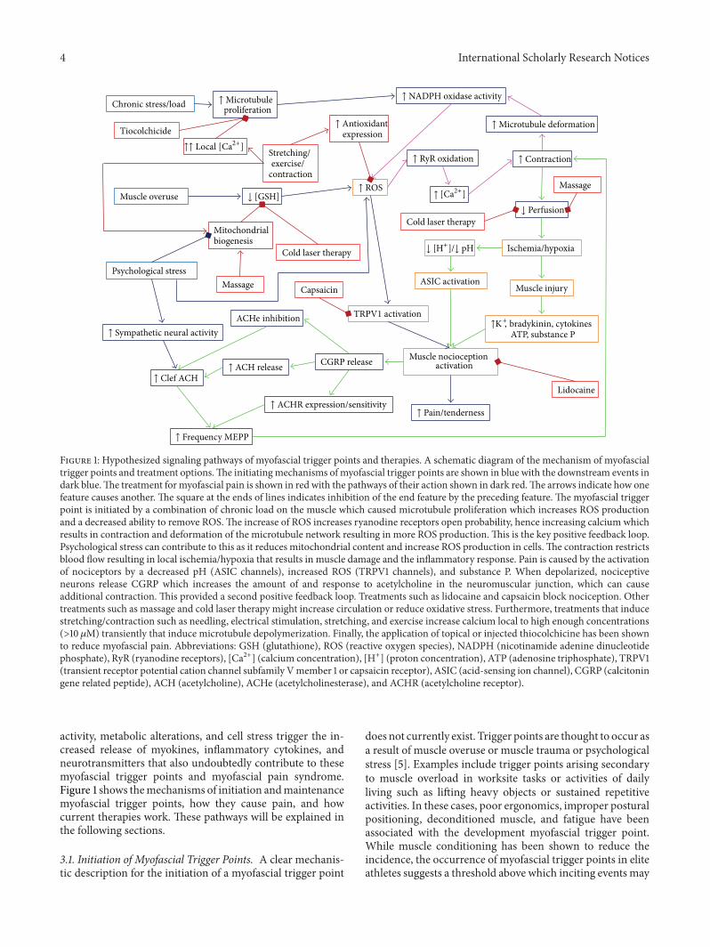
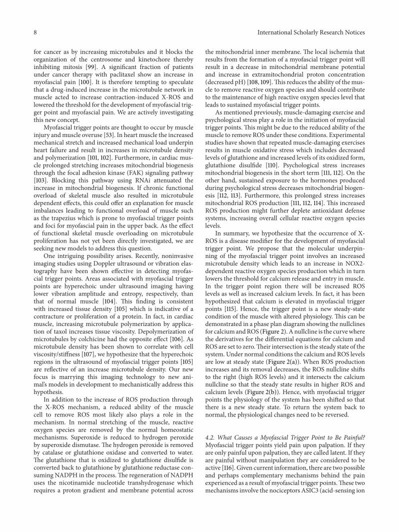
Figure 1: Hypothesized signaling pathways of myofascial trigger points and therapies. A schematic diagram of the mechanism of myofascialtrigger points and treatment options.The initiatingmechanisms of myofascial trigger points are shown in blue with the downstream events indark blue.The treatment formyofascial pain is shown in red with the pathways of their action shown in dark red.The arrows indicate how onefeature causes another. The square at the ends of lines indicates inhibition of the end feature by the preceding feature. The myofascial triggerpoint is initiated by a combination of chronic load on the muscle which caused microtubule proliferation which increases ROS productionand a decreased ability to remove ROS.The increase of ROS increases ryanodine receptors open probability, hence increasing calcium whichresults in contraction and deformation of the microtubule network resulting in more ROS production.This is the key positive feedback loop.Psychological stress can contribute to this as it reduces mitochondrial content and increase ROS production in cells.The contraction restrictsblood flow resulting in local ischemia/hypoxia that results in muscle damage and the inflammatory response. Pain is caused by the activationof nociceptors by a decreased pH (ASIC channels), increased ROS (TRPV1 channels), and substance P. When depolarized, nociceptiveneurons release CGRP which increases the amount of and response to acetylcholine in the neuromuscular junction, which can causeadditional contraction. This provided a second positive feedback loop. Treatments such as lidocaine and capsaicin block nociception. Othertreatments such as massage and cold laser therapy might increase circulation or reduce oxidative stress. Furthermore, treatments that inducestretching/contraction such as needling, electrical stimulation, stretching, and exercise increase calcium local to high enough concentrations(>10𝜇M) transiently that induce microtubule depolymerization. Finally, the application of topical or injected thiocolchicine has been shownto reduce myofascial pain. Abbreviations: GSH (glutathione), ROS (reactive oxygen species), NADPH (nicotinamide adenine dinucleotidephosphate), RyR (ryanodine receptors), [Ca2+] (calcium concentration), [H+] (proton concentration), ATP (adenosine triphosphate), TRPV1(transient receptor potential cation channel subfamily Vmember 1 or capsaicin receptor), ASIC (acid-sensing ion channel), CGRP (calcitoningene related peptide), ACH (acetylcholine), ACHe (acetylcholinesterase), and ACHR (acetylcholine receptor).
activity, metabolic alterations, and cell stress trigger the in-creased release of myokines, inflammatory cytokines, andneurotransmitters that also undoubtedly contribute to thesemyofascial trigger points and myofascial pain syndrome.Figure 1 shows themechanisms of initiation andmaintenancemyofascial trigger points, how they cause pain, and howcurrent therapies work. These pathways will be explained inthe following sections.
3.1. Initiation of Myofascial Trigger Points. A clear mechanis-tic description for the initiation of a myofascial trigger point
does not currently exist. Trigger points are thought to occur asa result of muscle overuse or muscle trauma or psychologicalstress [5]. Examples include trigger points arising secondaryto muscle overload in worksite tasks or activities of dailyliving such as lifting heavy objects or sustained repetitiveactivities. In these cases, poor ergonomics, improper posturalpositioning, deconditioned muscle, and fatigue have beenassociated with the development myofascial trigger point.While muscle conditioning has been shown to reduce theincidence, the occurrence of myofascial trigger points in eliteathletes suggests a threshold above which inciting events may
International Scholarly Research Notices 5
initiate a myofascial trigger point [44]. Psychological stressmay complement these mechanisms in the development ofmyofascial trigger point [45, 46].
Another important consideration is that, in some healthyindividuals and athletes, muscle fatigue or trauma does notalways result in myofascial trigger points. Instead they canresult in stiffness, soreness, and pain that usually resolvethemselves after a few days. This also supports a thresholdfor occurrence and/or possibly a cofactor that promotes theinitiation of a myofascial trigger point. Here, clues to themechanistic events that initiate myofascial trigger points maybe gained from our knowledge that comorbid conditionssuch as aging, disease, and stress increase the incidence ofmyofascial trigger points. For example, myofascial triggerpoints are thought to underlie the spontaneous pain patterin individuals suffering from fibromyalgia [47, 48]. Triggerpoints also have been observed with an increased frequencyin patients suffering from reflex sympathetic dystrophywhichis thought to arise from the unique emotional and psycho-logical condition of these patients and psychological stress inother patients [45, 46, 49, 50]. Additionally,myofascial triggerpoints may arise secondary to iatrogenic causes as certaincancer therapeutic regimens (i.e., taxanes and pacilitaxil)induce myofascial pain [51, 52].
Myofascial trigger points are more common under con-ditions of psychological stress [46]. In fact, myofascial triggerpoints display increasedmyogenic activity, while the adjacentmuscle remained silent under psychological stress [45]. Psy-chological stress results in an increase of certain hormonesand increase of sympathetic neural stimulation. It is believedthat the increase in hormones and sympathetic stimulationduring this condition leads to increase in release of acetyl-choline at the neuromuscular junction contributing to thecontraction of themotor units involved in a trigger point [53].This and other mechanisms that initiate a myofascial triggerpoint must feed into the mechanisms for their persistencedescribed in the next section.
3.2. Mechanisms for Persistence of Myofascial Trigger Points.The persistence of myofascial trigger points requires a self-sustaining positive feed-forward process. Simons presentedthe integrated hypothesis for myofascial trigger points tooffer an explanation [4]. The integrated hypothesis is a six-link chain that starts with step (1): the abnormal release ofacetylcholine. This triggers step (2): increased muscle fibertension which is seen as the taut band found in a myofascialtrigger point. The taut band is thought to constrict bloodflow that leads to step (3): local hypoxia. The reduced oxygendisrupts mitochondrial energy metabolism reducing ATPand leads to step (4): tissue distress and step (5): the releaseof sensitizing substances.These sensitizing substances lead topain by activation of nociceptors (pain receptors) and alsolead to step (6): autonomic modulation that then potentiatesstep (1): abnormal acetylcholine release.
More recently this hypothesis has been expanded byGerwin and coworkers [53]. It suggests more specific detailsof the feedback loop. For example, sympathetic nervoussystem activity augments acetylcholine release as well as the
local hypoperfusion caused by the muscle contraction. Theresulting ischemia/hypoxia leads to acidification (decreasedpH). Experiments have shown that injections of acidic salineof pH 4 can cause muscle pain through activation of musclepain receptors called acid-sensing ion channels (ASIC3)[54, 55]. While this low pH is much lower than that seenduring ischemia, a smaller physiological decrease in pH hasbeen shown to activate ASIC3 channels [56]. The prolongedischemia/hypoxia also leads to muscle injury resulting inthe release of potassium, bradykinins, cytokines, ATP, andsubstance P which might stimulate nociceptors in the muscle[53, 57]. The end result is the tenderness and pain observedwith myofascial trigger points accompanied by calcitoningene-related peptide (CGRP). Depolarization of nociceptiveneurons causes the release of CGRP [58]. CGRP inhibitsacetylcholine esterase and upregulates the amount of acetyl-choline receptors and release of acetylcholine.This nonquan-tal spontaneous acetylcholine release at themotor end plate asa result of CGRP is termed as acetylcholine leakage [59].Thisdiffers from the other modes of acetylcholine release suchas simulation induced multiquantal release resulting in anend plate potential (EPP) and spontaneous quantal releasesresulting in a miniature end plate potential (MEPP) [59].Thetheory also postulates CGRP release from nerve terminalswith the same targets. Furthermore, a decrease in pH canalso cause an increase in acetylcholine release [60].The resultis increased acetylcholine in the nerve terminal, synapticcleft, and increased motor endplate potentials resulting inmore contraction [61, 62]. The model also suggests thatpsychological stress also increases acetylcholine release intothe neuromuscular junction.
These expanded hypotheses presented in the previousparagraph are based upon experimental and clinical evidenceand understanding when possible [53, 63]. Using microdial-ysis samples of the chemical milieu of the muscle can beobtained. In microdialysis, a hollow needle filled with anabsorbing gel and with a semipermeable membrane at itstip is inserted into the myofascial trigger point. Another isinserted into adjacent normal muscle for comparison. Ions,signaling molecules, and proteins diffuse into the gel thatdoes not leave the needle. These are assayed upon removalof the needle. While this method allows access to the muscleinterior, it cannot differentiate between the intracellularand extracellular spaces. Such experiments, hence, can givean idea of the chemical species in the muscle tissue butnot specifically describe the intracellular conditions of theskeletal muscle cell.
The positive feedback loop in the above mechanismrequires that there be sustained stimulation of the mus-cle motor unit due to increased acetylcholine release anddecreased acetylcholinesterase activity. However, it appearsthat acetylcholine releasemight not be required for sustainingtrigger points. In studies comparing the efficacy of motornerve block using lidocaine injection compared to intramus-cular stimulation using dry needling, the group receiving theintramuscular stimulation showed more than 40% greaterimprovement than did the lidocaine injection group [64].Furthermore, lidocaine shows a dose-dependent decreasein miniature end plate potential, acetylcholine release, and
6 International Scholarly Research Notices
acetylcholine sensitivity [65, 66]. Therefore, there might beanother mechanism that provides the positive feedback thatsustains a myofascial trigger point. However, if the sponta-neous nonquantal acetylcholine release is a direct result ofCGRP, it might be too small to measure and, since it doesnot involve nerve depolarization, would be unaffected bylidocaine.
3.3. Biophysical Perspectives of Current Therapies for Myofas-cial Trigger Points. There are several therapies currentlyused to treat myofascial trigger points including massage,stretching, dry needling/injections, electrical stimulation,cold laser treatment, and ultrasound. There are severalmassage treatments that relax myofascial trigger points suchas passive rhythmic release, active rhythmic release, andtrigger point pressure release [4]. This paper is not intendedto be a comprehensive discussion of drugs that are usedto treat trigger points. What seems to be common to thetherapies is that they alleviate muscle stiffness and pain andmay be combined with therapies that stretch and improvemetabolism at the hypercontracted trigger point region.
3.3.1. Massage. It is widely believed that massage increasesblood flow. For example, in one study massage of the lowerleft extremity in young females increase blood flow in thetibial artery as measured by Doppler ultrasound [67]. Ifthis occurred on the local level in muscle, this would inprinciple break the cycle in the above theories. However,this finding does not necessarily affect the microcirculationwhich is thought to be constricted in a myofascial triggerpoint. Furthermore, another study of the benefits of massageafter exercise focused more on the muscle and found thatthere was no significant increase in circulation after massage[68]. Instead, massage activated the mechanotransductionsignaling pathways FAK and ERK (focal adhesion kinaseand extracellular signal-regulated kinase, resp.), decreasedinflammatory cytokines, and increased mitochondrial bio-genesis [68]. Mitochondrial biogenesis by increasing theamount of mitochondria would improve energy metabolismin the muscle. The NADPH oxidase/Rac1 increases theautophosphorylation of FAK. FAK is a scaffold for EGF-mediated signaling including activation of ERK. Further-more, inhibition of NADPH oxidase/Rac1 increases focaladhesions [69]. ERK activation potentiates FAK-stimulatedremoval of focal adhesions [70]. Focal adhesions have beenimplicated as a component of stiffness in aortic smoothmuscle cells [71]. Therefore, one way massage may reducemuscle stiffness is by the reduction of focal adhesions throughactivation of the FAK pathway.
3.3.2. Stretching. Stretching of muscle involves a series ofstretching exercises of the muscle where pain is experienced[4]. The reason that this works is not clearly understood.Anecdotally, it is thought to increase blood flow to muscle;however, it is unclear if this is supported by experimentalstudies. In fact, stretching of muscle transiently decreasesblood flow proportionally to the amount of stretching [72–74]. This is due in part to the longitudinal stretching of
the blood vessels running in the direction of the musclefibers [73, 75]. It is also due to the compression of the bloodvessels by the increase in intramuscular pressure [76, 77].On the other hand, muscle stretching training may actuallyincrease circulation as shown in ballet trained individuals[78]. This might explain the observed long-term benefit.There also might be other mechanisms involved. Recentexperiments have shown that stretching skeletal myocytesactivates NADPH oxidase [79]. This occurs through themicrotubules serving as mechanotransducers that interactwith Rac1 that activates NADPH oxidase. This results in theproduction of reactive oxygen species that increase the openprobability of the ryanodine receptors with no increase inchannel conductance through the oxidation of thiol groupson the protein [80–83]. In muscular dystrophic cells thisresponse is greater [79] due to the increase in microtubuledensity in these myocytes [84]. It should be noted thatreactive oxygen species from any source displays an increasein ryanodine receptor open probability. For example, reactiveoxygen species from hypoxanthine/xanthine oxidase has alsobeen shown to increase ryanodine receptor open probability[85]. In addition, this presents the possibility that stretchingof muscle, similar to massage, activates the FAK and ERKpathways.
3.3.3. Dry Needling/Injections. The insertion of a needle(acupuncture) can release a myofascial trigger point if theinsertion of a needle into the trigger point elicits a localtwitch. This local twitch involves a transient increase inactivity in the muscle band containing the trigger point.Furthermore, it is considered to be a spinal reflex since spinalcord transection between the brain and the level of the triggerpoint does not affect the response [86]. A local twitch at thesite is thought to stretch the muscle fibers at that location[87]. The relaxation of the muscle after twitch is thoughtto relieve constriction of the capillaries which restores themicrocirculation. This reoxygenates the muscle at the siteof the trigger point breaking the positive feedback. Recentstudies indicate that dry needling increases blood flow andoxygenation to the muscle band containing the trigger pointand not the rest of the muscle [88].
3.3.4. Electrical Stimulation. Electrical stimulation placeselectrode across the muscle affected by a trigger point andrapidly causes contractions by depolarizing the muscle. Thegoal of this therapy is to increase the size and frequencyof the twitches that could have been elicited by needling[87]. Once again this might work through the mechanismof stretching. In fact, muscle stimulation was shown to bemore effective in treating trigger points than application oflidocaine [64]. It has been proposed that working the musclein this fashion could be more like exercising the muscle.For example, in one study voluntary and electrically evokedisometric contractions showed similar oxygen demand atmaximum intensity although the torque generated by volun-tary isometric contraction was 40% of the torque generatedby electrically evoked isometric contraction [89].
International Scholarly Research Notices 7
3.3.5. Cold Laser Therapy. Cold laser therapy also knownas low level light therapy exposes the myofascial triggerpoint to near infrared light. It has been shown to workclinically reducing pain and rigidity and increasing mobility[90, 91]. This study suggests that the increased motion leadsto an increase in microcirculation. Others claim that coldlaser therapy is thought to energize the mitochondria. Inmouse embryonic fibroblasts low level light therapy increasedintracellular ATP and reactive oxygen species levels [92].The source of the increased reactive oxygen species is likelymitochondria [93]. Cold laser therapy has been shown toreduce oxidative stress in skeletal muscle [94].
3.3.6. Ultrasound Therapy. Ultrasound is often used to treatmyofascial pain and trigger points. However, the benefitsare unclear. While exercise and massage seem to reducepain and the number and size of myofascial trigger points,conventional ultrasound did not result in pain reduction [95].On the other hand, high powered ultrasound before stretch-ing increases mobility more than conventional ultrasound[96]. This might be due to the heating effect of ultrasound.For example, thermal ultrasound has been shown to reducestiffness of myofascial trigger points [97].
4. New Mechanistic Theory for MyofascialTrigger Points
In light of the discussion above, it is clear that the currenttheory for the mechanisms behind myofascial pain is notsufficient to fully explain the syndrome. As the myofascialtrigger point appears central to the onset and persistence ofmyofascial pain syndrome, we have focused attention on themuscle fiber level in an attempt to reveal new mechanisticinsight. Here we present mechanistic findings in muscle thatdemonstrate how mechanical stress acts to trigger excesscalcium release in muscle via a novel mechanotransductionpathway. With this new pathway as a foundation, we putforth a novel mechanistic hypothesis for the initiation andpersistence of myofascial trigger points that extends thecurrent theories discussed above.
4.1. What Initiates and Sustains a Myofascial Trigger Point?The essence of this question is what positive feedback mech-anisms exist that can sustain a myofascial trigger point onceinitiated. Based on the models and mechanisms discussedabove, the local and persistent hypercontracture of themuscleappears to be critical to the myofascial trigger point. At thecellular level, a persistent neural activation may act to initiatea local and sustained contraction; however, fatigue of themuscle would ensue much as in a highly trained athlete withhigh motivation that is eventually unable to sustain muscleactivity. Rather, the local contracture of the muscle mustoccur secondary to the normal neuromuscular activation andarise due to regenerative feed-forward processes within themuscle cell. At the most basic level, this situation wouldnecessitate a mechanism that permitted regenerative calciumrelease within the myofibers that escaped from the nor-mal inhibitory processes that govern central and peripheral
muscle fatigue. It would be most practical for this feed-forwardmechanism to take advantage of any aberrant activity(contraction dependent mechanical stress, calcium release,and altered metabolic signaling) as an initiation trigger andas a mechanism to sustain its activity.
X-ROS signaling is anewly characterized mechanoac-tivated ROS-dependent signaling cascade in cardiac andskeletal muscle. In X-ROS signaling mechanical deformationof the microtubule network acts as a mechanotransductionelement to activate the NADPH oxidase (NOX2) whichproduces ROS. The ROS oxidizes RyRs and increases theiropen probability resulting in increases in Ca2+ release fromthe sarcoplasmic reticulum. The Ca2+ mobilization resultingfrom mechanical stretch through this pathway is X-ROS sig-naling. In heart, X-ROS acts locally to affect the sarcoplasmicreticulum (SR) Ca2+ release channels (ryanodine receptors,ryanodine receptors) and “tunes” excitation-contraction cou-pling Ca2+ signaling during physiological behavior but canpromoteCa2+-dependent arrhythmias during pathologywithX-ROS in excess [79]. In skeletal muscle, X-ROS sensitizesCa2+-permeable sarcolemmal “transient receptor potential”(TRP) channels, a pathway critical for sustaining SR loadduring repetitive contractions. When in excess, X-ROS inskeletal muscle is maladaptive as shown in diseases suchas Duchenne muscular dystrophy (DMD) and dysferlinopa-thy which both have altered calcium signaling as majormechanistic underpinnings. Importantly, work in DMD byKhairallah et al. suggests that the development of X-ROS (i.e.,enhancement in the expression of microtubule protein andNOX2 and its resultant increase in mechanoactivated ROS)is a secondary process that is temporally associated with theseverity of the disease and not a primary cause of the disease[84, 98]. In that regard the enhancement in X-ROS was adisease modifier that increased the severity of the disease bylowering the threshold for calcium release in the muscle.
The above mechanism purports that excessive contrac-tion dependent stress acts through themicrotubule cytoskele-tal elements to activate NADPH oxidase to produce ROS.The subsequent ROS sensitization of ryanodine receptors andsarcolemmal calcium influx channels increases myoplasmiccalcium concentration and contraction leading to morestretch. Based on this model, one reason why the occurrenceof myofascial trigger points may be less common in healthyindividuals is due to the absence of the feed-forward trigger,excess microtubules, or NOX2 that serves to generate X-ROS. This hypothesis then proposes that the threshold fordeveloping myofascial pain and myofascial trigger points islower with the trigger present as a critical amount of X-ROS activity that would serve to lower the threshold forcalcium release activation such that spontaneous or regen-erative calcium release generation promoted can initiate thecontractures which underscore the myofascial trigger point.
Our hypothesis that the microtubule cytoskeleton andX-ROS may play a role in the mechanism of myofascialtrigger point came from work that suggests that microtubuleproliferation have been associated with either myofascialtrigger points or myofascial pain or both. Taxane basedchemotherapy, for example, pacilitaxel, is a common therapy
8 International Scholarly Research Notices
for cancer as by increasing microtubules and it blocks theorganization of the centrosome and kinetochore therebyinhibiting mitosis [99]. A significant fraction of patientsunder cancer therapy with paclitaxel show an increase inmyofascial pain [100]. It is therefore tempting to speculatethat a drug-induced increase in the microtubule network inmuscle acted to increase contraction-induced X-ROS andlowered the threshold for the development of myofascial trig-ger point and myofascial pain. We are actively investigatingthis new concept.
Myofascial trigger points are thought to occur by muscleinjury andmuscle overuse [53]. In heart muscle the increasedmechanical stretch and increased mechanical load underpinheart failure and result in increases in microtubule densityand polymerization [101, 102]. Furthermore, in cardiac mus-cle prolonged stretching increases mitochondrial biogenesisthrough the focal adhesion kinase (FAK) signaling pathway[103]. Blocking this pathway using RNAi attenuated theincrease in mitochondrial biogenesis. If chronic functionaloverload of skeletal muscle also resulted in microtubuledependent effects, this could offer an explanation for muscleimbalances leading to functional overload of muscle suchas the trapezius which is prone to myofascial trigger pointsand foci for myofascial pain in the upper back. As the effectof functional skeletal muscle overloading on microtubuleproliferation has not yet been directly investigated, we areseeking new models to address this question.
One intriguing possibility arises. Recently, noninvasiveimaging studies using Doppler ultrasound or vibration elas-tography have been shown effective in detecting myofas-cial trigger points. Areas associated with myofascial triggerpoints are hyperechoic under ultrasound imaging havinglower vibration amplitude and entropy, respectively, thanthat of normal muscle [104]. This finding is consistentwith increased tissue density [105] which is indicative of acontracture or proliferation of a protein. In fact, in cardiacmuscle, increasing microtubule polymerization by applica-tion of taxol increases tissue viscosity. Depolymerization ofmicrotubules by colchicine had the opposite effect [106]. Asmicrotubule density has been shown to correlate with cellviscosity/stiffness [107], we hypothesize that the hyperechoicregions in the ultrasound of myofascial trigger points [105]are reflective of an increase microtubule density. Our newfocus is marrying this imaging technology to new ani-mal’s models in development to mechanistically address thishypothesis.
In addition to the increase of ROS production throughthe X-ROS mechanism, a reduced ability of the musclecell to remove ROS most likely also plays a role in themechanism. In normal stretching of the muscle, reactiveoxygen species are removed by the normal homeostaticmechanisms. Superoxide is reduced to hydrogen peroxideby superoxide dismutase. The hydrogen peroxide is removedby catalase or glutathione oxidase and converted to water.The glutathione that is oxidized to glutathione disulfide isconverted back to glutathione by glutathione reductase con-suming NADPH in the process.The regeneration of NADPHuses the nicotinamide nucleotide transhydrogenase whichrequires a proton gradient and membrane potential across
the mitochondrial inner membrane. The local ischemia thatresults from the formation of a myofascial trigger point willresult in a decrease in mitochondrial membrane potentialand increase in extramitochondrial proton concentration(decreased pH) [108, 109].This reduces the ability of themus-cle to remove reactive oxygen species and should contributeto the maintenance of high reactive oxygen species level thatleads to sustained myofascial trigger points.
As mentioned previously, muscle-damaging exercise andpsychological stress play a role in the initiation of myofascialtrigger points. This might be due to the reduced ability of themuscle to remove ROS under these conditions. Experimentalstudies have shown that repeated muscle-damaging exercisesresults in muscle oxidative stress which includes decreasedlevels of glutathione and increased levels of its oxidized form,glutathione disulfide [110]. Psychological stress increasesmitochondrial biogenesis in the short term [111, 112]. On theother hand, sustained exposure to the hormones producedduring psychological stress decreases mitochondrial biogen-esis [112, 113]. Furthermore, this prolonged stress increasesmitochondrial ROS production [111, 112, 114]. This increasedROS production might further deplete antioxidant defensesystems, increasing overall cellular reactive oxygen specieslevels.
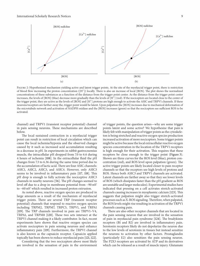
In summary, we hypothesize that the occurrence of X-ROS is a disease modifier for the development of myofascialtrigger point. We propose that the molecular underpin-ning of the myofascial trigger point involves an increasedmicrotubule density which leads to an increase in NOX2-dependent reactive oxygen species production which in turnlowers the threshold for calcium release and entry in muscle.In the trigger point region there will be increased ROSlevels as well as increased calcium levels. In fact, it has beenhypothesized that calcium is elevated in myofascial triggerpoints [115]. Hence, the trigger point is a new steady-statecondition of the muscle with altered physiology. This can bedemonstrated in a phase plan diagram showing the nullclinesfor calciumandROS (Figure 2). Anullcline is the curvewherethe derivatives for the differential equations for calcium andROS are set to zero.Their intersection is the steady state of thesystem. Under normal conditions the calcium and ROS levelsare low at steady state (Figure 2(a)). When ROS productionincreases and its removal decreases, the ROS nullcline shiftsto the right (high ROS levels) and it intersects the calciumnullcline so that the steady state results in higher ROS andcalcium levels (Figure 2(b)). Hence, with myofascial triggerpoints the physiology of the system has been shifted so thatthere is a new steady state. To return the system back tonormal, the physiological changes need to be reversed.
4.2. What Causes a Myofascial Trigger Point to Be Painful?Myofascial trigger points yield pain upon palpation. If theyare only painful upon palpation, they are called latent. If theyare painful without manipulation they are considered to beactive [116]. Given current information, there are two possibleand perhaps complementary mechanisms behind the painexperienced as a result ofmyofascial trigger points.These twomechanisms involve the nociceptors ASIC3 (acid-sensing ion
International Scholarly Research Notices 9
[ROS]
[ROS] nullcline
[Ca2+] nullcline
[Ca2
+]
(a)
[ROS]
[ROS] nullcline
[Ca2+] nullcline
[Ca2
+]
(b)
Figure 2: Hypothesized mechanism yielding active and latent trigger points. At the site of the myofascial trigger point, there is restrictionof blood flow increasing the proton concentration ([H+]) locally. There is also an increase of local [ROS]. The plot shows the normalizedconcentrations of these substances as a function of the distance from the trigger point center. As the distance from the trigger point centerincreases, the levels of [ROS] (blue) decrease more gradually than the levels of [H+] (red). If the nociceptors are located close to the center ofthe trigger point, they are active as the levels of [ROS] and [H+] protons are high enough to activate the ASIC and TRPV1 channels. If theseneurons/receptors are farther away the, trigger point would be latent. Upon palpation the [ROS] increases due to mechanical deformation ofthe microtubule network and activation of NADPH oxidase and the [ROS] increases (green) so that the nociceptors see sufficient ROS to beactivated.
channel) and TRPV1 (transient receptor potential) channelin pain sensing neurons. These mechanisms are describedbelow.
The local sustained contraction in a myofascial triggerpoint can result in restriction of local circulation which cancause the local ischemia/hypoxia and the observed changescaused by it such as increased acid accumulation resultingin a decrease in pH. In experiments in rabbit gastrocnemiusmuscle, the intracellular pH dropped from 7.0 to 6.6 during4 hours of ischemia [108]. In the extracellular fluid the pHchanges from 7.3 to 6.36 during the same time period due tothe accumulation of lactic acid.There are four ASIC channels:ASIC1, ASIC2, ASIC3, and ASIC4. However, only ASIC3seems to be involved in inflammatory pain [117, 118]. ThispH drop is enough to fully activate the nociceptive ASIC3channels in nearby neurons [56]. The pH changes seemed tolevel off due to a drop in membrane potential from −90mVto −60mV which resulted in increased proton extrusion.
As noted above, reactive oxygen species are produced inlarge amounts as a result of the mechanism of myofascialtrigger points. There are several TRP (transient receptorpotential) channels that respond to reactive oxygen speciesincluding TRPM2, TRPM7, TRPC5, TRPV1, and TRPA1[119]. The TRP channels involved in pain are TRPV1–V4,TRPA1, and TRPM8 [120]. These two sets intersect at theTRPV1 channel making it a likely contributor. In fact, recentexperiments have shown that reactive oxygen species canactivate nociceptors in pain sensing neurons and enhanceinflammatory pain [119]. Furthermore, the TRPV1 channelis also known as the capsaicin receptor. Capsaicin appliedtopically has been shown to reduce myofascial pain [121, 122].
Considering that the two nociceptors above most likelyare involved in the sensation of pain in the environment
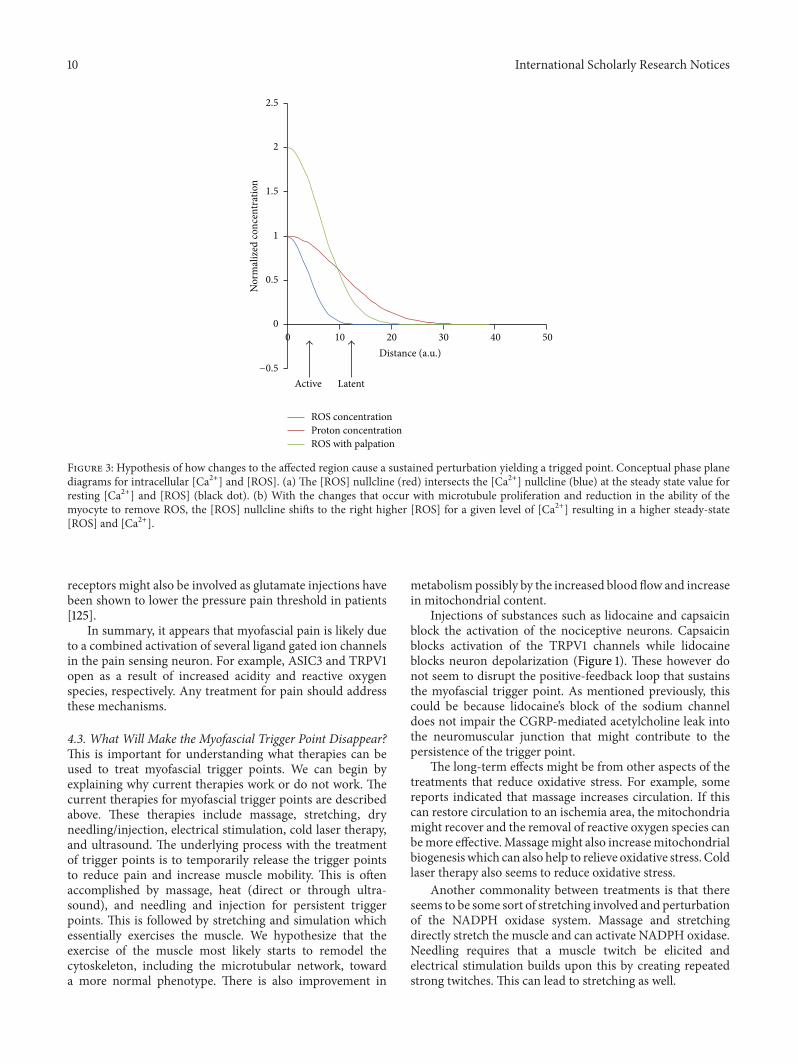
of trigger points, the question arises—why are some triggerpoints latent and some active? We hypothesize that pain islikely felt withmanipulation of trigger points as the cytoskele-ton is being stretched and reactive oxygen species productionincreased activation ofmore nociceptors. Some trigger pointsmight be active because the local extracellular reactive oxygenspecies concentration in the location of the TRPV1 receptorsis high enough for their activation. This requires that thesereceptors be close enough to the trigger point (Figure 3).Shown are three curves for the ROS level (blue), proton con-centration (red), and ROS level upon palpation (green). Theactive trigger points are likely located closer to pain receptorchannels so that the receptors see high levels of protons andROS. Hence both ASIC3 and TRPV1 channels are activated.Latent channels are farther away so that they see lower levelsof ROS (which dissipates faster than the pH gradient as ROSare unstable and largermolecules). Experimental studies haveindicated that pressing on a cell activates stretch-activatedchannels causing increases inmyoplasmic calcium [123].Thissuggests that palpation might also trigger stretch-activatedprocesses such asX-ROS signaling.Therefore, when palpated,the ROS levels might rise resulting in activation of the TRPV1channels causing pain.
There are also other receptor channels that are located inthe pain sensing neuron that are involved in the sensationof pain in myofascial pain syndrome [124]. The bradykininreceptors (B1 and B2) are involved in inflammatory pain.Serotonin receptors likely do not depolarize the neuron dueto the low levels of serotonin in tissues but instead sensitizethe neurons to activation by other factors. Prostaglandin(particularly E2) also sensitizes the pain sensing neuron.The P2X3 receptors are activated by ATP and its derivativeswhich can be released as a result of muscle injury. Glutamate
10 International Scholarly Research Notices
Nor
mal
ized
conc
entr
atio
n
Distance (a.u.)
Active Latent
0
0.5
1
1.5
2
2.5
0 10 20 30 40 50
ROS concentrationProton concentrationROS with palpation
−0.5
Figure 3: Hypothesis of how changes to the affected region cause a sustained perturbation yielding a trigged point. Conceptual phase planediagrams for intracellular [Ca2+] and [ROS]. (a) The [ROS] nullcline (red) intersects the [Ca2+] nullcline (blue) at the steady state value forresting [Ca2+] and [ROS] (black dot). (b) With the changes that occur with microtubule proliferation and reduction in the ability of themyocyte to remove ROS, the [ROS] nullcline shifts to the right higher [ROS] for a given level of [Ca2+] resulting in a higher steady-state[ROS] and [Ca2+].
receptors might also be involved as glutamate injections havebeen shown to lower the pressure pain threshold in patients[125].
In summary, it appears that myofascial pain is likely dueto a combined activation of several ligand gated ion channelsin the pain sensing neuron. For example, ASIC3 and TRPV1open as a result of increased acidity and reactive oxygenspecies, respectively. Any treatment for pain should addressthese mechanisms.
4.3. What Will Make the Myofascial Trigger Point Disappear?This is important for understanding what therapies can beused to treat myofascial trigger points. We can begin byexplaining why current therapies work or do not work. Thecurrent therapies for myofascial trigger points are describedabove. These therapies include massage, stretching, dryneedling/injection, electrical stimulation, cold laser therapy,and ultrasound. The underlying process with the treatmentof trigger points is to temporarily release the trigger pointsto reduce pain and increase muscle mobility. This is oftenaccomplished by massage, heat (direct or through ultra-sound), and needling and injection for persistent triggerpoints. This is followed by stretching and simulation whichessentially exercises the muscle. We hypothesize that theexercise of the muscle most likely starts to remodel thecytoskeleton, including the microtubular network, towarda more normal phenotype. There is also improvement in
metabolism possibly by the increased blood flow and increasein mitochondrial content.
Injections of substances such as lidocaine and capsaicinblock the activation of the nociceptive neurons. Capsaicinblocks activation of the TRPV1 channels while lidocaineblocks neuron depolarization (Figure 1). These however donot seem to disrupt the positive-feedback loop that sustainsthe myofascial trigger point. As mentioned previously, thiscould be because lidocaine’s block of the sodium channeldoes not impair the CGRP-mediated acetylcholine leak intothe neuromuscular junction that might contribute to thepersistence of the trigger point.
The long-term effects might be from other aspects of thetreatments that reduce oxidative stress. For example, somereports indicated that massage increases circulation. If thiscan restore circulation to an ischemia area, the mitochondriamight recover and the removal of reactive oxygen species canbemore effective.Massagemight also increasemitochondrialbiogenesiswhich can also help to relieve oxidative stress. Coldlaser therapy also seems to reduce oxidative stress.
Another commonality between treatments is that thereseems to be some sort of stretching involved and perturbationof the NADPH oxidase system. Massage and stretchingdirectly stretch the muscle and can activate NADPH oxidase.Needling requires that a muscle twitch be elicited andelectrical stimulation builds upon this by creating repeatedstrong twitches. This can lead to stretching as well.
International Scholarly Research Notices 11
Exercise has also been shown to help alleviate myofascialtrigger points. During exercise there are transient elevationsof calcium that are likely higher at peak than those seenin the muscle during a myofascial trigger point. Calcium-dependent regulator protein inhibited polymerization ofmicrotubules at physiological calcium concentrations [126,127]. This value is in the range of 10–100 𝜇M which is seennear the calcium release sites in muscle. At the site of calciumrelease during a calcium spark, calcium is predicted to beas high as 150 𝜇M by computational modeling [128, 129].Therefore, exercise might reduce microtubule proliferationand hence ROS production from the NADPH oxidase.Furthermore, exercise also acts to increase the ability ofthe muscle to remove ROS. Moderate exercise upregulatesantioxidant enzymes [130, 131].
Exercise also leads to mitochondrial biogenesis [132]. Afinal benefit of exercise is that it increases mitochondrialfunction as measured respiration and content as measured bycitrate synthase activity [133].
If the hypothesis that increased microtubule polymer-ization results in a pathologic increase in reactive oxygenspecies production through the NAPDH oxidase complex,then treatments that reduce microtubule polymerizationshould show benefit for treating myofascial trigger points.In fact, administration of colchicine and related compoundsseems to reduce myofascial pain. The application of topicalthiocolchicoside reduced pain in patients with acute cervicalmyofascial pain to alleviate back pain in patients [134]. Oralcolchicine, however, did not seem to alleviate back pain in astudy by Schnebel and Simmons [135]. Another study by Sim-mons and coworkers indicated that intravenous colchicineadministration reduced lower back pain in patients [136].These findings seem to support the idea that microtubulesplay a major role in myofascial pain syndrome.
5. Conclusions
Recent progress in experimental studies has provided awealth of information that can be used to gain understandingof the molecular mechanisms of myofascial pain syndrome.Only through improved understanding of the molecularand subcellular pathways behind this disorder can noveltherapeutics be discovered. This improved comprehensionmight also help guide current treatment protocols for optimalbenefit. However, many details of the signaling pathwaysinvolved remain yet unclear and further study is needed.Finally, the analysis presented here suggests that colchicineis a likely therapeutic that should be explored further as atreatment for myofascial pain.
Glossary
ASIC: Acid-sensing ion channel with isoformsASIC1, ASIC2, ASIC3, and ASIC4
ATP: Adenosine triphosphateCGRP: Calcitonin gene related peptideEPP: End plate potentialETC: Electron transport chain
ERK: Extracellular signal-related kinaseFAK: Focal adhesion kinasegp91phox: Another name for NOX2H2O2: Hydrogen peroxide
MEPP: Miniature end plate potentialNADPH: Nicotinamide adenine dinucleotide phos-
phateNOX: NADPH oxidase with isoforms NOX2 and
NOX4p22: Regulatory subunits of NOX2p47phox: Regulatory subunits of NOX2p67phox: Regulatory subunits of NOX2p40phox: Regulatory subunits of NOX2PLA2: Phospholipase A2Rac1: Ras-related C3 botulinum toxin substrate 1
acting as regulatory subunits of NOX2Rho: A small prokaryotic protein involved in the
termination of transcriptionROS: Reactive oxygen speciesRyR: Ryanodine receptorRyR1: Ryanodine receptor type 1SOD: Superoxide dismutaseTRP: Transient receptor potential channel
with isoforms TRPM2, TRPM7, TRPC5,TRPV1, TRPV2, TRPV3, TRPV4, TRPA1,and TRPM8
XO: Xanthine oxidaseX-ROS: Stretch activatedROSproduction byNOX2
to potential calcium signaling.
Conflict of Interests
The author declares that there is no conflict of interestsregarding the publication of this paper.
Acknowledgments
The author would like to give special thanks to ProfessorChrisWard of theUniversity ofMaryland School ofMedicinefor his essential discussions and his comments and sugges-tions for the paper. The author would also like to thankSiddhartha Sikdar for the discussions on this topic.This workwas supported in part by the National Institutes of HealthGrants 5R01AR057348 and 5R01HL105239.
References
[1] D. G. Simons, “Clinical and etiological update of myofascialpain from trigger points,” Journal of Musculoskeletal Pain, vol.4, no. 1-2, pp. 93–121, 1996.
[2] J. Fleckenstein, D. Zaps, L. J. Ruger et al., “Discrepancy betweenprevalence and perceived effectiveness of treatment methodsin myofascial pain syndrome: results of a cross-sectional,nationwide survey,” BMC Musculoskeletal Disorders, vol. 11,article 32, 2010.
[3] L. H. Gerber, S. Sikdar, K. Armstrong et al., “A systematiccomparison between subjects with no pain and pain associatedwith active myofascial trigger points,” PM & R: The Journal of
12 International Scholarly Research Notices
Injury, Function, and Rehabilitation, vol. 5, no. 11, pp. 931–938,2013.
[4] D. G. Simons, “Review of enigmatic MTrPs as a common causeof enigmatic musculoskeletal pain and dysfunction,” Journalof Electromyography and Kinesiology, vol. 14, no. 1, pp. 95–107,2004.
[5] D. G . Simons, J. G. Travell, L. S. Simons, and B. D. Cummings,Travell & Simons’ Myofascial Pain and Dysfunction: The TriggerPoint Manual, Lippincott Williams &Wilkins, 1998.
[6] J. D. Bullock, “Relative afferent pupillary defect in the ”better“eye,” Journal of Clinical Neuro-Ophthalmology, vol. 10, no. 1, pp.45–51, 1990.
[7] D. Celik and E. K. Mutlu, “Clinical implication of latentmyofascial trigger point,” Current Pain and Headache Reports,vol. 17, no. 8, article 353, 2013.
[8] H. Y. Ge, L. Arendt-Nielsen, and P. Madeleine, “Acceler-ated muscle fatigability of latent myofascial trigger points inhumans,” Pain Medicine, vol. 13, no. 7, pp. 957–964, 2012.
[9] H.-Y. Ge, C. Fernandez-de-las-Penas, and S.-W. Yue, “Myofas-cial trigger points: spontaneous electrical activity and its conse-quences for pain induction and propagation,”ChineseMedicine,vol. 6, article 13, 2011.
[10] R. Baron, “Peripheral neuropathic pain: from mechanisms tosymptoms,” Clinical Journal of Pain, vol. 16, no. 2, pp. S12–S20,2000.
[11] R. Baron, “Mechanisms of disease: neuropathic pain—a clinicalperspective,”Nature Clinical Practice Neurology, vol. 2, no. 2, pp.95–106, 2006.
[12] P. M. Hopkins, “Skeletal muscle physiology,” Continuing Educa-tion in Anaesthesia, Critical Care and Pain, vol. 6, no. 1, pp. 1–6,2006.
[13] E. Rios and G. Brum, “Involvement of dihydropyridine recep-tors in excitation-contraction coupling in skeletal muscle,”Nature, vol. 325, no. 6106, pp. 717–720, 1987.
[14] E. M. Capes, R. Loaiza, and H. H. Valdivia, “Ryanodinereceptors,” Skeletal Muscle, vol. 1, no. 1, article 18, 2011.
[15] M. S. Jafri, S. J. Dudycha, and B. O’Rourke, “Cardiac energymetabolism: models of cellular respiration,” Annual Review ofBiomedical Engineering, vol. 3, pp. 57–81, 2001.
[16] H. A. Krebs andW. A. Johnson, “Metabolism of ketonic acids inanimal tissues,”The Biochemical Journal, vol. 31, no. 4, pp. 645–660, 1937.
[17] P. Mitchell, “Coupling of phosphorylation to electron andhydrogen transfer by a chemi-osmotic type of mechanism,”Nature, vol. 191, no. 4784, pp. 144–148, 1961.
[18] H. Hoppeler, O. Mathieu, R. Krauer, H. Claassen, R. B. Arm-strong, and E. R.Weibel, “Design of themammalian respiratorysystem. VI. Distribution of mitochondria and capillaries invarious muscles,” Respiration Physiology, vol. 44, no. 1, pp. 87–111, 1981.
[19] K. Schwerzmann, H. Hoppeler, S. R. Kayar, and E. R. Weibel,“Oxidative capacity of muscle and mitochondria: correlation ofphysiological, biochemical, and morphometric characteristics,”Proceedings of the National Academy of Sciences of the UnitedStates of America, vol. 86, no. 5, pp. 1583–1587, 1989.
[20] H. Tsutsui, S. Kinugawa, and S. Matsushima, “Oxidative stressand heart failure,” American Journal of Physiology: Heart andCirculatory Physiology, vol. 301, no. 6, pp. H2181–H2190, 2011.
[21] G. S. Supinski and L. A. Callahan, “Free radical-mediated skele-tal muscle dysfunction in inflammatory conditions,” Journal ofApplied Physiology, vol. 102, no. 5, pp. 2056–2063, 2007.
[22] S. K. Powers and M. J. Jackson, “Exercise-induced oxidativestress: cellularmechanisms and impact onmuscle force produc-tion,” Physiological Reviews, vol. 88, no. 4, pp. 1243–1276, 2008.
[23] M. Bayeva and H. Ardehali, “Mitochondrial dysfunction andoxidative damage to sarcomeric proteins,”CurrentHypertensionReports, vol. 12, no. 6, pp. 426–432, 2010.
[24] L. P. Michaelson, G. Shi, C. W. Ward, and G. G. Rodney,“Mitochondrial redox potential during contraction in singleintact muscle fibers,” Muscle & Nerve, vol. 42, no. 4, pp. 522–529, 2010.
[25] G. K. Sakellariou, A. Vasilaki, J. Palomero et al., “Studiesof mitochondrial and nonmitochondrial sources implicatenicotinamide adenine dinucleotide phosphate oxidase(s) in theincreased skeletal muscle superoxide generation that occursduring contractile activity,” Antioxidants and Redox Signaling,vol. 18, no. 6, pp. 603–621, 2013.
[26] D. A. Stofan, L. A. Callahan, A. F. DiMarco, D. E. Nethery, andG. S. Supinski, “Modulation of release of reactive oxygen speciesby the contracting diaphragm,” American Journal of Respiratoryand Critical Care Medicine, vol. 161, no. 3, part 1, pp. 891–898,2000.
[27] M. C. Gomez-Cabrera, G. L. Close, A. Kayani, A. McArdle, J.Vina, and M. J. Jackson, “Effect of xanthine oxidase-generatedextracellular superoxide on skeletal muscle force generation,”American Journal of Physiology: Regulatory Integrative andComparative Physiology, vol. 298, no. 1, pp. R2–R8, 2010.
[28] M. Gomez-Cabrera, C. Borras, F. V. Pallardo, J. Sastre, L. L. Ji,and J. Vina, “Decreasing xanthine oxidase-mediated oxidativestress prevents useful cellular adaptations to exercise in rats,”The Journal of Physiology, vol. 567, no. 1, pp. 113–120, 2005.
[29] S. K. Powers, W. B. Nelson, and M. B. Hudson, “Exercise-induced oxidative stress in humans: cause and consequences,”Free Radical Biology and Medicine, vol. 51, no. 5, pp. 942–950,2011.
[30] M. C. Gong, S. Arbogast, Z. Guo, J. Mathenia, W. Su, and M.B. Reid, “Calcium-independent phospholipase A
2modulates
cytosolic oxidant activity and contractile function in murineskeletal muscle cells,” Journal of Applied Physiology, vol. 100, no.2, pp. 399–405, 2006.
[31] S. T. Russell, H. Eley, andM. J. Tisdale, “Role of reactive oxygenspecies in protein degradation in murine myotubes inducedby proteolysis-inducing factor and angiotensin II,” CellularSignalling, vol. 19, no. 8, pp. 1797–1806, 2007.
[32] H. Gissel, “The role of Ca2+ in muscle cell damage,” Annals ofthe New York Academy of Sciences, vol. 1066, pp. 166–180, 2005.
[33] M. J. Jackson, “Control of reactive oxygen species production incontracting skeletal muscle,” Antioxidants and Redox Signaling,vol. 15, no. 9, pp. 2477–2486, 2011.
[34] A. Sirker, M. Zhang, and A. M. Shah, “NADPH oxidasesin cardiovascular disease: Insights from in vivo models andclinical studies,” Basic Research in Cardiology, vol. 106, no. 5, pp.735–747, 2011.
[35] C. X. C. Santos, N. Anilkumar, M. Zhang, A. C. Brewer, and A.M. Shah, “Redox signaling in cardiac myocytes,” Free RadicalBiology and Medicine, vol. 50, no. 7, pp. 777–793, 2011.
[36] A. Akki, M. Zhang, C. Murdoch, A. Brewer, and A. M. Shah,“NADPH oxidase signaling and cardiac myocyte function,”Journal of Molecular and Cellular Cardiology, vol. 47, no. 1, pp.15–22, 2009.
[37] A. Stanley, K. Thompson, A. Hynes, C. Brakebusch, and F.Quondamatteo, “NADPH oxidase complex-derived reactive
International Scholarly Research Notices 13
oxygen species, the actin cytoskeleton, and Rho GTPases in cellmigration,” Antioxidants & Redox Signaling, vol. 20, no. 13, pp.2026–2042, 2013.
[38] P. L. Hordijk, “Regulation of NADPH oxidases: the role of Racproteins,”Circulation Research, vol. 98, no. 4, pp. 453–462, 2006.
[39] T. Wittmann and C. M. Waterman-Storer, “Cell motility: canRho GTPases and microtubules point the way?” Journal of CellScience, vol. 114, part 21, pp. 3795–3803, 2001.
[40] Y.-C. Poh, S. Na, F. Chowdhury, M. Ouyang, Y. Wang, and N.Wang, “Rapid activation of Rac GTPase in living cells by forceis independent of Src,”PLoSONE, vol. 4, no. 11, Article ID e7886,2009.
[41] J. Dommerholt, C. Bron, and J. Franssen, “Myofascial triggerpoints: an evidence-informed review,” The Journal of Manualand Manipulative Therapy, vol. 14, no. 4, pp. 203–221, 2006.
[42] D. G. Simons and W. C. Stolov, “Microscopic features andtransient contraction of palpable bands in canine muscle,”American Journal of Physical Medicine, vol. 55, no. 2, pp. 65–88,1976.
[43] J. Borg-Stein and D. G. Simons, “Focused review: myofascialpain,” Archives of Physical Medicine and Rehabilitation, vol. 83,supplement 1, no. 3, pp. S40–S47, 2002.
[44] A. Hidalgo-Lozano, C. Fernandez-de-las-Penas, C. Calderon-Soto, A. Domingo-Camara, P. Madeleine, and M. Arroyo-Morales, “Elite swimmers with and without unilateral shoulderpain: mechanical hyperalgesia and active/latent muscle triggerpoints in neck-shoulder muscles,” Scandinavian Journal ofMedicine and Science in Sports, vol. 23, no. 1, pp. 66–73, 2013.
[45] W.H.McNulty, R.N.Gevirtz, D. R.Hubbard, andG.M. Berkoff,“Needle electromyographic evaluation of trigger point responseto a psychological stressor,” Psychophysiology, vol. 31, no. 3, pp.313–316, 1994.
[46] B. Jaeger, “Myofascial trigger point pain,” The Alpha Omegan,vol. 106, no. 1-2, pp. 14–22, 2013.
[47] H. Y. Ge, “Prevalence of myofascial trigger points in fibromyal-gia: the overlap of two common problems,” Current Pain andHeadache Reports, vol. 14, no. 5, pp. 339–345, 2010.
[48] F. Wolfe, D. G. Simons, J. Fricton et al., “The fibromyalgiaand myofascial pain syndromes: a preliminary study of ten-der points and trigger points in persons with fibromyalgia,myofascial pain syndrome and no disease,” The Journal ofRheumatology, vol. 19, no. 6, pp. 944–951, 1992.
[49] C. Z. Hong, “Specific sequential myofascial trigger point ther-apy in the treatment of a patient withmyofascial pain syndromeassociated with reflex sympathetic dystrophy,” AustralasianChiropractic & Osteopathy, vol. 9, 7, no. 1, p. 11, 2000.
[50] D. V. Nelson and D. M. Novy, “Psychological characteristicsof reflex sympathetic dystrophy versus myofascial pain syn-dromes,” Regional Anesthesia, vol. 21, no. 3, pp. 202–208, 1996.
[51] N. Akkaya, N. S. Atalay, S. T. Selcuk, H. Alkan, N. Catalbas, andF. Sahin, “Frequency of fibromyalgia syndrome in breast cancerpatients,” International Journal of Clinical Oncology, vol. 18, no.2, pp. 285–292, 2013.
[52] M. Schrier, D. Amital, Y. Arnson et al., “Association offibromyalgia characteristics in patients with non-metastaticbreast cancer and the protective role of resilience,” Rheumatol-ogy International, vol. 32, no. 10, pp. 3017–3023, 2012.
[53] R. D. Gerwin, J. Dommerholt, and J. P. Shah, “An expansionof Simons’ integrated hypothesis of trigger point formation,”Current Pain and Headache Reports, vol. 8, no. 6, pp. 468–475,2004.
[54] K. A. Sluka, M. P. Price, N. M. Breese, C. L. Stucky, J. A.Wemmie, and M. J. Welsh, “Chronic hyperalgesia induced byrepeated acid injections in muscle is abolished by the loss ofASIC3, but not ASIC1,” Pain, vol. 106, no. 3, pp. 229–239, 2003.
[55] K. A. Sluka, A. Kalra, and S. A. Moore, “Unilateral intramus-cular injections of acidic saline produce a bilateral, long-lastinghyperalgesia,”Muscle & Nerve, vol. 24, no. 1, pp. 37–46, 2001.
[56] O. Alijevic and S. Kellenberger, “Subtype-specific modulationof acid-sensing ion channel (ASIC) function by 2-guanidine-4-methylquinazoline,” The Journal of Biological Chemistry, vol.287, no. 43, pp. 36059–36070, 2012.
[57] J. Reinohl, U. Hoheisel, T. Unger, and S. Mense, “Adenosinetriphosphate as a stimulant for nociceptive and non-nociceptivemuscle group IV receptors in the rat,” Neuroscience Letters, vol.338, no. 1, pp. 25–28, 2003.
[58] A. Capuano, D. Curro, C. Dello Russo et al., “Nociceptin(1–13)NH
2inhibits stimulated calcitonin-gene- related-peptide
release from primary cultures of rat trigeminal ganglia neu-rones,” Cephalalgia, vol. 27, no. 8, pp. 868–876, 2007.
[59] R. D. Gerwin, “The taut band and other mysteries of thetrigger point: an examination of themechanisms relevant to thedevelopment and maintenance of the trigger point,” Journal ofMusculoskeletal Pain, vol. 16, no. 1-2, pp. 115–121, 2008.
[60] R. S. Fitzgerald, M. Shirahata, and I. Chang, “The impact ofPCO2and H+ on the release of acetylcholine from the cat
carotid body,” Neuroscience Letters, vol. 397, no. 3, pp. 205–209,2006.
[61] D.G. Simons, C. Z.Hong, andL. S. Simons, “Endplate potentialsare common to midfiber myofacial trigger points,” AmericanJournal of Physical Medicine & Rehabilitation, vol. 81, no. 3, pp.212–222, 2002.
[62] C. Couppe, A. Midttun, J. Hilden, U. Jørgensen, P. Oxholm, andA. Fuglsang-Frederiksen, “Spontaneous needle electromyo-graphic activity in myofascial trigger points in the infraspinatusmuscle: a blinded assessment,” Journal of Musculoskeletal Pain,vol. 9, no. 3, pp. 7–16, 2001.
[63] J. P. Shah and E. A. Gilliams, “Uncovering the biochemicalmilieu of myofascial trigger points using in vivo microdialysis:an application of muscle pain concepts to myofascial painsyndrome,” Journal of Bodywork and Movement Therapies, vol.12, no. 4, pp. 371–384, 2008.
[64] H. Ga, H. J. Koh, J. H. Choi, and C. H. Kim, “Intramuscular andnerve root stimulation vs lidocaine: injection to trigger points inmyofascial pain syndrome,” Journal of Rehabilitation Medicine,vol. 39, no. 5, pp. 374–378, 2007.
[65] E. L. Post, S. M. Sarracino, S. D. Gergis, and M. D. Sokoll,“Comparative effects of etidocaine and lidocaine on nerve andneuromuscular conduction in the frog,” Acta AnaesthesiologicaScandinavica, vol. 26, no. 5, pp. 463–467, 1982.
[66] P. Alberts and S. O. Ogren, “Effects of alaproclate, potas-sium channel blockers, and lidocaine on the release of 3H-acetylcholine from the guinea-pig ileum myenteric plexus,”Pharmacology & Toxicology, vol. 65, no. 1, pp. 25–32, 1989.
[67] F. Taspinar, U. B. Aslan, N. Sabir, and U. Cavlak, “Implemen-tation of matrix rhythm therapy and conventional massage inyoung females and comparison of their acute effects on circula-tion,” Journal of Alternative and Complementary Medicine, vol.19, no. 10, pp. 826–832, 2013.
[68] J. D. Crane, D. I. Ogborn, C. Cupido et al., “Massage therapyattenuates inflammatory signaling after exercise-induced mus-cle damage.,” Science Translational Medicine, vol. 4, no. 119, pp.119–ra13, 2012.
14 International Scholarly Research Notices
[69] L. I. Flinder, O. A. Timofeeva, C. M. Rosseland, L. Wierød,H. S. Huitfeldt, and E. Skarpen, “EGF-induced ERK-activationdownstream of FAK requires rac1-NADPH oxidase,” Journal ofCellular Physiology, vol. 226, no. 9, pp. 2267–2278, 2011.
[70] D. J. Webb, K. Donais, L. A. Whitmore et al., “FAK-Srcsignalling through paxillin, ERK andMLCK regulates adhesiondisassembly,”Nature Cell Biology, vol. 6, no. 2, pp. 154–161, 2004.
[71] E. S. Pronker, T. C. Weenen, H. Commandeur, E. H. Claassen,andA.D.Osterhaus, “Risk in vaccine research and developmentquantified,” PLoS ONE, vol. 8, no. 3, Article ID e57755, 2013.
[72] G. S. Supinski, H. Bark, A. Guanciale, and S. G. Kelsen, “Effectof alterations in muscle fiber length on diaphragm blood flow,”Journal of Applied Physiology, vol. 60, no. 5, pp. 1789–1796, 1986.
[73] D. C. Poole, T. I. Musch, and C. A. Kindig, “In vivo microvas-cular structural and functional consequences of muscle lengthchanges,” The American Journal of Physiology—Heart and Cir-culatory Physiology, vol. 272, no. 5, pp. H2107–H2114, 1997.
[74] D. G. Welsh and S. S. Segal, “Muscle length directs sympatheticnerve activity and vasomotor tone in resistance vessels ofhamster retractor,” Circulation Research, vol. 79, no. 3, pp. 551–559, 1996.
[75] M. Nakao and S. S. Segal, “Muscle length alters geometryof arterioles and venules in hamster retractor,” The AmericanJournal of Physiology—Heart and Circulatory Physiology, vol.268, no. 1, pp. H336–H344, 1995.
[76] A. Kirkebo and A. Wisnes, “Regional tissue fluid pressure inrat calf muscle during sustained contraction or stretch,” ActaPhysiologica Scandinavica, vol. 114, no. 4, pp. 551–556, 1982.
[77] B. T. Ameredes andM. A. Provenzano, “Regional intramuscularpressure development and fatigue in the canine gastrocnemiusmuscle in situ,” Journal of Applied Physiology, vol. 83, no. 6, pp.1867–1876, 1997.
[78] A. Otsuki, E. Fujita, S. Ikegawa, and M. Kuno-Mizumura,“Muscle oxygenation and fascicle length during passive musclestretching in ballet-trained subjects,” International Journal ofSports Medicine, vol. 32, no. 7, pp. 496–502, 2011.
[79] B. L. Prosser, R. J. Khairallah, A. P. Ziman, C. W. Ward,and W. J. Lederer, “X-ROS signaling in the heart and skeletalmuscle: stretch-dependent local ROS regulates [Ca2+]
𝑖,” Journal
of Molecular and Cellular Cardiology, vol. 58, no. 1, pp. 172–181,2013.
[80] A. V. Zima and L. A. Blatter, “Redox regulation of cardiaccalcium channels and transporters,” Cardiovascular Research,vol. 71, no. 2, pp. 310–321, 2006.
[81] K. Anzai, K. Ogawa, A. Kuniyasu, T. Ozawa, H. Yamamoto,and H. Nakayama, “Effects of hydroxyl radical and sulfhydrylreagents on the open probability of the purified cardiac ryan-odine receptor channel incorporated into planar lipid bilayers,”Biochemical and Biophysical Research Communications, vol. 249,no. 3, pp. 938–942, 1998.
[82] E. Niggli, N. D. Ullrich, D. Gutierrez, S. Kyrychenko, E.Polakova, andN. Shirokova, “Posttranslational modifications ofcardiac ryanodine receptors: Ca2+ signaling and EC-coupling,”Biochimica et Biophysica Acta:Molecular Cell Research, vol. 1833,no. 4, pp. 866–875, 2013.
[83] A. Boraso and A. J. Williams, “Modification of the gating ofthe cardiac sarcoplasmic reticulum Ca(2+)-release channel byH2O2 and dithiothreitol,”TheAmerican Journal of Physiology—Heart and Circulatory Physiology, vol. 267, no. 3, part 2, pp.H1010–H1016, 1994.
[84] R. J. Khairallah, G. Shi, F. Sbrana et al., “Microtubules underliedysfunction in duchenne muscular dystrophy,” Science Signal-ing, vol. 5, no. 236, article ra56, 2012.
[85] M. Kawakami and E. Okabe, “Superoxide anion radical-triggered Ca2+ release from cardiac sarcoplasmic reticulumthrough ryanodine receptor Ca2+ channel,”Molecular Pharma-cology, vol. 53, no. 3, pp. 497–503, 1998.
[86] M. H. Rivner, “The neurophysiology of myofascial pain syn-drome,” Current Pain and Headache Reports, vol. 5, no. 5, pp.432–440, 2001.
[87] J. Chu, “The local mechanism of acupuncture,” Chinese MedicalJournal, vol. 65, no. 7, pp. 299–302, 2002.
[88] B. Cagnie, T. Barbe, E. De Ridder, J. Van Oosterwijck, A. Cools,and L. Danneels, “The influence of dry needling of the trapeziusmuscle on muscle blood flow and oxygenation,” Journal ofManipulative and Physiological Therapeutics, vol. 35, no. 9, pp.685–691, 2012.
[89] M. Muthalib, M. Jubeau, G. Y. Millet, K. Nosaka, and N.A. Maffiuletti, “Comparison between electrically evoked andvoluntary isometric contractions for biceps brachii muscleoxidative metabolism using near-infrared spectroscopy,” Euro-pean Journal of Applied Physiology, vol. 107, no. 2, pp. 235–241,2009.
[90] A. Hakguder, M. Birtane, S. Gurcan, S. Kokino, and F. N. Tura,“Efficacy of low level laser therapy inmyofascial pain syndrome:an algometric and thermographic evaluation,” Lasers in Surgeryand Medicine, vol. 33, no. 5, pp. 339–343, 2003.
[91] Z. Simunovic, “Low level laser therapy with trigger pointstechnique: a clinical study on 243 patients,” Journal of ClinicalLaser Medicine and Surgery, vol. 14, no. 4, pp. 163–167, 1996.
[92] N. Li, H. H.Nguyen,M. Byrom, andA.D. Ellington, “Inhibitionof cell proliferation by an anti-EGFR aptamer,” PLoS ONE, vol.6, no. 6, Article ID e20299, 2011.
[93] H. Zhao, D. Xing, and Q. Chen, “New insights of mitochondriareactive oxygen species generation and cell apoptosis inducedby low dose photodynamic therapy,” European Journal of Can-cer, vol. 47, no. 18, pp. 2750–2761, 2011.
[94] C. Ferraresi, M. R. Hamblin, and N. A. Parizotto, “Low-levellaser (light) therapy (LLLT) on muscle tissue: performance,fatigue and repair benefited by the power of light,” Photonics &Lasers in Medicine, vol. 1, no. 4, pp. 267–286, 2012.
[95] A. N. Gam, S. Warming, L. H. Larsen et al., “Treatmentof myofascial trigger-points with ultrasound combined withmassage and exercise—a randomised controlled trial,”Pain, vol.77, no. 1, pp. 73–79, 1998.
[96] J. Majlesi and H. Unalan, “High-power pain threshold ultra-sound technique in the treatment of active myofascial trig-ger points: a randomized, double-blind, case-control study,”Archives of Physical Medicine and Rehabilitation, vol. 85, no. 5,pp. 833–836, 2004.
[97] D. O. Draper, C. Mahaffey, D. Kaiser, D. Eggett, and J. Jarmin,“Thermal ultrasound decreases tissue stiffness of trigger pointsin upper trapezius muscles,” PhysiotherapyTheory and Practice,vol. 26, no. 3, pp. 167–172, 2010.
[98] MP.Wicklund, “Themuscular dystrophies,”Continuum, vol. 19,no. 6 Muscle Disease, pp. 1535–1570, 2013.
[99] M. de Brabander, G. Geuens, R. Nuydens, R. Willebrords, andJ. de Mey, “Taxol induces the assembly of free microtubules inliving cells and blocks the organizing capcity of the centrosomesand kinetochores,” Proceedings of the National Academy ofSciences of the United States of America, vol. 78, no. 9, pp. 5608–5612, 1981.
International Scholarly Research Notices 15
[100] J. A. Garrison, J. S. McCune, R. B. Livingston et al., “Myalgiasand arthralgias associated with paclitaxel,”Oncology, vol. 17, no.2, pp. 271–288, 2003.
[101] X. Yutao,W. Geru, B. Xiaojun, G. Tao, andM. Aiqun, “Mechan-ical stretch-induced hypertrophy of neonatal rat ventricularmyocytes is mediated by 𝛽1-integrin-microtubule signalingpathways,” European Journal of Heart Failure, vol. 8, no. 1, pp.16–22, 2006.
[102] E. A. Schroder, K. Tobita, J. P. Tinney, J. K. Foldes, and B. B.Keller, “Microtubule involvement in the adaptation to alteredmechanical load in developing chick myocardium,” CirculationResearch, vol. 91, no. 4, pp. 353–359, 2002.
[103] T. F. Tornatore, A. P. Dalla Costa, C. F. M. Z. Clemente etal., “A role for focal adhesion kinase in cardiac mitochondrialbiogenesis induced by mechanical stress,” American Journal ofPhysiology: Heart and Circulatory Physiology, vol. 300, no. 3, pp.H902–H912, 2011.
[104] S. Sikdar, J. P. Shah, T. Gebreab et al., “Novel applications ofultrasound technology to visualize and characterize myofascialtrigger points and surrounding soft tissue,” Archives of PhysicalMedicine and Rehabilitation, vol. 90, no. 11, pp. 1829–1838, 2009.
[105] D. Turo, P. Otto, J. P. Shah et al., “Ultrasonic characterization ofthe upper trapezius muscle in patients with chronic neck pain,”Ultrasonic Imaging, vol. 35, no. 2, pp. 173–187, 2013.
[106] S. Yamamoto, H. Tsutsui, M. Takahashi et al., “Role of micro-tubules in the viscoelastic properties of isolated cardiacmuscle,”Journal of Molecular and Cellular Cardiology, vol. 30, no. 9, pp.1841–1853, 1998.
[107] K. Nagayama and T. Matsumoto, “Contribution of actin fila-ments and microtubules to quasi-in situ tensile properties andinternal force balance of cultured smooth muscle cells on asubstrate,”American Journal of Physiology—Cell Physiology, vol.295, no. 6, pp. C1569–C1578, 2008.
[108] H. Hagberg, “Intracellular pH during ischemia in skeletalmuscle: relationship to membrane potential, extracellular pH,tissue lactic acid and ATP,” Pflugers Archiv, vol. 404, no. 4, pp.342–347, 1985.
[109] M. L. Brandao, J. E. S. Roselino, C. E. Piccinato, and J. Cherri,“Mitochondrial alterations in skeletal muscle submitted to totalischemia,” The Journal of Surgical Research, vol. 110, no. 1, pp.235–240, 2003.
[110] M. G. Nikolaidis, V. Paschalis, G. Giakas et al., “Decreasedblood oxidative stress after repeated muscle-damaging exer-cise,” Medicine and Science in Sports and Exercise, vol. 39, no.7, pp. 1080–1089, 2007.
[111] I. Manoli, S. Alesci, M. R. Blackman, Y. A. Su, O. M. Rennert,and G. P. Chrousos, “Mitochondria as key components of thestress response,” Trends in Endocrinology and Metabolism, vol.18, no. 5, pp. 190–198, 2007.
[112] M. Picard, “Pathways to aging: the mitochondrion at theintersection of biological and psychosocial sciences,” Journal ofAging Research, vol. 2011, Article ID 814096, 11 pages, 2011.
[113] S. J. H. Biddle, T. Gorely, and D. J. Stensel, “Health-enhancingphysical activity and sedentary behaviour in children andadolescents,” Journal of Sports Sciences, vol. 22, no. 8, pp. 679–701, 2004.
[114] V. Brower, “Mind–body research moves towards the main-stream,” EMBO Reports, vol. 7, no. 4, pp. 358–361, 2006.
[115] C. Bron and J. D. Dommerholt, “Etiology of myofascial triggerpoints,” Current Pain and Headache Reports, vol. 16, no. 5, pp.439–444, 2012.
[116] D. J. Alvarez and P. G. Rockwell, “Trigger points: diagnosis andmanagement,” American Family Physician, vol. 65, no. 4, pp.653–660, 2002.
[117] E. Deval, X. Gasull, J. Noel et al., “Acid-Sensing Ion Channels(ASICs): pharmacology and implication in pain,” PharmacologyandTherapeutics, vol. 128, no. 3, pp. 549–558, 2010.
[118] E. Deval, J. Noel, N. Lay et al., “ASIC3, a sensor of acidic andprimary inflammatory pain,”The EMBO Journal, vol. 27, no. 22,pp. 3047–3055, 2008.
[119] M. Ibi, K. Matsuno, D. Shiba et al., “Reactive oxygen speciesderived from NOX1/NADPH oxidase enhance inflammatorypain,” Journal of Neuroscience, vol. 28, no. 38, pp. 9486–9494,2008.
[120] M. Sisignano, DL. Bennett, G. Geisslinger, and K. Scholich,“TRP-channels as key integrators of lipid pathways in nocicep-tive neurons,” Progress in Lipid Research, vol. 53, pp. 93–107,2013.
[121] J. H. Cho, M. Brodsky, E. J. Kim et al., “Efficacy of a 0.1%capsaicin hydrogel patch for myofascial neck pain: a double-blinded randomized trial,” PainMedicine, vol. 13, no. 7, pp. 965–970, 2012.
[122] Y. C. Lin, T. S. Kuan, P. C. Hsieh, W. J. Yen, W. C. Chang, and S.M. Chen, “Therapeutic Effects of lidocaine patch on myofascialpain syndrome of the upper trapezius: A randomized, double-blind, placebo-controlled study,” American Journal of PhysicalMedicine and Rehabilitation, vol. 91, no. 10, pp. 871–882, 2012.
[123] W. Sigurdson, A. Ruknudin, and F. Sachs, “Calcium imagingof mechanically induced fluxes in tissue-cultured chick heart:role of stretch-activated ion channels,”The American Journal ofPhysiology—Heart and Circulatory Physiology, vol. 262, no. 4,pp. H1110–H1115, 1992.
[124] R. D. Gerwin, “Myofascial pain syndrome,” in Muscle Pain:Understanding the Mechanisms, S. Mense and R. D. Gerwin,Eds., pp. 25–33, Springer, Heidelberg, Germany, 2010.
[125] B. E. Cairns, P. Svensson, K. Wang et al., “Ketamine attenuatesglutamate-induced mechanical sensitization of the massetermuscle in human males,” Experimental Brain Research, vol. 169,no. 4, pp. 467–472, 2006.
[126] S. A. Berkowitz and J. Wolff, “Intrinsic calcium sensitivityof tubulin polymerization. The contributions of temperature,tubulin concentration, and associated proteins,” The Journal ofBiological Chemistry, vol. 256, no. 21, pp. 11216–11223, 1981.
[127] J. M. Marcum, J. R. Dedman, B. R. Brinkley, and A. R. Means,“Control of microtubule assembly-disassembly by calcium-dependent regulator protein,” Proceedings of the NationalAcademy of Sciences of the United States of America, vol. 75, no.8, pp. 3771–3775, 1978.
[128] E. A. Sobie, K. W. Dilly, J. dos Santos Cruz, W. J. Lederer,and M. S. Jafri, “Termination of cardiac Ca2+ sparks: aninvestigative mathematical model of calcium-induced calciumrelease,” Biophysical Journal, vol. 83, no. 1, pp. 59–78, 2002.
[129] G. S. B. Williams, A. C. Chikando, H. M. Tuan, E. A. Sobie, W.J. Lederer, and M. S. Jafri, “Dynamics of calcium sparks andcalcium leak in the heart,” Biophysical Journal, vol. 101, no. 6,pp. 1287–1296, 2011.
[130] P. M. Abruzzo, F. Esposito, C. Marchionni et al., “Moderateexercise training induces ros-related adaptations to skeletalmuscles,” International Journal of Sports Medicine, vol. 34, no.8, pp. 676–687, 2013.
[131] L. L. Ji, “Modulation of skeletal muscle antioxidant defenseby exercise: role of redox signaling,” Free Radical Biology &Medicine, vol. 44, no. 2, pp. 142–152, 2008.
16 International Scholarly Research Notices
[132] AP. Russell, VC. Foletta, RJ. Snow, and GD. Wadley, “Skeletalmuscle mitochondria: a major player in exercise, health anddisease,” Biochimica et Biophysica Acta, vol. 1840, no. 4, pp.1276–1284, 2014.
[133] D. J. Bishop, C. Granata, and N. Eynon, “Can we optimise theexercise training prescription to maximise improvements inmitochondria function and content?” Biochimica et BiophysicaActa, vol. 1840, no. 4, pp. 1266–1275, 2013.
[134] M. R. Rask, “Colchicine use in 6000 patients with disk diseaseand other related resistantly-painful spinal disorders,” Journalof Neurological and Orthopaedic Medicine and Surgery, vol. 10,no. 4, pp. 291–298, 1989.
[135] B. E. Schnebel and J. W. Simmons, “The use of oral colchicinefor low-back pain: a double-blind study,” Spine, vol. 13, no. 3, pp.354–357, 1988.
[136] J. W. Simmons, W. P. Harris, C. W. Koulisis, and S. J. Kimmich,“Intravenous colchicine for low-back pain: a double-blindstudy,” Spine, vol. 15, no. 7, pp. 716–717, 1990.

Submit your manuscripts athttp://www.hindawi.com
Hindawi Publishing Corporationhttp://www.hindawi.com Volume 2014
Anatomy Research International
PeptidesInternational Journal of
Hindawi Publishing Corporationhttp://www.hindawi.com Volume 2014
Hindawi Publishing Corporation http://www.hindawi.com
International Journal of
Volume 2014
Zoology
Hindawi Publishing Corporationhttp://www.hindawi.com Volume 2014
Molecular Biology International
GenomicsInternational Journal of
Hindawi Publishing Corporationhttp://www.hindawi.com Volume 2014
The Scientific World JournalHindawi Publishing Corporation http://www.hindawi.com Volume 2014
Hindawi Publishing Corporationhttp://www.hindawi.com Volume 2014
BioinformaticsAdvances in
Marine BiologyJournal of
Hindawi Publishing Corporationhttp://www.hindawi.com Volume 2014
Hindawi Publishing Corporationhttp://www.hindawi.com Volume 2014
Signal TransductionJournal of
Hindawi Publishing Corporationhttp://www.hindawi.com Volume 2014
BioMed Research International
Evolutionary BiologyInternational Journal of
Hindawi Publishing Corporationhttp://www.hindawi.com Volume 2014
Hindawi Publishing Corporationhttp://www.hindawi.com Volume 2014
Biochemistry Research International
ArchaeaHindawi Publishing Corporationhttp://www.hindawi.com Volume 2014
Hindawi Publishing Corporationhttp://www.hindawi.com Volume 2014
Genetics Research International
Hindawi Publishing Corporationhttp://www.hindawi.com Volume 2014
Advances in
Virolog y
Hindawi Publishing Corporationhttp://www.hindawi.com
Nucleic AcidsJournal of
Volume 2014
Stem CellsInternational
Hindawi Publishing Corporationhttp://www.hindawi.com Volume 2014
Hindawi Publishing Corporationhttp://www.hindawi.com Volume 2014
Enzyme Research
Hindawi Publishing Corporationhttp://www.hindawi.com Volume 2014
International Journal of
Microbiology


![Current Trends in Medicine - Somato Publications · myofascial pain [28,29]. Myofascial pain is associated with myofascial trigger points (MTPs), muscles in sustained contraction](https://img.dokumen.tips/doc/110x75/5e43a3f992ffb312756e8245/current-trends-in-medicine-somato-publications-myofascial-pain-2829-myofascial.jpg)