Embed Size (px)
Citation preview

Review ArticleLeft Atrial Appendage: Physiology, Pathology, and Role asa Therapeutic Target
Damiano Regazzoli,1 Francesco Ancona,1 Nicola Trevisi,2 Fabrizio Guarracini,2
Andrea Radinovic,2 Michele Oppizzi,1 Eustachio Agricola,1 Alessandra Marzi,2
Nicoleta Carmen Sora,2 Paolo Della Bella,2 and Patrizio Mazzone2
1Non-Invasive Cardiology, Cardio-Thoracic-Vascular Department, San Raffaele Scientific Institute, Via Olgettina 60,20132 Milano, Italy2Arrhythmology and Electrophysiology Unit, Cardio-Thoracic-Vascular Department, San Raffaele Scientific Institute,Via Olgettina 60, 20132 Milano, Italy
Correspondence should be addressed to Damiano Regazzoli; [email protected]
Received 28 November 2014; Revised 22 January 2015; Accepted 25 January 2015
Academic Editor: Kimimasa Tobita
Copyright © 2015 Damiano Regazzoli et al. This is an open access article distributed under the Creative Commons AttributionLicense, which permits unrestricted use, distribution, and reproduction in any medium, provided the original work is properlycited.
Atrial fibrillation (AF) is the most common clinically relevant cardiac arrhythmia. AF poses patients at increased risk ofthromboembolism, in particular ischemic stroke. The CHADS2 and CHA2DS2-VASc scores are useful in the assessment ofthromboembolic risk in nonvalvular AF and are utilized in decision-making about treatment with oral anticoagulation (OAC).However, OAC is underutilized due to poor patient compliance and contraindications, especially major bleedings. The Virchowtriad synthesizes the pathogenesis of thrombogenesis in AF: endocardial dysfunction, abnormal blood stasis, and alteredhemostasis. This is especially prominent in the left atrial appendage (LAA), where the low flow reaches its minimum. The LAAis the remnant of the embryonic left atrium, with a complex and variable morphology predisposing to stasis, especially during AF.In patients with nonvalvular AF, 90% of thrombi are located in the LAA. So, left atrial appendage occlusion could be an interestingand effective procedure in thromboembolism prevention in AF. After exclusion of LAA as an embolic source, the remaining risk ofthromboembolism does not longer justify the use of oral anticoagulants. Various surgical and catheter-based methods have beendeveloped to exclude the LAA. This paper reviews the physiological and pathophysiological role of the LAA and catheter-basedmethods of LAA exclusion.
1. Introduction
The Left atrial appendage (LAA) has a complex anatomicalstructure that is distinct from the rest of the left atrium asit has different embryologic, anatomic, and pathophysiologiccharacteristics.
LAA is a remnant of the embryonic left atrium [1], whilethe rest of the left atrial cavity derives from an outgrowth ofthe pulmonary veins.
In order to define LAA anatomy and topographic rela-tionships, multidetector computerized tomography (CT),and its high definition and transesophageal echocardio-gram (TEE), in particular with the development of three-dimensional reconstructions, are the most accurate non-invasive imaging modalities. Cardiac magnetic resonance
imaging (MRI) is an emerging technique in order to detectthrombi as well as LAA sizing [2], but its use in the clinicalsetting remains limited mainly due to its high costs and poortemporal resolution.
LAA is not just an embryologic remnant, but it seems toplay an important role in the regulation of heart rate and fluidbalance [3].
On the other hand, LAA has a key role in the throm-boembolic risk [4] associated with atrial fibrillation (AF)and it could also have a possible triggering effect of atrialtachyarrhythmias [5].
Because of this role in physiology and pathophysiology,LAA is recently gaining attention as a therapeutic targetespecially in thromboembolism prevention in patients withAF.
Hindawi Publishing CorporationBioMed Research InternationalVolume 2015, Article ID 205013, 13 pageshttp://dx.doi.org/10.1155/2015/205013
2 BioMed Research International
(a) (b)
Figure 1: Left appendage anatomical relationship, as seen with cardiac CT scan (a) and 3D transesophageal echocardiography (b). LAA: leftatrial appendage; LSPV: left superior pulmonary vein; CA: circumflex artery; MV: mitral valve.
In fact, it is the most common source of cardioembolicstroke in AF as LAA thrombus is present in up to 15% ofpatients in AF [6] and 90% of thrombus formation in non-valvular AF is in LAA [7].
For this reason it has been defined as the “most lethalhuman appendage” [8] causing significant mortality andmorbidity in AF patients.
CHADS2 score (cardiac failure, hypertension, age, dia-betes, diabetes, and stroke) and the more recent CHA2DS2-VASc score (with the addition of gender, vascular disease) areuseful tools to stratify thromboembolic risk in AF patients,guiding the decision for anticoagulation therapy [9, 10]. Infact, oral anticoagulation (OAC) has been shown to signif-icantly reduce the risk of thromboembolism in numerousstudies [11].
However, due to poor patient compliance, contraindica-tions, and potential bleeding complications, OAC is under-utilized in AF [12].
So, in certain clinical scenarios, when anticoagulation iscontraindicated or has a high risk, LAA percutaneous closureis a safe and effective measure to prevent thromboembolism.
Considering that only 10% of the clinically relevantemboli in nonvalvular AF do not originate in the LAA[13], with the exclusion of LAA as an embolic source, theremaining small risk does not require any longer OAC withits inherent risk for side effects, especially major bleedings.Numerousmethods have been developed to exclude the LAA,surgically or percutaneously, LAA [14, 15].
2. Physiology and Pathophysiology of the LAA
2.1. Anatomy and Physiology. The LAA is a remnant of theembryonic left atrium [1], lying in the left atrioventriculargroove and in close relation with the left circumflex artery,with the left superior pulmonary vein posteriorly, with themitral valve annulus medially, and with the left phrenic nervelaterally (Figure 1).
Anatomical studies have described numerous shapes ofthe LAA: as a long, narrow, tubular, and hooked structure[17].
The shape of the LAA ostium is typically elliptical(68.9%), with a long diameter ranging from 10 to 40mm anda maximal depth ranging from 16 to 51mm [18]. A roundshape is present only in 5.7% of cases. Interestingly, ostiumdiameters showed minimal changes during different phasesof the cardiac cycle in sinus rhythm (maximal change 1 to2mm), while no change was observed during AF [19].
Veinot et al. [20] have examined 500 anatomical findings:in more than two-thirds of cases, LAA is composed of twoor more lobes, located in different planes. Classically, thelobes head toward the atrioventricular groove and the basalsurface of the left ventricle.This has to be kept inmind duringimaging studies in order to rule out intracavitary thrombus:failure to view all the lobes or incomplete visualization of alobe may account for underdiagnosis of LAA thrombosis.
Recently, a CT based study classified LAA morphologyon the basis of the presence of a bend, giving to the LAAan appearance similar to a chicken wing (48% of cases).Others possible morphologies are cactus shape (30%), with adominant central lobe and secondary lobes extending fromthe central lobe in both superior and inferior directions;windsock shape (19%), with 1 dominant lobe; cauliflowershape (3%), with limited overall length and complex internalcharacteristics [21].
Histologically, the LAA has a single layer of endotheliumand contains pectinate muscles with variable thickness [19].The anterolateral wall, close to the mitral valve, has theminimum thickness (0.5mm): particular care should betaken to avoid perforation during invasive procedures.
As said before, the LAA does not seem to be just anembryologic remnant, a useless appendage. The LAA isresponsible for several functions: it acts as a reservoir duringleft ventricular systole, a conduit for blood transiting fromthe pulmonary veins to the left ventricle during early diastole,

BioMed Research International 3
(a) (b)
Figure 2: Left atrial appendage emptying velocity during normal sinus rhythm (a) and during atrial fibrillation (b).
an active contractile chamber that augments left ventricularfilling in late diastole, and a suction source that refills itselfin early systole [22]. In fact, the LAA seems more distensiblethan the rest of the atrium and it could act as a vol-ume reserve. Experimental findings during cardiac surgerydemonstrated how LAA temporary exclusion augments LApressure [23]. It is also possible that the LAA could contributeto stroke volume, due to its intrinsic contractile capability[24].
The LAA also has an endocrine role: it contains stretch-sensitive receptors that are able to influence heart rate andnatriuretic peptides secretion in response to change in atrialpressure. A quantitative analysis of atrial natriuretic peptides(ANP) in excised LAAs revealed a content of approximately30% of all cardiac ANP [25]. Experimental infusion of fluidin the LAA results in diuresis and natriuresis and increasedheart rate, supporting a significant role of the LAA inregulating normal cardiac physiology [26].
However, little is known about these functions in apathological LAA, as seen during AF or after LAA closure.
The LAA has a distinct pattern of contraction, extensivelystudied with TEE [27]. It has an augmented contractility inrespect to the atrium; typically, it has a biphasic pattern ofemptying, a first passive phase in protodiastole and a secondactive phase during left atrial contraction and a prominentmonophasic pattern of filling (Figure 2(a)). During atrialfibrillation, the pattern is characterized by a rapid alternationof emptying and filling, with lower velocities (Figure 2(b)).
Abnormalities of the LAA function observed at TEE inAF (perturbations of LAA emptying peak flow velocity, LAAfractional area change and LAA velocities <0.2m/sec), areassociated with the occurrence of spontaneous echo-contrastand thrombus formation resulting from blood stagnation inthe LA.These findings have been shown to be associated withthe occurrence of ischemic strokes in several clinical reports[28].
2.2. Role of the LAA in Cardiac Pathophysiology. As men-tioned before, the LAA is the most common source ofcardioembolic stroke in nonvalvular AF (up to 90% of cases)[7].
The reasons why this happens are multiple, summed upby the Virchow triad [29]. First of all, the risk of thrombus
formation depends on the hemodynamic function of theLAA. Three LAA flow patterns have been described:
(1) type I, characterized by a regular biphasic emptyingpattern, occurring in sinus rhythm;
(2) type II, characterized by a saw-tooth emptying pat-tern, occurring in some patients with atrial fibrilla-tion;
(3) type III, without any active emptying pattern, typi-cally occurring during AF. This is associated with thehighest incidence of spontaneous echo-contrast andthrombus [30].
A reduced LAA peak flow velocity is considered as oneof the strongest independent predictors of an increasedthromboembolic risk [31].
Furthermore a prothrombotic and hypercoagulable statein AF has been demonstrated, manifested by increasedblood levels of markers, reflecting coagulation activity (e.g.,prothrombin fragments 1 and 2, fibrinopeptide A, thrombin–antithrombin complexes, and D dimer) [32].
Eventually, atrial fibrillation leads to damages, fibrosis,and inflammation of the endothelium of the left atrium,especially of the LAA [33].
In addition to these factors, LAA shape and size havealso been recently evaluated as additive risk factors: in factspontaneous echo-contrast is most likely found in larger LAAwith more complex anatomies [21].
3. LAA as a Target for ThromboembolicRisk Prevention
Although antiarrhythmic drugs and catheter ablation areeffective in symptomatic relief for patients with atrial fib-rillation, the prevention of thromboembolic events is stillentrusted to oral anticoagulation (vitamin K antagonists,VKA), irrespective of the rhythm management strategy.
With the recent emergence of new antithrombotic drugs(i.e., dabigatran, rivaroxaban, apixaban, and edoxaban), itbecame clear that VKA, although being more effective thanaspirin and combination aspirin-clopidogrel, is often not welltolerated by patients, has a very narrow therapeutic range,and has a high risk for bleeding complications. However,

4 BioMed Research International
(a) (b)
(c) (d)
Figure 3: Left atrial appendage occlusion devices: PLAATO device, no longer available (a), Amplatzer Cardiac Plug (b), Watchman (c), andLariat device (d).
the drugs mentioned above do not completely solve the riskof bleeding related to antithrombotic therapy.
This is why, over the years, several clinical trials haveassessed the feasibility and efficacy of LAA occlusion as a toolfor thromboembolism prevention in AF. The first interven-tions were performed by surgical ligation or removal of theLAA during valvular operations. In fact, LAA obliterationwas first suggested as an addition to mitral valvotomy, evenbefore the advent of cardiopulmonary bypass [4]. Aboutfifty years later, interest in surgical LAA exclusion increasedafter the development of Maze procedure, performed byCox, that was a reliable solution for the treatment of AFand included atrial appendage removal [34]. Since then, theprocedure has evolved in two directions: LAA exclusion withsutures on the epicardial or endocardial surface and LAAexcision through staples or removal and oversew.The surgicalliterature on LAA closure consists primarily of retrospectivecase series and, regardless of the approach used, showed thatincomplete LAA closure may be worse than no closure [35].All available studies show a failure rate between 10% [36] and55% [37] with the consequent effect of potentially increasingthe incidence of stroke.
The vast majority of patients, however, suffers fromnonvalvular atrial fibrillation and has no indications forcardiac surgery: this is why in the last decade percutaneousapproaches for LAA occlusion were developed. Obstruction
of the LAA orifice with an occlusion device [38] or per-cutaneous suture ligation using an endocardial/epicardialapproach [39] is the two alternatives (Figure 3).
The first percutaneous LAA occlusion was performedby the electrophysiologist Michael Lesh with a device calledPLAATO (Percutaneous LeftAtrial AppendageTranscatheterOccluder) on 30 August 2001 [40].
After that, several studies using the PLAATO device havebeen published: in the international multicenter feasibilitytrials [41], device implantation was successful in 108 of111 patients (97.3%) with only one cardiac tamponade andone major vascular complication. The postprocedural strokeincidence was lower than that projected by the clinical scores(mean CHADS2 score 2.5). In fact, the incidence of themajorand minor stroke was under 2% during a mean follow-upperiod of about one year.
Further studies showed that the PLAATO deviceappeared to be effective in reducing the stroke risk in patientswith AF (stroke incidence 2.3%/year versus 6.6%/year aspredicted by CHADS2 score), with a small risk of majorperiprocedural events (procedural success 90%, cardiac tam-ponade 3.3%, acute mortality 1.1%, and device embolization0.6%) [42].
However, the PLAATO device has been discontinued forcommercial reasons and then it was withdrawn from themarket.

BioMed Research International 5
The Amplatzer Cardiac Plug (ACP) has the longest clin-ical follow-up among the currently available LAA occluders[43].
Trials have shown that complications, such as pericardialeffusion leading to cardiac tamponade, occurred in 2% ofpatients, as did the neurological events (Table 1).
Technical success was 97% and a relevant thrombuson the device during follow-up TEE was seen in 3%. Thepercentage of residual peridevice flow is very low: at 6months, TEE is about 1%, probably due to the device’s peculiarshape, with a disk that occludes the so-called “mouth” of theLAA. Generally, antithrombotic therapy after ACPs implantrelies on double antiplatelet therapy instead of OAC.
The second generation of Amplatzer device, the so-calledAmulet device, has recently been introduced in the market:Amulet data are available in only a few patients, although itwas used in over 250 patients in Europe. The device [44, 45]has new features as compared to the first generation ACP as itshows good deliverability (96%–100% of procedural success)with 0%–5% pericardial effusion incidence andwithout acutestrokes or device embolization.
The Watchman device is the only one evaluated inprospective, controlled, randomized trials (Table 2) exam-ining its efficacy and safety (PROTECT AF trial [38] andPREVAIL trial [46]).
In the PROTECT AF, that enrolled 707 patients for 1065patient-years of follow-up, a relative risk reduction of 46%of ischemic strokes and systemic thromboembolism (from2.85 to 1.53) was observed in comparison to the controlgroup, although a higher rate of adverse safety events wasnoted, mainly due to periprocedural complications such aspericardial effusion and procedural stroke typically related toair embolism.
Also the PREVAIL study, in patients with higher risk,demonstrated low-early and long-term primary and safetyevent rates.
Furthermore, a cost-efficacy analysis was carried out andshowed that LAA occlusion was cost-effective when com-pared to warfarin and dabigatran (but only marginally withthe latter drug) [47].
A subanalysis of patients enrolled in PROTECT AFshowed that residual peridevice flow is possible after deviceimplantation. However, small peridevice residual flow doesnot seem to have an impact on safety and clinical efficacy ofWatchman implantation [48].
TheWaveCrest device has recently received a CEmark aswell, and it seems to provide a more superficial deploymentwith little or no manipulation within the LAA body. TheWaveCrest device consists of a nitinol structure withoutexposed metal hub and with a foam layer facing the LAA topromote rapid organization and a PTFE layer facing the LA toreduce thrombus formation. Procedural and follow-up dataare not available yet for this device.
The Lariat combined endocardial/epicardial suture lig-ation of the LAA uses a combination of transseptal place-ment of a temporary balloon in the LAA, magnet-tippedguidewires inserted into the LAA and the pericardial space,and a closure snare device.This device demonstrated success-ful LAA closure in a canine model [49]. Studies in the human
population [50, 51] in almost 200 patients showed a goodprocedural success (93-94%). Early major complicationswere higher as compared to fully percutaneous devices: theincidence of pericardial effusion requiring pericardiocentesiswas between 11% and 20%, and the incidence of majorbleeding was 9% while another 9% of patients suffered LAAperforation needing open chest surgery. At the follow-upincidence of stroke,myocardial infarctionwas under 3%/year.Furthermore, LAA exclusion with this device appears toreduce AF burden [52], thus confirming the role of the LAAin triggering AF.
4. Imaging for Left Atrial AppendageOcclusion
Adequate imagingmodalities are essential for successful LAAocclusion, by guiding preprocedural planning and periproce-dural assessment and follow-up.
This usually requires TEE or CT. TEE is crucial in guidingthe procedure of LAA occlusions [53], and it is recognized asthe gold standard for it.
4.1. Preprocedural Assessment. At first, it is important toconfirm the absence of thrombi in LAA before the procedure,in order to avoid a possible embolization with sheath ordevice manipulation.
The imaging technique that is more validated and moreoften used for this purpose is TEE: in some patients, theremay still be difficulties in differentiating prominent pectinatemuscles from LAA thrombi. However, the incidence of LAAthrombus (Figure 4(a)) or sludge (Figure 5) among patientsundergoing AF ablation who have been adequately anticoag-ulated was found to be very low, and it is well correlated withthe CHA2DS2-VASc score [54].
Dual-enhanced cardiac CT [55] and cardiac MRI [56]could also be useful for this purpose (Figure 4(b)).
Spontaneous echocardiographic contrast is diagnosedin the presence of smoke-like echoes with a characteristicswirling motion, when the gain settings have been increasedin a stepwise manner.
A thrombus is diagnosed if an echo reflecting mass isevident in more than one imaging plane, with independentmobility.
If a thrombus is detected inside the LAA, it is prudentto optimize anticoagulation and reassess LAA status after 4weeks of optimal anticoagulation therapy.
In case of persistence, it is possible to surgically removethe thrombus and exclude the LAA. Percutaneous procedureshave also been performed in this setting with an embolicprotection device in the supra-aortic trunks [57].
However, it seems prudent not to perform this procedurein case of LAA thrombosis.
Preprocedural TEE guides the decision of the devicesize: multiplane views (midesophageal 0∘, 45∘, 90∘, and 135∘)characterize LAA shape and morphology, facilitated by 3-dimensional reconstructions (Figure 6(a)).

6 BioMed Research International
Table1:Re
sults
with
theW
atchman
device
from
Meier
etal.[16].
Trial
Patie
nts
Patie
nts
device/
control
Com
ments
Average
CHADS2
Score
Average
CHA2D
S2-
VASc
Score
Medical
therapy
Efficacy
events
Safety
events
Successfu
lim
plantatio
n
Mean
follo
w-up
(mon
ths)
No
warfarin
Prim
aryeffi
cacy
eventrate
(per
100
patie
nt-years)
Safetyevent
rate
Pilotstudy
6666/0
Non
rand
omized
coho
rtof
patie
nts
undergoing
Watchman
implantatio
n
1.8±1.1
Warfarin
plus
ASA
for4
5days
andASA
forlife
Death,
stroke,
syste
mic
embo
lism,
andmajor
bleeding
88%
73±25
91%
Actualstr
oke
rateof
0.5%
4-device
embo
lization
PROTE
CTAF
707
463/244
warfarin
Rand
omized
noninferiority
trial
2.2±1.2
3.4
Warfarin
plus
ASA
for4
5days,
DAPT
for
6mon
ths,
andASA
forlife
Com
posite
endp
oint
ofstr
oke,
cardiovas-
cular
death,and
syste
mic
embo
lism
Device
embo
liza-
tion,
major
bleeding
events,
and
peric
ardial
effusion
88%
18±10
43.4±21.7
94%
CAP
registr
y46
046
0/0
Non
rand
omized
registr
yof
patie
nts
undergoing
Watchman
implantatio
n
2.4±1.2
Warfarin
plus
ASA
for4
5days,
DAPT
for
6mon
ths,
andASA
forlife
PROTE
CTAF
protocol
PROTE
CTAF
protocol
95%
25.4±10.0
95%
2
ASA
Pregistr
y150
150/0
Treatp
atients
contraindicated
forw
arfarin
2.8
4.4±1.7
DAPT
for
6mon
ths
andASA
forlife
Stroke
rate
per100
patie
nt-
years
95%
100%
2
PREV
AIL
407
269/138
Similarto
PROTE
CTAF
with
revised
inclu
sion
criteria
2.6±1.0
Similarto
PROTE
CTAF
Stroke,
embo
lism
orun
ex-
plained
death
Samea
sPR
OTE
CTAFwith
in7days
95.1%
Mod
elled
to18
mon
ths;
only58
actually
reached18
mon
ths
14
BioMed Research International 7
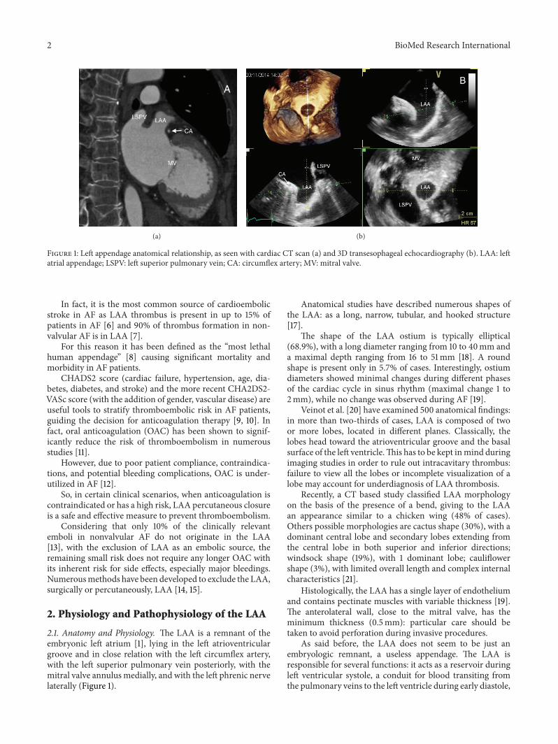
Table2:AC
Pregistrie
sincomparis
onwith
PROTE
CTAF.
In-hospital
Follo
w-up
Registr
yPatie
nts
Mean
age
(year5
)
Mean
CHADS2
score
Technical
success
Stroke
Peric
ardial
effusion
conservativ
e
Tampo
nade
(drainage)
Device
embo
lization
Death
(all
causes)
Total
adverse
events
Device
embo
lization
Peric
ardial
effusion
Thrombu
son
device
Stroke
Death
Italian
100
100/100
02/100
00
2/100
Registry
100%
2%2%
DualC
entre
131
131/1
310
1/131
00
01/1
31Ham
burg
100%
1%0.8%
Bern
ACPEU
Post
204
74±9
2.6±1.3
197/204
03/204
30
6/204
10
5/204
Market
97%
1.5%
2.9%
2.4%
Registr
ySpanish
3575±6
2.4±1.3
34/35
00
00
00
00
5/35
1/35
3/35
Registr
y97%
14%
3%9%
Initial
143
74±9
—132/137
3/143
4/143
5/143
2/143
010/14
3Eu
ropean
96%
2.1%
3%3.5%
1.4%
7%Ex
perie
nce
Bern
LAA
100
72±10
2.5±1.3
98/10
01/1
002/100
1/100
2/100
06/100
Occlusio
n98%
2%1%
2%6%
Registr
yInitialAs
ian
2068±9
2.3±1.3
19/20
00
00
0∗
——
——
—Ex
perie
nce
95%
Canadian
5274±8
3(2–4
)51/52
01/5
21/5
21/5
20
2/52
01/5
20
1/52
3/52
Registry
98%
2%2%
2%4%
2%2%
6%PR
OTE
CTAF
463
72±9
2.2±1.2
408/463
5/463
8/463
22/463
3/463
036/463
2/463
016/694
21/705
88%
1%1%
5%1%
8%0.4%
2.3%
3.0%
∗
:Airem
bolism
inrig
htcoronary
artery,one
esop
hagealinjury
durin
gTO
E.

8 BioMed Research International
(a) (b)
Figure 4: Left atrial appendage thrombosis (arrow), as seen with transesophageal echocardiography (a) and CT scan (b).
Figure 5: Severe spontaneous smoke effect (sludge) in left atrialappendage.
It has to be kept inmind that 2DTEE underestimates truedimensions in comparison to 3D TEE or CT measurements(Figure 6(b)) [58].
The maximal width of the LAA ostium is measured fromthe level of the left circumflex coronary artery up to a pointat 1-2 cm from the tip of the left superior pulmonary veinlimbus.
The maximal depth is measured from the ostium line tothe apex of the LAA. Sizing tables are available for both theWatchman and ACP devices.
The size of the chosen device should at least be 10–20%larger than the measured diameter: a correct oversizing isessential in order to avoid peridevice flow after deployment;on the other side, excessive oversizing may result in acompression of the left circumflex artery.
It is worth noting that the ostium of the LAA is typicallyelliptical, while all available occluders have a round shape,possibly accounting for incomplete sealing of the device andpossible cause of leakages.
ACP has to be preferred if the depth of the LAA is smallerthan the width of the ostium, because the placement of aWatchman device may result in an unstable position.
Preprocedural TEE evaluation could also be useful tobetter assess the thromboembolic risk of the patient. LAAdimensions, LAA velocities, left atrial dimensions and fibro-sis, left ventricular dysfunction, spontaneous echo-contrast,and aortic plaque (especially in aortic arc) have been asso-ciated with an increased thromboembolic risk [59]. In par-ticular, the presence of left atrial abnormalities is associated
with an embolic risk of 7.8%/year, aswell as aCHA2DS2-VAScscore of 5.
All these data could be useful to guide decisionson thromboembolic risk prevention in case of borderlineCHA2DS2-VASc scores.
4.2. Procedural Imaging to Guide Left Atrial AppendageOcclusion. TEE allows us to visualize the LAA during theprocedure: it is essential for the positioning and deploymentof the device.
The majority of centers perform the procedure undergeneral anesthesia with TEE and fluoroscopic guidance.There are only few reports of intracardiac echocardiographicguidance during percutaneous LAA occlusion.
TEE facilitates the transseptal puncture (Figure 7) andespecially 3D TEE can provide a real-time full view of theLAA, the shape of the ostium (Figure 8), with accuratemeasurements of the landing zone.
A final decision on device size is based on the informationcollected with all imaging modalities: echocardiography,fluoroscopy, and CT.
After deployment, a tug test should be performed demon-strating simultaneousmovement of the device and appendage(Figure 9). Optimally, the device should not protrude>4–7mm beyond the LAA ostium, and residual flow shouldbe <5mm by color Doppler with a compression grade of 8–20%, expressed in percent comparing the diameter of theimplanted device with the unconstricted diameter indicatedby themanufacturer.When optimal positioning is confirmed,the device is released. Rare device embolization after mobi-lization of the patient has been observed.
Following successful device deployment, the pericardiumis evaluated for effusion.
4.3. Follow-Up Imaging. Postprocedural imaging aims toassess device position, peridevice residual flow in the LAA,and thrombus formation on the device.
TEE (Figure 10) andCT can both be used for this purpose.In the PROTECT AF trial, serial TEE imaging was
performed at 45 days, 6 months, and 1 year following implant[38].
Residual peridevice flow is common (41% at 45 days) inpatients treated with theWatchman device. It is unclear if this

BioMed Research International 9
(a) (b)
Figure 6: Left atrial appendage measures with transesophageal echocardiography (a) and CT scan (b).
Figure 7: Real-time 3D echocardiography during transseptal punc-ture. The tip of the catheter (arrow) is passing from the right atrium(RA) to the left atrium (LA), through interatrial septum.
Figure 8: Progress of the delivery system in the left atrial appendage.
could be related to possible thromboembolic events, sincenew thrombi may be formed in the distal LAA pouch [48].Of note in the PROTECT AF trial, patients with a perideviceflow did not have a worse clinical outcome, regardless of thechosen antithrombotic therapy (warfarin, double antiplateletagents, ASA).
With Amplatzer devices, as the disc of the device typicallycovers the entire LAA ostium (pacifier principle), residualperidevice leaks are less frequent.
4.4. Anticoagulation after Implantation. Postprocedural anti-thrombotic therapy with warfarin or dual antiplatelet drugs
Figure 9: The image shows the so-called “tug test.” An AmplatzerCardiac Plug is pulled before the deployment.During thismaneuver,the distal part of the device (“disk,” arrow) is put in tension, while thedistal part (“lobe”) remains anchored in left atrial appendage.
Figure 10: An Amplatzer Cardiac Plug six months after implant,with perfect sealing and endothelization.
is recommended after implantation to avoid thrombus for-mation on the device until completion of endothelialization,provided there are no contraindications. For the Watchmandevice, the antithrombotic protocol of the PROTECT AFtrial is adopted: 45 days after implantation warfarin wasdiscontinued and substituted byASA+ clopidogrel if the TEEshowed the absence of thrombi or a residual peridevice flowof <5mm in width; clopidogrel was stopped if the 6 monthsTEE follow-up demonstrated no complications.
Usually, one antiplatelet agent is continued indefinitely,as most patients are elderly with evidence of atheroscleroticdisease, although the bleeding risk must be considered.

10 BioMed Research International
The PREVAIL trial and ASAP study provided evidencesfor double antiplatelet therapy instead of VKA for theWatchman device.
Observational studieswith theACP followed a regimen ofclopidogrel + ASA for 1month and acetylsalicylic acid for 3 to6 months [60], borrowing the antithrombotic protocol fromthe experience with the Amplatzer PFOOccluder. In patientswho are treated with antiplatelets drugs, it is reasonable toperform an imaging test (TEE or CT scan) before clopidogreltermination and again if ASA cessation is planned.
Incomplete LAA occlusion could create, theoretically,a thrombus-containing pocket with a source of possiblesystemic embolization. Anyway, as mentioned above, smallresidual shunts (<5mm) are usually considered irrelevantand may close spontaneously with time. When all patientswith residual shunts are included, the stroke risk is no differ-ent compared with patients in whom the LAA is completelyoccluded regardless of antithrombotic therapy [48].However,this remains a field open for debate.
5. Indications for Left AtrialAppendage Occlusion
PROTECT AF and PREVAIL randomized controlled trialswere included in the recent ESC focused guidelines on strokeprevention in patients with atrial fibrillation. In fact, theysuggest to use the CHA2DS2-VASc risk score >1 as thethreshold value for considering LAA occlusion [16].
Individual risk-benefit evaluation is fundamental, bear-ing in mind that the use of OAC has a primary recommenda-tion.
5.1. When Anticoagulation Is Not Possible. Patients with ahigh thromboembolic risk (CHA2DS2-VASc score of ≥2)but contraindication to systemic anticoagulation (e.g., his-tory of intracranial or life threatening bleeding, coagulationdisorders) represent the most accepted indication for LAAocclusion. In a survey of European centers, themost commonindication for LAA closure was an absolute contraindicationto OAC [61].
So far, no randomized trials targeting this specific groupof patients are available; in fact, this is the result of severalobservational studies and registries. However, the significantbleeding risk of dual antiplatelet therapy, indicated for 1or 6 months after implantation, has to be considered [62].Generally, this is only for a short time, thus reducing thecumulative risk of major bleeding events.
In patients who cannot receive any antiplatelet agent, theLariat technique can be considered.
5.2. When Oral Anticoagulation Is Possible. This is the onlyindication, as cited above, that is based on randomizedcontrolled trials. Those patients, in whom OAC or NOACare considered to pose an unacceptable bleeding risk (HAS-BLED ≥ 3), but with high stroke risk (CHA2DS2-VASc scoreof ≥ 2), should be considered for LAA occlusion.
The possibility of LAA occlusion should be discussedwith the patient, remembering that anticoagulation currentlyremains the standard of therapy.
Patients should be elucidated about the possibility oftherapy with NOAC, that, compared to OAC, have at least anequivalent and probably improved efficacy, with lower rate ofintracranial and, for some, lower overall bleeding risk.
It should be emphasized that we do not have any directdata comparing NOAC with LAA occlusion.
Ultimately, the decision belongs to a well-informedpatient in collaboration with the physician.
The HAS-BLED score does not characterize the bleedingrisk in certain categories of patients (e.g., patients withcancer or chronic inflammatory bowel disease): to these, LAAocclusion may also be offered.
Another possible scenario in which LAA occlusion couldbe helpful is the setting of triple anticoagulant therapy dueto coronary stent interventions in AF patients as it poses asignificant rise in bleeding risk [63].
End-stage renal failure poses patients at a high strokerisk and high bleeding risk: LAA occlusion could be adebatable alternative, keeping in mind that all NOAC arecontraindicated with creatinine clearance < 15mL/min andwarfarin could increase tissue calcification and enhancedatherosclerosis in this setting.
5.3. As a Complement to Anticoagulation. The combinationof LAA occlusion and OAC is debated and occasionally per-formed in patients with embolic events despite the adequateantithrombotic therapy, provided that there are no otherplausible causes (e.g., patients with mechanical prostheticvalves with evidence of thrombus in the LAA).
6. Conclusions
TheLAA is considered the “most lethal human appendage” asit causes significant mortality and morbidity in AF patients.We have to learn more about its complex role in physiologyand pathology.
However, LAA occlusion is becoming an interesting toolin reducing thromboembolic risk in certain categories ofpatients, as it has become a safe and effective procedure.
Indeed, we need new randomized prospective trialscomparing LAA procedure with NOAC, as they have a safetyprofile better than old OAC.
Other interesting aspects that warrant investigation arethe clinical significance of residual peridevice flow and thecorrect antithrombotic therapy after LAA closure.
In this way, we can shed light on thromboembolismmanagement in AF, in order to improve our knowledge whenchoosing between different measures to reduce the risk ofcatastrophic strokes.
Conflict of Interests
The authors declare that there is no conflict of interestsregarding the publication of this paper.
BioMed Research International 11
Acknowledgement
The authors want to sincerely thank St. Jude Medical ItaliaSpa for the scientific and economic support supplied to thewriting of this paper.
References
[1] H. M. F. Sherif, “The developing pulmonary veins and leftatrium: implications for ablation strategy for atrial fibrillation,”European Journal of Cardio-Thoracic Surgery, vol. 44, no. 5,Article ID ezt098, pp. 792–799, 2013.
[2] S. Qamruddin, J. Shinbane, J. Shriki, and T. Z. Naqvi, “Leftatrial appendage: structure, function, imaging modalities andtherapeutic options,” Expert Review of Cardiovascular Therapy,vol. 8, no. 1, pp. 65–75, 2010.
[3] B. D. Hoit, Y. Shao, and M. Gabel, “Influence of acutely alteredloading conditions on left atrial appendage flow velocities,”Journal of the American College of Cardiology, vol. 24, no. 4, pp.1117–1123, 1994.
[4] J. L. Madden, “Resection of the left auricular appendix; aprophylaxis for recurrent,”The Journal of the American MedicalAssociation, vol. 140, no. 9, pp. 769–772, 1949.
[5] Y. Takahashi, P. Sanders, M. Rotter, and M. Haıssaguerre,“Disconnection of the left atrial appendage for elimination offoci maintaining atrial fibrillation,” Journal of CardiovascularElectrophysiology, vol. 16, no. 8, pp. 917–919, 2005.
[6] R. Mahajan, A. G. Brooks, T. Sullivan et al., “Importance ofthe underlying substrate in determining thrombus location inatrial fibrillation: implications for left atrial appendage closure,”Heart, vol. 98, no. 15, pp. 1120–1126, 2012.
[7] J. L. Blackshear and J. A. Odell, “Appendage obliteration toreduce stroke in cardiac surgical patients with atrial fibrillation,”Annals of Thoracic Surgery, vol. 61, no. 2, pp. 755–759, 1996.
[8] W. D. Johnson, A. K. Ganjoo, C. D. Stone, R. C. Srivyas, andM. Howard, “The left atrial appendage: our most lethal humanattachment! Surgical implications,” European Journal of Cardio-thoracic Surgery, vol. 17, no. 6, pp. 718–722, 2000.
[9] B. F. Gage, A. D. Waterman, W. Shannon, M. Boechler, M. W.Rich, and M. J. Radford, “Validation of clinical classificationschemes for predicting stroke: results from the National Reg-istry of Atrial Fibrillation,”The Journal of the American MedicalAssociation, vol. 285, no. 22, pp. 2864–2870, 2001.
[10] G. Y. H. Lip, R. Nieuwlaat, R. Pisters, D. A. Lane, and H. J.G. M. Crijns, “Refining clinical risk stratification for predictingstroke and thromboembolism in atrial fibrillation using a novelrisk factor-based approach: the Euro Heart Survey on atrialfibrillation,” Chest, vol. 137, no. 2, pp. 263–272, 2010.
[11] R. G. Hart, O. Benavente, R. McBride, and L. A. Pearce,“Antithrombotic therapy to prevent stroke in patients with atrialfibrillation: a meta-analysis,” Annals of Internal Medicine, vol.131, no. 7, pp. 492–501, 1999.
[12] T. J. Bungard, W. A. Ghali, K. K. Teo, F. A. McAlister, and R.T. Tsuyuki, “Why do patients with atrial fibrillation not receivewarfarin?” Archives of Internal Medicine, vol. 160, no. 1, pp. 41–46, 2000.
[13] L. M. Tsai, J. H. Chen, L. J. Lin, and Y. J. Yang, “Roleof transesophageal echocardiography in detecting left atrialthrombus and spontaneous echo contrast in patientswithmitralvalve disease or non-rheumatic atrial fibrillation,” Journal of theFormosan Medical Association, vol. 89, no. 4, pp. 270–274, 1990.
[14] T. Lewalter, R. Ibrahim, B. Albers, and A. J. Camm, “Anupdate and current expert opinions on percutaneous left atrialappendage occlusion for stroke prevention in atrial fibrillation,”Europace, vol. 15, no. 5, pp. 652–656, 2013.
[15] S. Chatterjee, J. C. Alexander, P. J. Pearson, andT. Feldman, “Leftatrial appendage occlusion: lessons learned from surgical andtranscatheter experiences,” Annals of Thoracic Surgery, vol. 92,no. 6, pp. 2283–2292, 2011.
[16] B. Meier, Y. Blaauw, A. A. Khattab et al., “EHRA/EAPCI expertconsensus statement on catheter-based left atrial appendageocclusion,” Europace, vol. 16, no. 10, pp. 1397–1416, 2014.
[17] G. Ernst, C. Stollberger, F. Abzieher et al., “Morphology of theleft atrial appendage,” Anatomical Record, vol. 242, no. 4, pp.553–561, 1995.
[18] P. Su, K. P. McCarthy, and S. Y. Ho, “Occluding the left atrialappendage: anatomical considerations,”Heart, vol. 94, no. 9, pp.1166–1170, 2008.
[19] P. Santangeli, L. Di Biase, R. Horton, J. D. Burkhardt, andA. Natale, “CT imaging to assess the left atrial appendageanatomy: clinical implications,” in Computed Tomography—Clinical Applications, L. Saba, Ed., InTech Open Access Publish-ing, Vienna, Austria, 2012.
[20] J. P. Veinot, P. J. Harrity, F. Gentile et al., “Anatomy of the normalleft atrial appendage: a quantitative study of age-related changesin 500 autopsy hearts: implications for echocardiographicexamination,” Circulation, vol. 96, no. 9, pp. 3112–3115, 1997.
[21] L. di Biase, P. Santangeli, M. Anselmino et al., “Does the leftatrial appendage morphology correlate with the risk of strokein patients with atrial fibrillation? Results from a MulticenterStudy,” Journal of the American College of Cardiology, vol. 60,no. 6, pp. 531–538, 2012.
[22] P. Barbier, S. B. Solomon, N. B. Schiller, and S. A. Glantz, “Leftatrial relaxation and left ventricular systolic function determineleft atrial reservoir function,” Circulation, vol. 100, no. 4, pp.427–436, 1999.
[23] T. Tabata, T. Oki, H. Yamada et al., “Role of left atrial appendagein left atrial reservoir function as evaluated by left atrialappendage clamping during cardiac surgery,” American Journalof Cardiology, vol. 81, no. 3, pp. 327–332, 1998.
[24] P. Massoudy, S. Beblo, P. Raschke, S. Zahler, and B. F. Becker,“Influence of intact left atrial appendage on hemodynamicparameters of isolated guinea pig heart,” European Journal ofMedical Research, vol. 3, no. 10, pp. 470–474, 1998.
[25] C. Chapeau, J. Gutkowska, P. W. Schiller et al., “Localization ofimmunoreactive synthetic atrial natriuretic factor (ANF) in theheart of various animal species,” Journal of Histochemistry andCytochemistry, vol. 33, no. 6, pp. 541–550, 1985.
[26] C. T. Kappagoda, R. J. Linden, and H. M. Snow, “The effectof distending the atrial appendages on urine flow in the dog,”Journal of Physiology, vol. 227, no. 1, pp. 233–242, 1972.
[27] C. Pollick and D. Taylor, “Assessment of left atrial appendagefunction by transesophageal echocardiography: implicationsfor the development of thrombus,”Circulation, vol. 84, no. 1, pp.223–231, 1991.
[28] M. Zabalgoitia, J. L.Halperin, L. A. Pearce, J. L. Blackshear, R.W.Asinger, and R. G. Hart, “Transesophageal echocardiographiccorrelates of clinical risk of thromboembolism in nonvalvularatrial fibrillation,” Journal of the American College of Cardiology,vol. 31, no. 7, pp. 1622–1626, 1998.
[29] T. Watson, E. Shantsila, and G. Y. Lip, “Mechanisms of throm-bogenesis in atrial fibrillation: Virchow’s triad revisited,” TheLancet, vol. 373, no. 9658, pp. 155–166, 2009.

12 BioMed Research International
[30] M. A. Garcia-Fernandez, E. G. Torrecilla, D. S. Roman et al.,“Left atrial appendage Doppler flow patterns: implications onthrombus formation,” American Heart Journal, vol. 124, no. 4,pp. 955–961, 1992.
[31] T. Takada, M. Yasaka, K. Nagatsuka, K. Minematsu, and T.Yamaguchi, “Blood flow in the left atrial appendage and embolicstroke in nonvalvular atrial fibrillation,” European Neurology,vol. 46, no. 3, pp. 148–152, 2001.
[32] H. Inoue, T.Nozawa, K.Okumura, L. Jong-Dae, A. Shimizu, andK. Yano, “Prothrombotic activity is increased in patients withnonvalvular atrial fibrillation and risk factors for embolism,”Chest, vol. 126, no. 3, pp. 687–692, 2004.
[33] A. Frustaci, C. Chimenti, F. Bellocci, E. Morgante, M. A. Russo,and A. Maseri, “Histological substrate of atrial biopsies inpatients with lone atrial fibrillation,” Circulation, vol. 96, no. 4,pp. 1180–1184, 1997.
[34] J. L. Cox, R. B. Schuessler, H. J. D’Agostino Jr. et al., “The surgicaltreatment of atrial fibrillation: III. Development of a definitivesurgical procedure,” Journal of Thoracic and CardiovascularSurgery, vol. 101, no. 4, pp. 569–583, 1991.
[35] A. G. Dawson, S. Asopa, and J. Dunning, “Should patientsundergoing cardiac surgerywith atrial fibrillation have left atrialappendage exclusion?” Interactive Cardiovascular and ThoracicSurgery, vol. 10, no. 2, pp. 306–311, 2010.
[36] M.A.Garcıa-Fernandez, E. Perez-David, J. Quiles et al., “Role ofleft atrial appendage obliteration in stroke reduction in patientswith mitral valve prosthesis: a transesophageal echocardio-graphic study,” Journal of the American College of Cardiology,vol. 42, no. 7, pp. 1253–1258, 2003.
[37] J. S. Healey, E. Crystal, A. Lamy et al., “Left Atrial AppendageOcclusion Study (LAAOS): results of a randomized controlledpilot study of left atrial appendage occlusion during coronarybypass surgery in patients at risk for stroke,”TheAmericanHeartJournal, vol. 150, no. 2, pp. 288–293, 2005.
[38] D. R. Holmes, V. Y. Reddy, Z. G. Turi et al., “Percutaneousclosure of the left atrial appendage versus warfarin therapyfor prevention of stroke in patients with atrial fibrillation: arandomised non-inferiority trial,”TheLancet, vol. 374, no. 9689,pp. 534–542, 2009.
[39] K. Bartus, J. Bednarek, J. Myc et al., “Feasibility of closed-chestligation of the left atrial appendage in humans,” Heart Rhythm,vol. 8, no. 2, pp. 188–193, 2011.
[40] H. Sievert, M. D. Lesh, T. Trepels et al., “Percutaneous left atrialappendage transcatheter occlusion to prevent stroke in high-risk patients with atrial fibrillation: early clinical experience,”Circulation, vol. 105, no. 16, pp. 1887–1889, 2002.
[41] S. H. Ostermayer, M. Reisman, P. H. Kramer et al., “Per-cutaneous Left Atrial Appendage Transcatheter Occlusion(PLAATO System) to prevent stroke in high-risk patients withnon-rheumatic atrial fibrillation: results from the internationalmulti-center feasibility trials,” Journal of the American College ofCardiology, vol. 46, no. 1, pp. 9–14, 2005.
[42] Y. Bayard, H. Omran, P. Neuzil et al., “PLAATO (PercutaneousLeft Atrial Appendage Transcatheter Occlusion) for preventionof cardioembolic stroke in non-anticoagulation eligible atrialfibrillation patients: results from the European PLAATO study,”EuroIntervention, vol. 6, no. 2, pp. 220–226, 2010.
[43] F. Nietlispach, S. Gloekler, R. Krause et al., “Amplatzer leftatrial appendage occlusion: single center 10-year experience,”Catheterization and Cardiovascular Interventions, vol. 82, no. 2,pp. 283–289, 2013.
[44] S. C. Lam, S. Bertog, S. Gafoor et al., “Left atrial appendageclosure using the amulet device: an initial experience with thesecond generation amplatzer cardiac plug,” Catheterization andCardiovascular Interventions, vol. 85, no. 2, pp. 297–303, 2015.
[45] X. Freixa, A. Abualsaud, J. Chan et al., “Left atrial appendageocclusion: initial experience with the Amplatzer Amulet,” Inter-national Journal of Cardiology, vol. 174, no. 3, pp. 492–496, 2014.
[46] D. R. Holmes, S. Kar, M. J. Price et al., “Prospective randomizedevaluation of the watchman left atrial appendage closure devicein patients with atrial fibrillation versus long-term warfarintherapy,” Journal of the American College of Cardiology, vol. 64,no. 1, pp. 1–12, 2013.
[47] S. M. Singh, A. Micieli, and H. C. Wijeysundera, “Economicevaluation of percutaneous left atrial appendage occlusion,dabigatran, and warfarin for stroke prevention in patients withnonvalvular atrial fibrillation,” Circulation, vol. 127, no. 24, pp.2414–2423, 2013.
[48] J. F. Viles-Gonzalez, S. Kar, P. Douglas et al., “The clinicalimpact of incomplete left atrial appendage closure with thewatchman device in patients with atrial fibrillation: a PRO-TECT AF (percutaneous closure of the left atrial appendageversus warfarin therapy for prevention of stroke in patients withatrial fibrillation) substudy,” Journal of the American College ofCardiology, vol. 59, no. 10, pp. 923–929, 2012.
[49] R. J. Lee, K. Bartus, and S. J. Yakubov, “Catheter-based left atrialappendage (LAA) ligation for the prevention of embolic eventsarising from the LAA initial experience in a canine model,”Circulation: Cardiovascular Interventions, vol. 3, no. 3, pp. 224–229, 2010.
[50] M. A. Miller, S. R. Gangireddy, S. K. Doshi et al., “Multicenterstudy on acute and long-term safety and efficacy of percuta-neous left atrial appendage closure using an epicardial suturesnaring device,” Heart Rhythm, vol. 11, no. 11, pp. 1853–1859,2014.
[51] M. J. Price, D. N. Gibson, S. J. Yakubov et al., “Early safety andefficacy of percutaneous left atrial appendage suture ligation:results from the U.S. transcatheter LAA ligation consortium,”Journal of the American College of Cardiology, vol. 64, no. 6, pp.565–572, 2014.
[52] M. R. Afzal, A. Kanmanthareddy, M. Earnest et al., “Impactof left atrial appendage exclusion using an epicardial ligationsystem (LARIAT) on atrial fibrillation burden in patients withcardiac implantable electronic devices,” Heart Rhythm, vol. 12,no. 1, pp. 52–59, 2015.
[53] B. Meier, I. Palacios, S. Windecker et al., “Transcatheter leftatrial appendage occlusion with Amplatzer devices to obviateanticoagulation in patients with atrial fibrillation,” Catheteriza-tion andCardiovascular Interventions, vol. 60, no. 3, pp. 417–422,2003.
[54] S. Puwanant, B. C. Varr, K. Shrestha et al., “Role of the CHADS2score in the evaluation of thromboembolic risk in patients withatrial fibrillation undergoing transesophageal echocardiogra-phy before pulmonary vein isolation,” Journal of the AmericanCollege of Cardiology, vol. 54, no. 22, pp. 2032–2039, 2009.
[55] J. Hur, Y. J. Kim, H.-J. Lee et al., “Cardioembolic stroke: dual-energy cardiac CT for differentiation of left atrial appendagethrombus and circulatory stasis,” Radiology, vol. 263, no. 3, pp.688–695, 2012.
[56] H. Ohyama, N. Hosomi, T. Takahashi et al., “Comparison ofmagnetic resonance imaging and transesophageal echocardio-graphy in detection of thrombus in the left atrial appendage,”Stroke, vol. 34, no. 10, pp. 2436–2439, 2003.

BioMed Research International 13
[57] S. S. I. Bokhari, P. Martinez-Clark, J. P. Zambrano, M. Tracy,and W. W. O’Neill, “Percutaneous mechanical thrombectomyof left atrial appendage thrombus with bilateral neuro-embolicprotection followed by closure of left atrial appendage,” EuroIn-tervention, vol. 8, no. 3, pp. 408–409, 2012.
[58] G. Nucifora, F. F. Faletra, F. Regoli et al., “Evaluation of the leftatrial appendage with real-time 3-dimensional transesophagealechocardiography: implications for catheter-based left atrialappendage closure,” Circulation: Cardiovascular Imaging, vol. 4,no. 5, pp. 514–523, 2011.
[59] The Stroke Prevention in Atrial Fibrillation Investigators Com-mittee on Echocardiography, “Transesophageal echocardio-graphic correlates of thromboembolism in high- risk patientswith nonvalvular atrial fibrillation,”Annals of InternalMedicine,vol. 128, no. 8, pp. 639–687, 1998.
[60] E. E. Guerios, M. Schmid, S. Gloekler et al., “Left atrialappendage closure with the Amplatzer cardiac plug in patientswith atrial fibrillation,” Arquivos Brasileiros de Cardiologia, vol.98, no. 6, pp. 528–536, 2012.
[61] G. Y. H. Lip, N. Dagres, A. Proclemer, J. H. Svendsen, L. Pison,and C. Blomstrom-Lundqvist, “Left atrial appendage occlusionfor stroke prevention in atrial fibrillation in Europe: results ofthe EuropeanHeart RhythmAssociation survey,” Europace, vol.15, no. 1, pp. 141–143, 2013.
[62] J. S. Healey, R. G. Hart, J. Pogue et al., “Risks and benefits oforal anticoagulation compared with clopidogrel plus aspirin inpatients with atrial fibrillation according to stroke risk: the atrialfibrillation clopidogrel trial with irbesartan for prevention ofvascular events (ACTIVE-W),” Stroke, vol. 39, no. 5, pp. 1482–1486, 2008.
[63] K.Huber, K. Juhani Airaksinen, T. Cuisset, F.Marın, A. Rubboli,and G. Y. H. Lip, “Antithrombotic therapy in patients withatrial fibrillation undergoing coronary stenting: similarities anddissimilarities between northAmerica and Europe,”Thrombosisand Haemostasis, vol. 106, no. 4, pp. 569–571, 2011.

Submit your manuscripts athttp://www.hindawi.com
Stem CellsInternational
Hindawi Publishing Corporationhttp://www.hindawi.com Volume 2014
Hindawi Publishing Corporationhttp://www.hindawi.com Volume 2014
MEDIATORSINFLAMMATION
of
Hindawi Publishing Corporationhttp://www.hindawi.com Volume 2014
Behavioural Neurology
EndocrinologyInternational Journal of
Hindawi Publishing Corporationhttp://www.hindawi.com Volume 2014
Hindawi Publishing Corporationhttp://www.hindawi.com Volume 2014
Disease Markers
Hindawi Publishing Corporationhttp://www.hindawi.com Volume 2014
BioMed Research International
OncologyJournal of
Hindawi Publishing Corporationhttp://www.hindawi.com Volume 2014
Hindawi Publishing Corporationhttp://www.hindawi.com Volume 2014
Oxidative Medicine and Cellular Longevity
Hindawi Publishing Corporationhttp://www.hindawi.com Volume 2014
PPAR Research
The Scientific World JournalHindawi Publishing Corporation http://www.hindawi.com Volume 2014
Immunology ResearchHindawi Publishing Corporationhttp://www.hindawi.com Volume 2014
Journal of
ObesityJournal of
Hindawi Publishing Corporationhttp://www.hindawi.com Volume 2014
Hindawi Publishing Corporationhttp://www.hindawi.com Volume 2014
Computational and Mathematical Methods in Medicine
OphthalmologyJournal of
Hindawi Publishing Corporationhttp://www.hindawi.com Volume 2014
Diabetes ResearchJournal of
Hindawi Publishing Corporationhttp://www.hindawi.com Volume 2014
Hindawi Publishing Corporationhttp://www.hindawi.com Volume 2014
Research and TreatmentAIDS
Hindawi Publishing Corporationhttp://www.hindawi.com Volume 2014
Gastroenterology Research and Practice
Hindawi Publishing Corporationhttp://www.hindawi.com Volume 2014
Parkinson’s Disease
Evidence-Based Complementary and Alternative Medicine
Volume 2014Hindawi Publishing Corporationhttp://www.hindawi.com