Embed Size (px)
Citation preview

Int J Clin Exp Med 2018;11(5):4371-4388www.ijcem.com /ISSN:1940-5901/IJCEM0061760
Review ArticleDifferences between open-wedge versus closed-wedge high tibial osteotomy on clinical and radiological outcomes and adverse events
Ying-Jie Deng1*, Yuan Yuan2*, Yan-Ming Ren3*, Qi Li4, Duan Wang4, Yang Yang5,6, Chao You3, Rui Fang1, Dong-Mei Zhao7
1Department of Orthopedics, Hospital of Traditional Chinese Medicine Affiliated to Xinjiang Medical University, Urumqi 830000, P.R. China; 2Department of Radiology, West China Hospital, Sichuan University, Chengdu 610041, P.R. China; 3Department of Neurosurgery, West China Hospital, Sichuan University, Chengdu, P.R. China; 4Department of Orthopedics, West China Hospital/West China School of Medicine, Sichuan University, Chengdu, P.R. China; 5State Key Laboratory of Biotherapy, West China Hospital, Sichuan University and National Collab-orative Innovation Center, Chengdu, P.R. China; 6State Key Laboratory of Oral Diseases, West China Hospital of Stomatology, Sichuan University, Chengdu, P.R. China; 7Department of Obstetrics and Gynecology, West China Second University Hospital, Sichuan University, Chengdu 610041, P.R. China. *Equal contributors.
Received July 18, 2017; Accepted January 9, 2018; Epub May 15, 2018; Published May 30, 2018
Abstract: High tibial osteotomy (HTO) is a widely accepted procedure to treat medial knee osteoarthritis, which is performed either with open-wedge (OWO) or closed-wedge osteotomy (CWO). This meta-analysis was designed to evaluate whether OWO had an advantage over CWO regarding the clinical and radiographic results, and complica-tions. Multiple comprehensive databases, including PubMed, Embase, Web of Science and Cochrane Library, were searched from their inception to October 2017. A total of 28 publications reporting 24 clinical trials were finally eligible. The OWO showed significantly greater posterior tibial slope compared to CWO, and the result remained unchanged over time. Moreover, patellar height decreased after OWO, but increased after CWO. No significant dif-ference was detected for complications, such as infection, re-operation, nonunion, and revision to joint arthroplasty. No difference was found between them regarding the improvement of pain and knee function. Although both tech-niques led to the good and comparable clinical results, we recommended OWO as an alternatively effective treat-ment option for selected younger patients, which is easier to perform and to convert to TKA, avoids tibiofibular joint disruption and common peroneal nerve palsy, and permits multiplanar correction, as compared to CWO. However, additional well-designed studies are required to identify fixation type and augment selection in OWO.
Keywords: Total knee arthroplasty, high tibial osteotomy, clinical outcome, meta-analysis
Introduction
Varus deformity of knee joint increases the risk of progression of medial compartment osteoar-thritis (OA) [1, 2]. High tibial osteotomy (HTO) is a well-established surgical technique for indi-viduals with medial OA and varus deformity. It can shift the mechanical axis towards the lat-eral compartment to change the load distribu-tion across the knee and weaken the stress concentration in the medial compartment of knee [3, 4]. Two techniques, such as closed-wedge osteotomy (CWO) and open-wedge oste-otomy (OWO), are commonly available [5-7]. Many studies showed good clinical and radio-logical results for both OWO and CWO tech-niques [8-10]. The CWO has several disadvan-tages, such as more demanding subsequent
TKA, the need for fibular osteotomy, more neu-rovascular complications, and bone stock loss [5, 6, 11]. Compared to CWO, OWO is described later in order to avoid fibular osteotomy and thereby reduce the rate of co-morbidity related to it. In addition, with the introduction of spe-cific locked implants and bone-substituting bio-materials, OWO has gained popularity in recent years, because it has several advantages over CWO, including rapid rehabilitation, easier sub-sequent TKA, and easier adjustment of align-ment correction [12-15]. Nevertheless, OWO can result in some complications, such as rela-tively high nonunion rates at the osteotomy site, patella baja and increased posterior tibial slope [5, 6, 16]. In addition, many of these stud-ies contained relatively small cohorts and dem-

Differences between open-wedge versus closed-wedge high tibial osteotomy
4372 Int J Clin Exp Med 2018;11(5):4371-4388
onstrated inconsistent outcomes [2, 17-20]. Thus, no superiority of any technique over the other has been proven, and the choice remains basically on the surgeon’s preference.
Several meta-analyses were published on this topic without conclusive results [21-24]. Smith et al. reported that no difference was identified for any clinical outcome or complication between OWO and CWO [24]. However, Sun et al. demonstrated that CWO led to a higher inci-dence of opposite cortical fracture, and OWO provided higher accuracy of correction [21]. Unfortunately, these studies contained some methodological shortcomings, errors in inclu-sion criteria and data extraction, and high het-erogeneity. Not only did these studies have these limitations, but also their conclusions were inconsistent (Table 1). Two meta-analyses only investigated the radiographic results between OWO and CWO [22, 25]. But we think that the “safety” and “clinical outcomes” of the two techniques should also be the key words. Considering all these issues, whether OWO has advantages over CWO for the patients with medial compartment OA remains controversial, and it is impossible to give clear advice on which method to adopt. Recently, numerous studies on this topic have been published with inconclusive results [2, 11, 17, 19, 26-33]. Moreover, two investigations with longer follow-up time have been published recently, so we can conduct subgroup analysis to evaluate whether the clinical and radiological effect of HTO changes with time [2, 17].
Thus, we conducted this meta-analysis to determine whether OWO was superior to CWO with respect to: 1) clinical outcomes; 2) radio-logical outcomes; and 3) complications. Fur- thermore, to perform a reliable and compre-hensive evaluation of the advantages and dis-advantages of these two procedures, we added many new statistical indicators that have not been reported before.
Materials and methods
This meta-analysis design was based on the Preferred Reporting Items for Systematic Re- views and Meta-Analyses (PRISMA) prospec- tively.
Literature search
We systematically searched multiple electronic databases including PubMed, Embase, Web of
Science, and Cochrane Library from their incep-tion to 29th October 2017. The search strate-gies adopted included the following terms: (“High tibial osteotomy” OR “Tibial osteotomy” OR “Osteotomy”) AND (“Open” OR “Close” OR “Closed” OR “Closing”).
After the initial electronic search, Google Scholar and the citation lists of the studies included were checked for relevant articles. Searches of relevant journals, medical associa-tion and society web sites were also conducted. Each eligible study’s corresponding author was contacted via e-mail to obtain any further infor-mation. There were no restrictions in terms of the date, research site, or status of publi- cation.
Study selection
All randomized controlled trials (RCTs) and non-RCTs (n-RCTs) were eligible for inclusion if they satisfied the search strategy and compared OWO to CWO regarding the following outcomes: Clinical outcomes (length of hospital stay, oper-ation time, patient satisfaction, Hospital for Special Surgery (HSS) knee score, Lysholm knee score, knee range of motion (ROM), knee pain visual analog score (VAS); Radiographic outcomes (patellar height measured by three different methods, posterior tibial slope angle, hip-knee-ankle (HTA) angle, and mean angle of correction); Complications (infection, revision surgery, non-union, common peroneal nerve palsy, tibial plateau fracture, and revision to TKA). Any cadaver and animal study, comment, letter, editorial, protocol, guideline, and review papers were excluded.
Study identification was performed based on the predefined eligibility criteria. After eliminat-ing duplications, two reviewers independently screened the titles and abstracts of all studies that were identified with the search strategy and discarded those that were obviously ineli-gible. If suitability could not be determined, the full article was assessed. Any disagreement was resolved through discussion.
Data extraction
The data were collated independently by two reviewers onto a predefined data extraction form. Variables recorded included: patient demographics, surgical technique, implant used, follow-up period, methodology, complica-

Differences between open-wedge versus closed-wedge high tibial osteotomy
4373 Int J Clin Exp Med 2018;11(5):4371-4388
Table 1. Details of meta-analyses and systematic review published on this subjectAuthor Year Studies included Patients (knees) Indicators Conclusions Main ShortcomingSmith et al. 2011 12 pubications (9
clinical trials)621 (642) Clinical and
radiological results
1. No significant difference was found for any clinical outcome; 2. Significantly greater posterior tibial slope and reduced patellar height and hip-knee-ankle angle following opening-wedge HTO
1. Numerous studies on this subject have been published with inconclusive results; 2. Two investigations with longer follow-up time have been published
Brouwer et al. 2014 21 pubications (14 clinical trials)
1065 Clinical and radiological
results
1. No evidence suggests differences between different oste-otomy techniques; 2. No evidence shows whether an osteotomy is more effective than alternative surgical treatment such as unicompartmental knee replacement or non-operative treatment
1. CWO was compared with other types of HTO such as OWO, combined high tibial osteotomy, etc.; 2. All the osteotomy techniques except CWO were considered as identical method
Bin et al. 2016 23 studies 1150 Patellar height
1. The patellar height decreased after OWO, except when as-sessed by ISI; 2. Patellar height was unchanged after closing wedge HTO, regardless of the measurement method
1. They did not use sensitivity analysis to investigate the origin of high heterogeneity, thereby resulting in an un-stable result; 2. The meta-analysis included OCS, PCS and RCS, but subgroup analysis based on article type was not conducted to evaluate the stability of the meta-analysis; 3. These included studies with different follow-up times was pooled for analysis. They should conduct subgroup analysis to evaluate whether the clinical and radiological effect of HTO changes with time; 4. They only focused on the patellar height, we think that “safety” of the two techniques should be the key words
Sun et al. 2016 23 publications (17 clinical trials)
1444 (1483) Clinical and radiological
results
1. OWO increased the posterior slope angle and decreased the patellar height, and provided higher accuracy of correction; 2. CWO led to a higher incidence of opposite cortical fracture.
1. They stated that they excluded the duplicated publica-tions and chose one of them, but two duplicated publica-tions were included in this study; 2. The significant differ-ence between the groups regarding complications was conducted based on two publications. They concluded that CWO led to a higher incidence of opposite cortical fracture, which was not credit; 3. They used “Cochrane tool” to assessing risk of bias for RCTs and n-RCTs; 4. A sensitivity analysis was conducted to compare the out-comes between OWO and CWO for RCTs only. With a small number of studies, authors have suggested that random effects models may not be reliable irrespective of the homogeneity test; 5. The random-effects model should be compared with the fixed-effects model
Nha et al. 2016 27 studies 1260 Posterior tibial slope
1. Posterior tibial slope increased after OWO and decreased after CWO; 2. Both osteotomy techniques may have little effect on the biomechanics of the cruciate ligaments
1. They did not use sensitivity analysis to investigate the origin of high heterogeneity, thereby resulting in an un-stable result; 2. The meta-analysis included OCS, PCS and RCS, but subgroup analysis based on article type was not conducted to evaluate the stability of the meta-analysis; 3. These included studies with different follow-up times was pooled for analysis. They should conduct subgroup analysis to evaluate whether the clinical and radiological effect of HTO changes with time; 4. They only focused on the patellar height, we think that “safety” of the two techniques should be the key words
CWO, closed wedge high tibial osteotomy; OWO, open wedge high tibial osteotomy; HTO, high tibial osteotomy; PCS, prospective comparison study; RCS, retrospective comparison study; OCS, observational case series.

Differences between open-wedge versus closed-wedge high tibial osteotomy
4374 Int J Clin Exp Med 2018;11(5):4371-4388
tions, and clinical and radiographic outcomes. Any disagreement unresolved after discussion was decided by a third reviewer. In addition, the study authors were contacted by email to request these variables that were not provided in the publications. Missing standard devia-tions were calculated through the confidence intervals (CI) or range of values available in the studies.
Special focus was paid on knee function out-comes between OWO and CWO. Therefore, the primary outcomes were severity of pain, the improvement in HSS and ROM. While, other clinical outcomes, radiographic assessment, and complications were regarded as secondary outcomes.
Assessment of study quality
Two independent reviewers evaluated the methodological quality of eligible studies using
the modified Jadad scale for RCTs and the Newcastle-Ottawa Scale for n-RCTs. The modi-fied Jadad scale contains four questions evalu-ating random sequence generation (2 points), allocation concealment (2 points), blinding appropriateness (2 points), and dropouts or withdrawn description (1 point). The total score is 7 points, and a study with 4-7 points is con-sidered as a high-quality study. The Newcastle-Ottawa Scale awards stars depending on the level of bias, which includes low (1 star), high, and unclear bias. We assessed each study on 3 criteria: the selection of the study groups (0-4 stars), the comparability of the groups (0-2 stars), and the ascertainment of the exposure or outcome of interest (0-3 stars). Total scores range from 0 to 9 points. A study with scores of 7-9 points is regarded as a high-quality study. Any discrepancies were settled by consensus or by consultation with a third reviewer.
Figure 1. Flow chart showing study identification, inclusion and exclusion.

Differences between open-wedge versus closed-wedge high tibial osteotomy
4375 Int J Clin Exp Med 2018;11(5):4371-4388
Statistical analysis
The statistical analysis was performed using Review Manager 5.3 software (The Nordic Cochrane Center, The Cochrane Collaboration) and a P-value <0.05 was considered to be sig-nificant. For each eligible study, the results of dichotomous variables were presented as odds ratios (OR) with corresponding 95% CI. If con-tinuous variables were measured in the same way between studies, the mean differences (MD) were used. Otherwise, standardized mean differences (Std MD) with 95% CI values were adopted.
The assessment of heterogeneity was conduct-ed with use of the chi-squared test and I2 statis-tic to determine appropriateness for meta-anal-ysis. If statistical heterogeneity was moderate (P<0.1 or I2>50%), a random-effects model was chosen to estimate the overall effect sizes. When these conditions were not satisfied, fixed-effects model was used. Moreover, publi-cation bias was assessed with funnel plots and the Egger test of the primary outcomes. Subgroup analysis was performed for different time points (<1 year or >1 year). Moreover, sen-sitivity analysis was carried out to investigate the potential sources of heterogeneity and to determine the effect of study quality on the results.
Results
Literature search and population characteris-tics
The process of study selection was showed in Figure 1. We totally identified two hundred and ninety-seven records via our search strategy. Another three studies were identified through manual searching. After screening the search results, eleven investigations had been pub-lished by the four independent research teams. To eliminate the effect of duplicate data, we attempted to contact each author in order to confirm whether the data in their studies were reported from the same patient cohorts. One study [8] was a duplicate of a previous study presenting the same parameters [34]. However, eight publications reported the results of differ-ent parameters from three separate studies [2, 11, 12, 34-38]. In addition, two investigations were conducted as an update of two previous reports after longer follow-up time [2, 17, 20,
34]. Therefore, these studies were included in this meta-analysis, but we only analyzed the population characteristics of these four indi-vidual patient cohorts once. After removing duplications and reading the full-text, a total of 10 RCTs and 18 n-RCTs met our inclusion crite-ria (Figure 1).
Table 2 displayed the detailed characteristics of the studies. A total of 2701 evaluable knees were available for analysis in this study. These 24 eligible studies included 1368 knees that underwent closed-wedge HTO and 1335 knees that underwent open-wedge HTO. All publica-tions were published in English from 1999 to 2016. The mean age of the patients ranged from 34 to 60 years. And, 44.7% of the patients were males. Although the osteotomies were fix-ated using plates or staples in the majority of the studies, we included all different fixation methods to compare the two approaches. The evidence-based methodological assessment was varied. Table 2 presented the results of modified Jadad and the Newcastle-Ottawa clin-ical critical appraisal. Nineteen of the twenty-four studies achieved a high-quality score (modified Jadad ≥4 or Newcastle-Ottawa ≥7) (Table 2). We used data reporting a change from baseline as our effect index in primary outcomes (VAS, HSS and ROM).
Primary outcomes
VAS pain score
Data on 336 knees (including 166 knees that underwent open-wedge HTO and 170 knees that underwent closed-wedge HTO) were pooled from seven trials in eight publications [2, 17, 18, 20, 27, 34, 35, 39] analyzing the VAS pain scores. Pooled results illustrated no significant difference in the improvement of knee pain in the OWO group, when compared to the CWO group (MD=-0.15, 95% CI=-0.22 to 0.52; P=0.24; I2=40%). Considering that the origin of heterogeneity may be attributed to the duration of follow-up, subgroup analysis was conducted based on different follow-up time (Group A: less than 1 year follow-up; Group B: more than 1 year follow-up). No statistically significant dif-ference was identified either in Group A (P=0.45; I2=34%) or in Group B (P=0.68; I2=35%) with low heterogeneity (Figure 2 and Table 3).

Differences between open-wedge versus closed-wedge high tibial osteotomy
4376 Int J Clin Exp Med 2018;11(5):4371-4388
Table 2. Characteristics of the included studies
Author Year StudyCases (n) Age (mean) Gender (M/F) Fixation
Quality scoreCW OW CW OW CW OW CW OW
Kim et al. 2016 RCT 30 30 54 54 10/20 9/21 Stepped plate TomoFix plate 6Van Egmond et al. 2016 RCT 19 19 NA NA NA NA Locked plate Locked plate 4Nerhus et al. 2015 RCT 35 35 30-60 30-60 NA NA Puddu titanium plate Staples 6Duivenvoorden 1 et al. 2014 RCT 45 36 50 50 27/18 21/33 Staples Puddu plate 4Gaasbeek et al. 2010 RCT 25 25 50 47 16/9 14/11 Locked plate Locked plate 4Luites et al. 2009 RCT 19 23 53 53 NA NA TF plates and screws TF plates and screws 5Brouwer 1 et al. 2006 RCT 47 45 51 50 24/20 27/20 Staples Puddu plate 4Brouwer 2 et al. 2005 RCT 24 26 53 48 12/12 20/6 Staples Puddu plate 4Magyar 1 et al. 1999 RCT 15 18 53 55 12/3 10/8 Bone staple Orthofix OF-Garche Fixtior 5Magyar 2 et al. 1999 RCT 25 25 55 55 NA NA Bone staple External fixation 5Agarwala et al. 2016 PCS 25 23 55 56 13/10 13/12 Locked plate and screws Locked plate and screws 6Duivenvoorden 2 et al. 2015 RCS 354 112 49 48 NA NA Three different staples Puddu plate/Tomofix plate 7Portner et al. 2014 RCS 18 26 46 44 15/3 20/6 Staple gun Plate and screws 7Deie et al. 2014 RCS 12 9 58 58 3/9 3/6 Plate and screws Plate and screws 6Tabrizi et al. 2013 PCS 21 21 37 35 12/4 13/3 Plates L or T plates 8Bae et al. 2013 RCS 78 30 58 56 4/70 2/25 Miniplate staple Puddu plate 6Amzallag et al. 2013 PCS 97 224 50 53 NA NA NA NA 8Songet I.H. al 2012 RCS 50 50 60 58 12/38 10/40 Stepped staples Wedge plates 7Ducat et al. 2012 PCS 92 210 50 52 NA NA NA NA 7Magnussen et al. 2011 RCS 32 32 59 54 21/9 22/10 Blade and screws Tomofix plate 8Song E.K. et al. 2010 RCS 104 90 57 51 16/88 21/69 Two staples Aescula plates 6Hankemeier et al. 2010 RCS 26 35 53 44 NA NA Screw-plate fixation Fixed-angle plates 6El-Azab 1 et al. 2010 RCS 50 50 NA NA NA NA L-plate Self-locking plate 7Schiedel et al. 2009 RCS 30 31 34 45 18/7 19/10 T-Clamp and half pins Wedge staple 6Van den Bekerom et al. 2008 PCS 20 20 52 52 9/11 10/10 AO/ASIF L-plate Modified puddu Plate 8Schaefer et al. 2008 RCS 66 90 47 46 NA NA Wedge Blount’s staples T-Clamp and half pins 7El-Azab 2 et al. 2008 RCS 60 60 NA NA NA NA L-plate Non-locking/locking plate 7Hoell et al. 2005 RCS 57 51 52 46 40/17 32/19 Coventry Puddu plate 8TF, TomoFix; CWO, closed wedge high tibial osteotomy; OWO, open wedge high tibial osteotomy; RCT, randomized controlled trial; RCS, retrospective cohort study; PCS, prospective cohort study; NA, not avail-able.

Differences between open-wedge versus closed-wedge high tibial osteotomy
4377 Int J Clin Exp Med 2018;11(5):4371-4388
ROM
Data on 571 knees in six trials [18, 31, 40-43] (including 277 knees in OWO Group and 294 knees in CWO Group) were analyzed with use of ROM for knee function assessment. There was no significant difference between the two groups in terms of the improvement of ROM (MD=-0.75, 95% CI=-1.92 to 0.41; P=0.2), and no significant heterogeneity between studies was seen (P=0.79, I2=0%) (Figure 3 and Table 3).
HSS
Data on 594 knees (including 290 knees that underwent OWO and 304 knees that under-went CWO) in seven trials [2, 17, 20, 31, 34, 35, 42, 43] were analyzed with regard to the HSS for overall clinical assessment. The findings demonstrated no significant difference be- tween the two groups with respect to the improvement of HSS score (MD=-0.12, 95% CI=-1.01 to 0.76; P=0.78) with low heterogene-ity (P=0.33, I2=13%) (Figure 3 and Table 3). Furthermore, in the subgroup analysis of follow-up time, there was no significant difference in Group A (MD=1.04, 95% CI=-2.39 to 0.32; P=0.13; I2=0%) and Group B (MD=0.71, 95% CI=-0.61 to 1.71; P=0.2; I2=7%).
Secondary outcomes
Clinical outcomes
There was no statistically significant difference in Wallgren-Tegner score [28, 35] (MD=0.87,
95% CI=-0.02 to 1.77; P=0.06), Lysholm score [28, 35, 39, 40] (MD=1.83, 95% CI=-0.99 to 4.65; P=0.20), or leg length (MD=-0.38, 95% CI=-10.18 to 9.43; P=0.94) between open-wedge and closed-wedge HTO with no hetero-geneity (Table 3). In addition, no statistically significant difference between the two groups was identified with regard to complete weight bearing time [28, 40] (P=0.08), hospital stay [11, 20, 35] (P=0.25), and surgery time [11, 20, 28, 40] (P=0.83) with moderate heterogeneity (Table 3). Furthermore, the CWO group was associated with statistically higher patient sat-isfaction score compared with OWO group (P=0.0001), while no significant difference between the two groups was found in terms of the number of satisfied patients (P=0.33) with low heterogeneity (Table 3).
Radiological outcomes
Posterior tibial slope angle: Data on 1304 knees were pooled from twelve trials in four-teen publications [17-20, 26, 28, 29, 32, 36-38, 40, 42, 44] analyzing posterior tibial slope angle. There was a significantly greater poste-rior tibial slope angle in open-wedge HTO group compared to closed-wedge HTO group at any follow-up time point (Group A: MD=3.64, P<0.0001, I2=86%; Group B: MD=5.64, P= 0.009, I2=70%) with moderate heterogeneity (Figure 4 and Table 4).
HKA angle and Mean angle of correction: Data on 1226 knees in fifteen studies [2, 16-20,
Figure 2. Forest plot of the improvement of VAS knee pain score between OWO and CWO (OWO open-wedge oste-otomy, CWO closed-wedge osteotomy, CI confidence interval, df degrees of freedom).
Differences between open-wedge versus closed-wedge high tibial osteotomy
4378 Int J Clin Exp Med 2018;11(5):4371-4388
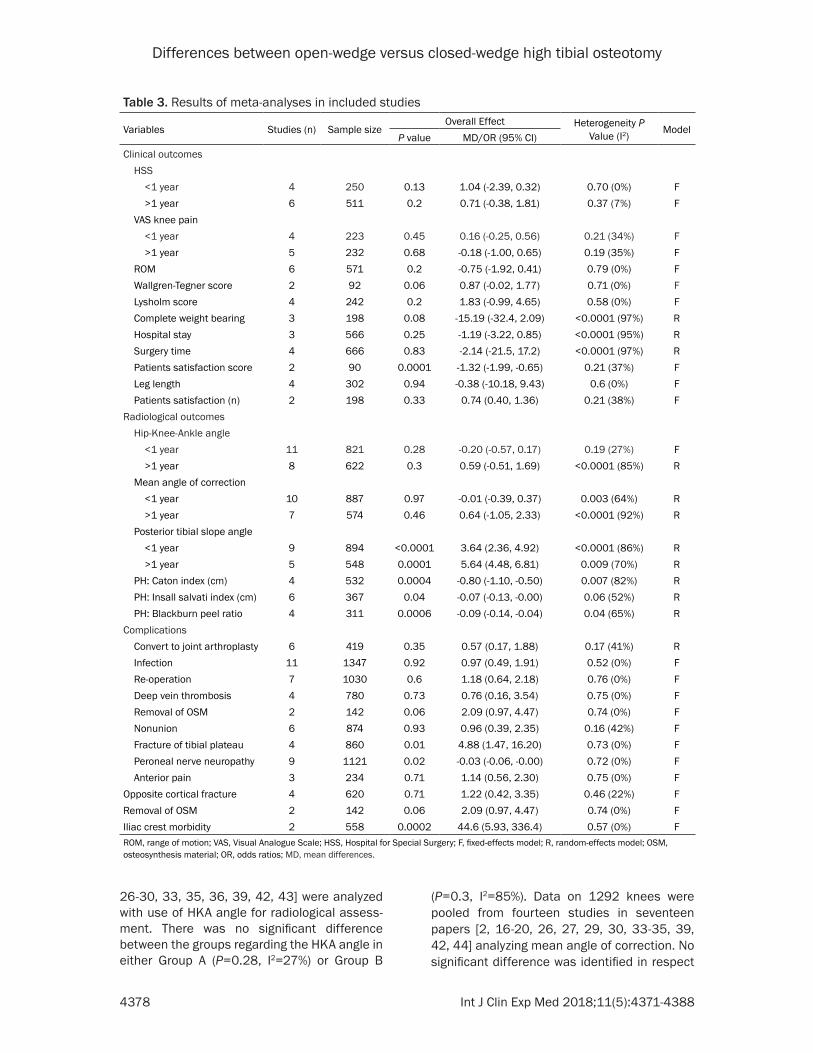
Table 3. Results of meta-analyses in included studies
Variables Studies (n) Sample sizeOverall Effect Heterogeneity P
Value (I2) ModelP value MD/OR (95% CI)
Clinical outcomes HSS <1 year 4 250 0.13 1.04 (-2.39, 0.32) 0.70 (0%) F >1 year 6 511 0.2 0.71 (-0.38, 1.81) 0.37 (7%) F VAS knee pain <1 year 4 223 0.45 0.16 (-0.25, 0.56) 0.21 (34%) F >1 year 5 232 0.68 -0.18 (-1.00, 0.65) 0.19 (35%) F ROM 6 571 0.2 -0.75 (-1.92, 0.41) 0.79 (0%) F Wallgren-Tegner score 2 92 0.06 0.87 (-0.02, 1.77) 0.71 (0%) F Lysholm score 4 242 0.2 1.83 (-0.99, 4.65) 0.58 (0%) F Complete weight bearing 3 198 0.08 -15.19 (-32.4, 2.09) <0.0001 (97%) R Hospital stay 3 566 0.25 -1.19 (-3.22, 0.85) <0.0001 (95%) R Surgery time 4 666 0.83 -2.14 (-21.5, 17.2) <0.0001 (97%) R Patients satisfaction score 2 90 0.0001 -1.32 (-1.99, -0.65) 0.21 (37%) F Leg length 4 302 0.94 -0.38 (-10.18, 9.43) 0.6 (0%) F Patients satisfaction (n) 2 198 0.33 0.74 (0.40, 1.36) 0.21 (38%) FRadiological outcomes Hip-Knee-Ankle angle <1 year 11 821 0.28 -0.20 (-0.57, 0.17) 0.19 (27%) F >1 year 8 622 0.3 0.59 (-0.51, 1.69) <0.0001 (85%) R Mean angle of correction <1 year 10 887 0.97 -0.01 (-0.39, 0.37) 0.003 (64%) R >1 year 7 574 0.46 0.64 (-1.05, 2.33) <0.0001 (92%) R Posterior tibial slope angle <1 year 9 894 <0.0001 3.64 (2.36, 4.92) <0.0001 (86%) R >1 year 5 548 0.0001 5.64 (4.48, 6.81) 0.009 (70%) R PH: Caton index (cm) 4 532 0.0004 -0.80 (-1.10, -0.50) 0.007 (82%) R PH: Insall salvati index (cm) 6 367 0.04 -0.07 (-0.13, -0.00) 0.06 (52%) R PH: Blackburn peel ratio 4 311 0.0006 -0.09 (-0.14, -0.04) 0.04 (65%) RComplications Convert to joint arthroplasty 6 419 0.35 0.57 (0.17, 1.88) 0.17 (41%) R Infection 11 1347 0.92 0.97 (0.49, 1.91) 0.52 (0%) F Re-operation 7 1030 0.6 1.18 (0.64, 2.18) 0.76 (0%) F Deep vein thrombosis 4 780 0.73 0.76 (0.16, 3.54) 0.75 (0%) F Removal of OSM 2 142 0.06 2.09 (0.97, 4.47) 0.74 (0%) F Nonunion 6 874 0.93 0.96 (0.39, 2.35) 0.16 (42%) F Fracture of tibial plateau 4 860 0.01 4.88 (1.47, 16.20) 0.73 (0%) F Peroneal nerve neuropathy 9 1121 0.02 -0.03 (-0.06, -0.00) 0.72 (0%) F Anterior pain 3 234 0.71 1.14 (0.56, 2.30) 0.75 (0%) FOpposite cortical fracture 4 620 0.71 1.22 (0.42, 3.35) 0.46 (22%) FRemoval of OSM 2 142 0.06 2.09 (0.97, 4.47) 0.74 (0%) FIliac crest morbidity 2 558 0.0002 44.6 (5.93, 336.4) 0.57 (0%) FROM, range of motion; VAS, Visual Analogue Scale; HSS, Hospital for Special Surgery; F, fixed-effects model; R, random-effects model; OSM, osteosynthesis material; OR, odds ratios; MD, mean differences.
26-30, 33, 35, 36, 39, 42, 43] were analyzed with use of HKA angle for radiological assess-ment. There was no significant difference between the groups regarding the HKA angle in either Group A (P=0.28, I2=27%) or Group B
(P=0.3, I2=85%). Data on 1292 knees were pooled from fourteen studies in seventeen papers [2, 16-20, 26, 27, 29, 30, 33-35, 39, 42, 44] analyzing mean angle of correction. No significant difference was identified in respect

Differences between open-wedge versus closed-wedge high tibial osteotomy
4379 Int J Clin Exp Med 2018;11(5):4371-4388
Figure 4. Forest plot of posterior tibial angle value between OWO and CWO (OWO open-wedge osteotomy, CWO closed-wedge osteotomy, CI confidence interval, df degrees of freedom).
Figure 3. Forest plot of clinical knee function between OWO and CWO. A) for the improvement of ROM; B) for the improvement of HSS (OWO open-wedge osteotomy, CWO closed-wedge osteotomy, CI confidence interval, df degrees of freedom).
Differences between open-wedge versus closed-wedge high tibial osteotomy
4380 Int J Clin Exp Med 2018;11(5):4371-4388
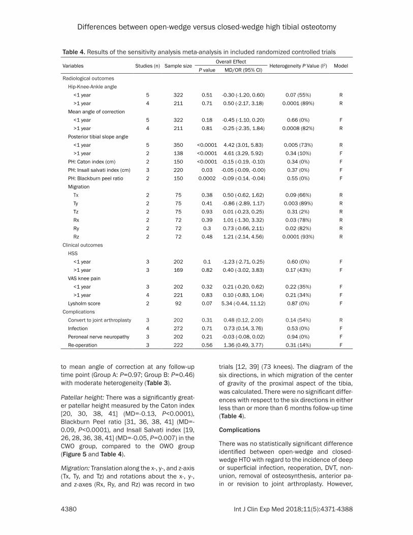
Table 4. Results of the sensitivity analysis meta-analysis in included randomized controlled trials
Variables Studies (n) Sample sizeOverall Effect
Heterogeneity P Value (I2) ModelP value MD/OR (95% CI)
Radiological outcomes Hip-Knee-Ankle angle <1 year 5 322 0.51 -0.30 (-1.20, 0.60) 0.07 (55%) R >1 year 4 211 0.71 0.50 (-2.17, 3.18) 0.0001 (89%) R Mean angle of correction <1 year 5 322 0.18 -0.45 (-1.10, 0.20) 0.66 (0%) F >1 year 4 211 0.81 -0.25 (-2.35, 1.84) 0.0008 (82%) R Posterior tibial slope angle <1 year 5 350 <0.0001 4.42 (3.01, 5.83) 0.005 (73%) R >1 year 2 138 <0.0001 4.61 (3.29, 5.92) 0.34 (10%) F PH: Caton index (cm) 2 150 <0.0001 -0.15 (-0.19, -0.10) 0.34 (0%) F PH: Insall salvati index (cm) 3 220 0.03 -0.05 (-0.09, -0.00) 0.37 (0%) F PH: Blackburn peel ratio 2 150 0.0002 -0.09 (-0.14, -0.04) 0.55 (0%) F Migration Tx 2 75 0.38 0.50 (-0.62, 1.62) 0.09 (66%) R Ty 2 75 0.41 -0.86 (-2.89, 1.17) 0.003 (89%) R Tz 2 75 0.93 0.01 (-0.23, 0.25) 0.31 (2%) R Rx 2 72 0.39 1.01 (-1.30, 3.32) 0.03 (78%) R Ry 2 72 0.3 0.73 (-0.66, 2.11) 0.02 (82%) R Rz 2 72 0.48 1.21 (-2.14, 4.56) 0.0001 (93%) RClinical outcomes HSS <1 year 3 202 0.1 -1.23 (-2.71, 0.25) 0.60 (0%) F >1 year 3 169 0.82 0.40 (-3.02, 3.83) 0.17 (43%) F VAS knee pain <1 year 3 202 0.32 0.21 (-0.20, 0.62) 0.22 (35%) F >1 year 4 221 0.83 0.10 (-0.83, 1.04) 0.21 (34%) F Lysholm score 2 92 0.07 5.34 (-0.44, 11.12) 0.87 (0%) FComplications Convert to joint arthroplasty 3 202 0.31 0.48 (0.12, 2.00) 0.14 (54%) R Infection 4 272 0.71 0.73 (0.14, 3.76) 0.53 (0%) F Peroneal nerve neuropathy 3 202 0.21 -0.03 (-0.08, 0.02) 0.94 (0%) F Re-operation 3 222 0.56 1.36 (0.49, 3.77) 0.31 (14%) F
to mean angle of correction at any follow-up time point (Group A: P=0.97; Group B: P=0.46) with moderate heterogeneity (Table 3).
Patellar height: There was a significantly great-er patellar height measured by the Caton index [20, 30, 38, 41] (MD=-0.13, P<0.0001), Blackburn Peel ratio [31, 36, 38, 41] (MD=-0.09, P<0.0001), and Insall Salvati index [19, 26, 28, 36, 38, 41] (MD=-0.05, P=0.007) in the CWO group, compared to the OWO group (Figure 5 and Table 4).
Migration: Translation along the x-, y-, and z-axis (Tx, Ty, and Tz) and rotations about the x-, y-, and z-axes (Rx, Ry, and Rz) was record in two
trials [12, 39] (73 knees). The diagram of the six directions, in which migration of the center of gravity of the proximal aspect of the tibia, was calculated. There were no significant differ-ences with respect to the six directions in either less than or more than 6 months follow-up time (Table 4).
Complications
There was no statistically significant difference identified between open-wedge and closed-wedge HTO with regard to the incidence of deep or superficial infection, reoperation, DVT, non-union, removal of osteosynthesis, anterior pa- in or revision to joint arthroplasty. However,

Differences between open-wedge versus closed-wedge high tibial osteotomy
4381 Int J Clin Exp Med 2018;11(5):4371-4388
closed-wedge HTO was associated with lower incidence of intra-operative fracture of the tibi-al plateau (MD=4.88, P=0.01) and iliac crest morbidity (MD=44.6, P=0.0002) with no het-erogeneity (Table 3). In addition, CWO showed higher incidence of common peroneal nerve palsy compared with OWO (MD=-0.03, P=0.02).
Sensitivity analysis
To validate our results, we conducted sensitivi-ty analyses to determine the effect of study quality on the results. Firstly, the random-effects model was compared with the fixed-effects model, and the statistically similar results were obtained in respect to any out-come (data not shown). Secondly, a sensitivity analysis was performed to compare the out-comes between OWO group and CWO group for RCTs only. Compared to RCTs outcomes alone using the sensitivity analysis, no statistically significant difference was detected from this secondary sensitivity analysis to the main anal-ysis in respect to clinical and radiological out-comes, suggesting the stability of our meta-
analysis (Table 4). However, the sensitivity analysis did indicate that there was no signifi-cantly higher incidence of the common pero-neal nerve palsy following CWO.
Publication bias
Publication bias was assessed through the Funnel plot. The shape of the funnel plots anal-yses on the improvement of VAS, HSS and ROM did not reveal basically asymmetric distribu-tion, indicating that bias was minimal (Figure 6).
Discussion
The most important findings of this study were that significantly lower posterior tibial slope, greater patellar height measurements, and decreased incidence of patellar baja were observed following CWO, when compared to OWO. However, there were no significant differ-ences regarding HKA angle, mean angle of cor-rection and leg length between the two groups. Moreover, no significant differences were iden-tified with respect to clinical outcomes.
Figure 5. Forest plot of patellar height value between OWO and CWO. A) for patellar height measured by Caton-Des-champs index; B) for patellar height measured by Insall-Salvati; C) for patellar height measured by Blackburne-Peel index (OWO open-wedge osteotomy, CWO closed-wedge osteotomy, CI confidence interval, df degrees of freedom).

Differences between open-wedge versus closed-wedge high tibial osteotomy
4382 Int J Clin Exp Med 2018;11(5):4371-4388
The results showed that posterior slope increases after open-wedge HTO and decreas-es after closed-wedge HTO. The unique ana-tomic geometry of the proximal tibia may cause the change of posterior tibial slope following HTO [36, 45, 46]. Increased tibial slope follow-ing OWO can produce an anterior translation in tibial resting position and increase the strain in the anterior cruciate ligament (ACL) [47, 48], while decreased slope following CWO can increase the strain in the posterior cruciate lig-ament (PCL) [49]. Thus, some studies reported that CWO was a therapeutic option for those with concomitant ACL injuries or insufficiency, while OWO was an appropriate method for those with PCL deficiency [46, 47, 50]. However, the study Nha et al. found that the change in posterior tibial slope was less than 5° (approximately 2°), indicating that both open- and closed-wedge HTO may have little effect on the in situ forces in the cruciate ligaments [22].
Three reference methods (Caton-Deschamps, Insall-Salvati or Blackburne-Peel index) were used to evaluate patellar height in these eligi-ble studies. The findings demonstrated that patellar height decreased after OWO and
Figure 6. Funnel plots of primary outcomes. A) for the improvement of VAS knee pain score; B) for the improvement of ROM; C) for the improvement of HSS.
increased after CWO using any measure. A pos-sible interpretation is the shift of tibial tubercle at the distal section of the osteotomy. The CWO could elevate the tibial tuberosity due to a prox-imalisation of the proximal tibia and result in an increase in patellar height. Conversely, the OWO could lower the tibial tuberosity and resulted in a decrease in patella height [36]. Decreasing patellar height would result in patella baja, thereby generating technical diffi-culties during TKA, such as patella eversion and lateral compartment exposure. However, there was no difference regarding the improve-ment of pain and knee function outcomes (VAS pain score, ROM, and HSS) between OWO and CWO, suggesting that the difference in the patellar height has little influence on the clini-cal outcomes.
The HKA angle was calculated on standing whole leg radiographs, which was defined as the angle between the mechanical axis of the femur and tibia. A RCT by Brouwer et al. showed more accurate correction after CWO, compared to OWO [34]. Nevertheless, Magyar et al. reported that OWO with external fixator could lead to higher accuracy of the correction since

Differences between open-wedge versus closed-wedge high tibial osteotomy
4383 Int J Clin Exp Med 2018;11(5):4371-4388
it provides continuous radiographic evaluation [12]. Hankemeier et al. reported that it was dif-ficult to correct such subtle difference in CWO [16]. Moreover, Gaasbeek et al. reported that no loss of correction was identified after one-year follow-up, suggesting that both OWO and CWO resulted in an accurate correction [20]. In this study, there was no statistically significant difference regarding the HKA angle and mean angle of correction at any follow-up time. Interestingly, our result was not consistent with the findings of the previous meta-analysis by Smith et al. suggesting that open-wedge HTO provided superior anatomical correction and might be interpreted by the following reasons: Firstly, two publications were conducted as an update of two previous studies, which had a longer follow-up time than previous studies did. The results of these studies suggested that the radiographic and clinical outcomes might change over time. Therefore, it is necessary to conduct subgroup analysis based on follow- up duration. Secondly, the small sample was included in the previous meta-analysis on this topic. Therefore, the results may reduce the power to reveal a reliable relationship. Thirdly, the sensitivity analysis including only two trials changed the results regarding HKA angle and mean angle of correction in previous meta-analysis. However, the sensitivity analysis was conducted through two different methods in this meta-analysis, and the results were consis-tent with the previous results regarding clinical and radiographic results, suggesting the results of this study were stable. In addition, the accu-racy of the correction was highly important in the long-term results of HTO, such as fixation stability.
As for complications, closed-wedge HTO implied lateral muscle detachment, higher risk of pero-neal nerve injury, more demanding subsequent TKA, bone stock loss and fibular osteotomy or proximal tibiofibular joint disruption. For the above-mentioned disadvantages of CWO, OWO was regarded as a safe and reproducible proce-dure and gained the popularity for being a wide-ly accepted alternative option [51]. However, OWO was not free from some complications, such as nonunion, the necessity of bone graft, disease transmission and possible loss of cor-rection [52]. In this meta-analysis, OWO showed higher incidence of intra-operative fracture of tibial plateau. Interpretation of the outcomes must be made with caution because the sam-
ple size was relatively small and the data were combined from one RCT and three n-RCTs, which may produce confounding factors and affect the validity. In addition, the only eligible RCT by Duivenvoorden et al. demonstrated that there was no difference between the two groups regarding intra-operative fracture of tibial plateau [2]. Therefore, we may not draw the conclusion that OWO increased the risk of intra-operative fracture of tibial plateau, com-pared with CWO. During open-wedge HTO, we may release the soft-tissues medially, leave appropriately lateral cortex intact and create an osteotomy parallel to the posterior tibial slope in the sagittal plane to decrease the risk of this complication [42].
No significant difference was identified for the incidence of opposite cortical fracture between the two groups. Interestingly, these findings were inconsistent with the previous meta-anal-yses, and we believe our meta-analysis was more accurate and comprehensive [21]. This might be interpreted by the following reasons: Firstly, the previous meta-analysis by Sun et al. only included two n-RCTs, But the present study included four studies and had a larger sample size than the study by Sun et al. which made a more robust conclusion. Secondly, they did not use sensitivity analysis to investigate the origin of high heterogeneity, thereby resulting in an unstable result. In our study, sensitivity analy-sis was utilized to further investigate the signifi-cant heterogeneity in our study, and the results were in line with the previous analysis. Thirdly, the aforementioned meta-analysis included two duplicated publications [8, 10], which may have substantial effect on the results. In this meta-analysis, the overall result showed signifi-cantly lower rate of common peroneal nerve palsy in OWO group. Nevertheless, the sensitiv-ity analysis showed no significant difference between the two groups. In the three RCTs, the rate of injury to the peroneal nerve during CWO was 4% (5/125), but with OWO this complica-tion was avoided. Several factors may have contributed to this complication during CWO, including the improper position of or excessive pressure utilized with a retractor or the improp-er detachment of muscle from the lateral side of the proximal tibia in proximal tibiofibular dis-ruption [42]. CWO was technically harder than OWO, thereby orthopedic surgeons may need more time to master the surgical technique of CWO and minimize the risk of common perone-

Differences between open-wedge versus closed-wedge high tibial osteotomy
4384 Int J Clin Exp Med 2018;11(5):4371-4388
al nerve palsy [52]. In addition, Han et al. in a systematic review demonstrated that more sur-gical technical concerns were identified in TKA conversion from CWO than from OWO group [23]. Robertsson et al. compared the results of primary TKR and HTO revision to TKR regarding clinical outcomes and demonstrated that the risk of revision was significantly higher after the previous CWO than primary TKR, whereas OWO had no effect on the outcome [53].
Another major adverse event was iliac crest morbidity, which was caused by harvesting bone to fill the osseous gap in opening wedge osteotomy. Autograft was regarded as the most successful bone filling material due to its os- teoinductive, osteoconductive and osteogenic properties, as compared to allograft. The over-all result showed significantly higher rate of iliac crest morbidity in OWO group. However, we must treat the result with caution due to the small sample size. If bone grafting at the iliac crest could be avoided, the rate of such compli-cation could be decreased. Some surgeons rec-ommended that the cancellous autograft was only applied for opening wedges <12.5 mm, and allograft or synthetic bone substitute (hydroxyapatite, β-tricalcium phosphate, and bone cement etc.) was utilized for all cases or only for larger opening wedges, which could decrease the incidence of complications relat-ed to autograft donor site morbidity [54]. Some studies demonstrated that platelet-rich plasma (PRP), bone marrow stromal cells and growth factor combined with bone graft or substitute showed encouraging results regarding main-taining the desired correction and long-term stability and allowing early weight bearing [6, 52]. However, no clear evidence was found that whether PRP and augmentation showed superi-ority of decreasing union rates compared with autologous iliac crest graft.
Reliable fixation was fundamental in achieving good results in open-wedge HTO, but the most stable fixation system was still controversial. External fixators and plates (conventional, lock-ing, long or short plates) are the most common-ly used fixation devices. Numerous biomechan-ical studies have compared different implants. Agneskirchner et al. in their investigation on four different plates concluded that rigid long plate fixator with angle-stable locking bolts yields the best results [55]. In addition, Spahn et al. compared the four different fixation devic-
es and reported that implants with a spacer had superior biomechanical properties and seem to be more reliable in predictable mainte-nance of correction [56]. Some studies showed better biomechanical properties and early weight bearing in long-locking plate, but it was bulky, expensive and frequently require hard-ware removal in many cases [52]. Spacer plates show more reliable fixation and lower risk of hardware removal and correction loss [6]. Although The results are still inconclusive, the gold standards may be locked plates and autol-ogous graft based on the current evidence. Similar results were described by Amendola et al. [57] and Bonasia et al. [52].
According to literature, the carefully preopera-tive planning and selection of ideal candidate for HTO is essential. In general, a slight valgus correction (5°-6° degree) is associated with better results. However, the achievement of a neutral alignment is recommended in young patients and athletes [48]. Surgeons should be aware of several risk factors related to poor outcomes, such as bad range of motion, old inactive patients (>65) [58], severely medial isolated osteoarthritis (more than grade III according to Ahlbäck classification) [59], joint instability [60], patellofemoral arthrosis [5, 6] and lateral tibial thrust [15, 46].
This study has several strengths. Firstly, the extensive search strategy and broad inclusion criteria were applied to search all related litera-ture and ensured thoroughness. Secondly, we screened duplicate articles to minimize review-ers’ bias. The sample size in this review was quite bigger than that in previous meta-analy-sis to permit subgroup analysis, which provide a relatively unbiased picture of the results. Thirdly, since different validated scoring sys-tems may lead to unclear functional assess-ment findings and moderate heterogeneity, we evaluate the knee function with the use of more complete scoring systems than previous meta-analyses, including ROM, HSS, Wallgren-Tegner score, and Lysholm score. In addition, we used the change in knee functional assessments from baseline as our effect index to assess actual improvement in knee function, which eliminated the influence of different baselines. The pooled results found no difference regard-ing knee function between OWO and CWO. However, no study was available to investigate whether there was a difference regarding time to occupational or sporting pursuits. Therefore,

Differences between open-wedge versus closed-wedge high tibial osteotomy
4385 Int J Clin Exp Med 2018;11(5):4371-4388
the basis of this suggestion of longevity of reha-bilitation remains unclear, and future studies will have to investigate the cost-effectiveness by assessing the rehabilitation time.
We note several potential limitations in this meta-analysis. Firstly, some of the eligible stud-ies were observational comparison studies, leading to some inherent heterogeneity due to uncontrolled bias. In addition, heterogeneity may come from some risk factors, such as age, gender, fixation position, the reliability of radio-graphs, and especially fixation methods. Some studies comparing the biomechanical proper-ties and stability of the different fixation devic-es had been published, but the most reliable fixation system still remains controversial [55, 61]. Secondly, sample sizes were not calculat-ed based on power analysis. Therefore, this may possibly result in type II statistical error. Thirdly, it was impossible to blind the observer assessing the radiographs regarding HTO type [19]. As a result, there remains a possibility that assessor bias has a substantial effect on the clinical outcomes recorded.
This meta-analysis confirmed that posterior tibial slope increased and patellar height de- creased following OWO. Conversely, posterior tibial slope decreased and patellar height increased following CWO. Although both tech-niques led to the good and comparable clinical results, we recommend OWO as an alternati- vely effective treatment option for selected younger patients, which was easier to perform and to convert to TKA, avoids tibiofibular joint disruption and common peroneal nerve palsy, and permits multiplanar correction, as com-pared to CWO. However, additional well-desi- gned studies and long-term follow-up data are required to identify the ideal candidates, the type of fixation and augment selection in OWO.
Disclosure of conflict of interest
None.
Address correspondence to: Dong-Mei Zhao, De- partment of Obstetrics and Gynecology, West China Second University Hospital, Sichuan University, Chengdu 610041, P.R. China. Tel: +86-28-85501- 323; Fax: +86-28-85542425; E-mail: [email protected]; Dr. Rui Fang, Department of Orthopedics, Hospital of Traditional Chinese Medicine Affiliated
to Xinjiang Medical University, Urumqi 830000, P.R. China. Tel: +86-28-84423231; Fax: +86-28-84422623; E-mail: [email protected]; Dr. Chao You, Department of Neurosurgery, West China Hospital, Sichuan University, Chengdu 610041, P.R. China. Tel: +86-28-85422490; Fax: +86-28-8516- 4009; E-mail: [email protected]
References
[1] Sharma L, Song J, Felson DT, Cahue S, Shami-yeh E and Dunlop DD. The role of knee align-ment in disease progression and functional decline in knee osteoarthritis. JAMA 2001; 286: 188-195.
[2] Duivenvoorden T, Brouwer RW, Baan A, Bos PK, Reijman M, Bierma-Zeinstra SM and Ver-haar JA. Comparison of closing-wedge and opening-wedge high tibial osteotomy for medi-al compartment osteoarthritis of the knee: a randomized controlled trial with a six-year fol-low-up. J Bone Joint Surg Am 2014; 96: 1425-1432.
[3] Gaasbeek R, Welsing R, Barink M, Verdon-schot N and van Kampen A. The influence of open and closed high tibial osteotomy on dynamic patellar tracking: a biomechanical study. Knee Surg Sports Traumatol Arthrosc 2007; 15: 978-984.
[4] Whitehead TS, Willits K, Bryant D, Giffin JR and Fowler PJ. Impact of medial opening or lateral closing tibial osteotomy on bone resection and posterior cruciate ligament integrity during knee arthroplasty. J Arthroplasty 2009; 24: 979-989.
[5] McNamara I, Birmingham TB, Fowler PJ and Giffin JR. High tibial osteotomy: evolution of re-search and clinical applications--a Canadian experience. Knee Surg Sports Traumatol Ar-throsc 2013; 21: 23-31.
[6] Lee DC and Byun SJ. High tibial osteotomy. Knee Surg Relat Res 2012; 24: 61-69.
[7] Lobenhoffer P and Agneskirchner JD. Improve-ments in surgical technique of valgus high tibi-al osteotomy. Knee Surg Sports Traumatol Ar-throsc 2003; 11: 132-138.
[8] van Raaij TM, Brouwer RW, de Vlieger R, Reij-man M and Verhaar JA. Opposite cortical frac-ture in high tibial osteotomy: lateral closing compared to the medial opening-wedge tech-nique. Acta Orthop 2008; 79: 508-514.
[9] Hanada M, Takahashi M, Koyama H and Mat-suyama Y. Comparison of the change in patel-lar height between opening and closed wedge high tibial osteotomy: measurement with a new method. Eur J Orthop Surg Traumatol 2014; 24: 567-570.
[10] van Egmond N, van Grinsven S, van Loon CJ, Gaasbeek RD and van Kampen A. Better clini-

Differences between open-wedge versus closed-wedge high tibial osteotomy
4386 Int J Clin Exp Med 2018;11(5):4371-4388
cal results after closed- compared to open-wedge high tibial osteotomy in patients with medial knee osteoarthritis and varus leg align-ment. Knee Surg Sports Traumatol Arthrosc 2014; 24: 198-206.
[11] Duivenvoorden T, van Diggele P, Reijman M, Bos PK, van Egmond J, Bierma-Zeinstra SM and Verhaar JA. Adverse events and survival after closing- and opening-wedge high tibial osteotomy: a comparative study of 412 pa-tients. Knee Surg Sports Traumatol Arthrosc 2015; 32: 100-108.
[12] Magyar G, Toksvig-Larsen S and Lindstrand A. Changes in osseous correction after proximal tibial osteotomy: radiostereometry of closed- and open-wedge osteotomy in 33 patients. Acta Orthop Scand 1999; 70: 473-477.
[13] Nakamura E, Mizuta H, Kudo S, Takagi K and Sakamoto K. Open-wedge osteotomy of the proximal tibia with hemicallotasis. J Bone Joint Surg Br 2001; 83: 1111-1115.
[14] Han SB, Bae JH, Lee SJ, Jung TG, Kim KH, Kwon JH and Nha KW. Biomechanical proper-ties of a new anatomical locking metal block plate for opening wedge high tibial osteotomy: uniplane osteotomy. Knee Surg Relat Res 2014; 26: 155-161.
[15] Cho SW, Kim DH, Lee GC, Lee SH and Park SH. Comparison between Autogenous Bone Graft and Allogenous Cancellous Bone Graft in Me-dial Open Wedge High Tibial Osteotomy with 2-Year follow-up. Knee Surg Relat Res 2013; 25: 117-125.
[16] Hankemeier S, Mommsen P, Krettek C, Jagodz-inski M, Brand J, Meyer C and Meller R. Accu-racy of high tibial osteotomy: comparison be-tween open- and closed-wedge technique. Knee Surg Sports Traumatol Arthrosc 2010; 18: 1328-1333.
[17] van Egmond N, van Grinsven S, van Loon CJ, Gaasbeek RD and van Kampen A. Better clini-cal results after closed- compared to open-wedge high tibial osteotomy in patients with medial knee osteoarthritis and varus leg align-ment. Knee Surg Sports Traumatol Arthrosc 2016; 24: 34-41.
[18] Kim JI, Kim BH, Lee KW, Lee O, Han HS, Lee S and Lee MC. Lower limb length discrepancy af-ter high tibial osteotomy: prospective random-ized controlled trial of lateral closing versus medial opening wedge osteotomy. Am J Sports Med 2016; 44: 3095-3102.
[19] Nerhus TK, Ekeland A, Solberg G, Sivertsen EA, Madsen JE and Heir S. Radiological outcomes in a randomized trial comparing opening wedge and closing wedge techniques of high tibial osteotomy. Knee Surg Sports Traumatol Arthrosc 2015; 25: 348-406
[20] Gaasbeek RD, Nicolaas L, Rijnberg WJ, van Loon CJ and van Kampen A. Correction accu-
racy and collateral laxity in open versus closed wedge high tibial osteotomy. a one-year ran-domised controlled study. Int Orthop 2010; 34: 201-207.
[21] Sun H, Zhou L, Li F and Duan J. Comparison between closing-wedge and opening-wedge high tibial osteotomy in patients with medial knee osteoarthritis: a systematic review and meta-analysis. J Knee Surg 2016; 30: 223-229
[22] Nha KW, Kim HJ, Ahn HS and Lee DH. Change in posterior tibial slope after open-wedge and closed-wedge high tibial osteotomy: a meta-analysis. Am J Sports Med 2016; 44: 209-216
[23] Han JH, Yang JH, Bhandare NN, Suh DW, Lee JS, Chang YS, Yeom JW and Nha KW. Total knee arthroplasty after failed high tibial oste-otomy: a systematic review of open versus closed wedge osteotomy. Knee Surg Sports Traumatol Arthrosc 2016; 24: 2567-2577.
[24] Smith TO, Sexton D, Mitchell P and Hing CB. Opening- or closing-wedged high tibial osteoto-my: a meta-analysis of clinical and radiological outcomes. Knee 2011; 18: 361-368.
[25] Bin SI, Kim HJ, Ahn HS, Rim DS and Lee DH. Changes in patellar height after opening wedge and closing wedge high tibial osteoto-my: a meta-analysis. Arthroscopy 2016; 32: 2393-2400.
[26] Portner O. High tibial valgus osteotomy: clos-ing, opening or combined? Patellar height as a determining factor. Clin Orthop Relat Res 2014; 472: 3432-3440.
[27] Deie M, Hoso T, Shimada N, Iwaki D, Nakamae A, Adachi N and Ochi M. Differences between opening versus closing high tibial osteotomy on clinical outcomes and gait analysis. Knee 2014; 21: 1046-1051.
[28] Tabrizi A, Soleimanpour J, Sadighi A and Zare AJ. A short term follow up comparison of genu varum corrective surgery using open and closed wedge high tibial osteotomy. Malays Or-thop J 2013; 7: 7-12.
[29] Bae DK, Song SJ, Kim HJ and Seo JW. Change in limb length after high tibial osteotomy us- ing computer-assisted surgery: a comparative study of closed- and open-wedge osteotomies. Knee Surg Sports Traumatol Arthrosc 2013; 21: 120-126.
[30] Amzallag J, Pujol N, Maqdes A, Beaufils P, Ju-det T and Catonne Y. Patellar height modifica-tion after high tibial osteotomy by either medial opening-wedge or lateral closing-wedge oste-otomies. Knee Surg Sports Traumatol Arthrosc 2013; 21: 255-259.
[31] Song IH, Song EK, Seo HY, Lee KB, Yim JH and Seon JK. Patellofemoral alignment and anteri-or knee pain after closing- and opening-wedge valgus high tibial osteotomy. Arthroscopy 2012; 28: 1087-1093.

Differences between open-wedge versus closed-wedge high tibial osteotomy
4387 Int J Clin Exp Med 2018;11(5):4371-4388
[32] Ducat A, Sariali E, Lebel B, Mertl P, Hernigou P, Flecher X, Zayni R, Bonnin M, Jalil R, Amzallag J, Rosset P, Servien E, Gaudot F, Judet T and Catonne Y. Posterior tibial slope changes after opening- and closing-wedge high tibial osteoto-my: a comparative prospective multicenter study. Orthop Traumatol Surg Res 2012; 98: 68-74.
[33] Magnussen RA, Lustig S, Demey G, Neyret P and Servien E. The effect of medial opening and lateral closing high tibial osteotomy on leg length. Am J Sports Med 2011; 39: 1900-1905.
[34] Brouwer RW, Bierma-Zeinstra SM, van Raaij TM and Verhaar JA. Osteotomy for medial com-partment arthritis of the knee using a closing wedge or an opening wedge controlled by a puddu plate. a one-year randomised, con-trolled study. J Bone Joint Surg Br 2006; 88: 1454-1459.
[35] Magyar G, Ahl TL, Vibe P, Toksvig-Larsen S and Lindstrand A. Open-wedge osteotomy by hemi-callotasis or the closed-wedge technique for osteoarthritis of the knee. a randomised study of 50 operations. J Bone Joint Surg Br 1999; 81: 444-448.
[36] Brouwer RW, Bierma-Zeinstra SM, van Koever-inge AJ and Verhaar JA. Patellar height and the inclination of the tibial plateau after high tibial osteotomy. the open versus the closed-wedge technique. J Bone Joint Surg Br 2005; 87: 1227-1232.
[37] El-Azab H, Halawa A, Anetzberger H, Imhoff AB and Hinterwimmer S. The effect of closed- and open-wedge high tibial osteotomy on tibial slope: a retrospective radiological review of 120 cases. J Bone Joint Surg Br 2008; 90: 1193-1197.
[38] El-Azab H, Glabgly P, Paul J, Imhoff AB and Hin-terwimmer S. Patellar height and posterior tib-ial slope after open- and closed-wedge high tibial osteotomy: a radiological study on 100 patients. Am J Sports Med 2010; 38: 323-329.
[39] Luites JW, Brinkman JM, Wymenga AB and van Heerwaarden RJ. Fixation stability of opening- versus closing-wedge high tibial osteotomy: a randomised clinical trial using radiostereome-try. J Bone Joint Surg Br 2009; 91: 1459-1465.
[40] Hoell S, Suttmoeller J, Stoll V, Fuchs S and Gos-heger G. The high tibial osteotomy, open ver-sus closed wedge, a comparison of methods in 108 patients. Arch Orthop Trauma Surg 2005; 125: 638-643.
[41] Schiedel F, Probst A, Buller TC and Rodl R. The postoperative patella height: a comparison of additive and subtractive high tibial osteotomy in correcting the genu varum. Arch Orthop Trauma Surg 2009; 129: 1271-1277.
[42] Song EK, Seon JK, Park SJ and Jeong MS. The complications of high tibial osteotomy: closing-
versus opening-wedge methods. J Bone Joint Surg Br 2010; 92: 1245-1252.
[43] Agarwala S, Sobti A, Naik S and Chaudhari S. Comparison of closing-wedge and opening-wedge high tibial osteotomies for medial com-partment osteoarthritis of knee in asian popu-lation: mid-term follow-up. J Clin Orthop Trau- ma 2016; 7: 272-275.
[44] Schaefer TK, Majewski M, Hirschmann MT and Friederich NF. Comparison of sagittal and fron-tal plane alignment after open- and closed-wedge osteotomy: a matched-pair analysis. J Int Med Res 2008; 36: 1085-1093.
[45] Noyes FR, Goebel SX and West J. Opening wedge tibial osteotomy: the 3-triangle method to correct axial alignment and tibial slope. Am J Sports Med 2005; 33: 378-387.
[46] Giffin JR, Vogrin TM, Zantop T, Woo SL and Harner CD. Effects of increasing tibial slope on the biomechanics of the knee. Am J Sports Med 2004; 32: 376-382.
[47] Feucht MJ, Mauro CS, Brucker PU, Imhoff AB and Hinterwimmer S. The role of the tibial slope in sustaining and treating anterior cruci-ate ligament injuries. Knee Surg Sports Trau-matol Arthrosc 2013; 21: 134-145.
[48] Amis AA. Biomechanics of high tibial osteoto-my. Knee Surg Sports Traumatol Arthrosc 2013; 21: 197-205.
[49] Petrigliano FA, Suero EM, Voos JE, Pearle AD and Allen AA. The effect of proximal tibial slope on dynamic stability testing of the posterior cruciate ligament- and posterolateral corner-deficient knee. Am J Sports Med 2012; 40: 1322-1328.
[50] Hohmann E, Bryant A and Imhoff AB. The ef-fect of closed wedge high tibial osteotomy on tibial slope: a radiographic study. Knee Surg Sports Traumatol Arthrosc 2006; 14: 454-459.
[51] Brinkman JM, Lobenhoffer P, Agneskirchner JD, Staubli AE, Wymenga AB and van Heer-waarden RJ. Osteotomies around the knee: patient selection, stability of fixation and bone healing in high tibial osteotomies. J Bone Joint Surg Br 2008; 90: 1548-1557.
[52] Bonasia DE, Governale G, Spolaore S, Rossi R and Amendola A. High tibial osteotomy. Curr Rev Musculoskelet Med 2014; 7: 292-301.
[53] Robertsson O, W-Dahl A. The risk of revision after TKA is affected by previous HTO or UKA. Clin Orthop Relat Res 2015; 473: 90-93.
[54] Ruzbarsky JJ, Dare DM and Marx RG. Closing verses opening wedge high tibial osteotomy: an evidence-based review. HSS J 2015; 11: 291-3.
[55] Agneskirchner JD, Freiling D, Hurschler C and Lobenhoffer P. Primary stability of four differ-ent implants for opening wedge high tibial os-
Differences between open-wedge versus closed-wedge high tibial osteotomy
4388 Int J Clin Exp Med 2018;11(5):4371-4388
teotomy. Knee Surg Sports Traumatol Arthrosc 2006; 14: 291-300.
[56] Spahn G, Muckley T, Kahl E and Hofmann GO. Biomechanical investigation of different inter-nal fixations in medial opening-wedge high tib-ial osteotomy. Clin Biomech (Bristol, Avon) 2006; 21: 272-278.
[57] Amendola A and Bonasia DE. Results of high tibial osteotomy: review of the literature. Int Or-thop 2010; 34: 155-160.
[58] Rossi R, Bonasia DE and Amendola A. The role of high tibial osteotomy in the varus knee. J Am Acad Orthop Surg 2011; 19: 590-599.
[59] Bonasia DE, Dettoni F, Sito G, Blonna D, Mar-motti A, Bruzzone M, Castoldi F and Rossi R. Medial opening wedge high tibial osteotomy for medial compartment overload/arthritis in the varus knee: prognostic factors. Am J Sports Med 2014; 42: 690-698.
[60] Segal NA, Buckwalter JA and Amendola A. Oth-er surgical techniques for osteoarthritis. Best Pract Res Clin Rheumatol 2006; 20: 155-176.
[61] Zhim F, Laflamme GY, Viens H, Saidane K and Yahia L. Biomechanical stability of high tibial opening wedge osteotomy: internal fixation versus external fixation. Clin Biomech (Bristol, Avon) 2005; 20: 871-876.