Embed Size (px)
Citation preview
Review ArticleCircadian System and Melatonin Hormone:Risk Factors for Complications during Pregnancy
F. J. Valenzuela,1,2 J. Vera,1,2 C. Venegas,1,2 F. Pino,1 and C. Lagunas1,2
1Department of Basic Sciences, Universidad del Bıo-Bıo, Campus Fernando May, Avenida Andres Bello s/n, Chillan, Chile2Grupo de Ciencias Biotecnologicas, Basic Sciences Department, Universidad del Bıo-Bıo, Avenida Andres Bello s/n, Chillan, Chile
Correspondence should be addressed to F. J. Valenzuela; [email protected]
Received 29 August 2014; Accepted 8 February 2015
Academic Editor: Enrique Hernandez
Copyright © 2015 F. J. Valenzuela et al.This is an open access article distributed under the Creative Commons Attribution License,which permits unrestricted use, distribution, and reproduction in any medium, provided the original work is properly cited.
Pregnancy is a complex and well-regulated temporal event in which several steps are finely orchestrated including implantation,decidualization, placentation, and partum and any temporary alteration has serious effects on fetal and maternal health.Interestingly, alterations of circadian rhythms (i.e., shiftwork) have been correlated with increased risk of preterm delivery,intrauterine growth restriction, and preeclampsia. In the last few years evidence is accumulating that the placenta may have afunctional circadian system and express the clock genes Bmal1, Per1-2, and Clock. On the other hand, there is evidence that thehuman placenta synthesizes melatonin, hormone involved in the regulation of the circadian system in other tissues. Moreover, isunknown the role of this local production ofmelatonin andwhether this production have a circadian pattern. Available informationindicates that melatonin induces in placenta the expression of antioxidant enzymes catalase and superoxide dismutase, prevents theinjury produced by oxidative stress, and inhibits the expression of vascular endothelial growth factor (VEGF) a gene that in othertissues is controlled by clock genes. In this review we aim to analyze available information regarding clock genes and clock genescontrolled genes such as VEGF and the possible role of melatonin synthesis in the placenta.
1. Introduction
Pregnancy is a complex and well-regulated temporal eventin which several steps are finely orchestrated includingimplantation, decidualization, placentation, and partum [1].The chronological transitions are critical for a normal preg-nancy and any temporary alteration may have detrimentaleffects for fetal development and/or maternal health [2–4].The placenta is the unit of communication and exchangebetween mother and fetus. This organ is in charge ofbidirectional transference and metabolism of hormones,nutrients, and gases (oxygen/CO
2) [5]. The major site of
complication in pregnancy is the placenta and themain causeof development of obstetric syndrome is the placentation[6]. The impaired placentation causes spontaneous abortion,preeclampsia, preterm birth, and placental abruption [7].Moreover, placenta mediates the maternal-fetal interactionin the regulation of glucocorticoids, human placental lac-togen (hPL), human chorionic gonadotropin (hCG), andprogesterone and estriol, among others [5, 8]. In this regard,
hormonal production and activity is regulated by a circadiansystem, which, in fact, is composed by a family of genesnamed “clock genes” (Bmal1, Clock, Per1-3, and Cry1-2) [9].
2. Circadian Rhythms and Pregnancy
The circadian time-keeping system is actively engaged inthe maintenance of normal physiology, not only in adults,but also during development [10]. Within an individual, thepeak and trough of the rhythms for different physiologicalvariables occurs at different clock times. For instance, inhumans under normal light-dark condition, cortisol peaksat 08 h, while temperature peaks at 14–17 h, and melatoninat 02 h [11]. Similarly, during normal pregnancy differentcircadian rhythm are observed in themother such as temper-ature [12, 13], leukocytes count, blood pressure [13], circadianpattern of weight gain [14], rhythms of uterine contraction,blood flow [15], and intra-amniotic fluid pressure [5, 15]. Thefinal output of the circadian system during pregnancy is thelabor. Humans and monkeys (diurnal animals) show a peak
Hindawi Publishing CorporationObstetrics and Gynecology InternationalVolume 2015, Article ID 825802, 10 pageshttp://dx.doi.org/10.1155/2015/825802
2 Obstetrics and Gynecology International
in the second middle of the night and early in the morning[3, 16, 17]. The rat and mice (nocturnal animals) show a timebirth in the afternoon or final hours of the day [18, 19]. Animportant factor during the pregnancy is photoperiod, andlight exposition during night hours (inhibition of melatoninproduction) is able to modify the hours of labor in monkeysand rats [18, 20]. At level of fetus, circadian rhythms of fetalheart rate and tachycardia are observed in twin pregnancy,showing a peak during light hours [21], showing that both themother and the fetus have circadian rhythms.
Placenta during the pregnancy has important function ofbeing in charge of bidirectional transference and metabolismof hormones, nutrients, and gases (oxygen/CO
2) [5]. Some
hormones produced by placenta show a circadian rhythmsuch as human chorionic gonadotropin (hCG) showing apeak at 12–15 h [8, 22, 23]. Progesterone and the products ofaromatase from placenta which convert dehydroepiandros-terone sulphate (DHES) to estriol and estradiol [5] andplacental lactogen show a circadian pattern in junctional andlabyrinthine zones in the rat placenta [24].
The phase relation between the circadian rhythms ofdifferent physiological variables in the 24 h cycles generatesan internal temporal order [25, 26], and recent data showthat alterations of circadian rhythms correlated to increasedsusceptibility to cancer in humans [27]. In addition, it hasbeen reported that incidence of breast cancer increasessignificantly in womenworking in shifts, being higher amongindividuals who spend more years and hours per weekworking at night [28]. In this sense, during human pregnancyseveral reports suggest alterations of circadian rhythms arecorrelated to increased susceptibility to pregnancy diseaseand ameta-analysis published by Bonzini et al. (2011) showedthe impacts of shiftwork in those women [4].Thus, shiftworkwas associated with an increased risk of small for gestationalage (<10th percentile) and low birth weight and reportedeleven studies showing elevated risk for preterm birth [4]. Inanimal, pregnant rats exposed to light-dark cycle that mimicsshiftwork showed an increase of fat weight and changes inpeak hours of plasmatic glucose and leptine in three-month-old offspring [29].
In mammals, circadian rhythms are commanded by acentral clock located in the Suprachiasmatic Nucleus of theHypothalamus (SCN) acting on peripheral circadian clockslocated in almost every tissue of the body, for example, inthe adrenal gland [30]. In both the SCN and peripheraltissues, the circadian oscillation depends on a transcrip-tion/translation feedback loop of a group of genes collectivelynamed “clock genes.” This family of clock genes includethe transcription factors BMAL1 and CLOCK; the proteinsencoded by genesPer1-3,Cry1-2 and the enzyme casein kinase1 epsilon (CK1𝜀) [31] (see Figure 1).Themutation of any of theclock genes causes severe disruptions in circadian rhythms[32]. The heterodimer composed of CLOCK-BMAL1 proteinis a positive regulator and binds to the E-box sequences(CACGTG) of the promoters of Per and Cry, inducingtheir expression. The negative regulator is a complex of theproteins PER and CRY which translocate to the nucleus andby protein-protein interaction with CLOCK-BMAL1 inhibitsthe transcription of Per and Cry. Translocation to the nucleus
PER and CRY requires the formation of a complex with CK1𝜀and provides a delay in the system to achieve a period of 24 h[33]. Clock genes are expressed inmultiple tissues: heart, liver,kidney, pancreas, muscle, pars tuberalis, adrenal gland, andisolated cells such as fibroblasts and cardiomyocytes [30, 34–41].
Circadian clock genes are expressed in the placenta ofrats and mice [42, 43] and in the cell line of humantrophoblast [44] previously stimulated by serum shock, apotent stimulator of the circadian system such as what hasbeen described in fibroblast [45], immortalized human breastepithelial cell [46], or hepatoma cells [47]. The circadianexpression of two genes potentially controlled by clock geneshas been also shown in the placenta, the vascular endothelialgrowth factor (VEGF), and placental lactogen (PL-II). Thus,in the cell line of human trophoblast stimulated with serumshock, the VEGF is expressed with a circadian pattern [44].Besides in culture of rat placenta stimulated by serum shock,a circadian rhythm of PL-II expression reaching a peak at04:00 hrs in the junctional zone and at 16:00 hrs in labyrinthzones has been showed [24].
In humans, alterations in the levels of VEGF and humanPL (hPL-II) proteins have been proposed as risk mark-ers for preeclampsia or placental dysfunction [48–50] andwe speculate that the chronodisruption might be part ofpathophysiological process during pregnancies diseases. Thevascular endothelial growth factor A (VEGF-A) has beenrelated with occurrence of pregnancy pathologies such aspreeclampsia [48, 51]. VEGF-A is a protein that is undercontrol of the complex CLOCK-BMAL. Thus, the promoterof VEGF has four putative E-box elements (CANNTG)to respond to clock genes, showing a circadian pattern ofexpression in implanted tumor cell with a peak during lighthours [52]. The in vitro transcription of VEGF cotransfectedwith CLOCK-BMAL increases the level of VEGF protein[52]. On the other hand, the transient expression of Per2and Cry1 inhibits the expression of VEGF [52]. In vivoexperiments have showed that implanted tumor cells in miceare subordinated to SCN of the host animal.These cells showcircadian rhythms of expression of clock genes and VEGF,observing a peak for the latter during the light hours, in apattern similar to that observed for Bmal1 [52]. Consideringthat both reduced expression and activity of VEGF in theplacenta [51] and altered circadian rhythms are associatedwith pathologies of pregnancy [4, 53], we could speculate thatthe circadian expression of clock genes would be controllingmany placental functions in both normal and pathologicalplacenta.This assumption would be difficult to test in humanbeing; therefore investigation should include culture cell oranimal models.
Nevertheless, an important question is whether in theculture of fresh human placenta, the clock gene expressionis maintained as in vivo condition (i.e., circadian peripheraloscillator). In regard, we demonstrated the expression ofthe clock genes Bmal1 and Per2 during 36 hours in explantcultures of the adrenal gland without stimulation of serumshock, suggesting that the adrenal gland is a peripheraloscillator [30]. Whether the placenta contains a peripheralclock able to sustain an oscillation in vitro in absence of

Obstetrics and Gynecology International 3
Nucleus
Ribosome
CytoplasmCircadian rhythms of cellular function,
that is,
Clock controlled genes
Inhibition of Per andCry expressionPer-Cry
Per-Cry
Per-CryPer-Cry
Bmal-Clock
Bmal-Clock
P
P
PUb
Degradation
CK1
Per and Cry
Hexokinase,VEGF
Figure 1: Molecular Circuit of Circadian Oscillator. See details in the text. Positive regulation of clock genes Bmal1 and Clock stimulatepromoter of negative regulators Per 1-2, Cry 1-2 and controlled clock genes Hexokinase and VEGF.
synchronizing stimuli is unknown. However, Frigato et al.(2009) showed that the circadian expression of Per2 isstimulated by serum shock in culture of trophoblast [44],suggesting that placental cell in culture might maintain thecircadian oscillation observed in vivo.
In vivo, oscillation of clock genes in some peripheralorgans requires the SCN. Guo et al. (2006) demonstratedin hamsters that oscillatory expression of the clock genesPer1, Per2, and Bmal1 in the heart, liver, kidney, renal cortex,adrenal medulla, muscle, and spleen is eliminated by ablatingthe SCN [54]. Transplantation of SCN restored the circadianoscillation of clock genes only in the liver and kidney,suggesting that the synchronizing signal from the SCN tothe other organs involves neural pathways. These couldinclude the autonomic nervous system or corticosterone andmelatonin rhythms that are not restored by SCN transplants[54]. Since the placenta is not innervated but expressesmelatonin receptors [55], we believe that melatonin couldregulate the expression of clock genes in the placenta.
3. Melatonin and the Placenta
The pineal gland synthesizes melatonin, a lipophilic indol-eamine hormone, increasing immediately in response tolight-off. This increase gives chronobiotic information to thebody for its circadian organization [11]. The extension of
melatonin during the night is directly proportional to pho-toperiod (winter or summer), which in turn may modulatethe gonadotropin axis and the time of mating sheep [35, 56].A second function proposed for melatonin is as homeostatichormone, regulating several aspects of fetal physiology. Forinstance, during the pregnancy, maternal melatonin providesa chronobiotic signal for the fetus and also plays a rolein the development and maintenance of the fetal adrenalfunction under conditions suitable for fetal life, an effectthat may also involve other fetal systems as shown in fetalsheep [9]. A third function of melatonin in placenta is tomodulate the redox status, via both direct scavenger activityfor radical species such as hydroxyl, alkoxyl, peroxyl, andnitric oxide (NO) [11, 57, 58], or regulating the expressionof antioxidant enzymes such as catalase and manganesesuperoxide dismutase [59]. Additionally, in adult tissuesmelatonin has been shown to increase the expression ofseveral genes such as Bax, p53, p21-27, caspases 3, 8, and 9[60], NeuroD1, Pbef/Nampt, Hif1𝛼 [61], SGK, Nf𝜅bia, DNA-damage-inducible transcript 4, C/EBP-𝛿, pdk4, Ets-1, HSP[62], HOXA4, FOXO1A, GTFIIF1, PPAR𝛿, and TCEB3 [63]or decrease the expression of genes Bax, Bcl-2 [64]; P4507𝛼 (CYP7B) [65]; metalloproteinase 9, 3 (MMP-9, MMP-3)[66]; heat shock proteins HSP [62]; ZNF33A and PHF15[63]. Moreover, we have found that melatonin inhibitedthe expression of Per1, Bmal1, and PGC1𝛼 in response to
4 Obstetrics and Gynecology International
Melatonin Melatonin
PKCcascades
PKA
CREB
CREB
CRE
P
DAG
Gq
IP3
Transcription isactivated
cyclaseAdenylyl
cAMPATP
MT1Gi
N N
C
PIP2 PLCMT2
C
Ca++
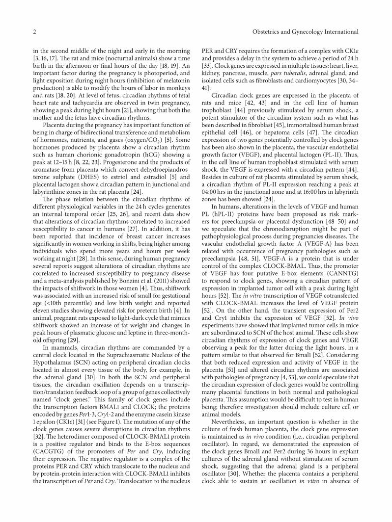
Figure 2: Signaling of MT1 and MT2. Both are G protein-coupled receptors. MT1 is associated with Gi protein and inhibition of adenylylcyclase. MT2 receptor is associated with PKC stimulation and increase of calcium associated with IP3 (for details see Dubocovich et al. [11]).
adrenocorticotropin (ACTH) in humans and sheep [38, 67],strongly suggesting a loop of regulation between melatoninproduction and clock genes expression.
Melatonin acts through membrane receptors (see Fig-ure 2), although some effects of melatonin could be medi-ated by binding to endogenous ligand, the orphan nuclearhormone receptor superfamily RZR/ROR [68].There are twomelatonin membrane receptors named MT1 and MT2. Bothare G protein-coupled receptors. Thus, MT1 is associatedwith (i) Gi and inhibition of adenylyl cyclase with decreaseof cAMP, (ii) stimulation of potassium channels, and (iii)increase of Ca2+ via phospholipase C (PLC), whereas theMT2 receptor is associated with (a) Protein Kinase C (PKC)stimulation or (b) increase of calcium associated with IP
3.
Melatonin receptors MT1 and MT2 are present in many tis-sues [11, 69] although their function is not quite understood.However, their participation has been described in severalmechanisms as indicated below.
3.1. Chronobiotic and Homeostatic Effects of Melatonin. Invivo treatment (2 hours) withmelatonin increases the expres-sion of Cry-2 in rat pars tuberalis [36] and decreases in theamplitude of the peak of Per1, suggesting an inhibitory effectof melatonin on this gene expression. In sheep, in whichthe secretion of melatonin was abolished by exposure tocontinuous light, expression of the clock genes Per 1-2 andBmal1 continued in the pars tuberalis. However, melatonin
treatment at any point in the day for a period of 3 hoursinducedmRNA expression of Cry-1 and inhibited the expres-sion of Per 1-2 and Bmal1, effects that were not observed inthe SCN [70]. In capuchinmonkey, adrenal explantsmaintainan oscillatory expression of Bmal1 and Per2 for at least 36hours in culture, and the treatment withmelatonin decreasedthe expression of Bmal1 and Per2 [30]. Similar effects havebeen shown in the rat fetal adrenal in culture [40] andrecently we detected that melatonin inhibit the expressionof Per1 and Bmal1 in response to ACTH in newborn sheepand human adrenal gland [38, 67]. Moreover, melatonin viamelatonin receptor MT1 and MT2 can modify the levels ofpro- or antiapoptotic proteins such as Bax andBcl-2 in humanneuroblastoma cells [64]; similarly in placenta, treatmentwith 10 𝜇Mof melatonin in villous trophoblast cells increasesthe survival via inhibition of loss ofmitochondrialmembranepotential and stimulating the formation of complex Bax/Bcl-2 (intrinsic via), expression of caspase-9, and the activation ofROCK1 [71].
3.2. Antioxidant Effects of Melatonin. Melatonin has a potentscavenger activity over hydroxyl, alkoxyl, and peroxyl rad-icals, as well as over species derived from nitrogen suchas nitric oxide (NO) radicals [11, 57, 58]. In this regard,Milczarek et al. (2010) reported in placentas obtained afterdelivery a potent antioxidant effect of melatonin, specifically
Obstetrics and Gynecology International 5
preventing the NADPH and iron dependent lipid peroxida-tion in the mitochondria [72]. Moreover, in studies of fetalgrowth restriction in animal model by placental ischemia,increased placental level of 8-hydroxy-2-deoxyguanosinehas been detected (8-OHdG, i.e., a marker of a markerof oxidative DNA damage) [73] compared with controls.Both growth restriction and DNA damage were revertedwhen the rats received an oral dose of melatonin. Similarly,in rat undernourished pregnancy at day 20 of gestation,the fetal biometry showed lower values for fetal bodyweight and fetal body/placental weight ratio; moreover atendency to a higher value of melatonin in maternal andfetal plasma is observed. Placentas from undernourishedpregnancy showed no changes in the expression of antiox-idant enzymes Mn-SOD, catalase, and GPx-1. However,the melatonin treatment during the pregnancy restores theplacental efficiency at level of fetal body weight and fetalbody/placental weight and induces the protein expression ofMn-SOD and catalase [59], suggesting that melatonin couldbe a candidate for protection/treatment of diseases charac-terized by placental ischemia such as intrauterine growthrestriction, preeclampsia [73], or undernourished pregnancy[59]. Indeed, recent evidences have described that melatoninadministration improved fetal-placental hemodynamic [74]and increased umbilical blood flow, an effect associatedwith “NO-dependent mechanisms” [75], as what occurs incotyledonary placental arteries, via increased sensitivity tovasorelaxation agents such as bradykinin and lower contrac-tile response to noradrenaline [76]. Additionally, melatoninadministration reverted the increment of lipid peroxidationin the placenta and liver of mother and fetus exposed tocholestasis of pregnancy [77]. Interestingly, in human it hasbeen described that oral melatonin administration increasedglutathione peroxidase (GSH-Px) in the placenta of Japanesewomen with pregnancies of 7 and 9 weeks of gestation [78].
Protective effects of other antioxidant agents on fetalgrowth and development strongly support the protectiveeffects of melatonin in adverse pregnancy being due to itsantioxidant rather than antioxidant-independent properties,for example, developmental programming of cardiovasculardysfunction by prenatal hypoxia and oxidative stress.
3.3. Melatonin and Vascular Remodeling. In rats, it has beendescribed that melatonin regulate the levels of NO andVEGF in the nervous system, that is, in the choroid plexus,cerebellum, periventricular white matter, and hippocam-pus [79–82]. Moreover, melatonin treatment for short orlong periods of time inhibits the endogenous expressionof VEGF and hypoxia induced factor 1 alpha (HIF-1𝛼)in tumor cells [83]. In addition, melatonin induces theexpression of VEGF and matrix metalloproteinase- (MMP-)2 in extrapineal tissues such as gastric mucosa [66]. Alsomelatonin increases bone defect repair in rabbits [84]. Allthese indirect evidences suggest that melatonin may controlvessel formation. Nevertheless, other reports have showedreduced tumor angiogenesis in mice treated with melatonin[85], as well as reduced human umbilical vein endothelial cellproliferation/migration induced by VEGF [86, 87].
4. Extrapineal Production of Melatonin:The Placenta
The critical enzymes for the synthesis of melatonin arearylalkylamine N-acetyltransferase (AA-NAT) and hydrox-yindole O-methyltransferase (HIOMT). These enzymes areexpressed in the major site of synthesis of melatonin, in thepineal gland with a circadian pattern of activity for AA-NAT [11], and in the human placenta [88]. Therefore, thehuman placenta can be considered as an extrapineal sourceof melatonin similar to retina [89] and lymphocytes [90].In this regard, expression (mRNA and protein) of AA-NATand HIOMT has been detected in both cell line of humanplacental trophoblasts and human term placentas [71, 88, 91].In addition, other reports have described the presence ofMT1 and MT2 in total human placenta [55] or placentalendothelial cells [88]. Interestingly, MT1 is expressed at highlevels in the junctional zone during day hours (16:00 hrs) andin the labyrinth during night hours (04:00 hrs) in mice [24].
We do not know whether the production of melatoninin the human placenta changes with the hours of the day.Interestingly, in normal pregnancies, serum levels of mela-tonin increase progressively until 32 weeks of gestation anddecrease prior to delivery reaching the lowest levels at 2days postpartum [92–95]. Moreover, the level of melatonin ishigher in human twins pregnancies [92], as well as it is corre-latedwith the number of pups in animalmodels [94], suggest-ing a relationship between placental volume and melatoninlevel. Nevertheless, in human pregnancies associated withplacental alteration, the maternal circadian production ofmelatonin is lost and it is associated with diminished levelsof melatonin. For example, circadian alteration has beenobserved in humans at the level of diastolic blood pressure,plasma concentration, and circadian production ofmelatoninduring preeclampsia. After pregnancy, these women showeda normal circadian diastolic blood pressure but maintainedaltered rhythm for melatonin [53]. In contrast to humans,AA-NAT is not expressed in the placenta of rats, and anincrease of maternal melatonin is a consequence of placentalfactor released into the circulation,whichwould stimulate thematernal pineal gland [94].
Although melatonin has multiple effects on placentalfunction, including induction of the expression in under-nourished pregnancy of antioxidant enzymes such as super-oxide dismutase (Mn-SOD) and catalase [59], prevention ofoxidative stress-mediated injury during placental ischemia[73], inhibition of hCG release in trophoblast cells [91],and inhibition of formation of proapoptotic complex [71],the role of this local production in the human placenta isnot well understood and opens the possibility of autocrine,intracrine, or paracrine effects. Although there are no directevidences, other studies using lymphoid cells, which expressthe enzymes NAT and HIOMT and produce 5-fold moremelatonin than pineal gland, have described that melatoninproduced locally has a minor effect on melatonin serum levelbut has a local role incrementing the IL-2 production [90].This last effect was inhibited by luzindole and CGP 55644, anantagonist of membrane and nuclear receptors of melatonin[96]. Then, it is feasible that melatonin can be modulating

6 Obstetrics and Gynecology International
Melatonin
NAT
HIOMT
Trophoblast
Placental endothelial cell
??
Clock genes MT1
?
Pineal gland (mother)
Melatonin
Circadian expression??
Auto
crin
e effe
ct
Para
crin
e effe
ct
?
Clock controlledgenes, that is, VEGF
MT2
Signal transduction
Figure 3: Autocrine and paracrine effects of melatonin over circadian system and enzyme of synthesis of melatonin localized in trophoblastand endothelial cells.
the circadian system in the placenta, see Figure 3, producingchanges in clock genes that may control VEGF or enzymesNAT and HIOMT. We encourage the scientific communitywith this idea.
5. Conclusion
Participation of the circadian regulatory system has beendescribed as a feedback regulatory loop where melatonin isdownregulating the clock genes. In turn, clock genes upregu-late the expression of output genes such as VEGF, SOD, NAT,and HIOMT. Current evidences describe that the placentais a nonpineal organ, which synthesizes melatonin, and theactivity of this organ is regulated by a circadian system.Moreover, an impaired circadian system is associated with analtered production of melatonin; however, the effect of thisalteration on clock gene expression or output genes (VEGF)has not been described. We believe that circadian systemand melatonin are a keystone molecule in the placentalphysiology, butmore studies are necessary in order to test thisidea. In this regard, we propose that melatonin may controlclock gene expression (Bmal1, Per1-3, Cry1-2, and Clock) andoutput genes (VEGF) during normal pregnancies.
Glossary
BMAL1: Aryl hydrocarbon receptor nucleartranslocator-like
CLOCK: Circadian locomotor output cycles kaputPER: Homolog of period, DrosophilaE-BOX: Promoter sequence for binding of clock-bmal1
complex (CACGTG)CREB: cAMP response element-binding proteinPGC1𝛼: Peroxisome proliferator-activated
receptor-gamma, coactivator 1𝛼.
Conflict of Interests
None of the authors have a conflict of interests.
Acknowledgment
This paper received funding Conicyt 79112027 (Chile).
References
[1] J. Cha, X. Sun, and S. K. Dey, “Mechanisms of implantation:strategies for successful pregnancy,”NatureMedicine, vol. 18, no.12, pp. 1754–1767, 2012.
[2] P. H. Andraweera, G. A. Dekker, and C. T. Roberts, “Thevascular endothelial growth factor family in adverse pregnancyoutcomes,” Human Reproduction Update, vol. 18, no. 4, ArticleID dms011, pp. 436–457, 2012.
[3] J. Olcese, “Circadian aspects of mammalian parturition: areview,” Molecular and Cellular Endocrinology, vol. 349, no. 1,pp. 62–67, 2012.

Obstetrics and Gynecology International 7
[4] M. Bonzini, K. T. Palmer, D. Coggon, M. Carugno, A. Cromi,and M. M. Ferrario, “Shift work and pregnancy outcomes:a systematic review with meta-analysis of currently availableepidemiological studies,” BJOG, vol. 118, no. 12, pp. 1429–1437,2011.
[5] M. Seron-Ferre, C. A. Ducsay, and G. J. Valenzuela, “Circadianrhythms during pregnancy,” Endocrine Reviews, vol. 14, no. 5,pp. 594–609, 1993.
[6] W. P. Mutter and S. A. Karumanchi, “Molecular mechanismsof preeclampsia,”Microvascular Research, vol. 75, no. 1, pp. 1–8,2008.
[7] R. Romero, J. P. Kusanovic, T. Chaiworapongsa, and S. S.Hassan, “Placental bed disorders in preterm labor, pretermPROM, spontaneous abortion and abruptio placentae,” BestPractice and Research: Clinical Obstetrics and Gynaecology, vol.25, no. 3, pp. 313–327, 2011.
[8] S. Rotmensch, C. Celentano, N. Elliger et al., “Diurnal variationof human chorionic gonadotropin 𝛽-core fragment concentra-tions in urine during second trimester of pregnancy,” ClinicalChemistry, vol. 47, no. 9, pp. 1715–1717, 2001.
[9] M. Seron-Ferre, G. J. Valenzuela, and C. Torres-Farfan, “Cir-cadian clocks during embryonic and fetal development,” BirthDefects Research Part C: Embryo Today: Reviews, vol. 81, no. 3,pp. 204–214, 2007.
[10] M. Seron-Ferre, N. Mendez, L. Abarzua-Catalan et al., “Circa-dian rhythms in the fetus,” Molecular and Cellular Endocrinol-ogy, vol. 349, no. 1, pp. 68–75, 2012.
[11] M. L. Dubocovich, P. Delagrange, D. N. Krause, D. Sugden,D. P. Cardinali, and J. Olcese, “International union of basicand clinical pharmacology. LXXV.Nomenclature, classification,and pharmacology of G protein-coupled melatonin receptors,”Pharmacological Reviews, vol. 62, no. 3, pp. 343–380, 2010.
[12] M. Seron-Ferre, M. L. Forcelledo, C. Torres-Farfan et al.,“Impact of chronodisruption during primate pregnancy on thematernal and newborn temperature rhythms,” PLoS ONE, vol.8, no. 2, Article ID e57710, 2013.
[13] J. Malek, K. Suk, M. Brestak, and V. Maly, “Daily rhythm ofleukocytes, blood pressure, pulse rate, and temperature duringpregnancy.,” Annals of the New York Academy of Sciences, vol.98, pp. 1018–1041, 1962.
[14] M. Barr Jr., “Prenatal growth of Wistar rats: circadian periodic-ity of fetal growth late in gestation,” Teratology, vol. 7, no. 3, pp.283–288, 1973.
[15] M. A. Morgan, S. L. Silavin, R. A. Wentworth et al., “Differentpatterns of myometrial activity and 24-H rhythms in myome-trial contractility in the gravid baboon during the second halfof pregnancy,” Biology of Reproduction, vol. 46, no. 6, pp. 1158–1164, 1992.
[16] M. Vatish, P. J. Steer, A. M. Blanks, M. Hon, and S. Thornton,“Diurnal variation is lost in preterm deliveries before 28 weeksof gestation,” BJOG: An International Journal of Obstetrics andGynaecology, vol. 117, no. 6, pp. 765–767, 2010.
[17] J. Malek, J. Gleich, and V. Maly, “Characteristics of the dailyrhythm of menstruation and labor.,” Annals of the New YorkAcademy of Sciences, vol. 98, pp. 1042–1055, 1962.
[18] H. Takayama, Y. Nakamura, H. Tamura et al., “Pineal gland(melatonin) affects the parturition time, but not luteal functionand fetal growth, in pregnant rats,” Endocrine Journal, vol. 50,no. 1, pp. 37–43, 2003.
[19] C. K. Ratajczak, M. Asada, G. C. Allen et al., “Generationof myometrium-specific Bmal1 knockout mice for parturitionanalysis,” Reproduction, Fertility and Development, vol. 24, no.5, pp. 759–767, 2012.
[20] C. A. Ducsay and S. M. Yellon, “Photoperiod regulation ofuterine activity and melatonin rhythms in the pregnant rhesusmacaque,” Biology of Reproduction, vol. 44, no. 6, pp. 967–974,1991.
[21] Y. Maeda, M. Muro, M. Shono, H. Shono, and T. Iwasaka,“Diurnal rhythms in fetal heart rate baseline and sustained fetaltachycardia in twin pregnancy,” Early Human Development, vol.82, no. 10, pp. 637–644, 2006.
[22] L. Dıaz-Cueto, J. P. Mendez, J. Barrios-de-Tomasi et al.,“Amplitude regulation of episodic release, in vitro biological toimmunological ratio, and median charge of human chorionicgonadotropin in pregnancy,” Journal of Clinical Endocrinologyand Metabolism, vol. 78, no. 4, pp. 890–897, 1994.
[23] S. T. Nakajima, T. Mcauliffe, and M. Gibson, “The 24-hourpattern of the levels of serumprogesterone and immunoreactivehuman chorionic Gonadotropin in Normal Early Pregnancy,”Journal of Clinical Endocrinology and Metabolism, vol. 71, no. 2,pp. 345–353, 1990.
[24] C. K. Lee, D. H. Moon, C. S. Shin et al., “Circadian expressionof Mel
1𝑎and PL-II genes in placenta: effects of melatonin on
the PL-II gene expression in the rat placenta,” Molecular andCellular Endocrinology, vol. 200, no. 1-2, pp. 57–66, 2003.
[25] M. C. Moore-Ede, “Physiology of the circadian timing system:predictive versus reactive homeostasis,”TheAmerican Journal ofPhysiology—Regulatory Integrative and Comparative Physiology,vol. 250, no. 5, part 2, pp. R737–R752, 1986.
[26] I. Edery, “Circadian rhythms in a nutshell,” Physiol Genomics,vol. 3, no. 2, pp. 59–74, 2000.
[27] C.-M. Hsu, S.-F. Lin, C.-T. Lu, P.-M. Lin, and M.-Y. Yang,“Altered expression of circadian clock genes in head and necksquamous cell carcinoma,”Tumor Biology, vol. 33, no. 1, pp. 149–155, 2012.
[28] E. S. Schernhammer, F. Laden, F. E. Speizer et al., “Rotatingnight shifts and risk of breast cancer in women participating inthe nurses’ health study,” Journal of theNational Cancer Institute,vol. 93, no. 20, pp. 1563–1568, 2001.
[29] T. J. Varcoe, N.Wight, A. Voultsios,M.D. Salkeld, andD. J. Ken-naway, “Chronic phase shifts of the photoperiod throughoutpregnancy programs glucose intolerance and insulin resistancein the rat,” PLoS ONE, vol. 6, no. 4, Article ID e18504, 2011.
[30] F. J. Valenzuela, C. Torres-Farfan, H. G. Richter et al., “Clockgene expression in adult primate suprachiasmatic nuclei andadrenal: is the adrenal a peripheral clock responsive to mela-tonin?” Endocrinology, vol. 149, no. 4, pp. 1454–1461, 2008.
[31] J. C. Dunlap, J. J. Loros, Y. Liu, and S. K. Crosthwaite, “Eukary-otic circadian systems: cycles in common,”Genes to Cells, vol. 4,no. 1, pp. 1–10, 1999.
[32] K. Bae, X. Jin, E. S. Maywood, M. H. Hastings, S. M. Reppert,and D. R. Weaver, “Differential functions ofmPer1,mPer2, andmPer3 in the SCN circadian clock,” Neuron, vol. 30, no. 2, pp.525–536, 2001.
[33] M. Akashi, Y. Tsuchiya, T. Yoshino, and E. Nishida, “Control ofintracellular dynamics of mammalian period proteins by caseinkinase I epsilon (CKIepsilon) and CKIdelta in cultured cells,”Molecular and Cellular Biology, vol. 22, no. 6, pp. 1693–1703,2002.

8 Obstetrics and Gynecology International
[34] M. Stratmann and U. Schibler, “Properties, entrainment, andphysiological functions of mammalian peripheral oscillators,”Journal of Biological Rhythms, vol. 21, no. 6, pp. 494–506, 2006.
[35] G. A. Lincoln, H. Andersson, and D. Hazlerigg, “Clock genesand the long-term regulation of prolactin secretion: evidencefor a photoperiod/circannual timer in the pars tuberalis,”Journal of Neuroendocrinology, vol. 15, no. 4, pp. 390–397, 2003.
[36] H. Dardente, J. S. Menet, V.-J. Poirel et al., “Melatonin inducesCry1 expression in the pars tuberalis of the rat,”Molecular BrainResearch, vol. 114, no. 2, pp. 101–106, 2003.
[37] D. R. Lemos, J. L. Downs, and H. F. Urbanski, “Twenty-four-hour rhythmic gene expression in the rhesus macaque adrenalgland,” Molecular Endocrinology, vol. 20, no. 5, pp. 1164–1176,2006.
[38] C. Campino, F. J. Valenzuela, C. Torres-Farfan et al., “Melatoninexerts direct inhibitory actions on ACTH responses in thehuman adrenal gland,” Hormone and Metabolic Research, vol.43, no. 5, pp. 337–342, 2011.
[39] C. Torres-Farfan, V. Rocco, C. Monso et al., “Maternal mela-tonin effects on clock gene expression in a nonhuman primatefetus,” Endocrinology, vol. 147, no. 10, pp. 4618–4626, 2006.
[40] C. Torres-Farfan, N. Mendez, L. Abarzua-Catalan, N. Vilches,G. J. Valenzuela, and M. Seron-Ferre, “A circadian clockentrained by melatonin is ticking in the rat fetal adrenal,”Endocrinology, vol. 152, no. 5, pp. 1891–1900, 2011.
[41] H. Oster, S. Damerow, S. Kiessling et al., “The circadian rhythmof glucocorticoids is regulated by a gating mechanism residingin the adrenal cortical clock,” Cell Metabolism, vol. 4, no. 2, pp.163–173, 2006.
[42] M. D. Wharfe, P. J. Mark, and B. J. Waddell, “Circadianvariation in placental and hepatic clock genes in rat pregnancy,”Endocrinology, vol. 152, no. 9, pp. 3552–3560, 2011.
[43] S. Akiyama, H. Ohta, S. Watanabe et al., “The uterus sustainsstable biological clock during pregnancy,” The Tohoku Journalof Experimental Medicine, vol. 221, no. 4, pp. 287–298, 2010.
[44] E. Frigato, L. Lunghi,M. E. Ferretti, C. Biondi, andC. Bertolucci,“Evidence for circadian rhythms in human trophoblast cell linethat persist in hypoxia,” Biochemical and Biophysical ResearchCommunications, vol. 378, no. 1, pp. 108–111, 2009.
[45] A. Balsalobre, L. Marcacci, and U. Schibler, “Multiple signalingpathways elicit circadian gene expression in cultured Rat-1fibroblasts,” Current Biology, vol. 10, no. 20, pp. 1291–1294, 2000.
[46] S. Xiang, L. Mao, T. Duplessis et al., “Oscillation of clock andclock controlled genes induced by serum shock in human breastepithelial and breast cancer cells: regulation by melatonin,”Breast Cancer: Basic and Clinical Research, vol. 6, no. 1, pp. 137–150, 2012.
[47] A. Balsalobre, F. Damiola, and U. Schibler, “A serum shockinduces circadian gene expression in mammalian tissue culturecells,” Cell, vol. 93, no. 6, pp. 929–937, 1998.
[48] F. J. Valenzuela, A. Perez-Sepulveda, M. J. Torres, P. Correa, G.M. Repetto, and S. E. Illanes, “Pathogenesis of preeclampsia: thegenetic component,” Journal of Pregnancy, vol. 2012, Article ID632732, 8 pages, 2012.
[49] P. J. Dutton, L. K. Warrander, S. A. Roberts et al., “Predictorsof poor perinatal outcome following maternal perception ofreduced fetal movements—a prospective cohort study,” PLoSONE, vol. 7, no. 7, Article ID e39784, 2012.
[50] R. V. Anthony, S. L. Pratt, R. Liang, and M. D. Holland,“Placental-fetal hormonal interactions: impact on fetal growth,”Journal of Animal Science, vol. 73, no. 6, pp. 1861–1871, 1995.
[51] Y. Wang and S. Zhao, Vascular Biology of the Placenta, Morgan& Claypool Life Sciences, San Rafael, Calif, USA, 2010.
[52] S. Koyanagi, Y. Kuramoto, H. Nakagawa et al., “A molec-ular mechanism regulating circadian expression of vascularendothelial growth factor in tumor cells,” Cancer Research, vol.63, no. 21, pp. 7277–7283, 2003.
[53] A. L. Tranquilli, A. Turi, S. R. Giannubilo, and E. Garbati,“Circadian melatonin concentration rhythm is lost in pregnantwomen with altered blood pressure rhythm,” GynecologicalEndocrinology, vol. 18, no. 3, pp. 124–129, 2004.
[54] H. Guo, J. M. Brewer, M. N. Lehman, and E. L. Bittman,“Suprachiasmatic regulation of circadian rhythms of geneexpression in hamster peripheral organs: effects of transplantingthe pacemaker,”The Journal of Neuroscience, vol. 26, no. 24, pp.6406–6412, 2006.
[55] D. Lanoix, R. Ouellette, and C. Vaillancourt, “Expression ofmelatoninergic receptors in human placental choriocarcinomacell lines,” Human Reproduction, vol. 21, no. 8, pp. 1981–1989,2006.
[56] S. R. Pandi-Perumal, V. Srinivasan, G. J. M. Maestroni, D. P.Cardinali, B. Poeggeler, and R. Hardeland, “Melatonin: nature’smost versatile biological signal?” FEBS Journal, vol. 273, no. 13,pp. 2813–2838, 2006.
[57] A. Galano, D. X. Tan, and R. J. Reiter, “Melatonin as a naturalally against oxidative stress: a physicochemical examination,”Journal of Pineal Research, vol. 51, no. 1, pp. 1–16, 2011.
[58] R. J. Reiter, D.-X. Tan, L. C. Manchester, S. D. Paredes, J. C.Mayo, and R.M. Sainz, “Melatonin and reproduction revisited,”Biology of Reproduction, vol. 81, no. 3, pp. 445–456, 2009.
[59] H. G. Richter, J. A. Hansell, S. Raut, and D. A. Giussani,“Melatonin improves placental efficiency and birth weight andincreases the placental expression of antioxidant enzymes inundernourished pregnancy,” Journal of Pineal Research, vol. 46,no. 4, pp. 357–364, 2009.
[60] C. H. Kim and Y.-M. Yoo, “Melatonin induces apoptotic celldeath via p53 in LNCaP cells,” Korean Journal of Physiology andPharmacology, vol. 14, no. 6, pp. 365–369, 2010.
[61] S. M. Dupre, D. W. Burt, R. Talbot et al., “Identification ofmelatonin-regulated genes in the ovine pituitary pars tuberalis,a target site for seasonal hormone control,” Endocrinology, vol.149, no. 11, pp. 5527–5539, 2008.
[62] E. H. Sharman, S. C. Bondy, K. G. Sharman, D. Lahiri, C.W. Cotman, and V. M. Perreau, “Effects of melatonin and ageon gene expression in mouse CNS using microarray analysis,”Neurochemistry International, vol. 50, no. 2, pp. 336–344, 2007.
[63] E. Ha, E. Han, H. J. Park et al., “Microarray analysis of tran-scription factor gene expression in melatonin-treated humanperipheral bloodmononuclear cells,” Journal of Pineal Research,vol. 40, no. 4, pp. 305–311, 2006.
[64] W. Wisessmith, P. Phansuwan-Pujito, P. Govitrapong, and B.Chetsawang, “Melatonin reduces induction of Bax, caspase andcell death in methamphetamine-treated human neuroblastomaSH-SY5Y cultured cells,” Journal of Pineal Research, vol. 46, no.4, pp. 433–440, 2009.
[65] K. Tsutsui, S. Haraguchi, K. Inoue et al., “Identification, biosyn-thesis, and function of 7𝛼-hydroxypregnenolone, a new keyneurosteroid controlling locomotor activity, in nonmammalianvertebrates,” Annals of the New York Academy of Sciences, vol.1163, pp. 308–315, 2009.

Obstetrics and Gynecology International 9
[66] K. Ganguly and S. Swarnakar, “Induction of matrix metal-loproteinase-9 and -3 in nonsteroidal anti-inflammatory drug-induced acute gastric ulcers in mice: regulation by melatonin,”Journal of Pineal Research, vol. 47, no. 1, pp. 43–55, 2009.
[67] F. J. Valenzuela, H. E. Reynolds, C. Torres-Farfan, G. J. Valen-zuela, andM. Seron-Ferre, “Melatonin inhibition of the cortisolresponse to aCth may be exerted through period circadianprotein homolog 1 (Per1),” Revista Chilena de Endocrinologıa yDiabetes, vol. 5, no. 1, pp. 6–12, 2012.
[68] B. Claustrat, J. Brun, and G. Chazot, “The basic physiology andpathophysiology of melatonin,” Sleep Medicine Reviews, vol. 9,no. 1, pp. 11–24, 2005.
[69] C. Ekmekcioglu, “Melatonin receptors in humans: biologicalrole and clinical relevance,” Biomedicine and Pharmacotherapy,vol. 60, no. 3, pp. 97–108, 2006.
[70] J. D. Johnston, B. B. Tournier, H. Andersson, M. Masson-Pevet,G. A. Lincoln, and D. G. Hazlerigg, “Multiple effects of mela-tonin on rhythmic clock gene expression in the mammalianpars tuberalis,” Endocrinology, vol. 147, no. 2, pp. 959–965, 2006.
[71] D. Lanoix, P. Guerin, and C. Vaillancourt, “Placental melatoninproduction and melatonin receptor expression are altered inpreeclampsia: new insights into the role of this hormone inpregnancy,” Journal of Pineal Research, vol. 53, no. 4, pp. 417–425, 2012.
[72] R. Milczarek, A. Hallmann, E. Sokołowska, K. Kaletha, and J.Klimek, “Melatonin enhances antioxidant action of 𝛼-tocopherol and ascorbate against NADPH- and iron-dependentlipid peroxidation in human placental mitochondria,” Journalof Pineal Research, vol. 49, no. 2, pp. 149–155, 2010.
[73] R. Nagai, K. Watanabe, A. Wakatsuki et al., “Melatonin pre-serves fetal growth in rats by protecting against ischemia/reperfusion-induced oxidative/nitrosative mitochondrial dam-age in the placenta,” Journal of Pineal Research, vol. 45, no. 3, pp.271–276, 2008.
[74] C. O. Lemley, A. M. Meyer, L. E. Camacho et al., “Melatoninsupplementation alters uteroplacental hemodynamics and fetaldevelopment in an ovine model of intrauterine growth restric-tion,” The American Journal of Physiology—Regulatory Integra-tive and Comparative Physiology, vol. 302, no. 4, pp. R454–R467,2012.
[75] A. S. Thakor, E. A. Herrera, M. Seron-Ferre, and D. A.Giussani, “Melatonin and vitamin C increase umbilical bloodflow via nitric oxide-dependent mechanisms,” Journal of PinealResearch, vol. 49, no. 4, pp. 399–406, 2010.
[76] P. Shukla, C. O. Lemley, N. Dubey, A. M. Meyer, S. T. O’Rourke,and K. A. Vonnahme, “Effect of maternal nutrient restrictionand melatonin supplementation from mid to late gestationon vascular reactivity of maternal and fetal placental arteries,”Placenta, vol. 35, no. 7, pp. 461–466, 2014.
[77] M. J. Perez, B. Castano, J. M. Gonzalez-Buitrago, and J. J.G. Marin, “Multiple protective effects of melatonin againstmaternal cholestasis-induced oxidative stress and apoptosis inthe rat fetal liver-placenta-maternal liver trio,” Journal of PinealResearch, vol. 43, no. 2, pp. 130–139, 2007.
[78] Y. Okatani, A. Wakatsuki, K. Shinohara, C. Kaneda, and T.Fukaya, “Melatonin stimulates glutathione peroxidase activityin human chorion,” Journal of Pineal Research, vol. 30, no. 4, pp.199–205, 2001.
[79] V. Sivakumar, J. Lu, E. A. Ling, and C. Kaur, “Vascular endothe-lial growth factor and nitric oxide production in response
to hypoxia in the choroid plexus in neonatal brain,” BrainPathology, vol. 18, no. 1, pp. 71–85, 2008.
[80] C. Kaur, V. Sivakumar, Y. Zhang, and E. A. Ling, “Hypoxia-induced astrocytic reaction and increased vascular permeabilityin the rat cerebellum,” Glia, vol. 54, no. 8, pp. 826–839, 2006.
[81] C. Kaur, V. Sivakumar, J. Lu, F. R. Tang, and E. A. Ling,“Melatonin attenuates hypoxia-induced ultrastructural changesand increased vascular permeability in the developing hip-pocampus,” Brain Pathology, vol. 18, no. 4, pp. 533–547, 2008.
[82] C. Kaur, V. Sivakumar, and E. A. Ling, “Melatonin protectsperiventricular white matter from damage due to hypoxia,”Journal of Pineal Research, vol. 48, no. 3, pp. 185–193, 2010.
[83] M. Dai, P. Cui, M. Yu, J. Han, H. Li, and R. Xiu, “Melatoninmodulates the expression of VEGF and HIF-1𝛼 induced byCoCl2in cultured cancer cells,” Journal of Pineal Research, vol.
44, no. 2, pp. 121–126, 2008.[84] M. P. Ramırez-Fernandez, J. L. Calvo-Guirado, J. E.-M. S. de-Val
et al., “Melatonin promotes angiogenesis during repair of bonedefects: a radiological and histomorphometric study in rabbittibiae,” Clinical Oral Investigations, vol. 17, no. 1, pp. 147–158,2013.
[85] K.-J. Kim, J.-S. Choi, I. Kang, K.-W. Kim, C.-H. Jeong, and J.-W. Jeong, “Melatonin suppresses tumor progression by reducingangiogenesis stimulated by HIF-1 in a mouse tumor model,”Journal of Pineal Research, vol. 54, no. 3, pp. 264–270, 2013.
[86] V. Alvarez-Garcıa, A. Gonzalez, C. Martınez-Campa, C.Alonso-Gonzalez, and S. Cos, “Melatoninmodulates aromataseactivity and expression in endothelial cells,” Oncology Reports,vol. 29, no. 5, pp. 2058–2064, 2013.
[87] V. Alvarez-Garcıa, A. Gonzalez, C. Alonso-Gonzalez, C.Martınez-Campa, and S. Cos, “Regulation of vascular endothe-lial growth factor by melatonin in human breast cancer cells,”Journal of Pineal Research, vol. 54, no. 4, pp. 373–380, 2013.
[88] D. Lanoix, H. Beghdadi, J. Lafond, andC. Vaillancourt, “Humanplacental trophoblasts synthesize melatonin and express itsreceptors,” Journal of Pineal Research, vol. 45, no. 1, pp. 50–60,2008.
[89] P. M. Iuvone, G. Tosini, N. Pozdeyev, R. Haque, D. C. Klein,and S. S. Chaurasia, “Circadian clocks, clock networks, ary-lalkylamine N-acetyltransferase, and melatonin in the retina,”Progress in Retinal and Eye Research, vol. 24, no. 4, pp. 433–456,2005.
[90] A. Carrillo-Vico, J. R. Calvo, P. Abreu et al., “Evidence ofmelatonin synthesis by human lymphocytes and its physiolog-ical significance: possible role as intracrine, autocrine, and/orparacrine substance.,”The FASEB Journal, vol. 18, no. 3, pp. 537–539, 2004.
[91] S. Iwasaki, K. Nakazawa, J. Sakai et al., “Melatonin as alocal regulator of human placental function,” Journal of PinealResearch, vol. 39, no. 3, pp. 261–265, 2005.
[92] Y. Nakamura, H. Tamura, S. Kashida et al., “Changes of serummelatonin level and its relationship to feto-placental unit duringpregnancy,” Journal of Pineal Research, vol. 30, no. 1, pp. 29–33,2001.
[93] H. Tamura, Y. Nakamura, M. P. Terron et al., “Melatonin andpregnancy in the human,” Reproductive Toxicology (Elmsford,N.Y.), vol. 25, no. 3, pp. 291–303, 2008.
[94] H. Tamura, H. Takayama, Y. Nakamura, R. J. Reiter, and N.Sugino, “Fetal/placental regulation of maternal melatonin inrats,” Journal of Pineal Research, vol. 44, no. 3, pp. 335–340, 2008.

10 Obstetrics and Gynecology International
[95] F. Wierrani, W. Grin, B. Hlawka, A. Kroiss, andW. Grunberger,“Elevated serummelatonin levels during human late pregnancyand labour,” Journal of Obstetrics and Gynaecology, vol. 17, no. 5,pp. 449–451, 1997.
[96] A. Carrillo-Vico, P. J. Lardone, J. M. Fernandez-Santos et al.,“Human lymphocyte-synthesized melatonin is involved in theregulation of the interleukin-2/interleukin-2 receptor system,”The Journal of Clinical Endocrinology &Metabolism, vol. 90, no.2, pp. 992–1000, 2005.

Submit your manuscripts athttp://www.hindawi.com
Stem CellsInternational
Hindawi Publishing Corporationhttp://www.hindawi.com Volume 2014
Hindawi Publishing Corporationhttp://www.hindawi.com Volume 2014
MEDIATORSINFLAMMATION
of
Hindawi Publishing Corporationhttp://www.hindawi.com Volume 2014
Behavioural Neurology
EndocrinologyInternational Journal of
Hindawi Publishing Corporationhttp://www.hindawi.com Volume 2014
Hindawi Publishing Corporationhttp://www.hindawi.com Volume 2014
Disease Markers
Hindawi Publishing Corporationhttp://www.hindawi.com Volume 2014
BioMed Research International
OncologyJournal of
Hindawi Publishing Corporationhttp://www.hindawi.com Volume 2014
Hindawi Publishing Corporationhttp://www.hindawi.com Volume 2014
Oxidative Medicine and Cellular Longevity
Hindawi Publishing Corporationhttp://www.hindawi.com Volume 2014
PPAR Research
The Scientific World JournalHindawi Publishing Corporation http://www.hindawi.com Volume 2014
Immunology ResearchHindawi Publishing Corporationhttp://www.hindawi.com Volume 2014
Journal of
ObesityJournal of
Hindawi Publishing Corporationhttp://www.hindawi.com Volume 2014
Hindawi Publishing Corporationhttp://www.hindawi.com Volume 2014
Computational and Mathematical Methods in Medicine
OphthalmologyJournal of
Hindawi Publishing Corporationhttp://www.hindawi.com Volume 2014
Diabetes ResearchJournal of
Hindawi Publishing Corporationhttp://www.hindawi.com Volume 2014
Hindawi Publishing Corporationhttp://www.hindawi.com Volume 2014
Research and TreatmentAIDS
Hindawi Publishing Corporationhttp://www.hindawi.com Volume 2014
Gastroenterology Research and Practice
Hindawi Publishing Corporationhttp://www.hindawi.com Volume 2014
Parkinson’s Disease
Evidence-Based Complementary and Alternative Medicine
Volume 2014Hindawi Publishing Corporationhttp://www.hindawi.com


![melatonin - Department of Chemistry€¦ · functioning circadian rhythm. Melatonin supplements are made synthetically and can be purchased without a prescription [1]. History of](https://img.dokumen.tips/doc/110x75/5f091d457e708231d4254be9/melatonin-department-of-chemistry-functioning-circadian-rhythm-melatonin-supplements.jpg)