Embed Size (px)
Citation preview

Retrospective Reconstruction ofRadiation Doses of
Chernobyl Liquidators byElectron Paramagnetic Resonance
Scientific Center of Radiation MedicineAcademy of Medical Sciences, Ukraine


Retrospective Reconstruction ofRadiation Doses of
Chernobyl Liquidators byElectron Paramagnetic Resonance
Authored by
Scientific Center of Radiation MedicineAcademy of Medical Sciences, Ukraine
254050, Kiev-50, Melnikova 53
Vadim V. Chumak, Ilia A. Likhtarev,Sergey S. Sholom, Larisa F. Pasalskaya,
and Yuri V. Pavlenko
Published by
Armed Forces Radiobiology Research InstituteBethesda, Maryland, USA
Editor and NIS Initiatives CoordinatorGlen I. Reeves, M.D.

Cleared for public release: distribution unlimited.
AFRRI Contract Report 97–2Printed December 1997
Defense Nuclear Agency Contract DNA001–95–C–0017
For information about this publication, write Armed Forces Radiobiology ResearchInstitute, 8901 Wisconsin Avenue, Bethesda, MD 20889–5603, USA, or telephone011–301–295–0377, or send electronic mail to [email protected]. Find moreinformation about AFRRI on the Internet’s World Wide Web at http://www.afrri.usuhs.mil.
This and other AFRRI publications are available to qualified users from the DefenseTechnical Information Center, Attention: OCP, 8725 John J. Kingman Road, Suite0944, Fort Belvoir, VA 22060–6218; telephone 703–767–8274. Others may contactthe National Technical Information Service, 5285 Port Royal Road, Springfield, VA22161; telephone 703–487–4650. AFRRI publications are also available from univer-sity libraries and other libraries associated with the U.S. Government’s DepositoryLibrary System.

Preface
On April 26, 1986, Reactor #4 at the Chernobyl Nuclear Power Plantnear Pripyat, Ukraine, exploded, releasing millions of curies of radio-active materials into the environment. The reaction was swift, with fire-fighters and medics being mobilized within hours of the accident. Afterinitial care of the injured and instituting measures to prevent furtherexposure of the general population in the vicinity, attention was turnedto cleanup of the damaged reactor and the radioactive debris. Hun-dreds of thousands of workers (called “liquidators”) were employed inthe cleanup. Authorities were aware of the risks of immediate andlong-term health effects to these people and took measures to limit thedose received.
With the passage of time, the liquidators have developed leukemia,solid tissue neoplasms, cardiovascular disease, and other illnesses.The question of what relationship these illnesses, which also occur inunexposed populations, bear to the radiation exposure received atChernobyl naturally arises. This question is vitally important, not onlyfor compensation purposes, but also for advancing our knowledge ofthe effects of protracted radiation exposure on human health and forsetting or reevaluating safety standards. But the critical first step infinding the answer is accurately ascertaining what dose was actuallyreceived. Physical dosimeters were not always used, and were not al-ways used reliably, during the several operations involved in the Cher-nobyl cleanup. It is necessary to employ accurate, reliable biologicalindicators of radiation effects to reconstruct exposure received.
The implications of using electron paramagnetic resonance (EPR)analysis as one such state-of-the-art technique in performing dose re-construction clearly go beyond Chernobyl. There are other areas ofthe world with widespread environmental contamination at dose lev-els sufficient to cause adverse health effects, such as along the TechaRiver in the Southern Urals region of Russia and in the areas sur-rounding the former nuclear weapons test site near Semipalatinsk,Kazakstan. There have also been accidents involving small numbers
iii

of individuals where the actual dose received is, for one reason or an-other, not accurately known.
Because of the importance of a means of accurately and precisely esti-mating cumulative radiation exposure for epidemiologic studies, theArmed Forces Radiobiology Research Institute (AFRRI) elected to fundthis study. The authors are highly competent investigators who alsohave connections with similarly skilled independent scientists whocould refine their techniques and improve their results. In addition,they have access to the data from the Chernobyl liquidators accessibleto followup, most of whom are now in Ukraine. Although the scope ofthis study was limited, its results should provide a significant step inimproving the utility of EPR in dose reconstruction as well as in gettinga clearer picture of the magnitude of the radiation exposure actually re-ceived at the world’s most tragic reactor accident.
Glen I. Reeves, M.D.Editor and NIS Initiatives CoordinatorAFRRI
iv
Retrospective Reconstruction of Radiation Doses by EPR

Contents
Preface . . . . . . . . . . . . . . . . . . . . . . . . . . . . . . . . . . . . . . . . . . . . . iiiAbstract . . . . . . . . . . . . . . . . . . . . . . . . . . . . . . . . . . . . . . . . . . . . . 1Introduction. . . . . . . . . . . . . . . . . . . . . . . . . . . . . . . . . . . . . . . . . . 3Task 1. Development of a Routine High-Performance
EPR-Dosimetric Technique. . . . . . . . . . . . . . . . . . . . . . . . . 5EPR Measurements, Spectra Analysis, and Dose Reconstruction 5Sample Collection. . . . . . . . . . . . . . . . . . . . . . . . . . . . . . . . . . . 8Sample Preparation . . . . . . . . . . . . . . . . . . . . . . . . . . . . . . . . . 8
Task 2. Quality Assurance Program . . . . . . . . . . . . . . . . . . . . . . . 11Stage 1. Intercalibration With Homogenized Samples . . . . . . . 11
Intercomparison Design . . . . . . . . . . . . . . . . . . . . . . 12Methods and Results. . . . . . . . . . . . . . . . . . . . . . . . . 12
Stage 2. Intercalibration With Whole TeethIrradiated Under Laboratory Conditions . . . . . . . . . 14
Intercomparison Design . . . . . . . . . . . . . . . . . . . . . . 15Methods and Results. . . . . . . . . . . . . . . . . . . . . . . . . 15
Stage 3. Intercomparison of Teeth From Liquidators . . . . . . . . 17Intercomparison Design . . . . . . . . . . . . . . . . . . . . . . 17Methods and Results. . . . . . . . . . . . . . . . . . . . . . . . . 18
Discussion . . . . . . . . . . . . . . . . . . . . . . . . . . . . . . . . . . . . . . . 19Task 3. Test of Practical Dose Determination . . . . . . . . . . . . . . . . 21
Dose Reconstruction . . . . . . . . . . . . . . . . . . . . . . . . . . . . . . . 21System Development . . . . . . . . . . . . . . . . . . . . . . . . . . . . . . . 27
Discussion . . . . . . . . . . . . . . . . . . . . . . . . . . . . . . . . . . . . . . . . . . 29Effects of Medical X Rays . . . . . . . . . . . . . . . . . . . . . . . . . . . . 29Effects of UV Light . . . . . . . . . . . . . . . . . . . . . . . . . . . . . . . . . 30Nonlinearity of Dose Response Curves . . . . . . . . . . . . . . . . . . 31
Summary . . . . . . . . . . . . . . . . . . . . . . . . . . . . . . . . . . . . . . . . . . . 33References . . . . . . . . . . . . . . . . . . . . . . . . . . . . . . . . . . . . . . . . . . 35Appendix—Identification Form for Tooth Sampling . . . . . . . . . . . . 37
v


Abstract
Accurate, reliable dose reconstruction is a critical component in theepidemiological followup of liquidators. Dosimetry of teeth by electronparamagnetic resonance (EPR) is a state-of-the-art laboratory tech-nique that is key to this effort. The Scientific Center of Radiation Medi-cine (SCRM) has developed and refined this technique in order to meetthe practical demands of large-scale epidemiologic followup of theChernobyl liquidators. Independent analysis using similar technologywas performed by investigators at the University of Utah and showedgood correlation with the SCRM results. The lower limit of detection forreliable dose reconstruction was 100 mGy. Techniques were applied tosamples from approximately 135 liquidators involved in cleanup activi-ties within the first 2 years after the Chernobyl accident in 1986. Meandose was 287 mGy, geometric mean was 205 mGy, and median dosevalue was 200 mGy. The reconstructed dose values range from 30 to2220 mGy. Correlation of results between the two institutions was gen-erally within 17%. This report also addresses some confounding factors(previous medical x-ray exposures, ultraviolet light effects on anteriorteeth, nonlinearity of dose response curves below 100 mGy) and how todeal with them.
Key words: dosimetry, retrospective, EPR, technique, doses, liquidators,Chernobyl
1


Introduction
Electron paramagnetic resonance (EPR) dosimetry using teeth is gener-ally accepted as a highly attractive method for reconstructing individ-ual radiation doses long after exposure [1]. However, until recently,EPR dosimetry was generally considered a unique state-of-the-artlaboratory procedure unsuitable for practical dose reconstruction.Moreover, the accuracy and consistency of results produced by thistechnique were not proven.
A tool for dose reconstruction is acutely needed, particularly in theChernobyl situation. Although these dose records are incomplete andnot all liquidators know their doses, an official dose record is includedin most of the identification forms of cleanup workers. The objective ofthe effort funded by DNA (contract number DNA001–95–C–0017) wasto develop reliable retrospective estimates of radiation doses receivedby the Chernobyl liquidators. This goal was approached in three stages.
First, the Scientific Center of Radiation Medicine (SCRM) developedthe EPR dosimetry technique to provide reliable and efficient recon-struction of doses. Each of the basic steps of EPR-dosimetric method-ology was subjected to rigorous analysis and optimization. Methodo-logical research and development of the technique are far beyond thescope of this contract, but the technique has been explicitly presentedelsewhere [2,3] and is widely accepted worldwide. Figure 1 illustratesthat every step incorporated a number of innovations and specific fea-tures. This improved version of EPR dosimetric technique was em-ployed for both routine dose reconstruction and interlaboratorycross-calibration.
Second, a sophisticated cross-calibration effort was undertaken in or-der to assure the quality of results. This effort included a series of inter-nal tests as well as intercomparisons with an experienced counterpartin the USA.
3

Third, after completion of the above tasks, routine reconstruction ofdoses to liquidators began. The doses to 135 individuals were assessedin accordance with the technique designed in Task One. These resultswere entered into a database and are available for researchers.
4
Retrospective Reconstruction of Radiation Doses by EPR
Figure 1. Principal steps of EPR dosimetry and innovative features of the SCRM version ofthe EPR technique

Task 1
Development of a Routine High-Performance EPR-Dosimetric Technique
Extensive scientific and technological investigations were conducted inorder to make EPR dosimetry usable as a routine tool for dose recon-struction. Special attention was paid to ensuring reliable results. Withrespect to the demands of epidemiological followup, optimal EPR tech-nique must meet the following requirements:
• Sensitivity of the technique and accuracy of the results must beadequate to meet the practical needs of post-Chernobyl followup.
• The results produced by the technique must be consistent withother (independent) dosimetric methods and internal standards.
• The technique must be reproducible at different times and in otherlaboratories.
• Performance of the technique must be high enough to meet practi-cal demands.
EPR Measurements, Spectra Analysis, and DoseReconstruction
A brief profile of the EPR measurement procedure is presented in table1. The use of the Mn:MgO spectrometric standard allows for objectivecontrol of stability of the system and ease in accounting for possibledrifts and deviations. The standard is used for calibration of the instru-ment in terms of both sensitivity and g factor. The empty resonator sig-nal is recorded daily and subtracted as instrument background noisein each series of measurements.
5

6
Retrospective Reconstruction of Radiation Doses by EPR
Table 1. Brief profile of the EPR measurement procedure used atSCRM
Feature Characteristics
1.Instrument BRUKER ECS–106
2.Laboratory irradiator 137Cs
3.Buildup for secondary electrons +
4.Number of additional irradiations 5
5.Dose increment/cumulative dose ∆D = 174 for Dx < 500 mGyvalues for calibration curve ∆D = 348 for Dx ≥ 500 mGy
6.Method of best fit of the Linear regressioncalibration curve
7.Sample preparation +
8.Parameters of EPR registration:Microwave frequency 9.81 GHzMicrowave power 10 mWCenter field 348 mTSweep width 10.0 mTModulation frequency 100 kHzModulation amplitude 0.23 mTConversion time 164 msTime constant 328 msNumber of scans 15Measurement time 60 and 45 minutes
See item 5
9.Processing of measuredspectra and detection ofradiation-induced (RI)signals
Subtraction of standard back-ground signal by selectivelyvarying its intensity and fixing gfactor and width
10.Type of standardbackground signal
Spectrum of the mixture of non-irradiated teeth from students18–25 years old
11.Use of presumably nonirra- —diated samples as reference
12.Approach to the errorpropagation
For doses less than 500 mGy,the error is determined by un-controlled impurities of enamelwith maximum intensityequivalent to RI signal from 50to 80 mGy dose; for higherdoses, error is determined bystatistical error of RI signaldetermination.

The procedure for mathematical processing was based on investigationof the principal background signal variability. We discovered that back-ground signals of different teeth differ only in terms of intensity, while gfactors and width of lines for background signals are constant for allteeth. Therefore, we use a standard background signal, which was ob-tained from a mixture of several dozen nonirradiated teeth that were ex-tracted from young people (18–25 years old), in order to minimize thenatural background dose.
The optimal procedure for subtraction of the principal background sig-nal using the BRUKER ECS–106 or similar instrument is as follows:
1. The standard background spectrum is shifted by the constant mag-netic field relative to the sample spectrum, using an Mn:MgOmarker until the g factors of both spectra coincide.
2. The amplitude of the standard background spectrum is adjusted tocoincide with the sample spectrum.
3. The two spectra are subtracted while keeping the Mn:MgO signalconstant.
4. The resultant subtracted g factors of the maximum and minimumcomponents of the suspected radiation-induced (RI) signal are com-pared to the positions of the relevant points determined for high-dose signals.
5. If points coincide, the intensity of the original radiation signal ismeasured and the value is used as the first experimental point inthe individual calibration curve. If no coincidence occurs, no confi-dent dose reconstruction is possible, and the tooth is considered tobe exposed to a dose of less than 0.1 Gy.
This rather conservative approach to spectrum interpretation ensuresagainst the measurement of artifacts and misleading readouts.
To account for individual radiosensitivity of teeth, an internal standardwas used. Each sample was exposed to additional doses under con-trolled laboratory conditions. The 137Cs secondary standard irradiator,calibrated in terms of absorbed dose in air using an 8 mm plastic screenfor buildup of secondary electrons, was used for this purpose. The ad-ditional doses and the results of subsequent measurement produce thecalibration curve of the individual tooth. The intersection of this curvewith the abscissa corresponds to the amount of exposure.
7
Development of a Routine High-Performance EPR-Dosimetric Technique

Sample Collection
The following samples were collected for use in both practical dose re-construction and interlaboratory cross-calibration:
• 69 teeth from liquidators (40 were multiple teeth from 15 individu-als)
• 19 teeth from unexposed young people in Ukraine
• 6 teeth from unexposed people in the United States
Teeth from cleanup workers were collected in the course of routine den-tal treatment in the Liquidators’ Clinic in Kiev, which is dedicated to themedical and dental care of liquidators. All teeth were extracted for clini-cal reasons only; none was collected solely for dosimetric purposes. Theextraction procedure was performed by a skilled dental surgeon whohad been appropriately instructed. Every sample was accompanied bya Tooth ID Form (see appendix), which contains ID information on thepatient, a history of occupational and medical exposure, activities andlength of stay in the restricted zone, and the officially recorded dosevalue. After extraction, the teeth were washed with water and dried atroom temperature. Each sample was transferred to our laboratory in aclearly labeled, individual container.
Sample Preparation
A new, elaborate method of sample preparation was one of the most ef-ficient innovations introduced into the EPR-dosimetric technique. Ap-plication of this procedure leads to the substantial reduction of back-ground EPR signals that normally are superimposed on the radiation-induced signal in tooth enamel, making detection of doses below 0.5 Gydifficult.
When samples arrive at the laboratory, they are registered in the logand computer database. Then sample preparation begins. Since theproperties of the original material vary, the degree of purificationneeded to obtain an optimal specimen may vary. Accordingly, purifica-tion is normally performed in several progressive steps.
1. Removal of the tooth root.
2. Splitting of the tooth into its inner and outer parts with a diamondsaw (this step is taken in order to take possible medical x-rayexposures into account).
3. Fragmentation of the tooth into 1– to 2-mm particles.
8
Retrospective Reconstruction of Radiation Doses by EPR

4. Chemical treatment of the tooth with KOH alkaline in an ultrasonicbath for 4 to 7 days to remove dentine and organic components oftooth enamel.
5. Removal of the remainder of the dentine (especially in the toothparts with the highest curvature) with a hard-alloy dental drill.
6. Crushing of samples to 0.1 to 0.25 mm-sized grains.
7. Additional purification of tooth enamel using a heavy liquid, sodiumpolytungstate, with a specific weight of 2.92–2.94 g/cm3.
8. Several washings of samples with distilled water; the last washingtakes several hours under ultrasonic processing.
9. If needed, visual control of samples using a binocular microscope toremove nonenamel inclusions.
The failure of any given procedure leads to application of further treat-ment. This algorithm is graphically presented in figure 2.
9
Development of a Routine High-Performance EPR-Dosimetric Technique
STEP 1.
Crush bulk tooth into grains of 1-2 mm size.
Treat with NaOH under ultrasound and temperature 60 C over the 24-hour period.o
STEP 2.
Crush to smaller grains (0.1-0.25mm).
Treat with NaOH (as in STEP 1)over 8- to 12-hour period.
Wash in distilled water underultrasound for 2 hours.
STEP 3.
Separate heavy liquid (sodiumpolytungstate; liquids of 2.65 g/cmand 2.85 g/cm densities are usedfor removal of light and heavyinclusions, respectively.
3
3
STEP 4.
Manually remove nonenamel paramagnetic particles under visual control.
Record EPR spectrum.
Is samplepure enough?
Was STEP 2done?
Was STEP 3done?
Was STEP 4done?
ENDNO
NO
NO
NO
YES
YES
YES
YES
Figure 2. Flow chart of the sample-preparation process

Figure 3 shows the EPR spectra of the same low-dose sample recordedbefore and after application of the purification procedure. Clearly, thespectrum was dramatically improved, making dose reconstructionwith the sample possible.
Another important issue is evaluating the contribution of medical xrays to the measured cumulative dose. Oversensitivity of tooth mate-rial to low-energy photons is well known, causing serious difficultiesin determining Chernobyl-related doses. As much as a 7 to 1 differ-ence in deposited dose in enamel versus that in soft tissue representsa threat to the utility of EPR as a dosimeter. The results of a study of thisproblem are presented elsewhere [4], giving a clue to a solution to thispotentially severe problem.
10
Retrospective Reconstruction of Radiation Doses by EPR
Figure 3. Effect of sample purification. EPR spectra (A) be-fore and (B) after purification of the low-dose sample

Task 2
Quality Assurance Program
Dr. Ed Haskell of the Division of Radiobiology, College of Medicine, Uni-versity of Utah (UU), agreed to take part in the cross-calibration stud-ies. These were performed in three stages, namely:
Stage 1: Intercalibration using samples of tooth enamel with uniformproperties that were exposed to known radiation doses underlaboratory conditions
Stage 2: Intercalibration using whole teeth exposed in vitro
Stage 3: Intercomparison using liquidator’s teeth accidentally exposedin vivo
Each stage was designed to more closely approximate reality. Thus, thefirst stage dealt with rather ideal samples while the third intercompari-son involved full-scale dose reconstruction using teeth specificallyfrom exposed individuals. Interpretation of the results became increas-ingly difficult with each stage, as the number of uncertainty factors in-creased. However, the overall results appear to have engendered confi-dence in the adequacy of the dose assessments by EPR dosimetry withteeth.
Stage 1. Intercalibration With Homogenized Samples
For the first stage, the simplest intercalibration was performed withunrealistic but extremely uniform samples. This stage could be consid-ered as a check for the reproducibility of the technique for practicaldose reconstruction. The significant advantage of the intercomparisondesign was the possibility of objectively judging the results—no
11

uncertainty factors could lead to a fuzzy interpretation of the dosedeterminations.
Intercomparison Design
The main purpose of this intercalibration1 was to test the results pro-duced by different techniques using samples with well-known and uni-form properties, thus allowing for objective evaluation of results. Sincesample preparation techniques in different laboratories vary signifi-cantly, only the minimum necessary treatment was applied to toothsamples.
A large number of nonirradiated human teeth with minimum back-ground doses were treated mechanically in order to extract toothenamel in the form of 0.1– to 0.25-mm grains. These were mixed to-gether to form homogeneous material. This material was then dividedinto 100 mg portions and forwarded for irradiation to the IAEA Labora-tory in Siebersdorf, Austria. The samples were irradiated with dose lev-els of about 100, 250, 500, and 1000 mGy. Those receiving doses up tothe 500 mGy level were irradiated with a 137Cs source at a dose rate of800 Gy/min. Those at the 1000 mGy level were exposed to a 60Co sourceat a dose rate of 200 mGy/min. Sets of samples with five different doselevels (unknown to the participants) were shipped to SCRM and UU. Theparticipants were invited to use their own EPR dosimetric routines (in-cluding chemical treatment of samples, EPR measurement, and interpre-tation of spectra) to determine the exposures received by the samples.
Methods and Results
The intercomparison revealed significant variations in the experimen-tal techniques used for dose reconstruction with EPR of tooth enamel.The SCRM technique is shown in table 1. The major differences in theUU technique were as follows.
• The background signal was not subtracted. The intensity of the RIsignal was determined as the difference of intensity at the pointscorresponding to the g factor of the first maximum and first mini-mum of the RI signal.
• The samples were measured 10 times each, shaking the tube afterevery measurement.
12
Retrospective Reconstruction of Radiation Doses by EPR
1 This cross-calibration was performed as part of the First International Inter-calibration of EPR dosimetry with teeth, which was sponsored in part by CECcontract COSU–CT93–0051.
• Laboratory irradiation was performed for those samples that dem-onstrated the most significant variations. For these samples (8 of14), the value of the initial RI signal was determined as the intersec-tion of the calibration curve with the ordinate axis. For the remain-der, the initial intensity of the RI signal was assessed as the mean of10 measurements. At this point, three samples with minimum RIsignal values were considered as empirical zero. Doses of othersamples were determined by subtracting the average intensity ofthree low-dose signals from the RI signal intensity. The results weredivided by the value of the average calibration factor derived fromthe analyses of eight samples.
• The residual signal, which corresponded to the average dose ofthree nonirradiated samples, was estimated to be 68 mGy.
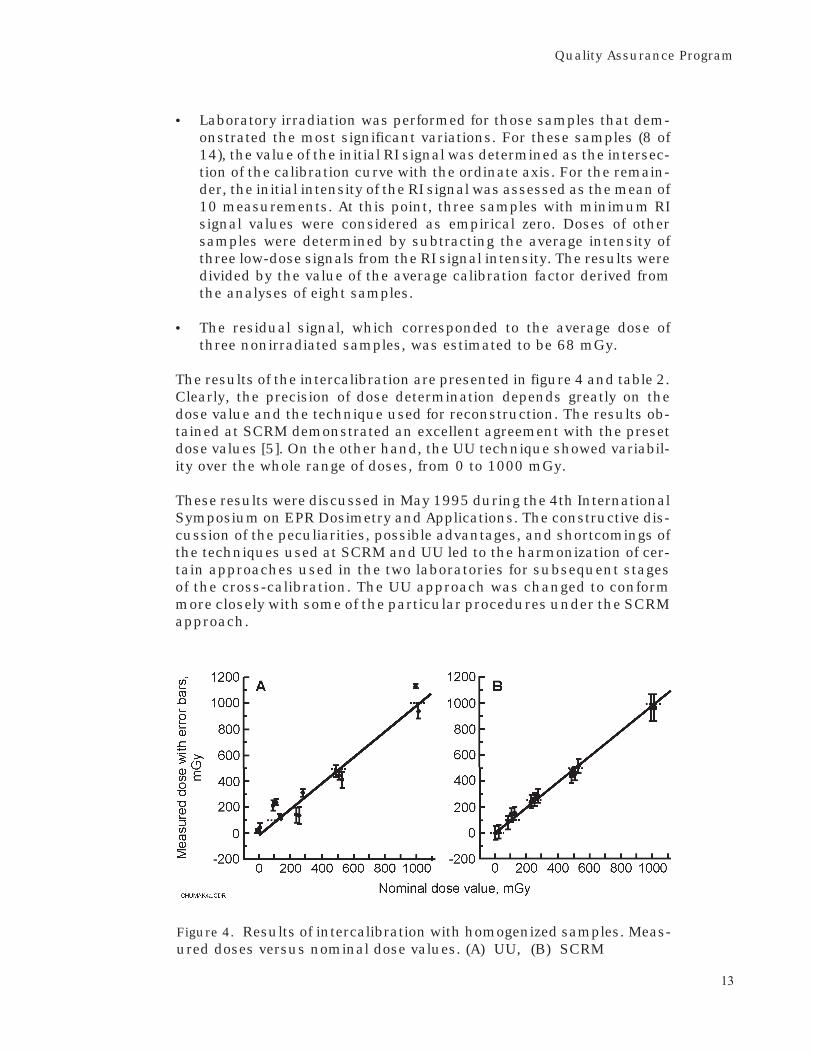
The results of the intercalibration are presented in figure 4 and table 2.Clearly, the precision of dose determination depends greatly on thedose value and the technique used for reconstruction. The results ob-tained at SCRM demonstrated an excellent agreement with the presetdose values [5]. On the other hand, the UU technique showed variabil-ity over the whole range of doses, from 0 to 1000 mGy.
These results were discussed in May 1995 during the 4th InternationalSymposium on EPR Dosimetry and Applications. The constructive dis-cussion of the peculiarities, possible advantages, and shortcomings ofthe techniques used at SCRM and UU led to the harmonization of cer-tain approaches used in the two laboratories for subsequent stagesof the cross-calibration. The UU approach was changed to conformmore closely with some of the particular procedures under the SCRMapproach.
13
Quality Assurance Program
Figure 4. Results of intercalibration with homogenized samples. Meas-ured doses versus nominal dose values. (A) UU, (B) SCRM

Stage 2. Intercalibration With Whole TeethIrradiated Under Laboratory Conditions
The main purpose of stage 2 was to address the possible influence ofsample preparation procedures on the results obtained using differenttechniques.
14
Retrospective Reconstruction of Radiation Doses by EPR
Table 2. Results of intercalibration using homogenized samplesirradiated in vitro
Participant Sample Measured dose Nominal dosenumber value, mGy value, mGy
University 87 20.7±17 0of Utah 88 49.8±28 0
90 –93±9 021 132±15 1004 209±39 1006 237±26 100
35 137±58 25043 138±68 25058 312±31 25075 413±67 50066 448±34 50056 479±49 500
106 940±60 1000100 1128±21 1000
SCRM 80 0±50 081 10±50 08 80±50 100
16 140±50 10023 150±50 10036 240±50 25037 260±50 25042 290±50 25060 440±50 50068 460±50 50069 520±50 50076 970±100 1000
108 970±100 1000
Intercomparison Design
The sample set consisted of six teeth, collected by a local dentist in theUnited States. The prior dose of x rays was unknown. Each tooth wassplit in two at the plane perpendicular to the jaw contour. One half ofeach tooth was irradiated using a 60Co source with a dose rate of about10 Gy/h. Measurements were considered to be dependent only uponthe total dose, not the dose rate. Three dose levels between 100 and 500mGy were used; dose was unknown to the participants. One half ofeach tooth remained unirradiated in order to provide a postmeasure-ment check for detectable dose due to dental x rays as well as to providea check should anomalies appear in the spectra or the results.
The participants knew that the pairs of teeth numbered 1 and 4, 2 and5, and 3 and 6 were irradiated with equal doses in order to provide di-rect comparison of the results with the equivalent laboratory-addeddose.2 Teeth 4, 5, and 6 were shipped to the SCRM; the others remainedat UU. Shipping was done by express mail specially labeled to avoid x-ray inspection and thus minimize transportation dose. Participantswere instructed to apply their customary EPR dosimetric technique.
Methods and Results
The samples of tooth enamel were prepared as described in Task 1above. The tooth size was sufficient to perform separate analyses of theouter and inner parts in order to control, and if possible, account forunreported x-ray examinations.
The EPR spectrum of tooth 6 differed significantly from the expectedshape of the signal of irradiated enamel. Chemical processing withNaOH alkali (8 hours in an ultrasonic bath at 60 oC) was used to purifythe enamel (see figure 2). Although the subsequent EPR analysis re-vealed a noticeable improvement, the shape of the signal was still dis-torted. The specimen was examined under the microscope, and oneparamagnetic nonenamel particle was located and removed. That sig-nificantly improved the signal, making the sample appropriate for dosereconstruction.
A special effort was made to control for possible x-ray exposure priorto intercalibration. Although the results of this examination do notallow for the reliable quantitative assessment of lifetime dose, thereare strong indications of both a dose gradient (i.e., the surface of thetooth nearest the x-ray source receives a higher dose than the surface
15
Quality Assurance Program
2 The total dose to be measured consisted of two principal components: un-known lifetime dose of the tooth donor and the known dose added at thelaboratory.
opposite the source, which is typical for dental x-ray examinations) andnon-zero readouts in nonirradiated parts of the teeth. The findings (ta-ble 3) are not statistically significant and are given only as guidance todemonstrate general tendencies. Unfortunately, x-ray doses for all theteeth were below the threshold of reliable dose reconstruction with theEPR technique, and therefore these figures could not be used to correctthe results of the intercalibration.
UU measurements were taken at a microwave power of 2 mW (a methodchosen after a series of tests as the most promising one to minimizenoise and, therefore, uncertainty of the dose determination). An analy-sis of expected uncertainties was performed before analysis, and thenumber of spectra to be collected at each power was set at 42 for the ir-radiated samples, 12 at the additive dose level of 1 Gy, and 6 at the 10-Gy additive dose level. Samples were stored for a minimum of 12 hoursat room temperature following each irradiation in a 60Co irradiator at adose rate of 10 Gy/h (5 mm of aluminum was used for electronbuildup). Dose increments were 210, 435, 435, 435, 870 mGy.
Both sets of dose determinations are presented in table 4. As expected,the results of the second stage intercalibration were not as clear-cut asthose of stage 1, and they could not be interpreted definitely. Both labo-ratories demonstrated good agreement (within 17%) with nominal
16
Retrospective Reconstruction of Radiation Doses by EPR
Table 3. Dose assessments for different parts of teeth (SCRM)(in mGy)
Tooth Inner part External part Inner part External part Mean dose ofnumber (unexposed) (unexposed) (exposed) (exposed) exposed half
T4 30 50 230 240 230±50
T5 30 50 230 270 250±50
T6 20 50 290 300 300±70
Table 4. Intercomparison of whole teeth exposed in vitro
Laboratory-Group Sample added dose Laboratory Measured dose
mGy mGy
1 T1 171 UU 190±50Τ4 SCRM 230±50
2 T2 256 UU 180±50T5 SCRM 250±50
3 T3 200 UU 190±100T6 SCRM 300±70

dose values, although the results produced by the SCRM techniquetended to overestimate the doses. The latter may be explained by thecontribution of the lifetime dose (particularly medical x-ray exposure)to the total dose to the tooth, which was determined by the dosereconstruction. Neither data on x-ray examination nor age of patientswas available, making assessment of this component of the total doseimpossible. Since the detected doses corresponding to the preinter-calibration history of the teeth were found to be below the thresholdof reliable dose reconstruction, the correction of the results was,unfortunately, impossible.
Stage 3. Intercomparison of Teeth From Liquidators
The third stage of cross-calibration was designed to test the capabilityof the two techniques to perform dose reconstruction using teeth ex-posed in vivo. Since the actual doses were unknown and, therefore, ab-solute validation of the results is impossible, the expected yield of thiseffort was a consistency check.
Intercomparison Design
The initial intention was to provide both laboratories with identicalsamples exposed in vivo. Three groups of samples from liquidators to-taling 34 specimens were shipped to the United States:
• 13 halves of large teeth (molars)—the remainders were retained atSCRM
• 15 teeth from pairs extracted simultaneously from the same indi-vidual and therefore presumably having the same doses
• 6 samples in the form of pieces of mechanically separated toothenamel
Due to the limited time that could be allocated by UU for examination ofthe samples, the number of dose reconstructions was reduced to five.The specimens selected (numbers X23, X24, X25, X26, and X28) wererepresented by granular samples of tooth enamel. For each sample, theinitial separation of tooth enamel was performed at SCRM using a steeldental drill. After the removal of dentine, the pieces of enamel were col-lected and the whole sample was divided into parts of about 100 mgeach for independent determination of dose by the participants. Thecharacteristic size of enamel grains was about 500 micrometers, al-though the dimensions of individual particles varied from hundreds ofmicrons to several millimeters.
17
Quality Assurance Program

Methods and Results
SCRM used basically the same standard technique described above.The technique used at UU was somewhat modified. The parametersused for the second EPR intercomparison of the liquidators’ teeth wereas follows: 8-mW microwave power, 5-Gauss modulation amplitude,20-second conversion time, 35-Gauss sweep width, 168-ms time con-stant, and 105 gain.
The dose reconstruction was done using the spectra from the samples,a baseline enamel sample with negligible dose, and an empty EPRtube. The spectrum of the tube was subtracted from each spectrumtaken of all the samples and the baseline samples. This was done inproportion to the number of scans taken in each enamel spectrum,that is, an enamel spectrum composed of 12 sweeps was corrected bysubtracting the spectrum of the empty tube, which itself was normal-ized to 12 sweeps. The spectra of the samples were then precisely nor-malized to the standard of 10 sweeps and 100 mg per spectrum by thenormalization factor:
(10 sweeps/# of sweeps taken) • (100 mg sample weight)
This adjustment was not necessary for the baseline sample as it wasprecisely weighed before measurement. The spectrum of the baselinesample was then subtracted from all the spectra of the irradiated andunirradiated enamel samples. From these resulting background andbaseline-free spectra, we did the dose reconstruction on the g-perpendicular signal extremes using standard least squares fitting anderror propagation for the dose estimates and errors, respectively. Theadditive dose technique was employed using only one applied dose of 5or 10 Gy (10 Gy if the sample mass was less than 35 mg). The number ofspectra taken at each dose was 20 to 25 for the zero dose and 12 for theone applied dose (25 if the sample mass was less than 35 mg).
In this intercomparison, an additional check of the purity of the sam-ples was performed using the EPR spectra recorded before additionalirradiation and the spectrum of a milk tooth as the standard of thebackground signal. Two (X24 and X25) of five samples had satisfac-tory purity. Three others were subjected to treatment with an NaOHsolution. After the chemical treatment, the shape of the signal hadimproved. Two of the samples (X26 and X28) were considered to bepurified completely. Although the purity of the third one (X23) hadimproved, it still had some distortions in the spectrum. Because ofsignificant loss of mass (it had decreased from 70 to 33 mg), we de-cided to refrain from further purification and proceed with EPRmeasurements.
18
Retrospective Reconstruction of Radiation Doses by EPR

This third stage of cross-calibration had relatively poor results (table 5).The doses, determined in different laboratories, coincided within de-clared uncertainty ranges for only two individuals out of five. The re-sults from the two laboratories differed significantly (up to 60%) forsome of the samples. The results of this intercomparison are discussedelsewhere [6].
At the present stage of the intercomparison, it is impossible to deter-mine the major reasons for these discrepancies. Adequate interpreta-tion of the results requires additional investigation and, possibly, con-ducting the intercomparison with a partially modified design. One pos-sibility could be to use teeth from individuals whose doses have beenassessed by independent methods of retrospective dosimetry (such as aFISH test or analytical dose reconstruction).
Discussion
The cross-calibration performed within Task 2 of the project was thefirst international, full-scale effort to harmonize EPR-dosimetric tech-niques developed in Ukrainian and US laboratories as well as to per-form quality assurance of this method. The three stages of cross-calibration, for the most part, covered all degrees of complexity andadequacy of approaches to dose reconstruction. Generally positive, theresults of the cross-calibration have proven the applicability of EPRdosimetry to practical reconstruction of individual doses.
The reproducibility of the results of the different versions of EPR tech-nique that were designed and used on different continents is, at worst,within the 60% standard deviation interval. Clearly, even with thisconservative and potentially improvable uncertainty interval, EPR do-simetry could produce more accurate dose assessments than anyother method of retrospective dose reconstruction to be used in apost-Chernobyl epidemiological followup. Moreover, stage 2 of thecross-calibration experimentally demonstrated that lifetime diagnos-tic x-ray examinations may lead to overestimation of doses within only
19
Quality Assurance Program
Table 5. Results of intercomparison with teeth of liquidatorsexposed in vivo
Sample SCRM, Gy UU, Gy
X23 0.36±0.05 0.67±0.10
X24 1.42±0.14 1.60±0.24
X25 1.08±0.11 1.56±0.23
X26 1.50±0.15 1.56±0.23
X28 0.48±0.05 1.18±0.18

30% limits. This important point, which needs additional investigation,could resolve positively the greatest concern presently associated withthe use of EPR dosimetry for reconstruction of individual doses amongthe liquidators.
20
Retrospective Reconstruction of Radiation Doses by EPR
Task 3
Test of Practical Dose Determination
A system for retrospective reconstruction of doses received by theChernobyl liquidators was tested in Task 3.
Dose Reconstruction
The teeth from liquidators were collected in the course of dental surgi-cal practice in the Kiev central liquidators’ polyclinics. Extracted teethwere accompanied by special ID forms (see appendix) reflecting the per-sonal data necessary for tracing the individual, information about oc-cupational contacts with ionizing radiation, lifetime medical x-ray ex-amination of the head, data on location of the extracted tooth, and thediagnosis leading to extraction. After extraction, the teeth were pre-served in formalin in small bottles. Periodically (approximately once amonth), the teeth were transported to the Laboratory of External Expo-sure Dosimetry for storage, processing, and determination of radiationdoses.
Upon arrival, all teeth were subjected to preprocessing, including rins-ing in distilled water and drying at 80 oC. The tooth root was separatedand, if necessary, residuals of soft tissues and damaged areas of teethwere removed. Then, teeth were placed in intermediate storage underroom conditions.
The dose determination cycle began with chemical treatment as dis-cussed in Task 1 and illustrated in figure 2. The samples of pure toothenamel were subjected to measurements, including recording of EPR-spectra (with parameters as indicated in table 1) and laboratory irra-diation with preset doses. Individual calibration curves were plotted forall measured samples, and doses were determined accounting for in-dividual radiosensitivity of enamel. It was found that calibration
21
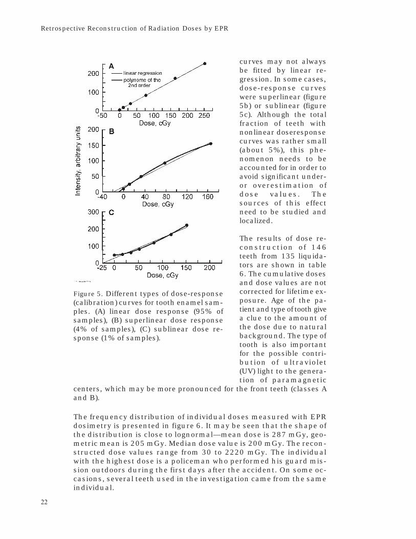
curves may not alwaysbe fitted by linear re-gression. In some cases,dose-response curveswere superlinear (figure5b) or sublinear (figure5c). Although the totalfraction of teeth withnonlinear doseresponsecurves was rather small(about 5%), this phe-nomenon needs to beaccounted for in order toavoid significant under-or overestimation ofdose values. Thesources of this effectneed to be studied andlocalized.
The results of dose re-construction of 146teeth from 135 liquida-tors are shown in table6. The cumulative dosesand dose values are notcorrected for lifetime ex-posure. Age of the pa-tient and type of tooth givea clue to the amount ofthe dose due to naturalbackground. The type oftooth is also importantfor the possible contri-bution of ultraviolet(UV) light to the genera-tion of paramagnetic
centers, which may be more pronounced for the front teeth (classes Aand B).
The frequency distribution of individual doses measured with EPRdosimetry is presented in figure 6. It may be seen that the shape ofthe distribution is close to lognormal—mean dose is 287 mGy, geo-metric mean is 205 mGy. Median dose value is 200 mGy. The recon-structed dose values range from 30 to 2220 mGy. The individualwith the highest dose is a policeman who performed his guard mis-sion outdoors during the first days after the accident. On some oc-casions, several teeth used in the investigation came from the sameindividual.
22
Retrospective Reconstruction of Radiation Doses by EPR
Figure 5. Different types of dose-response(calibration) curves for tooth enamel sam-ples. (A) linear dose response (95% ofsamples), (B) superlinear dose response(4% of samples), (C) sublinear dose re-sponse (1% of samples).

Table 6. Individual dose values reconstructed in course of EPRdosimetric exercise
Sample Age at Beginning ofnumber/ time of tooth X–ray cleanup work Type of Total doseID code extraction examinations (year) tooth* (cGy)
1 2 3 4 5 6
177/13862 60 - 86 A 39
178/16042 53 - 86 C 16
180/17568 25 - 86 C 5.5
182/2069 56 + 86 C 13
183/59 45 - 86 C 24
185/10709 43 + 86 C 12
187/15961 61 - 86 C 19
190/17871 64 - 86 CC 14
C 29
192/17099 60 - 86 C 25
194/20457 45 - 86 C 10
195/13337 58 - A86 34
198/12006 55 + 86 B 15
199/20491 44 + 86 C 11
200/7107 46 - 86 C 10
201/4385 43 - 86 C 17
202/1304 56 + 86 C 28
204/1801 47 + 86 C 8
205/ 40 + 86 C 13
208/9731 56 - 86 C 12
301/15518 60 + 86 C 19
302/16012 58 - 86 C 28
303/13827 39 86 U 6
304/13473 55 + 86 C 15
305/7826 39 86 A 31
307/4513 48 - 86 C 8
278/14877 40 - 86 C 4
28/10058 55 + 86 C 12
281/14571 57 - 86 C 11
284/17631 57 - 86 A 31
A 16
287/19245 57 - 86 C 18
298/8822 66 - 86 B 34
33/483 57 - 86 A 46
8/4416 44 + 86 U 18
A 21
4/13930 53 + 86 C 45
C 67
39/15579 62 + 86 B 78
16/4689 52 + 86 C 38
9/7821 55 - 86 C 53
*A - incisors and canines, B - premolars, C - molars, U - unknown
23
Test of Practical Dose Determination

Table 6. Continued
1 2 3 4 5 6
10/11338 53 + 86 A 40
15/13919 59 - 86 C 22
29/16047 53 + 86 C 19
24/6902 55 - 86 A 64
53/1926 54 + 86 C 18
46/7125 46 - 87 C 13
97/10075 56 - 86 A 55
98/199 52 - 86 A 59
43/17164 55 + 87 C 120
67/16344 38 + 86 A 66
138/17320 41 + 87 C 9
151/3598 64 + 86 C 12
132/3915 66 - 86 A 23
64/3978 40 + 86 C 27
84/2743 54 - 87 C 20
181/9287 63 + 86 B 14
6C 53 - 86 C 142
19C 62 - 86 C 25
105/11735 44 - 86 C 16
106/9138 44 - 86 C 20
108/573 62 + 86 C 30
C 23
7/1670 62 + 86 A 30
71/763 64 - 86 C 18
72/18249 42 - 86 C 3.5
78/18414 30 + 86 C 5
C 8
79/15694 79 - 86 C 13
82/7452 49 - 86 C 5
83/18187 49 + 86 C 13
86/17174 50 - 87 C 12
87/15171 55 - 86 C 6
35/3602 28 + 86 C 20
41/4068 37 + 86 C 18
42/3304 55 - 86 C 7
44/8329 47 - 86 B 18
61/8329 B 28
45/8203 60 + 86 B 20
49/9037 65 + 86 C 19
56/9737 56 - 86 A 29
59/7727 45 - 86 C 8
50/7727 C 6
65/4299 50 + 86 C 9
68/10373 50 + 87 C 8
*A - incisors and canines, B - premolars, C - molars
24
Retrospective Reconstruction of Radiation Doses by EPR

Table 6. Continued
1 2 3 4 5 6
69/17092 60 - 86 C 8
109/17545 45 - 87 C 21
113/8025 29 - 87 C 6
123/13820 52 + 86 A 50
125/10396 55 + 86 C 14
129/13494 57 + 86 A 24
130/15240 64 - 86 A 60
142/5009 68 - 86 A 71
145/4768 56 - 86 A 35
148/16267 49 + 87 C 67
153/13349 60 + 86 C 96
209/1408 40 - 86 U 47
212/8456 46 - 86 A 39
216/8544 45 - 86 C 25
217/13506 43 - C 13
218/13953 59 86 U 20
219/15067 64 + 86 U 13
223/3467 57 - 86 C 23
224/18355 71 - 87 C 65
225/17113 56 - 87 C 14
227/18318 55 - 86 C 10
228/8369 53 + 86 C 14
249/8369 54 C 39
230/2721 57 - 86 C 12
231/13381 63 - 86 C 195
234/16478 63 - 86 C 16
235/8152 56 + 86 C 23
236/4325 47 - 86 A 27
238/9123 62 - 86 A 70
247/14939 43 - 86 C 18
250/6863 55 + 86 C 21
C 15
251/7583 45 + 86 A 23
263/8939 64 - 86 A 35
300/17933 66 + 86 C 40
81/10298 33 - 89 C 8
308/19933 51 - 87 C 7
309/8120 56 - 88 C 13
312/2043 61 + 86 C 23
313/16137 68 - 87 C 26
314/4571 58 + 86 U 64
317/21873 55 - 86 C 50
320/7004 48 86 C 6
321/20577 44 - 87 C 7
*A - incisors and canines, B - premolars, C - molars, U - unknown
25
Test of Practical Dose Determination

Table 6. Continued
1 2 3 4 5 6
322/15777 38 - 86 A 36
B 31
325/23887 42 + 86 C 8
326/15707 64 - 86 A 42
330/17012 61 + 86 C 12
331/8719 33 + 86 C 10
332/14533 59 - 86 C 35
334/3713 57 + 86 C 12
335/15336 51 - 86 B 3
337/4722 68 - 86 A 21
340/13695 52 - 86 C 32
C 20
2C 86 U 57
17C 40 86 U 30
18C 35 86 U 222
20C 41 86 U 40
21C 86 U 30
22C 37 1586 U
*A - incisors and canines, B - premolars, C - molars, U - unknown
The doses generally depend on the amount of time spent working in the30-km zone. As may be seen from table 6, most of the liquidators in-volved in the current dose reconstruction effort began their work in1986. Median dose of this group is 211 mGy, while doses to liquidatorsof 1987 and later years are lower—164 mGy. Maximum doses to liqui-dators from 1986 and 1987 were 2220 mGy and 1200 mGy, respec-tively. This observation is in good agreement with the fact that the mostdose-intensive activities were performed during the first months after
26
Retrospective Reconstruction of Radiation Doses by EPR
Figure 6. Frequency distribution of individual doses to liquidatorsdetermined by EPR dosimetry of teeth.

the accident, when dose rate levels were much higher and most of thecleanup work took place.
System Development
Practical demand dictates a need for reconstruction of radiation dosesto the large groups of liquidators included in cohorts studied for epide-miological followup. The possibility of long-term storage of tooth sam-ples together with increasing performance of the EPR-dosimetric tech-nique make this task feasible.
However, since the teeth used for dose reconstruction are extracted formedical reasons only, sampling is a random and relatively infrequentevent. Besides, the process of natural tooth loss is an important factor,reducing the available sampling population over time.
Therefore, a systematic approach to dose reconstruction from teeth, in-cluding sample acquisition, is required. For longitudinal epidemiologi-cal followup of an exposed population, the problem of availability ofsamples may be solved by organizing a widespread network for ac-quisition of teeth extracted from the members of the studied cohort.This network should be based on centers with a high density of liqui-dators and other heavily exposed populations. Since the productivityof EPR dosimetry is limited and not yet sufficient to process all thepotential influx of samples in real time, a central bank of bioprobesshould be established for acquisition, storage, processing, and re-trieval of tooth material. Potentially, every participant of this studiedcohort sooner or later would be covered by this effort, yielding toothsamples to the bioprobe bank. Teeth from those individuals who wereincluded in the study cohort and have died could be received in thecourse of autopsy. Dose values, reconstructed by means of EPR, couldbe entered in the personal dosimetric file of the individual for access byradioepidemiologists.
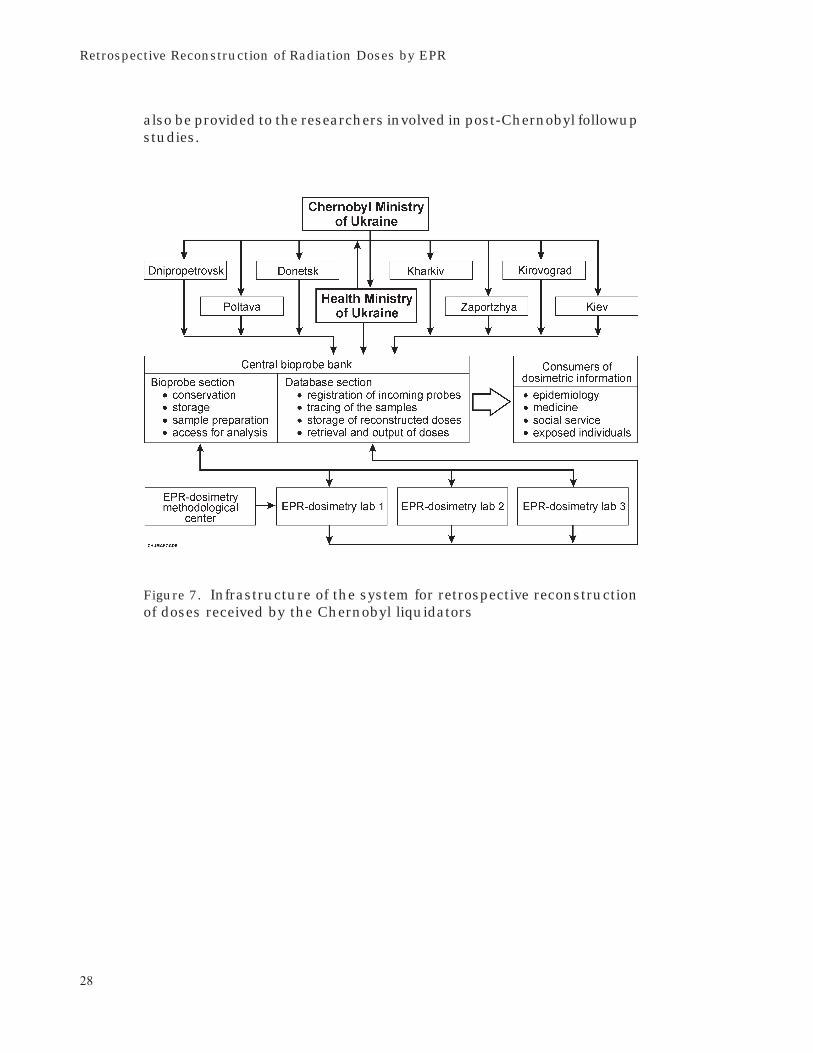
Development of such an infrastructure for dose reconstruction is un-derway now in Ukraine. The acquisition network (figure 7) would bebased on special liquidators’ hospitals in seven regional centers, cover-ing about 45% of the heavily exposed cleanup workers. The samples,along with ID forms, would be transferred to the central bioprobe bankfor long-term storage and subsequent processing.
The role of the central bioprobe bank is to coordinate activities in acqui-sition of teeth, EPR dosimetry, and data management on a nationalscale. Results of the ongoing EPR dose reconstruction will be forwardedto the National Registry and sent in parallel to the local health care bod-ies in order to provide feedback to individuals whose teeth had beensubmitted for examination. Access to the individual dose records will
27
Test of Practical Dose Determination
also be provided to the researchers involved in post-Chernobyl followupstudies.
28
Retrospective Reconstruction of Radiation Doses by EPR
Figure 7. Infrastructure of the system for retrospective reconstructionof doses received by the Chernobyl liquidators

Discussion
Although performance and capability were tested in a series of cross-calibrations and routine dose reconstructions, several issues of key im-portance need to be resolved prior to extensive use of EPR dosimetrywith teeth for followup studies. These yet unresolved problems maythreaten the utility of EPR dosimetry. Some of these effects have beenknown for a long time; others were discovered recently. Among theseare the well-known effect of enhanced sensitivity to low-energy photonsand the recently reported generation of paramagnetic centers by UVlight [7]. Nonlinearity of dose-response curves in the dose range below 1Gy was observed by the authors of this report only during the dose re-construction exercise and is yet unpublished.
Effects of Medical X Rays
Irradiation of tooth enamel with low-energy photons may lead to sub-stantial (up to seven times) overestimation of the tissue-absorbed dose.This effect has pronounced energy dependence, with the highest over-sensitivity at 60 keV. The signals from paramagnetic centers producedby high-energy (accidental) and low-energy (medical x-ray) photons areidentical, making discrimination of these signals by means of EPR im-possible. As a result, a dose measured by EPR is the sum of an acciden-tal component (dose of interest) and a component due to medical expo-sure. The degree of significance of the latter depends on the relativevalue, which is a function of incidence energy, dose per examination,and number of examinations. This means that the type of x-ray appara-tus used in the dental practice is very important, determining, after all,the degree of significance of the x-ray component.
In order to clarify this issue, it is necessary to conduct a systematic in-vestigation of the effects connected with x-ray exposure. This investiga-
29

tion should include both experimental and theoretical evaluation ofdose responses prompted by different types of x-ray examination, in-cluding different geometry, x-ray apparatus, and dose per examina-tion. The issue of doses deposited on neighboring and opposite teethshould be studied also. The contribution to the total tooth dose fromdifferent types of x-ray examination (e.g., gamma-tomography, pano-ramic diagnostics of the mandible, skull and sinus films) and radio-therapy is still unknown and requires special investigation. This workwill demand the use of both mathematical and physical phantoms forthe simulation of realistic situations.
Since x-ray practices are different in Ukraine and the United States,special attention in this research should be paid to comparison of thesetwo cases and the development of approaches to the solution of thisproblem. Intercomparisons using teeth exposed to x rays or exposedto mixed fields would be useful as well. Such studies should eitherconclude that the x-ray contribution to the tooth dose is insignificant(and establish the limits of the application of this assumption) or elserecommend how to mitigate or account for the effect of x-rayexamination.
Analysis of different teeth from the same person may be useful for un-derstanding of the effects of x-ray examination on different types ofteeth and other factors. Among the 135 individuals studied in the pres-ent research, multiple teeth were available from 11 persons. In ninecases, teeth were extracted simultaneously, with presumably equal ac-cidental doses. In two cases (individuals 190/17871 and 284/17631),the doses determined using different teeth showed a discrepancy abovethe 40% standard deviation accuracy granted by the technique. Sincethe teeth in both cases were of the same type, and x-ray examinationwas not reported on the ID form, this phenomenon could not be ex-plained by the contribution of x-ray exposure or variations in the type oftooth. Some as yet unknown effects may be responsible for such devia-tion. Unfortunately, the limited scope of research and similarities inlifetime exposure and type of teeth give little material for analysis. How-ever, so far, the largest deviation of doses determined for similar teethwas 52%, which is not a particularly large error, considering errorstypical for other methods of retrospective dosimetry.
Effects of UV Light
Another phenomenon which may affect the reliability of dose recon-struction with tooth enamel is the generation of paramagnetic centersby UV light. Information about the role and the qualitative and quanti-tative characteristics of this effect is quite contradictory. This effect wasfirst discovered and reported by Ivannikov et al. [7] in 1995. The series
30
Retrospective Reconstruction of Radiation Doses by EPR

of experiments conducted worldwide to study this effect brought noclarification.
According to existing information and our own data, the centers gener-ated in tooth enamel have position and shape very similar to those ofradiation-induced centers. That means that discrimination of UV- andradiation-induced signals by spectrometric means may be difficult. It isexpected that UV irradiation effects are most pronounced for frontteeth; such factors as time spent outdoors and elevation of the livingarea above sea level may also influence the degree of this effect.
At present, the problem of UV irradiation needs to be approached in asystematic way; this phenomenon must be studied from the point ofview of its physical, spectrometric, and kinetic (half-life) properties.Processes of generation of paramagnetic centers as a function of wave-length and intensity of UV light and decay of these centers should be in-vestigated in order to obtain a clear view of this effect.
Study of spectrometric properties (e.g., saturation of the signals) mayyield an approach to discrimination of UV- and radiation-induced sig-nals by means of EPR technique. Investigation of depth profiles of UV-generated signals in teeth for different energies of UV photons and theUV component of daylight should help explain attenuation of UV lightin enamel and could be used for target etching of exposed fractions oftooth enamel. Recommendations concerning accounting for and miti-gating this effect should be issued as a final point of this research.
Nonlinearity of Dose Response Curves
Nonlinearity of dose response curves in the dose range below 1 Gy wasobserved in some teeth in the course of the dose reconstruction exer-cise in this project. Before, saturation of the dose-response curve wasobserved only at doses above 10 Gy; below this range, the curve wasconsidered to be linear, and this property is widely used for extrapola-tion of calibration curves in the low-dose regions. Moreover, the tech-niques based on the utility of a single calibration factor (withoutadditive dose) critically depend on linearity of the dose-responsefunction.
Nonlinearity of dose response curves may have a significant influenceon the results of dose reconstruction. Not accounting for nonlinearity ofcalibration curves leads to substantial under- or overestimation of in-dividual doses (as illustrated in figure 5). Advanced study of this ef-fect, investigation of factors having impact on the dose-responsecurve, and the development of methods for extrapolating additive-dose curves are necessary for accurate and reliable retrospective do-simetry using teeth as a natural dosimeter. Since this effect takes
31
Discussion

place in only about 5% of cases, the scope of dose reconstructionshould be large enough to provide consistent and statistically signifi-cant conclusions.
32
Retrospective Reconstruction of Radiation Doses by EPR

Summary
During the 14-month period covered by this contract, extensive re-search and technological developments were performed at SCRM AMSUkraine in close collaboration with the University of Utah, USA. As a re-sult of this effort, EPR dosimetry with teeth was brought to the level of asemiroutine technique for evaluation of doses received by individualsheavily exposed after the Chernobyl accident.
Special attention was paid to quality assurance for this high-technology method in order to provide accurate and reliable individualdose assessments. The quality assurance program included several in-ternational cross-calibrations using a variety of specimens, from pul-verized tooth enamel in the beginning to whole teeth from liquidatorsexposed in vivo during the final phase of intercomparison.
Since the limited availability of samples from the individuals of interestis one of the important bottlenecks of EPR dosimetry now, a completesystem for the reconstruction of doses to liquidators must include ameans for acquiring the samples. Therefore, an organization patternfor acquisition of teeth extracted by medical prescription from theChernobyl liquidators was presented. This infrastructure is being im-plemented in Ukraine now.
The semiroutine technique developed and adopted over the period ofconsideration was used for retrospective dosimetry of a considerablegroup of liquidators. In total, doses were reconstructed for 135 indi-viduals who took part in the Chernobyl cleanup in 1986-87. The cohortof liquidators studied was assembled randomly in the course of dentalsurgery in the Kiev central liquidators’ polyclinic.
Analysis of the data obtained revealed that the mean dose of this groupis 287 mGy, ranging to the highest value of 2220 mGy. This is signifi-
33

cantly higher than the officially reported mean dose of 110 mGy. There-fore, the widely accepted opinion that the official records are of lowquality and underestimate the actual doses was supported by this firstretrospective dosimetry effort involving an appreciable number of sub-jects. The fact that doses reconstructed instrumentally are muchhigher than those officially recorded gives additional justification forthe investment in development and performance of retrospective do-simetry, particularly EPR.
However, recent research and developments in the field of EPR dosime-try make obvious a need for further investigations. From the pragmaticpoint of view, these investigations should be conducted along the fol-lowing lines:
• Investigation and development of approaches to account for EPRsignals induced by lifetime medical x-ray exposure
• Comprehensive study of the effects in tooth enamel caused by UVlight
• Investigation of the factors causing nonlinearity of the dose-response function in the dose range below 1 Gy and development ofapproaches to account for this effect in dose determination
• Cross-validation of EPR dosimetry with independent methods ofretrospective dosimetry; this may be achieved by parallel applica-tion of different methods (e.g., EPR, FISH, and analytical) to thesame objects
• Methodological research aimed at improving the technological ca-pabilities of EPR dosimetry and enhancing the productivity of thetechnique.
Completion and success of the outlined efforts will bring EPR dosimetryfrom a quite exotic methodology to an ordinary dosimetric routine likegamma-spectroscopy and alpha counting.
34

References
1. Romanyukha AA, Ignatiev EA, Degteva MO, Kozheurov VP, WieserA, Jacob P (1996) Radiation doses from Ural Region. Scientific Cor-respondence. Nature 381:199–200
2. Chumak V, Sholom S, Likhtarev I (1995) Semi-routine ESR-dosimetry technique currently used in Ukraine. Presented at the4th International Symposium on ESR Dosimetry and Application,Munich, Germany, May 15–19, 1995
3. Chumak V, Sholom S, Pasalskaya L, Pavlenko Yu (1995) Ukrainianversion of the EPR-dosimetric technique: An approach to the rou-tine dose reconstruction. Second Workshop on Dose Reconstruc-tion, Bad-Honnef, Germany, November 20–22, 1995
4. Sholom S, Chumak V, Pavlenko Yu (1995) An account of diagnosticx-ray exposure in the problem of retrospective ESR dosimetry. Pre-sented at the 4th International Symposium on ESR Dosimetry andApplication, Munich, Germany, May 15–19, 1995.
5. Chumak V, Baran N, Bugai A, Dubovsky S, Fedosov I, Finin V, Has-kell E, Hayes R, Ivannikov A, Kenner G, Kirilov V, Khamidova L,Kolesnik S, Liidja G, Lippmmaa E, Maksimenko V, Meijer E,Pasalskaya L, Past J, Puskar J, Sholom S, Skvortzov V, Vaher U,Wieser A (1995) The first international intercomparison of EPR-dosimetry with teeth: First results. Presented at the 4th Interna-tional Symposium on ESR Dosimetry and Application, Munich,Germany, May 15–19, 1995
6. Haskell EH, Kenner GH, Hayes RB, Sholom S, Chumak V (1995) AnEPR intercomparison using teeth irradiated prior to crushing. Sec-ond Workshop on Dose Reconstruction, Bad-Honnef, Germany, No-vember 20–22, 1995
35

7. Ivannikov A, Skvortzov V, Khamidova L, Eichhoff U (1995) Develop-ment of tooth enamel EPR spectroscopy method for individual do-simetry. Presented at the 4th International Symposium on ESR Do-simetry and Application, Munich, Germany, May 15–19, 1995
36
Retrospective Reconstruction of Radiation Doses by EPR

Appendix
Identification Form for Tooth Sampling
1. Complete affiliation of the hospital whichperformed extraction
2. ID number ___________________________ 3. Date of extraction ___/___/___
N General information Fragment 1
1 Family name
2 First name
3 Second name
4 Sex ( male - 1, female - 2)
5 Date of birth
6 Liquidators pass (series and number)
7 Year of work in Chernobyl
8 Dose value, officially recorded (if available)
9 Date of evacuation from the 30-km zone
10 From what settlement
N Postal address at present time Fragment 2
1 ZIP code
2 Region
3 District
4 Town
5 Street
6 House
7 Building
8 Appartment
37

4. Places of stay since the accident (region, district, settlement)(1986 in all details, afterwards - reflect locations with period of stay more than 3 months ).
Year SettlementPeriod of stay
Arrival Departure
5. Professional contact with radiation (including military service) _______________________________
_________________________________________________________________________________________
6. Information about x-ray examinations of skull, jaws, teeth (dates, type, approximate number
dur-
ing life span): _____________________________________________________________________________
_________________________________________________________________________________________
7. General deseases affecting solid tissues of tooth _____________________________________________
_________________________________________________________________________________________
_________________________________________________________________________________________
_________________________________________________________________________________________
_________________________________________________________________________________________
8. Location of the tooth and reason of extraction:
8 7 6 5 4 3 2 1 1 2 3 4 5 6 7 8
38
Retrospective Reconstruction of Radiation Doses by EPR

9. Affiliation during the Chernobyl recovery activities
_________________________________________________________________________________________
_________________________________________________________________________________________
_________________________________________________________________________________________
_________________________________________________________________________________________
_________________________________________________________________________________________
_________________________________________________________________________________________
_________________________________________________________________________________________
10. Notes ____________________________________________________________________________________
_________________________________________________________________________________________
_________________________________________________________________________________________
_________________________________________________________________________________________
_________________________________________________________________________________________
_________________________________________________________________________________________
_________________________________________________________________________________________
_________________________________________________________________________________________
_________________________________________________________________________________________
_________________________________________________________________________________________
_________________________________________________________________________________________
11. Name of physician who extracted the tooth _________________________________________________
_________________________________________________________________________________________
39
Appendix

40
DISTRIBUTION LIST
DEPARTMENT OF DEFENSE
ARMED FORCES RADIOBIOLOGY RESEARCH INSTITUTEATTN: PUBLICATIONS BRANCHATTN: LIBRARY
ARMY/AIR FORCE JOINT MEDICAL LIBRARYATTN: DASG-AAFJML
ASSISTANT TO THE SECRETARY OF DEFENSEATTN: AEATTN: HA(IA)
DEFENSE SPECIAL WEAPONS AGENCYATTN: TITLATTN: DDIRATTN: RAEMATTN: MID
DEFENSE TECHNICAL INFORMATION CENTERATTN: ACQUISITIONATTN: ADMINISTRATOR
FIELD COMMAND DEFENSE SPECIAL WEAPONS AGENCYATTN: DASIACATTN: FCIEO
INTERSERVICE NUCLEAR WEAPONS SCHOOLATTN: DIRECTOR
LAWRENCE LIVERMORE NATIONAL LABORATORYATTN: LIBRARY
UNDER SECRETARY OF DEFENSE (ACQUISITION)ATTN: OUSD(A)/R&E
UNIFORMEDSERVICESUNIVERSITYOFTHEHEALTHSCIENCESATTN: LIBRARY
DEPARTMENT OF THE ARMY
HARRY DIAMOND LABORATORIESATTN: SLCSM-SE
OFFICE OF THE SURGEON GENERALATTN: MEDDH-N
U.S. ARMY AEROMEDICAL RESEARCH LABORATORYATTN: SCIENCE SUPPORT CENTER
U.S. ARMY CHEMICAL RESEARCH, DEVELOPMENT, &ENGINEERING CENTER
ATTN: SMCCR-RST
U.S. ARMY INSTITUTE OF SURGICAL RESEARCHATTN: COMMANDER
U.S. ARMYMEDICALDEPARTMENTCENTERANDSCHOOLATTN: MCCS-FCM
U.S.ARMYMEDICALRESEARCHANDMATERIELCOMMANDATTN: COMMANDER
U.S. ARMYMEDICALRESEARCH INSTITUTEOFCHEMICALDEFENSE
ATTN: MCMR-UV-R
U.S. ARMY NUCLEAR AND CHEMICAL AGENCYATTN: MONA-NU
U.S. ARMY RESEARCH INSTITUTE OF ENVIRONMENTALMEDICINE
ATTN: DIRECTOR OF RESEARCH
U.S. ARMY RESEARCH LABORATORYATTN: DIRECTOR
WALTER REED ARMY INSTITUTE OF RESEARCHATTN: DIVISION OF EXPERIMENTAL THERAPEUTICS
DEPARTMENT OF THE NAVY
BUREAU OF MEDICINE & SURGERYATTN: CHIEF
NAVAL AEROSPACE MEDICAL RESEARCH LABORATORYATTN: COMMANDING OFFICER
NAVAL MEDICAL RESEARCH AND DEVELOPMENT COMMANDATTN: CODE 42
NAVAL MEDICAL RESEARCH INSTITUTEATTN: LIBRARY
NAVAL RESEARCH LABORATORYATTN: LIBRARY
OFFICE OF NAVAL RESEARCHATTN: BIOLOGICAL & BIOMEDICAL S&T
DEPARTMENT OF THE AIR FORCE
BROOKS AIR FORCE BASEATTN: AL/OEBZATTN: OEHL/RZATTN: USAFSAM/RZB
OFFICE OF AEROSPACE STUDIESATTN: OAS/XRS
OFFICE OF THE SURGEON GENERALATTN: HQ AFMOA/SGPTATTN: HQ USAF/SGES
U.S. AIR FORCE ACADEMYATTN: HQ USAFA/DFBL
U.S. AIR FORCE OFFICE OF SCIENTIFIC RESEARCHATTN: DIRECTOR OF CHEMISTRY & LIFE SCIENCES
OTHER FEDERAL GOVERNMENT
ARGONNE NATIONAL LABORATORYATTN: ACQUISITIONS
BROOKHAVEN NATIONAL LABORATORYATTN: RESEARCH LIBRARY, REPORTS SECTION
CENTER FOR DEVICES AND RADIOLOGICAL HEALTHATTN: DIRECTOR
GOVERNMENT PRINTING OFFICEATTN: DEPOSITORY ADMINISTRATION BRANCHATTN: CONSIGNED BRANCH
LIBRARY OF CONGRESSATTN: UNIT X
LOS ALAMOS NATIONAL LABORATORYATTN: REPORT LIBRARY
NATIONAL AERONAUTICS AND SPACE ADMINISTRATIONATTN: RADLAB
NATIONAL AERONAUTICS AND SPACE ADMINISTRATIONGODDARD SPACE FLIGHT CENTER
ATTN: LIBRARY
NATIONAL CANCER INSTITUTEATTN: RADIATION RESEARCH PROGRAM
NATIONAL DEFENSE UNIVERSITYATTN: LIBRARY TECHNICAL SERVICES
U.S. DEPARTMENT OF ENERGYATTN: LIBRARY
U.S. EMBASSY, MOSCOWATTN: DEPARTMENT OF ENVIRONMENT, HEALTH,
OCEANS, AND FISHERIES
U.S. FOOD AND DRUG ADMINISTRATIONATTN: WINCHESTER ENGINEERING AND
ANALYTICAL CENTER
U.S. NUCLEAR REGULATORY COMMISSIONATTN: LIBRARY
RESEARCH AND OTHER ORGANIZATIONS
ACADEMY OF MEDICAL SCIENCES, SCIENTIFIC CENTER OFRADIATION MEDICINE
ATTN: V. CHUMAK
AUSTRALIAN DEFENCE FORCEATTN: SURGEON GENERAL
AUTRE, INC.ATTN: PRESIDENT
BRITISH LIBRARYATTN: ACQUISITIONS UNIT
CENTREDERECHERCHESDUSERVICEDESANTEDESARMEESATTN: DIRECTOR
FEDERAL ARMED FORCES DEFENSE SCIENCE AGENCY FOR NBCPROTECTION
ATTN: LIBRARY
FOA NBC DEFENCEATTN: LIBRARY
INHALATION TOXICOLOGY RESEARCH INSTITUTEATTN: LIBRARY
INSTITUTE OF NUCLEAR MEDICINE AND ALLIED SCIENCESATTN: DIRECTOR
INSTITUTE OF RADIOBIOLOGY, ARMED FORCESMEDICAL ACADEMY
ATTN: DIRECTOR
OAK RIDGE ASSOCIATED UNIVERSITIESATTN: MEDICAL LIBRARY
RESEARCH CENTER OF SPACECRAFT RADIATION SAFETYATTN: DIRECTOR
RUTGERS UNIVERSITYATTN: LIBRARY OF SCIENCE AND MEDICINE
UNIVERSITY OF CALIFORNIAATTN: DIRECTOR, INSTITUTE OF TOXICOLOGY &
ENVIRONMENTAL HEALTHATTN: LIBRARY,LAWRENCEBERKELEYLABORATORY
UNIVERSITY OF CINCINNATIATTN: UNIVERSITY HOSPITAL, RADIOISOTOPE
LABORATORY
XAVIER UNIVERSITY OF LOUISIANAATTN: COLLEGE OF PHARMACY

REPORT DOCUMENTATION PAGE Form Approved
OMB No. 0704-0188
Public reporting burden for this collection of information is estimated to average 1 hour per response, including the time for reviewing instructions, searching existing data sources gathering and gath-
ering and maintaining the data needed, and completing and reviewing the collection of information. Send comments regarding this burden estimate or any other aspect of this collection of informa-
tion, including suggestions for reducing the burden, to Washington Headquarters Services. Directorate for Information Operations and Reports, 1215 Jefferson Davis Highway, Suite 1204, Arling-
ton, VA 22202-4302, and to the Office of Management and Budget, Paperwork Reduction Project (0704-0188), Washington, DC 20503.
1. AGENCY USE ONLY (Leave blank) 2. REPORT DATE
December 1997
3. REPORT TYPE AND DATES COVERED
Contract Report4. TITLE AND SUBTITLE
Retrospective Reconstruction of Radiation Doses of Chernobyl Liquidators byElectron Paramagnetic Resonance
5. FUNDING NUMBERS
6. AUTHORS
Chumak V.V., Likhtarev I.A., Sholom S.S., Pasalskaya L.F., Pavlenko Y.V.
7. PERFORMING ORGANIZATION NAME(S) AND ADDRESS(ES)
Armed Forces Radiobiology Research Institute
8901 Wisconsin Avenue
Bethesda, MD 20889-5603
8. PERFORMING ORGANIZATION
REPORT NUMBER
CR97-2
9. SPONSORING/MONITORING AGENCY NAME(S) AND ADDRESS(ES) 10. SPONSORING/MONITORING
AGENCY REPORT NUMBER
11. SUPPLEMENTARY NOTES
12a. DISTRIBUTION/AVAILABILITY STATEMENT
Approved for public release; distribution unlimited.
12b. DISTRIBUTION CODE
13. ABSTRACT (Maximum 200 words)
Accurate, reliable dose reconstruction is a critical component in the epidemiological followup of liquidators.Dosimetry of teeth by electron paramagnetic resonance (EPR) is a state-of-the-art laboratory technique that iskey to this effort. The Scientific Center of Radiation Medicine (SCRM) has developed and refined this tech-nique in order to meet the practical demands of large-scale epidemiologic followup of the Chernobyl liquida-tors. Independent analysis using similar technology was performed by investigators at the University of Utahand showed good correlation with the SCRM results. The lower limit of detection for reliable dose reconstruc-tion was 100 mGy. Techniques were applied to samples from approximately 135 liquidators involved incleanup activities within the first 2 years after the Chernobyl accident in 1986. Mean dose was 287 mGy, geo-metric mean was 205 mGy, and median dose value was 200 mGy. The reconstructed dose values range from 30to 2220 mGy. Correlation of results between the two institutions was generally within 17%. This report also ad-dresses some confounding factors (previous medical x-ray exposures, ultraviolet light effects on anterior teeth,nonlinearity of dose response curves below 100 mGy) and how to deal with them.
14. SUBJECT TERMS 15. NUMBER OF PAGES
5616. PRICE CODE
17. SECURITY CLASSIFICATION
OF REPORT
UNCLASSIFIED
18. SECURITY CLASSIFICATION
OF THIS PAGE
UNCLASSIFIED
19. SECURITY CLASSIFICATION
OF ABSTRACT
UNCLASSIFIED
20. LIMITATION OF
ABSTRACT
UL
NSN 7540-01-280-5500 Standard Form 298 (Rev.2-89)Prescribed By ANSI Sta. 239-18

CLASSIFIED BY:
DECLASSIFY ON:
SECURITY CLASSIFICATION OF THIS PAGE
SECURITY CLASSIFICATION OF THIS PAGE

This publication is a product of the AFRRI Information Services Division.
Editor/desktop publisher: Jane Myers, Janus Associates.