Embed Size (px)
Citation preview
CASE REPORT Open Access
Retroperitoneal abscess with pylephlebitiscaused by lumbar acupuncture: a casereportHayemin Lee, Kiyoung Sung and Jinbeom Cho*
Abstract
Background: Retroperitoneal abscess (RA) is an unusual life-threatening disease that has insidious and occultpresentations. Although the incidence of this disease is low, diagnosis and treatment are challenging due to itsnonspecific presentation and the complex anatomy of the retroperitoneal space. Recently, we experienced onecase of a RA with extensive thrombophlebitis of the portal venous system.
Case presentation: An 80-year-old male presented to the emergency room with symptoms and signs of septicshock; however, the decision making for diagnosis and treatment was difficult, as no clinical and radiologicalevidence supported key findings regarding the origin of sepsis. Although this patient eventually recovered aftersurgical drainage, we suggested that more straightforward diagnostic and treatment procedures were required inthis patient to avoid possible critical complications. Through a retrospective review of operative findings, patienthistory, and microbiology, we found that the RA in this patient was caused by lumbar acupuncture, which is usuallyperformed for the management of chronic back pain with long needles.
Conclusion: Early surgical intervention should be considered for RA whenever the patient does not respond tobroad-spectrum antibiotic treatment. Acupuncture is a possible cause of otherwise unexplained soft tissueinfections, such as RA, especially in Asian countries.
Keywords: Retroperitoneal abscess, Acupuncture, Sepsis
BackgroundRetroperitoneal abscess (RA) is an uncommon diseasethat is mainly caused by perinephric inflammation, infec-tions of the gastrointestinal tract, and postoperativecomplications [1]. Patients usually have comorbidities,such as diabetes mellitus, malignancy, and renal failure.These characteristics seem to contribute to a fatal out-come of this disease. We recently treated a patient whoexhibited septic shock of unknown origin. This patientwas eventually confirmed to have RA through severaldiagnostic work-ups and recovered after surgical drain-age; however, definitive treatment was delayed due todiagnostic uncertainty, and the outcome could have beenfatal. Furthermore, lumbar acupuncture might havecaused RA in this patient. Acupuncture, which is used in
traditional medicine, is an accepted treatment forchronic musculoskeletal pain [2]. Acupuncture is recom-mended to be performed by well-trained healthcareprofessionals.Here, we report on this rare but critical case to discuss
optimal diagnostic and treatment strategies for RA. Thisis the first reported case of RA caused by acupuncture.
Case presentationAn 80-year-old male patient was admitted to the emer-gency medical center of our hospital based on a com-plaint of myalgia and abdominal pain. According to thepatient and his daughter, the patient had no known co-morbidities, including psychiatric disorders, immune de-ficiency or trauma-related problems. The patient washemodynamically unstable; he was hypotensive, and hisbody temperature was increased to 40.7 °C upon the firstexamination. As the patient exhibited jaundice with ab-normal laboratory findings (total bilirubin, 5.46 mg/dL;
© The Author(s). 2019 Open Access This article is distributed under the terms of the Creative Commons Attribution 4.0International License (http://creativecommons.org/licenses/by/4.0/), which permits unrestricted use, distribution, andreproduction in any medium, provided you give appropriate credit to the original author(s) and the source, provide a link tothe Creative Commons license, and indicate if changes were made. The Creative Commons Public Domain Dedication waiver(http://creativecommons.org/publicdomain/zero/1.0/) applies to the data made available in this article, unless otherwise stated.
* Correspondence: [email protected] of Surgery, Bucheon St. Mary’s Hospital, College of Medicine,The Catholic University of Korea, 327, Sosa-ro, Bucheon-si, Gyeonggi-do14647, South Korea
Lee et al. BMC Surgery (2019) 19:145 https://doi.org/10.1186/s12893-019-0613-6
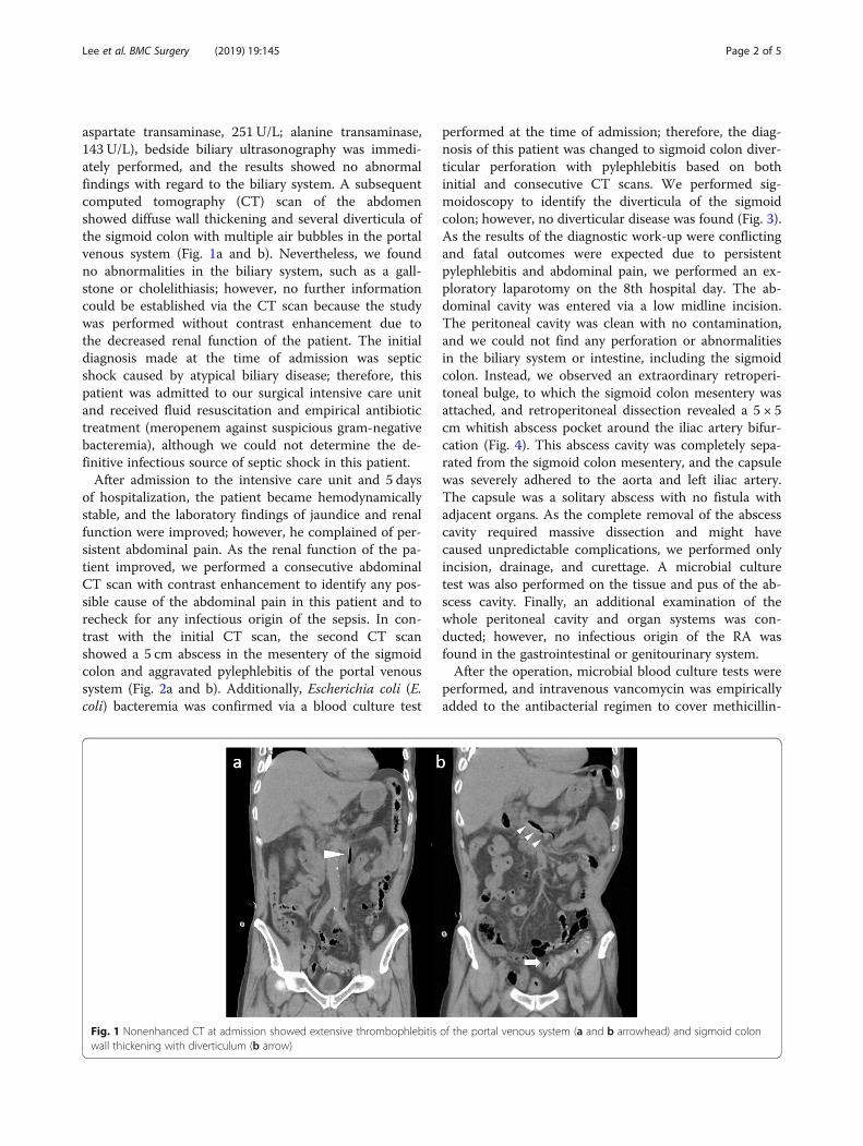
aspartate transaminase, 251 U/L; alanine transaminase,143 U/L), bedside biliary ultrasonography was immedi-ately performed, and the results showed no abnormalfindings with regard to the biliary system. A subsequentcomputed tomography (CT) scan of the abdomenshowed diffuse wall thickening and several diverticula ofthe sigmoid colon with multiple air bubbles in the portalvenous system (Fig. 1a and b). Nevertheless, we foundno abnormalities in the biliary system, such as a gall-stone or cholelithiasis; however, no further informationcould be established via the CT scan because the studywas performed without contrast enhancement due tothe decreased renal function of the patient. The initialdiagnosis made at the time of admission was septicshock caused by atypical biliary disease; therefore, thispatient was admitted to our surgical intensive care unitand received fluid resuscitation and empirical antibiotictreatment (meropenem against suspicious gram-negativebacteremia), although we could not determine the de-finitive infectious source of septic shock in this patient.After admission to the intensive care unit and 5 days
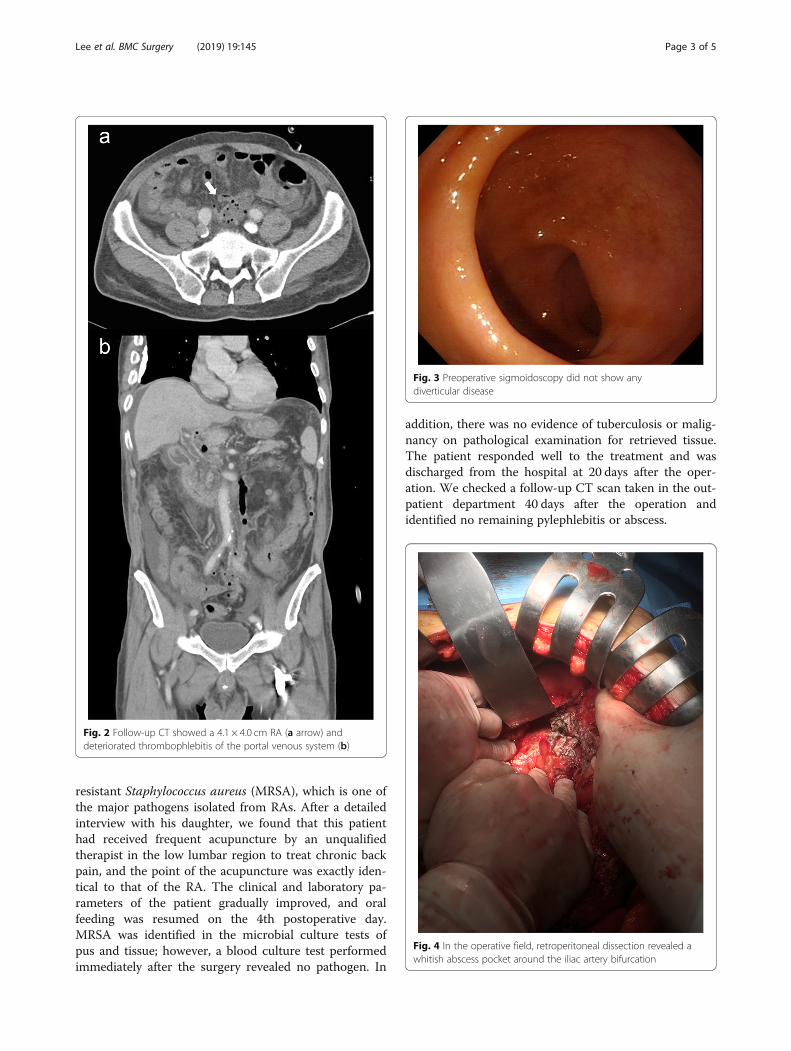
of hospitalization, the patient became hemodynamicallystable, and the laboratory findings of jaundice and renalfunction were improved; however, he complained of per-sistent abdominal pain. As the renal function of the pa-tient improved, we performed a consecutive abdominalCT scan with contrast enhancement to identify any pos-sible cause of the abdominal pain in this patient and torecheck for any infectious origin of the sepsis. In con-trast with the initial CT scan, the second CT scanshowed a 5 cm abscess in the mesentery of the sigmoidcolon and aggravated pylephlebitis of the portal venoussystem (Fig. 2a and b). Additionally, Escherichia coli (E.coli) bacteremia was confirmed via a blood culture test
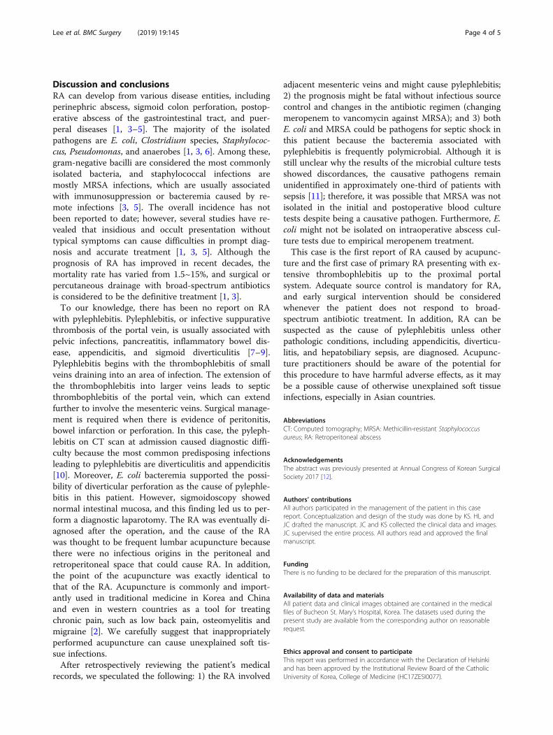
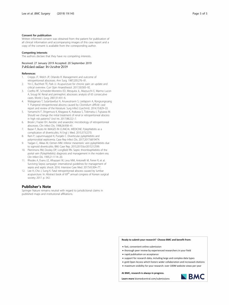
performed at the time of admission; therefore, the diag-nosis of this patient was changed to sigmoid colon diver-ticular perforation with pylephlebitis based on bothinitial and consecutive CT scans. We performed sig-moidoscopy to identify the diverticula of the sigmoidcolon; however, no diverticular disease was found (Fig. 3).As the results of the diagnostic work-up were conflictingand fatal outcomes were expected due to persistentpylephlebitis and abdominal pain, we performed an ex-ploratory laparotomy on the 8th hospital day. The ab-dominal cavity was entered via a low midline incision.The peritoneal cavity was clean with no contamination,and we could not find any perforation or abnormalitiesin the biliary system or intestine, including the sigmoidcolon. Instead, we observed an extraordinary retroperi-toneal bulge, to which the sigmoid colon mesentery wasattached, and retroperitoneal dissection revealed a 5 × 5cm whitish abscess pocket around the iliac artery bifur-cation (Fig. 4). This abscess cavity was completely sepa-rated from the sigmoid colon mesentery, and the capsulewas severely adhered to the aorta and left iliac artery.The capsule was a solitary abscess with no fistula withadjacent organs. As the complete removal of the abscesscavity required massive dissection and might havecaused unpredictable complications, we performed onlyincision, drainage, and curettage. A microbial culturetest was also performed on the tissue and pus of the ab-scess cavity. Finally, an additional examination of thewhole peritoneal cavity and organ systems was con-ducted; however, no infectious origin of the RA wasfound in the gastrointestinal or genitourinary system.After the operation, microbial blood culture tests were
performed, and intravenous vancomycin was empiricallyadded to the antibacterial regimen to cover methicillin-
Fig. 1 Nonenhanced CT at admission showed extensive thrombophlebitis of the portal venous system (a and b arrowhead) and sigmoid colonwall thickening with diverticulum (b arrow)
Lee et al. BMC Surgery (2019) 19:145 Page 2 of 5
resistant Staphylococcus aureus (MRSA), which is one ofthe major pathogens isolated from RAs. After a detailedinterview with his daughter, we found that this patienthad received frequent acupuncture by an unqualifiedtherapist in the low lumbar region to treat chronic backpain, and the point of the acupuncture was exactly iden-tical to that of the RA. The clinical and laboratory pa-rameters of the patient gradually improved, and oralfeeding was resumed on the 4th postoperative day.MRSA was identified in the microbial culture tests ofpus and tissue; however, a blood culture test performedimmediately after the surgery revealed no pathogen. In
addition, there was no evidence of tuberculosis or malig-nancy on pathological examination for retrieved tissue.The patient responded well to the treatment and wasdischarged from the hospital at 20 days after the oper-ation. We checked a follow-up CT scan taken in the out-patient department 40 days after the operation andidentified no remaining pylephlebitis or abscess.
Fig. 2 Follow-up CT showed a 4.1 × 4.0 cm RA (a arrow) anddeteriorated thrombophlebitis of the portal venous system (b)
Fig. 3 Preoperative sigmoidoscopy did not show anydiverticular disease
Fig. 4 In the operative field, retroperitoneal dissection revealed awhitish abscess pocket around the iliac artery bifurcation
Lee et al. BMC Surgery (2019) 19:145 Page 3 of 5
Discussion and conclusionsRA can develop from various disease entities, includingperinephric abscess, sigmoid colon perforation, postop-erative abscess of the gastrointestinal tract, and puer-peral diseases [1, 3–5]. The majority of the isolatedpathogens are E. coli, Clostridium species, Staphylococ-cus, Pseudomonas, and anaerobes [1, 3, 6]. Among these,gram-negative bacilli are considered the most commonlyisolated bacteria, and staphylococcal infections aremostly MRSA infections, which are usually associatedwith immunosuppression or bacteremia caused by re-mote infections [3, 5]. The overall incidence has notbeen reported to date; however, several studies have re-vealed that insidious and occult presentation withouttypical symptoms can cause difficulties in prompt diag-nosis and accurate treatment [1, 3, 5]. Although theprognosis of RA has improved in recent decades, themortality rate has varied from 1.5~15%, and surgical orpercutaneous drainage with broad-spectrum antibioticsis considered to be the definitive treatment [1, 3].To our knowledge, there has been no report on RA
with pylephlebitis. Pylephlebitis, or infective suppurativethrombosis of the portal vein, is usually associated withpelvic infections, pancreatitis, inflammatory bowel dis-ease, appendicitis, and sigmoid diverticulitis [7–9].Pylephlebitis begins with the thrombophlebitis of smallveins draining into an area of infection. The extension ofthe thrombophlebitis into larger veins leads to septicthrombophlebitis of the portal vein, which can extendfurther to involve the mesenteric veins. Surgical manage-ment is required when there is evidence of peritonitis,bowel infarction or perforation. In this case, the pyleph-lebitis on CT scan at admission caused diagnostic diffi-culty because the most common predisposing infectionsleading to pylephlebitis are diverticulitis and appendicitis[10]. Moreover, E. coli bacteremia supported the possi-bility of diverticular perforation as the cause of pylephle-bitis in this patient. However, sigmoidoscopy showednormal intestinal mucosa, and this finding led us to per-form a diagnostic laparotomy. The RA was eventually di-agnosed after the operation, and the cause of the RAwas thought to be frequent lumbar acupuncture becausethere were no infectious origins in the peritoneal andretroperitoneal space that could cause RA. In addition,the point of the acupuncture was exactly identical tothat of the RA. Acupuncture is commonly and import-antly used in traditional medicine in Korea and Chinaand even in western countries as a tool for treatingchronic pain, such as low back pain, osteomyelitis andmigraine [2]. We carefully suggest that inappropriatelyperformed acupuncture can cause unexplained soft tis-sue infections.After retrospectively reviewing the patient’s medical
records, we speculated the following: 1) the RA involved
adjacent mesenteric veins and might cause pylephlebitis;2) the prognosis might be fatal without infectious sourcecontrol and changes in the antibiotic regimen (changingmeropenem to vancomycin against MRSA); and 3) bothE. coli and MRSA could be pathogens for septic shock inthis patient because the bacteremia associated withpylephlebitis is frequently polymicrobial. Although it isstill unclear why the results of the microbial culture testsshowed discordances, the causative pathogens remainunidentified in approximately one-third of patients withsepsis [11]; therefore, it was possible that MRSA was notisolated in the initial and postoperative blood culturetests despite being a causative pathogen. Furthermore, E.coli might not be isolated on intraoperative abscess cul-ture tests due to empirical meropenem treatment.This case is the first report of RA caused by acupunc-
ture and the first case of primary RA presenting with ex-tensive thrombophlebitis up to the proximal portalsystem. Adequate source control is mandatory for RA,and early surgical intervention should be consideredwhenever the patient does not respond to broad-spectrum antibiotic treatment. In addition, RA can besuspected as the cause of pylephlebitis unless otherpathologic conditions, including appendicitis, diverticu-litis, and hepatobiliary sepsis, are diagnosed. Acupunc-ture practitioners should be aware of the potential forthis procedure to have harmful adverse effects, as it maybe a possible cause of otherwise unexplained soft tissueinfections, especially in Asian countries.
AbbreviationsCT: Computed tomography; MRSA: Methicillin-resistant Staphylococcusaureus; RA: Retroperitoneal abscess
AcknowledgementsThe abstract was previously presented at Annual Congress of Korean SurgicalSociety 2017 [12].
Authors’ contributionsAll authors participated in the management of the patient in this casereport. Conceptualization and design of the study was done by KS. HL andJC drafted the manuscript. JC and KS collected the clinical data and images.JC supervised the entire process. All authors read and approved the finalmanuscript.
FundingThere is no funding to be declared for the preparation of this manuscript.
Availability of data and materialsAll patient data and clinical images obtained are contained in the medicalfiles of Bucheon St. Mary’s Hospital, Korea. The datasets used during thepresent study are available from the corresponding author on reasonablerequest.
Ethics approval and consent to participateThis report was performed in accordance with the Declaration of Helsinkiand has been approved by the Institutional Review Board of the CatholicUniversity of Korea, College of Medicine (HC17ZESI0077).
Lee et al. BMC Surgery (2019) 19:145 Page 4 of 5
Consent for publicationWritten informed consent was obtained from the patient for publication ofall clinical information and accompanying images of this case report and acopy of the consent is available from the corresponding author.
Competing interestsThe authors declare that they have no competing interests.
Received: 27 January 2019 Accepted: 20 September 2019
References1. Crepps JT, Welch JP, Orlando R. Management and outcome of
retroperitoneal abscesses. Ann Surg. 1987;205:276–81.2. Yin C, Buchheit TE, Park JJ. Acupuncture for chronic pain: an update and
critical overview. Curr Opin Anaesthesiol. 2017;30:583–92.3. Coelho RF, Schneider-Monteiro ED, Mesquita JL, Mazzucchi E, Marmo Lucon
A, Srougi M. Renal and perinephric abscesses: analysis of 65 consecutivecases. World J Surg. 2007;31:431–6.
4. Wataganara T, Sutantawibul A, Anuwutnavin S, Leelaporn A, RongrungruangY. Puerperal retroperitoneal abscess caused by Clostridium difficile: casereport and review of the literature. Surg Infect (Larchmt). 2014;15:829–33.
5. Yamamichi F, Shigemura K, Kitagawa K, Arakawa S, Tokimatsu I, Fujisawa M.Should we change the initial treatment of renal or retroperitoneal abscessin high risk patients? Urol Int. 2017;98:222–7.
6. Brook I, Frazier EH. Aerobic and anaerobic microbiology of retroperitonealabscesses. Clin Infect Dis. 1998;26:938–41.
7. Bazan F, Busto M. IMAGES IN CLINICAL MEDICINE. Pylephlebitis as acomplication of diverticulitis. N Engl J Med. 2015;373:2270.
8. Ram P, Lapumnuaypol K, Punjabi C. Diverticular pylephlebitis andpolymicrobial septicemia. Case Rep Infect Dis. 2017;2017:6819474.
9. Yazgan C, Akkas M, Ozmen MM. Inferior mesenteric vein pylephlebitis dueto sigmoid diverticulitis. BMJ Case Rep. 2015;2015:bcr2015212599.
10. Plemmons RM, Dooley DP, Longfield RN. Septic thrombophlebitis of theportal vein (Pylephlebitis): diagnosis and management in the modern era.Clin Infect Dis. 1995;21:1114–20.
11. Rhodes A, Evans LE, Alhazzani W, Levy MM, Antonelli M, Ferrer R, et al.Surviving Sepsis campaign: international guidelines for management ofsepsis and septic shock: 2016. Intensive Care Med. 2017;43:304–77.
12. Lee H, Cho J, Sung K. Fatal retroperitoneal abscess caused by lumbaracupuncture. In: Abstract book of 69th annual congress of Korean surgicalsociety; 2017. p. 542.
Publisher’s NoteSpringer Nature remains neutral with regard to jurisdictional claims inpublished maps and institutional affiliations.
Lee et al. BMC Surgery (2019) 19:145 Page 5 of 5