Embed Size (px)
Citation preview

VS02CH10-Palczewski ARI 29 September 2016 10:52
Retinoids and Retinal DiseasesPhilip D. Kiser1,2 and Krzysztof Palczewski11Department of Pharmacology, Cleveland Center for Membrane and Structural Biology,School of Medicine, Case Western Reserve University, Cleveland, Ohio 44106;email: [email protected], [email protected] Stokes Cleveland VA Medical Center, Cleveland, Ohio 44106
Annu. Rev. Vis. Sci. 2016. 2:197–234
First published online as a Review in Advance onJuly 18, 2016
The Annual Review of Vision Science is online atvision.annualreviews.org
This article’s doi:10.1146/annurev-vision-111815-114407
Copyright c© 2016 by Annual Reviews.All rights reserved
Keywords
retinoids, visual cycle, rhodopsin, cone pigments, RPE65, LRAT, retinoldehydrogenases, A2E, visual cycle modulator, age-related maculardegeneration
Abstract
Recent progress in molecular understanding of the retinoid cycle in mam-malian retina stems from painstaking biochemical reconstitution studies sup-ported by natural or engineered animal models with known genetic lesionsand studies of humans with specific genetic blinding diseases. Structural andmembrane biology have been used to detect critical retinal enzymes andproteins and their substrates and ligands, placing them in a cellular context.These studies have been supplemented by analytical chemistry methods thathave identified small molecules by their spectral characteristics, often in con-junction with the evaluation of models of animal retinal disease. It is from thisbackground that rational therapeutic interventions to correct genetic defectsor environmental insults are identified. Thus, most presently accepted mod-ulators of the retinoid cycle already have demonstrated promising resultsin animal models of retinal degeneration. These encouraging signs indicatethat some human blinding diseases can be alleviated by pharmacologicalinterventions.
197
Click here to view this article'sonline features:
• Download figures as PPT slides• Navigate linked references• Download citations• Explore related articles• Search keywords
ANNUAL REVIEWS Further
Ann
u. R
ev. V
is. S
ci. 2
016.
2:19
7-23
4. D
ownl
oade
d fr
om w
ww
.ann
ualr
evie
ws.
org
Acc
ess
prov
ided
by
Cas
e W
este
rn R
eser
ve U
nive
rsity
on
10/2
1/16
. For
per
sona
l use
onl
y.

VS02CH10-Palczewski ARI 29 September 2016 10:52
RAL: retinal
RPE: retinal pigmentepithelium
1. DISCOVERY OF VISUAL PIGMENT REGENERATION PATHWAYS
The sole action of light in visual sensation is to photoisomerize the 11-cis-retinal (11-cis-RAL)chromophore of photoreceptor visual pigments in the retina to an all-trans configuration (Wald1968). Conformational changes in the visual pigment elicited by this isomerization allow thereceptor to couple to its cognate G protein and initiate the phototransduction cascade (Palczewski2006). The absorption of light by visual pigments destroys their chromophore properties in aprocess referred to as photochemical bleaching (Wald 1968). Sustained vision thus relies on amechanism for regenerating ground-state visual pigments (Kiser et al. 2014, McBee et al. 2001).This regeneration pathway was originally referred to as the visual cycle, although the more recentterm retinoid cycle is probably a more accurate description of the process (the terms are usedinterchangeably in this article).
Research into this area of visual science was initiated in the late 1800s with the work of Boll andKuhne (Ripps 2008) who discovered the presence of a substance in the rod-dominant frog retinathat they named visual purple, speculated to be the light-responsive substance of vision. Exposureof the purplish-red retina to light resulted in its progressive bleaching over the course of severalseconds. Kuhne made the critical discovery that the color of the retina could be restored if it waspositioned back in contact with the retinal pigment epithelium (RPE) and placed in the dark, thusestablishing the existence of a two-cell type system required for visual pigment regeneration.
The molecular basis of these processes was revealed by the research of George Wald andhis colleagues (Hubbard & Wald 1952, Wald 1968) in the mid-1900s (Figure 1). They foundthat the chromophore of the visual pigments is a vitamin A derivative known as retinaldehydeor retinene, as it was called then. This group also elucidated the multistep photochemistry ofrhodopsin activation and laid out the first detailed scheme of chemical reactions involved in visualchromophore regeneration (Figure 1a) (see also the sidebar, Chemical Reactions of the RetinoidCycle, and Figure 2). Following photoactivation, visual pigments release their chromophore
a b
Figure 1The visual cycle pathway as proposed by George Wald. (a) Reactions involved in rhodopsin bleaching and regeneration of the visualchromophore. Panel a reproduced with permission from the George Wald Nobel lecture, copyright The Nobel Foundation. (b) Apicture of George Wald following the announcement that he had won the 1967 Nobel Prize in Physiology or Medicine. Photographcourtesy of Dr. John Dowling (Harvard University).
198 Kiser · Palczewski
Ann
u. R
ev. V
is. S
ci. 2
016.
2:19
7-23
4. D
ownl
oade
d fr
om w
ww
.ann
ualr
evie
ws.
org
Acc
ess
prov
ided
by
Cas
e W
este
rn R
eser
ve U
nive
rsity
on
10/2
1/16
. For
per
sona
l use
onl
y.

VS02CH10-Palczewski ARI 29 September 2016 10:52
CHEMICAL REACTIONS OF THE RETINOID CYCLE
Reactions of the visual cycle take place at the membrane–cytoplasm interface. Vitamin A, a fat-soluble vitamincentral to this cycle, is available to the enzymes because it partitions mostly into membranes. Yet access to othersoluble substrates and products, for example, water, is also needed. Thus, these enzymes reside within membranesas transmembrane or membrane-associated proteins. The visual cycle depends on a few reactions that could oc-cur partially in membranes at physiological temperatures. These include redox reactions, Schiff base formationand hydrolysis, esterification, and stereospecific isomerizations (Figure 2). The movement of retinoids betweenmembranes is enabled by retinoid-binding protective proteins and active ATP-driven transporters (ABCA4), or itis driven by LRAT-mediated esterification. The transfer of all-trans-ROL from photoreceptors to the RPE occursby passive diffusion because retinyl esters have a propensity to form insoluble lipid droplets, termed retinosomes.All-trans-ROL taken up from the circulation via the STRA6 transporter is also esterified and stored in retinosomes.11-cis-RAL transfer from the RPE to photoreceptors is driven by a nearly irreversible reaction between this retinoidand opsins.
ROL: retinol
LRAT:lecithin:retinolacyltransferase
RPE65: retinalpigmentepithelium–specific65 kDa protein
as all-trans-RAL which is reduced to all-trans-retinol (all-trans-ROL) within photoreceptor cellouter segments (Dowling 1960). The latter compound is then transferred to the RPE where itundergoes esterification by a microsomal enzyme (Andrews & Futterman 1964), lecithin:retinolacyltransferase (LRAT) (Ruiz et al. 1999), to form all-trans-retinyl esters. During dark adaption,the retinyl esters are gradually converted back to 11-cis-RAL, which combines with apo-opsinsto regenerate light-responsive, ground-state visual pigments (Dowling 1960). In the 1980s, aphospholipid membrane-dependent retinoid isomerase activity was discovered in RPE cellularfractions (Bernstein et al. 1987, Deigner et al. 1989), some 50 years after Wald’s first descriptionof the visual cycle pathway. This retinoid isomerase activity was eventually linked to an RPE-specific protein called RPE65 (retinal pigment epithelium–specific 65 kDa protein) ( Jin et al.2005; Moiseyev et al. 2005; Redmond et al. 1998, 2005).
The reactions described above comprise the classical visual cycle essential for both rod andcone cell function in humans (Figure 3) ( Jacobson et al. 2007, Saari 2012). Moreover, there isexperimental support for the existence of a second pathway for 11-cis-RAL biosynthesis in theretina (Fleisch et al. 2008, Wang et al. 2009). This intraretinal visual cycle specifically suppliescone photoreceptors with quantities of visual chromophore sufficient for operation under pho-topic conditions (Wang & Kefalov 2011). The existence of such an alternative pathway was firstsuggested by experiments demonstrating that cone photoreceptors in isolated frog retinas regen-erated in the dark, unlike rod photoreceptors which depend on the RPE for such regeneration(Goldstein 1970). Defined enzymatic activities thought important for cone-specific regenerationhave been identified in the retinas of cone-dominant species (Figure 3) (Mata et al. 2002, Munizet al. 2009). More recently, candidate enzymes of this pathway have been proposed, although theirphysiological relevance remains to be determined (Tables 1 and 2). In this article, we primarilyfocus on progress made in understanding the classical RPE-based retinoid cycle, as well as diseasesthat result from the disruption of this pathway.
2. VISUAL PIGMENTS AND NONVISUAL OPSINS
Rhodopsin is the sole visual pigment of rod photoreceptors. Owing to its natural abundance inthe rod-dominant retinas of many common laboratory animals and cattle (Palczewski 2006), this
www.annualreviews.org • The Visual Cycle and Its Modulators 199
Ann
u. R
ev. V
is. S
ci. 2
016.
2:19
7-23
4. D
ownl
oade
d fr
om w
ww
.ann
ualr
evie
ws.
org
Acc
ess
prov
ided
by
Cas
e W
este
rn R
eser
ve U
nive
rsity
on
10/2
1/16
. For
per
sona
l use
onl
y.

VS02CH10-Palczewski ARI 29 September 2016 10:52
Redox reactions
H
NAD(P)H + H+ NAD(P)+ NAD(P)H + H+NAD(P)+
All-trans-retinal All-trans-retinolRDH 11-cis-retinol 11-cis-retinal11-cis-RDH
Schiff base formation and hydrolysis
RhoOpsin
NH2
11-cis-retinal H2O
Rho*
All-trans-retinal
Opsin
+ NH2
H2O
Trans-esterification Isomerization
PC sn1-Lyso-PC
LRAT
H2O HO
O
R
RPE65All-trans-pro-(R)-C15 11-cis-pro-(S)-C15
O
H
OHOOH
NH+
OH
N
HO
H
O OP
ON+
O
H
O
O– O–nO
On
HO OP
ON+
H
O
O
On
OH O
O
n
O
H1 H2 O
ROH
H1 H2
Figure 2Reactions that constitute the retinoid cycle. The asterisk denotes the active signaling state of rhodopsin (meta-II rhodopsin).Abbreviations: LRAT, lecithin:retinol acyltransferase; NAD, nicotinamide adenine dinucleotide; PC, phosphatidylcholine; RDH,retinol dehydrogenase; Rho, rhodopsin; RPE, retinal pigment epithelium.
pigment is the most thoroughly characterized member of the mammalian opsin protein family.In rod outer segments, most of the rhodopsin resides in paracrystalline arrays within intracellulardisks, of which in mice there are approximately 600 per outer segment. Rhodopsin constitutesan integral component of the rod outer segment structure as demonstrated by the absence ofphotoreceptor outer segments in Rho−/− mice (Humphries et al. 1997). The rod outer segmentplasma membrane also contains a subpopulation of rhodopsin with distinct properties as comparedwith intradiscal rhodopsin (Kessler et al. 2014).
The lipophilic nature of rhodopsin was first noted by Kuhne (1977), who observed that it couldbe solubilized in a spectrally intact form by treating rod outer segments with bile detergents.Determination of the bovine rhodopsin amino acid sequence (Hargrave et al. 1983, Nathans &Hogness 1983) has revealed seven distinct stretches of hydrophobic amino acids of appropriatelength to be embedded as α-helices in a phospholipid membrane. This topology was confirmedby the projection structure of rhodopsin (Schertler et al. 1993), which provided the first detailedinformation concerning the configuration of the α-helical bundle of this protein. Shortly afterelucidation of the rhodopsin primary structure, the gene encoding the β1-adrenergic receptor(β1-AR) was identified, which revealed that these two proteins belong to the G protein–coupledreceptor superfamily (Dixon et al. 1986). Like the β1-AR, rhodopsin activation has been shown
200 Kiser · Palczewski
Ann
u. R
ev. V
is. S
ci. 2
016.
2:19
7-23
4. D
ownl
oade
d fr
om w
ww
.ann
ualr
evie
ws.
org
Acc
ess
prov
ided
by
Cas
e W
este
rn R
eser
ve U
nive
rsity
on
10/2
1/16
. For
per
sona
l use
onl
y.

VS02CH10-Palczewski ARI 29 September 2016 10:52
11-cis-RAL-opsin
All-trans-RAL-opsin
All-trans-RAL
All-trans-ROL
All-trans-RE 11-cis-ROL
11-cis-RAL
RPE65
RDH5LRAT
RDH8
RDH8*
hν
PhotoreceptorRPE
Conephotoreceptor
Müller glia
Classical visual cycle Intraretinal visual cycle
11-cis-ROL
11-cis-RAL-opsin
All-trans-RAL-opsin
All-trans-RAL
All-trans-ROL
11-cis-RE
11-cis-ROL
hν
11-cis-RAL
DES1*
MFAT*
RDH14*
Retinal pigment epithelium
Rods Cones
Müller gliaapical processes
Figure 3Retinoid transformations and enzymes of the classical and intraretinal visual cycles. (Left) The classical visual cycle involving enzymeslocated in the RPE and photoreceptor outer segments. This pathway supports both rod and cone cell function. (Right) Reactionscomprising the putative intraretinal visual cycle that provides cones with a privileged source of 11-cis-RAL. This pathway is thought toinvolve enzymes located in Muller glia and cone photoreceptors. Candidate enzymes of the pathway are shown in blue and marked byasterisks to indicate that their physiological involvement in the pathway has not yet been established. Abbreviations: DES1,dihydroceramide desaturase 1; hv, light; LRAT, lecithin:retinol acyltransferase; MFAT, multifunctional O-acyltransferase; RAL,retinal; RDH, retinol dehydrogenase; RE, retinyl ester; ROL, retinol; RPE, retinal pigment epithelium.
to stimulate downstream second messenger–generating enzymes (PDE6 and adenylyl cyclase for,respectively, rhodopsin and β1-AR) by catalyzing the exchange of guanosine triphosphate forguanosine diphosphate in an intermediary signaling, heterotrimeric G protein called transducin(Kwok-Keung Fung & Stryer 1980).
In addition to rhodopsin, there are a number of other light-activated opsin molecules expressedin the human retina. These include cone visual pigments, which mediate color vision, the retinalG protein–coupled receptor (RGR), which is involved in mobilizing retinyl esters within the RPE(Radu et al. 2008, Wenzel et al. 2005), melanopsin, expressed in photosensitive ganglion cells thattrigger the pupillary light response (Lucas et al. 2003, Panda et al. 2003), and related proteins,which are involved in circadian rhythmicity (Van Gelder 2008). These light receptors belong to thebroad type 2 opsin family and typically signal through G proteins (Ernst et al. 2014). Type 1 opsinsare a second group of retinylidene proteins expressed in lower organisms that exhibit significantstructural similarity to type 2 opsins, including a seven-transmembrane-spanning α-helical foldand a retinal-binding lysine (Lys) residue located at a position equivalent to Lys296 in rhodopsin(Spudich et al. 2000). Type 1 opsins function as light-driven ion channels or pumps, or mediatephototaxis through activation of transducer molecules. Type 1 and 2 opsins lack a discerniblesequence identity to each other and differ in the configurations of their α-helical bundles. Thisfinding seems to indicate that the two opsin families arose via convergent evolution, with theequivalent positioning of the lysine being a critical structural feature that had to be achieved twiceduring evolution from two ancestral sequences (Spudich et al. 2000). However, a recent studydemonstrating that functional rhodopsin can be formed from mutants in which this lysine residue
www.annualreviews.org • The Visual Cycle and Its Modulators 201
Ann
u. R
ev. V
is. S
ci. 2
016.
2:19
7-23
4. D
ownl
oade
d fr
om w
ww
.ann
ualr
evie
ws.
org
Acc
ess
prov
ided
by
Cas
e W
este
rn R
eser
ve U
nive
rsity
on
10/2
1/16
. For
per
sona
l use
onl
y.

VS02CH10-Palczewski ARI 29 September 2016 10:52
Tab
le1
Gen
esof
the
reti
noid
cycl
e
Gen
ena
me
Gen
esy
mbo
land
stru
ctur
eaD
isab
ling
mut
atio
nsin
hum
ans:
phen
otyp
ee,g
Kno
ckou
tm
ouse
phen
otyp
eSe
ctio
nin
this
revi
ew
Rho
dops
inR
HO
,5ex
ons
loca
lized
onch
rom
osom
e3q
21-q
24A
ccou
nts
for
20–3
0%of
auto
som
aldo
min
antR
Pb ;
mor
eth
an10
0di
stin
ctm
utat
ions
;the
Pro
23H
isca
uses
∼10%
ofau
toso
mal
dom
inan
tR
Pin
the
Uni
ted
Stat
es;a
utos
omal
rece
ssiv
eR
Pm
utat
ions
are
rare
c
(≤1%
)
Rap
idro
dce
llde
gene
ratio
n,la
ckof
RO
S,su
bseq
uent
cone
dege
nera
tion
Sect
ion
2
Ops
in1f
(con
epi
gmen
ts),
long
-wav
ese
nsiti
ve;o
psin
1(c
one
pigm
ents
),m
ediu
m-w
ave
sens
itive
;op
sin
1(c
one
pigm
ents
),sh
ort-
wav
ese
nsiti
ve
OPN
1LW
,6ex
ons
loca
lized
onch
rom
osom
eX
q28;
OPN
1MW
,6ex
ons
loca
lized
onch
rom
osom
eX
q28;
OPN
1SW
,5ex
ons
loca
lized
onch
rom
osom
e7q
32.1
Pro
tano
pia
(1%
ofm
ales
)and
prot
anom
aly
(1%
ofm
ales
,0.0
1%of
fem
ales
):la
ckin
gor
blue
-shi
fted
long
-wav
elen
gth-
sens
itive
retin
alco
nes;
deut
eran
opia
(1%
ofm
ales
)an
dde
uter
anom
aly
(mos
tcom
mon
type
:6%
ofm
ales
,0.4
%of
fem
ales
):la
ckin
gor
mut
ated
med
ium
-wav
elen
gth
cone
s;tr
itano
pia
(<1%
ofm
ales
and
fem
ales
)and
trita
nom
aly
(equ
ally
rare
for
mal
esan
dfe
mal
es,0
.01%
for
both
):la
ckin
gor
mut
ated
shor
t-w
avel
engt
hco
nes
Lar
gedi
ffere
nces
note
dbe
twee
nco
lor
visi
onof
hum
ans
and
rode
nts
Sect
ion
2
Mel
anop
sin
OPN
4,11
exon
slo
caliz
edon
chro
mos
ome
10q2
2Se
ason
alaf
fect
ive
diso
rder
(pre
vale
nce
unkn
own)
Affe
cts
the
mag
nitu
deof
circ
adia
ncl
ock
phot
icre
spon
ses
(but
isno
tes
sent
ial)
Sect
ion
2
Ret
inal
Gpr
otei
n–co
uple
dre
cept
or
RG
R,7
exon
slo
caliz
edon
chro
mos
ome
10q2
3.1
Aut
osom
alre
cess
ive
RP
(ove
rall
1in
3,00
0–7,
000
peop
le,b
utm
utat
ions
inth
eR
GR
gene
are
rare
at≤1
%)
Cha
nges
inre
tinoi
dco
nten
t,sp
ecifi
cally
the
isom
eric
com
posi
tion
ofre
tinyl
este
rs
Sect
ion
2
AT
P-b
indi
ngca
sset
te,
subf
amily
A(A
BC
1),
mem
ber
4
ABC
A4,
50ex
ons
loca
lized
onch
rom
osom
e1p
22.1
Rec
essi
veSt
arga
rdtd
isea
se(p
reva
lenc
eof
∼1in
10,0
00);
juve
nile
-an
dla
te-o
nset
rece
ssiv
em
acul
ardy
stro
phy;
auto
som
alre
cess
ive
RP
(2–5
%);
rece
ssiv
efu
ndus
flavi
mac
ulat
us;r
eces
sive
cone
–rod
dyst
roph
y;ag
e-re
late
dm
acul
arde
gene
ratio
n
Am
inor
phen
otyp
ein
mic
e;ac
cum
ulat
ion
ofal
l-tr
ans-
RA
Lad
duct
sw
ithag
e
Sect
ion
3
202 Kiser · Palczewski
Ann
u. R
ev. V
is. S
ci. 2
016.
2:19
7-23
4. D
ownl
oade
d fr
om w
ww
.ann
ualr
evie
ws.
org
Acc
ess
prov
ided
by
Cas
e W
este
rn R
eser
ve U
nive
rsity
on
10/2
1/16
. For
per
sona
l use
onl
y.

VS02CH10-Palczewski ARI 29 September 2016 10:52
Stim
ulat
edby
retin
oic
acid
6ST
RA
6,19
exon
slo
caliz
edon
chro
mos
ome
15q2
4.1
Mic
roph
thal
mia
,syn
drom
e9
(MC
OP
S9),
ara
recl
inic
alen
tity
that
also
caus
espu
lmon
ary
hypo
plas
ia;
only
afe
wch
ildre
nsu
rviv
e
Mic
ear
evi
able
whe
nbr
edon
diet
sre
plet
ew
ithvi
tam
inA
,but
disp
lay
mar
kedl
yre
duce
dle
vels
ofoc
ular
retin
oids
;mal
form
atio
nsin
the
chor
oid
and
RP
E;e
arly
cone
phot
orec
epto
rce
llde
ath
Sect
ion
3
Ret
inol
dehy
drog
enas
e5
RD
H5,
5ex
ons
loca
lized
onch
rom
osom
e12
q13-
q14
Rec
essi
vefu
ndus
albi
punc
tatu
s;fe
atur
esla
te-o
nset
rece
ssiv
eco
nedy
stro
phy
(pre
vale
nce
1in
2,00
0)
Rdh
5−/−
mic
eha
vede
laye
dda
rkre
cove
ryat
high
blea
chin
gle
vels
;ala
rge
incr
ease
in11
/13-
cis-r
etin
yles
ters
also
has
been
note
d
Sect
ion
4
Ret
inol
dehy
drog
enas
e8
RD
H8,
6ex
ons
loca
lized
onch
rom
osom
e19
p13.
2N
ohu
man
retin
aldi
seas
eha
sbe
enas
soci
ated
with
mut
atio
nsof
this
gene
Sign
ifica
ntac
cum
ulat
ion
ofal
l-tr
ans-
RA
L,a
ndde
laye
dre
cove
ryof
rod
func
tion
has
been
note
dfo
llow
ing
expo
sure
tobr
ight
light
Sect
ion
4
Ret
inol
dehy
drog
enas
e10
RD
H10
,7ex
ons
loca
lized
onch
rom
osom
e8q
21.1
1N
ohu
man
retin
aldi
seas
eha
sbe
enas
soci
ated
with
mut
atio
nsof
this
gene
Rdh
10−/
−le
ads
toea
rly
embr
yoni
cle
thal
itySe
ctio
n4
Ret
inol
dehy
drog
enas
e11
RD
H11
,7ex
ons
loca
lized
onch
rom
osom
e14
q24.
1O
nefa
mily
iden
tified
with
auto
som
alre
cess
ive
RP
disp
laye
dfa
cial
dysm
orph
olog
y,de
velo
pmen
tal
dela
y,an
dsh
orts
tatu
re
Min
oref
fect
s;si
ngle
-flas
hE
RG
sof
Rdh
11−/
−m
ice
show
edno
rmal
resp
onse
sun
der
dark
-an
dlig
ht-a
dapt
edco
nditi
ons,
but
exhi
bite
dde
laye
dda
rkad
apta
tion
afte
rbl
each
ing
leve
lsof
light
Sect
ion
4
Ret
inol
dehy
drog
enas
e12
RD
H12
,9ex
ons
loca
lized
onch
rom
osom
e14
q24.
1L
CA
dw
ithse
vere
child
hood
retin
aldy
stro
phy
(4%
ofal
lcas
es);
auto
som
aldo
min
antR
P(r
are)
Mic
edi
spla
ysl
owed
kine
tics
ofal
l-tr
ans-
RA
Lre
duct
ion,
dela
yed
dark
adap
tatio
n,in
crea
sed
susc
eptib
ility
tolig
ht-i
nduc
edph
otor
ecep
tor
apop
tosi
s
Sect
ion
4
Ret
inol
dehy
drog
enas
e13
RD
H13
,12
exon
slo
caliz
edon
chro
mos
ome
19q1
3.42
No
hum
anre
tinal
dise
ase
has
been
asso
ciat
edw
ithm
utat
ions
ofth
isge
ne
No
obvi
ous
phen
otyp
e;in
mic
e,in
tens
elig
htex
posu
reca
used
swol
len
mito
chon
dria
with
disr
upte
dcr
ista
e
Sect
ion
4
Ret
inol
dehy
drog
enas
e14
RD
H14
,2ex
ons
loca
lized
onch
rom
osom
e2p
24.2
No
hum
anre
tinal
dise
ase
has
been
asso
ciat
edw
ithm
utat
ions
ofth
isge
ne
Not
gene
rate
dSe
ctio
n4 (C
ontin
ued
)
www.annualreviews.org • The Visual Cycle and Its Modulators 203
Ann
u. R
ev. V
is. S
ci. 2
016.
2:19
7-23
4. D
ownl
oade
d fr
om w
ww
.ann
ualr
evie
ws.
org
Acc
ess
prov
ided
by
Cas
e W
este
rn R
eser
ve U
nive
rsity
on
10/2
1/16
. For
per
sona
l use
onl
y.

VS02CH10-Palczewski ARI 29 September 2016 10:52
Tab
le1
(Con
tinu
ed)
Gen
ena
me
Gen
esy
mbo
land
stru
ctur
eaD
isab
ling
mut
atio
nsin
hum
ans:
phen
otyp
ee,g
Kno
ckou
tm
ouse
phen
otyp
eSe
ctio
nin
this
revi
ew
Ret
inol
dehy
drog
enas
e;de
hydr
ogen
ase/
redu
ctas
e(S
DR
fam
ily)
mem
ber
3(r
etSD
R)
DH
RS3
,8ex
ons
loca
lized
onch
rom
osom
e1p
36.1
No
hum
anre
tinal
dise
ase
has
been
asso
ciat
edw
ithm
utat
ions
ofth
isge
ne
Mic
ela
ckin
gth
isge
nedi
ebe
fore
wea
ning
and
exhi
bita
ltere
dre
tinoi
dm
etab
olis
m,a
ndhe
art,
cran
iofa
cial
,an
dsk
elet
alde
fect
s
Sect
ion
4
Lec
ithin
:ret
inol
acyl
tran
sfer
ase
LRA
T,3
exon
slo
caliz
edon
chro
mos
ome
4q32
.1E
arly
onse
taut
osom
alre
cess
ive
RP
;L
CA
(≤1%
ofal
lcas
es)
Lrat
−/−
mic
eex
hibi
tpup
illar
yco
nstr
ictio
nan
dha
vetr
ace
leve
lsof
all-
tran
s-re
tinyl
este
rsin
thei
rliv
er,
lung
,eye
,and
bloo
d;sc
otop
ican
dph
otop
icE
RG
sar
ese
vere
lyat
tenu
ated
atan
earl
yag
e
Sect
ion
5
Ret
inoi
dis
omer
ase
RPE
65,1
4ex
ons
loca
lized
onch
rom
osom
e1p
31L
CA
(or
earl
yon
seta
utos
omal
rece
ssiv
eR
P(2
–5%
ofal
lcas
es)w
ithan
estim
ated
prev
alen
ceof
∼1in
80,0
00;R
PE
65L
CA
isth
ough
tto
repr
esen
tabo
ut6%
ofal
lLC
Aca
ses
Rpe
65−/
−m
ice
exhi
bitp
oor
rod
func
tion;
they
lack
rhod
opsi
nan
dov
erac
cum
ulat
eal
l-tr
ans-
retin
yles
ters
Sect
ion
6
Ret
inol
-bin
ding
prot
ein
4R
BP4,
6ex
ons
loca
lized
onch
rom
osom
e10
q23.
33Fe
atur
esR
PE
atro
phy
with
nigh
tbl
indn
ess
and
redu
ced
visu
alac
uity
;sc
otop
ican
dph
otop
icE
RG
sar
eab
sent
orre
duce
d(e
xtre
mel
yra
re)
Rbp
4−/−
mic
eha
velo
wse
rum
retin
olle
vels
(12.
5%of
wild
-typ
ean
imal
s)w
ithlo
wre
tinoi
dle
vels
inth
eey
e,an
dim
pair
edre
tinal
func
tion
and
visu
alac
uity
;the
yca
nre
cove
rno
rmal
visi
onon
lyaf
ter
seve
ral
mon
ths
ona
vita
min
A–s
uffic
ient
diet
Sect
ion
7
Ret
inal
dehy
de-b
indi
ngpr
otei
n1
(CR
AL
BP
)R
LBP1
,10
exon
slo
caliz
edon
chro
mos
ome
15q2
6A
utso
mal
rece
ssiv
eR
P;r
eces
sive
New
foun
dlan
dro
d–co
nedy
stro
phy;
rece
ssiv
eB
othn
iady
stro
phy;
rece
ssiv
ere
tiniti
spu
ncta
taal
besc
ens
(≤1%
ofal
lcas
es)
Rlb
p1−/
−m
ice
disp
lay
rhod
opsi
nre
gene
ratio
n;11
-cis-
RA
Lpr
oduc
tion
and
dark
adap
tatio
nar
ede
laye
dby
>10
-fol
d;no
phot
orec
epto
rde
gene
ratio
nha
sbe
enob
serv
ed
Sect
ion
7
204 Kiser · Palczewski
Ann
u. R
ev. V
is. S
ci. 2
016.
2:19
7-23
4. D
ownl
oade
d fr
om w
ww
.ann
ualr
evie
ws.
org
Acc
ess
prov
ided
by
Cas
e W
este
rn R
eser
ve U
nive
rsity
on
10/2
1/16
. For
per
sona
l use
onl
y.
VS02CH10-Palczewski ARI 29 September 2016 10:52
Cel
lula
rre
tinol
-bin
ding
prot
ein
1(C
RB
P1)
RBP
1,6
exon
slo
caliz
edon
chro
mos
ome
3q23
No
hum
anre
tinal
dise
ase
has
been
asso
ciat
edw
ithm
utat
ions
ofth
isge
ne
Rbp
1−/−
mic
ear
ehe
alth
yan
dfe
rtile
,w
ithab
outa
50%
redu
ctio
nof
retin
yles
ter
accu
mul
atio
nin
thei
rhe
patic
stel
late
cells
;CR
BP
1is
requ
ired
for
effic
ient
retin
yles
ter
synt
hesi
san
dst
orag
e
Sect
ion
7
Inte
rpho
tore
cept
or(in
ters
titia
l)re
tinol
bind
ing
prot
ein
(IR
BP
)
RBP
3,4
exon
slo
caliz
edon
chro
mos
ome
10q1
1.2
Aut
osom
alre
cess
ive
RP
(≤1%
ofal
lca
ses)
Irbp
−/-
mic
ere
veal
som
elo
ssof
phot
orec
epto
rnu
clei
;the
rate
sof
reco
very
of11
-cis-
RA
Lan
dof
rege
nera
tion
ofrh
odop
sin
inth
eda
rkin
Irbp
−/-
mic
ear
esi
mila
rto
thos
eof
wild
-typ
em
ice;
dele
tion
ofIR
BP
redu
ces
the
ampl
itude
and
slow
sth
eki
netic
sof
mou
seM
-an
dL
-con
eph
otor
espo
nses
,but
cone
adap
tatio
nto
brig
ht,s
tead
ylig
htan
dth
eki
netic
sof
cone
dark
adap
tatio
nar
eun
affe
cted
;IR
BP
does
not
incr
ease
pigm
entr
egen
erat
ion
and
isno
tcri
tical
for
mou
seM
-an
dL
-con
efu
nctio
nin
brig
htlig
ht
Sect
ion
7
Abb
revi
atio
ns:A
TP
,ade
nosi
netr
ipho
spha
te;C
RA
LB
P,c
ellu
lar
retin
alde
hyde
-bin
ding
prot
ein;
ER
G,e
lect
rore
tinog
raph
(y);
His
,his
tidin
e;L
CA
,Leb
erco
ngen
itala
mau
rosi
s;P
ro,p
rolin
e;R
AL
,re
tinal
;RO
S,ro
dou
ter
segm
ents
;RP
,ret
initi
spi
gmen
tosa
;RP
E,r
etin
alpi
gmen
tepi
thel
ium
;SD
R,s
hort
-cha
inde
hydr
ogen
ase/
redu
ctas
e.a N
omen
clat
ure
acco
rdin
gto
HU
GO
Gen
eN
omen
clat
ure
Com
mitt
ee.
bA
utos
omal
dom
inan
tret
initi
spi
gmen
tosa
(1in
14,0
00).
c Aut
osom
alre
cess
ive
retin
itis
pigm
ento
sa(1
in20
,000
).dR
eces
sive
Leb
erco
ngen
itala
mau
rosi
s(2
–3ca
ses
per
100,
000)
.e F
orup
date
din
form
atio
nab
outm
utat
ions
and
the
nove
ltyof
gene
sid
entifi
edas
bein
gas
soci
ated
with
retin
aldi
seas
es,c
onsu
ltth
ecu
rate
dR
etN
et(h
ttps
://s
ph.u
th.e
du/r
etne
t/).
f For
upda
ted
info
rmat
ion
abou
tmut
atio
nsca
usin
gch
ange
sin
colo
rvi
sion
,con
sult
Cau
ses
ofC
olor
’spa
geon
caus
esan
din
cide
nce
ofco
lor
blin
dnes
s(h
ttp:
//w
ww
.web
exhi
bits
.org
/cau
seso
fcol
or/2
C.h
tml).
g Non
synd
rom
icR
Pby
mod
eof
inhe
rita
nce:
auto
som
aldo
min
antR
P,1
5–25
%;a
utos
omal
rece
ssiv
eR
P,5
–20%
;X-l
inke
dR
P,5
–15%
;unk
now
n,40
–50%
;dig
enic
RP
,ver
yra
re(f
rom
Ret
Net
http
s://
sph.
uth.
edu/
retn
et/)
.
www.annualreviews.org • The Visual Cycle and Its Modulators 205
Ann
u. R
ev. V
is. S
ci. 2
016.
2:19
7-23
4. D
ownl
oade
d fr
om w
ww
.ann
ualr
evie
ws.
org
Acc
ess
prov
ided
by
Cas
e W
este
rn R
eser
ve U
nive
rsity
on
10/2
1/16
. For
per
sona
l use
onl
y.

VS02CH10-Palczewski ARI 29 September 2016 10:52
Tab
le2
Pro
tein
sof
the
reti
noid
cycl
e
Pro
tein
Loc
aliz
atio
n3D
stru
ctur
eFu
ncti
onSe
ctio
nin
this
revi
ew
Rho
dops
inR
odce
lls;m
embr
ane-
boun
din
rod
oute
rse
gmen
tsX
-ray
stru
ctur
eav
aila
ble;
ase
ven-
tran
smem
bran
ehe
lical
prot
ein
11-c
is-re
tinyl
iden
e-bi
ndin
gpr
otei
n;a
rod
phot
orec
epto
rlig
htre
cept
orth
attr
igge
rsph
otot
rans
duct
ion
Sect
ion
2
Con
epi
gmen
tsC
one
cells
;mem
bran
e-bo
und
inco
neou
ter
segm
ents
Mod
elba
sed
onbo
vine
rhod
opsi
nst
ruct
ure
isav
aila
ble;
seve
n-tr
ansm
embr
ane
helic
alpr
otei
ns
11-c
is-re
tinyl
iden
e-bi
ndin
gpr
otei
ns;
cone
phot
orec
epto
rlig
htre
cept
ors
that
trig
ger
phot
otra
nsdu
ctio
n
Sect
ion
2
Mel
anop
sin
Asu
bset
ofga
nglio
nce
lls;
cellu
lar
mem
bran
e-bo
und
Mod
elba
sed
onin
vert
ebra
terh
odop
sin
stru
ctur
eis
avai
labl
e;a
seve
n-tr
ansm
embr
ane
helic
alpr
otei
n
Are
tinyl
iden
e-bi
ndin
gpr
otei
n;a
phot
orec
epto
rlig
htre
cept
orth
attr
igge
rsG
qph
otot
rans
duct
ion,
cont
rols
slee
p–aw
ake
patt
ern
Sect
ion
2
RG
RR
PE
Mod
elba
sed
onin
vert
ebra
terh
odop
sin
stru
ctur
eis
avai
labl
e;a
seve
n-tr
ansm
embr
ane
helic
alpr
otei
n
Are
tinyl
iden
e-bi
ndin
gpr
otei
n;a
phot
orec
epto
rlig
htre
cept
orth
attr
igge
rsG
qph
otot
rans
duct
ion;
func
tion
still
uncl
ear
Sect
ion
2
AB
CA
4R
odan
dco
neou
ter
segm
ent
rim
regi
ons
An
18A
reso
lutio
nst
ruct
ure
ofA
BC
A4
isol
ated
from
bovi
nero
dou
ter
segm
ents
was
dete
rmin
edby
elec
tron
mic
rosc
opy
and
sing
le-p
artic
lere
cons
truc
tion;
am
inim
umof
four
dom
ains
isre
quir
edfo
rfu
nctio
nala
ctiv
ity:t
wo
tran
smem
bran
edo
mai
ns,e
ach
with
six
pred
icte
dm
embr
ane-
span
ning
helic
esan
dtw
onu
cleo
tide-
bind
ing
dom
ains
,and
two
intr
adis
cald
omai
ns,e
ach
linke
dto
one
ofth
etr
ansm
embr
ane
dom
ains
An
AB
Cim
port
er;t
rans
fers
retin
oid
from
the
intr
adis
cals
pace
toth
ecy
topl
asm
Sect
ion
3
STR
A6
Inth
eR
PE
,ST
RA
6is
loca
lized
toth
eba
sola
tera
lmem
bran
es;
also
inbr
ain,
chor
oid
plex
usm
icro
vess
els,
test
is,s
plee
n,ki
dney
,pla
cent
a,an
dth
efe
mal
ere
prod
uctiv
etr
act
9–11
tran
smem
bran
ehe
lical
segm
ents
pred
icte
d;lim
ited
stru
ctur
alin
form
atio
nav
aila
ble
RB
P4
rece
ptor
;tra
nspo
rts
all-
tran
s-R
OL
driv
enby
intr
acel
lula
rL
RA
Tac
tivity
Sect
ion
3
206 Kiser · Palczewski
Ann
u. R
ev. V
is. S
ci. 2
016.
2:19
7-23
4. D
ownl
oade
d fr
om w
ww
.ann
ualr
evie
ws.
org
Acc
ess
prov
ided
by
Cas
e W
este
rn R
eser
ve U
nive
rsity
on
10/2
1/16
. For
per
sona
l use
onl
y.

VS02CH10-Palczewski ARI 29 September 2016 10:52
RD
H5a
Abu
ndan
texp
ress
ion
inth
eR
PE
From
the
SDR
fam
ily;t
wo
dom
ains
:one
bind
sN
AD
(P)(
Ros
sman
nfo
ld)a
ndth
ese
cond
bind
ssu
bstr
ate
Red
oxre
actio
nsp
ecifi
cto
11-c
is-R
AL
/11-
cis-R
OL
;N
AD
-spe
cific
Sect
ion
4
RD
H8a
Abu
ndan
tin
rod
and
cone
oute
rse
gmen
tsFr
omth
eSD
Rfa
mily
;tw
odo
mai
ns:o
nebi
nds
NA
D(P
)(R
ossm
ann
fold
)and
the
othe
rbi
nds
subs
trat
e
Red
oxre
actio
nsp
ecifi
cto
all-
tran
s-R
AL
/all-
tran
s-R
OL
;NA
DP
spec
ific
Sect
ion
4
RD
H10
aB
road
lyex
pres
sed
From
the
SDR
fam
ily;t
wo
dom
ains
:one
bind
sN
AD
(P)(
Ros
sman
nfo
ld)a
ndth
eot
her
bind
ssu
bstr
ate
Red
oxre
actio
nsp
ecifi
cto
all-
tran
s-R
AL
/all-
tran
s-R
OL
orcis
-RA
L/c
is-R
OL
(dua
lspe
cific
ity);
NA
DP
dinu
cleo
tide
spec
ifici
ty
Sect
ion
4
RD
H11
aB
road
lyex
pres
sed;
high
leve
lsin
pros
tate
epith
eliu
mFr
omth
eSD
Rfa
mily
;tw
odo
mai
ns:o
nebi
nds
NA
D(P
)(R
ossm
ann
fold
)and
the
othe
rbi
nds
subs
trat
e
Red
oxre
actio
nsp
ecifi
cto
all-
tran
s-R
AL
/all-
tran
s-R
OL
orcis
-RA
L/c
is-R
OL
(dua
lspe
cific
ity);
NA
DP
spec
ific
Sect
ion
4
RD
H12
aE
xpre
ssed
inro
dan
dco
nein
ner
segm
ents
,ski
n,ce
rvic
alep
ithel
ium
,liv
er,a
ndpl
atel
ets
From
the
SDR
fam
ily;t
wo
dom
ains
:one
bind
sN
AD
(P)(
Ros
sman
nfo
ld)a
ndth
eot
her
bind
ssu
bstr
ate
Red
oxre
actio
nsp
ecifi
cto
all-
tran
s-R
AL
/all-
tran
s-R
OL
orcis
-RA
L/c
is-R
OL
(dua
lspe
cific
ity);
NA
DP
spec
ific
Sect
ion
4
RD
H13
aSu
gges
ted
mito
chon
dria
llo
caliz
atio
nFr
omth
eSD
Rfa
mily
;tw
odo
mai
ns:o
nebi
nds
NA
D(P
)(R
ossm
ann
fold
)and
the
othe
rbi
nds
subs
trat
e
Red
oxre
actio
nsp
ecifi
cto
all-
tran
s-R
AL
/all-
tran
s-R
OL
orcis
-RA
L/c
is-R
OL
(dua
lspe
cific
ity);
NA
DP
spec
ific
Sect
ion
4
RD
H14
aN
otfu
llyes
tabl
ishe
dFr
omth
eSD
Rfa
mily
;tw
odo
mai
ns:o
nebi
nds
NA
D(P
)(R
ossm
ann
fold
)and
the
othe
rbi
nds
subs
trat
e
Red
oxre
actio
nsp
ecifi
cto
all-
tran
s-R
AL
/all-
tran
s-R
OL
orcis
-RA
L/c
is-R
OL
(dua
lspe
cific
ity);
NA
DP
spec
ific
Sect
ion
4
DH
RS3
aV
ery
broa
dex
pres
sion
,with
high
esti
nliv
er,s
moo
thm
uscl
e,an
dco
neou
ter
segm
ents
From
the
SDR
fam
ily;t
wo
dom
ains
:one
bind
sN
AD
(P)(
Ros
sman
nfo
ld)a
ndth
eot
her
bind
ssu
bstr
ate
Red
oxre
actio
nsp
ecifi
cto
all-
tran
s-R
AL
/all-
tran
s-R
OL
orcis
-RA
L/c
is-R
OL
(dua
lspe
cific
ity);
NA
DP
spec
ific;
low
spec
ific
activ
ityst
imul
ated
byR
DH
10
Sect
ion
4
LR
AT
bV
ery
broa
dex
pres
sion
;enr
iche
din
hepa
ticst
ella
tece
llsan
dth
eR
PE
Dim
eric
,with
each
mon
omer
com
pose
dof
afo
ur-s
tran
d,an
tipar
alle
lβ-s
heet
and
thre
eα
-hel
ices
,sim
ilar
toN
lpC
/P60
thio
lpro
teas
es,w
itha
Cys
-His
-His
cata
lytic
tria
d
Est
erifi
catio
nof
RO
Ls;
spec
ifici
tyto
war
dsn
1ph
osph
olip
idtr
ansf
erof
fatt
yac
idch
ains
Sect
ion
5 (Con
tinue
d)
www.annualreviews.org • The Visual Cycle and Its Modulators 207
Ann
u. R
ev. V
is. S
ci. 2
016.
2:19
7-23
4. D
ownl
oade
d fr
om w
ww
.ann
ualr
evie
ws.
org
Acc
ess
prov
ided
by
Cas
e W
este
rn R
eser
ve U
nive
rsity
on
10/2
1/16
. For
per
sona
l use
onl
y.
VS02CH10-Palczewski ARI 29 September 2016 10:52
Tab
el2
(Con
tinu
ed)
Pro
tein
Loc
aliz
atio
n3D
stru
ctur
eFu
ncti
onSe
ctio
nin
this
revi
ew
RP
E65
Exp
ress
ion
limite
dto
the
RP
ESe
ven-
blad
edβ
-pro
pelle
r,m
onot
opic
ally
boun
dto
endo
plas
mic
retic
ulum
mem
bran
es
Are
tinoi
dis
omer
ase
that
cata
lyze
sth
eco
nver
sion
ofal
l-tr
ans-
retin
yles
ters
into
11-c
is-R
OL
Sect
ion
6
RB
P4
Exp
ress
edin
liver
;low
erex
pres
sion
inad
ipoc
ytes
The
over
allf
old
cons
ists
ofa
core
β-b
arre
lofe
ight
up-a
nd-d
own
β-s
tran
ds,a
nN
-ter
min
alco
il,an
da
C-t
erm
inal
α-h
elix
follo
wed
bya
coil
regi
on(li
poca
linfa
mily
)
Car
rier
ofal
l-tr
ans-
RO
Lin
aco
mpl
exw
ithtr
anst
hyre
tinSe
ctio
n7
CR
AL
BP
Exp
ress
edin
RP
Ean
dM
ulle
rce
llsO
vera
llpr
otei
nfo
lds
sim
ilar
toth
epr
otot
ypic
alSE
C14
-lik
edo
mai
nst
ruct
ure
exhi
bite
dby
α-t
ocop
hero
ltr
ansf
erpr
otei
n;th
eN
-ter
min
alα
-do
mai
nco
mpr
ises
five
helic
es;t
heC
-ter
min
alα
βα
dom
ain
cont
ains
aβ
-she
etw
ithon
ean
tipar
alle
land
four
para
llels
tran
ds(β
1–β
5)an
dsi
xhe
lices
Hig
hly
sele
ctiv
eca
rrie
rof
11-c
is-R
AL
;al
sobi
nds
11-c
is-R
OL
and
9-cis
-ret
inoi
ds
Sect
ion
7
CR
BP
1E
xpre
ssed
broa
dly
atlo
wle
vels
Com
pact
ion
oftw
oan
tipar
alle
lβ-s
heet
sfo
rms
anor
thog
onal
barr
elC
ellu
lar
RB
PSe
ctio
n7
IRB
Pc
Exp
ress
edin
phot
orec
epto
rs,
pine
algl
and
Four
hom
olog
ous
dom
ains
;the
dom
ain
stru
ctur
eis
sim
ilar
toph
otos
yste
mII
D1
C-t
erm
inal
proc
essi
ngpr
otea
sean
dth
een
oyl-
CoA
isom
eras
e/hy
drat
ase
fam
ily(a
thre
e-he
lixbu
ndle
follo
wed
bya
smal
lβ
-str
and
conn
ecte
dto
ala
rge
six-
stra
nded
,mix
edβ
-she
etsu
bdom
ain
with
four
helic
espa
cked
agai
nsto
nesi
dean
da
fifth
helix
)
An
extr
acel
lula
rpr
otei
n;tr
ansp
orte
rof
retin
oids
betw
een
the
RP
Ean
dph
otor
ecep
tor
cells
;pos
sibl
yin
volv
edin
lipid
bind
ing
Sect
ion
7
Abb
revi
atio
ns:A
BC
A4,
aden
osin
etr
ipho
spha
te-b
indi
ngca
sset
tetr
ansp
orte
r4;
CR
AL
BP
,cel
lula
rre
tinal
dehy
de-b
indi
ngpr
otei
n;C
RB
P,c
ellu
lar
retin
al-b
indi
ngpr
otei
n1;
Cys
,cys
tein
e;H
is,
hist
idin
e;IR
BP
,int
erph
otor
ecep
tor
retin
oid-
bind
ing
prot
ein;
LR
AT
,lec
ithin
:ret
inol
acyl
tran
sfer
ase;
NA
D,n
icot
inam
ide
aden
ine
dinu
cleo
tide;
RA
L,r
etin
al;R
BP
,ret
inol
-bin
ding
prot
ein;
RD
H,r
etin
olde
hydr
ogen
ase;
RG
R,r
etin
alG
prot
ein–
coup
led
rece
ptor
;RO
L,r
etin
ol;R
PE
,ret
inal
pigm
ente
pith
eliu
m;S
DR
,sho
rt-c
hain
dehy
drog
enas
e/re
duct
ase;
STR
A6,
stim
ulat
edby
retin
oic
acid
6.a S
truc
ture
dete
rmin
edon
lyfo
rse
vera
loth
erm
embe
rsof
the
SDR
fam
ily.
bSt
ruct
ures
ofa
hybr
idor
rela
ted
prot
eins
wer
ede
term
ined
.c S
truc
ture
ofth
ew
hole
prot
ein
isun
know
n.
208 Kiser · Palczewski
Ann
u. R
ev. V
is. S
ci. 2
016.
2:19
7-23
4. D
ownl
oade
d fr
om w
ww
.ann
ualr
evie
ws.
org
Acc
ess
prov
ided
by
Cas
e W
este
rn R
eser
ve U
nive
rsity
on
10/2
1/16
. For
per
sona
l use
onl
y.

VS02CH10-Palczewski ARI 29 September 2016 10:52
RP: retinitispigmentosa
has been relocated within the helical bundle suggests that type 1 and 2 opsins descended from acommon ancestral sequence (Devine et al. 2013).
Rhodopsin, like all known vertebrate visual pigments, senses light through a Schiff base–linked11-cis-retinylidene chromophore (Hubbard & Wald 1952). The absorption of light by this chro-mophore promotes transition of electrons from ground-state to excited-state molecular orbitals.It is this change in electron density distribution that allows the cis–trans isomerization to proceed.Notably, photoisomerization of the 11-cis chromophore is extremely rapid, occurring within 200fs after light absorption. Torsional vibrations of the bound 11-cis-retinylidene occur over a similartime scale, and vibrational coherence has been observed in the all-trans-retinylidene photoproduct(Wang et al. 1994). These findings imply that isomerization involves small movements, mainlywithin the center of the all-trans-RAL molecule, and that these movements are virtually unim-peded by the chromophore-binding pocket. Moreover, the vibronic coupling explains, in part, thehigh quantum yield (0.67) of rhodopsin photoisomerization.
The ligand-binding pocket of rhodopsin, resolved in atomic detail by determination of itscrystal structure (Figure 4) (Palczewski et al. 2000), exerts a profound influence on the opticalabsorbance properties of the bound retinylidene chromophore via distortions of the planar polyenestructure, as well as by electrostatic influences, a phenomenon referred to as the opsin shift.The protonation state of the Schiff base is one well-characterized factor that determines theabsorbance spectrum of rhodopsin. In rhodopsin’s ground state, the Schiff base is protonated, withthe glutamate113 side chain carboxylate serving as the counterion (Nathans 1990, Sakmar et al.1989). Upon photoisomerization, the Schiff base loses its proton to form the signaling-competentMeta II rhodopsin state, with a blue-shifted absorbance spectrum (Wald 1968).
Crystal structures of rhodopsin, including Meta II–like states, indicate that the isomerizationof retinylidene is communicated to the G protein–interacting surface via alterations in a water-mediated hydrogen bonding network (Angel et al. 2009, Choe et al. 2011). The primary result ofthese changes is movement of the cytosolic ends of α-helices V and VI, making the binding sitefor transducin accessible. Crystal structures of rhodopsin in complex with transducin are neededto fully resolve the mechanism of information transfer from the ligand-binding pocket to the Gprotein.
Mutations in the opsin gene result in a spectrum of eyesight disorders, the most severe of whichis referred to as retinitis pigmentosa (RP). A RHO mutation resulting in a Pro → His substitutionat position 23 in the amino acid sequence (P23H) was the first reported pathological mutation inthis gene, and it is a particularly common cause of inherited autosomal dominant RP (Dryja et al.1990). This P23H rhodopsin is highly susceptible to degradation within the endoplasmic reticulum(ER), impeding rhodopsin trafficking to the outer segments, disrupting disc morphogenesis, and,ultimately, causing photoreceptor cell death (Sakami et al. 2011).
3. RETINOID TRANSPORTERS: STRA6 AND ABCA4
3.1. Stimulated by Retinoic Acid 6
The passage of lipophilic retinoids, such as all-trans-ROL, between the two leaflets of phospho-lipid biological membranes can occur by simple diffusion. Nevertheless, there are two knownlocations within the retina where selective and efficient retinoid trafficking across phospholipidmembranes relies on the activity of integral membrane transporter proteins, namely the apicalplasma membrane of the RPE, where all-trans-ROL is obtained from the circulation, and thephotoreceptor outer segment disks, where large quantities of all-trans-RAL are generated duringlight exposure (Figure 5).
www.annualreviews.org • The Visual Cycle and Its Modulators 209
Ann
u. R
ev. V
is. S
ci. 2
016.
2:19
7-23
4. D
ownl
oade
d fr
om w
ww
.ann
ualr
evie
ws.
org
Acc
ess
prov
ided
by
Cas
e W
este
rn R
eser
ve U
nive
rsity
on
10/2
1/16
. For
per
sona
l use
onl
y.

VS02CH10-Palczewski ARI 29 September 2016 10:52
IIIIII
IIII
IVIV
IIVV
VIVI
VIIVII
VIIIVIII
Lys296
11-cis-retinylidene
Cytosol
Intradiscal(extracellular)
Figure 4Crystal structure of ground-state bovine rhodopsin. The protein backbone is shown in a schematicrepresentation with the 11-cis-retinylidene chromophore and covalently linked lysine (Lys296) side chaindisplayed as sticks and spheres. The α-helices are numbered based on their order in the polypeptidesequence. The red and blue lines demarcate the approximate width of the phospholipid membrane bilayer.The figure was generated with PyMol (Schrodinger, https://www.pymol.org/) using the rhodopsin atomiccoordinates deposited in the Protein Data Bank under accession code 1U19.
RBP: retinol-bindingprotein
STRA6: stimulatedby retinoic acid 6
RA: retinoic acid
After years of controversy regarding its existence, the receptor for holo-retinol-binding protein4 (RBP4) was identified in 2007 as the protein known as stimulated by retinoic acid 6 (STRA6)(Kawaguchi et al. 2007), so named because of its retinoic acid (RA)-inducible nature (Bouilletet al. 1997). This multipass transmembrane protein is conserved in vertebrates, but bears nosignificant sequence homology to any known protein family (Figure 5a). All-trans-ROL (vitaminA) mainly circulates in the plasma in complex with RBP4, a 21 kDa protein, which in turn forms acomplex with transthyretin (TTR) that prevents the renal excretion of RBP4 (Figure 5b). STRA6contains an extracellular loop that mediates its interaction with holo-RBP4 to allow extraction ofall-trans-ROL from the RBP4-binding pocket (Figure 5b) (Kawaguchi et al. 2008). Liberated all-trans-ROL then traverses a passageway formed by the STRA6 transmembrane α-helical segments,of which there are likely nine. Free all-trans-ROL then is picked up on the cytoplasmic side ofthe membrane by cellular retinal-binding protein 1 (CRBP1), which mediates the diffusion of all-trans-ROL to the ER, where it is metabolized (Figure 5b). Unlike most small molecule facilitative
210 Kiser · Palczewski
Ann
u. R
ev. V
is. S
ci. 2
016.
2:19
7-23
4. D
ownl
oade
d fr
om w
ww
.ann
ualr
evie
ws.
org
Acc
ess
prov
ided
by
Cas
e W
este
rn R
eser
ve U
nive
rsity
on
10/2
1/16
. For
per
sona
l use
onl
y.

VS02CH10-Palczewski ARI 29 September 2016 10:52
a b
I
N
C
II III IV V VI VII VIII IX
Extracellular
Intracellular
RBD321 360
667
517
CN
I II III IV V VI VII VIII IX X XI XII
ECD1 ECD2
CD1 CD2
50 650 1,390 1,680
850 1,370 1,9002,273
c d
TTR
TTR RBP +atROL
RBP +atROL STRA6
CRBP1+ atROL
LRAT
CRBP1+ atROL
Vasculature
Basolateral plasmamembrane
Endoplasmicreticulum
Retinosome
atREsatREs
atROLatROL
RDH8
Normal ABCA4loss of function
RDH8
All-trans-retinol
All-trans-retinaltoxicity
To RPE
ABCA4 ABCA4
atRAL
Phosphatidylethanolamine
Visualpigments
Visualpigments
Figure 5Structure and function of retinoid transporters essential for retinal health and function. (a) Topology diagram of the holo-RBPreceptor, STRA6. The RBD (orange) resides between transmembrane helices VI and VII. Numbering indicates positions within thehuman amino acid sequence. (b) The role of STRA6 in the uptake of atROL from holo-RBP into a target tissue, such as the RPE.(c) Topological diagram of ABCA4. The large ECDs are glycosylated at multiple positions, as indicated by red asterisks. Two CDscontain Walker A and B motifs responsible for adenosine triphosphate binding and hydrolysis (shown in a schematic representation)that drive the transport function of the protein. Numbering indicates positions within the human amino acid sequence. (d ) Role ofABCA4 in the metabolism of atRAL. atRAL (red sticks) released from photoactivated visual pigments (red schematic) into theexocytoplasmic leaflet of the disk membrane is inaccessible to RDH8, the enzyme that converts atRAL into atROL. atRAL readilyforms a Schiff base conjugate with phosphatidylethanolamine (black sticks with blue headgroup). The Ret-PE adduct is translocated to thecytosolic leaflet through the action of ABCA4, where it dissociates to yield free atRAL that can be metabolized by RDH8 tononelectrophilic atROL. Loss-of-function mutations in ABCA4, causative of Stargardt disease, result in the accumulation of atRAL andRet-PE, which can undergo secondary reactions to form potentially toxic bis-retinoids and protein–retinoid conjugates that ultimatelylead to retinal degeneration. Panels b and d adapted from Kiser et al. (2014). Abbreviations: ABCA4, adenosine triphosphate-bindingcassette transporter 4; atRAL, all-trans-retinal; atRE, all-trans-retinol ester(s); atROL, all-trans-retinol; CD, cytosolic domain; CRBP,cellular retinal-binding protein; ECD, extracellular domain; LRAT, lecithin:retinol acyltransferase; RDH8, retinol dehydrogenase 8;RBP, retinol-binding protein; RBD, RBP-binding domain; Ret-PE, retinal-phosphatidylethanolamine Schiff base adduct; RPE, retinalpigment epithelium; STRA6, stimulated by retinoic acid 6; TTR, transthyretin.
transporters, substrate flow is not governed by the concentration gradient of free substrate. Instead,the flow depends on the concentrations of the holo- and apo-forms of the retinoid-binding proteinsRBP and CRBP1 (Kawaguchi et al. 2011). Besides its transport function, STRA6 also directlycatalyzes the release of all-trans-ROL from holo-RBP into the transport passageway (Kawaguchiet al. 2011).
STRA6—which is expressed in several tissues, including the eye, brain, placenta, and testis,both during development as well as in adulthood—is particularly abundant in tight junction–linkedepithelium and endothelial cell layers that form blood–organ barriers (Bouillet et al. 1997). In the
www.annualreviews.org • The Visual Cycle and Its Modulators 211
Ann
u. R
ev. V
is. S
ci. 2
016.
2:19
7-23
4. D
ownl
oade
d fr
om w
ww
.ann
ualr
evie
ws.
org
Acc
ess
prov
ided
by
Cas
e W
este
rn R
eser
ve U
nive
rsity
on
10/2
1/16
. For
per
sona
l use
onl
y.

VS02CH10-Palczewski ARI 29 September 2016 10:52
RDH: retinoldehydrogenase
ABCA4: adenosinetriphosphate(ATP)-bindingcassette transporter 4
SGD:Stargardt disease
RPE, STRA6 expression is restricted to the basolateral membrane, where it mediates vitamin Auptake from the choriocapillaris (Kawaguchi et al. 2007).
Mutations in STRA6 are associated with a severe developmental disorder known as theMatthew-Wood syndrome (Golzio et al. 2007, Pasutto et al. 2007). This disease, typically lethalin early childhood, is characterized by a range of developmental abnormalities, including anoph-thalmia or microphthalmia; heart, lung, and urogenital malformations; short stature; and facialdysmorphism. Similar malformations have been observed in a STRA6 knockdown zebrafish modelof Matthew-Wood syndrome (Isken et al. 2008), whereas Stra6−/− mice exhibit a more mild andrestricted phenotype, primarily affecting the eye (Amengual et al. 2014, Ruiz et al. 2012). Domi-nant negative mutations in RBP4 that enhance STRA6–apo-RBP4 binding affinity have also beenlinked to congenital eye malformations (Chou et al. 2015). Notably, maternal inheritance of thisclass of mutations is linked to greater disease penetrance, likely due to partial blockade of placentalSTRA6 adversely affecting maternal to fetal transfer of vitamin A.
3.2. Adenosine Triphosphate-Binding Cassette Transporter 4
After photobleaching, all-trans-RAL released from activated visual pigments can diffuse into theouter or inner leaflets of disk membranes. In the former case, all-trans-RAL is directly avail-able to all-trans-retinol dehydrogenase (RDH) enzymes that quickly convert it to vitamin A(Rattner et al. 2000). RDH8 is such an enzyme, but all-trans-RAL released into the inner leafletis not accessible and thus persists as a reactive electrophile in disk membranes. All-trans-RALis prone to undergoing reactions with primary amine-containing compounds, such as proteins(which can directly cause cellular toxicity), small molecular drugs (Maeda et al. 2012), and phos-phatidylethanolamine (PE). Although formation of the all-trans-RAL-PE Schiff base conjugate isreversible, a small portion of the conjugates undergo secondary reactions with an additional all-trans-RAL molecule that result in the irreversible formation of a number of bis-retinoid adducts,the most thoroughly characterized of which are A2E and all-trans-RAL-dimer (Kim et al. 2007).To prevent the accumulation of such potentially pathological side products, vertebrate photore-ceptors express an approximately 250 kDa adenosine triphosphate (ATP)-driven transporter calledABCA4 (for ATP-binding cassette transporter 4, also known as ABCR) that belongs to the largeABC transporter family (Figure 5c) (Illing et al. 1997, Tsybovsky et al. 2010). This flippase isresponsible for translocating all-trans-RAL-PE Schiff base conjugates from the intradiscal to thecytosolic side of the disk membrane, where they dissociate and the released all-trans-RAL canbe reduced by all-trans-RDHs to promote retinoid cycle entry (Figure 5c,d ) (Quazi et al. 2012).11-cis-RAL-PE adducts also can accumulate during 11-cis-RAL excess and undergo condensationinto toxic bis-retinoids (Quazi & Molday 2014). ABCA4 localizes specifically to the rim regionsof rod photoreceptor disks, hence its prior designation as a Rim protein (Papermaster et al. 1978),possibly because of the large size of its extracellular domains, which could exclude the protein fromthe narrow interior of the disks’ central regions (Figure 5c) (Tsybovsky et al. 2013). ABCA4 isalso expressed in cone photoreceptor outer segments and thus could participate in retinoid cyclingwithin cones (Molday et al. 2000).
Loss-of-function mutations in ABCA4 result in a severe, juvenile-onset autosomal recessiveretinopathy called Stargardt disease (SGD), characterized by the loss of central vision, dis-ruption of retinal structure, and accumulation of retinoid-containing lipofuscin-like material(Figure 5d ) (Allikmets et al. 1997b, Sun et al. 2000). Additionally, ABCA4 mutations havebeen associated with an increased risk of age-related macular degeneration (Allikmets et al.1997a). Abca4−/− mice display some features associated with SGD, although the phenotypes aremild relative to the human disease. However, a combined knockout of Abca4 with Rdh8 greatly
212 Kiser · Palczewski
Ann
u. R
ev. V
is. S
ci. 2
016.
2:19
7-23
4. D
ownl
oade
d fr
om w
ww
.ann
ualr
evie
ws.
org
Acc
ess
prov
ided
by
Cas
e W
este
rn R
eser
ve U
nive
rsity
on
10/2
1/16
. For
per
sona
l use
onl
y.

VS02CH10-Palczewski ARI 29 September 2016 10:52
accentuates this retinal phenotype and the susceptibility to all-trans-RAL phototoxicity (Maedaet al. 2009a).
ABCA4 gene therapy for the treatment of SGD is an active and promising area of research(Cideciyan et al. 2009). The large size of the ABCA4 coding sequence precludes the use of tra-ditional adeno-associated virus (AAV) vectors for gene delivery to the retina. This limitation hasbeen circumvented by using dual AAV vectors containing portions of the ABCA4 coding sequencethat reconstitute the complete coding sequence in situ via concatemerization (Trapani et al. 2014).Nonviral DNA nanoparticles, lacking the size limitations associated with AAV vectors, have alsobeen successfully employed for delivering ABCA4 into the retinas of Abca4−/− mice (Han et al.2012). The application of systems pharmacology has fostered the discovery of signaling pathwaynetworks that can be pharmacologically modulated to treat SGD (Chen et al. 2013). This ap-proach has been enabled by the advent of in vivo two-photon microscopy imaging of the retina,used in conjunction with traditional imaging and histological modalities, as a means to track diseasepathogenesis and treatment efficacy (Maeda et al. 2014).
4. RETINOL DEHYDROGENASES: REVERSIBLE REDOXCHEMISTRY OF RETINOIDS
11-cis-RAL contains an electrophilic aldehyde needed for covalent binding to the lysine residuelocated in the orthosteric pocket of visual opsins. However, the chemical trans–cis reisomerizationreaction of the visual cycle occurs at the alcohol level of oxidation (Bernstein & Rando 1986).Additionally, all-trans-RAL released from photoactivated visual pigments is cytotoxic at highconcentrations and must be rapidly converted to nonelectrophilic all-trans-ROL to maintain thehealth of the retina (Chen et al. 2012, Maeda et al. 2009a). For these reasons, enzymes capable ofcatalyzing the interconversion of retinoids between their aldehyde and alcohol forms are essentialcomponents of the visual cycle (Table 3) (reviewed in Kiser et al. 2012b, Parker & Crouch 2010).
All-trans-RAL and all-trans-ROL interconversion, using nicotinamide adenine dinucleotides(NADs) as cofactors, has a low energy barrier and is reversible under physiological conditions.Whether an enzyme acts as a RAL reductase or an RDH is thus governed primarily by mass action.In most cells, the ratio of [NAD+] to [NADH] is much greater than 1, such that in vivo enzymespreferentially using this cofactor tend to convert ROLs to RALs. The [NADP+]/[NADPH] ratiodepends on the cell type, but is usually much lower than 1, meaning that enzymes with a preferencefor this cofactor, with some exceptions, act as reductases.
Although various oxidoreductase enzymes are capable of catalyzing these transformations,RDH members of the short-chain dehydrogenase/reductase (SDR) superfamily are the major invivo contributors to these processes. SDRs constitute a large superfamily of oxidoreductases thatmetabolize a variety of endogenous and exogenous substrates—including retinoids, steroids, poly-ols, and xenobiotics—utilizing NAD(P)(H) as a source of redox equivalents. Based on sequencehomology, all members of the RDH subfamily are predicted to adopt a classical SDR structureconsisting of an N-terminal Rossmann fold that houses the nucleotide-binding site and a variableC-terminal domain where retinoids and other substrates bind. The topology of RDH membranebinding is an active area of research. These enzymes bind to membranes either through hydropho-bic sequences at their N or C termini, or both, or by posttranslationally attached fatty acyl chains.Several members of this enzyme class are encoded in the human and mouse genomes and, in manycases, there is overlapping substrate specificity between orthologs. Such overlap has complicatedthe identification of the specific RDHs most responsible for catalyzing visual cycle redox chemistryin vivo. Nevertheless, knockout mouse studies and RDH mutations in humans with various retinaldiseases have helped clarify the relative physiological importance of the various RDH members.
www.annualreviews.org • The Visual Cycle and Its Modulators 213
Ann
u. R
ev. V
is. S
ci. 2
016.
2:19
7-23
4. D
ownl
oade
d fr
om w
ww
.ann
ualr
evie
ws.
org
Acc
ess
prov
ided
by
Cas
e W
este
rn R
eser
ve U
nive
rsity
on
10/2
1/16
. For
per
sona
l use
onl
y.

VS02CH10-Palczewski ARI 29 September 2016 10:52
Table 3 Transcript reads across different RNA sequencing experiments with whole eye tissue from 1-month-old B6 andRho−/− mice and the retina and cultured primary RPE cells from B6 micea
GenesWhole eye from
B6 mice Retina from B6 miceWhole eye from
Rho−/− miceRPE cells from
B6 mice
Retinol dehydrogenases
Rdh5 177.38 122.91 166.30 32.47
Rdh8 67.4 46.78 7.54 0.18
Rdh10 76.79 52.17 38.52 8.29
Rdh11 19.27 20.52 11.07 27.14
Rdh12 195.4 131.39 13.57 0.73
Rdh13 8.79 8.14 6.50 8.41
Rdh14 21.09 19.62 17.93 7.88
Dhrs3 104.7 41.73 34.11 8.28Retinol-binding proteins
Rbp4 2.05 0.53 0.90 1.57
Rlbp1 354.00 226.98 137.88 36.86
Rbp1 90.74 12.36 103.43 26.73
Rbp2 6.20 0.08 122.79 0.00
Rbp3 654.90 1093.27 174.28 0.05Control genes from the retina and RPE
Rpe65 114.07 57.02 25.44 1.94
Gcap1 1063.27 687.74 205.92 1.09
Abbreviations: Dhrs, dehydrogenase/reductase family member; Gcap, guanylate cyclase–activating protein; Rbp, retinol-binding protein; Rdh, retinoldehydrogenase; Rlbp, retinal binding protein; Rpe, retinal pigment epithelium.aAll listed transcripts were detected at a level of 1 FPKM or higher (fragments per kilobase of exon per million fragments mapped) in RNA sequencingdata (average of three biological replicates) from whole eye samples and indicated tissues.
4.1. Reduction of All-trans-Retinal to All-trans-Retinol in Photoreceptor Cells
The first enzymatic step of the visual cycle is the conversion of all-trans-RAL into all-trans-ROL,which occurs mostly in photoreceptor outer segments (Zimmerman et al. 1975). RDH8 (alsoknown as photoreceptor RDH) is an abundant enzyme in rod and cone photoreceptor outer seg-ments. This protein displays selectivity for all-trans-retinoid substrates, and utilizes NADP(H)as a cofactor, signifying that it catalyzes reduction in cells (Rattner et al. 2000). In vitro exper-iments have indicated that RDH8 is the main contributor to RAL reductase activity in mousephotoreceptor membranes. Rdh8−/−mice accumulate all-trans-RAL and exhibit slightly delayeddark adaption following exposure to intense light. But when reared under nonextreme lightingconditions, these mice have structurally and functionally intact retinas, revealing that other en-zymes can compensate for the loss of RDH8 enzymatic activity at this step of the retinoid cycle(Maeda et al. 2005). Indeed, in humans no RDH8 mutation has yet been associated with disease.In vivo, effective RDH8 function depends on the ABCA4 transporter, which flips N-retinylidene-phosphatidylethanolamine conjugates to the cytosolic leaflet of disks where they can dissociate,and the released all-trans-RAL becomes accessible to the RDH8 active site (Quazi et al. 2012).The partnership between these two proteins has been demonstrated in Rdh8−/−Abca4−/− mice,which exhibit a much greater susceptibility toward light-induced retinal degeneration and bis-retinoid accumulation (Maeda et al. 2009a). RDH12, a photoreceptor-specific RDH expressed in
214 Kiser · Palczewski
Ann
u. R
ev. V
is. S
ci. 2
016.
2:19
7-23
4. D
ownl
oade
d fr
om w
ww
.ann
ualr
evie
ws.
org
Acc
ess
prov
ided
by
Cas
e W
este
rn R
eser
ve U
nive
rsity
on
10/2
1/16
. For
per
sona
l use
onl
y.

VS02CH10-Palczewski ARI 29 September 2016 10:52
inner segments, accepts trans- and cis-retinoid isomers as well as some other lipophilic aldehydesand alcohols as substrates (Haeseleer et al. 2002). This RDH exhibits preferential utilization ofNADP(H) as a cofactor and in vivo operates in the reductive direction (Haeseleer et al. 2002).The Rdh12−/− mouse exhibits delayed dark adaption and a paradoxical increase in the rate of11-cis-RAL production. However, its most pronounced phenotype is an enhanced susceptibilityto light-induced all-trans-RAL toxicity (Maeda et al. 2006b). Consistent with this strong murinephenotype, mutations in RDH12 cause RP in humans ( Janecke et al. 2004). RDH12 probablyprotects photoreceptors from toxicity associated with electrophilic aldehydes. Dual knockout ofRdh8 and Rdh12 in mice eliminates more than 98% of all-trans-RAL reductase activity in mousephotoreceptors. These mice exhibit delayed dark adaptation and slowly progressive rod–conedystrophy, but can still regenerate visual pigments, indicating the presence of additional RALreductases in mouse retina (Maeda et al. 2007).
4.2. Oxidation of 11-cis-Retinol to 11-cis-Retinal in the RetinalPigment Epithelium
Following trans–cis isomerization, the nascent 11-cis-ROL product must be oxidized to the alde-hyde form to enable regeneration of photoreceptor visual pigments. In the classical retinoid cycle,this step takes place within the smooth ER of the RPE, catalyzed by cis-RDHs (Zimmerman et al.1975), the most important of which is RDH5 (Simon et al. 1995). This integral membrane enzymeuses NAD(H) as a cofactor (Simon et al. 1995, Zimmerman 1976). Rdh5−/- mice exhibit a mildphenotype consisting of delayed dark adaptation at high bleaching light levels and the accumu-lation of ocular 11/13-cis-ROL/retinyl esters (Driessen et al. 2000). In humans, loss-of-functionRDH5 mutations cause a mild retinal disease known as fundus albipunctatus, characterized bynight blindness, delayed dark adaptation, and the presence of small, pale yellow deposits in thesubretinal space (Yamamoto et al. 1999). That the retinoid cycle is slowed only in the settingof an RDH5 loss-of-function mutation indicates that additional enzymes are capable of carryingout oxidation of 11-cis-ROL in the retina ( Jang et al. 2000). RDH11, a dual-substrate specificityRDH, is one such enzyme that contributes to 11-cis-RAL reduction in the RPE (Haeseleer et al.2002, Kim et al. 2005). Recently, RDH11 mutations have been identified as a cause of syndromicRP, indicating that this enzyme could also have a role in controlling RA levels during development(Xie et al. 2014). RDH10 also is expressed in the RPE (Wu et al. 2002), where it contributes to 11-cis-ROL oxidation (Farjo et al. 2009). Additionally, RDH10 plays a decisive part in regulating RAlevels and, hence, developmental processes, as evidenced by the embryonic lethality in Rdh10−/-
mice (Farjo et al. 2011).
4.3. Retinol–Retinal Interconversion in Cone Photoreceptors and Muller Cells
A current model of the Muller cell–based cone visual cycle posits key roles for RDHs in theselective delivery of visual chromophore to cone visual pigments (Kaylor et al. 2013). RetinalSDR1 (or RetSDR1), also known as DHRS3, is an all-trans-specific, NADP(H)-dependent RDHwith retinal expression restricted to cone photoreceptor outer segments (Haeseleer et al. 1998). Itspotential in vivo function in the retina is to reduce all-trans-RAL released from photoactivated conevisual pigments in conjunction with RDH8. RetSDR1 also metabolizes a number of nonretinoidcompounds, such as steroids and various xenobiotics. This enzyme also is essential for controllingRA levels, as evidenced by the developmental defects and gestational lethality in Dhrs3−/− mice.Interestingly, retSDR1 and a related RDH also involved in RA biosynthesis, RDH10, modulateeach other’s activity to precisely control RA levels (Figure 6) (Adams et al. 2014).
www.annualreviews.org • The Visual Cycle and Its Modulators 215
Ann
u. R
ev. V
is. S
ci. 2
016.
2:19
7-23
4. D
ownl
oade
d fr
om w
ww
.ann
ualr
evie
ws.
org
Acc
ess
prov
ided
by
Cas
e W
este
rn R
eser
ve U
nive
rsity
on
10/2
1/16
. For
per
sona
l use
onl
y.
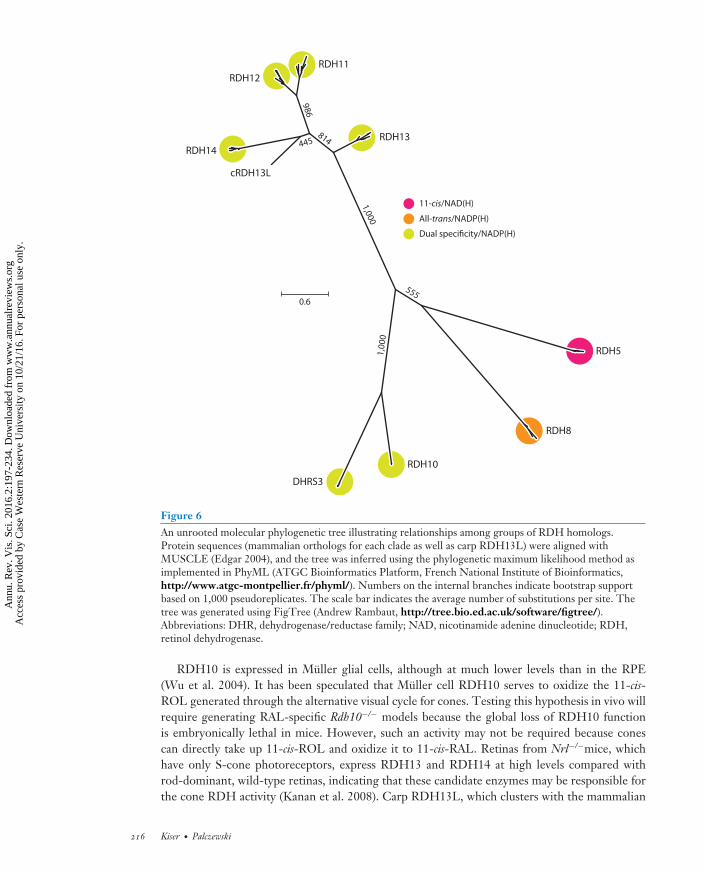
VS02CH10-Palczewski ARI 29 September 2016 10:52
986
445
1,00
0
555
1,000
814
0.6
RDH14
cRDH13L
RDH12RDH11
RDH13
RDH5
RDH8
RDH10
DHRS3
11-cis/NAD(H)
All-trans/NADP(H)
Dual specificity/NADP(H)
Figure 6An unrooted molecular phylogenetic tree illustrating relationships among groups of RDH homologs.Protein sequences (mammalian orthologs for each clade as well as carp RDH13L) were aligned withMUSCLE (Edgar 2004), and the tree was inferred using the phylogenetic maximum likelihood method asimplemented in PhyML (ATGC Bioinformatics Platform, French National Institute of Bioinformatics,http://www.atgc-montpellier.fr/phyml/). Numbers on the internal branches indicate bootstrap supportbased on 1,000 pseudoreplicates. The scale bar indicates the average number of substitutions per site. Thetree was generated using FigTree (Andrew Rambaut, http://tree.bio.ed.ac.uk/software/figtree/).Abbreviations: DHR, dehydrogenase/reductase family; NAD, nicotinamide adenine dinucleotide; RDH,retinol dehydrogenase.
RDH10 is expressed in Muller glial cells, although at much lower levels than in the RPE(Wu et al. 2004). It has been speculated that Muller cell RDH10 serves to oxidize the 11-cis-ROL generated through the alternative visual cycle for cones. Testing this hypothesis in vivo willrequire generating RAL-specific Rdh10−/− models because the global loss of RDH10 functionis embryonically lethal in mice. However, such an activity may not be required because conescan directly take up 11-cis-ROL and oxidize it to 11-cis-RAL. Retinas from Nrl−/−mice, whichhave only S-cone photoreceptors, express RDH13 and RDH14 at high levels compared withrod-dominant, wild-type retinas, indicating that these candidate enzymes may be responsible forthe cone RDH activity (Kanan et al. 2008). Carp RDH13L, which clusters with the mammalian
216 Kiser · Palczewski
Ann
u. R
ev. V
is. S
ci. 2
016.
2:19
7-23
4. D
ownl
oade
d fr
om w
ww
.ann
ualr
evie
ws.
org
Acc
ess
prov
ided
by
Cas
e W
este
rn R
eser
ve U
nive
rsity
on
10/2
1/16
. For
per
sona
l use
onl
y.

VS02CH10-Palczewski ARI 29 September 2016 10:52
RDH14 group (Figure 6), has been identified as a major 11-cis-RDH in cone photoreceptors. Thein vivo relevance of RDH13 and RDH14 to mammalian cone photoreceptor function remains tobe elucidated.
5. RETINYL ESTER SYNTHASE(LECITHIN: RETINOL ACYLTRANSFERASE)
The RPE is the major site of vitamin A storage within the retina. Following photobleaching,vitamin A generated by the activity of photoreceptor RDHs is rapidly transferred from the retinato the RPE (Dowling 1960). Additionally, the RPE takes up vitamin A from holo-RBP4 circu-lating in the choriocapillaris through the STRA6 transporter expressed on its basolateral surface(Amengual et al. 2012, Isken et al. 2008, Kawaguchi et al. 2007). Both of these processes aredriven by the esterification of vitamin A. The resulting all-trans-retinyl esters are efficiently storedin lipid droplet–like structures within the RPE referred to as retinosomes (Imanishi et al. 2004).These esters also serve as substrates for the retinoid isomerization step of the visual cycle. LRATis the main enzyme responsible for catalyzing the formation of retinyl esters in the eye and manyother tissues. LRAT activity, in which an acyl group is transferred from the sn1 position of phos-phatidylcholine (lecithin) to ROLs, was first identified in microsomal membrane fractions fromhepatocytes and the RPE (MacDonald & Ong 1988, Saari & Bredberg 1989). This reaction differsfrom that of a second retinyl ester synthase activity present in humans in which the retinyl esteris synthesized from a fatty-acyl-CoA instead of a phospholipid. Notably, LRAT is fairly promis-cuous in its choice of acyl acceptors and can readily esterify or amidate a number of primaryalcohol and amine compounds, including the retinoid cycle inhibitor retinylamine (Golczak et al.2005a). LRAT-catalyzed acyl transfer occurs via a ping-pong mechanism featuring a covalentacyl-enzyme intermediate involving a conserved cysteine residue (Cys161 in the human sequence)(Mondal et al. 2000). Phylogenetic analysis has revealed that LRAT belongs to the NlpC/P60protein superfamily, whose members feature a papain-like catalytic triad consisting of a nucle-ophilic cysteine residue, a histidine residue that acts as a general base, and a third polar residue(usually histidine or asparagine) (Anantharaman & Aravind 2003). Three-dimensional structuresof LRAT family proteins have confirmed that they adopt a papain-like thiol peptidase fold, despitehaving no discernible sequence similarity to those enzymes (Figure 7). Compared with classicalNlpC/P60 enzymes in which the nucleophilic cysteine residue resides on the N terminus, LRAT-like members are circularly permuted, with the catalytic cysteine relocated to the C-terminalend (Anantharaman & Aravind 2003). Besides LRAT, the human genome encodes seven otherLRAT-like proteins: five H-Ras-like tumor suppressors (HRASLS) and two neurosensory pro-teins (NSE1 and 2). The former group of proteins exhibits phospholipase A2 and phospholipidacyltransferase activities, whereas the latter remains uncharacterized. LRAT and its homologs arebitopic integral membrane enzymes anchored to lipid bilayers via a C-terminal transmembraneα-helix. An amphipathic N-terminal region can also mediate monotopic membrane interactions(Moise et al. 2007). LRAT differs from the other LRAT-like family members by the presence ofan approximately 30 residue insertion located near the middle of the protein sequence. Graftingthis LRAT-specific region into the HRASLS3 sequence has resulted in a chimeric protein withsn1-specific LRAT activity akin to that of native LRAT (Golczak et al. 2015). The crystal structureof this chimeric protein has revealed that the LRAT-specific sequence mediates the formation ofa homodimeric assembly in which the C-terminal α-helix containing the catalytic cysteine residueis domain swapped compared with native, monomeric HRASLS3 (Figure 7) (Golczak et al. 2015).The structure is consistent with biochemical work demonstrating that LRAT forms functionalhomodimers on microsomal membranes. Dimer formation results in structural changes around
www.annualreviews.org • The Visual Cycle and Its Modulators 217
Ann
u. R
ev. V
is. S
ci. 2
016.
2:19
7-23
4. D
ownl
oade
d fr
om w
ww
.ann
ualr
evie
ws.
org
Acc
ess
prov
ided
by
Cas
e W
este
rn R
eser
ve U
nive
rsity
on
10/2
1/16
. For
per
sona
l use
onl
y.

VS02CH10-Palczewski ARI 29 September 2016 10:52
Cys125
His24His24
His36His36
Heptanoyl chain
Swapped helices
LRAT-specific insertion
Figure 7Crystal structure of the lecithin:retinol acyltransferase (LRAT)–H-Ras-like tumor suppressor 3 chimera.The two chains of the homodimer are shown in a schematic representation (blue and green). TheLRAT-specific insertion (red ) forms a β-sheet that mediates dimer formation. This region also provides apathway for the exchange of the C-terminal α-helical regions within the dimer pair. The histidine (His) andcysteine (Cys) residues constituting the catalytic triad are shown as sticks. The catalytic Cys125 residue isacylated as a result of the crystals being grown in the presence of the acyl donor, diheptanoylphosphatidylcholine. The figure was generated with PyMol (Schrodinger, https://www.pymol.org/).
the catalytic cysteine residue in which the active site pocket with enhanced hydrophobicity is moredeeply embedded in the protein. These characteristics have been postulated to impede water entryinto the pocket such that acyltransferase activity is favored over hydrolase activity (Golczak et al.2015).
Studies in Lrat−/- mice have revealed that this enzyme is critical for ocular retinoid homeosta-sis and photoreceptor health (Batten et al. 2004). Essentially, these mice lack all ocular retinoids,consistent with an indispensable role for LRAT in the uptake and retention of vitamin A by theeye. Only trace levels of retinyl esters in liver, the other major vitamin A storage site in the body,are present in these mice. Due to the lack of retinyl ester substrate needed for the retinoid isomer-ization step of the retinoid cycle, Lrat−/− mice have severely impaired electroretinographic (ERG)and pupillary light responses. These in vivo results are consistent with in vitro data showing that in-hibition of LRAT activity blocks the biosynthesis of 11-cis-ROL by the retinoid isomerase RPE65.Lrat−/− mice have shortened rod outer segments at 8 weeks of age and develop a progressive lossof cone photoreceptors that can be rescued by administration of the artificial chromophore pro-drug 9-cis-retinyl acetate (Maeda et al. 2009b). Despite the major disruption in vitamin A storagecaused by the loss of LRAT function, these treated mice are otherwise viable and fertile. Con-sistent with the knockout mouse phenotype, loss-of-function mutations in LRAT cause RP inhumans (Thompson et al. 2001). 9-cis-Retinyl acetate and LRAT gene replacement therapy areunder investigation as treatments for RP associated with loss-of-function LRAT mutations (Battenet al. 2005, Koenekoop et al. 2014).
218 Kiser · Palczewski
Ann
u. R
ev. V
is. S
ci. 2
016.
2:19
7-23
4. D
ownl
oade
d fr
om w
ww
.ann
ualr
evie
ws.
org
Acc
ess
prov
ided
by
Cas
e W
este
rn R
eser
ve U
nive
rsity
on
10/2
1/16
. For
per
sona
l use
onl
y.
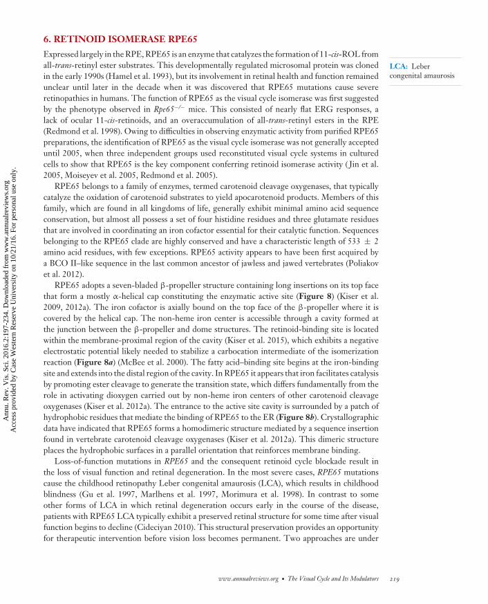
VS02CH10-Palczewski ARI 29 September 2016 10:52
LCA: Lebercongenital amaurosis
6. RETINOID ISOMERASE RPE65
Expressed largely in the RPE, RPE65 is an enzyme that catalyzes the formation of 11-cis-ROL fromall-trans-retinyl ester substrates. This developmentally regulated microsomal protein was clonedin the early 1990s (Hamel et al. 1993), but its involvement in retinal health and function remainedunclear until later in the decade when it was discovered that RPE65 mutations cause severeretinopathies in humans. The function of RPE65 as the visual cycle isomerase was first suggestedby the phenotype observed in Rpe65−/− mice. This consisted of nearly flat ERG responses, alack of ocular 11-cis-retinoids, and an overaccumulation of all-trans-retinyl esters in the RPE(Redmond et al. 1998). Owing to difficulties in observing enzymatic activity from purified RPE65preparations, the identification of RPE65 as the visual cycle isomerase was not generally accepteduntil 2005, when three independent groups used reconstituted visual cycle systems in culturedcells to show that RPE65 is the key component conferring retinoid isomerase activity ( Jin et al.2005, Moiseyev et al. 2005, Redmond et al. 2005).
RPE65 belongs to a family of enzymes, termed carotenoid cleavage oxygenases, that typicallycatalyze the oxidation of carotenoid substrates to yield apocarotenoid products. Members of thisfamily, which are found in all kingdoms of life, generally exhibit minimal amino acid sequenceconservation, but almost all possess a set of four histidine residues and three glutamate residuesthat are involved in coordinating an iron cofactor essential for their catalytic function. Sequencesbelonging to the RPE65 clade are highly conserved and have a characteristic length of 533 ± 2amino acid residues, with few exceptions. RPE65 activity appears to have been first acquired bya BCO II–like sequence in the last common ancestor of jawless and jawed vertebrates (Poliakovet al. 2012).
RPE65 adopts a seven-bladed β-propeller structure containing long insertions on its top facethat form a mostly α-helical cap constituting the enzymatic active site (Figure 8) (Kiser et al.2009, 2012a). The iron cofactor is axially bound on the top face of the β-propeller where it iscovered by the helical cap. The non-heme iron center is accessible through a cavity formed atthe junction between the β-propeller and dome structures. The retinoid-binding site is locatedwithin the membrane-proximal region of the cavity (Kiser et al. 2015), which exhibits a negativeelectrostatic potential likely needed to stabilize a carbocation intermediate of the isomerizationreaction (Figure 8a) (McBee et al. 2000). The fatty acid–binding site begins at the iron-bindingsite and extends into the distal region of the cavity. In RPE65 it appears that iron facilitates catalysisby promoting ester cleavage to generate the transition state, which differs fundamentally from therole in activating dioxygen carried out by non-heme iron centers of other carotenoid cleavageoxygenases (Kiser et al. 2012a). The entrance to the active site cavity is surrounded by a patch ofhydrophobic residues that mediate the binding of RPE65 to the ER (Figure 8b). Crystallographicdata have indicated that RPE65 forms a homodimeric structure mediated by a sequence insertionfound in vertebrate carotenoid cleavage oxygenases (Kiser et al. 2012a). This dimeric structureplaces the hydrophobic surfaces in a parallel orientation that reinforces membrane binding.
Loss-of-function mutations in RPE65 and the consequent retinoid cycle blockade result inthe loss of visual function and retinal degeneration. In the most severe cases, RPE65 mutationscause the childhood retinopathy Leber congenital amaurosis (LCA), which results in childhoodblindness (Gu et al. 1997, Marlhens et al. 1997, Morimura et al. 1998). In contrast to someother forms of LCA in which retinal degeneration occurs early in the course of the disease,patients with RPE65 LCA typically exhibit a preserved retinal structure for some time after visualfunction begins to decline (Cideciyan 2010). This structural preservation provides an opportunityfor therapeutic intervention before vision loss becomes permanent. Two approaches are under
www.annualreviews.org • The Visual Cycle and Its Modulators 219
Ann
u. R
ev. V
is. S
ci. 2
016.
2:19
7-23
4. D
ownl
oade
d fr
om w
ww
.ann
ualr
evie
ws.
org
Acc
ess
prov
ided
by
Cas
e W
este
rn R
eser
ve U
nive
rsity
on
10/2
1/16
. For
per
sona
l use
onl
y.

VS02CH10-Palczewski ARI 29 September 2016 10:52
a b
His241His241
His313His313
His527His527
His180His180
Retinoid-bindingpocket
Acyl-bindingpocket
Helical cap
Membrane-binding surface
Active siteentranceIron cofactorIron cofactor
Figure 8Orthogonal views of the crystal structure of retinal pigment epithelium–specific 65 kDa protein. (a) View down the β-propeller axis,with the seven blades indicated by distinct colors. The iron cofactor (orange sphere) is coordinated on the propeller axis by fourconserved histidine (His) residues. The purple surface delineates the active site pocket. (b) Rotating the structure by 90◦ along thehorizontal axis shows the helical cap on the top face of the β-propeller. The active site entrance is surrounded by a patch of mostlyhydrophobic residues that mediate the membrane binding critical for substrate uptake. The structural representations were generatedwith PyMol (Schrodinger, https://www.pymol.org/) using the coordinates deposited under Protein Data Bank accession code 4F2Z.
IRBP:interphotoreceptorretinoid-bindingprotein
CRALBP: cellularretinaldehyde-bindingprotein
investigation: RPE65 gene therapy to restore a functional RPE-based retinoid cycle (Acland et al.2001, Bainbridge et al. 2008, Maguire et al. 2008) and treatment with an artificial chromophore(9-cis-RAL) to bypass the metabolic blockade caused by the lack of RPE65 activity (Koenekoopet al. 2014, Van Hooser et al. 2000). Both approaches have shown therapeutic promise in clinicaltrials but require further optimization.
7. THE RETINOID-BINDING PROTEINS
The efficient movement of retinoids between and within the photoreceptors, RPE, and Mullerglia is required for proper visual cycle function (Table 3). Because retinoids are lipophilic com-pounds prone to destruction via off-pathway reactions, their diffusion within the retina dependscritically on retinoid-binding proteins that mobilize these compounds from membrane stores andprotect them from degradation. Four such binding proteins are known to be critical for visualfunction: serum RBP4, cellular RBP1 (CRBP1 or RBP1), interphotoreceptor retinoid-bindingprotein (IRBP or RBP3), and cellular retinaldehyde-binding protein (CRALBP) (Figure 9).Although these proteins are not homologous and are structurally diverse, they all are water solublemolecules that possess internal lipophilic pockets into which retinoids can bind and be protectedfrom degradation.
7.1. Retinol-Binding Protein 4
RBP4 is the major carrier responsible for distributing hepatic vitamin A obtained from dietarysources to peripheral tissues such as the eye. This approximately 21 kDa member of the lipocalinprotein family circulates in the plasma in complex with the tetrameric protein TTR, which prevents
220 Kiser · Palczewski
Ann
u. R
ev. V
is. S
ci. 2
016.
2:19
7-23
4. D
ownl
oade
d fr
om w
ww
.ann
ualr
evie
ws.
org
Acc
ess
prov
ided
by
Cas
e W
este
rn R
eser
ve U
nive
rsity
on
10/2
1/16
. For
per
sona
l use
onl
y.
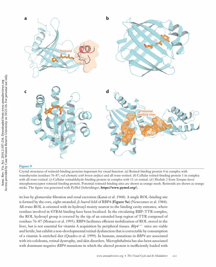
VS02CH10-Palczewski ARI 29 September 2016 10:52
a b
c d
RetinoidRetinoid
Potentialbinding sites
Potentialbinding sites
Figure 9Crystal structures of retinoid-binding proteins important for visual function. (a) Retinol-binding protein 4 in complex withtransthyretin (residues 76–87; red schematic with brown surface) and all-trans-retinol. (b) Cellular retinol-binding protein 1 in complexwith all-trans-retinol. (c) Cellular retinaldehyde-binding protein in complex with 11-cis-retinal. (d ) Module 2 from Xenopus laevisinterphotoreceptor retinoid-binding protein. Potential retinoid-binding sites are shown as orange mesh. Retinoids are shown as orangesticks. The figure was generated with PyMol (Schrodinger, https://www.pymol.org/).
its loss by glomerular filtration and renal excretion (Kanai et al. 1968). A single ROL-binding siteis formed by the core, eight-stranded, β-barrel fold of RBP4 (Figure 9a) (Newcomer et al. 1984).All-trans-ROL is oriented with its hydroxyl moiety nearest to the binding cavity entrance, whereresidues involved in STRA6 binding have been localized. In the circulating RBP–TTR complex,the ROL hydroxyl group is covered by the tip of an extended loop region of TTR composed ofresidues 76–87 (Monaco et al. 1995). RBP4 facilitates efficient mobilization of ROL stored in theliver, but is not essential for vitamin A acquisition by peripheral tissues. Rbp4−/− mice are viableand fertile, but exhibit a non-developmental retinal dysfunction that is correctable by consumptionof a vitamin A–enriched diet (Quadro et al. 1999). In humans, mutations in RBP4 are associatedwith iris coloboma, retinal dystrophy, and skin disorders. Microphthalmia has also been associatedwith dominant-negative RBP4 mutations in which the altered protein is inefficiently loaded with
www.annualreviews.org • The Visual Cycle and Its Modulators 221
Ann
u. R
ev. V
is. S
ci. 2
016.
2:19
7-23
4. D
ownl
oade
d fr
om w
ww
.ann
ualr
evie
ws.
org
Acc
ess
prov
ided
by
Cas
e W
este
rn R
eser
ve U
nive
rsity
on
10/2
1/16
. For
per
sona
l use
onl
y.

VS02CH10-Palczewski ARI 29 September 2016 10:52
all-trans-ROL yet binds to STRA6 with a higher affinity than wild-type RBP4, as discussed in thesection on STRA6 (Chou et al. 2015).
7.2. Cellular Retinol-Binding Protein 1
Following passage through the STRA6 transporter, or by other means, all-trans-ROL is trans-ported inside the cell in complex with CRBP1. Like RBP4, CRBP1 is a small, water soluble proteinthat contains a single ROL-binding site formed by its β-barrel fold (Figure 9b) (Cowan et al.1993). Of the retinoid isomers, it binds all-trans-ROL with the highest affinity, consistent withvitamin A being the physiological ligand (MacDonald & Ong 1987, Ong & Chytil 1975). In con-trast to holo-RBP4, the hydroxyl moiety of all-trans-ROL is buried within the binding pocket ofCRBP1, such that it is not solvent accessible. This structural finding indicates that all-trans-ROLmust dissociate from the protein before it can be metabolized, although kinetic data have indicatedthat the holo–CRBP1 complex might be the substrate for all-trans-ROL esterification. CRBP1 isexpressed in many sites throughout the body, whereas its paralog, CRBP2, is found mainly in theintestine. Within the retina, CRBP1 expression is highest in the RPE (Saari et al. 1977). Elevatedlevels of all-trans-ROL in the retina and a lack of retinyl ester production in the RPE of Rbp1−/−
mice are consistent with a key role for this binding protein in retinoid metabolism within the eye(Saari et al. 2002).
7.3. Cellular Retinaldehyde-Binding Protein
CRALBP is the key protein responsible for binding 11-cis-retinoids generated via the RPE andMuller cell–based visual cycles and delivering them to the plasma membrane for transport tophotoreceptors (Bunt-Milam & Saari 1983, Saari et al. 1977). As isolated from bovine neuralretina, CRALBP contains both 11-cis-RAL and 11-cis-ROL as bound ligands, whereas 11-cis-RALis the only retinoid associated with RPE-derived CRALBP (Saari et al. 1982). 9-cis-Retinoidsare the only other isomers capable of binding CRALBP with high affinity. The strong bindingselectivity for these two isomers has been explained by the presence of a curved binding pocketthat has high complementarity to the 11-cis- and 9-cis-configurations (Figure 9c) (He et al. 2009).This structure also has revealed a cationic surface that likely interacts with negatively chargedphospholipids to enable retinoid transfer to the plasma membrane. Rlbp1−/− mice exhibit delayeddark adaption consistent with a key role of CRALBP in efficient visual cycle function (Saari et al.2001). Mutations in human RLBP1 are associated with several different retinopathies, including RP(Maw et al. 1997), Bothnia dystrophy, retinitis punctata albescens, and Newfoundland rod–conedystrophy (Travis et al. 2007).
7.4. Interphotoreceptor Retinoid-Binding Protein
Photoreceptors secrete an approximately 136 kDa lipoglycoprotein called IRBP that mediatesintercellular retinoid transfer (Bunt-Milam & Saari 1983, Lai et al. 1982). This protein has acomplex structure consisting of four homologous modules that arose from an internal quadru-plication (Fong & Bridges 1988). IRBP has an elongated appearance in negative-stained electronmicroscopy images and undergoes major conformational changes in the presence of retinoidligands. Unlike the other retinoid-binding proteins, IRBP possesses three retinoid-binding sitesand can bind both all-trans- and 11-cis-retinoids with high affinity. The structure of an IRBPmodule from Xenopus has revealed that the modules adopt a two-domain architecture (Loew &Gonzalez-Fernandez 2002). The retinoid-binding sites within the modules have not yet been
222 Kiser · Palczewski
Ann
u. R
ev. V
is. S
ci. 2
016.
2:19
7-23
4. D
ownl
oade
d fr
om w
ww
.ann
ualr
evie
ws.
org
Acc
ess
prov
ided
by
Cas
e W
este
rn R
eser
ve U
nive
rsity
on
10/2
1/16
. For
per
sona
l use
onl
y.

VS02CH10-Palczewski ARI 29 September 2016 10:52
A2E, otherlipid adducts
A2E, otherlipid adducts
RPE65 inhibitorsRPE65 inhibitors
All-trans-RAL trapsAll-trans-RAL trapsC20-2H-ROLC20-2H-ROL
CyclodextrinsCyclodextrins
All-trans-RALAll-trans-RAL
All-trans-ROLAll-trans-ROL
All-trans-REAll-trans-RE 11-cis-ROL11-cis-ROL
11-cis-RAL11-cis-RAL
11-cis-RAL-opsin11-cis-RAL-opsin
b
Holo-RBP4Holo-RBP4
SFDSFD
LRATRP/LCA
RPE65RP/LCARPE65
RP/LCA
All-trans-ROLsupplementAll-trans-ROLsupplement
All-trans-RALAll-trans-RAL
All-trans-ROLAll-trans-ROL
All-trans-REAll-trans-RE 11-cis-ROL11-cis-ROL
11-cis-RAL11-cis-RAL
11-cis-RAL-opsin11-cis-RAL-opsin
9-cis-RAL9-cis-RAL
IsorhodopsinIsorhodopsin
a
Holo-RBP4Holo-RBP4
RBP4antagonists
RBP4antagonists
All-trans-RAL adductsNADPH oxidase activation
Orange: theraputicsBrown: diseases
Figure 10Therapeutic strategies for treating retinal diseases. (a) Retinoid supplementation for diseases such as SFD affecting ROL delivery to theRPE or generation of a visual chromophore (LRAT or RPE65 RP/LCA). (b) Strategies to reduce the formation of excess atRAL, itslipid condensation products, or both, to facilitate their clearance from the eye. Abbreviations: LCA, Leber congenital amaurosis;LRAT, lecithin:retinol acyltransferase; NAD, nicotinamide adenine dinucleotide; RAL, retinal; RBP, retinol-binding protein; ROL,retinol; RP, retinitis pigmentosa; RPE, retinal pigment epithelium; SFD, Sorsby’s fundus dystrophy.
revealed, but two candidate binding pockets are apparent from the crystal structure: one at thejunction between the two domains and the other within the C-terminal domain of the module(Figure 9d ). Consistent with an important role for IRBP in retinoid trafficking within the retina,mutations in the RBP3 gene are associated with autosomal recessive RP (den Hollander et al. 2009).
8. VISUAL CYCLE MODULATORS
Defects in most genes involved in the retinoid cycle are associated with specific retinopathies(Tables 1 and 2). Here, we highlight research conducted on modulators of the visual cycle for thetreatment of retinal diseases (Figure 10). Many of these agents are based on the retinoid structure;thus their specific delivery to the eye can employ the same retinoid mechanism, for example,transport by RBP4, delivery via STRA6, or enhancement of retention through esterification oramidation in the retina with the help of LRAT (reviewed in Palczewski 2010). These strategies aredescribed below along with some key references (Figure 10). As this field rapidly evolves, furtherprogress likely will be revealed in the literature.
8.1. Retinol Supplementation
Uncomplicated vitamin A deficiency can be overcome by providing therapeutic doses of the vi-tamin, either as free ROL or retinyl esters (reviewed in Kiser et al. 2014). Improvement in visual
www.annualreviews.org • The Visual Cycle and Its Modulators 223
Ann
u. R
ev. V
is. S
ci. 2
016.
2:19
7-23
4. D
ownl
oade
d fr
om w
ww
.ann
ualr
evie
ws.
org
Acc
ess
prov
ided
by
Cas
e W
este
rn R
eser
ve U
nive
rsity
on
10/2
1/16
. For
per
sona
l use
onl
y.

VS02CH10-Palczewski ARI 29 September 2016 10:52
function upon vitamin A supplementation has also been observed in a mouse model with a specificrhodopsin mutation related to RP (Sibulesky et al. 1999). In these cases, all-trans-ROL or its activecis form acted as a chaperone for the mutated protein, although there was no follow-up to theseinitial studies (Figure 10a).
A mutation in the metalloproteinase inhibitor 3 gene (TIMP3) causes thickening of Bruch’smembrane, which is between the photoreceptor layer and its blood supply, leading to chronicdeprivation of vitamin A in a disease known as Sorsby’s fundus dystrophy. A dose of 50,000IU/day of vitamin A administered orally diminishes night blindness in patients with early stages ofthis disease ( Jacobson et al. 1995). Similarly, treatment with pharmacological doses of vitamin Arestored vision in Stra6−/- mice lacking adequate vitamin A supplementation to the eye (Amengualet al. 2014).
8.2. 9-cis-Retinal and 11-cis-Retinal Supplementation
When 11-cis-RAL cannot be generated because of retinoid cycle blockade, as with a nonfunctionalor suppressed retinoid isomerase or LRAT, supplementation with pharmacological doses of 9-cis-RAL can dramatically improve visual function in animal models (Batten et al. 2004, Van Hooseret al. 2000, 2002). A successful clinical trial using this therapeutic strategy has recently beenreported (Koenekoop et al. 2014). Treatment with cis-retinoid appears to be useful in preventingthe deterioration of vision in aging mice (Maeda et al. 2006c), raising the possibility that a similartreatment could successfully preserve vision in older humans (Figure 10a).
8.3. Inhibitors of Retinal Pigment Epithelium–Specific 65 kDa Protein
Another idea is that slowing the regeneration of visual pigments by inhibiting the retinoid cyclewill decrease the amount of free all-trans-RAL and its toxic condensation products (Radu et al.2003). [Importantly, profound suppression of visual pigment regeneration is detrimental to retinalhealth because free opsin has constitutive signaling activity that eventually causes the death ofphotoreceptor cells, as mentioned in Section 5 (Woodruff et al. 2003).] 13-cis-RA was the initialcompound found to decrease A2E formation (Radu et al. 2003). The first highly selective andpotent inhibitor discovered was retinylamine (Golczak et al. 2005b). The systematic comparisonof different inhibitors has been reported (Maeda et al. 2006a) and, based on those results, emixustatwas developed by Acucela Inc. (Bavik et al. 2015), but human clinical trials have revealed significantside effects (Kubota et al. 2012). Finally, recent studies have questioned the mechanism of actionof emixustat and suggested that sequestration of all-trans-RAL should be considered as discussedin Section 8.5 (Figure 10b) (Zhang et al. 2015).
8.4. Reducing the Supply of Vitamin A to the Eye
Dissociation of the RBP4–TTR complex allows filtration of RBP4 through renal glomeruli andelimination of vitamin A from the circulation. Thus, fenretinide, an amide of RA, had been hy-pothesized to have an antagonistic effect on the RBP4–TTR complex (Mata et al. 2013). However,it has been shown that the mechanism of action likely involves a slow hydrolysis of the amide alongwith upregulation of STRA6 and LRAT, thus decreasing circulating all-trans-ROL and increasingcellular vitamin A uptake by peripheral tissues. Nonretinoid antagonists of the RBP4 complex arebeing developed (Dobri et al. 2013).
224 Kiser · Palczewski
Ann
u. R
ev. V
is. S
ci. 2
016.
2:19
7-23
4. D
ownl
oade
d fr
om w
ww
.ann
ualr
evie
ws.
org
Acc
ess
prov
ided
by
Cas
e W
este
rn R
eser
ve U
nive
rsity
on
10/2
1/16
. For
per
sona
l use
onl
y.

VS02CH10-Palczewski ARI 29 September 2016 10:52
8.5. Sequestration of All-trans-Retinal to Prevent Toxicity
A series of studies have demonstrated that one candidate toxin in the retinoid cycle is all-trans-RAL released from photoactivated visual pigments (reviewed in Kiser et al. 2014). All-trans-RALtoxicity can be lowered if a Schiff base is rapidly and reversibly formed with primary amines. Inthis case, the free aldehyde would be decreased, allowing RDHs to convert it into an alcohol.In mouse models of retinal degeneration, the peak concentration of free all-trans-RAL has beenlowered with primary amine-containing, US Food and Drug Administration-approved drugs thatdid not inhibit chromophore regeneration because they were nonretinoid compounds. To validatethis strategy, Schiff base adducts between all-trans-RAL and these amines have been identified bymass spectrometry (Maeda et al. 2012). This option is ready to be tested in humans (Figure 10b).
8.6. Elimination of A2E
Severe and rapid bleaching of visual pigments produces free all-trans-RAL that overwhelms thecapacity of retinal cells to reduce this aldehyde to all-trans-ROL. During visual transduction, all-trans-RAL can react with itself and lipids. The resulting condensation products and their oxidizedderivatives reportedly are toxic to the RPE (reviewed in Sparrow et al. 2012). Intravitreal treatmentwith β-cyclodextrins reduced bis-retinoids in the RPE of mice by 48% (Nociari et al. 2014). Thesenovel prophylactic results are encouraging, but a beneficial effect on the health of the RPE hasyet to be demonstrated (Figure 10b).
8.7. Use of Isotope Effects to Slow Production of A2E
What if A2E formation could be selectively blocked without affecting visual cycle kinetics? Con-sideration of the mechanistic details of A2E formation has revealed that one of the rate-limitingsteps is the subtraction of a proton. Thus, the rate of this reaction could be changed if the hydro-gen atom is replaced by a deuterium atom (a kinetic isotope effect). Long-term administration ofdeuterium-enriched vitamin A, namely C20-D3–vitamin A, slowed the formation of all-trans-RALcondensation products (Ma et al. 2011). However, this conceptually elegant idea has many caveats,including the difficulty in replacing all vitamin A in liver and eye, uncertainty about whether allcondensation products will display the same isotopic effect as A2E formation, and whether slow-ing the reaction would make a significant impact on condensation product formation. Finally, ifall-trans-RAL is the primary toxic agent that kills photoreceptor and RPE cells, then deuteriumreplacement would not be expected to have any therapeutic effect (Figure 10b).
9. CONCLUSIONS
The past three decades have led to a much greater molecular understanding of the retinoid cycle.Individual proteins have been well characterized by biochemical and biophysical methods. Thephysiological functions of these proteins have been clarified from analyses of human visual diseaseresulting from their inadequate expression, activation or altered properties and their correspondingtransgenic animal models. Such animal models also are indispensable in testing new concepts forcorrecting genetic deficiencies by using pharmacological, genetic, or stem-cell-based approaches.
High-resolution structures provide detailed information on how the macromolecules of theretinoid cycle work. The utility of atomic information is exemplified by rhodopsin, the first Gprotein–coupled receptor whose structure was elucidated; by LRAT, through the use of hybrid
www.annualreviews.org • The Visual Cycle and Its Modulators 225
Ann
u. R
ev. V
is. S
ci. 2
016.
2:19
7-23
4. D
ownl
oade
d fr
om w
ww
.ann
ualr
evie
ws.
org
Acc
ess
prov
ided
by
Cas
e W
este
rn R
eser
ve U
nive
rsity
on
10/2
1/16
. For
per
sona
l use
onl
y.

VS02CH10-Palczewski ARI 29 September 2016 10:52
approaches to expose its mechanism of catalysis; and with CRALBP, which in complex with RALprovided precise information as to its substrate specificity.
Historical knowledge about the chemistry of vision in the retina was obtained in three phases.First, the elegant work on the chemistry of vision by George Wald placed vision in a proper molec-ular perspective. Second, retinoid-binding proteins and enzymes were identified in the complexphotoreceptor–RPE two-cell type system. Finally, molecular and structural characterizations ofvisual pathway components, supported by genetic approaches, have been, and continue to be,especially fruitful. This latest phase also has benefited greatly from mechanism-based pharmacol-ogy. As summarized in this review, visual cycle modulators will continue to advance to clinicaltrials, and confidence is building that these compounds will become efficacious therapies for somecurrently incurable retinal-blinding diseases.
SUMMARY POINTS
1. Structural and chemical bases for how the retinoid cycle components perform theirphysiological functions are presented.
2. Human retinal diseases that result from disruption of visual cycle components and animalmodels of these disorders are summarized.
3. Therapeutically aimed visual cycle modulation strategies include (a) supplementing theeye in animal models and humans that have defective retinoid cycles with all-trans- and-cis isomers, (b) reducing vitamin A delivery to the eye and inhibiting retinoid isomerase,(c) slowing A2E formation by involving isotope effects, (d ) removing all-trans-RAL con-densation products, and, finally, (e) sequestrating all-trans-RAL to eliminate its toxicityand reduce the formation of its condensation products.
FUTURE ISSUES
1. High-resolution structures of IRBP, RDHs, ABCA4, and STRA6 are needed to under-stand how these proteins participate in the movement and metabolism of retinoids.
2. Additional visual cycle modulators should be identified that support retinal function with-out inhibiting RPE65 and prevent pathology resulting from genetic and environmentalfactors.
3. The precise sequence of events that leads to rhodopsin regeneration and chromophorehydrolysis after photoisomerization requires further characterization.
4. The molecular and genetic basis of cone chromophore regeneration must becharacterized.
5. The factors governing the incorporation of retinyl esters into retinosomes must be elu-cidated as well as those governing their reentry into the retinoid cycle.
6. The molecular steps in recycling chromophores phagocytized by the RPE (Kevany& Palczewski 2010) must be defined as well as their utilization in regenerating visualpigments.
7. Cone visual pigment structures and the mechanisms of their spectral tuning should beelucidated.
226 Kiser · Palczewski
Ann
u. R
ev. V
is. S
ci. 2
016.
2:19
7-23
4. D
ownl
oade
d fr
om w
ww
.ann
ualr
evie
ws.
org
Acc
ess
prov
ided
by
Cas
e W
este
rn R
eser
ve U
nive
rsity
on
10/2
1/16
. For
per
sona
l use
onl
y.

VS02CH10-Palczewski ARI 29 September 2016 10:52
DISCLOSURE STATEMENT
K.P. is an inventor of US Patent 8722669 (Compounds and methods of treating ocular disorders)and US Patent 20080275134 (Methods for treatment of retinal degenerative disease) issued to CaseWestern Reserve University (CWRU), whose values may be affected by this publication. CWRUmay license this technology for commercial development. K.P. is a member of the scientific boardof Vision Medicines, Inc., which develops visual cycle modulators. K.P. is Chief Scientific Officerat Polgenix, Inc., and an inventor of US Patent 7706863 (Methods for assessing a physiologicalstate of a mammalian retina) and US Patent 8346345 B2 (Methods for assessing a physiologicalstate of a mammalian retina), whose values may be affected by this publication.
ACKNOWLEDGMENTS
We thank Dr. Leslie T. Webster Jr., Dr. Grzegorz Palczewski, and members of the Palczewskilaboratory for helpful comments on this manuscript. We thank Dr. Debarshi Mustafi and Dr. BrianKevany for RNA sequencing data. This work was supported by funding from the US NationalInstitutes of Health grants EY009339 and EY025451 (to K.P.), the Arnold and Mabel BeckmanFoundation, the Foundation Fighting Blindness (to K.P.), and the US Department of VeteransAffairs grant BX002683 (to P.D.K.). K.P. is the John H. Hord Professor of Pharmacology.
LITERATURE CITED
Acland GM, Aguirre GD, Ray J, Zhang Q, Aleman TS, et al. 2001. Gene therapy restores vision in a caninemodel of childhood blindness. Nat. Genet. 28:92–95
Adams MK, Belyaeva OV, Wu L, Kedishvili NY. 2014. The retinaldehyde reductase activity of DHRS3is reciprocally activated by retinol dehydrogenase 10 to control retinoid homeostasis. J. Biol. Chem.289:14868–80
Allikmets R, Shroyer NF, Singh N, Seddon JM, Lewis RA, et al. 1997a. Mutation of the Stargardt diseasegene (ABCR) in age-related macular degeneration. Science 277:1805–7
Allikmets R, Singh N, Sun H, Shroyer NF, Hutchinson A, et al. 1997b. A photoreceptor cell-specific ATP-binding transporter gene (ABCR) is mutated in recessive Stargardt macular dystrophy. Nat. Genet. 15:236–46
Amengual J, Golczak M, Palczewski K, von Lintig J. 2012. Lecithin:retinol acyltransferase is critical for cellularuptake of vitamin A from serum retinol-binding protein. J. Biol. Chem. 287:24216–27
Amengual J, Zhang N, Kemerer M, Maeda T, Palczewski K, Von Lintig J. 2014. STRA6 is critical for cellularvitamin A uptake and homeostasis. Hum. Mol. Genet. 23:5402–17
Anantharaman V, Aravind L. 2003. Evolutionary history, structural features and biochemical diversity of theNlpC/P60 superfamily of enzymes. Genome Biol. 4:R11
Andrews JS, Futterman S. 1964. Metabolism of the retina. V. The role of microsomes in vitamin A esterificationin the visual cycle. J. Biol. Chem. 239:4073–76
Angel TE, Gupta S, Jastrzebska B, Palczewski K, Chance MR. 2009. Structural waters define a functionalchannel mediating activation of the GPCR, rhodopsin. PNAS 106:14367–72
Bainbridge JW, Smith AJ, Barker SS, Robbie S, Henderson R, et al. 2008. Effect of gene therapy on visualfunction in Leber’s congenital amaurosis. N. Engl. J. Med. 358:2231–39
Batten ML, Imanishi Y, Maeda T, Tu DC, Moise AR, et al. 2004. Lecithin–retinol acyltransferase is essentialfor accumulation of all-trans-retinyl esters in the eye and in the liver. J. Biol. Chem. 279:10422–32
Batten ML, Imanishi Y, Tu DC, Doan T, Zhu L, et al. 2005. Pharmacological and rAAV gene therapy rescueof visual functions in a blind mouse model of Leber congenital amaurosis. PLOS Med. 2:1177–89
Bavik C, Henry SH, Zhang Y, Mitts K, McGinn T, et al. 2015. Visual cycle modulation as an approach towardpreservation of retinal integrity. PLOS ONE 10:e0124940
Bernstein PS, Law WC, Rando RR. 1987. Isomerization of all-trans-retinoids to 11-cis-retinoids in vitro.PNAS 84:1849–53
www.annualreviews.org • The Visual Cycle and Its Modulators 227
Ann
u. R
ev. V
is. S
ci. 2
016.
2:19
7-23
4. D
ownl
oade
d fr
om w
ww
.ann
ualr
evie
ws.
org
Acc
ess
prov
ided
by
Cas
e W
este
rn R
eser
ve U
nive
rsity
on
10/2
1/16
. For
per
sona
l use
onl
y.

VS02CH10-Palczewski ARI 29 September 2016 10:52
Bernstein PS, Rando RR. 1986. In vivo isomerization of all-trans- to 11-cis-retinoids in the eye occurs at thealcohol oxidation state. Biochemistry 25:6473–78
Bouillet P, Sapin V, Chazaud C, Messaddeq N, Decimo D, et al. 1997. Developmental expression pattern ofStra6, a retinoic acid–responsive gene encoding a new type of membrane protein. Mech. Dev. 63:173–86
Bunt-Milam AH, Saari JC. 1983. Immunocytochemical localization of two retinoid-binding proteins in ver-tebrate retina. J. Cell Biol. 97:703–12
Chen Y, Okano K, Maeda T, Chauhan V, Golczak M, et al. 2012. Mechanism of all-trans-retinal toxicity withimplications for Stargardt disease and age-related macular degeneration. J. Biol. Chem. 287:5059–69
Chen Y, Palczewska G, Mustafi D, Golczak M, Dong Z, et al. 2013. Systems pharmacology identifies drugtargets for Stargardt disease–associated retinal degeneration. J. Clin. Investig. 123:5119–34
Choe HW, Kim YJ, Park JH, Morizumi T, Pai EF, et al. 2011. Crystal structure of metarhodopsin II. Nature471:651–55
Chou CM, Nelson C, Tarle SA, Pribila JT, Bardakjian T, et al. 2015. Biochemical basis for dominant inheri-tance, variable penetrance, and maternal effects in RBP4 congenital eye disease. Cell 161:634–46
Cideciyan AV. 2010. Leber congenital amaurosis due to RPE65 mutations and its treatment with gene therapy.Prog. Retin. Eye Res. 29:398–427
Cideciyan AV, Swider M, Aleman TS, Tsybovsky Y, Schwartz SB, et al. 2009. ABCA4 disease progression anda proposed strategy for gene therapy. Hum. Mol. Genet. 18:931–41
Cowan SW, Newcomer ME, Jones TA. 1993. Crystallographic studies on a family of cellular lipophilictransport proteins: refinement of P2 myelin protein and the structure determination and refinement ofcellular retinol-binding protein in complex with all-trans-retinol. J. Mol. Biol. 230:1225–46
Deigner PS, Law WC, Canada FJ, Rando RR. 1989. Membranes as the energy source in the endergonictransformation of vitamin A to 11-cis-retinol. Science 244:968–71
den Hollander AI, McGee TL, Ziviello C, Banfi S, Dryja TP, et al. 2009. A homozygous missense mutationin the IRBP gene (RBP3) associated with autosomal recessive retinitis pigmentosa. Investig. Ophthalmol.Vis. Sci. 50:1864–72
Devine EL, Oprian DD, Theobald DL. 2013. Relocating the active-site lysine in rhodopsin and implicationsfor evolution of retinylidene proteins. PNAS 110:13351–55
Dixon RA, Kobilka BK, Strader DJ, Benovic JL, Dohlman HG, et al. 1986. Cloning of the gene and cDNAfor mammalian β-adrenergic receptor and homology with rhodopsin. Nature 321:75–79
Dobri N, Qin Q, Kong J, Yamamoto K, Liu Z, et al. 2013. A1120, a nonretinoid RBP4 antagonist, inhibitsformation of cytotoxic bisretinoids in the animal model of enhanced retinal lipofuscinogenesis. Investig.Ophthalmol. Vis. Sci. 54:85–95
Dowling JE. 1960. Chemistry of visual adaptation in the rat. Nature 188:114–18Driessen CA, Winkens HJ, Hoffmann K, Kuhlmann LD, Janssen BP, et al. 2000. Disruption of the 11-cis-
retinol dehydrogenase gene leads to accumulation of cis-retinols and cis-retinyl esters. Mol. Cell. Biol.20:4275–87
Dryja TP, McGee TL, Reichel E, Hahn LB, Cowley GS, et al. 1990. A point mutation of the rhodopsin genein one form of retinitis pigmentosa. Nature 343:364–66
Edgar RC. 2004. MUSCLE: multiple sequence alignment with high accuracy and high throughput. NucleicAcids Res. 32:1792–97
Ernst OP, Lodowski DT, Elstner M, Hegemann P, Brown LS, Kandori H. 2014. Microbial and animalrhodopsins: structures, functions, and molecular mechanisms. Chem. Rev. 114:126–63
Farjo KM, Moiseyev G, Nikolaeva O, Sandell LL, Trainor PA, Ma JX. 2011. RDH10 is the primary enzymeresponsible for the first step of embryonic vitamin A metabolism and retinoic acid synthesis. Dev. Biol.357:347–55
Farjo KM, Moiseyev G, Takahashi Y, Crouch RK, Ma JX. 2009. The 11-cis-retinol dehydrogenase activity ofRDH10 and its interaction with visual cycle proteins. Investig. Ophthalmol. Vis. Sci. 50:5089–97
Fleisch VC, Schonthaler HB, von Lintig J, Neuhauss SC. 2008. Subfunctionalization of a retinoid-bindingprotein provides evidence for two parallel visual cycles in the cone-dominant zebrafish retina. J. Neurosci.28:8208–16
Fong SL, Bridges CD. 1988. Internal quadruplication in the structure of human interstitial retinol-bindingprotein deduced from its cloned cDNA. J. Biol. Chem. 263:15330–34
228 Kiser · Palczewski
Ann
u. R
ev. V
is. S
ci. 2
016.
2:19
7-23
4. D
ownl
oade
d fr
om w
ww
.ann
ualr
evie
ws.
org
Acc
ess
prov
ided
by
Cas
e W
este
rn R
eser
ve U
nive
rsity
on
10/2
1/16
. For
per
sona
l use
onl
y.
VS02CH10-Palczewski ARI 29 September 2016 10:52
Golczak M, Imanishi Y, Kuksa V, Maeda T, Kubota R, Palczewski K. 2005a. Lecithin:retinol acyltransferaseis responsible for amidation of retinylamine, a potent inhibitor of the retinoid cycle. J. Biol. Chem.280:42263–73
Golczak M, Kuksa V, Maeda T, Moise AR, Palczewski K. 2005b. Positively charged retinoids are potent andselective inhibitors of the trans–cis isomerization in the retinoid (visual) cycle. PNAS 102:8162–67
Golczak M, Sears AE, Kiser PD, Palczewski K. 2015. LRAT-specific domain facilitates vitamin A metabolismby domain swapping in HRASLS3. Nat. Chem. Biol. 11:26–32
Goldstein EB. 1970. Cone pigment regeneration in the isolated frog retina. Vis. Res. 10:1065–68Golzio C, Martinovic-Bouriel J, Thomas S, Mougou-Zrelli S, Grattagliano-Bessieres B, et al. 2007. Matthew-
Wood syndrome is caused by truncating mutations in the retinol-binding protein receptor gene STRA6.Am. J. Hum. Genet. 80:1179–87
Gu SM, Thompson DA, Srikumari CR, Lorenz B, Finckh U, et al. 1997. Mutations in RPE65 cause autosomalrecessive childhood-onset severe retinal dystrophy. Nat. Genet. 17:194–97
Haeseleer F, Huang J, Lebioda L, Saari JC, Palczewski K. 1998. Molecular characterization of a novel short-chain dehydrogenase/reductase that reduces all-trans-retinal. J. Biol. Chem. 273:21790–99
Haeseleer F, Jang GF, Imanishi Y, Driessen CA, Matsumura M, et al. 2002. Dual-substrate specificity shortchain retinol dehydrogenases from the vertebrate retina. J. Biol. Chem. 277:45537–46
Hamel CP, Tsilou E, Pfeffer BA, Hooks JJ, Detrick B, Redmond TM. 1993. Molecular cloning and expressionof RPE65, a novel retinal pigment epithelium-specific microsomal protein that is post-transcriptionallyregulated in vitro. J. Biol. Chem. 268:15751–57
Han Z, Conley SM, Makkia RS, Cooper MJ, Naash MI. 2012. DNA nanoparticle-mediated ABCA4 deliveryrescues Stargardt dystrophy in mice. J. Clin. Investig. 122:3221–26
Hargrave PA, McDowell JH, Curtis DR, Wang JK, Juszczak E, et al. 1983. The structure of bovine rhodopsin.Biophys. Struct. Mech. 9:235–44
He XQ, Lobsiger J, Stocker A. 2009. Bothnia dystrophy is caused by domino-like rearrangements in cellularretinaldehyde-binding protein mutant R234W. PNAS 106:18545–50
Hubbard R, Wald G. 1952. Cis–trans isomers of vitamin A and retinene in the rhodopsin system. J. Gen.Physiol. 36:269–315
Humphries MM, Rancourt D, Farrar GJ, Kenna P, Hazel M, et al. 1997. Retinopathy induced in mice bytargeted disruption of the rhodopsin gene. Nat. Genet. 15:216–19
Illing M, Molday LL, Molday RS. 1997. The 220-kDa Rim protein of retinal rod outer segments is a memberof the ABC transporter superfamily. J. Biol. Chem. 272:10303–10
Imanishi Y, Batten ML, Piston DW, Baehr W, Palczewski K. 2004. Noninvasive two-photon imaging revealsretinyl ester storage structures in the eye. J. Cell Biol. 164:373–83
Isken A, Golczak M, Oberhauser V, Hunzelmann S, Driever W, et al. 2008. RBP4 disrupts vitamin A uptakehomeostasis in a STRA6-deficient animal model for Matthew-Wood syndrome. Cell Metab. 7:258–68
Jacobson SG, Aleman TS, Cideciyan AV, Heon E, Golczak M, et al. 2007. Human cone photoreceptordependence on RPE65 isomerase. PNAS 104:15123–28
Jacobson SG, Cideciyan AV, Regunath G, Rodriguez FJ, Vandenburgh K, et al. 1995. Night blindness inSorsby’s fundus dystrophy reversed by vitamin A. Nat. Genet. 11:27–32
Janecke AR, Thompson DA, Utermann G, Becker C, Hubner CA, et al. 2004. Mutations in RDH12 encodinga photoreceptor cell retinol dehydrogenase cause childhood-onset severe retinal dystrophy. Nat. Genet.36:850–54
Jang GF, McBee JK, Alekseev AM, Haeseleer F, Palczewski K. 2000. Stereoisomeric specificity of the retinoidcycle in the vertebrate retina. J. Biol. Chem. 275:28128–38
Jin M, Li S, Moghrabi WN, Sun H, Travis GH. 2005. Rpe65 is the retinoid isomerase in bovine retinalpigment epithelium. Cell 122:449–59
Kanai M, Raz A, Goodman DS. 1968. Retinol-binding protein: the transport protein for vitamin A in humanplasma. J. Clin. Investig. 47:2025–44
Kanan Y, Kasus-Jacobi A, Moiseyev G, Sawyer K, Ma JX, Al-Ubaidi MR. 2008. Retinoid processing in coneand Muller cell lines. Exp. Eye Res. 86:344–54
Kawaguchi R, Yu J, Honda J, Hu J, Whitelegge J, et al. 2007. A membrane receptor for retinol binding proteinmediates cellular uptake of vitamin A. Science 315:820–25
www.annualreviews.org • The Visual Cycle and Its Modulators 229
Ann
u. R
ev. V
is. S
ci. 2
016.
2:19
7-23
4. D
ownl
oade
d fr
om w
ww
.ann
ualr
evie
ws.
org
Acc
ess
prov
ided
by
Cas
e W
este
rn R
eser
ve U
nive
rsity
on
10/2
1/16
. For
per
sona
l use
onl
y.

VS02CH10-Palczewski ARI 29 September 2016 10:52
Kawaguchi R, Yu J, Ter-Stepanian M, Zhong M, Cheng G, et al. 2011. Receptor-mediated cellular uptakemechanism that couples to intracellular storage. ACS Chem. Biol. 6:1041–51
Kawaguchi R, Yu J, Wiita P, Honda J, Sun H. 2008. An essential ligand-binding domain in the membranereceptor for retinol-binding protein revealed by large-scale mutagenesis and a human polymorphism.J. Biol. Chem. 283:15160–68
Kaylor JJ, Yuan Q, Cook J, Sarfare S, Makshanoff J, et al. 2013. Identification of DES1 as a vitamin A isomerasein Muller glial cells of the retina. Nat. Chem. Biol. 9:30–36
Kessler C, Tillman M, Burns ME, Pugh EN Jr. 2014. Rhodopsin in the rod surface membrane regeneratesmore rapidly than bulk rhodopsin in the disc membranes in vivo. J. Physiol. 592:2785–97
Kevany BM, Palczewski K. 2010. Phagocytosis of retinal rod and cone photoreceptors. Physiology 25:8–15Kim SR, Jang YP, Jockusch S, Fishkin NE, Turro NJ, Sparrow JR. 2007. The all-trans-retinal dimer series
of lipofuscin pigments in retinal pigment epithelial cells in a recessive Stargardt disease model. PNAS104:19273–78
Kim TS, Maeda A, Maeda T, Heinlein C, Kedishvili N, et al. 2005. Delayed dark adaptation in 11-cis-retinoldehydrogenase-deficient mice: a role of RDH11 in visual processes in vivo. J. Biol. Chem. 280:8694–704
Kiser PD, Farquhar ER, Shi W, Sui X, Chance MR, Palczewski K. 2012a. Structure of RPE65 isomerase ina lipidic matrix reveals roles for phospholipids and iron in catalysis. PNAS 109:E2747–56
Kiser PD, Golczak M, Lodowski DT, Chance MR, Palczewski K. 2009. Crystal structure of native RPE65,the retinoid isomerase of the visual cycle. PNAS 106:17325–30
Kiser PD, Golczak M, Maeda A, Palczewski K. 2012b. Key enzymes of the retinoid (visual) cycle in vertebrateretina. Biochim. Biophys. Acta 1821:137–51
Kiser PD, Golczak M, Palczewski K. 2014. Chemistry of the retinoid (visual) cycle. Chem. Rev. 114:194–232Kiser PD, Zhang J, Badiee M, Li Q, Shi W, et al. 2015. Catalytic mechanism of a retinoid isomerase essential
for vertebrate vision. Nat. Chem. Biol. 11:409–15Koenekoop RK, Sui R, Sallum J, van den Born LI, Ajlan R, et al. 2014. Oral 9-cis retinoid for childhood
blindness due to Leber congenital amaurosis caused by RPE65 or LRAT mutations: an open-label phase1b trial. Lancet 384:1513–20
Kubota R, Boman NL, David R, Mallikaarjun S, Patil S, Birch D. 2012. Safety and effect on rod function ofACU-4429, a novel small-molecule visual cycle modulator. Retina 32:183–88
Kuhne W. 1977. Chemical processes in the retina. Vis. Res. 17:1269–316Kwok-Keung Fung B, Stryer L. 1980. Photolyzed rhodopsin catalyzes the exchange of GTP for bound GDP
in retinal rod outer segments. PNAS 77:2500–4Lai YL, Wiggert B, Liu YP, Chader GJ. 1982. Interphotoreceptor retinol-binding proteins: possible transport
vehicles between compartments of the retina. Nature 298:848–49Loew A, Gonzalez-Fernandez F. 2002. Crystal structure of the functional unit of interphotoreceptor retinoid
binding protein. Structure 10:43–49Lucas RJ, Hattar S, Takao M, Berson DM, Foster RG, Yau KW. 2003. Diminished pupillary light reflex at
high irradiances in melanopsin-knockout mice. Science 299:245–47Ma L, Kaufman Y, Zhang J, Washington I. 2011. C20-D3-vitamin A slows lipofuscin accumulation and
electrophysiological retinal degeneration in a mouse model of Stargardt disease. J. Biol. Chem. 286:7966–74
MacDonald PN, Ong DE. 1987. Binding specificities of cellular retinol-binding protein and cellular retinol-binding protein, type II. J. Biol. Chem. 262:10550–56
MacDonald PN, Ong DE. 1988. A lecithin:retinol acyltransferase activity in human and rat liver. Biochem.Biophys. Res. Commun. 156:157–63
Maeda A, Golczak M, Chen Y, Okano K, Kohno H, et al. 2012. Primary amines protect against retinaldegeneration in mouse models of retinopathies. Nat. Chem. Biol. 8:170–78
Maeda A, Maeda T, Golczak M, Chou S, Desai A, et al. 2009a. Involvement of all-trans-retinal in acutelight-induced retinopathy of mice. J. Biol. Chem. 284:15173–83
Maeda A, Maeda T, Golczak M, Imanishi Y, Leahy P, et al. 2006a. Effects of potent inhibitors of the retinoidcycle on visual function and photoreceptor protection from light damage in mice. Mol. Pharmacol. 70:1220–29
230 Kiser · Palczewski
Ann
u. R
ev. V
is. S
ci. 2
016.
2:19
7-23
4. D
ownl
oade
d fr
om w
ww
.ann
ualr
evie
ws.
org
Acc
ess
prov
ided
by
Cas
e W
este
rn R
eser
ve U
nive
rsity
on
10/2
1/16
. For
per
sona
l use
onl
y.

VS02CH10-Palczewski ARI 29 September 2016 10:52
Maeda A, Maeda T, Imanishi Y, Kuksa V, Alekseev A, et al. 2005. Role of photoreceptor-specific retinoldehydrogenase in the retinoid cycle in vivo. J. Biol. Chem. 280:18822–32
Maeda A, Maeda T, Imanishi Y, Sun W, Jastrzebska B, et al. 2006b. Retinol dehydrogenase (RDH12) protectsphotoreceptors from light-induced degeneration in mice. J. Biol. Chem. 281:37697–704
Maeda A, Maeda T, Palczewski K. 2006c. Improvement in rod and cone function in mouse model of Fundusalbipunctatus after pharmacologic treatment with 9-cis-retinal. Investig. Ophthalmol. Vis. Sci. 47:4540–46
Maeda A, Maeda T, Sun W, Zhang H, Baehr W, Palczewski K. 2007. Redundant and unique roles of retinoldehydrogenases in the mouse retina. PNAS 104:19565–70
Maeda A, Palczewska G, Golczak M, Kohno H, Dong Z, et al. 2014. Two-photon microscopy reveals earlyrod photoreceptor cell damage in light-exposed mutant mice. PNAS 111:E1428–37
Maeda T, Cideciyan AV, Maeda A, Golczak M, Aleman TS, et al. 2009b. Loss of cone photoreceptors causedby chromophore depletion is partially prevented by the artificial chromophore pro-drug, 9-cis-retinylacetate. Hum. Mol. Genet. 18:2277–87
Maguire AM, Simonelli F, Pierce EA, Pugh EN Jr., Mingozzi F, et al. 2008. Safety and efficacy of gene transferfor Leber’s congenital amaurosis. N. Engl. J. Med. 358:2240–48
Marlhens F, Bareil C, Griffoin JM, Zrenner E, Amalric P, et al. 1997. Mutations in RPE65 cause Leber’scongenital amaurosis. Nat. Genet. 17:139–41
Mata NL, Lichter JB, Vogel R, Han Y, Bui TV, Singerman LJ. 2013. Investigation of oral fenretinide fortreatment of geographic atrophy in age-related macular degeneration. Retina 33:498–507
Mata NL, Radu RA, Clemmons RC, Travis GH. 2002. Isomerization and oxidation of vitamin A in cone-dominant retinas: a novel pathway for visual-pigment regeneration in daylight. Neuron 36:69–80
Maw MA, Kennedy B, Knight A, Bridges R, Roth KE, et al. 1997. Mutation of the gene encoding cellularretinaldehyde-binding protein in autosomal recessive retinitis pigmentosa. Nat. Genet. 17:198–200
McBee JK, Kuksa V, Alvarez R, de Lera AR, Prezhdo O, et al. 2000. Isomerization of all-trans-retinol tocis-retinols in bovine retinal pigment epithelial cells: dependence on the specificity of retinoid-bindingproteins. Biochemistry 39:11370–80
McBee JK, Palczewski K, Baehr W, Pepperberg DR. 2001. Confronting complexity: the interlink of photo-transduction and retinoid metabolism in the vertebrate retina. Prog. Retin. Eye Res. 20:469–529
Moise AR, Golczak M, Imanishi Y, Palczewski K. 2007. Topology and membrane association of lecithin:retinolacyltransferase. J. Biol. Chem. 282:2081–90
Moiseyev G, Chen Y, Takahashi Y, Wu BX, Ma JX. 2005. RPE65 is the isomerohydrolase in the retinoidvisual cycle. PNAS 102:12413–18
Molday LL, Rabin AR, Molday RS. 2000. ABCR expression in foveal cone photoreceptors and its role inStargardt macular dystrophy. Nat. Genet. 25:257–58
Monaco HL, Rizzi M, Coda A. 1995. Structure of a complex of two plasma proteins: transthyretin and retinol-binding protein. Science 268:1039–41
Mondal MS, Ruiz A, Bok D, Rando RR. 2000. Lecithin retinol acyltransferase contains cysteine residuesessential for catalysis. Biochemistry 39:5215–20
Morimura H, Fishman GA, Grover SA, Fulton AB, Berson EL, Dryja TP. 1998. Mutations in the RPE65gene in patients with autosomal recessive retinitis pigmentosa or Leber congenital amaurosis. PNAS95:3088–93
Muniz A, Betts BS, Trevino AR, Buddavarapu K, Roman R, et al. 2009. Evidence for two retinoid cycles inthe cone-dominated chicken eye. Biochemistry 48:6854–63
Nathans J. 1990. Determinants of visual pigment absorbance: identification of the retinylidene Schiff ’s basecounterion in bovine rhodopsin. Biochemistry 29:9746–52
Nathans J, Hogness DS. 1983. Isolation, sequence analysis, and intron–exon arrangement of the gene encodingbovine rhodopsin. Cell 34:807–14
Newcomer ME, Jones TA, Aqvist J, Sundelin J, Eriksson U, et al. 1984. The three-dimensional structure ofretinol-binding protein. EMBO J. 3:1451–54
Nociari MM, Lehmann GL, Perez Bay AE, Radu RA, Jiang Z, et al. 2014. Beta cyclodextrins bind, stabilize,and remove lipofuscin bisretinoids from retinal pigment epithelium. PNAS 111:E1402–8
Ong DE, Chytil F. 1975. Specificity of cellular retinol-binding protein for compounds with vitamin A activity.Nature 255:74–75
www.annualreviews.org • The Visual Cycle and Its Modulators 231
Ann
u. R
ev. V
is. S
ci. 2
016.
2:19
7-23
4. D
ownl
oade
d fr
om w
ww
.ann
ualr
evie
ws.
org
Acc
ess
prov
ided
by
Cas
e W
este
rn R
eser
ve U
nive
rsity
on
10/2
1/16
. For
per
sona
l use
onl
y.

VS02CH10-Palczewski ARI 29 September 2016 10:52
Palczewski K. 2006. G protein–coupled receptor rhodopsin. Annu. Rev. Biochem. 75:743–67Palczewski K. 2010. Retinoids for treatment of retinal diseases. Trends Pharmacol. Sci. 31:284–95Palczewski K, Kumasaka T, Hori T, Behnke CA, Motoshima H, et al. 2000. Crystal structure of rhodopsin:
a G protein–coupled receptor. Science 289:739–45Panda S, Provencio I, Tu DC, Pires SS, Rollag MD, et al. 2003. Melanopsin is required for non-image-forming
photic responses in blind mice. Science 301:525–27Papermaster DS, Schneider BG, Zorn MA, Kraehenbuhl JP. 1978. Immunocytochemical localization of a
large intrinsic membrane protein to the incisures and margins of frog rod outer segment disks. J. CellBiol. 78:415–25
Parker RO, Crouch RK. 2010. Retinol dehydrogenases (RDHs) in the visual cycle. Exp. Eye Res. 91:788–92Pasutto F, Sticht H, Hammersen G, Gillessen-Kaesbach G, Fitzpatrick DR, et al. 2007. Mutations in STRA6
cause a broad spectrum of malformations including anophthalmia, congenital heart defects, diaphragmatichernia, alveolar capillary dysplasia, lung hypoplasia, and mental retardation. Am. J. Hum. Genet. 80:550–60
Poliakov E, Gubin AN, Stearn O, Li Y, Campos MM, et al. 2012. Origin and evolution of retinoid isomerizationmachinery in vertebrate visual cycle: hint from jawless vertebrates. PLOS ONE 7:e49975
Quadro L, Blaner WS, Salchow DJ, Vogel S, Piantedosi R, et al. 1999. Impaired retinal function and vitaminA availability in mice lacking retinol-binding protein. EMBO J. 18:4633–44
Quazi F, Lenevich S, Molday RS. 2012. ABCA4 is an N-retinylidene-phosphatidylethanolamine and phos-phatidylethanolamine importer. Nat. Commun. 3:925
Quazi F, Molday RS. 2014. ATP-binding cassette transporter ABCA4 and chemical isomerization protectphotoreceptor cells from the toxic accumulation of excess 11-cis-retinal. PNAS 111:5024–29
Radu RA, Hu J, Peng J, Bok D, Mata NL, Travis GH. 2008. Retinal pigment epithelium–retinal G proteinreceptor-opsin mediates light-dependent translocation of all-trans-retinyl esters for synthesis of visualchromophore in retinal pigment epithelial cells. J. Biol. Chem. 283:19730–38
Radu RA, Mata NL, Nusinowitz S, Liu X, Sieving PA, Travis GH. 2003. Treatment with isotretinoin inhibitslipofuscin accumulation in a mouse model of recessive Stargardt’s macular degeneration. PNAS 100:4742–47
Rattner A, Smallwood PM, Nathans J. 2000. Identification and characterization of all-trans-retinol dehy-drogenase from photoreceptor outer segments, the visual cycle enzyme that reduces all-trans-retinal toall-trans-retinol. J. Biol. Chem. 275:11034–43
Redmond TM, Poliakov E, Yu S, Tsai JY, Lu Z, Gentleman S. 2005. Mutation of key residues of RPE65abolishes its enzymatic role as isomerohydrolase in the visual cycle. PNAS 102:13658–63
Redmond TM, Yu S, Lee E, Bok D, Hamasaki D, et al. 1998. Rpe65 is necessary for production of 11-cis-vitamin A in the retinal visual cycle. Nat. Genet. 20:344–51
Ripps H. 2008. The color purple: milestones in photochemistry. FASEB J. 22:4038–43Ruiz A, Mark M, Jacobs H, Klopfenstein M, Hu J, et al. 2012. Retinoid content, visual responses, and ocular
morphology are compromised in the retinas of mice lacking the retinol-binding protein receptor, STRA6.Investig. Ophthalmol. Vis. Sci. 53:3027–39
Ruiz A, Winston A, Lim YH, Gilbert BA, Rando RR, Bok D. 1999. Molecular and biochemical characterizationof lecithin retinol acyltransferase. J. Biol. Chem. 274:3834–41
Saari JC. 2012. Vitamin A metabolism in rod and cone visual cycles. Annu. Rev. Nutr. 32:125–45Saari JC, Bredberg DL. 1989. Lecithin:retinol acyltransferase in retinal pigment epithelial microsomes.
J. Biol. Chem. 264:8636–40Saari JC, Bredberg L, Garwin GG. 1982. Identification of the endogenous retinoids associated with three
cellular retinoid-binding proteins from bovine retina and retinal pigment epithelium. J. Biol. Chem.257:13329–33
Saari JC, Bunt AH, Futterman S, Berman ER. 1977. Localization of cellular retinal-binding protein in bovineretina and retinal pigment epithelium, with a consideration of the pigment epithelium isolation technique.Investig. Ophthalmol. Vis. Sci. 16:797–806
Saari JC, Nawrot M, Garwin GG, Kennedy MJ, Hurley JB, et al. 2002. Analysis of the visual cycle in cellularretinol-binding protein type I (CRBPI ) knockout mice. Investig. Ophthalmol. Vis. Sci. 43:1730–35
232 Kiser · Palczewski
Ann
u. R
ev. V
is. S
ci. 2
016.
2:19
7-23
4. D
ownl
oade
d fr
om w
ww
.ann
ualr
evie
ws.
org
Acc
ess
prov
ided
by
Cas
e W
este
rn R
eser
ve U
nive
rsity
on
10/2
1/16
. For
per
sona
l use
onl
y.

VS02CH10-Palczewski ARI 29 September 2016 10:52
Saari JC, Nawrot M, Kennedy BN, Garwin GG, Hurley JB, et al. 2001. Visual cycle impairment in cellu-lar retinaldehyde binding protein (CRALBP) knockout mice results in delayed dark adaptation. Neuron29:739–48
Sakami S, Maeda T, Bereta G, Okano K, Golczak M, et al. 2011. Probing mechanisms of photoreceptordegeneration in a new mouse model of the common form of autosomal dominant retinitis pigmentosadue to P23H opsin mutations. J. Biol. Chem. 286:10551–67
Sakmar TP, Franke RR, Khorana HG. 1989. Glutamic acid-113 serves as the retinylidene Schiff base counter-ion in bovine rhodopsin. PNAS 86:8309–13
Schertler GF, Villa C, Henderson R. 1993. Projection structure of rhodopsin. Nature 362:770–72Sibulesky L, Hayes KC, Pronczuk A, Weigel-DiFranco C, Rosner B, Berson EL. 1999. Safety of <7500 RE
(<25000 IU) vitamin A daily in adults with retinitis pigmentosa. Am. J. Clin. Nutr. 69:656–63Simon A, Hellman U, Wernstedt C, Eriksson U. 1995. The retinal pigment epithelial-specific 11-cis retinol
dehydrogenase belongs to the family of short chain alcohol dehydrogenases. J. Biol. Chem. 270:1107–12Sparrow JR, Gregory-Roberts E, Yamamoto K, Blonska A, Ghosh SK, et al. 2012. The bisretinoids of retinal
pigment epithelium. Prog. Retin. Eye Res. 31:121–35Spudich JL, Yang CS, Jung KH, Spudich EN. 2000. Retinylidene proteins: structures and functions from
archaea to humans. Annu. Rev. Cell Dev. Biol. 16:365–92Sun H, Smallwood PM, Nathans J. 2000. Biochemical defects in ABCR protein variants associated with human
retinopathies. Nat. Genet. 26:242–46Thompson DA, Li Y, McHenry CL, Carlson TJ, Ding X, et al. 2001. Mutations in the gene encoding lecithin
retinol acyltransferase are associated with early-onset severe retinal dystrophy. Nat. Genet. 28:123–24Trapani I, Colella P, Sommella A, Iodice C, Cesi G, et al. 2014. Effective delivery of large genes to the retina
by dual AAV vectors. EMBO Mol. Med. 6:194–211Travis GH, Golczak M, Moise AR, Palczewski K. 2007. Diseases caused by defects in the visual cycle: retinoids
as potential therapeutic agents. Annu. Rev. Pharmacol. Toxicol. 47:469–512Tsybovsky Y, Molday RS, Palczewski K. 2010. The ATP-binding cassette transporter ABCA4: structural and
functional properties and role in retinal disease. Adv. Exp. Med. Biol. 703:105–25Tsybovsky Y, Orban T, Molday RS, Taylor D, Palczewski K. 2013. Molecular organization and ATP-induced
conformational changes of ABCA4, the photoreceptor-specific ABC transporter. Structure 21:854–60Van Gelder RN. 2008. Non-visual photoreception: sensing light without sight. Curr. Biol. 18:R38–R39Van Hooser JP, Aleman TS, He YG, Cideciyan AV, Kuksa V, et al. 2000. Rapid restoration of visual pigment
and function with oral retinoid in a mouse model of childhood blindness. PNAS 97:8623–28Van Hooser JP, Liang Y, Maeda T, Kuksa V, Jang GF, et al. 2002. Recovery of visual functions in a mouse
model of Leber congenital amaurosis. J. Biol. Chem. 277:19173–82Wald G. 1968. The molecular basis of visual excitation. Nature 219:800–7Wang JS, Estevez ME, Cornwall MC, Kefalov VJ. 2009. Intra-retinal visual cycle required for rapid and
complete cone dark adaptation. Nat. Neurosci. 12:295–302Wang JS, Kefalov VJ. 2011. The cone-specific visual cycle. Prog. Retin. Eye Res. 30:115–28Wang Q, Schoenlein RW, Peteanu LA, Mathies RA, Shank CV. 1994. Vibrationally coherent photochemistry
in the femtosecond primary event of vision. Science 266:422–24Wenzel A, Oberhauser V, Pugh EN Jr., Lamb TD, Grimm C, et al. 2005. The retinal G protein–coupled
receptor (RGR) enhances isomerohydrolase activity independent of light. J. Biol. Chem. 280:29874–84Woodruff ML, Wang Z, Chung HY, Redmond TM, Fain GL, Lem J. 2003. Spontaneous activity of opsin
apoprotein is a cause of Leber congenital amaurosis. Nat. Genet. 35:158–64Wu BX, Chen Y, Fan J, Rohrer B, Crouch RK, Ma JX. 2002. Cloning and characterization of a novel all-trans
retinol short-chain dehydrogenase/reductase from the RPE. Investig. Ophthalmol. Vis. Sci. 43:3365–72Wu BX, Moiseyev G, Chen Y, Rohrer B, Crouch RK, Ma JX. 2004. Identification of RDH10, an all-trans
retinol dehydrogenase, in retinal Muller cells. Investig. Ophthalmol. Vis. Sci. 45:3857–62Xie YA, Lee W, Cai C, Gambin T, Noupuu K, et al. 2014. New syndrome with retinitis pigmentosa is caused
by nonsense mutations in retinol dehydrogenase RDH11. Hum. Mol. Genet. 23:5774–80Yamamoto H, Simon A, Eriksson U, Harris E, Berson EL, Dryja TP. 1999. Mutations in the gene encoding
11-cis retinol dehydrogenase cause delayed dark adaptation and fundus albipunctatus. Nat. Genet. 22:188–91
www.annualreviews.org • The Visual Cycle and Its Modulators 233
Ann
u. R
ev. V
is. S
ci. 2
016.
2:19
7-23
4. D
ownl
oade
d fr
om w
ww
.ann
ualr
evie
ws.
org
Acc
ess
prov
ided
by
Cas
e W
este
rn R
eser
ve U
nive
rsity
on
10/2
1/16
. For
per
sona
l use
onl
y.
VS02CH10-Palczewski ARI 29 September 2016 10:52
Zhang J, Kiser PD, Badiee M, Palczewska G, Dong Z, et al. 2015. Molecular pharmacodynamics of emixustatin protection against retinal degeneration. J. Clin. Investig. 125:2781–94
Zimmerman WF. 1976. Subcellular distribution of 11-cis retinol dehydrogenase activity in bovine pigmentepithelium. Exp. Eye Res. 23:159–64
Zimmerman WF, Lion F, Daemen FJ, Bonting SL. 1975. Biochemical aspects of the visual process. XXX.Distribution of stereospecific retinol dehydrogenase activities in subcellular fractions of bovine retina andpigment epithelium. Exp. Eye Res. 21:325–32
RELATED RESOURCES
Ambati J, Fowler BJ. 2012. Mechanisms of age-related macular degeneration. Neuron 75:26–39Ernst OP, Lodowski DT, Elstner M, Hegemann P, Brown LS, Kandori H. 2014. Microbial and
animal rhodopsins: structures, functions, and molecular mechanisms. Chem. Rev. 114:126–63Filipek S, Stenkamp RE, Teller DC, Palczewski K. 2003. G protein–coupled receptor rhodopsin:
a prospectus. Annu. Rev. Physiol. 65:851–79Fritsche LG, Fariss RN, Stambolian D, Abecasis G R, Curcio C A, Swaroop A. 2014. Age-related
macular degeneration: genetics and biology coming together. Annu. Rev. Genom. Hum. Genet.15:151–71
Holmstrom KM, Finkel T. 2014. Cellular mechanisms and physiological consequences of redox-dependent signaling. Nat. Rev. Mol. Cell Biol. 15:411–21
Jacobs GH, Nathans J. 2009. The evolution of primate color vision. Sci. Am. 300:56–63Kevany BM, Palczewski K. 2010. Phagocytosis of retinal rod and cone photoreceptors. Physiology
25:8–15Pearring JN, Salinas RY, Baker SA, Arshavsky VY. 2013. Protein sorting, targeting and trafficking
in photoreceptor cells. Prog. Retin. Eye Res. 36:24–51Rodieck RW. 1998. The First Steps in Seeing. Sunderland, MA: Sinauer Assoc.Sundermeier TR, Palczewski K. 2012. The physiological impact of microRNA gene regulation
in the retina. Cell. Mol. Life Sci. 69:2739–50
234 Kiser · Palczewski
Ann
u. R
ev. V
is. S
ci. 2
016.
2:19
7-23
4. D
ownl
oade
d fr
om w
ww
.ann
ualr
evie
ws.
org
Acc
ess
prov
ided
by
Cas
e W
este
rn R
eser
ve U
nive
rsity
on
10/2
1/16
. For
per
sona
l use
onl
y.

VS02-FrontMatter ARI 28 September 2016 19:14
Annual Review ofVision Science
Volume 2, 2016ContentsThe Road to Certainty and Back
Gerald Westheimer � � � � � � � � � � � � � � � � � � � � � � � � � � � � � � � � � � � � � � � � � � � � � � � � � � � � � � � � � � � � � � � � � � � � � � � � � � � � � 1
Experience-Dependent Structural Plasticity in the Visual SystemKalen P. Berry and Elly Nedivi � � � � � � � � � � � � � � � � � � � � � � � � � � � � � � � � � � � � � � � � � � � � � � � � � � � � � � � � � � � � � �17
Strabismus and the Oculomotor System: Insights from Macaque ModelsVallabh E. Das � � � � � � � � � � � � � � � � � � � � � � � � � � � � � � � � � � � � � � � � � � � � � � � � � � � � � � � � � � � � � � � � � � � � � � � � � � � � � � � �37
Corollary Discharge and Oculomotor Proprioception: CorticalMechanisms for Spatially Accurate VisionLinus D. Sun and Michael E. Goldberg � � � � � � � � � � � � � � � � � � � � � � � � � � � � � � � � � � � � � � � � � � � � � � � � � � � � �61
Mechanisms of Orientation Selectivity in the Primary Visual CortexNicholas J. Priebe � � � � � � � � � � � � � � � � � � � � � � � � � � � � � � � � � � � � � � � � � � � � � � � � � � � � � � � � � � � � � � � � � � � � � � � � � � � � �85
Perceptual Learning: Use-Dependent Cortical PlasticityWu Li � � � � � � � � � � � � � � � � � � � � � � � � � � � � � � � � � � � � � � � � � � � � � � � � � � � � � � � � � � � � � � � � � � � � � � � � � � � � � � � � � � � � � � � 109
Early Visual Cortex as a Multiscale Cognitive BlackboardPieter R. Roelfsema and Floris P. de Lange � � � � � � � � � � � � � � � � � � � � � � � � � � � � � � � � � � � � � � � � � � � � � � � 131
Ocular Photoreception for Circadian Rhythm Entrainment in MammalsRussell N. Van Gelder and Ethan D. Buhr � � � � � � � � � � � � � � � � � � � � � � � � � � � � � � � � � � � � � � � � � � � � � � � 153
Probing Human Visual Deficits with Functional Magnetic ResonanceImagingStelios M. Smirnakis � � � � � � � � � � � � � � � � � � � � � � � � � � � � � � � � � � � � � � � � � � � � � � � � � � � � � � � � � � � � � � � � � � � � � � � 171
Retinoids and Retinal DiseasesPhilip D. Kiser and Krzysztof Palczewski � � � � � � � � � � � � � � � � � � � � � � � � � � � � � � � � � � � � � � � � � � � � � � � � � 197
Understanding Glaucomatous Optic Neuropathy: The Synergy BetweenClinical Observation and InvestigationHarry A. Quigley � � � � � � � � � � � � � � � � � � � � � � � � � � � � � � � � � � � � � � � � � � � � � � � � � � � � � � � � � � � � � � � � � � � � � � � � � � � 235
Vision and AgingCynthia Owsley � � � � � � � � � � � � � � � � � � � � � � � � � � � � � � � � � � � � � � � � � � � � � � � � � � � � � � � � � � � � � � � � � � � � � � � � � � � � � 255
Electrical Stimulation of the Retina to Produce Artificial VisionJames D. Weiland, Steven T. Walston, and Mark S. Humayun � � � � � � � � � � � � � � � � � � � � � � � � 273
v
Ann
u. R
ev. V
is. S
ci. 2
016.
2:19
7-23
4. D
ownl
oade
d fr
om w
ww
.ann
ualr
evie
ws.
org
Acc
ess
prov
ided
by
Cas
e W
este
rn R
eser
ve U
nive
rsity
on
10/2
1/16
. For
per
sona
l use
onl
y.

VS02-FrontMatter ARI 28 September 2016 19:14
Evolution of Concepts and Technologies in Ophthalmic Laser TherapyDaniel Palanker � � � � � � � � � � � � � � � � � � � � � � � � � � � � � � � � � � � � � � � � � � � � � � � � � � � � � � � � � � � � � � � � � � � � � � � � � � � � 295
Low Vision and Plasticity: Implications for RehabilitationGordon E. Legge and Susana T.L. Chung � � � � � � � � � � � � � � � � � � � � � � � � � � � � � � � � � � � � � � � � � � � � � � � � 321
The Human Brain in Depth: How We See in 3DAndrew E. Welchman � � � � � � � � � � � � � � � � � � � � � � � � � � � � � � � � � � � � � � � � � � � � � � � � � � � � � � � � � � � � � � � � � � � � � � 345
Visual Object Recognition: Do We (Finally) Know More Now Than WeDid?Isabel Gauthier and Michael J. Tarr � � � � � � � � � � � � � � � � � � � � � � � � � � � � � � � � � � � � � � � � � � � � � � � � � � � � � � 377
3D DisplaysMartin S. Banks, David M. Hoffman, Joohwan Kim, and Gordon Wetzstein � � � � � � � � � 397
Capabilities and Limitations of Peripheral VisionRuth Rosenholtz � � � � � � � � � � � � � � � � � � � � � � � � � � � � � � � � � � � � � � � � � � � � � � � � � � � � � � � � � � � � � � � � � � � � � � � � � � � � � 437
Visual ConfidencePascal Mamassian � � � � � � � � � � � � � � � � � � � � � � � � � � � � � � � � � � � � � � � � � � � � � � � � � � � � � � � � � � � � � � � � � � � � � � � � � � 459
Errata
An online log of corrections to Annual Review of Vision Science articles may be found athttp://www.annualreviews.org/errata/vision
vi Contents
Ann
u. R
ev. V
is. S
ci. 2
016.
2:19
7-23
4. D
ownl
oade
d fr
om w
ww
.ann
ualr
evie
ws.
org
Acc
ess
prov
ided
by
Cas
e W
este
rn R
eser
ve U
nive
rsity
on
10/2
1/16
. For
per
sona
l use
onl
y.