Embed Size (px)
Citation preview
Retina1 Clinical anatomy and retinal Micro-circulation2 Characteristics of Normal Fundus, Optic Disc,
Dr Mahmood Fauzi MBBS MS FCLI
ASSIST PROF OPHTHALMOLOGY AL MAAREFA COLLEGE
Objectives
Explain the clinical anatomy of retina and retinal microcirculation
Enumerate retinal diagnostic procedures-Retinal examination
Describe patho-physiology and clinical presentation and management of retinal vascular disorders ie- (a) Periphlebitis (b) Central retinal artery/ vein occlusion (c) Retinopathy-diabetic and hypertensive
• Other retinal conditions(Macular Degeneration,Retinitis Pigmentosa, Retinal Detachment, Retinal Dystrophy,Retinoblastoma)
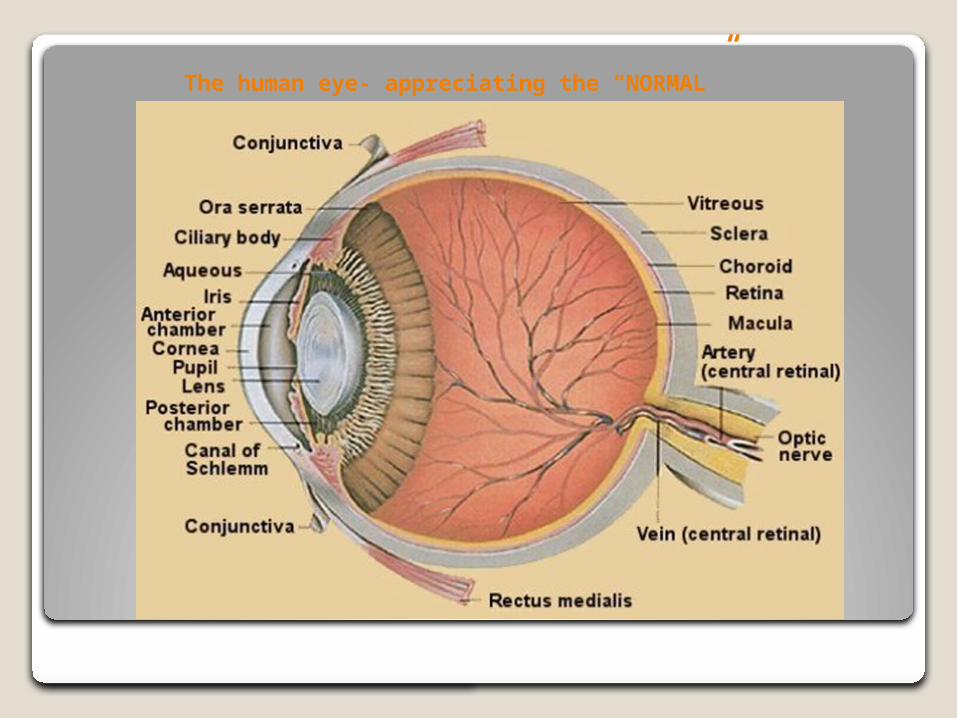
The human eye- appreciating the “NORMAL”

DEVELOPMENT OF THE RETINA During the fourth week of gestation the optic vesicle is converted into a double-
layered optic cup The conversion of the optic vesicle to the optic cup is due to the differential growth
of the walls of the vesicle. The retina develops from the two parts of the optic cup i. the retinal pigment epithelium develops from the outer layer of the optic cup, ii. the neurosensory retina develops from the inner layer of the optic cup.
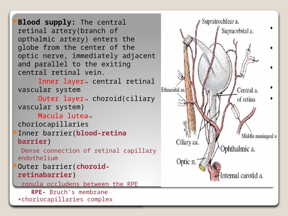
Blood supply: The central retinal artery(branch of opthalmic artery) enters the globe from the center of the optic nerve, immediately adjacent and parallel to the exiting central retinal vein.
Inner layer→ central retinal vascular system
Outer layer→ choroid(ciliary vascular system)
Macula lutea→ choriocapillaries Inner barrier(blood–retina
barrier) Dense connection of retinal capillary
endotheliumOuter barrier(choroid-
retinabarrier) zonula occludens between the RPE RPE- Bruch’s membrane
+choriocapillaries complex
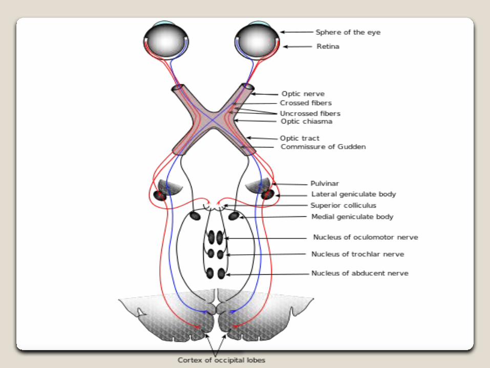
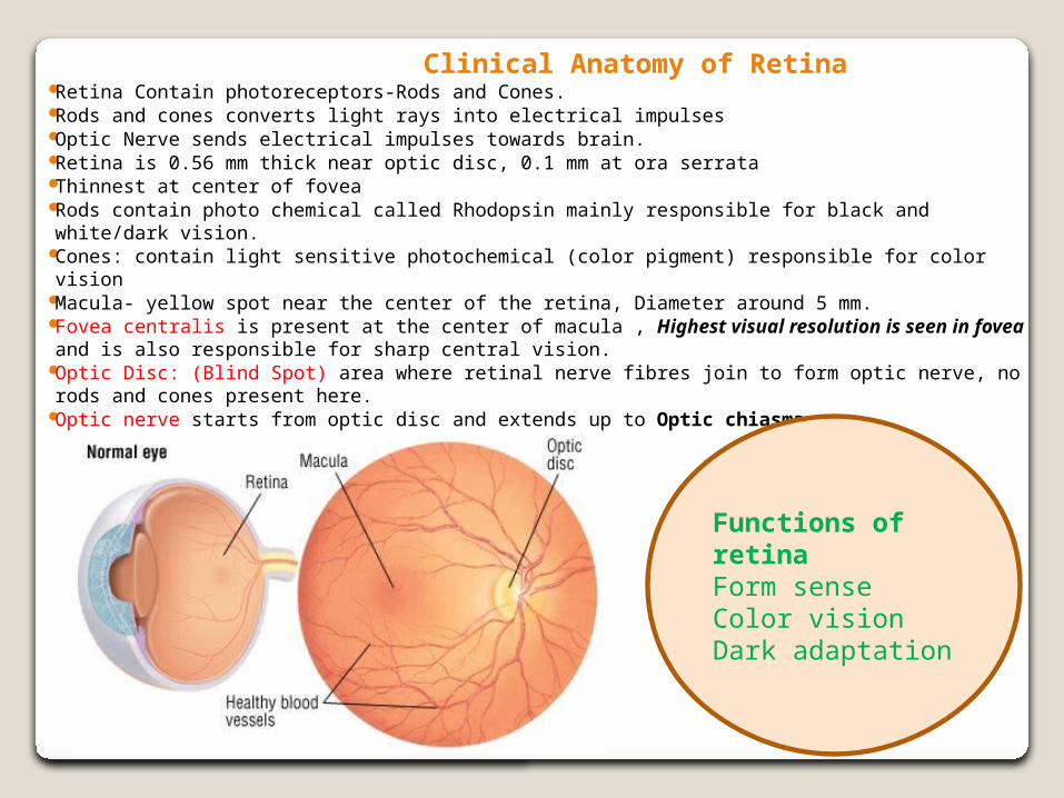
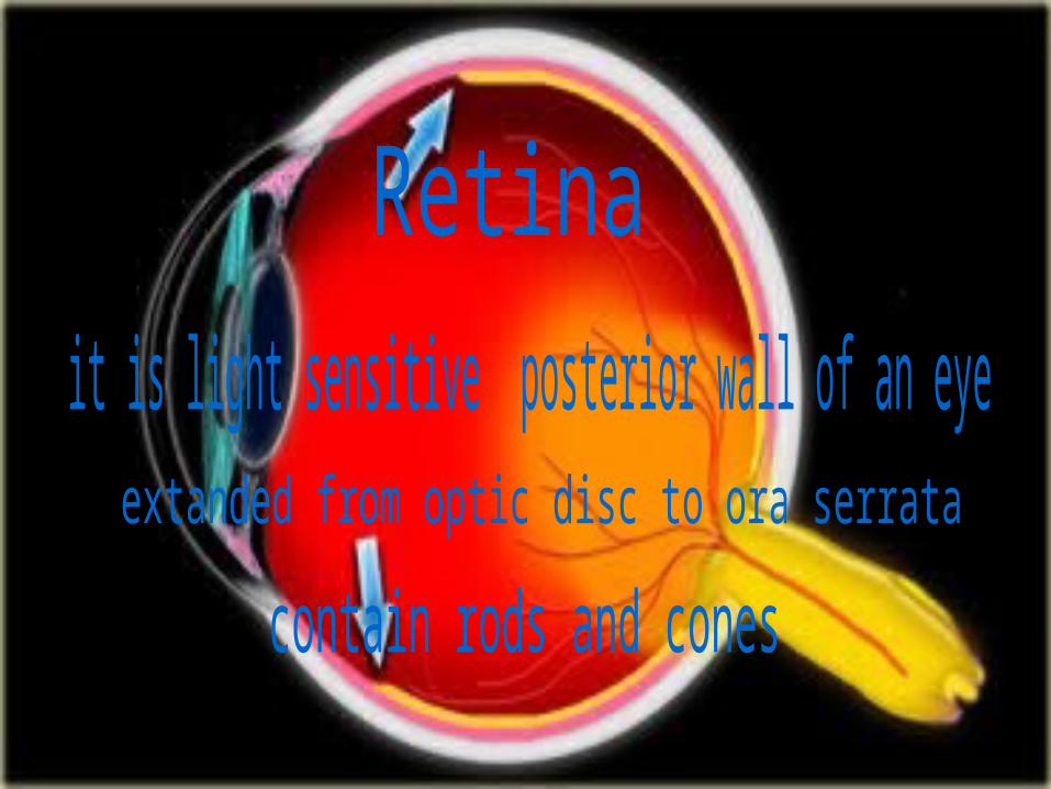
Clinical Anatomy of Retina Retina Contain photoreceptors-Rods and Cones. Rods and cones converts light rays into electrical impulses Optic Nerve sends electrical impulses towards brain. Retina is 0.56 mm thick near optic disc, 0.1 mm at ora serrata Thinnest at center of fovea Rods contain photo chemical called Rhodopsin mainly responsible for black and white/dark
vision. Cones: contain light sensitive photochemical (color pigment) responsible for color vision Macula- yellow spot near the center of the retina, Diameter around 5 mm. Fovea centralis is present at the center of macula , Highest visual resolution is seen in
fovea and is also responsible for sharp central vision. Optic Disc: (Blind Spot) area where retinal nerve fibres join to form optic nerve, no rods and
cones present here. Optic nerve starts from optic disc and extends up to Optic chiasma
Functions of retinaForm senseColor visionDark adaptation

DISCRIPTION OF FUNDUS1 Size, Shape and Borders of the Optic Disc. Optic disc size is about 1.92 millimeters vertically by 1.76 millimeters horizontally..
Shape of optic disc is a vertically oval normally a sharply defined, yellowish- orange structure and forms head of optic nerve.
Edge of optic disc is known as peripapillary area which may be hyperpigmented or scalloped pale
Temporal creasent is seen in myopia
2 Cup to Disc Ratio---1:3 or 0.3:1 central depression at optic disc known as the optic or physiologic cup
3 Relative size of the Arteries(smaller) and Veins(darker)--- 2:3 (veins are 1.5 times wider than arteries)
4 Texture of the Retina-- retina is normally completely transparent without any intrinsic color
5 Color of the Retina---Orangeish color is from vasculature of the choroid.
6 Trace the vascular structure to the equator of the retina.
7 Macula- color and size

Differentiating arteries from veins & Identifying Optic disc, Macula, Fovea Arteries are lighter in color. Shinier due to light reflex. Narrower in caliber than veins. Optic disc is at the post. pole of eye ball. Oval in shape and pale in color. Optic disc aka Blind spot–area where optic nerve leaves eyeball. Arteries and veins radiate outwards from the disc in a tree like fashion. Identify disc-to-cup ratio The pink rim of disc contains nerve fibers. The white cup is a pit with no nerve fibers. Macula is less vascular zone ,temporally located,1.5DD away from disc.
Central avascular zone is Fovea
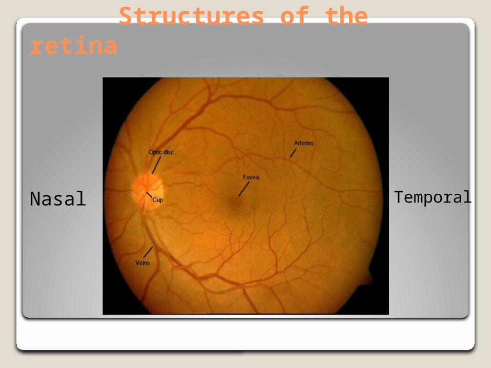
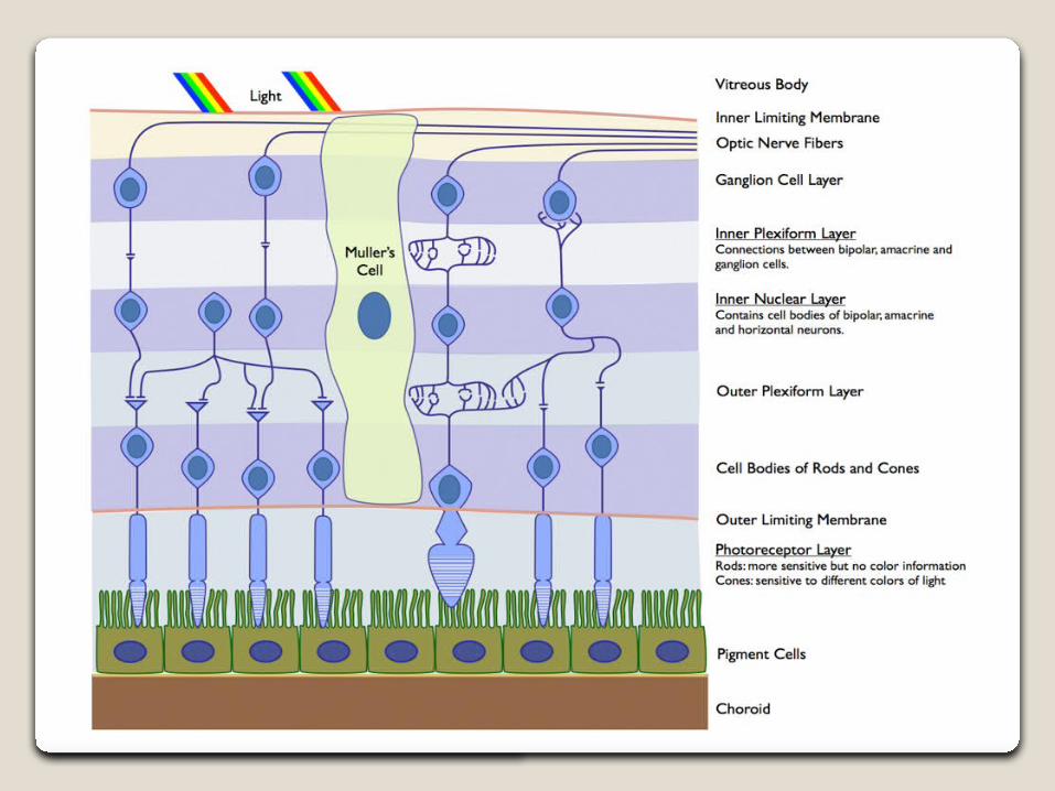
Structures of the retina
Nasal Temporal
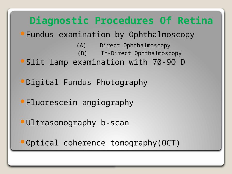
Diagnostic Procedures Of Retina Fundus examination by Ophthalmoscopy (A) Direct Ophthalmoscopy (B) In-Direct OphthalmoscopySlit lamp examination with 70-9O D
Digital Fundus Photography
Fluorescein angiography
Ultrasonography b-scan
Optical coherence tomography(OCT)
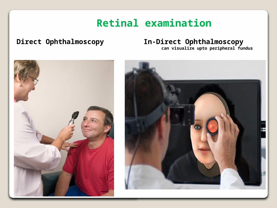
Retinal examinationIn-Direct Ophthalmoscopy can visualize upto peripheral fundus
Direct Ophthalmoscopy
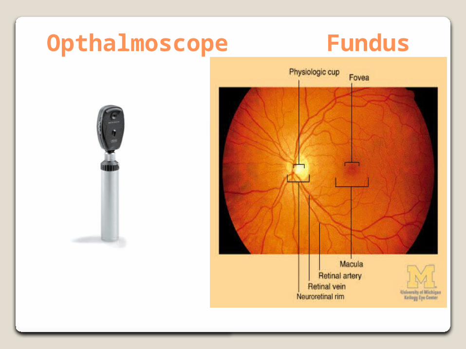
Opthalmoscope Fundus
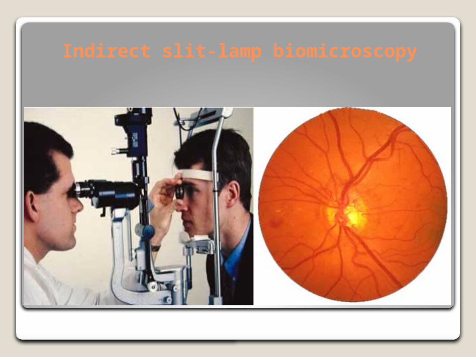
Indirect slit-lamp biomicroscopy
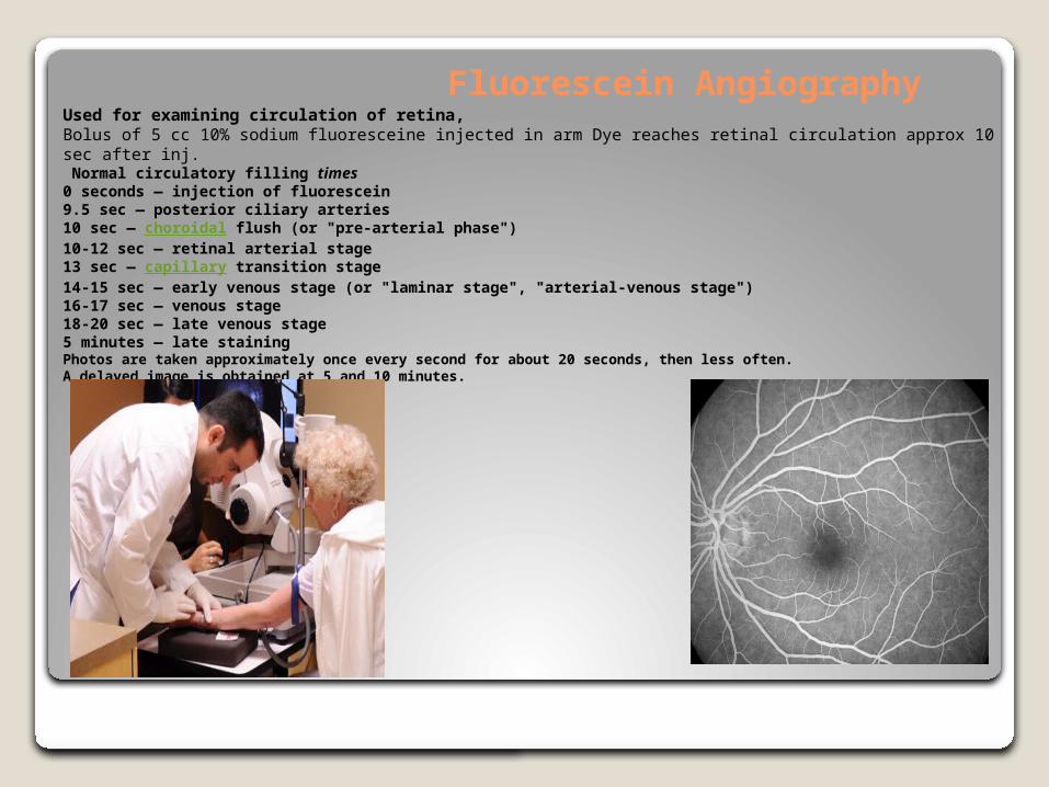
Fluorescein AngiographyUsed for examining circulation of retina,Bolus of 5 cc 10% sodium fluoresceine injected in arm Dye reaches retinal circulation approx 10 sec after inj. Normal circulatory filling times 0 seconds — injection of fluorescein9.5 sec — posterior ciliary arteries10 sec — choroidal flush (or "pre-arterial phase")10-12 sec — retinal arterial stage13 sec — capillary transition stage14-15 sec — early venous stage (or "laminar stage", "arterial-venous stage")16-17 sec — venous stage18-20 sec — late venous stage5 minutes — late stainingPhotos are taken approximately once every second for about 20 seconds, then less often. A delayed image is obtained at 5 and 10 minutes.
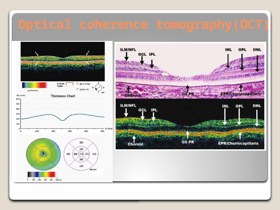
Optical coherence tomography(OCT)
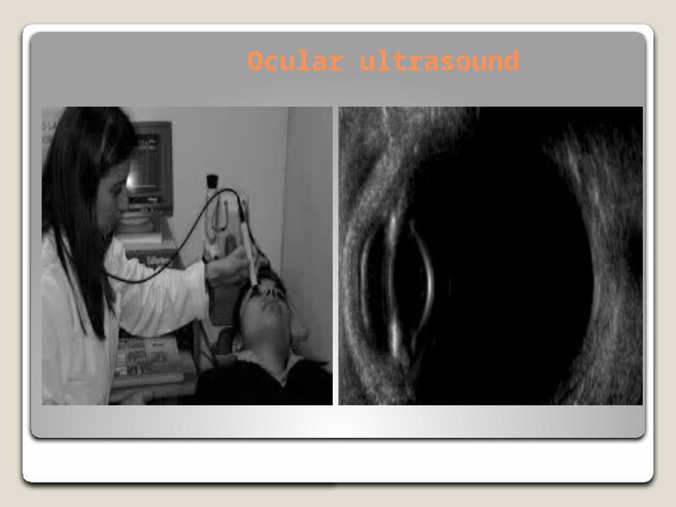
Ocular ultrasound
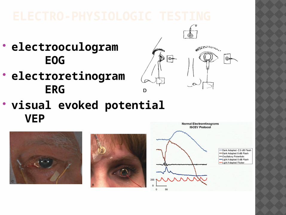
ELECTRO-PHYSIOLOGIC TESTING
electrooculogram EOG
electroretinogram ERG
visual evoked potential VEP

Retinal vascular disorders

Periphlebitis retinae Inflammation of the wall of the retinal veins commonly due to
1. tuberculosis (Mycobacterium tuberculosis).2. Sarcoidosis, 3. multiple sclerosis,4. Eales Disease (“periphlebitis retinae).
Cause hemorrhages in retina and vitreous Commonly effect adult(20-30 year) Cause sudden loss of vision due to vitreous hemorrhage Treatment Control the basic etiology Corticosteroid help to control inflammation Photocoagulation (laser) of leakage area
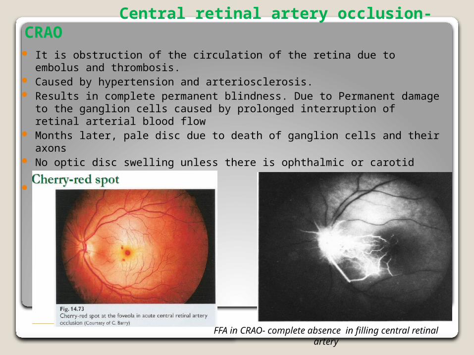
Central retinal artery occlusion-CRAO It is obstruction of the circulation of the retina due to embolus and
thrombosis. Caused by hypertension and arteriosclerosis. Results in complete permanent blindness. Due to Permanent damage to the
ganglion cells caused by prolonged interruption of retinal arterial blood flow Months later, pale disc due to death of ganglion cells and their axons No optic disc swelling unless there is ophthalmic or carotid artery occlusion True ophthalmic emergency
FFA in CRAO- complete absence in filling central retinal artery
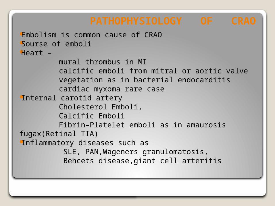
PATHOPHYSIOLOGY OF CRAO Embolism is common cause of CRAO Sourse of emboli Heart – mural thrombus in MI calcific emboli from mitral or aortic valve vegetation as in bacterial endocarditis cardiac myxoma rare case Internal carotid artery Cholesterol Emboli, Calcific Emboli Fibrin–Platelet emboli as in amaurosis fugax(Retinal TIA) Inflammatory diseases such as SLE, PAN,Wageners granulomatosis, Behcets disease,giant cell arteritis
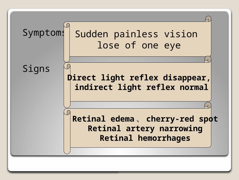
Symptoms
Signs
Sudden painless vision lose of one eye
Direct light reflex disappear, indirect light reflex normal
Retinal edema、 cherry-red spotRetinal artery narrowing
Retinal hemorrhages
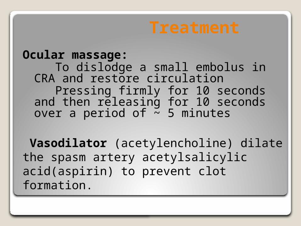
Treatment
Ocular massage: To dislodge a small embolus in CRA and restore circulation Pressing firmly for 10 seconds and then releasing for 10 seconds over a period of ~ 5 minutes
Vasodilator (acetylencholine) dilate the spasm artery acetylsalicylic acid(aspirin) to prevent clot formation.
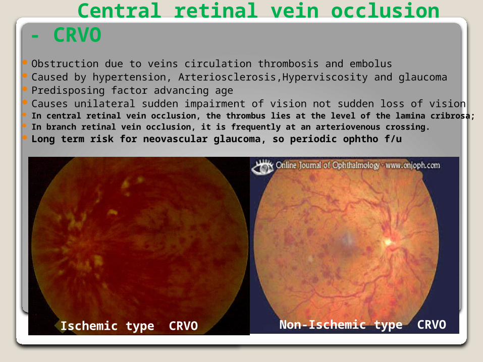
Central retinal vein occlusion - CRVO
Obstruction due to veins circulation thrombosis and embolus Caused by hypertension, Arteriosclerosis,Hyperviscosity and glaucoma Predisposing factor advancing age Causes unilateral sudden impairment of vision not sudden loss of vision In central retinal vein occlusion, the thrombus lies at the level of the lamina cribrosa; In branch retinal vein occlusion, it is frequently at an arteriovenous crossing. Long term risk for neovascular glaucoma, so periodic ophtho f/u
Ischemic type CRVO Non-Ischemic type CRVO
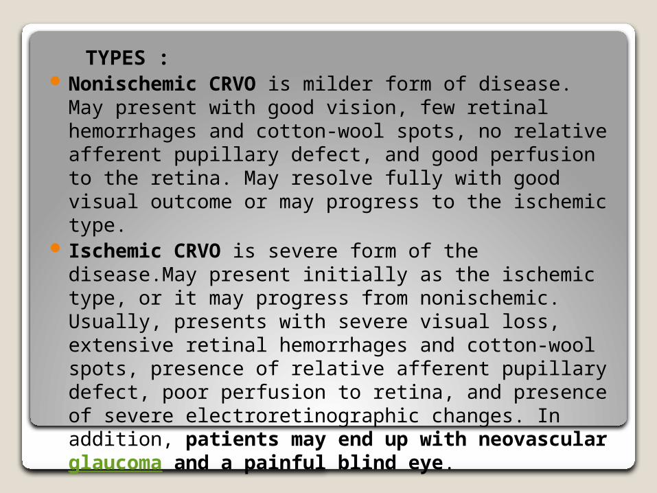
TYPES : Nonischemic CRVO is milder form of disease. May
present with good vision, few retinal hemorrhages and cotton-wool spots, no relative afferent pupillary defect, and good perfusion to the retina. May resolve fully with good visual outcome or may progress to the ischemic type.
Ischemic CRVO is severe form of the disease.May present initially as the ischemic type, or it may progress from nonischemic. Usually, presents with severe visual loss, extensive retinal hemorrhages and cotton-wool spots, presence of relative afferent pupillary defect, poor perfusion to retina, and presence of severe electroretinographic changes. In addition, patients may end up with neovascular glaucoma and a painful blind eye.

Non-Ischemic CRVO Ischemic CRVO
NON ISCHEMIC CRVO –Diffuse flame shaped retinal hemorrhage Tortuosity and engorgement of retinal veins
ISCHEMIC CRVO: diffuse capillary non perfusion—rubiosis iridis—neovascular glaucoma(NVG)
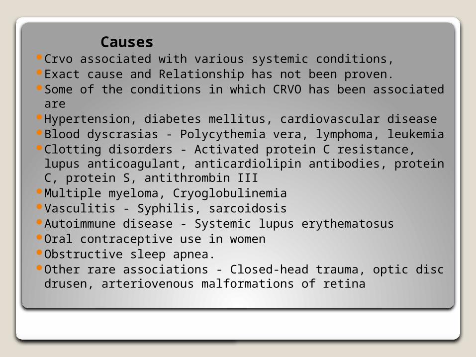
Causes Crvo associated with various systemic conditions, Exact cause and Relationship has not been proven. Some of the conditions in which CRVO has been associated
are Hypertension, diabetes mellitus, cardiovascular disease Blood dyscrasias - Polycythemia vera, lymphoma, leukemia Clotting disorders - Activated protein C resistance, lupus
anticoagulant, anticardiolipin antibodies, protein C, protein S, antithrombin III
Multiple myeloma, Cryoglobulinemia Vasculitis - Syphilis, sarcoidosis Autoimmune disease - Systemic lupus erythematosus Oral contraceptive use in women Obstructive sleep apnea.
Other rare associations - Closed-head trauma, optic disc drusen, arteriovenous malformations of retina
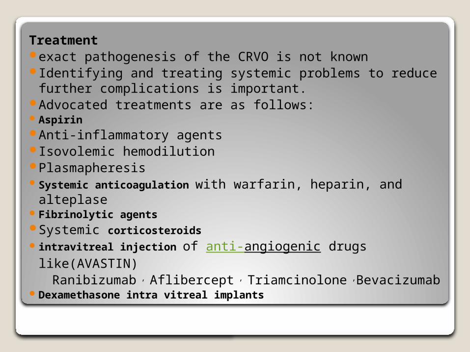
Treatment exact pathogenesis of the CRVO is not known Identifying and treating systemic problems to reduce
further complications is important. Advocated treatments are as follows: Aspirin Anti-inflammatory agents Isovolemic hemodilution Plasmapheresis Systemic anticoagulation with warfarin, heparin, and
alteplase Fibrinolytic agents Systemic corticosteroids intravitreal injection of anti-angiogenic drugs like(AVASTIN) Ranibizumab , Aflibercept , Triamcinolone ,Bevacizumab Dexamethasone intra vitreal implants
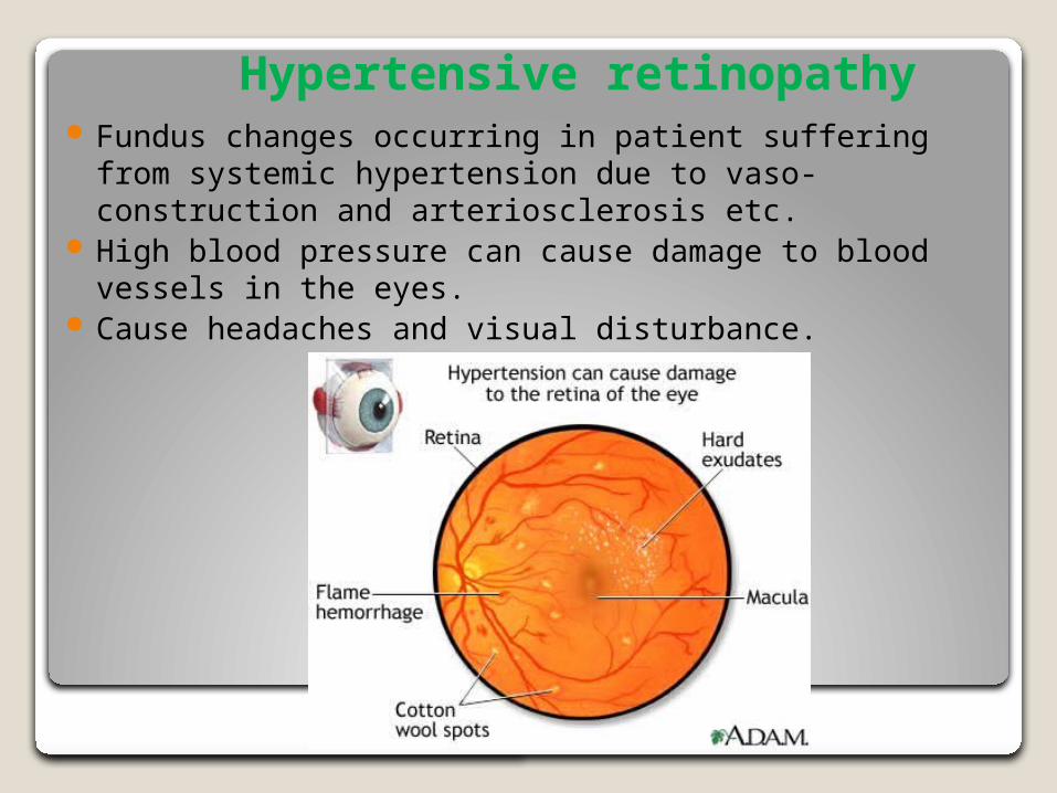
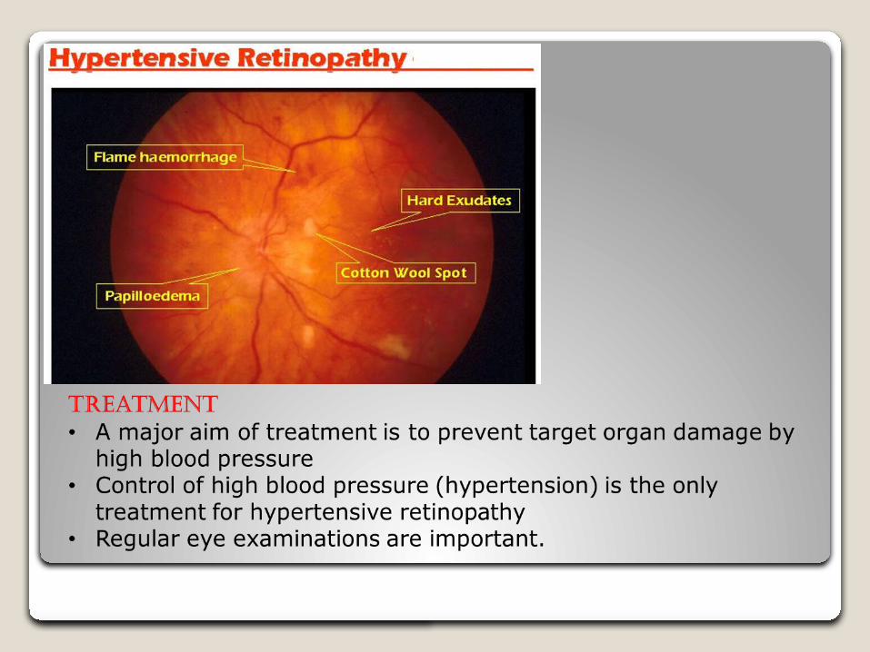
Hypertensive retinopathy Fundus changes occurring in patient suffering from
systemic hypertension due to vaso-construction and arteriosclerosis etc.
High blood pressure can cause damage to blood vessels in the eyes.
Cause headaches and visual disturbance.
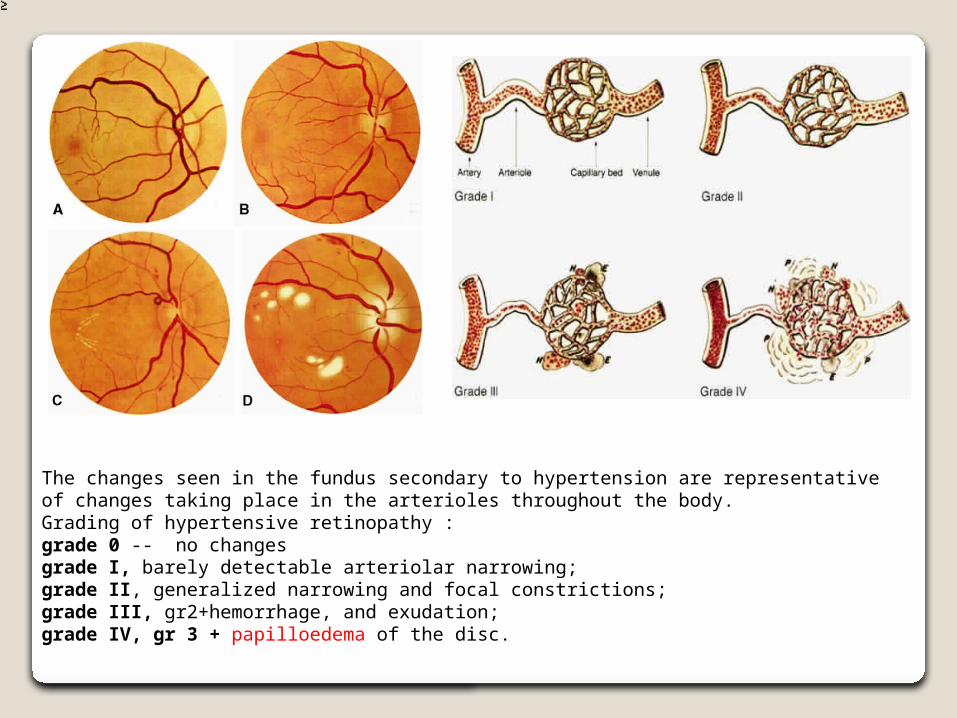
The changes seen in the fundus secondary to hypertension are representative of changes taking place in the arterioles throughout the body.Grading of hypertensive retinopathy : grade 0 -- no changesgrade I, barely detectable arteriolar narrowing; grade II, generalized narrowing and focal constrictions; grade III, gr2+hemorrhage, and exudation; grade IV, gr 3 + papilloedema of the disc.
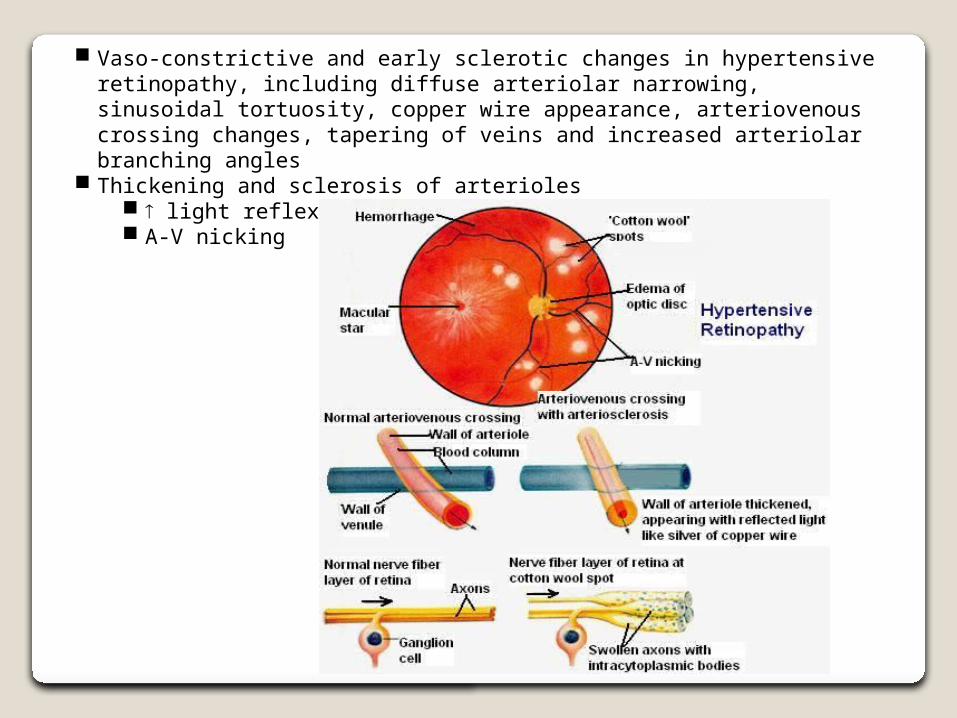
Vaso-constrictive and early sclerotic changes in hypertensive retinopathy, including diffuse arteriolar narrowing, sinusoidal tortuosity, copper wire appearance, arteriovenous crossing changes, tapering of veins and increased arteriolar branching angles
Thickening and sclerosis of arterioles light reflex width (copper silver wire) A-V nicking
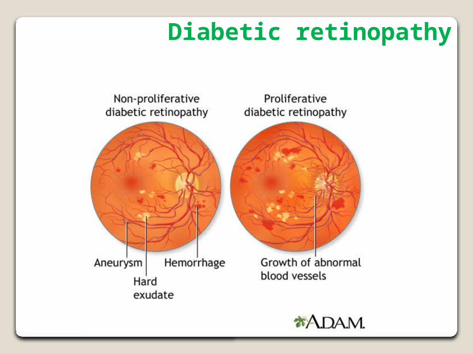
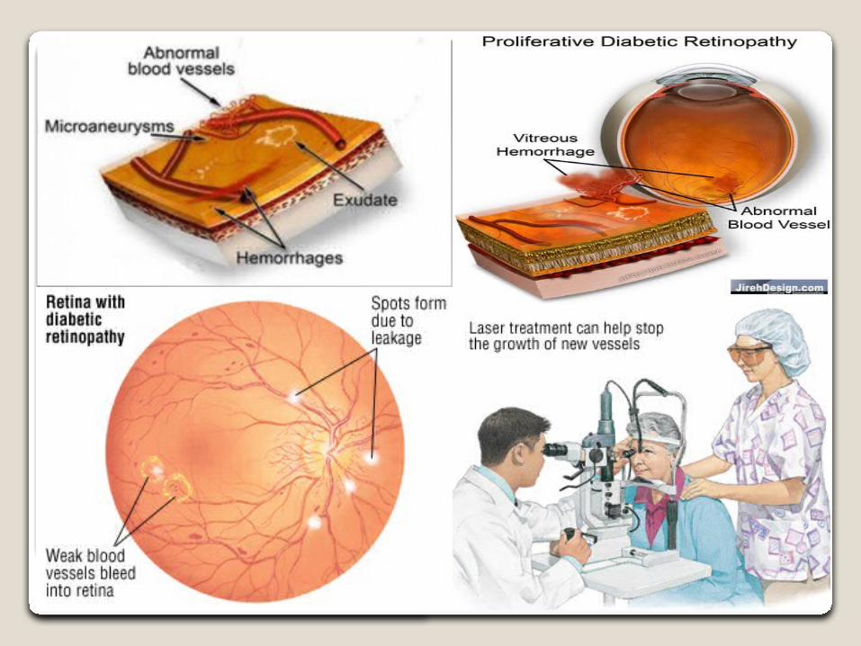
Diabetic retinopathy

PATHO-PHYSIOLOGY OF DIABETIC RETINOPATHY1 Diabetic retinopathy is a microangiopathy. Results in a thickening of the basement membrane of the vessels and loss of pericytes and vascular endothelial cells.
2 Hyperglycemia capillary closure retinal ischemia. Further course is triggered by hypoxia.
3 In the ischemic retina, angiogenic factors such as vascular endothelial growth factor (VEGF) and insulin-like growth factor-1 (IGF-1) are produced.
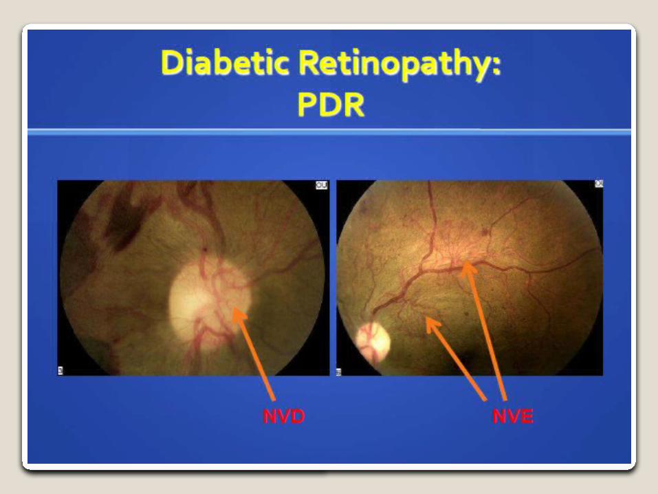
4 Ischemia + VEGF == new vessel formation
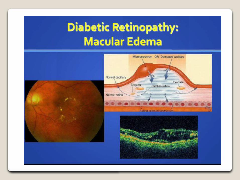
5 Breakdown of the blood–retina barrier + increased vascular permeability == macular edema.
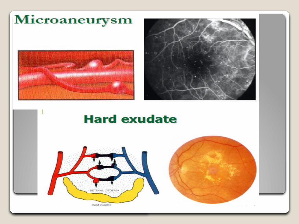
6 Hard Exudates representing lipid deposits in the retinaCotton-Wool spots and soft exudates representing nerve fiber infarction

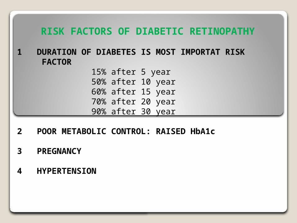
RISK FACTORS OF DIABETIC RETINOPATHY
1 DURATION OF DIABETES IS MOST IMPORTAT RISK FACTOR 15% after 5 year 50% after 10 year 60% after 15 year 70% after 20 year 90% after 30 year
2 POOR METABOLIC CONTROL: RAISED HbA1c
3 PREGNANCY
4 HYPERTENSION
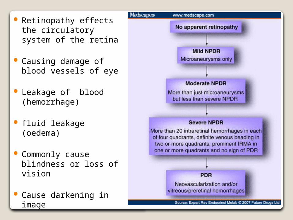
Retinopathy effects the circulatory system of the retina
Causing damage of blood vessels of eye
Leakage of blood (hemorrhage)
fluid leakage (oedema)
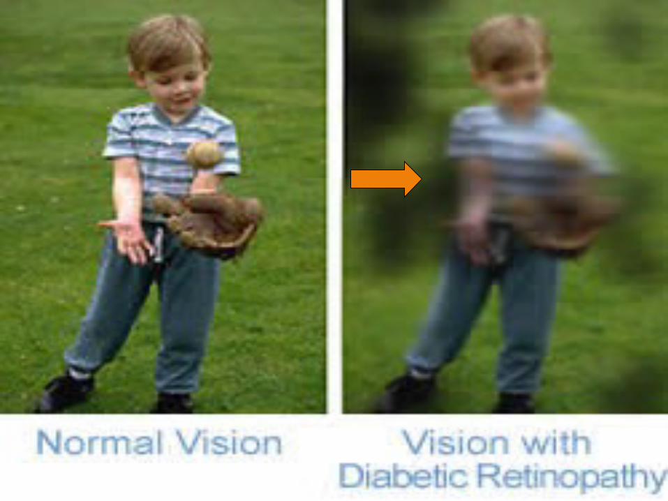
Commonly cause blindness or loss of vision
Cause darkening in image
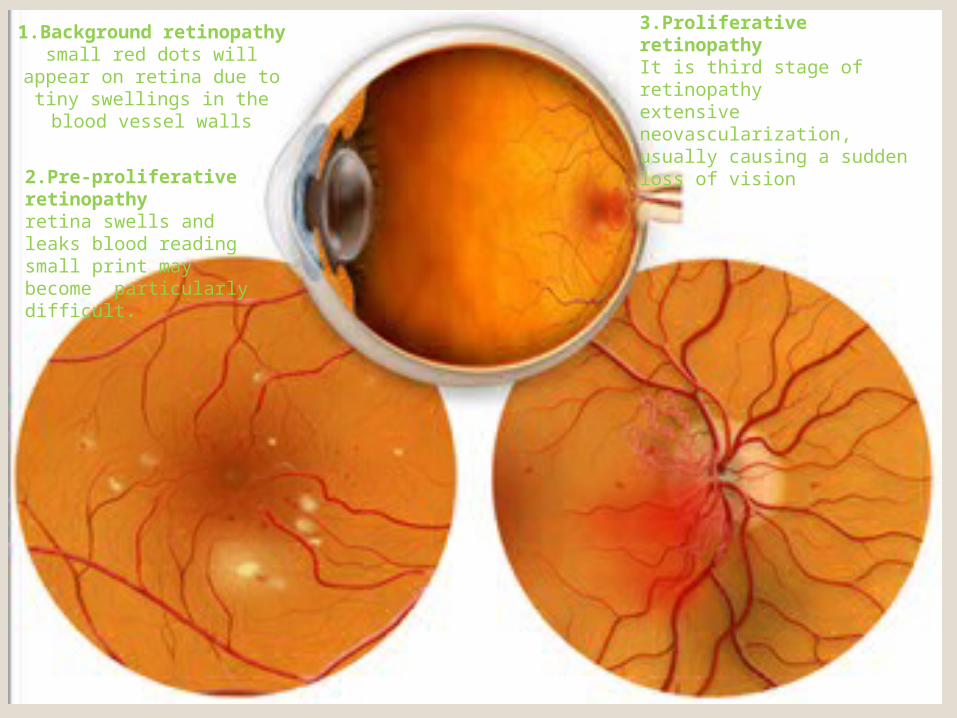
1.Background retinopathy
small red dots will appear on retina due to tiny
swellings in the blood vessel walls
3.Proliferative retinopathyIt is third stage of retinopathyextensive neovascularization, usually causing a sudden loss of vision
2.Pre-proliferative retinopathyretina swells and leaks blood reading small print may become particularly difficult.

TREATMENTBackground retinopathy Requires no treatment, but should have Regular
eye Examinations by Ophthalmologist
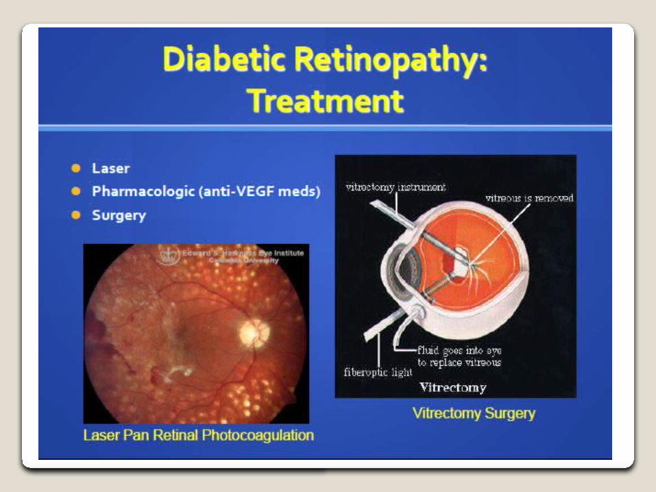
Pre-proliferative retinopathy also does not require treatment, Laser treatment can be an option if leakage begins
Proliferative retinopathy Argon Laser is used to 'burn' the abnormal blood
vessels to prevent further growth of new blood vessel
Argon Laser cannot restore any lost vision, but to prevent further growth

Diabetic Maculopathy
Involvement of fovea occur at any stage of retinopathy
Macular edema Macular hemorrhages Macular detachment
Other retinal disorders
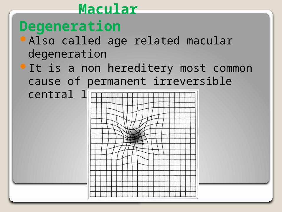
Macular Degeneration Also called age related macular
degenerationIt is a non hereditery most common cause
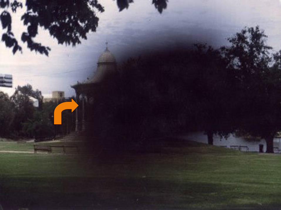
of permanent irreversible central loss of vision
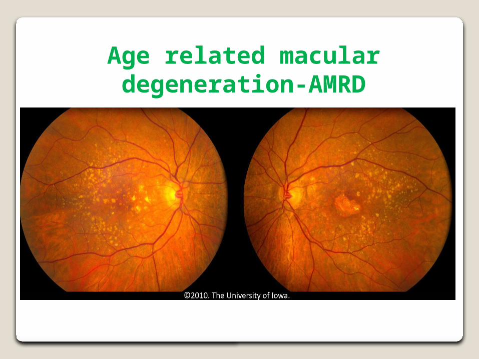
Age related macular degeneration-AMRD

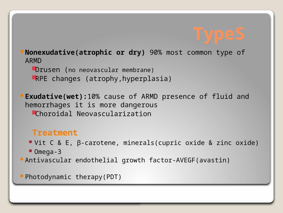
TypeS Nonexudative(atrophic or dry) 90% most common type of
ARMDDrusen (no neovascular membrane)RPE changes (atrophy,hyperplasia)
Exudative(wet):10% cause of ARMD presence of fluid and hemorrhages it is more dangerous
Choroidal Neovascularization
Treatment Vit C & E, β-carotene, minerals(cupric oxide & zinc oxide) Omega-3
Antivascular endothelial growth factor-AVEGF(avastin)
Photodynamic therapy(PDT)
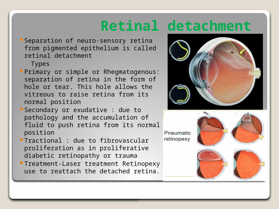
Retinal detachment Separation of neuro-sensory retina
from pigmented epithelium is called retinal detachment
Types Primary or simple or
Rhegmatogenous: separation of retina in the form of hole or tear. This hole allows the vitreous to raise retina from its normal position
Secondary or exudative : due to pathology and the accumulation of fluid to push retina from its normal position
Tractional : due to fibrovascular proliferation as in proliferative diabetic retinopathy or trauma
Treatment-Laser treatment Retinopexy use to reattach the detached retina.
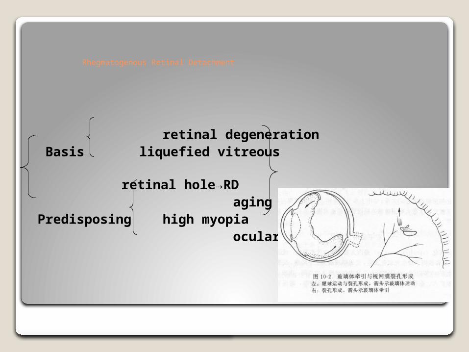
Rhegmatogenous Retinal Detachment
retinal degeneration Basis liquefied vitreous retinal hole→RD agingPredisposing high myopia ocular trauma

Resources http://
www.mayoclinic.org/diseases-conditions/retinal-diseases/basics/definition/con-20036725
http://www.sciencedirect.com/science/journal/13509462
http://www.snec.com.sg/eye-conditions-and-treatments/common-eye-conditions-and-procedures/Pages/retinal-vascular-disorders.aspx
http://www.academy.org.uk/lectures/barnard5.htm