Embed Size (px)
Citation preview

Vol. 43, No. 5APPLIED AND ENVIRONMENTAL MICROBIOLOGY, May 1982, p. 1006-10100099-2240/82/051006-05$02.00/0
Restriction Endonuclease Analysis of the Lactose Plasmid inStreptococcus lactis ML3 and Two Recombinant Lactose
PlasmidstPATRICIA M. WALSH't AND LARRY L. McKAYl*
Department of Genetics and Cell Biology' and Department ofFood Science and Nutrition,2 University ofMinnesota, St. Paul, Minnesota 55108
Received 28 October 1981/Accepted 20 January 1982
We investigated the molecular relationship between the 60-megadalton (Mdal)recombinant lactose plasmids in ML3 x LM2301 lactose-positive (Lac') trans-conjugants and the genetic material of Streptococcus lactis ML3. Lactosemetabolism is linked to the 33-Mdal plasmid pSK08 in ML3, and the recipientLM2301 is cured of plasmid DNA. The plasmids were analyzed with a series ofrestriction enzymes. We found that the 60-Mdal plasmids in Lac' transconjugantscontained pSK08 DNA, but were not simply dimers of pSK08. The 60-Mdalplasmids contained a segment of DNA not apparent in pSK08. The restrictionpatterns of the 60-Mdal plasmid in a Lac' nonclumping transconjugant and that ina Lac' clumping transconjugant were different. This suggested that there was amolecular difference between these two recombinant plasmids. We conclude thatthe segment of DNA in the 60-Mdal plasmids that was not present in pSK08 wasthe proposed transfer factor responsible for cell aggregation and high-frequencyconjugation.
The group N streptococci, which includeStreptococcus lactis, S. cremoris, and S. lactissubsp. diacetylactis, are gram-positive bacteriaused extensively in dairy fermentations. Boththe ability to ferment lactose and the ability todegrade casein are linked to plasmid DNA inmany strains of group N streptococci (1, 5, 6, 9).It is essential that the dairy industry have at itsdisposal microorganisms capable of fermentingmilk satisfactorily. Genetic approaches, such asconjugation, could potentially facilitate the de-velopment of improved strains for use as startercultures; therefore, the development of a systemof efficient conjugal plasmid transfer in lacticstreptococci would be useful.Although the conjugal transfer of plasmid
DNA in this group of bacteria generally occursat a low frequency (10-6 to 10-9 lactose-positive[Lac'] transconjugants per donor; 6, 10), high-frequency transfer of lactose metabolism associ-ated with donor cell aggregation in S. lactis 712(3) and in S. lactis ML3 (14) was recentlydescribed. In the S. lactis 712 system, the lac-tose plasmid was not initially identified in eitherthe donor or the transconjugants (3), but subse-quently Davies and Gasson (2) reported, thatsome transconjugants contained a 60-megadal-
t Scientific journal series paper no. 11820, Minnesota Agri-cultural Experiment Station, St. Paul, MN 55108.
t Present address: Eastern Regional Research Center, U.S.Department of Agriculture, Philadelphia, PA 19118.
ton (Mdal) plasmid not observed in S. lactis 712.We recently reported that a recombinant plas-mid is associated with cell aggregation and high-frequency conjugation in S. lactis ML3 (14).Although lactose metabolism is linked to a 33-Mdal plasmid, pSK08, in S. lactis ML3 (8),Lac' transconjugants resulting from matingsbetween ML3 and a Lac- plasmid-cured deriva-tive of S. lactis C2, designated LM2301, possessa single plasmid of approximately 60 Mdal (10,14). The majority of these transconjugants formaggregates in broth and transfer the ability toferment lactose at a frequency greater than 10-1per donor. Transconjugants which do not clumpmate at a much lower frequency. Our resultssuggested that the genes responsible for cellaggregation and high-frequency conjugationwere on the segment ofDNA which recombinedwith pSK08 (14).
In this report, we used restriction enzymes toanalyze the Lac plasmid pSK08 in S. lactis ML3and the recombinant plasmid in PW1, a Lac'nonclumping transconjugant, and in PW2, aLac' clumping transconjugant. We present evi-dence that the 60-Mdal plasmids are not simplydimers of pSK08 and that there is a moleculardifference between the plasmid in PW1 and thatin PW2.
MATERIALS AND METHODSMicrobial strains. The microorganisms used in this
study are maintained in our active stock culture collec-
1006
on April 25, 2020 by guest
http://aem.asm
.org/D
ownloaded from
LACTOSE PLASMID IN S. LACTIS ML3 1007
TABLE 1. Strains of S. lactis used in this study
Strain Plasmid Relevantdesignation compositiona phenotype Comment
ML3 33, 5.5, 2, 1 Lac' Parent culture (8)LM0230 Lac- Nitrosoguanidine and UV-induced mutant of S. lactis C2 cured of
plasmid DNA (7, 9)LM2301 Lac- Spontaneous streptomycin-resistant mutant of LM0230LM2302 Lac- Spontaneous erythromycin-resistant mutant of LM2301PW1 60 Lac' Nonclumping transconjugant of ML3 x LM2301 (14)PW2 60 Lac' Clumping transconjugant of ML3 x LM2301 (14)PN221 33 Lac' Spontaneous nonclumping derivative of PW2 (14)PC6 33 Lac' Nonclumping transconjugant of PN221 x LM2302 (this paper)a Plasmid molecular weight x 106.
tion through biweekly transfer at 21°C in M17 broth(13) containing 0.5% glucose or lactose. Table 1 de-scribes strains of S. lactis used.Large-volume lysis procedures. Strains to be lysed
were incubated at 32°C for 4 h in 1,600 ml of lysis broth(7). For the standard lysis procedure (7), cells werewashed with TES buffer (50 mM NaCI, 5 mM disodiumEDTA, and 30 mM Tris, pH 8.0) and divided into two250-ml centrifuge bottles. Cleared lysates were pre-pared by using a 20x volume of reagents per bottle.Because of the difficulty in obtaining plasmid DNAfrom PW1 and PW2, the Hansen-Olsen procedure forthe isolation of large plasmids (4) was adapted for lysisof S. lactis strains as previously described (14). Forthe large-volume lysis, the cells were washed withTES buffer and distributed among 8 to 12 40-mIpolypropylene centrifuge tubes. Twice the usual vol-ume of reagents per tube was used. The ethanol-precipitated DNA pellet from the cleared lysates or theDNA-polyethylene glycol pellet was suspended in 4 mlof TES buffer. Plasmid DNA was further purified in aCsCI-ethidium bromide density gradient or directly ona horizontal agarose gel.Dye-buoyant density gradient centrifugation. CsCl-
ethidium bromide density gradient centrifugation wasperformed similarly to the procedure of Klaenhammeret al. (7). Gradients were prepared with 7 g of CsCl(Kawecki Berylco Industries, Inc.), 2 ml of TESbuffer, 4 ml of ethanol-precipitated or polyethyleneglycol-concentrated DNA in TES buffer, and 1 ml ofethidium bromide (4 mg/ml in TES buffer). The gradi-ents were adjusted to a refractive index of 1.389 to 1.39with TES buffer and centrifuged in cellulose nitratetubes for 48 h at 43,000 rpm at 15°C in a Beckman type50 Ti rotor. After centrifugation, the plasmid band waswithdrawn from the tube, and ethidium bromide wasremoved from the sample as previously described (7).CsCl was removed by extensive dialysis against TESbuffer or TE buffer (10 mM Tris-1 mM disodiumEDTA, pH 7.8). Plasmid samples were concentratedagainst polyethylene glycol 6000 or by extraction withsec-butanol followed by ethanol precipitation.
Isolation of plasmid DNA from agarose gels. A 0.55 or0.63% agarose (SeaKem; Marine Colloids) gel in Tris-borate buffer (89 mM Tris, 2.5 mM disodium EDTA,and 89 mM boric acid, pH 8.0) containing 0.5 ,ug ofethidium bromide per ml was used for isolation of theplasmids in ML3. A 19-cm horizontal gel was used forisolation of pSK08, and a 40-cm gel was used to isolateall four plasmids in ML3. Approximately 400 ,ul of
DNA preparation in TES buffer plus 50 ,ul of trackingdye/stop solution (50% sucrose, 0.1 M disodiumEDTA, and 0.05% bromophenol blue) was loaded intoeach well (32 by 3 mm). DNA was electrophoresedinto the gel at 400 V for 10 min, and electrophoresiswas continued at 100 V (19-cm gel) or 150 V (40-cmgel), constant voltage, for approximately 16 h. Themigration of the DNA was monitored by using alongwave UV light (Blak Ray, UVL 56; UltravioletLight Products, Inc.).The hydroxylapatite (HAP) procedure of Tabak and
Flavell (12) was used to isolate plasmid DNA from thehorizontal gels. Hydroxylapatite (Bio-Rad Labora-tories; DNA-grade Bio-Gel HTP) was prepared inTris-borate buffer. Sephadex G-50 (fine; PharmaciaFine Chemicals, Inc.) columns were prepared in plas-tic 3-cm3 disposable syringes. Upon elution of theDNA from the HAP with 1 M sodium phosphatebuffer, pH 6.8, the DNA-containing fractions werepooled and concentrated with sec-butanol, which alsoremoved ethidium bromide. The samples were thenextensively dialyzed against TE buffer. After dialysis,samples were concentrated with sec-butanol to a finalvolume of 400 IL1 and ethanol precipitated (1/10 volumeof 2 M NaCl and 2 volumes of cold ethanol, storedovernight at -40°C). To pellet the DNA, the sampleswere spun for 20 min in an Eppendorf centrifuge(Brinkmann Instruments Inc.) at 4°C. The DNA pelletswere suspended in 10 mM Tris-0.1 mM disodiumEDTA, pH 8.0, and stored at -10°C until needed forrestriction experiments.
Restriction enzyme analysis. Restriction enzymeswere obtained from either Bethesda Research Labora-tories, Inc., Biotec, Inc., Miles Laboratories, Inc., orNew England Biolabs.
Restriction digestions were carried out in sterilemicrocentrifuge tubes in a total volume of 50 p.l. DNAin 10 mM Tris-0.1 mM disodium EDTA, pH 8.0, wascombined with 5 IL1 of a 1Ox stock solution of theappropriate assay buffer prepared according to suppli-er's directions, 1 ,ul of bovine serum albumin (nucle-ase-free; Bethesda Research Laboratories; 50 jig/p.l),enzyme, and enough distilled water to bring the finalvolume to 50 1I. The amount of enzyme varied de-pending on what was needed to obtain completedigestion. Generally, 20 to 60 U of enzyme per diges-tion was used to restrict plasmids isolated from anagarose gel. Less than 10 U per assay was used forplasmids isolated directly from CsCI-ethidium bromidegradients. The digestion mix was incubated at 37°C in
VOL. 43, 1982
on April 25, 2020 by guest
http://aem.asm
.org/D
ownloaded from

1008 WALSH AND McKAY
a Temp-Blok module heater (Lab-Line Instruments,Inc.) for approximately 3 h. The reaction was stoppedby the addition of 12.5 ,ul of tracking dye/stop solution.Lambda DNA (New England Biolabs) was digestedwith HindlIl alone or with both EcoRI and HindlIl.The restriction enzyme-generated fragments were
separated in a 19-cm horizontal gel containing either0.8 or 1.2% agarose in Tris-borate buffer. Sampleswere loaded into wells (11 by 3 mm) and subjected toelectrophoresis. The DNA was first electrophoresedinto the gel at 200 V for 10 min. Electrophoresis wasthen continued at 120 V for 1 to 2 h, and the voltagewas then lowered to 30 to 35 V for approximately 12 to16 h. The gel was stained after electrophoresis in asolution of 0.5 p.g of ethidium bromide per ml anddestained in distilled water. The gels were photo-graphed under UV light. Restriction enzyme-generat-ed fragments of lambda DNA were used to estimatethe molecular weight of the plasmid fragments.
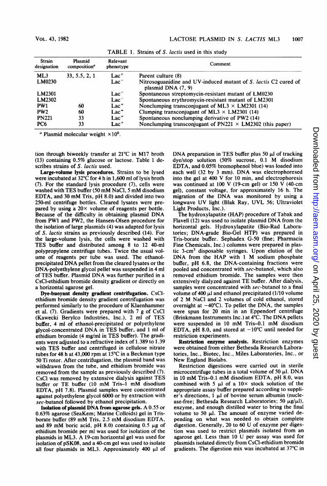
Restriction analysis of pSK08 and pPC6. The majorobjective of this study was to compare the 33-Mdallactose plasmid, pSK08, in S. lactis ML3 with pPW1and pPW2. The instability of pSK08 and the presenceof three other plasmids in ML3 prevented the purifica-tion of pSK08 by CsCl-ethidium bromide density gra-dient centrifugation. Therefore, pSK08 was isolateddirectly from an agarose gel after lysis of ML3 by thelarge-volume, modified Hansen-Olsen procedure. Thismethod, however, resulted in the presence of contami-nating chromosomal DNA and traces of the lower-molecular-weight ML3 plasmids in the pSK08 prepara-tion. The presence of the smaller ML3 plasmidsconfused the restriction patterns of pSK08. To allevi-ate this problem, a strain which only contained a 33-Mdal lactose plasmid was constructed. PC6 is a Lac'erythromycin-resistant transconjugant of PN221 xLM2302 which appears to contain a single plasmid of33 Mdal, designated pPC6 (P. M. Walsh, Ph.D. thesis,University of Minnesota, 1981). Plasmid DNA wasobtained from PC6 by the large-volume, modifiedHansen-Olsen lysis procedure followed by CsCl-ethi-dium bromide density gradient centrifugation. TheHhaI-HindIII-, HpaIl-, EcoRI-, and XbaI-generatedrestriction patterns of pSK08 were compared to thoseof pPC6. The HAP preparation of pSK08 in theabsence of restriction enzyme and the restriction di-gestion patterns of other plasmids isolated from ML3were included as controls. The plasmid pPC6 wasgenerally digested to completion with less than 10 U ofenzyme. The plasmids pSK08 and pPC6 had verysimilar digestion patterns for all enzymes tested. All ofthe very low-molecular-weight restriction fragmentsmatched, as shown in the HhaI (Fig. 1, wells A and B;wells K and J), Hind-III (Fig. 1, wells I and H; wells Rand S), and HpaII (Fig. 2, wells E and D; wells G andF) restriction patterns. The bands present in pSK08digests that were absent in pPC6 patterns could gener-ally be accounted for by the presence of other plas-mids of ML3 in the HAP preparation of pSK08 (Fig. 1,wells 0 and P; Fig. 2, well I). It appeared that the HAPpreparation of pSK08 contained the 2- and 5.5-Mdalplasmids and traces of the 1-Mdal plasmid. Digestionof the 2-Mdal plasmid with HhaI (Fig. 1, well L) anddigestion of the 5.5-Mdal plasmid with HhaI (Fig. 1,well M), HindIll (Fig. 1, well Q), and HpaII (Fig. 2,well H) could partially explain the presence of extrabands in pSK08 digests. Very faint bands that did not
J K LMNO P QR S T
>~5.5.5rC -2
FIG. 1. Agarose gel electrophoresis of HhaI-gener-ated restriction fragments of pSK08 (A, K), pPC6 (B,J), pPW1 (C), and pPW2 (D), the 2-Mdal plasmid fromML3 (L), and the 5.5-Mdal plasmid from ML3 (M);and HindIll-generated restriction fragments of pSK08(I, R), pPC6 (H, S), pPW1 (G), and pPW2 (F) and the5.5-Mdal plasmid of ML3 (Q). The 2-Mdal plasmid inthe absence of enzyme (N), the 5.5-Mdal plasmid inthe absence of enzyme (T), and the HAP preparationof pSK08 in the absence of enzyme (0, P) were used ascontrols. Lambda DNA was digested with EcoRI andHindlIl (E). The DNA fragments were separated in a0.8% agarose gel. oc, Open circular form of theplasmid.
match were probably due to the incomplete digestionof pSK08.Comparison of restriction patterns of the 33-Mdal
and the 60-Mdal plasmids. Plasmids pSK08 and pPC6were isolated as described above. Plasmids pPW1 andpPW2 were isolated by the large-volume, modifiedHansen-Olsen lysis procedure followed by CsCl-ethi-dium bromide density gradient centrifugation. Theplasmids pSK08, pPC6, pPW1, and pPW2 were digest-ed with HhaI (Fig. 1, wells A, B, C, and D, respective-ly), HindIll (Fig. 1, wells I, H, G, and F, respectively),HpaII (Fig. 2, wells E, D, C, and B, respectively),EcoRI (gel not shown), and Xbal (gel not shown). To
ABCDE FGH I
APPL. ENVIRON. MICROBIOL.
on April 25, 2020 by guest
http://aem.asm
.org/D
ownloaded from

LACTOSE PLASMID IN S. LACTIS ML3 1009
FIG. 2. Agarose gel electrophoresis of HpaII-gen-erated restriction fragments of pSK08 (E, G), pPC6(D, F), pPW1 (C), and pPW2 (B) and the 5.5-Mdalplasmid of ML3 (H). Lambda DNA was digested withEcoRI and Hindlll (A). The HAP preparation ofpSK08 in the absence of enzyme (I) was used as a
control. The DNA fragments were separated in a 0.8%agarose gel.
determine the relationship between the 33-Mdal andthe 60-Mdal lactose plasmids, the restriction patternsof pPC6, pPW1, and pPW2 were compared (Table 2).It is recognized that all of the restriction fragmentsmay not be resolved in the gel system used and thatsome of the bands may be doublets or may containmore than one fragment of the same molecular weight.However, similarities and differences among the plas-mids were apparent. The plasmids pPW1 and pPW2either contained all fragments detectable in pPC6 or
were missing only one fragment. Plasmids pPW1 andpPW2 also possessed fragments that were not detect-able in pPC6 digests. However, there were similaritiesand differences between pPW1 and pPW2 restrictiondigests. The molecular weights of the restriction en-
zyme-generated fragments of pPC6, and those notdetectable in pPC6 digests, but present in either pPW1or pPW2 or in both pPW1 and pPW2, were estimated
(Table 3). Except for the HhaI-generated fragments ofpPC6, the sum of the estimated molecular weights ofthe restriction fragments of pPC6 was approximatelyequal to the previously determined molecular weightof pSK08 (7, 8). The sum of the estimated molecularsizes of the HpaII, EcoRI, and XbaI restriction frag-ments not detectable in pPC6, but present in therecombinant plasmids pPW1 and pPW2, was approxi-mately 30 Mdal.
DISCUSSION
The molecular relationship between the 60-Mdal recombinant plasmids in transconjugantsPW1 and PW2 and the genetic material of S.lactis ML3 was investigated. From the compari-son of the restriction patterns of pPC6 andpSK08, it is concluded that they are identicalplasmids. For all enzymes tested, they producedsimilar digestion patterns. Differences could beaccounted for by the presence of the 2- and 5.5-Mdal plasmids in the HAP preparation of pSK08and by the incomplete digestion of pSK08. Al-though excess enzyme was used, complete di-gestions of pSK08 were not obtained, possiblydue to the presence of impurities resulting fromthe necessity of isolating this plasmid from an
agarose gel.The restriction analysis of pPC6 and pSK08
demonstrated the legitimacy of comparing pPC6to pPW1 and pPW2. Such a comparison re-
vealed that although the 60-Mdal plasmids con-
tained pPC6 DNA, they were not simply dimersof this plasmid. Both pPW1 and pPW2 digestscontained restriction fragments not detectable inpPC6. We suggest that the 60-Mdal plasmidscontain a segment of DNA, the proposed trans-fer factor (14), that is not present in pPC6. Insome of the restriction digests, a fragment pres-ent in pPC6 was not detectable in the restrictionpatterns of the 60-Mdal plasmids. This pPC6fragment could be the site of insertion for thetransfer factor. The missing pPC6 fragment was
not the same for both pPW1 and pPW2. Thissuggests that the formation of these two recom-
binant plasmids involved insertion of the DNAsegment at different sites on the lactose plasmid.Plasmids pPW1 and pPW2 contained restrictionfragments not detectable in pPC6 digests that
TABLE 2. Comparison of the restriction patterns of pPC6, pPW1, and pPW2Detectable fragment
Enzyme In pPC6 In pPW1 In pPW2 In pPW1 andNot in Not in Not in Not in pPW2 but
Total pPW1 pPW2 Total pPC6 Total pPC6 not in pPC6
HhaI 15 1 0 21 7 22 7 4HindIII 18 0 1 26 8 24 7 7HpaIl 17 1 1 21 5 22 6 4EcoRI 11 0 1 14 3 14 4 2XbaI 7 0 1 11 4 10 4 2
VOL. 43, 1982
on April 25, 2020 by guest
http://aem.asm
.org/D
ownloaded from
1010 WALSH AND McKAY
TABLE 3. Total molecular weight of detectablefragments in restriction digests of pPC6, pPW1, and
pPW2Mol wt (x 106)a of fragments detectable
Enzyme Not in pPC6 but in:In pPC6
pPW1 pPW2 Both
HhaI 18.7 14.6 14.8 3.8HindlII 34.5 15.3 12.9 12.9HpaII 31.3 33.6 34.0 25.9EcoRI 28.1 30.5 29.9 19.1XbaI 32 30.3 31.1 13.4
a Estimated from the migration rate relative toEcoRI and HindlII restriction digest fragments oflambda DNA.
were of the same molecular weight, but they alsocontained fragments that were different. Differ-ent insertion sites would account for two differ-ent fragments in each of the restriction patterns;however, the HhaI analysis (Fig. 1, wells C andD) indicated that pPW1 and pPW2 each con-tained three different fragments not found inpPC6. This suggests that there may be otherdifferences between pPW1 and pPW2 besidesthe site of insertion of the transfer factor. Nucle-ic acid hybridization studies could resolve thisquestion.The origin of the proposed transfer factor is
unknown. Recently, a DNA band estimated tobe 27 Mdal was detected in plasmid profiles ofML3 and Lac- derivatives of ML3 (11). It is notalways recovered in plasmid preparations, and itis being investigated to determine whether itrepresents covalently closed circular DNA. It isunknown whether this DNA is related to the 60-Mdal plasmids.As more is learned about the proposed trans-
fer factor of S. lactis ML3, the development ofan efficient system of plasmid transfer in thegroup N streptococci becomes a real possibility.Use of the Lac- derivative ofML3 as a recipientin conjugal matings with a S. cremoris C3 trans-ductant (11) and with S. diacetylactis (P. M.Walsh and L. L. McKay, unpublished data)resulted in Lac' clumping transconjugantswhich contained a recombinant plasmid. Ittherefore appears that the proposed ML3 trans-fer factor can translocate onto other lactoseplasmids. Exploitation of this transfer factor
could facilitate the genetic analysis of thesebacteria and aid in the construction of improvedstrains for use in dairy fermentations.
ACKNOWLEDGMENTS
This research was supported in part by the BiotechnologyGroup, Miles Laboratories, Inc., Elkhart, Ind. P.M.W. wassupported by a University of Minnesota Graduate Schooldoctoral dissertation fellowship.
LITERATURE CITED
1. Anderson, D. A., and L. L. McKay. 1977. Plasmids, lossof lactose metabolism and appearance of partial and fulllactose-fermenting revertants in Streptococcus cremorisBi. J. Bacteriol. 129:367-377.
2. Davies, F. L., and M. J. Gasson. 1981. Reviews of theprogress of dairy science: genetics of lactic acid bacteria.J. Dairy Res. 48:363-376.
3. Gasson, M. J., and F. L. Davies. 1980. High-frequencyconjugation associated with Streptococcus lactis donorcell aggregation. J. Bacteriol. 143:1260-1264.
4. Hansen, J. B., and R. H. Olsen. 1978. Isolation of largebacterial plasmids and characterization of the P2 incom-patability group plasmids pMG1 and pMG5. J. Bacteriol.135:227-238.
5. Kempler, G. M., and L. L. McKay. 1979. Characteriza-tion of plasmid deoxyribonucleic acid in Streptococcuslactis subsp. diacetylactis: evidence for plasmid-linkedcitrate utilization. Appl. Environ. Microbiol. 37:316-323.
6. Kempler, G. M., and L. L. McKay. 1979. Genetic evi-dence for plasmid-linked lactose metabolism in Strepto-coccus lactis subsp. diacetylactis. AppI. Environ. Micro-biol. 37:1041-1043.
7. Klaenhammer, T. R., L. L. McKay, and K. A. Baldwin.1978. Improved lysis of group N streptococci for isolationand rapid characterization of plasmid deoxyribonucleicacid. Appl. Environ. Microbiol. 35:592-600.
8. Kuhl, S. A., L. D. Larsen, and L. L. McKay. 1979. Plas-mid profiles of lactose-negative and proteinase-deficientmutants of Streptococcus lactis C10, ML3, and M18.Appl. Environ. Microbiol. 37:1193-1195.
9. McKay, L. L., K. A. Baldwin, and J. D. Efstathiou. 1976.Transductional evidence for plasmid linkage of lactosemetabolism in Streptococcus lactis C2. Appi. Environ.Microbiol. 32:45-52.
10. McKay, L. L., K. A. Baldwin, and P. M. Walsh. 1980.Conjugal transfer of genetic information in group N strep-tococci. Appl. Environ. Microbiol. 40:84-91.
11. Snook, R. J., L. L. McKay, and G. G. Ahlstrand. 1981.Transduction of lactose metabolism by Streptococcuscremoris C3 temperate phage. Appi. Environ. Microbiol.42:897-903.
12. Tabak, H. F., and R. A. Flavell. 1978. A method for therecovery of DNA from agarose gels. Nucleic Acids Res.5:2321-2332.
13. Terzaghi, B. E., and W. E. Sandine. 1975. Improvedmedium for lactic streptococci and their bacteriophages.Appl. Microbiol. 29:807-813.
14. Walsh, P. M., and L. L. McKay. 1981. Recombinantplasmid associated with cell aggregation and high-fre-quency conjugation in Streptococcus lactis ML3. J. Bac-teriol. 146:937-944.
APPL. ENVIRON. MICROBIOL.
on April 25, 2020 by guest
http://aem.asm
.org/D
ownloaded from