Embed Size (px)
Citation preview

R
RuG
C(a
b
e
ARRAA
MLSMRCCBLALJPJJCD
2
Respir. Med and Res 78 (2020) 100768
Available online at
ScienceDirectwww.sciencedirect.com
ecommendations
espiratory support in patients with COVID-19 (outside intensive carenit). A position paper of the Respiratory Support and Chronic Careroup of the French Society of Respiratory Diseases
. Rabeca,∗, J. Gonzalez-Bermejob on behalf the Respiratory Support, Chronic Care GroupGAVO2) of the French Society of Respiratory Diseases (SPLF)�
Service de Pneumologie et Soins Intensifs Respiratoires, Centre Hospitalier Universitaire de Dijon, 14, rue Gaffarel, 21000 Dijon, FranceService de Pneumologie, Médecine Intensive et Réanimation (Département R3S, Sorbonne Université, INSERM UMRS 1158 Neurophysiologie respiratoire,xpérimentale et clinique, Groupe Hospitalier Universitaire AP–HP Sorbonne Université, AP–HP site Pitié-Salpêtrière, Paris, France
a r t i c l e i n f o
rticle history:
a b s t r a c t
With first cases noted towards the end of 2019 in China, COVID-19 infection was rapidly become a
eceived 18 April 2020eceived in revised form 9 May 2020ccepted 10 May 2020vailable online 27 May 2020devastating pandemic. Even if most patients present with a mild to moderate form of the disease, theestimated prevalence of COVID-19-related severe acute respiratory failure (ARF) is 15–20% and 2–12%needed intubation and mechanical ventilation. In addition to mechanical ventilation some other tech-niques of respiratory support could be used in some forms of COVID-19 related ARF. This position paper ofthe Respiratory Support and Chronic Care Group of the French Society of Respiratory Diseases is intended
to help respiratory clinicians involved in care of COVID-19 pandemic in the rational use of non-invasive techniques such as oxygen therapy, CPAP, non-invasive ventilation and high flow oxygen therapy inmanaging patients outside intensive care unit (ICU). The aims are: (1) to focus both on the place of each∗ Corresponding author.E-mail address: [email protected] (C. Rabec).
� GAVO2 collaborators. Mercy (Pulmonologist) c , Metz, France
. Grassion (Pulmonologist)d , Bordeaux, France
. Pontier (Pulmonologist)e , Toulouse, France. Patout (Pulmonologist) f , Paris, France
. Luque (Biomedical Engineer)g , Jouy-en-Josas, France
. Delafosse (Pulmonologist)h , Eaubonne, France
. Raherison-Semjen (Pulmonologist)d , Bordeaux, France
. Maître (Pulmonologist) i , Créteil, France. Duthoit (Pulmonologist) j , Lille, France. Mendoza (Respiratory Technician) f , Paris, France. Jacquin (Respiratory Therapist)k , Saint-Priest, France.C. Borel (Respiratory Therapist) l , Grenoble, France. Cervantes (Pulmonologist) c , Metz, France.-P. Janssens (Pulmonologist)m , Genève, Switzerland.-F. Chabot (Pulmonologist)n , Nancy, France. Morelot-Panzini (Pulmonologist) f , Paris, France. Jaffuel (Pulmonologist)o , Montpellier, France
https://doi.org/10.1016/j.resmer.2020.100768590-0412/© 2020 SPLF and Elsevier Masson SAS. All rights reserved.

2 C. Rabec, J. Gonzalez-Bermejo / Respir. Med and Res 78 (2020) 100768
technique and in describing practical tips (types of devices and circuit assemblies) aimed to limit therisk of caregivers when using those techniques at high risk spreading of viral particles; (2) to propose a
anag
dc
Tttbsb
t4([
spiIdlvisott
1
1
pss(ptao2trtnfldbhe
step-by-step strategy to m
The SARS-CoV-2 has been identified as the agent of the pan-emic known as Coronavirus disease 2019 (COVID-19). The firstases were noted towards the end of 2019 in Wuhan, China.
COVID-19 infection is spontaneously resolutive in most cases.he clinical presentation can vary from mild respiratory symptomso severe pneumonia progressing to fulminant acute respira-ory failure (ARF) [1]. COVID-2019 pneumonia is characterized byilateral infiltrates, which can progress to diffuse alveolar conden-ations. In less severe patients, computed tomography (CT) showsilateral ground glass sub pleural opacities [2].
The estimated prevalence of COVID-19-related acute respira-ory failure (ARF) is 15–20% [1,3,4]. In different published series,1% had received O2, 4–13% of patients non-invasive ventilationNIV) and 2–12% needed intubation and mechanical ventilation1,3,5–7].
In addition to MV some other techniques of respiratory supportuch as non-invasive ventilation (NIV), continuous positive airwayressure (CPAP) or high flow oxygen therapy (HFOT) could be used
n some forms of ARF. Those techniques are currently applied inCU but also in a pulmonary department. But the fact that this pan-emics could go beyond the capacity of the health system, may
ead that those techniques might be applied in less specialized ser-ices, These position paper is intended to help respiratory cliniciansnvolved in care of COVID-19 pandemic in managing patients out-ide ICU. They cannot be regarded as recommendations and reflectnly authors experience during COVID-19 pandemics, their exper-ise in the field of respiratory support and their critical review ofhe literature.
. Available tools
.1. Oxygen therapy
Oxygen should be used in case of severe COVID-19 pneu-monia probably as soon as SpO2 < 92% with a SpO2 targetbetween 92 and 96%.
There are no randomized controlled studies concerning O2 inatients with COVID-19 but it is possible to extrapolate data fromtudies in severe ARF. A meta-analysis of 25 randomized controlledtudies has shown that a strategy with no upper limit on O2 flow“liberal” approach) increases the risk of in-hospital death com-ared to a conservative (“targeted” approach) [8]. This contrast withhe results of a recent randomized controlled trial (RCT) comparing
“liberal” O2 therapy strategy (target SpO2 > 96%) to a conservativene (target SpO2 > 88% < 92%). This study showed no difference in8-days survival but an over mortality at 90 days in the conserva-ive group [9]. Thus, based on those data, a target of > 92 < 96% seemseasonable [10]. There are no controlled studies concerning the bet-er interface to deliver O2. Based on clinical recommendations, aasal cannula should be used for mild hypoxia and, if an oxygenow >6 L min−1 is needed, a switch for a simple face mask set to
eliver 5 to 10 L min−1 should be proposed. A non-rebreather masketween 10 and 15 L min−1 should be used in patients remainingypoxemic despite using a simple mask. These latter interfaces cannsure FiO2 of 35–55% and 80–95% respectively FiO2 depending one ARF outside ICU.
flow and breathing pattern [11]. If available, it could be appropriateto use masks with filtered exhalation port (FiltamaskTM or simi-lar). Whatever the interface chosen, patient’s comfort and decreasecaregiver’s risk will be prioritized when choosing the interface.
1.2. Continuous Positive Airway Pressure (CPAP)
CPAP with added O2 could be used to improve oxy-genation, if conventional O2 failed and there is nourgent indication for intubation or as a surrogate whilewaiting for intubation.
This solution is simpler, less expensive and possibly lessharmful than NIV.
CPAP must be applied under a strict supervision of a trainedphysician. ICU team must be prevented and readily available.
The interfaces used to deliver CPAP must be those avail-able or those more familiar for the team. A helmet could bean alternative to limit exposition to droplets dispersion, andremains an option for expert teams Circuits and masks mustbe adapted to reduce caregiver’s risks (Fig. 1).
In hypoxemic ARF, applying an extrinsic positive end expira-tory pressure (PEEP) increases alveolar recruitment and improvesoxygenation. Furthermore, there is high level of evidence aboutthe efficacy of adding PEEP during invasive ventilation in ARDSpatients. Nevertheless, we lack information about the effective-ness of applying non-invasive PEEP. Experiences from Italian andChinese teams are shared but not published.
As CPAP is easier to use than NIV, more available and need lessexpertise, this technique could be offered as a first line therapyin selected patients, as a gap to invasive MV, in particular whenresources are limited or if there is no immediate access to inva-sive ventilation. A detail of CPAP-delivering devices, their limitsand advantages is showed in Fig. 2.
1.3. Non Invasive Ventilation (NIV)
NIV with added O2 could be used to improveoxygenation and/for providing ventilatory support, ifconventional O2 failed and there is no urgent indica-tion for intubation or as a surrogate while waiting forintubation.
In those cases NIV must be applied under a strict supervi-sion of a trained physician. ICU team must be prevented andreadily available.
NIV should be considered in patients who will not be admit-ted to ICU for intubation.
The interfaces used to deliver NIV must be those availableor those more familiar for the team A helmet could be an alter-native to limit exposition to droplets dispersion, and remains
an option for expert teams.Circuits and masks must be adapted to reduce caregiver’srisks (Fig. 2).

C. Rabec, J. Gonzalez-Bermejo / Respir. Med and Res 78 (2020) 100768 3
Fig. 1. Different ventilator circuit assemblies to reduce viral spreading. (a) Ventilator using double circuit with an integrated expiratory valve. The filter* must be interposedbetween the expiratory arm and the ventilator. (b) Ventilator using single-limb circuit with an active expiratory valve: the filter* must be interposed between the mask andthe expiratory valve. (c) Ventilator using single limb circuit with intentional leaks. Not-vented mask must be preferred if available. In this case a deported exhalation port(Whisper Swivel or similar) must be added. The filter* must be interposed between the mask and the deported exhalation port. If a non-vented mask is not available analternative is to seal the intentional leak of the vented mask. In the last case caution must be taken not to block anti-asphyxia valve. I: inspiratory arm; E: expiratory arm. *There are not published studies comparing the efficacy of different filters.
iding
iCorpptt
Fig. 2. Different devices prov
There are no published data concerning NIV use outside ICUn patients with COVID-19. NIV indications in ICU patients withOVID-19-related ARF range from 11 to 34% [12]. Meta-analysisf RCT conducted in severe hypoxemic ARF showed that NIV couldeduce the rate of intubation and mortality. However, they included
atients with immunosuppression, acute heart failure (AHF) andostoperative ARF [7,13–15]. Moreover, in ARF etiologies otherhan AHF, NIV has shown a high level of failure with higher mor-ality (28%) than those treated with O2 (23%) or HFOT (13%) [16],non-invasive CPAP therapy.
In a cohort of patients with Middle East respiratory syndrome,NIV was associated with better survival and a shorter length ofstay (LOS) compared patients who were intubated without previ-ous NIV. However, NIV showed a high failure rate (92.4%) requiringintubation [17]. Finally, associating NIV and prone position reduced
intubation in patients with moderate ARDS related to viral pneu-monia [18].On the other side, NIV could be harmful in those patients as itcould worsen lung damage due to high pressures and high VT and

4 espir.
crtNw
tibemahbser
tcic
oC
he(
idM
wfebu
m(fio
1
In the case of tracheostomized ventilator-dependent
C. Rabec, J. Gonzalez-Bermejo / R
ould delay intubation [19,20]. Moreover, NIV as CPAP is at highisk spreading of viral particles. In this field, WHO guidelines forhe management of ARF in COVID-19 advocate the use of CPAP orIV, provided that appropriate personal protective equipment isorn [21].
Hence NIV could be considered as a first line therapy outsidehe ICU in particular when resources are limited or if there is nommediate access to invasive ventilation. In those cases NIV muste applied under a strict supervision of a trained physician to detectarly and rapid worsening, a frequent condition in COVID-19 pneu-onia. ICU team must be prevented and readily available. When
patient is treated with NIV, monitoring should be intensified. Ifypoxemia worsens the patient must be transferred to ICU if eligi-le for intubation. This decision will integrate not only the clinicaleverity, but also underlying pathologies and the “living wills” ornd-of-life decisions (if available) of the patient and his family. Thisequires previous discussion on a case-by-case basis.
NIV should also be considered in patients no eligible to be admit-ed to ICU or in those with do not intubate (DNI) decision. In thoseases, NIV must be continued only if it is well tolerated and a benefits obtained. Otherwise, comfort measures only, including pharma-ological measures, may be applied to relieve dyspnea [22].
Some patients with underlying respiratory disease (COPD,besity hypoventilation, restrictive disease) complicated withOVID-19 pneumonia could benefit from de novo NIV.
In patients on long term NIV, treatment must be adapted duringospitalization, possibly by temporarily lowering the pressures ifxcessive leaks are present. Circuits and masks must be adaptedFig. 2).
When available a helmet interface could be applied. A random-zed controlled study conducted in ARDS has shown that NIV whenelivered by a helmet decrease intubation rate and mortality [23].oreover helmet limits viral spreading [24].It is imperative not to insist with NIV or O2 in patients that
orsen. This can lead to a delay in intubation, which can beatal. Increased vigilance is necessary since muscle exhaustion isxpressed late in those patients. In all cases, the intubation muste anticipated, carried out according to rigorous procedures andnder conditions limiting the risk of caregivers.
As NIV is at high risk spreading of viral particles circuits andasks must be adapted to reduce caregiver’s risks [14,25,26]
Fig. 2). Moreover, caution may be taken to ensure a good interfacetting for CPAP or NIV systems, to minimize widespread dispersionf exhaled air and reduce risk of airborne transmission.
.4. High Flow Oxygen Therapy (HFOT)
HFOT could be used to improve oxygenation, if con-ventional O2 failed and there is no urgent indication forintubation or as a surrogate while waiting for intuba-tion.
As HFOT is at high risk spreading of viral particles,
protective measures must be applied to reduce caregiver’sexposition.Med and Res 78 (2020) 100768
HFOT delivers a high-flow gas mixture (up to 70 L/min)with variable FiO2 (up to 100%) administered by a nasal can-nula. Compared to conventional oxygen HFOT can ensure aconstant and known FiO2. Other advantages are dead spacereduction and generation of a low PEEP level allowing alveolarrecruitment [27].
There are no published data concerning HFOT in patients withCOVID-19 pneumonia.
In a RCT conducted in severe hypoxemic ARF, HFOT reduced90-days mortality but not intubation rate compared to con-ventional oxygen [17]. Otherwise, a meta-analysis of 9 RCTshows a decrease in intubation rate but without improvingsurvival or length of stay [28–30] an important issue in thefield of COVID-19 pandemic considering an expected scarcity ofventilators.
As with NIV, adding prone position to HFOT may help to avoidintubation in patients with moderate ARDS related to viral pneu-monia [18].
The same precautions cited above for NIV should be taken ifHFOT is used as a first line therapy in patients qualifying for intu-bation.
Even if published studies did not demonstrate an increased riskfor caregivers when comparing HFOT to conventional O2 [31,32]to limit the risk of contamination, the following measures are sug-gested when using HFOT:
• ensure maximum sealing of the interface;• limit the flow rate to the minimum necessary. Prefer higher FiO2
instead of higher flow (i.e.: start at 30 L/min and increase FiO2 toensure target SaO2);
• patient must wear a surgical mask during care.
A detail of HFOT-delivering devices, their limits and advantagesis showed in Fig. 3.
1.5. Nebulized treatments
As nebulization is at high risk spreading of viral par-ticles, nebulized treatments should be limited as muchas possible.
Spray and powders should be preferred to provide inhaledtherapy, preferably by using a personal inhalation chamber. Ifnot possible, minimal distance of 1 m must be respected andthe room must aerate during nebulization.
1.6. Tracheal aspirations
patients a “closed suction system” must be used.

C. Rabec, J. Gonzalez-Bermejo / Respir. Med and Res 78 (2020) 100768 5
es pro
2
C
aefOa
2
••
•
•
Fig. 3. Different devic
. Patient management
The proposed flow chart strategy to manage ARF outside ICU inOVID-19 patients is showed in Fig. 4.
It is crucial to ensure an appropriate triage of patients at thedmission. As those patients are at risk of early and rapid wors-ning, it seems reasonable that those with a rapidly progressiveorm (i.e presenting a SpO2 < 94% and a RR > 30 while receiving > 6 L2) must be promptly assessed by the ICU staff to decide properllocation.
.1. Step by step approach
Check and treat comorbidities.Begin conventional oxygen therapy by using nasal prongs toobtain the target RR and SpO2:◦ if predominant oral breathing or intolerance to high flow, an
oxygen mask could be proposed,◦ masks with filtered exhalation port could be used if available,◦ there is no limit in terms of O2 flow rate but if > 6 L/min or a mask
with reservoir bag is needed, ICU team must be prevented andpromptly available,
◦ venturi masks should be precluded;then, if oxygen therapy failed, non-invasive respiratory assis-tance should be proposed with CPAP and/or HFOT as a firstchoice. NIV should be used as a second line therapy incase of CPAP or HFOT failure, and mainly if hypercapniadevelops;
HFOT could be an alternative in the absence of CPAP/NIV or asa therapeutic ceiling option (HFOT allows higher FiO2 but thereis hypothetically a greater risk of drops diffusion and low PEEPlevels are generated) [33].viding HFOT therapy.
• Close monitoring is needed during at least the first 48–72 hours,including SpO2, RR and clinical assessment (ventilatory mechan-ics/use of accessory muscles). Three issues highlighted bydifferent teams managing those patients requires particularattention:◦ an initially stable patient may suddenly become unstable (with
refractory hypoxemia and high fever),◦ a delayed re-aggravation was noted in a significant percentage
of patients (stability then rapid worsening after 48 h, up to 7days),
◦ it was described in some of these patients an impaired per-ception of dyspnea despite severe hypoxemia. A possibleexplanation is a neuroinvasive potential of COVID-19 thatcould spread in brainstem affecting respiratory center and/ormechano-/chemoreceptors [34].
• In cases with DNI decision, it is recommended to dis-pose an end-of-life sedation protocol to be applied ifpatient’s condition worsens (http://www.sfap.org/actualite/outils-et-ressources-soins-palliatifs-et-covid-19).
In summary: non- invasive respiratory support could be use-ful in treating COVID-19-related ARF. A rational use of differenttechniques (oxygen therapy, CPAP, NIV or HFOT) by a trained pul-monologist could allow to prevent clinical aggravation and reducethe risk of ICU admission. Then, those techniques should be con-sidered as a first line therapy outside the ICU in particular whenresources are limited or if there is no immediate access to inva-sive ventilation. A step-by step approach, under a strict supervision
of a trained physician, must be applied to detect early and rapidaggravation. In patients eligible for intubation. ICU team must beprevented and readily available to an immediate transfer to ICU assoon as the patient’s condition impairs.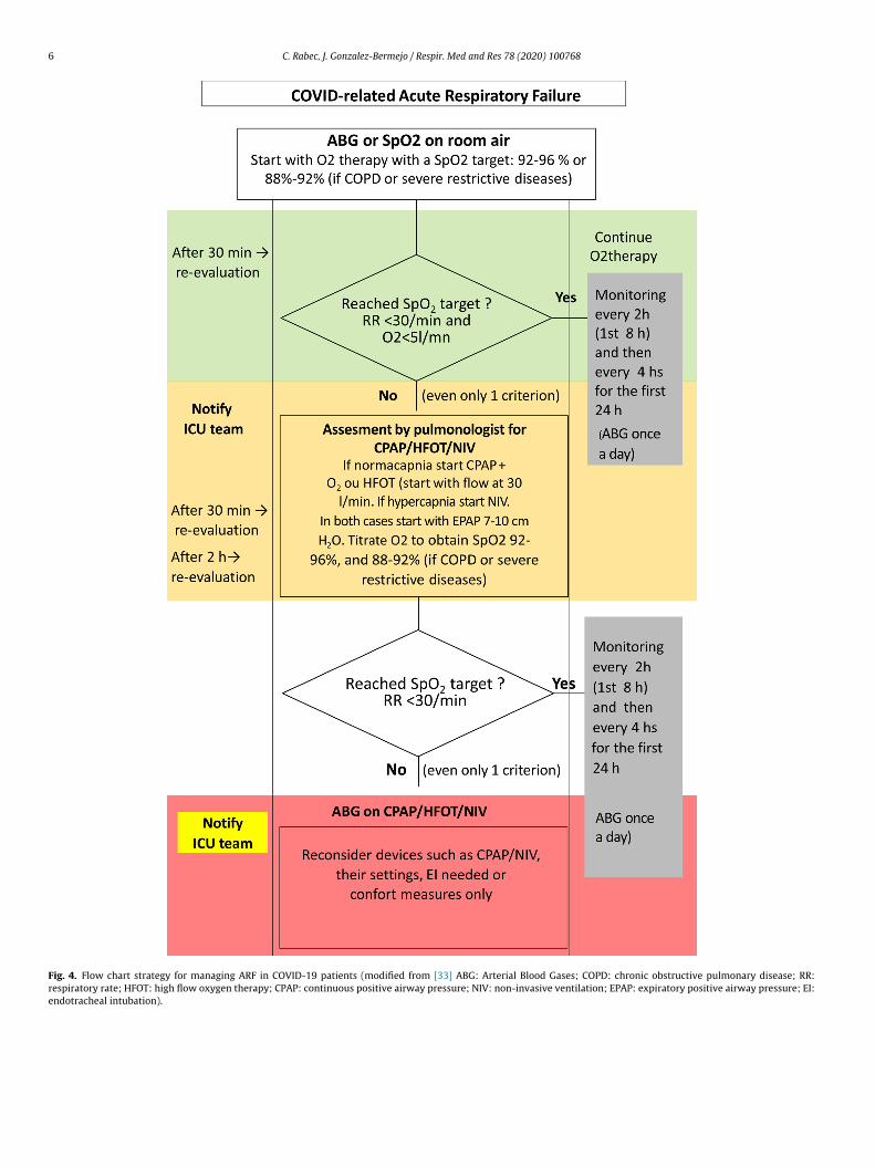
6 C. Rabec, J. Gonzalez-Bermejo / Respir. Med and Res 78 (2020) 100768
Fig. 4. Flow chart strategy for managing ARF in COVID-19 patients (modified from [33] ABG: Arterial Blood Gases; COPD: chronic obstructive pulmonary disease; RR:respiratory rate; HFOT: high flow oxygen therapy; CPAP: continuous positive airway pressure; NIV: non-invasive ventilation; EPAP: expiratory positive airway pressure; EI:endotracheal intubation).

espir.
D
i
A
(i
rt
ou
R
[
[
[
[
[
[
[
[
[
[
[
[
[
[
[
[
[
[
[
[
[
[
[
[33] http://www.aiponet.it/news/speciale-covid-19/2426-managing-the-
C. Rabec, J. Gonzalez-Bermejo / R
isclosure of interest
The authors have not supplied their declaration of competingnterest.
cknowledgements
Stefano Nava (Italy), Javiers Sayas (Spain), Michelle ChatwinUK), Manel Lujan (Spain), Annalisa Carlucci (Italy) for sharingnformation.
Special acknowledgement to the colleagues of the Italian Tho-acic Society (AIPO–ITS) and Italian Respiratory Society (SIP/IRS) forheir original idea of the flow chart proposed in this paper.
A French version of this document is availablenline in open format (http://www.splf.fr/wp-content/ploads/2020/04/RespiPreREA-SPLF-GAVO2avril2020.pdf).
eferences
[1] Wu Z, McGoogan JM. Characteristics of and important lessons from the Coro-navirus Disease 2019 (COVID-19) outbreak in China: summary of a report of72,314 cases from the Chinese center for disease control and prevention. JAMA2020.
[2] Wang Y, Dong C, Hu Y, Li C, Ren Q, Zhang X, et al. Temporal changes of CT find-ings in 90 patients with COVID-19 pneumonia: a longitudinal study. Radiology2020:200843.
[3] Guan W-J, Ni Z-Y, Hu Y, Liang W-H, Ou C-Q, He J-X, et al. Clinical characteristicsof Coronavirus Disease 2019 in China. N Engl J Med 2020.
[4] Shi Y, Yu X, Zhao H, Wang H, Zhao R, Sheng J. Host susceptibility to severeCOVID-19 and establishment of a host risk score: findings of 487 cases outsideWuhan. Crit Care 2020;24:108.
[5] Chen N, Zhou M, Dong X, Qu J, Gong F, Han Y, et al. Epidemiological and clinicalcharacteristics of 99 cases of 2019 novel coronavirus pneumonia in Wuhan,China: a descriptive study. Lancet 2020;395:507–13.
[6] Wang D, Hu B, Hu C, Zhu F, Liu X, Zhang J, et al. Clinical characteristics of 138 hos-pitalized patients with 2019 novel Coronavirus-infected pneumonia in Wuhan,China. JAMA 2020.
[7] Yang X, Yu Y, Xu J, Shu H, Xia J, Liu H, et al. Clinical course and outcomes ofcritically ill patients with SARS-CoV-2 pneumonia in Wuhan, China: a single-centered, retrospective, observational study. Lancet Respir Med 2020.
[8] Chu DK, Kim LH-Y, Young PJ, Zamiri N, Almenawer SA, Jaeschke R, et al.Mortality and morbidity in acutely ill adults treated with liberal versusconservative oxygen therapy (IOTA): a systematic review and meta-analysis.Lancet 2018;391:1693–705.
[9] Barrot L, Asfar P, Mauny F, Winiszewski H, Montini F, Badie J, et al. Liberal orconservative oxygen therapy for acute respiratory distress syndrome. N Engl JMed 2020;382:999–1008.
10] Alhazzani W, Møller MH, Arabi YM, Loeb M, Gong MN, Fan E, et al. Sur-viving sepsis campaign: guidelines on the management of critically illadults with Coronavirus Disease 2019 (COVID-19). Crit Care Med 2020,http://dx.doi.org/10.1097/CCM.0000000000004363 [Epub ahead of print].
11] O’Driscoll B, Howard LS, Earis J, Mak V. BTS guideline for oxygen use in adultsin healthcare and emergency settings. Thorax 2017;72:i1–90.
12] Grasselli G, Pesenti A, Cecconi M. Critical care utilization for the COVID-19 out-break in Lombardy, Italy: early experience and forecast during an emergency
response. JAMA 2020 [Epub 2020/03/14].13] Wang T, Zhang L, Luo K, He J, Ma Y, Li Z, et al. Noninvasive versus invasivemechanical ventilation for immunocompromised patients with acute res-piratory failure: a systematic review and meta-analysis. BMC Pulmon Med2016;16:129.
[
Med and Res 78 (2020) 100768 7
14] Xu X-P, Zhang X-C, Hu S-L, Xu J-Y, Xie J-F, Liu S-Q, et al. Noninvasive ventilationin acute hypoxemic nonhypercapnic respiratory failure: a systematic reviewand meta-analysis. Crit Care Med 2017;45:e727–33.
15] Zayed Y, Banifadel M, Barbarawi M, Kheiri B, Chahine A, Rashdan L, et al. Non-invasive oxygenation strategies in immunocompromised patients with acutehypoxemic respiratory failure: a pairwise and network meta-analysis of ran-domized controlled trials. J Intensive Care Med 2019 [885066619844713].
16] Frat J-P, Thille AW, Mercat A, Girault C, Ragot S, Perbet S, et al. High-flow oxygenthrough nasal cannula in acute hypoxemic respiratory failure. N Engl J Med2015;372:2185–96.
17] Alraddadi BM, Qushmaq I, Al-Hameed FM, Mandourah Y, Almekhlafi GA, JoseJ, et al. Noninvasive ventilation in critically ill patients with the Middle Eastrespiratory syndrome. Influenza Other Respir Viruses 2019;13:382–90.
18] Ding L, Wang L, Ma W, He H. Efficacy and safety of early prone positioning com-bined with HFNC or NIV in moderate to severe ARDS: a multi-center prospectivecohort study. Crit Care 2020;24:28.
19] Brochard L, Lefebvre J-C, Cordioli RL, Akoumianaki E, Richard J-CM. Noninvasiveventilation for patients with hypoxemic acute respiratory failure. Semin RespirCrit Care Med 2014;35:492–500.
20] Slutsky AS, Ranieri VM. Ventilator-induced lung injury. N Engl J Med2013;369:2126–36.
21] WHO Clinical management of severe acute respiratory infection when novelcoronavirus (2019-nCoV) infection is suspected: interim guidance; 2020https://www.apps.who.int/iris/handle/10665/330893.
22] Tripodoro VA, Rabec CA, De Vito EL. Withdrawing noninvasive ventilationat end-of-life care: is there a right time? Curr Opin Support Palliat Care2019;13:344–50.
23] Patel BK, Wolfe KS, Pohlman AS, Hall JB, Kress JP. Effect of noninvasive ventila-tion delivered by helmet vs face mask on the rate of endotracheal intubation inpatients with acute respiratory distress syndrome: a randomized clinical trial.JAMA 2016;315:2435–41.
24] Hui DS, Chow BK, Lo T, Ng SS, Ko FW, Gin T, et al. Exhaled air disper-sion during noninvasive ventilation via helmets and a total facemask. Chest2015;147:1336–43.
25] Fowler RA, Guest CB, Lapinsky SE, Sibbald WJ, Louie M, Tang P, et al. Transmis-sion of severe acute respiratory syndrome during intubation and mechanicalventilation. Am J Respir Crit Care Med 2004;169:1192–202.
26] Hui DSC, Zumla A. Severe acute respiratory syndrome: historical, epidemio-logic, and clinical features. Infect Dis Clin N Am 2019;33:869–89.
27] Drake MG. High-flow nasal cannula oxygen in adults: an evidence-basedassessment. Ann Am Thorac Soc 2018;15:145–55.
28] Ni Y-N, Luo J, Yu H, Liu D, Liang B-M, Liang Z-A. The effect of high-flow nasalcannula in reducing the mortality and the rate of endotracheal intubation whenused before mechanical ventilation compared with conventional oxygen ther-apy and noninvasive positive pressure ventilation. A systematic review andmeta-analysis. Am J Emerg Med 2018;36:226–33.
29] Ou X, Hua Y, Liu J, Gong C, Zhao W. Effect of high-flow nasal cannula oxygentherapy in adults with acute hypoxemic respiratory failure: a meta-analysis ofrandomized controlled trials. CMAJ 2017;189:E260–7.
30] Rochwerg B, Granton D, Wang DX, Helviz Y, Einav S, Frat JP, et al. High flownasal cannula compared with conventional oxygen therapy for acute hypox-emic respiratory failure: a systematic review and meta-analysis. Intensive CareMed 2019;45:563–72.
31] Leung CCH, Joynt GM, Gomersall CD, Wong WT, Lee A, Ling L, et al. Comparisonof high-flow nasal cannula versus oxygen face mask for environmental bacterialcontamination in critically ill pneumonia patients: a randomized controlledcrossover trial. J Hosp Infect 2019;101:84–7.
32] Raboud J, Shigayeva A, McGeer A, Bontovics E, Chapman M, Gravel D, et al. Riskfactors for SARS transmission from patients requiring intubation: a multicentreinvestigation in Toronto, Canada. PloS One 2010;5:e10717.
respiratory-care-of-patients-with-covid-19-english-version.html [updated2020-04-01 21:35:40].
34] Li Y-C, Bai W-Z, Hashikawa T. The neuroinvasive potential of SARS-CoV2 mayplay a role in the respiratory failure of COVID-19 patients. J Med Virol 2020.