Embed Size (px)
Citation preview

RESEARCH ARTICLE
Effect of O. porcinus Tick Salivary GlandExtract on the African Swine Fever VirusInfection in Domestic PigJennifer Bernard1,3, Evelyne Hutet1, Frédéric Paboeuf1, Tantely Randriamparany2,Philippe Holzmuller3, Renaud Lancelot3, Valérie Rodrigues3, Laurence Vial3, Marie-Frédérique Le Potier1*
1 Unité Virologie et Immunologie Porcines, Laboratoire de Ploufragan-Plouzané, Agence Nationale deSécurité Sanitaire (Anses), Univ Bretagne Loire, Ploufragan, France, 2 Laboratoire National de DiagnosticVétérinaire, Antananarivo, Madagascar, 3 UMRCMAEE "Contrôle des maladies animales exotiques etémergentes", Centre de coopération internationale en recherche agronomique pour le développement(CIRAD), Montpellier, France
AbstractAfrican swine fever is a haemorrhagic disease in pig production that can have disastrous
financial consequences for farming. No vaccines are currently available and animal slaugh-
tering or area zoning to restrict risk-related movements are the only effective measures to
prevent the spread of the disease.Ornithodoros soft ticks are known to transmit the African
swine fever virus (ASFV) to pigs in farms, following the natural epidemiologic cycle of the
virus. Tick saliva has been shown to modulate the host physiological and immunological
responses during feeding on skin, thus affecting viral infection. To better understand the
interaction between soft tick, ASFV and pig at the bite location and the possible influence of
tick saliva on pig infection by ASFV, salivary gland extract (SGE) ofOrnithodoros porcinus,co-inoculated or not with ASFV, was used for intradermal auricular inoculation. Our results
showed that, after the virus triggered the disease, pigs inoculated with virus and SGE pre-
sented greater hyperthermia than pigs inoculated with virus alone. The density of Langer-
hans cells was modulated at the tick bite or inoculation site, either through recruitment by
ASFV or inhibition by SGE. Additionally, SGE and virus induced macrophage recruitment
each. This effect was enhanced when they were co-inoculated. Finally, the co-inoculation of
SGE and virus delayed the early local spread of virus to the first lymph node on the inocula-
tion side. This study has shown that the effect of SGE was powerful enough to be quantified
in pig both on the systemic and local immune response. We believe this model should be
developed with infected tick and could improve knowledge of both tick vector competence
and tick saliva immunomodulation.
PLOS ONE | DOI:10.1371/journal.pone.0147869 February 1, 2016 1 / 19
OPEN ACCESS
Citation: Bernard J, Hutet E, Paboeuf F,Randriamparany T, Holzmuller P, Lancelot R, et al.(2016) Effect of O. porcinus Tick Salivary GlandExtract on the African Swine Fever Virus Infection inDomestic Pig. PLoS ONE 11(2): e0147869.doi:10.1371/journal.pone.0147869
Editor: Pedro L. Oliveira, Universidade Federal doRio de Janeiro, BRAZIL
Received: October 7, 2015
Accepted: December 13, 2015
Published: February 1, 2016
Copyright: © 2016 Bernard et al. This is an openaccess article distributed under the terms of theCreative Commons Attribution License, which permitsunrestricted use, distribution, and reproduction in anymedium, provided the original author and source arecredited.
Data Availability Statement: All relevant data arewithin the paper and its Supporting Information files.
Funding: This work was partly funded by theEuropean Union Seventh Framework Programme(FP7/2007-2013) under Grant agreement no.311931– ASFORCE, the General Council of the Côtesd’Armor department and the Regional Council ofLanguedoc Roussillon (Chercheur d’avenir project).JB received her Ph.D. grant from CIRAD andANSES. The funders had no role in study design,data collection and analysis, decision to publish, orpreparation of the manuscript.
IntroductionComplex interactions have been described in many host-vector-pathogen associations. Most ofthese associations are highly adaptive, especially for vectors and pathogens that try to escapehost defence responses by particular behavioural or immunological control strategies [1].
Ticks play an important role in pathogen transmission and emergence and are consideredof great importance in veterinary and public health domains [2]. As blood-feeding parasites,ticks have to develop high plasticity and resistance to host immune systems to achieve completeengorgement. Indeed, the host reacts to the dilacerations of its own skin barrier by ticks’mouthparts and activates an early response to infestation at the bite site [3]. This immuneresponse involves haemostasis and inflammation modulation, as well as the development ofcellular and humoral responses through the early recruitment of mononuclear phagocytic cellsand polymorphonuclear cells, the maturation of antigen-presenting cells (APCs), the activationof mast cells, and other modulation of antibodies, cytokines, chemokines and the complementsystem [4,5]. At the same time, ticks produce pharmacologically active molecules in their salivato escape host immune defences [6], incidentally improving pathogen transmission [7].
African swine fever (ASF) is a disease with disastrous economic consequences in pig pro-duction that can be transmitted by several soft tick species of the Ornithodoros genus (Acari,Argasidae). ASF is caused by a DNA virus of the Asfarviridae family [8], which induces lethalhaemorrhagic fever in domestic pigs. Quarantine of the affected areas and the slaughtering ofinfected or suspicious animals are currently the only reliable control strategies [9]. ASF ishighly contagious among pigs and the ASF virus (ASFV) can persist in the environment andfomites for weeks or even months. Ornithodoros ticks such as Ornithodoros erraticus in Spainand Portugal, and O.moubata sensu lato in eastern and southern Africa, are able to maintainand transmit the virus, and are competent vectors and reservoirs for ASFV [10]. These tickscan maintain ASFV for years and transmit the virus through different routes such as transovar-ial and/or sexual transmission from tick to tick, as well as horizontal transmission to suids viacontaminated saliva or coxal fluid [11]. Given their endophilous lifestyle, Ornithodoros ticksare sedentary and greatly dependent on their host habitat [12].
During the early stage of pig infection with ASFV, mononuclear phagocytic cells are themain targets for viral replication [13]. At the inoculation site, ASFV induces the recruitment ofmacrophages, as well as their maturation, and phagocytic and secretory activation [13], throughincreased production of cytokines such as TNF-α, IL-1 and IL-6 [14,15]. Macrophages haveessential functions in the host innate immune response and in the modulation of inflammationand maintenance of skin homeostasis [16,17]. Other important immune target cells for ASFVreplication are APCs, particularly Langerhans cells (LC), which have a sentinel role in the skin.Following infection, APCs mature in interdigitating dendritic cells and present ASFV antigen inlymph nodes [18]. However, specific ASFV contamination through tick bites and the action oftick saliva on ASFV infection in pigs have never been described. Only two studies investigatedimmunomodulation due toO.moubata saliva [19,20] but not in relation to ASFV or other path-ogen transmission. For several other tick-borne diseases, tick saliva was described as impairingthe chemo-attraction or maturation of macrophages, altering the function of dendritic cells(DCs) in the transportation of nonself antigens or reducing the ability of lymphocytes to prolif-erate [6,21,22]. Modulation of the production of cytokines, such as TNF-α, IFN-γ or IL-10, wasalso reported [4,23]. In the specific case of ASFV vectorial transmission, similar patterns can bepredicted with greater complexity, since these cells are also the main target of ASFV.
Since monocytes-macrophages and other APCs are recruited early when skin aggression orinfection occur [5], LCs are at the first line of defence, triggering the local immune responseand playing a key role in the achievement of the blood meal for ticks, and since monocytes-
Ornithodoros Salivary Effect on African Swine Fever Infection
PLOS ONE | DOI:10.1371/journal.pone.0147869 February 1, 2016 2 / 19
Competing Interests: The authors have declaredthat no competing interests exist.

macrophages and APCs are the target cells for ASFV replication [13,24], we chose to focus ourstudy on LCs and macrophage cells. The aim of this work was thus to determine whether thesaliva of O. porcinus (belonging to the O.moubata complex of species) was able to modulatethe immune response of domestic pigs infected by ASFV, using the intradermal injection oftick salivary gland extract (SGE) in the presence or absence of ASFV. Our observations focusedboth on the pig systemic immune response and on pig skin inflammation and cellular modula-tion (especially LCs and macrophages) at the tick bite location. Unlike previous studies, theassessment of such immune modulations was conducted on the natural hosts, domestic pigs,for soft tick vectors or ASFV and using what we believe to be a highly adapted tick-virus associ-ation with O. porcinus ticks collected fromMadagascar and a Madagascan ASF virus strain.
Materials and Methods
ASF virus isolateThe highly pathogenic ASFV isolate Ambaton02 (GenBank accession number BankIt1774827ANSES-MADA68322 KP144287) was originally isolated from an infected domestic pig inAmbatondrazaka, Madagascar, in 2002 and kindly provided by the Direction de la santé ani-male et du phytosanitaire (DSAPS)—Ministère de l'agriculture de l'élevage et de la pêche, Anta-nanarivo, Madagascar. The Ambaton02 isolate was classified as a haemadsorbing ASFV strainbelonging to the genotype II [25]. It was passaged three times in primary porcine alveolar mac-rophages before use in this experiment.
Soft ticksOrnithodoros porcinus domesticus ticks were collected from pig pens in Madagascar between2006 and 2010, under the supervision of DSAPS. ASFV DNA was detected naturally in somespecimens, using PCR [26]. A review of the literature indicated that this species was able tomaintain and transmit ASFV [27,28]. The ticks used for this study were shown to be ASFV-free before being reared at CIRAD, Montpellier, France. They were regularly fed on artificialmembrane with heparinised blood [29] and kept at 24°C, in 85% relative humidity, to completethe developmental cycle from nymph to adult stage.
For the experiments, 5 adult ticks were placed together in a Petri dish covered with a pieceof mosquito netting, to allow the ticks to bite the pig through the fabric. This device wasattached to the ear of a pig with adhesive tape for 2 hours to allow the ticks enough time tocomplete their blood meals. Another group of unengorged adult ticks was used to prepare thesalivary gland extract (SGE). They were dissected and salivary glands were removed, eachgland was crushed in 400 μL of Minimum Essential Medium (MEM) (BE12-611F, Ozyme,France), and the homogenates were clarified by centrifugation before inoculation. One adulttick SGE was choosen to simulate two feeding ticks.
PigsForty-eight Large-White pigs were obtained from the specific pathogen-free (SPF) breedingfacilities at ANSES, Ploufragan, France. Pigs of both sexes were eight weeks old and weighed30–35 kg at the time of inoculation. The experiment was performed in accordance with EUand French regulations on animal welfare in experimentation. The protocol was approvedunder number 20/12/12-15, by the French ethical committee for animal experimentation,named ComEth-Anses/ENVA/UPEC (agreement C2EA-16, Ministère de l’enseignementsupérieur et de la recherche, Paris, France). The procedure for the euthanasia of the animalswas based on an accepted method included in European Directive 2010/63/EU, using an
Ornithodoros Salivary Effect on African Swine Fever Infection
PLOS ONE | DOI:10.1371/journal.pone.0147869 February 1, 2016 3 / 19

anaesthetic overdose of 20 mg of sodium thiopental per kilogram of weight, administered viathe vena cava. All the pigs were maintained at BSL-3 security facilities throughout the experi-ment and fed ad libitum.
Experimental design and trial monitoringThe pigs were divided into 6 groups (Table 1). Two groups of pigs received an intradermalinoculation in one ear with virus alone (ID ASFV), or with virus and SGE (ID ASFV+SGE). Inaddition, four groups of pig did not receive any virus: i) one group was bitten by ASFV-freeticks (TICK), ii) one group received an intradermal inoculation of SGE (ID SGE), iii) onegroup received an intradermal inoculation of medium (ID MEM) and iv) one group was thenegative control group (NEG).
Except for the NEG group, each pig received 5 intradermal inoculations of 200 μl, or 5 ticks,on one ear. The other ear was kept as an internal control to account for individual variations ofcell counts (macrophages and Langerhans cells). For both the ID ASFV and ID ASFV+SGEgroups, 18 pigs received 104 50% haemadsorbing doses (HAD50) per pig called the high ASFVdose (HD) trial and 12 pigs received 102 HAD50/pig, called the low ASFV dose (LD) trial(Table 1).
The pigs were monitored daily as previously described [30] for rectal temperature and clini-cal signs (inappitence, recumbancy, skin haemorrhage, joint swelling, laboured breathing and/or coughing, ocular discharge, diarrhoea, blood in urine, vomiting), which were recorded andscored according to a scale from 0 to 5 per sign. The pigs were weighed regularly and beforeeuthanasia. Apart from the NEG group, one to three pigs were slaughtered at 1 hour and 48hours post inoculation (pi) in each group of pigs (Table 1). The other pigs were slaughteredbetween 5 and 8 days pi (dpi), as soon as the clinical score was equal or higher than 15. Onpost-mortem examination, gross lesions were observed and scored.
Sample collectionBlood samples were collected before inoculation and then at least twice a week depending onthe group, for several uses: (i) on heparin (Vacuette 9 ml clinical chemistry, lithium heparinGreiner Bio-One, Dutscher, France) for virus isolations, (ii) on EDTA (Vacuette 4 ml haema-tology, EDTA-K3 Greiner Bio-One Dutscher, France) for blood cell numbering (MS9 hematol-ogy analyzer, Melet Schloesing Laboratoires, Osny, France) and for ASFV genome real-timePCR detection.
Serum samples were purified from coagulated blood samples in dry tubes (Vacuette 8 ml ZSerum Sep Clot Activator, Greiner Bio-One, Dutscher, France) by centrifugation at 3000 g for5 min for cytokine quantification.
Lymphoid organs (spleen, tonsils and parotid lymph nodes) were collected at necropsy forvirus detection.
Table 1. Number of pigs per trial, treatment group and time post inoculation.
Group ID ASFV ID ASFV+SGE TICK ID SGE ID MEM NEG
ASFV inoculation HD trial LD trial HD trial LD trial No No No No
Nb of pig at 1hpi 9 6 9 6 10 3 1 4
Nb of pig at 48hpi 6 4 6 4 8 2 0 4
Nb of pig at 5–8 dpi 3 2 3 2 6 0 0 4
Nb: number (of pig alive); hpi: hours post-inoculation; dpi: days post-inoculation.
doi:10.1371/journal.pone.0147869.t001
Ornithodoros Salivary Effect on African Swine Fever Infection
PLOS ONE | DOI:10.1371/journal.pone.0147869 February 1, 2016 4 / 19

Both ears of each pig were collected less than 15 min after euthanasia to investigate localimmune response. Two skin biopsies were taken per ear using 8 mm punches, fixed in 4% para-formaldehyde overnight, immersed in 10%, 20% and 30% sucrose baths and snap-frozen inOCT compound (MM-France, Francheville, France). Serial cryosections (12 μm) were per-formed using a cryomicrotome (Microm HM 505 E-VAC, Francheville, France). Slides wereair-dried and labelled with anti-swine antibodies (Ab) and isotype-specific secondary antibod-ies and isotype control (IgG2b and IgG1, Dako, Les Ulis, France) for the immuno-histologicalstudy. Two other biopsies per pig were post-fixed in 4% paraformaldehyde overnight andembedded in paraffin according to the routine method applied at Labocea 22 (Ploufragan,France) for histological lesion analysis by a pathologist.
Virus detectionVirus isolation on a heparin blood sample was performed as described in the OIE diagnosticmanual [31]. The absence of any antagonist effect of SGE on the virus isolation was verified onpig alveolar macrophages (data not shown). Virus detection was carried out by real-time PCRas previously described [32] on EDTA blood samples and organs, after DNA extraction withthe DNeasy Blood and Tissue kit (Qiagen, Courtaboeuf, France). For each pig euthanized at 48hpi, four separate samples used for DNA extraction from the parotid lymph node on the sideof the inoculated ear (PLI). A sample of spleen, tonsils, and two different samples of parotidlymph nodes from the opposite side (PLO) were extracted. Organs that were late positive forASFV by real-time PCR (Ct > 40) were further analysed by a haemadsorption assay (HAD)using SPF primary porcine alveolar macrophages in a 96-well plate [31].
Viral genome loads, quantified by the cycle number threshold (Ct) detected by real-timePCR, were categorized as i) negative (Ct > 45), ii) weakly positive, close to the real-time PCRdetection threshold (37< Ct � 44), iii) positive (26< Ct � 36) and iv) strongly positive (15<Ct � 25).
Porcine cytokine quantificationIn pig sera, IL-6, IL-12 and TNF-α were measured using ready-to-use ELISA kits (R&D sys-tems, Minneapolis, USA) and haptoglobin quantification was carried out with the TRIDELTADevelopment LTD kit, (Eurobio, Courtaboeuf, France). IFN-α was quantified by a homemadeELISA test as previously described [33].
Immunofluorescence (IF) and histological staining on skin biopsiesFor IF, non-specific binding of antibodies (Ab) to tissue sections was blocked for 1 h in PBS con-taining 3% goat serum albumin (GSA). Slides were incubated overnight with primary monoclo-nal antibody (mAb) in PBS containing 10% GSA and 0.03% Triton X100, then washed in PBSbaths and incubated for 2 h with secondary Abs (Alexa Fluor1 goat anti-mouse, Invitrogen) atroom temperature. Finally, Hoechst stain solution (Sigma-Aldrich, St Louis, USA) was added tothe slides after PBS washes. Sections were mounted in Mowiol 4–88 medium (Sigma-Aldrich, StLouis, USA) for analysis using a fluorescent microscope. Microscopy observations were per-formed with an Olympus BX41 epifluorescence microscope (Scop Pro, Itteville, France). Imageswere recorded on an EXI Aqua camera (QImaging, Surrey, Canada) using Image Pro-Plus soft-ware (Media Cybernetics). Twelve serial skin sections of each of the 66 biopsies were systemati-cally examined and pictures were taken of representative sections. Ten to 25 fields were capturedfrom comparable regions of dermis in each biopsy at the same exposure and magnification bychromatic filter for panorama reconstruction. Autofluorescence intensity was subtracted beforepicture analysis. Manual counting of Langerhans cells (LC) was carried out by SWC3-Ab
Ornithodoros Salivary Effect on African Swine Fever Infection
PLOS ONE | DOI:10.1371/journal.pone.0147869 February 1, 2016 5 / 19

labelling (SWC3/CD172, IgG2b, porcine pan-myeloid, Clone 74-22-15A, SouthernBiotech) andby morphological discrimination on all epidermis transects of the opposite ear and above theinjured transect of the inoculated ear. LC density was expressed as LC μm-1. We systematicallydelineated rectangular counting frames of identical areas to assess the density of cells per frame.Macrophages were manually counted by co-labelling using SWC3-Ab and CD163-Ab (IgG1,Monocytes and macrophages, Clone MCA2311, AbD serotec) in 4 rectangular counting frameson the opposite ear: two in the dermis and the other two in deep dermis, and on the observedlesion area made by the needle or the tick bite for the inoculated ear (S1 Fig).
Haematoxylin-Eosin and saffron staining was used for histological lesion descriptions andscored using a semi-quantitative method according to severity and intensity of the lesions asoedema (1–3), haemorrhage (4–6), inflammation (7–9), dermitis (10–12), or necrosis (13–15).A descriptive analysis of 64 biopsies was performed on the mean total score per pig group.
According to observations of histological lesions and LC distribution along the epidermisbefore counting, biopsies were distributed in 5 patterns: i) “Mechanical” class, which includedbiopsies with epidermis disruption caused by needle introduction or tick mouthparts; this classwas characterized by a total absence of LCs in the disrupted region and normal density at eachextremity, ii) “Physiological” class, which showed a disappearance of LCs above the lesion withgradual reparation on both sides, iii) “Scab” class, which corresponded to phenomena ofwound healing; there were no LCs under the scab and gradual reparation on both sides, iv)“Deep” class, which only concerned inoculated groups with lesions in deep dermis but a non-modified LC distribution, and v) “No-effect” class, which presented a normal distribution ofLCs above the lesion in the dermis.
Statistical AnalysisWe fitted statistical models to the available data, thus utilizing most of the data and limitingthe number of tests. In all the models the factors of interest were the ASFV dose (categories: noASFV, low ASFV dose, high ASFV dose), tick saliva (categories: no saliva, tick bite, salivarygland extract) and time after inoculation (categories: 1 hpi and 48 hpi).
For the rectal temperature and clinical scores, preliminary exploratory data analysesrevealed sigmoid patterns for the response, with a delay before the onset of the response, fol-lowed by a steep rise and a plateau preceding the agonic stage. To model this pattern, we used a3-parameter nonlinear logistic model [34]. The first parameter (Ø1) was the plateau value (hor-izontal asymptote when time increased); the second parameter (Ø2) was the inflection point ofthe response curve; the last parameter (Ø3) was a shape parameter (steepness of the curvearound the inflection point). As the rectal temperature showed between-pig variations (range:39.1–39.8°C on the day of experimental infection), we modelled the temperature change fromthe inoculation day, rather than the actual rectal temperature.
The nonlinear logistic models were fitted using generalized least-squares (GLS), which pro-vided maximum-likelihood (ML) estimators. During the model building stage, this property wasused to compare fitted models with the Akaike information criterion (AIC) corrected for thesmall sample size [35,36]: AIC = −2 log(L) + 2 k and AICc = AIC + 2 k (k + 1) / (n − k − 1), whereL was the maximized likelihood, n was the number of observations and k was the number ofparameters (coefficients) in the model. AICc was used to compare models with the same response.Comparable models with lower AICc were considered as better than those with higher values.
Moreover, the GLS estimation method made it possible to account for correlations in themodel residuals related to repeated measurements made on the same pigs, as well as possibleheteroscedasticity in these residuals [34]. The pig was defined as a grouping factor, and ahomogeneous within-pig correlation structure was used for residuals.
Ornithodoros Salivary Effect on African Swine Fever Infection
PLOS ONE | DOI:10.1371/journal.pone.0147869 February 1, 2016 6 / 19

For the detection of ASFV DNA in parotid lymph nodes with real-time PCR, we consideredthe cycle number threshold (Ct) as a quantitative variable. We were also confronted withrepeated measurements in this analysis, with several values of Ct recorded on the same lymphnode, and two parotid lymph nodes per pig. We used a two-level linear mixed-effect model[37], with ASFV dose, tick saliva and time as the fixed effects, and two nested random effects(grouping factors) associated with the intercept: (i) pig, and (ii) parotid lymph node within pig.Models were fitted with an ML method [34].
For the analysis of Langerhans cells and macrophage density, the within-pig correlation ofresiduals was removed, taking each pig as its own control. Indeed, for each pig i, we computed:
• The cell density (Langerhans cells or macrophages) in non-inoculated (control) ears: di,ref =Si Уi,ref / Si Zi,ref where yi,ref was the cell count in region Zi,ref. For Langerhans cells, Zi,ref wasa line transect of known length (Zi,ref) () located in the epidermis. For macrophages, it was acounting frame of known area(Zi,ref),
• The cell density in inoculated (treated) ears: di,obs = Si Уi,obs / Si Zi,obs. For Langerhans cells,Zi,obs was a transect line segment chosen just above the inoculation point. For macrophages,the counting frame was selected close to the inoculation point in an area with a higher mac-rophage density as determined after a preliminary visual inspection,
• The difference in cell density δi = di,ref − di,obs. A positive value of δi indicated a decrease incell density. This difference in cell density was the response in subsequent statistical models.
We used linear models with adaptations required by response peculiarities. Indeed, δi was adifference of cell counts which typically showed a high range of values, with outliers (observa-tions very different from the population mean). To account for this, we used a robust linearmodel, in which outliers had a limited influence on coefficient estimates [38].
Model coefficients were fitted by iterated re-weighted least squares. Moreover, because ofthe small sample size (24 pigs allocated to different treatment categories), it was difficult to usethe asymptotic condition for the computation of p values associated with tests on model coeffi-cients. Instead, we used a bootstrap procedure on model residuals as described by Davison andHinkley [39]. In short, the estimated coefficients (robust linear model) and fitted values wereconsidered as fixed values. Residuals were sampled with replacement, and a new response wascomputed adding the fitted values to the sampled residuals. The robust linear model was thenrefitted with this new response, and the resulting coefficients were stored. This loop was iter-ated B times (with large values of B: typically 999 or 1,999, or more). The B sets of coefficientswere added to the original set. The 2.5% and 97.5% quantiles of each coefficient series of B+1values (or linear combination of these coefficients) were then used as 95% confidence intervals.
For instance, the fitted mean difference in cell density �̂d i was a linear combination of modelcoefficients. To know whether the combined effects of time, ASFV dose and tick saliva (includ-
ing possible interactions) significantly altered �̂d i (null hypothesis: �̂d i ¼ 0), we computed the B+1 values for a given group (e.g. tick saliva and high ASFV dose at 48 hpi), and assessedwhether the 95% confidence interval included the value of 0 (non-significant combined effectfor α = 0.05) or not (significant combined effect for α = 0.05).
Results
General pig immune response: clinical signs and pathophysiology1. Clinical scores and viraemia. Symptoms were only observed when the virus was inocu-
lated, with no clinical signs for the NEG, TICK and ID SGE groups throughout the experiment.
Ornithodoros Salivary Effect on African Swine Fever Infection
PLOS ONE | DOI:10.1371/journal.pone.0147869 February 1, 2016 7 / 19

The disease spread and clinical scores were similar between intradermally infected groupswhatever the virus doses (HD and LD trials), including first a loss of appetite at 1–3 dpi, thenhyperthermia the following day, at 2–4 dpi. Pigs that received the high ASFV dose were alleuthanized 5 or 6 dpi for ethical reasons, whereas the pigs inoculated with the lowest dose hadto be euthanized at 8 dpi. The onset of the disease, according to the clinical scores, was signifi-cantly delayed by one day in the LD trial compared to the HD trial (p = 5 10−3). The meandaily weight gain (MDWG) was evaluated and all the infected groups (ID ASFV, ID ASFV+-SGE) lost weight from 3 dpi whatever the virus doses expressed in mean ± standard deviation(SD), -0.9 ± 0.1 (n = 5) kg/day for the HD trial and -0.7 ± 0.1 (n = 4) kg/day for the LD trial,whereas the non-infected groups (NEG, TICK, ID SGE, ID MEM) gained weight up to 8 dpi(0.9 ± 0.1 (n = 7) kg/day).
The body temperature indicated that the pigs of the non-infected groups never displayedhyperthermia (T>40°C). For the other groups, the onset of fever occurred with a significantone day delay between pigs of the HD trial at 3.3 dpi and the LD trial at 4.6 dpi (p< 10−3, non-linear logistic model) (Fig 1B). In the HD and LD trials, higher mean rectal temperatures wereobserved for the ID ASFV+SGE groups over the first three days after the onset of hyperther-mia. To assess this effect, we considered both groups together and analysed the temperaturedata on the first three days after the onset of hyperthermia. We fitted the mean temperatureswith a generalized least squares linear regression model with a single fixed effect (tick saliva vs.no tick saliva) and pig as the grouping factor (within-pig homogeneous correlation structure).The fitted mean temperature of the ID ASFV+SGE group was significantly higher by 0.3°C(p = 0.032) than for the ID ASFV group.
The virus was detected in blood as early as 2 dpi whatever the virus dose. The virus load wasscored as “positive” to “strongly positive” for the 12 pigs slaughtered at that time in the HDtrial, but “weakly positive” for 5 out of the corresponding 8 pigs in the LD trial and “negative”for the last 3 pigs. This difference in detection level between trials was also found at 3 dpi.Then, from 4 dpi to the endpoint, no difference was found between groups, with or withoutSGE, in both trials (Fig 1A, left scale).
Fig 1. Clinical scores, viraemia and rectal temperature. (A) The scale for clinical score is shown on theleft, and the scale for individual real-time PCR results is shown on the right. Real-time PCR results (dark greytriangles) were categorized as negative (Ct > 45), weakly positive (44� Ct > 37), positive (36� Ct > 26) andstrongly positive (Ct < 25). (B) Changes in pig rectal temperature were recorded with respect to theinoculation day. The mean was estimated with a nonlinear logistic model fitted by generalized least squares.
doi:10.1371/journal.pone.0147869.g001
Ornithodoros Salivary Effect on African Swine Fever Infection
PLOS ONE | DOI:10.1371/journal.pone.0147869 February 1, 2016 8 / 19

2. Blood leukocyte count. Peripheral white blood cell counts for lymphocytes, monocytesand granulocytes were similar between pig groups before virus inoculation. Leucopoenia wasobserved at the onset of hyperthermia in all groups of infected pigs and showed a similar pat-tern with a recovery period during the hyperthermia phase (Table 2).
3. Gross pathological lesions. Post-mortem examination did not reveal any differencebetween ASFV infected groups, with jaundice and splenomegaly systematically observed at 5–8dpi. The most recurrent lesions for 15/20 pigs were severely congested or haemorrhagic lymphnodes. When pigs were slaughtered at 1 hpi, no lesions were recorded. A slight difference wasobserved at 48 hpi in the HD trial, with less reactive lymph nodes for the ID ASFV group thanfor ID ASFV+SGE, which also showed petechial lesions on the thymus. This distinction wasnot found for the LD trial.
4. Cytokine quantification in serum. Due to technical constraints, blood samples wereonly taken daily for the pigs receiving the low ASFV dose (with or without SGE). For these ani-mals, the response pattern was similar for IL-12, IFN-α and TNF-α (S2 Fig, columns 2 and 4):a rise in cytokines was observed at 4 dpi. Because of the sampling framework, we could onlyassess the effect of the ASFV dose for animals receiving SGE (S2 Fig, columns 3 and 4). At 3dpi, an increase in the TNF-α level was found in the HD trial, whereas it was significantlydelayed by one day in the LD trial (p = 0.02, Wilcoxon test). The same pattern was observed forIFN-α, IL-12, and I-L6. A rise was also observed for haptoglobin (data not shown) but itremained under the positivity threshold recommended by the manufacturer for detectinginflammation.
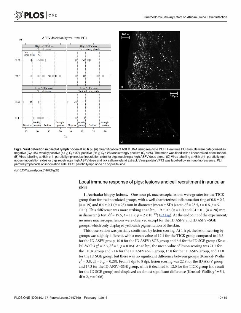
5. Virus detection in organs. At 1 hpi, no viral genome was detected in any lymphoidorgan. Conversely, all pigs euthanized at 5–8 dpi presented a very high level of viral genome intheir lymphoid organs. The results were more heterogeneous when pigs were euthanized at 48hpi. In parotid lymph nodes (Fig 2), the viral load was higher in the high ASFV dose trial thanin the low ASFV dose trial, for each side (inoculated or opposite side). However, this effect wasnot significant (t test, df = 7, t = -1.90, p = 0.10).
For opposite parotid lymph nodes (PLO), no difference was observed between the ID ASFVgroup and the ID ASFV+SGE group (Fig 2A, Wald test, w = 0.016, df = 1, p = 0.90). For inocu-lated parotid lymph nodes (PLIs), SGE had a large and significant effect (Fig 2A, Wald test:w = 4.6, df = 1, p = 0.031). The IF method on lymph node cryosections confirmed that PLIs ofthe ID ASFV group presented more labelled cells for the ASFV VP72 virus capsid protein thanthe ID ASFV+SGE group on a similar area of lymph node section (Fig 2B and 2C).
For the spleen and tonsils, no effect of SGE was observed (data not shown).
Table 2. Blood leucocyte mean count kinetics (x 106 cells/ml of blood) presented in relation to hyperthermia.
Group ID ASFV ID ASFV+SGE ID SGE*
ASFV inoculation HD trial LD trial HD trial LD trial No
Leucocyte count before hyperthermia1 7.4 ± 2.2 10.7 ± 2.3 9.2 ± 0.7 8.3 ± 2.4 11.1 ± 5.6
Leucocyte count the day of hyperthermia2 7.9 ± 2.1 5.4 ± 0.7 4.8 ± 1.5 5.5 ± 0.2 11.7 ± 4.7
Leucocyte count after hyperthermia3 17.4 ± 5.6 13 ± 7.9 16.1 ± 6.6 13.8 ± 7 14.9 ± 5.5
Data in x 106 cells/ml of blood, Mean ± SD;1: Incubation time before the onset of hyperthermia;2: day of the onset of hyperthermia;3: time after the onset of hyperthermia up to the end of the experiment,
*ID SGE group did not display hyperthermia, so results were separated in 3 periods of 2 days from 0 dpi to 6 dpi.
doi:10.1371/journal.pone.0147869.t002
Ornithodoros Salivary Effect on African Swine Fever Infection
PLOS ONE | DOI:10.1371/journal.pone.0147869 February 1, 2016 9 / 19
Local immune response of pigs: lesions and cell recruitment in auricularskin
1. Auricular biopsy lesions. One hour pi, macroscopic lesions were greater for the TICKgroup than for the inoculated groups, with a well characterized inflammation ring of 0.8 ± 0.2(n = 19) and 0.4 ± 0.1 (n = 25) mm in diameter (mean ± SD) (t test, df = 23.5, t = 6.6, p = 910−7). This difference was more striking at 48 hpi, 1.9 ± 0.5 (n = 19) and 0.4 ± 0.1 (n = 28) mmin diameter (t test, df = 19.5, t = 11.9, p = 2 x 10⁻¹⁰) (S3 Fig). At the endpoint of the experiment,no more macroscopic lesions were observed except for the ID ASFV and ID ASFV+SGEgroups, which only displayed yellowish pigmentation of the skin.
This observation was partially confirmed by lesion scoring. At 1 h pi, the lesion scoring bygroups was slightly different, with a mean value of 17.1 for the TICK group compared to 13.3for the ID ASFV group, 10.0 for the ID ASFV+SGE group and 6.5 for the ID SGE group (Krus-kal-Wallis χ² = 7.3, df = 3, p = 0.06). At 48 hpi, the mean value of lesion scoring was 21.7 forthe TICK group and 21.6 for the ID ASFV+SGE group, 13.8 for the ID ASFV group, and 11.0for the ID SGE group, but there was no significant difference between groups (Kruskal-Wallisχ² = 3.8, df = 3, p = 0.28). From 5 dpi to 8 dpi, lesion scoring was 22.8 for the ID ASFV groupand 17.3 for the ID AFSV+SGE group, while it declined to 12.0 for the TICK group (no resultfor the ID SGE group) and displayed an almost significant difference (Kruskal-Wallis χ² = 5.4,df = 2, p = 0.06).
Fig 2. Viral detection in parotid lymph nodes at 48 h pi. (A) Quantification of ASFV DNA using real-time PCR. Real-time PCR results were categorized asnegative (Ct> 45), weakly positive (44� Ct > 37), positive (36�Ct > 26) and strongly positive (Ct < 25). The mean was fitted with a linear mixed-effect model.(B) Virus labelling at 48 h pi in parotid lymph nodes (inoculation side) for pigs receiving a high ASFV dose alone. (C) Virus labelling at 48 h pi in parotid lymphnodes (inoculation side) for pigs receiving a high ASFV dose and tick salivary gland extract. Virus protein VP72 was labelled by immunofluorescence. PLI:parotid lymph node on inoculation side; PLO: parotid lymph node on opposite side.
doi:10.1371/journal.pone.0147869.g002
Ornithodoros Salivary Effect on African Swine Fever Infection
PLOS ONE | DOI:10.1371/journal.pone.0147869 February 1, 2016 10 / 19

2. Langerhans cell density. Preliminary investigations revealed no difference in Langerhanscell (LC) density between tick bites or SGE inoculation. These two groups of pigs were thereforegrouped together in the tick saliva category. The main results are shown in Figs 3 and 4A.
At 1 hpi, changes in LC density were related to all types of epidermis disruption exceptscabs (Fig 3A), independently of ASFV or tick saliva conditions. Indeed, no significant differ-ence in LC density was detected between the pigs in any group: Fig 4A, second row of eachgroup, Monte Carlo simulation, B = 1,999, p> 0.05.
At 48 hpi, most of the slices showing a decrease in LC density were from animals thatreceived tick saliva (ID ASFV+SGE, ID SGE, TICK): Fig 3B, right-hand side. Those with thelargest decrease (32/44) were categorized in the “Scab” or “Physiological” classes. Slices show-ing an increase had mostly been taken from pigs receiving ASFV only (Fig 3B, left-hand side).Most of them were categorized in the “Deep” class (16/24). The results from the robust linearmodel showed significant interaction between tick saliva and time (48 hpi): Monte Carlo simu-lation, B = 1,999, p = 0.006. As shown in Fig 4A, saliva significantly decreased LC density forany ASFV dose (including no virus): right-hand column, first row of each group, Monte Carlosimulation, B = 1,999, p<0.05. For pigs which did not receive tick saliva (Fig 4A, left-hand col-umn, first row of each group), the presence of ASFV tended to increase LC density, with ahigher density for the higher ASFV dose. However, this effect was not significant (Monte Carlosimulation, B = 1,999, p> 0.05).
Fig 3. Classification of biopsies by Langerhans cell density differences and associated histological lesion pattern. (A) Classification of biopsy at 1 hpi. (B) Classification of biopsy at 48 h pi. Results of manual count and observation carried out on each slide. Two main lesion patterns were observed, onedue to the tick bite or to the intradermal inoculation (pattern: mechanical or deep) and the other due to the physiological effect of saliva, SGE and virus(pattern: no-effect, physiological or scab).
doi:10.1371/journal.pone.0147869.g003
Ornithodoros Salivary Effect on African Swine Fever Infection
PLOS ONE | DOI:10.1371/journal.pone.0147869 February 1, 2016 11 / 19

3. Macrophage recruitment in the dermis. An example of histological observations isshown in S4 Fig. As for LC, observations from tick bites and SGE inoculations were groupedfor data analysis. The estimated macrophage density (robust linear model) did not show cellrecruitment at 1 hpi in any pig groups (Monte Carlo simulation, B = 1,999, p> 0.05), with theexception of pigs receiving tick saliva alone (Fig 4B). At 48 hpi, the results showed a strong andsignificant interaction between the low ASFV dose and time (Monte Carlo simulation,B = 1,999, p = 0.01). In addition, the interaction between saliva and time was significant(Monte Carlo simulation, B = 1,999, p = 0.03). All pig groups receiving ASFV and/or tick salivashowed significant macrophage recruitment, except the pig group treated with a high ASFVdose without tick saliva (Fig 4B, left-hand column, and first row of upper group: Monte Carlosimulation, B = 1,999, p> 0.05). The pig group that did not receive either ASFV or tick salivadid not show any macrophage recruitment (Fig 4B, left-hand column, and first row of lowergroup: Monte Carlo simulation, B = 1,999, p> 0.05).
DiscussionThe role of Ornithodoros ticks in the ASF epidemiological cycle is fairly well understood,whereas the effect of tick bites, infected or not, on the immune response to infection in domesticpigs has never been studied. To recreate as closely as possible natural conditions of contamina-tion by tick bites, we selected a combination of ticks and viral isolate collected from a restrictedgeographical zone (Madagascar), along with intradermal administration of a low virus dose pre-sumed to better mimic the quantity of virus inoculated by ticks during their blood meal [27].Extracts of O. porcinus salivary glands were used for intradermal inoculation, as it has been pre-viously showed that there was a good correlation between transcript and protein abundance insalivary gland and saliva [40]. This was confirmed by our experimental results, with similar LCand macrophage recruitment patterns between the SGE and TICK groups.
The disease course observed in pigs, was similar to that previously described in infection bythe ASF virus using other inoculation methods (intramuscular versus oro-nasal) [41–43]. This
Fig 4. Pattern of immunological cell density difference between inoculated ear and opposite ear. (A) Langerhans cell density difference 10−2 x densitydifference, μm-1. (B) Macrophage density difference 10−4 x density difference, μm-2. The mean was fitted with a robust linear model.
doi:10.1371/journal.pone.0147869.g004
Ornithodoros Salivary Effect on African Swine Fever Infection
PLOS ONE | DOI:10.1371/journal.pone.0147869 February 1, 2016 12 / 19

confirmed that the Madagascan ASFV strain therefore displayed a similar infection pattern tothat described with the Georgian strain, which is phylogenetically very close [25,44].
Effect of virus dose on the kinetics of immunological and symptomaticresponses in pigsOur results showed a correlation between the dose of virus inoculated (LD/HD trials) and thetime course of the effects observed, with a delay of one day in the immune response, onset ofclinical signs and virus dissemination in pig at 48 hpi, as previously described [43,45]. Theother result attributable to the virus dose was a difference in macrophage density detected inthe skin biopsies at the inoculation point at 48 hpi. It has been shown that the virus is capable,via the caspase-3 protein, of triggering the late activation of apoptosis between 24 and 48 hafter infection, enabling massive dissemination [46,47]. It may be that the biopsies analysed at48 hpi were taken at the time of this first and intense phase of virus dissemination, in whichcase the local disappearance of macrophages was probably due to induced cell death. A secondhypothesis concerns the kinetics of macrophage maturation: once stimulated by its interactionwith a pathogen, the macrophage undergoes profound changes [46,48]. SWC3/CD172-Ab, aporcine myelomonocytic marker used, in our study, to detect macrophages [18,49], is a homo-logue of the epitope SIRP-α [50], whose expression is regulated according to macrophage mat-uration [51,52]. A high virus dose can influence LC and macrophage maturation at 48 hpi [53]and induce an extinction of the SIPR-α signal, possibly reducing the detection of these cells inour samples.
The presence of SGE modulates the immune response in pigs1. SGE plus ASFV increased fever. The group of pigs receiving an intradermal inocula-
tion of SGE without virus displayed similar results to those from the TICK group, whether forimmune or physiological responses. When the pigs were inoculated with the virus, irrespectiveof the dose, SGE presence increased the degree of hyperthermia, whereas SGE did not affectthe systemic level of the proinflammatory cytokines TNF-α and IL-6. The pyrogenic substancehypothesis did not therefore appear to explain this difference, but it needs to be investigatedfurther, even though very little is currently known about fever-triggering mechanisms [5].
2. SGE had an immunomodulating effect on skin tissue lesions. During engorgement,the tick maintains a passageway to the outside via its saliva until 60 min [12], which is of greaterconsequence than a needle-mediated intradermal inoculation of a few seconds. This differencepartly explains the results of the lesion observations at 1 hpi, for which the TICK group had ahigher lesion score than the ID SGE group, a trend that was confirmed at 48 hpi. For inoculationwith a virus+SGE mixture, the lesion score at 48 hpi then became equivalent to that of the TICKgroup, thereby showing a greater immunomodulating effect of SGE in the presence of virus.This mechanism should probably be considered jointly with the observation of greater hyper-thermia in the groups of diseased pigs with SGE. To assess the scores of the biopsies at the endof the experiment (5 to 8 d pi), it should be remembered that the main symptom of the ASFvirus is rapid-onset haemorrhagic fever. Thus, the higher lesion level in the ID ASFV groupthan in the ID ASFV+SGE group can be explained by the physiological consequences due to thefast multiplication of the virus. However, most saliva molecules enable ticks to modulate thehaemostasis of their host [54] along with the associated scarring phenomena [6].
3. SGE promoted LC disappearance in the epidermis. It has been established, that LCs arerecruited by the ASF virus [18,24] and their migration may depend on the production of TNF-αinduced by macrophages and neutrophils [55]. Inversely, the immunomodulation by saliva at 48hpi was reflected in a clear reduction in the number of LCs. It is quite possible that exposing
Ornithodoros Salivary Effect on African Swine Fever Infection
PLOS ONE | DOI:10.1371/journal.pone.0147869 February 1, 2016 13 / 19

immature DCs to tick saliva leads to reduced migration and poor LC renewal [56]. In addition,LCs are sentinels with a limited radius of action from the epidermis, which are rapidly replacedby dendritic cells and macrophages in the case of deeper lesions of the dermis. The reductionobserved in the ID ASFV+SGE groups was lower than for the TICK group but remained statisti-cally similar, meaning that the salivary gland extract had an inhibiting impact on LC density,whereas it was inoculated more deeply than saliva during natural tick engorgement.
4. SGE promoted the recruitment of macrophages in the dermis. Once the epidermisand the LC barrier have been overcome, pathogens encounter a second line of defence mainlyconsisting of a massive arrival of immune cells (e.g. neutrophils, granulocytes then macro-phages) [6,17,57,58,59] However, the cell type involved early in the host-tick response alsodepends on the time since engorgement and the degree of host susceptibility [58,59]. The activ-ity of macrophages would appear to be inhibited in susceptible vertebrate hosts duringengorgement by hard ticks or SGE inoculation [22,60,61]. Only one study on the evasion strat-egy of Dermacentor variabilis shows substantial macrophage recruitment [62]. Our resultsshowed weak macrophage recruitment at 1 hpi whichever group of pigs was studied, that wasprobably the direct consequence of the lesions caused by inoculation or tick engorgement [17].Nevertheless, it clearly intensified at 48 hpi, that mean SGE and virus displayed macrophagerecruitment alone or co-inoculated. The nature of activation signals received by these macro-phages, being probably different in maturation or susceptibility to infection, could explain thedifferences observed between groups.
5. SGE delayed virus infection in the first lymph node. Once the virus enters the hostorganism, it spreads rapidly by way of infected antigen-presenting cells which travel via thelymph or blood towards target organs [33,62,63]. Our results indicated that dissemination ofthe ASFV was found to be less in the PLIs of the pigs for which the inoculum contained SGE,suggesting that saliva molecules act more on modulating APCs. Three hypotheses can be putforward to explain this early local phenomenon in vivo. The first is based on the idea of dilution[64], the share of APCs bearing antigens of the virus would appear to be reduced to the benefitof those bearing SGE antigens [65–67]. Marquet et al. [64] estimated that the number of LCsmigrating to the lymph nodes via the lymph amounted to around 11% of the afferent DCs. It isknown that APCs and especially LCs are able to present antigens of hard tick SGE to T lym-phocytes in lymph nodes in vitro and in vivo [65–67]. The second more likely hypothesiswould be that SGE acts on APCs via immunoregulation phenomena. Saliva molecules mightinduce APC cell death or inhibit their migration to the PLIs, making virus replication less effi-cient in the latter. However, APCs density in the lymph nodes was similar in presence orabsence of SGE (S5 Fig). The third hypothesis, which does not exclude the second, depends oncertain in vitro and in vivo studies showing that hard tick saliva is capable of downregulatingAPC functions [23,56,68]. For instance, SGE of O. porcinusmight influence the ability of APCsto present viral antigens to T cells in lymph nodes.
Limitations and prospectsOne aspect to consider in histological studies is the type of syringe administration. As we sawin our study, the inoculation depth and the concentrations inoculated are relatively dissimilarto those of tick bites [3]. In addition, the virus produced on pig macrophages did not suffer theselection pressures that its passage through the different tick tissue barriers might entail, partic-ularly the midgut and salivary glands. However, despite these conditions, we were able to seethat the effect of the virus on one hand, and the effect of SGE on the other, were strong enoughto be observable and quantifiable at the inoculation point on histological sections, and physio-logically in pigs. Another bias to consider was the SGE utilisation because the profile of secreted
Ornithodoros Salivary Effect on African Swine Fever Infection
PLOS ONE | DOI:10.1371/journal.pone.0147869 February 1, 2016 14 / 19
proteins in saliva is not exactly the same. However our experimental results and statistical anal-ysis, notably on the comparison between TICK and ID SGE group, showed that it was possibleto assimilate SGE effect to saliva effect.
However, the observed immune modulations still depend on the models studied and thearthropod-pathogen-host combinations concerned [69]. Barratt-Due et al. [70] showed thatthe OmCI molecule present in O.moubata saliva is able to modulate haemostasis in pigs invivo, attenuate the production of TNF-α and IL-6, and reduce neutrophil migration, whereastheir previous study had shown that TNF-α was not modulated by this protein in vitro in pigcells [71]. Even though some common patterns seem to occur, relationships can be difficult tomodel, making predictions about reactions problematical. Indeed, most vectors modulate theimmune system of the host, thus facilitating pathogen proliferation. Our study showed that themolecules contained in SGE may have partly caused an inhibiting effect on macrophage andAPC activation. However, we saw that SGE and tick saliva increased macrophage recruitmentin the dermis, which is likely to promote viral infection. Consequently, to better understandthe local effect of saliva, it would be interesting to investigate, as a priority, how the bite of atick infected with the ASFV affects modulation of the immune cells. Further research compar-ing expression levels for some cytokines of interest (TNF-α, IL-6, etc.) and their tissue location(dermis and lymph node), as well as the dissemination of SGE molecules in lesions, would helpto verify the hypotheses put forward. Another valuable aim of further study would be to acquirethe ability to keep track of changes in LCs and antigen-presenting cells. It would thus be possi-ble to improve our knowledge of the impact of SGE on macrophage recruitment and APCexchanges with lymph nodes. Lastly, it seems important to check that the infection patternsproduced are similar between the different Ornithodoros species of interest (O.moubata and O.erraticus in particular). The O. erraticus tick is known to cause greater inflammation andlesions in tissues than a bite from ticks of the O.moubata–O. porcinus complex [72]. Such astudy might help to more effectively characterize immunomodulation phenomena within thesame tick genus (e.g. Ornithodoros) in line with their vector ability.
Supporting InformationS1 Fig. Cell count method on skin biopsies from inoculated ears.Within the epidermis, thetransect for the Langerhans cell count is delineated in blue. Within the dermis and deep dermis,the macrophages were counted inside the areas delineated in yellow. The circular area corre-sponds to the area of the tick bite or inoculation point. The yellow rectangles are outside thearea where the tissue was disrupted. In the opposite ear, macrophages were only counted in theyellow rectangles as there was no tissue disruption and the transect extended throughout thebiopsy.(TIF)
S2 Fig. Cytokine response: dosage of TNF-α, INF-α, IL-6 and IL-12 in pig sera. For eachcytokine, whatever the pig group and day post infection, the results were transformed into apercentage of serum concentration, with the maximal observed concentration considered tohave 100% activity.(TIF)
S3 Fig. Histological slice of O. porcinus tick bite. (A) Tick bite at 1 h pi, black star showingthe tick bite, an arrow indicating the haemorrhagic area. (B) Tick bite 48 h pi with arrowsshowing extensive haemorrhagic area and dermis lesions. Staining: Hemacolor kit (MerckMillipore, Darmstadt, Germany)(TIF)
Ornithodoros Salivary Effect on African Swine Fever Infection
PLOS ONE | DOI:10.1371/journal.pone.0147869 February 1, 2016 15 / 19
S4 Fig. Effect of a tick bite and intradermal inoculation at 1 h pi in pig skin with SWC3/SC172 cell labelling. (A) Tick bite. (B) Intradermal inoculation. Two types of tissue lesionwere observed. The first was characterized by an area delimited by tissue disruption, collagendisruption and more abundant SWC3/CD172 cell labelling than in healthy tissue (yellowarrows). The second was sometimes observed inside the first, consisting of more intensivedamage with more abundant haemorrhage and SWC3/CD172 cell labelling (red arrows). Mostof the TICK biopsies presented both areas. However, statistical model analyses were only per-formed on the disrupted area indicated in the photo by the yellow arrows. Biopsies in the sam-ples with tick bites were performed to a depth of 550,0 μm (n = 14 biopsies on 7 pigs), unlikethe inoculated biopsies which were performed to a depth of 1052.8 ± 511.4 μm (n = 23 biopsieson 14 pigs).(TIF)
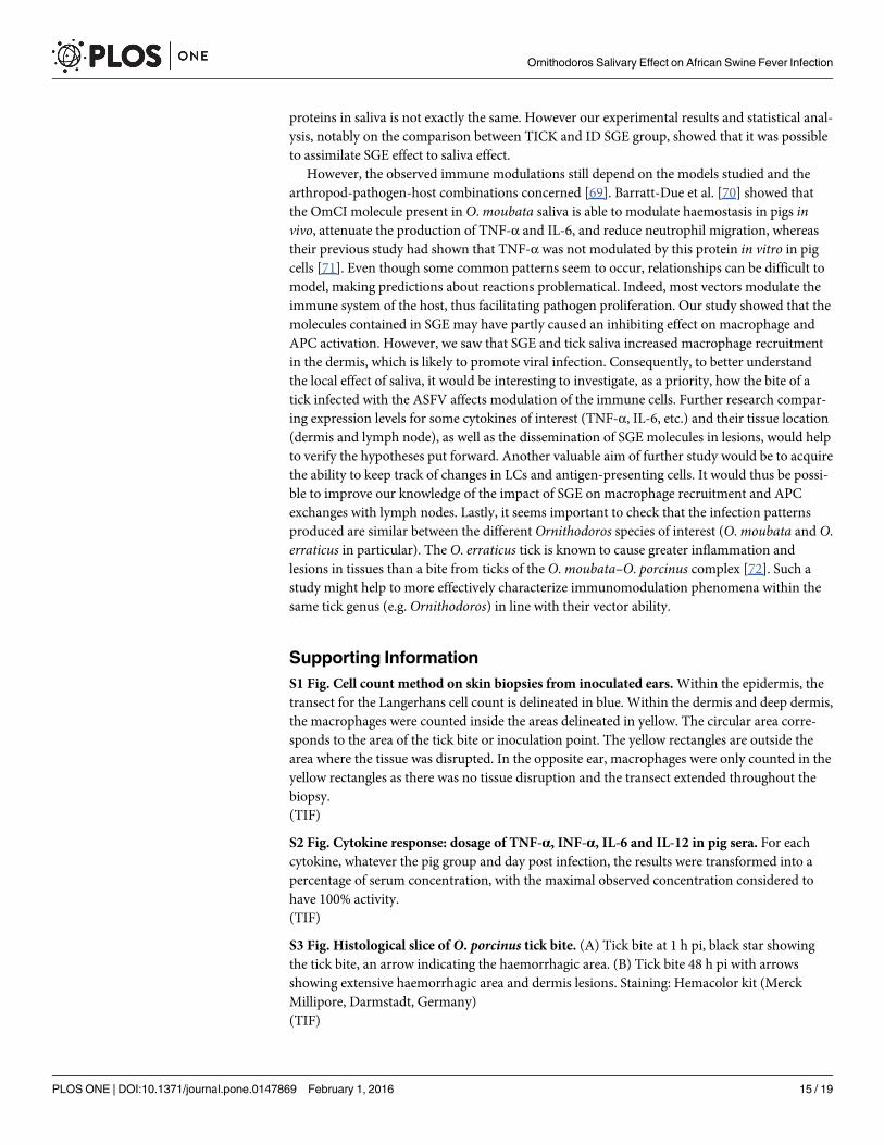
S5 Fig. Histological slice of parotid lymph node, inoculation side, at 48 h pi. (A) Parotidlymph node from inoculation side for pigs receiving a high ASFV dose and tick salivary glandextract. (B) Parotid lymph node from inoculation side for pigs receiving a high ASFV dosealone. Merging of histological slice labelled in red with S100-Ab (IgG1, interdigitating dendriticcells, Clone SH-B1, Sigma) [18] and in green with SWC3-Ab.(TIF)
AcknowledgmentsThe authors are grateful to André Keranflec’h and Jean-Marie Guionnet for animal care andsampling at ANSES. The authors are grateful to Dr Nadia Amenna-Bernard from Labocea 22for advice and expertise in the analysis of biopsy lesions; Chantal Ripoll from the histologyplatform of INM-RHEM, Montpellier, for advice and histology protocols; Frédéric Stachurskifrom CIRAD, Montpellier, for tick sampling and breeding in Madagascar, Vincent Michaudfrom CIRAD, Montpellier, for virus production and helpful discussions on virology, and JohnKerr (Coup de Puce Expansion) for revising the English text. The authors thank the Directionde la Santé Animale et du Phytosanitaire—Ministère de l'agriculture de l'élevage et de la pêche,Antananarivo, Madagascar for their permission to use the Madagascan isolate in this study.
Author ContributionsConceived and designed the experiments: MFLP JB LV. Performed the experiments: JB EH FPLVMFLP. Analyzed the data: JB RL PH VR LVMFLP. Contributed reagents/materials/analy-sis tools: LV EH FP TR JB MFLP. Wrote the paper: JB LV MFLP RL.
References1. Wikel SK, Alarcon-Chaidez FJ (2001) Progress toward molecular characterization of ectoparasite mod-
ulation of host immunity. Veterinary parasitology 101: 275–287. PMID: 11707302
2. Sonenshine DE, Roe RM (2013) Biology of ticks: Oxford University Press.
3. Frischknecht F (2007) The skin as interface in the transmission of arthropod-borne pathogens. CellMicrobiol 9: 1630–1640. PMID: 17490407
4. Brossard M, Wikel SK (1997) Immunology of interactions between ticks and hosts. Med Vet Entomol11: 270–276. PMID: 9330259
5. Owen JA, Punt J, Stranford SA, Jones PP (2013) Immunology 7th edition: WH Freeman and Com-pany, New York.
6. Francischetti IM, Sa-Nunes A, Mans BJ, Santos IM, Ribeiro JM (2009) The role of saliva in tick feeding.Front Biosci 14: 2051–2088.
7. Nuttall PA, Labuda M (2004) Tick-host interactions: saliva-activated transmission. Parasitology 129Suppl: S177–189. PMID: 15938511
Ornithodoros Salivary Effect on African Swine Fever Infection
PLOS ONE | DOI:10.1371/journal.pone.0147869 February 1, 2016 16 / 19

8. Dixon LK, Chapman DA, Netherton CL, Upton C (2013) African swine fever virus replication and geno-mics. Virus Res 173: 3–14. doi: 10.1016/j.virusres.2012.10.020 PMID: 23142553
9. Penrith ML, VoslooW (2009) Review of African swine fever: transmission, spread and control. J S AfrVet Assoc 80: 58–62. PMID: 19831264
10. Costard S, Mur L, Lubroth J, Sanchez-Vizcaino JM, Pfeiffer DU (2013) Epidemiology of African swinefever virus. Virus Res 173: 191–197. doi: 10.1016/j.virusres.2012.10.030 PMID: 23123296
11. Kleiboeker SB, Scoles GA (2001) Pathogenesis of African swine fever virus inOrnithodoros ticks.Anim Health Res Rev 2: 121–128. PMID: 11831434
12. Vial L (2009) Biological and ecological characteristics of soft ticks (Ixodida: Argasidae) and their impactfor predicting tick and associated disease distribution. Parasite-Journal De La Societe Francaise DeParasitologie 16: 191–202.
13. Gomez-Villamandos JC, Bautista MJ, Sanchez-Cordon PJ, Carrasco L (2013) Pathology of Africanswine fever: The role of monocyte-macrophage. Virus Res 173: 140–149. doi: 10.1016/j.virusres.2013.01.017 PMID: 23376310
14. Gomez del Moral M, Ortuno E, Fernandez-Zapatero P, Alonso F, Alonso C, Ezquerra A,et al. (1999)African swine fever virus infection induces tumor necrosis factor alpha production: implications in path-ogenesis. J Virol 73: 2173–2180. PMID: 9971800
15. Salguero FJ, Ruiz-Villamor E, Bautista MJ, Sanchez-Cordon PJ, Carrasco L, Gomez-Villamandos JC(2002) Changes in macrophages in spleen and lymph nodes during acute African swine fever: expres-sion of cytokines. Vet Immunol Immunopathol 90: 11–22. PMID: 12406651
16. Nestle FO, Di Meglio P, Qin JZ, Nickoloff BJ (2009) Skin immune sentinels in health and disease. NatRev Immunol 9: 679–691. doi: 10.1038/nri2622 PMID: 19763149
17. Shaw TJ, Martin P (2009) Wound repair at a glance. J Cell Sci 122: 3209–3213. doi: 10.1242/jcs.031187 PMID: 19726630
18. Gregg DA, Mebus CA, Schlafer DH (1995) Early infection of interdigitating dendritic cells in the piglymph node with African swine fever viruses of high and low virulence: immunohistochemical and ultra-structural studies. J Vet Diagn Invest 7: 23–30. PMID: 7779960
19. Astigarraga A, Oleaga-Perez A, Perez-Sanchez R, Baranda JA, Encinas-Grandes A (1997) Hostimmune response evasion strategies inOrnithodoros erraticus andO.moubata and their relationshipto the development of an antiargasid vaccine. Parasite Immunol 19: 401–410. PMID: 9347516
20. Ribeiro JM, Endris TM, Endris R (1991) Saliva of the soft tick,Ornithodoros moubata, contains anti-platelet and apyrase activities. Comp Biochem Physiol A Comp Physiol 100: 109–112. PMID: 1682082
21. Titus RG, Bishop JV, Mejia JS (2006) The immunomodulatory factors of arthropod saliva and the poten-tial for these factors to serve as vaccine targets to prevent pathogen transmission. Parasite Immunol28: 131–141. PMID: 16542315
22. Wikel S (2013) Ticks and tick-borne pathogens at the cutaneous interface: host defenses, tick counter-measures, and a suitable environment for pathogen establishment. Front Microbiol 4: 337. doi: 10.3389/fmicb.2013.00337 PMID: 24312085
23. Skallova A, Iezzi G, Ampenberger F, Kopf M, Kopecky J (2008) Tick saliva inhibits dendritic cell migra-tion, maturation, and function while promoting development of Th2 responses. J Immunol 180: 6186–6192. PMID: 18424740
24. Gregg DA, Schlafer DH, Mebus CA (1995) African swine fever virus infection of skin-derived dendriticcells in vitro causes interference with subsequent foot-and-mouth disease virus infection. J Vet DiagnInvest 7: 44–51. PMID: 7779963
25. Michaud V, Randriamparany T, Albina E (2013) Comprehensive phylogenetic reconstructions of Afri-can swine fever virus: proposal for a new classification and molecular dating of the virus. PLoS One 8:e69662. doi: 10.1371/journal.pone.0069662 PMID: 23936068
26. Ravaomanana J, Michaud V, Jori F, Andriatsimahavandy A, Roger F, Albina E,et al. (2010) First detec-tion of African swine fever virus in Ornithodoros porcinus in Madagascar and new insights into tick distri-bution and taxonomy. Parasit Vectors 3.
27. Kleiboeker SB, Burrage TG, Scoles GA, Fish D, Rock DL (1998) African swine fever virus infection inthe argasid host,Ornithodoros porcinus porcinus. Journal of Virology 72: 1711–1724. PMID: 9499019
28. Plowright W, Perry CT, Peirce MA, Parker J (1970) Experimental infection of the argasid tick,Ornitho-doros moubata porcinus, with African swine fever virus. Arch Gesamte Virusforsch 31: 33–50. PMID:5475061
29. Schwan EV, Hutton D, Shields KJ, Townson S (1991) Artificial feeding and successful reproduction inOrnithodoros moubata moubata (Murray, 1877) (Acarina: Argasidae). Exp Appl Acarol 13: 107–115.PMID: 1786742
Ornithodoros Salivary Effect on African Swine Fever Infection
PLOS ONE | DOI:10.1371/journal.pone.0147869 February 1, 2016 17 / 19
30. King K, Chapman D, Argilaguet JM, Fishbourne E, Hutet E, Cariolet R,et al. (2011) Protection of Euro-pean domestic pigs from virulent African isolates of African swine fever virus by experimental immuni-sation. Vaccine 29: 4593–4600. doi: 10.1016/j.vaccine.2011.04.052 PMID: 21549789
31. OIE (2012) African swine fever. Manual of Diagnostic Tests and Vaccines for Terrestrial Animals. Sev-enth Edition ed. pp. 1067–1079.
32. Tignon M, Gallardo C, Iscaro C, Hutet E, Van der Stede Y, Kolbasov D,et al. (2011) Development andinter-laboratory validation study of an improved new real-time PCR assay with internal control for detec-tion and laboratory diagnosis of African swine fever virus. J Virol Methods 178: 161–170. doi: 10.1016/j.jviromet.2011.09.007 PMID: 21946285
33. Jamin A, Gorin S, Le Potier MF, Kuntz-Simon G (2006) Characterization of conventional and plasmacy-toid dendritic cells in swine secondary lymphoid organs and blood. Vet Immunol Immunopathol 114:224–237. PMID: 16978709
34. Pinheiro J, Bates D (2006) Mixed-effects models in S and S-PLUS: Springer Science & BusinessMedia.
35. Hurvich CM, Tsai C-L (1995) Model selection for extended quasi-likelihood models in small samples.Biometrics: 1077–1084. PMID: 7548692
36. Burnham KP, Anderson DR (2002) Model selection and multimodel inference: a practical information-theoretic approach: Springer-Verlag.
37. Goldstein H (1999) Multilevel statistical models: Arnold Publishers.
38. VenablesW, Ripley B (2002) Modern Applied Statistics Using S. Springer, New York, NY, USA. pp.495.
39. Davison AC, Hinkley DV (1997) Bootstrap methods and their application: Cambridge university press.
40. Mans BJ, Andersen JF, Francischetti IM, Valenzuela JG, Schwan TG, Pham VM, et al. (2008) Compar-ative sialomics between hard and soft ticks: implications for the evolution of blood-feeding behavior.Insect BiochemMol Biol 38: 42–58. PMID: 18070664
41. Blome S, Gabriel C, Beer M (2013) Pathogenesis of African swine fever in domestic pigs and Europeanwild boar. Virus Res 173: 122–130. doi: 10.1016/j.virusres.2012.10.026 PMID: 23137735
42. Galindo-Cardiel I, Ballester M, Solanes D, Nofrarias M, Lopez-Soria S, Argilaguet JM, et al. (2013)Standardization of pathological investigations in the framework of experimental ASFV infections. VirusRes 173: 180–190. doi: 10.1016/j.virusres.2012.12.018 PMID: 23313935
43. Howey EB, O’Donnell V, de Carvalho Ferreira HC, Borca MV, Arzt J (2013) Pathogenesis of highly viru-lent African swine fever virus in domestic pigs exposed via intraoropharyngeal, intranasopharyngeal,and intramuscular inoculation, and by direct contact with infected pigs. Virus research 178: 328–339.doi: 10.1016/j.virusres.2013.09.024 PMID: 24076499
44. Guinat C, Reis A, Netherton CL, Goatley L, Pfeiffer DU, Dixon L, et al. (2014) Dynamics of Africanswine fever virus shedding and excretion in domestic pigs infected by intramuscular inoculation andcontact transmission. Vet Res 45: 93. doi: 10.1186/s13567-014-0093-8 PMID: 25256695
45. de Carvalho Ferreira HC, Backer JA, Weesendorp E, Klinkenberg D, Stegeman JA, et al. (2013) Trans-mission rate of African swine fever virus under experimental conditions. Vet Microbiol 165: 296–304.doi: 10.1016/j.vetmic.2013.03.026 PMID: 23664069
46. Alonso C, Galindo I, Cuesta-Geijo MA, Cabezas M, Hernaez B, Munoz-Moreno R, et al. (2013) Africanswine fever virus-cell interactions: From virus entry to cell survival. Virus Res 173: 42–57. doi: 10.1016/j.virusres.2012.12.006 PMID: 23262167
47. Sanchez EG, Quintas A, Nogal M, Castello A, Revilla Y (2013) African swine fever virus controls thehost transcription and cellular machinery of protein synthesis. Virus Res 173: 58–75. doi: 10.1016/j.virusres.2012.10.025 PMID: 23154157
48. Rutherford MS, Witsell A, Schook LB (1993) Mechanisms generating functionally heterogeneous mac-rophages: chaos revisited. J Leukoc Biol 53: 602–618. PMID: 8501399
49. Piriou-Guzylack L, Salmon H (2008) Membrane markers of the immune cells in swine: an update. VetRes 39: 54. doi: 10.1051/vetres:2008030 PMID: 18638439
50. Ezquerra A, Revilla C, Alvarez B, Perez C, Alonso F, Dominguez J (2009) Porcine myelomonocyticmarkers and cell populations. Dev Comp Immunol 33: 284–298. doi: 10.1016/j.dci.2008.06.002 PMID:18586052
51. Barclay AN (2009) Signal regulatory protein alpha (SIRPα)/CD47 interaction and function. Curr OpinImmunol 21: 47–52. doi: 10.1016/j.coi.2009.01.008 PMID: 19223164
52. Kong XN, Yan HX, Chen L, Dong LW, YangW, Liu Q, et al. (2007) LPS-induced down-regulation of sig-nal regulatory protein α contributes to innate immune activation in macrophages. J Exp Med 204:2719–2731. PMID: 17954568
Ornithodoros Salivary Effect on African Swine Fever Infection
PLOS ONE | DOI:10.1371/journal.pone.0147869 February 1, 2016 18 / 19

53. Salguero FJ, Sanchez-Cordon PJ, Nunez A, Fernandez de Marco M, Gomez-Villamandos JC (2005)Proinflammatory cytokines induce lymphocyte apoptosis in acute African swine fever infection. J CompPathol 132: 289–302. PMID: 15893987
54. Chmelar J, Calvo E, Pedra JH, Francischetti IM, Kotsyfakis M (2012) Tick salivary secretion as a sourceof antihemostatics. J Proteomics 75: 3842–3854. doi: 10.1016/j.jprot.2012.04.026 PMID: 22564820
55. Epaulard O, Adam L, Poux C, Zurawski G, Salabert N, Rosenbaum P, et al. (2014) Macrophage- andneutrophil-derived TNF-alpha instructs skin langerhans cells to prime antiviral immune responses. JImmunol 193: 2416–2426. doi: 10.4049/jimmunol.1303339 PMID: 25057007
56. Oliveira CJ, Cavassani KA, More DD, Garlet GP, Aliberti JC, Silva JS, et al. (2008) Tick saliva inhibitsthe chemotactic function of MIP-1α and selectively impairs chemotaxis of immature dendritic cells bydown-regulating cell-surface CCR5. Int J Parasitol 38: 705–716. PMID: 18023445
57. Theis JH, Budwiser PD (1974) Rhipicephalus sanguineus: sequential histopathology at the host-arthro-pod interface. Exp Parasitol 36: 77–105. PMID: 4846421
58. Johnston CM, Brown SJ (1985) Cutaneous and systemic cellular responses induced by the feeding ofthe argasid tickOrnithodoros parkeri. Int J Parasitol 15: 621–628. PMID: 4093235
59. McLaren DJ, Worms MJ, Brown SJ, Askenase PW (1983)Ornithodorus tartakovskyi: quantitation andultrastructure of cutaneous basophil responses in the guinea pig. Exp Parasitol 56: 153–168. PMID:6617800
60. Brossard M, Wikel SK (2004) Tick immunobiology. Parasitology 129 Suppl: S161–176. PMID:15940820
61. Fontaine A, Diouf I, Bakkali N, Misse D, Pages F, Fusai T, et al. (2011) Implication of haematophagousarthropod salivary proteins in host-vector interactions. Parasit Vectors 4: 187. doi: 10.1186/1756-3305-4-187 PMID: 21951834
62. Kramer CD, Poole NM, Coons LB, Cole JA (2011) Tick saliva regulates migration, phagocytosis, andgene expression in the macrophage-like cell line, IC-21. Exp Parasitol 127: 665–671. doi: 10.1016/j.exppara.2010.11.012 PMID: 21145320
63. Briant L, Despres P, Choumet V, Misse D (2014) Role of skin immune cells on the host susceptibility tomosquito-borne viruses. Virology 464–465: 26–32. doi: 10.1016/j.virol.2014.06.023 PMID: 25043586
64. Marquet F, Bonneau M, Pascale F, Urien C, Kang C, Schwartz-Cornil I, et al. (2011) Characterization ofdendritic cells subpopulations in skin and afferent lymph in the swine model. PLoS One 6: e16320. doi:10.1371/journal.pone.0016320 PMID: 21298011
65. Allen JR, Khalil HM, Wikel SK (1979) Langerhans cells trap tick salivary gland antigens in tick-resistantguinea pigs. J Immunol 122: 563–565. PMID: 153930
66. Nithiuthai S, Allen JR (1984) Significant changes in epidermal Langerhans cells of guinea-pigs infestedwith ticks (Dermacentor andersoni). Immunology 51: 133–141. PMID: 6228517
67. Nithiuthai S, Allen JR (1985) Langerhans cells present tick antigens to lymph node cells from tick-sensi-tized guinea-pigs. Immunology 55: 157–163. PMID: 3997201
68. Mason LM, Veerman CC, Geijtenbeek TB, Hovius JW (2014) Menage a trois: Borrelia, dendritic cells,and tick saliva interactions. Trends Parasitol 30: 95–103. doi: 10.1016/j.pt.2013.12.003 PMID:24388562
69. Xu Q, Seemanapalli SV, Reif KE, Brown CR, Liang FT (2007) Increasing the recruitment of neutrophilsto the site of infection dramatically attenuates Borrelia burgdorferi infectivity. J Immunol 178: 5109–5115. PMID: 17404293
70. Barratt-Due A, Thorgersen EB, Egge K, Pischke S, Sokolov A, Hellerud BC, et al. (2013) Combinedinhibition of complement C5 and CD14 markedly attenuates inflammation, thrombogenicity, and hemo-dynamic changes in porcine sepsis. J Immunol 191: 819–827. doi: 10.4049/jimmunol.1201909 PMID:23761634
71. Barratt-Due A, Thorgersen EB, Lindstad JK, Pharo A, Lissina O, Lambris JD, et al. (2011)Ornithodorosmoubata complement inhibitor is an equally effective C5 inhibitor in pigs and humans. The Journal ofImmunology 187: 4913–4919. doi: 10.4049/jimmunol.1101000 PMID: 21964028
72. Riek R, Lavoipierre M (1954) Reactions of the skin of laboratory animals to the bites of argasid ticks.Royal Soc Tropical Medicine pp. 8–9.
Ornithodoros Salivary Effect on African Swine Fever Infection
PLOS ONE | DOI:10.1371/journal.pone.0147869 February 1, 2016 19 / 19