Embed Size (px)
Citation preview

RESEARCH ARTICLE
De Novo Generated Human Red Blood Cellsin Humanized Mice Support Plasmodiumfalciparum InfectionAnburaj Amaladoss1¤, Qingfeng Chen1,2, Min Liu1, Sara K. Dummler1, Ming Dao1,3*,Subra Suresh4,5, Jianzhu Chen1,6, Peter R. Preiser1,7*
1 Infectious Diseases Interdisciplinary Research Group, Singapore-Massachusetts Institute of TechnologyAlliance for Research and Technology, Singapore, 138602, Singapore, 2 Humanised Mouse Unit, Institute ofMolecular and Cell Biology, Agency for Science, Technology and Research, Singapore, 138673, Singapore,3 Department of Materials Science and Engineering, Massachusetts Institute of Technology, Cambridge,MA, 02139, United States of America, 4 Department of Biomedical Engineering, Carnegie Mellon University,Pittsburgh, PA, 15213, United States of America, 5 Department of Materials Science and Engineering,Carnegie Mellon University, Pittsburgh, PA, 15213, United States of America, 6 The Koch Institute forIntegrative Cancer Research and Department of Biology, Massachusetts Institute of Technology, Cambridge,MA, 02139, United States of America, 7 School of Biological Sciences, Nanyang Technological University,Singapore, 637551, Singapore
¤ Current address: Temasek Polytechnic, School of Applied Science, Singapore, 529757, Singapore.* [email protected] (MD); [email protected] (PRP)
AbstractImmunodeficient mouse–human chimeras provide a powerful approach to study host spe-
cific pathogens like Plasmodium (P.) falciparum that causes human malaria. Existing
mouse models of P. falciparum infection require repeated injections of human red blood
cells (RBCs). In addition, clodronate lipsomes and anti-neutrophil antibodies are injected to
suppress the clearance of human RBCs by the residual immune system of the immunode-
ficient mice. Engraftment of NOD-scid Il2rg-/- mice with human hematopoietic stem cells
leads to reconstitution of human immune cells. Although human B cell reconstitution is
robust and T cell reconstitution is reasonable in the recipient mice, human RBC reconstitu-
tion is generally poor or undetectable. The poor reconstitution is mainly the result of a
deficiency of appropriate human cytokines that are necessary for the development and
maintenance of these cell lineages. Delivery of plasmid DNA encoding human erythropoie-
tin and interleukin-3 into humanized mice by hydrodynamic tail-vein injection resulted in sig-
nificantly enhanced reconstitution of erythrocytes. With this improved humanized mouse,
here we show that P. falciparum infects de novo generated human RBCs, develops into
schizonts and causes successive reinvasion. We also show that different parasite strains
exhibit variation in their ability to infect these humanized mice. Parasites could be detected
by nested PCR in the blood samples of humanized mice infected with P. falciparum K1 and
HB3 strains for 3 cycles, whereas in other strains such as 3D7, DD2, 7G8, FCR3 and
W2mef parasites could only be detected for 1 cycle. In vivo adaptation of K1 strain further
improves the infection efficiency and parasites can be detected by microscopy for 3 cycles.
The parasitemia ranges between 0.13 and 0.25% at the first cycle of infection, falls between
0.08 and 0.15% at the second cycle, and drops to barely detectable levels at the third cycle
PLOS ONE | DOI:10.1371/journal.pone.0129825 June 22, 2015 1 / 12
OPEN ACCESS
Citation: Amaladoss A, Chen Q, Liu M, Dummler SK,Dao M, Suresh S, et al. (2015) De Novo GeneratedHuman Red Blood Cells in Humanized Mice SupportPlasmodium falciparum Infection. PLoS ONE 10(6):e0129825. doi:10.1371/journal.pone.0129825
Editor: Claudio Romero Farias Marinho, Instituto deCiências Biomédicas / Universidade de São Paulo—USP, BRAZIL
Received: May 12, 2014
Accepted: May 18, 2015
Published: June 22, 2015
Copyright: © 2015 Amaladoss et al. This is an openaccess article distributed under the terms of theCreative Commons Attribution License, which permitsunrestricted use, distribution, and reproduction in anymedium, provided the original author and source arecredited.
Data Availability Statement: All relevant data arewithin the paper and its Supporting Information files.
Funding: This research was supported by theNational Research Foundation Singapore through theSingapore-MIT Alliance for Research andTechnology's Infectious Disease IRG researchprogramme. The funders had no role in study design,data collection and analysis, decision to publish, orpreparation of the manuscript.
Competing Interests: The authors have declaredthat no competing interests exist.

of infection. Compared to existing mouse models, our model generates human RBCs denovo and does not require the treatment of mice with immunomodulators.
IntroductionMalaria is caused by parasites of the Plasmodium species which are transmitted by infectedAnopheles mosquitoes. Plasmodium species are host specific, making it difficult to modelhuman parasite infection in laboratory animals. Therefore, most in vivo experimental studieshave been carried out with mouse and rodent Plasmodium strains. However, differences ininvasion and disease pathology between parasite species remain a major problem. The lack ofappropriate experimental models have hampered the evaluation of new drugs and vaccinesprior to clinical trials [1].
One approach to overcome this challenge is to supplement severe combined immunode-ficient (scid) mice with human RBCs. The resulting mice support a limited blood stage P. fal-ciparum infection [2–6]. The requirements to inject large volumes of human RBCs repeatedlyalong with treating mice with anti-neutrophil antibody and clodronate liposomes to suppressthe rapid clearance of the injected human RBCs by macrophages makes this a difficult systemto work with [7]. NOD-scid Il2rg-/- or NSG mice developed recently bears a targeted mutationat the interleukin-2 receptor (IL-2R) gamma chain locus. Macrophages are functionally imma-ture in NSG mice due to IL2-R gamma chain deficiency [8]. P. falciparum infection was alsoreported in NSG mice without the use of clodronate liposomes or anti-neutrophil antibody,but still requiring daily human RBC injection [9]. The best small animal model for malariainfection so far is the human RBC-supplemented, immune cell-optimized humanized (orRICH) mice that support multiple cycles of P. falciparum infection in the presence of a humanimmune system [10]. Nevertheless, it is still a formidable challenge to establish a malaria infec-tion model that does not require regular human RBC supplementation.
NSG mice have been reported to support an increased efficiency of human cell engraftment,including hematopoeitic stem cells (HSCs) [8, 11]. However, very few human RBCs are gener-ated in the recipient mice following engraftment of human HSCs [12]. Expressing human cyto-kines interleukin (IL)-3 and erythropoietin (EPO) in NSG recipient mice leads to an improvedreconstitution of human RBCs [13]. Using these improved humanized mice, we show here thatP. falciparum can cause multiple cycles of infection without any myelodepletion and withouthuman RBC supplementation.
Results
P. falciparum can infect and multiply in human RBCs generated in miceMice with>40% of human leukocyte reconstitution were injected with plasmids encodinghuman IL-3 and EPO. One month later mice were analysed for human RBC reconstitution.The human RBC reconstitution in mice used in this study ranged between 1.6 and 4.0% (S1Table). Interestingly reticulocytes account for 10 to 30% of human RBCs as measured by stain-ing the blood cells with anti-glycophorin antibodies and thiozol orange (S1 Fig). 100 μl ofwhole blood from these mice were used for ex vivo infection with 1x10⁶ mature P. falciparumschizonts (3D7 strain). Microscopic analysis of blood smears at 16 h showed ring stage infec-tion (S2A Fig) with a parasitemia of 0.02 to 1.6% (or 1 to 59.3% when normalised to humanRBC frequency in the whole blood). Different batches of mice as well as stem cells from differ-ent sources were used for each batch. The following factors may be related to the high variation
De NovoHuman RBCs in Humanized Mice Support Malaria Infection
PLOS ONE | DOI:10.1371/journal.pone.0129825 June 22, 2015 2 / 12

in parasitemia (S1 Table), i.e. genetic background of the donor, the quality of the hematopoi-etic stem cells, the efficiency of the hydrodynamic injection and therefore the IL-3/EPO expres-sion. The ex vivo culture was maintained for multiple cycles and different stages of parasitedevelopment, including early ring, late ring, trophozoites, and schizonts, were observed overtime (S2B–S2F Fig). Viable merozoites were produced as indicated by reinvasion of new RBCsat 64 h after the culture was initiated (S2G Fig). In contrast, blood from control NSG mice(recipient mice without any human hematopoietic stem cells engraftment) did not show anyinfection.
In order to confirm that the infected RBCs are in fact from humanized mouse origin andnot from contaminant RBCs carried along with purified schizonts, we performed a doublestaining experiment. RBCs from humanized mice and in vitro parasite culture (source of schiz-onts) were stained with CellTracker Red CMTPX and Green CMFDA (Life Technologies) fluo-rescent dyes, respectively. These dyes freely pass through cell membranes into the cells, wherethey transform into a cell-impermeable, fluorescent product which are retained for severaldays. After overnight culture of the purified schizonts with blood from humanized mice, theparasites were stained with DAPI and imaged under fluorescent microscope. The parasites(blue) were observed in RBCs stained with red confirming that the parasites invaded only thede novo generated human RBC from mice (Fig 1).
A previous study has shown that the P. falciparum 7G8 and Camp strains can invade mouseerythrocytes but development was blocked at the early ring stage, leading to the lysis of the
Fig 1. Infection of P. falciparum (3D7) in de novo generated human RBCs.RBCs from humanized mice and purified schizonts were stained with RedCMTPX and Green CMFDA dyes, respectively. After the overnight culture, the parasites were stained with DAPI and imaged under fluorescent microscope(Panel A). Panel B and C represent the images taken under Green (contaminant RBC from culture) and Red (humanized mice RBC) channels. A ring stageparasite can be seen in red stained RBC (Panel C) indicating the infection of de novo generated human RBC in humanized mice. Panel D and E representthe bright field and composite images respectively.
doi:10.1371/journal.pone.0129825.g001
De NovoHuman RBCs in Humanized Mice Support Malaria Infection
PLOS ONE | DOI:10.1371/journal.pone.0129825 June 22, 2015 3 / 12

infected RBCs [14]. In our study, multiple rounds of infection were observed with 3D7 strainin blood from humanized mice but not from NSG mice, suggesting that the infected cells arehuman RBCs only. Furthermore, the infected RBCs were stained positive for human glyco-phorin a/b (S3 Fig). These results show that P. falciparum can infect, develop and mature inthe de novo generated human RBCs in mice.
To test whether de novo generated human RBCs can be directly infected in vivo, humanizedmice were injected with 2x10⁷ purified schizonts (3D7) intravenously. The amount of injectedschizonts was calculated based on the ex vivo infection using 1x10⁶ schizonts in 100 μl bloodfrom humanized mice and approximately 2 ml blood per mouse. However, no in vivo infectionwas detected in the circulation by microscopy, flow cytometry or nested PCR. The nested PCRamplifies the conserved sequences of the small-subunit rRNA (ssrRNA) gene [15] and wouldallow us to detect very low levels of circulating parasites. This lack of infection could be due tothe fact that P. falciparum schizonts-infected RBCs are more rigid than the ring stage parasite-infected RBCs [16, 17], and therefore are more likely to be retained in the spleen before theyrupture and release the merozoites that can invade new RBCs. Therefore, we injected human-ized mice with ring stage parasites obtained from ex vivo infection of whole blood fromhumanized mice. The maximal parasitemia in the ex vivo cultures was 1.6% (among totalRBCs) and therefore the number of injected ring stage parasites in 100 μl was about 8x10⁶
[0.016 x 5x108 (total RBC in 100 μl mouse blood)]. Parasites were detected by nested PCR at 48h post infection (Fig 2). Because nuclear materials from killed parasites are rapidly clearedfrom the circulation and do not contribute significantly to PCR amplification [18], the resultindicate P. falciparum infection in humanized mice but at low efficiency.
Different strains of P. falciparum show variation in their ability to infectRBCs from humanized miceTo identify a parasite strain that can infect humanized mice more efficiently, we screened vari-ous P. falciparum strains along with a knob-less clone of 3D7 (3D7KL) and KAHRP k/o para-sites [19]. Ring stage parasites were prepared from ex vivo culture with 100 μl blood fromhumanized mice as described above. The level of parasitemia was considered low but detectablewhen at least 1 parasite could be detected in 100 microscopic fields. Humanized mice were
Fig 2. Detection of P. falciparum (3D7) infection in humanized mice by nested PCR.Humanized mice were injected intravenously with either live ringstage parasites (M1, M2 and M3) or killed ring stage parasites (M4, M5 and M6). 20 μl blood samples were collected immediately after injection or 48, 96 and144 h after injection and the presence of parasite DNA was assayed by nested PCR. Arrow indicates 205 bp PCR product. Genomic DNA prepared fromblood of an uninfected mouse was used as negative (-ve) control.
doi:10.1371/journal.pone.0129825.g002
De NovoHuman RBCs in Humanized Mice Support Malaria Infection
PLOS ONE | DOI:10.1371/journal.pone.0129825 June 22, 2015 4 / 12

injected intravenously with the culture containing the ring stage parasites and infection wasanalyzed at 48, 96 and 144 h after injection. Among the 9 strains tested, parasite PCR productsfor FCR3 and T994 strains were detected in blood samples taken immediately after injectionbut not after 48 h. Parasite PCR products for DD2, KAHRP k/o, 7G8, and W2mef strains weredetected at 48 h after injection. Parasite PCR products for HB3, K1 and 3D7KL strains weredetected at 96 h after injection. At 144 h after injection, parasite PCR products were detected inone of the two K1 and HB3 samples (Fig 3 and S4 Fig), suggesting that these parasites caninfect humanized mice for three cycles.
P. falciparum K1 SMG01 infection in humanized mice can be detectedby microscopy for multiple cyclesTo further increase efficiency of infection, i.e., detectable by microscopy, K1 strain was selectedby in vivo adaptation in NSG mice supplemented with human RBCs. Human RBC-supple-mented NSG mice infected with 2x10⁷ ring stage parasites showed 2% parasitemia 48 h afterinjection. Afterwards, however, parasitemia declined to undetectable level. After 16 days, para-sitemia was detected again by microscopy and reached 2% by day 20. The details of experimen-tal procedures of in vivo adaptation are shown in S5 Fig. Using the adapted K1 strain (namedSMG01) to infect new human RBC-supplemented NSG mice, parasitemia was consistentlydetected for over 10 days (S6 Fig).
We then examined whether SMG01 parasites can generate microscopy-detectable infectionin humanized mice where human RBCs are generated de novo. Unlike the parental K1 strain,SMG01 ring stage parasites were detected for three cycles by Giemsa staining of blood smear(Fig 4A–4C). The parasitemia ranged between 0.13 and 0.25% at the first cycle of infection, fellbetween 0.08 and 0.15% at the second cycle, and dropped to barely detectable levels at the thirdcycle of infection (Fig 4D). The parasitemia (%) versus human RBC percentage (huRBC%) isshown in Fig 4E, where the parasitemia/huRBC% (i.e., the parasitemia with respect to human
Fig 3. Infection of ex vivo cultured P. falciparumK1 strain in humanized mice.Humanized mice were infected with ex vivo cultured P. falciparum K1 ringstage parasites and the parasite PCR product (indicated by arrow) was determined using nested PCR at the indicated time points after parasite injection. 3out of 4 mice (M1, M3 and M4) showed infected human RBCs until the 3rd cycle. Genomic DNA prepared from blood of an uninfected mouse was used asnegative (-ve) control.
doi:10.1371/journal.pone.0129825.g003
De NovoHuman RBCs in Humanized Mice Support Malaria Infection
PLOS ONE | DOI:10.1371/journal.pone.0129825 June 22, 2015 5 / 12

Fig 4. Direct infection of humanized mice by selected P. falciparum SMG01.Humanized mice were injected intravenously with the ring stage parasitesof the in vivo adapted K1 P. falciparum strain SMG01. Thin blood smear was made and stained with Giemsa. Representative ring stage parasites are shown
De NovoHuman RBCs in Humanized Mice Support Malaria Infection
PLOS ONE | DOI:10.1371/journal.pone.0129825 June 22, 2015 6 / 12

RBCs) is 10.3% ± 2.4%. The low level of parasitemia could be attributed to the fact that therewere only 1.5 to 2.8% human RBCs in the circulation. As a result, short-lived free merozoitesare probably cleared before they have a chance to invade another human RBC to sustain theinfection. Nevertheless, these results show that human RBCs generated de novo in mice can beinfected by a selected P. falciparum K1 SMG01 parasite for at least 3 cycles.
DiscussionIn both human RBC-supplemented NSG mice and the humanized mice presented here, somestrains of P. falciparum are able to induce significant levels of infection. In the in vivo adapta-tion process, initial parasite clearance and reappearance after a period of time is observed.These results are in agreement with a previous report [20] and this may be due to the retentionof majority of the infected RBCs in the sinus cord of the spleen [21, 22]. Failure to detect newlyinvaded parasites after injection of schizonts could also be due to the reason that infected RBCsare retained in the spleen due to their increased rigidity. Studies have shown that the deform-ability of infected RBCs is a key factor affecting their clearance. Because infected RBCs becomemore rigid as the development of the parasite progresses [16, 17], they are likely being clearedmore rapidly [22]. Screening parasite strains and their further adaptation in mice may selectfor parasites that maintain higher deformability of the infected RBCs, although rigorous andsystematic experiments are needed to confirm this possibility. The in vivo adaptation of theparasite we used is similar to Angulo-Barturen et al [20]. However we showed infection in denovo generated huRBCs in a humanized mouse model. More recently Arnold et al [23]reported a NSG-IV model of malaria parasite infection, which does not require preadaptationof the parasites. However, these mice still required injection of clodronate liposomes alongwith human blood.
All existing mouse models of P. falciparum infection rely on supplementing immunode-ficient mice with human RBCs and/or the treatment with clodronate liposomes. In compari-son, the humanized mouse model used in this study generates de novo human RBCs as well ashuman immune cells from the same donor. For the first time the humanized mouse modelpresented here allows successful infection of P. falciparum in vivo without any human RBCsupplementation or treating mice with anti-neutrophil antibodies and clodronate liposomesto suppress clearance of human RBCs. The amount of de novo generated huRBCs is neverthe-less still fairly low. Further improvement of human RBC reconstitution in our model is neces-sary in order to sustain in vivo parasite infection for more than three cycles and significantlyincrease parasitemia levels. This model, after further development, has the potential to serve asa fully integrated humanized mouse model for studying host immune responses against P. fal-ciparum in which both the human immune system as well as the human RBCs have the sameorigin.
Methods
Humanized MiceNSG mice were purchased from the Jackson Laboratories (Bar Harbor, Maine) and maintainedunder specific pathogen-free conditions in the animal facilities at National University of Singa-pore, Singapore. Reconstitution of human blood lineage cells was carried out as described
at 48 h (A), 96 h (B) and 144 h (C) after infection. Quantification of parasitemia is presented in (D). Positive (+ve) reading means that the level of parasitemiawas low but detectable when at least 1 parasite could be detected in 100 microscopic fields. The parasitemia versus human RBC percentage (huRBC%) isshown in (E), where the average parasitemia/huRBC% is 10.3% ± 2.4%.
doi:10.1371/journal.pone.0129825.g004
De NovoHuman RBCs in Humanized Mice Support Malaria Infection
PLOS ONE | DOI:10.1371/journal.pone.0129825 June 22, 2015 7 / 12

previously [13]. Briefly, newborn NSG pups (less than 48 h old) were irradiated with 100 cGyusing a Gamma radiation source and injected intracardially with CD34+ HSCs from cordblood (1x10⁵ cells/recipient). Mice were analyzed for human leukocyte reconstitution at 10–12weeks of age. Mice with 40% or more human leukocyte reconstitution in the peripheral bloodmononuclear cells (PBMCs) were injected hydrodynamically with plasmids encoding humanIL-3 and EPO to improve human RBC reconstitution, as specified in detail in [13]. The level ofhuman RBCs in humanized mice was quantified by staining blood cells with FITC-conjugatedanti-human glycophorin a/b antibody (BioLegend, USA). Representative FACS data forhuman RBC characterization is presented in S1 Fig. Mice with a minimum of 1.5% humanRBC reconstitution were then used for subsequent experimentation. All studies involvinghuman HSC from cord blood and mice were approved by the institutional review board (IRB)and institutional animal care and use committee (IACUC) of the National University of Singa-pore. Studies involving mice were also approved by the committee on animal care (CAC) ofthe Massachusetts Institute of Technology.
P. falciparum cultureP. falciparum strains 3D7, DD2, HB3, K1, KAHRP k/o, 7G8, FCR3, T994 and W2Mef wereobtained fromMalaria Research and Reference Reagent Resource (MR4) and maintained inleukocyte-free human erythrocytes as described by Trager and Jensen [24]. P. falciparum3D7KL (knob-less) clone was generated by limiting dilution and selecting for knob-less clone.This clone does not produce knobs on the infected erythrocyte membrane and has increaseddeformability compared to the parental line (Li Ang, personal communication). The parasiteswere treated with trypsin/chymotrypsin or neuraminidase depending on the strain for one h at37°C with shaking before purification of schizonts in order to prevent the reinvasion of residualRBCs [25]. The late stage schizonts were purified by percoll gradient centrifugation accordingto Fernandez [26].
Ex vivo infection100 μl whole blood was collected via facial vein from humanized mice and washed twice withRPMI medium. RBCs were resuspended in 1 ml PRMI medium containing 0.5% heat-inacti-vated human serum. After addition of 1x10⁶ purified schizonts the mixture was incubated at37° C for 16 to 64 hrs and then analyzed for infection by Giemsa staining of blood smear.
Fluorescent staining of RBCsWhole blood collected from humanized mice via facial vein bleeding was washed with RPMIand incubated with 10 μMCellTracker Red CMTPX dye (Life Technologies) for 30 minutes at37°C under shaking condition. Similarly, the in vitro P. falciparum culture was also stainedwith CellTracker Green CMFDA dye. The humanized mouse RBCs and the P. falciparum cul-ture were washed with RPMI and the schizonts were purified. After co-culture of schizontswith blood from humanized mice overnight, the parasites were stained with DAPI and imagedunder fluorescent microscope.
RBC supplementation and adaptation of parasitesFor RBC supplementation, human blood was pelleted and resuspended in equal volume ofRPMI medium containing 50% heat-inactivated human serum (Invitrogen). 4 to 6 weeks oldmale or female NSG mice were each injected daily with 1 ml human RBCs intraperitoneally.When human RBCs reached ~20% as determined by anti-glycophorin a/b staining (after about
De NovoHuman RBCs in Humanized Mice Support Malaria Infection
PLOS ONE | DOI:10.1371/journal.pone.0129825 June 22, 2015 8 / 12
7–10 days), mice were used for in vivo adaptation of parasites as described by Angulo-Barturenet al [20]. To infect RBC-supplemented NSG mice, P. falciparum K1 strain of the parasite wascultured with human RBCs as described [24]. At the peak of ring stage infection, the culturewas harvested and 2x10⁷ ring stage parasites were injected intravenously per recipient mice.
Infection of humanized miceParasite infection of humanized mice was carried out in three ways. In the first approach, puri-fied schizonts were injected into humanized mice intravenously (2x10⁷ schizonts per mouse).In the second approach, 100 μl whole blood from humanized mice containing human RBCswas infected with 1x10⁶ schizonts overnight. The parasitemia in these infected cultures rangedbetween 0.02 and 1.6% (correspond to 2x10⁴ to 8x10⁶ parasites in total). The cultures werewashed with incomplete RPMI media and then injected intravenously into the same mousefrom which the blood was collected. In the third approach, the selected parasite K1 strain wasused to infect human RBC-supplemented NSG mice as above. When parasitemia reached ~2%,the blood was harvested, treated with tryspin/chymotrypsin and 2x10⁷ ring stage parasiteswere injected intravenously into humanized mice directly or from a frozen stock of K1 adaptedparasite strain.
Parasite detection by nested PCR and immunofluorescence assay (IFA)Parasite genomic DNA was extracted using the Easy-DNA Kit (Invitrogen) following the man-ufacturer’s protocol. Nested PCR was performed as described by Snounou et al [15]. GenomicDNA prepared from blood of uninfected mice was used as negative control in all PCR reac-tions. IFA was carried out as described by Blackman [27].
Supporting InformationS1 Fig. Reticulocytes among total human RBCs in humanized mice. The top panel showsrepresentative percentage of human RBCs in 4 different mice. The bottom panel shows thereticulocytes among human RBCs in the same mice. RBCs are enucleated and the RNA slowlydegrades as the cells mature. However reticulocytes (young RBCs) carry residual RNAs whichcan be stained with Thiozol Orange (TO). Since leukocytes are removed from the samples onlyreticulocytes will be stained. To differentiate human reticulocytes they are co-stained with antiglycophorin a/b (GPA/B) antibodies.(PDF)
S2 Fig. Ex vivo infection of human RBCs from humanized mice by P. falciparum (3D7).Parasites and whole blood from humanized mice were mixed and co-cultured. A thin smear ofthe culture was stained with Giemsa at different time points and visualized for infected RBCs.A) Representative image of Giemsa staining at 16 h after starting the culture showing twoinfected RBCs with ring stage parasites. B-G) Giemsa stained parasites showing different devel-opmental stages: purified schizonts from in vitro culture (B), newly invaded early ring stage at16 h (C), late ring at 24 h (D), trophozoite stage at 32 h (E), schizont stage at 48 h (F), and ringstage parasite 64 h after reinvasion of new uninfected RBCs (G). Representative data from 6mice are shown.(PDF)
S3 Fig. P. falciparum infects only human RBCs from humanized mice blood.Whole bloodfrom humanized mice were cultured with 3D7 parasites and thin smear of the culture wasstained with anti-human glycophorin a/b antibody and DAPI to stain the parasites. Shown arerepresentative images of DAPI (parasite) stain (A), anti-human glycophorin a/b stain (B), and
De NovoHuman RBCs in Humanized Mice Support Malaria Infection
PLOS ONE | DOI:10.1371/journal.pone.0129825 June 22, 2015 9 / 12
merged image (C). (D) is overexposure of (B) to visualize both human and mouse RBCs.(PDF)
S4 Fig. Infection of humanized mice with different P. falciparum strains. P. falciparumstrains DD2, HB3, K1 (A), KAHRP k/o, 7G8, FCR3 (B), T994, W2Mef and 3D7KL (C) wereused to infect humanized mice and the parasite PCR products with 205 bp size (indicated byarrow) were detected by nested PCR at the indicated time points after parasite infection. Para-site PCR products were detected in one of the two K1 and HB3 samples at 3rd infection cyclewhereas in other strains infected RBCs can only be detected until the 1st cycle of blood stageinfection. The last lane represents negative (-ve) control for which genomic DNA preparedfrom blood was used as template.(PDF)
S5 Fig. In vivo adaptation of P. falciparum K1 strain in NSG mice supplemented withhuman RBCs. NSG mice (M1 and M2) are supplemented daily with human RBCs by intraper-itoneal injection. Antibodies to glycophorin a/b (conjugated to FITC) was used to stain thehuman RBCs for quantification. When human RBC reconstitution reaches about 20% (on day9 as shown above), NSG mice were infected with 2x10⁷ ring stage parasites of P. falciparum K1strain. The supplementation of human RBCs was continued throughout the experiment and anincrease in human RBC reconstitution up to 89% could be seen in M1 on day 28. 48h afterinfection with the parasite, a parasitemia level of 2% was detected. Later on parasitemia, how-ever, decreased to undetectable levels. 16 days after infection parasitemia became detectable bymicroscopy again and reached 2% by day 20. This adapted P. falciparum K1 strain namedSMG01 was used for further studies.(PDF)
S6 Fig. Infection of human RBC supplemented NSGmice with adapted P. falciparum K1parasite strain SMG01. The selected and in vivo adapted parasite strain SMG01 can bedetected consistently in human RBC-supplemented NSG mice.(PDF)
S1 Table. Ex vivo infection of human RBCs from humanized mice by P. falciparum (3D7).Total human RBCs and parasitemia in 14 mice are shown. Parasitemia ranged between 0.02%(mouse 4) and 1.6% (mouse 6) or 1% to 59.3% when normalized to human RBCs.(PDF)
AcknowledgmentsWe thank Dr. William Hwang of Singapore Cord Blood Bank for providing cord blood,Malaria Research and Reference Reagent Resources (MR4) for various parasite strains, LanHiong Wong for technical assistance, and Dr. Farzad Olfat for general support. This researchwas supported by the National Research Foundation Singapore through the Singapore-MITAlliance for Research and Technology's Infectious Disease IRG research programme.
Author ContributionsConceived and designed the experiments: AA QC SKDMD SS JC PRP. Performed the experi-ments: AA QCML. Analyzed the data: AA QC SKDMD SS JC PRP. Contributed reagents/materials/analysis tools: AA QC SKDMD SS JC PRP. Wrote the paper: AA QC SKDMD SS JCPRP.
De NovoHuman RBCs in Humanized Mice Support Malaria Infection
PLOS ONE | DOI:10.1371/journal.pone.0129825 June 22, 2015 10 / 12
References1. Moreno A, Perignon JL, Morosan S, Mazier D, Benito A. Plasmodium falciparum-infected mice: more
than a tour de force. Trends Parasitol. 2007; 23(6):254–9. doi: 10.1016/j.pt.2007.04.004 PMID:17434343.
2. Moore JM, Kumar N, Shultz LD, Rajan TV. Maintenance of the human malarial parasite, Plasmodiumfalciparum, in scid mice and transmission of gametocytes to mosquitoes. J Exp Med. 1995; 181(6):2265–70. PMID: 7760012; PubMed Central PMCID: PMC2192049.
3. Tsuji M, Ishihara C, Arai S, Hiratai R, Azuma I. Establishment of a SCIDmousemodel having circulatinghuman red blood cells and a possible growth of Plasmodium falciparum in the mouse. Vaccine. 1995;13(15):1389–92. PMID: 8578814.
4. Badell E, Oeuvray C, Moreno A, Soe S, van Rooijen N, Bouzidi A, et al. Human malaria in immunocom-promised mice: an in vivo model to study defense mechanisms against Plasmodium falciparum. J ExpMed. 2000; 192(11):1653–60. PMID: 11104807; PubMed Central PMCID: PMC2193098.
5. Moreno A, Badell E, Van Rooijen N, Druilhe P. Human malaria in immunocompromised mice: new invivo model for chemotherapy studies. Antimicrob Agents Chemother. 2001; 45(6):1847–53. doi: 10.1128/AAC.45.6.1847–1853.2001 PMID: 11353636; PubMed Central PMCID: PMC90556.
6. Moreno A, Ferrer E, Arahuetes S, Eguiluz C, Van Rooijen N, Benito A. The course of infections andpathology in immunomodulated NOD/LtSz-SCID mice inoculated with Plasmodium falciparum labora-tory lines and clinical isolates. Int J Parasitol. 2006; 36(3):361–9. doi: 10.1016/j.ijpara.2005.10.012PMID: 16443227.
7. Sabater AM, Moreno M, Moreno FJ, Eguiluz C, van Rooijen N, Benito A. Experimental infection ofimmunomodulated NOD/LtSz-SCID mice as a newmodel for Plasmodium falciparum erythrocyticstages. Parasitol Res. 2005; 95(2):97–105. doi: 10.1007/s00436-004-1249-7 PMID: 15592938.
8. Shultz LD, Lyons BL, Burzenski LM, Gott B, Chen X, Chaleff S, et al. Human lymphoid and myeloid celldevelopment in NOD/LtSz-scid IL2R gamma null mice engrafted with mobilized human hemopoieticstem cells. J Immunol. 2005; 174(10):6477–89. PMID: 15879151.
9. Jimenez-Diaz MB, Mulet T, Viera S, Gomez V, Garuti H, Ibanez J, et al. Improved murine model ofmalaria using Plasmodium falciparum competent strains and non-myelodepleted NOD-scid IL2Rgam-manull mice engrafted with human erythrocytes. Antimicrob Agents Chemother. 2009; 53(10):4533–6.doi: 10.1128/AAC.00519-09 PMID: 19596869; PubMed Central PMCID: PMC2764183.
10. Chen Q, Amaladoss A, YeW, Liu M, Dummler S, Kong F, et al. Human natural killer cells control Plas-modium falciparum infection by eliminating infected red blood cells. Proc Natl Acad Sci U S A. 2014;111(4):1479–84. doi: 10.1073/pnas.1323318111 PMID: 24474774; PubMed Central PMCID:PMC3910619.
11. Shultz LD, Pearson T, King M, Giassi L, Carney L, Gott B, et al. Humanized NOD/LtSz-scid IL2 receptorcommon gamma chain knockout mice in diabetes research. Ann N Y Acad Sci. 2007; 1103:77–89. doi:10.1196/annals.1394.002 PMID: 17332083.
12. Hu Z, Van Rooijen N, Yang YG. Macrophages prevent human red blood cell reconstitution in immuno-deficient mice. Blood. 2011; 118(22):5938–46. doi: 10.1182/blood-2010-11-321414 PMID: 21926352;PubMed Central PMCID: PMC3228505.
13. Chen Q, Khoury M, Chen J. Expression of human cytokines dramatically improves reconstitution ofspecific human-blood lineage cells in humanized mice. Proc Natl Acad Sci U S A. 2009; 106(51):21783–8. doi: 10.1073/pnas.0912274106 PMID: 19966223; PubMed Central PMCID:PMC2789167.
14. Klotz FW, Chulay JD, Daniel W, Miller LH. Invasion of mouse erythrocytes by the human malaria para-site, Plasmodium falciparum. J Exp Med. 1987; 165(6):1713–8. doi: 10.1084/jem.165.6.1713 PMID:3295109; PubMed Central PMCID: PMC2188355.
15. Snounou G, Viriyakosol S, Zhu XP, Jarra W, Pinheiro L, Dorosario VE, et al. High-Sensitivity of Detec-tion of Human Malaria Parasites by the Use of Nested Polymerase Chain-Reaction. Mol Biochem Para-sit. 1993; 61(2):315–20. doi: 10.1016/0166-6851(93)90077-B PMID: WOS:A1993ME14900016.
16. Suresh S, Spatz J, Mills JP, Micoulet A, Dao M, Lim CT, et al. Connections between single-cell biome-chanics and human disease states: gastrointestinal cancer and malaria. Acta Biomater. 2005; 1(1):15–30. doi: 10.1016/j.actbio.2004.09.001 PMID: 16701777.
17. Diez-Silva M, Dao M, Han J, Lim CT, Suresh S. Shape and Biomechanical Characteristics of HumanRed Blood Cells in Health and Disease. MRS Bull. 2010; 35(5):382–8. doi: 10.1557/mrs2010.571PMID: 21151848; PubMed Central PMCID: PMC2998922.
18. Jarra W, Snounou G. Only viable parasites are detected by PCR following clearance of rodent malarialinfections by drug treatment or immune responses. Infect Immun. 1998; 66(8):3783–7. PMID: 9673262;PubMed Central PMCID: PMC108417.
De NovoHuman RBCs in Humanized Mice Support Malaria Infection
PLOS ONE | DOI:10.1371/journal.pone.0129825 June 22, 2015 11 / 12

19. Crabb BS, Cooke BM, Reeder JC, Waller RF, Caruana SR, Davern KM, et al. Targeted gene disruptionshows that knobs enable malaria-infected red cells to cytoadhere under physiological shear stress.Cell. 1997; 89(2):287–96. doi: 10.1016/S0092-8674(00)80207-X PMID: WOS:A1997WU88800015.
20. Angulo-Barturen I, Jimenez-Diaz MB, Mulet T, Rullas J, Herreros E, Ferrer S, et al. A murine model offalciparum-malaria by in vivo selection of competent strains in non-myelodepleted mice engrafted withhuman erythrocytes. PLoS One. 2008; 3(5):e2252. doi: 10.1371/journal.pone.0002252 PMID:18493601; PubMed Central PMCID: PMC2375113.
21. Huang S, Undisz A, Diez-Silva M, Bow H, Dao M, Han J. Dynamic deformability of Plasmodium falcipa-rum-infected erythrocytes exposed to artesunate in vitro. Integr Biol (Camb). 2013; 5(2):414–22. doi:10.1039/c2ib20161e PMID: 23254624; PubMed Central PMCID: PMC3615419.
22. Huang S, Amaladoss A, Liu M, Chen H, Zhang R, Preiser PR, et al. In vivo splenic clearance correlateswith in vitro deformability of red blood cells from Plasmodium yoelii-infected mice. Infect Immun. 2014;82(6):2532–41. Epub 31 March 2014 doi: 10.1128/IAI.01525-13 PMID: 24686065; PubMed CentralPMCID: PMC4019160.
23. Arnold L, Tyagi RK, Meija P, Swetman C, Gleeson J, Perignon JL, et al. Further improvements of the P.falciparum humanized mouse model. PLoS One. 2011; 6(3):e18045. doi: 10.1371/journal.pone.0018045 PMID: 21483851; PubMed Central PMCID: PMC3069031.
24. Trager W, Jensen JB. Human malaria parasites in continuous culture. Science. 1976; 193(4254):673–5. PMID: 781840
25. Thompson JK, Triglia T, Reed MB, Cowman AF. A novel ligand from Plasmodium falciparum that bindsto a sialic acid-containing receptor on the surface of human erythrocytes. Mol Microbiol. 2001; 41(1):47–58. PMID: 11454199.
26. Fernandez V. Enrichment of late-stage infected erythrocytes in 60% Percoll. In: Moll K, Ljungstrom I,Perimann H, Scherf A, Wahlgren M, editors. Methods in Malaria Research2008. p. 25.
27. BlackmanM. Formaldehyde fixation for immunofluorescence analysis (IFA) of P. falciparum. In: Moll K,Ljungstrom I, Perimann H, Scherf A, Wahlgren M, editors. Methods in Malaria Research2008. p. 73.
De NovoHuman RBCs in Humanized Mice Support Malaria Infection
PLOS ONE | DOI:10.1371/journal.pone.0129825 June 22, 2015 12 / 12

S1 Fig. Reticulocytes among total human RBCs in humanized mice. The top panel shows representative percentage of human RBCs in 4 different mice. The bottom panel shows the reticulocytes among human RBCs in the same mice. RBCs are enucleated and the RNA slowly degrades as the cells mature. However reticulocytes (young RBCs) carry residual RNAs which can be stained with Thiozol Orange (TO). Since leukocytes are removed from the samples only reticulocytes will be stained. To differentiate human reticulocytes they are co-stained with anti glycophorin a/b (GPA/B) antibodies.
Supporting Information

S2 Fig. Ex vivo infection of human RBCs from humanized mice by P. falciparum (3D7). Parasites and whole blood from humanized mice were mixed and co-cultured. A thin smear of the culture was stained with Giemsa at different time points and visualized for infected RBCs. A) Representative image of Giemsa staining at 16 h after starting the culture showing two infected RBCs with ring stage parasites. B-G) Giemsa stained parasites showing different developmental stages: purified schizonts from in vitro culture (B), newly invaded early ring stage at 16 h (C), late ring at 24 h (D), trophozoite stage at 32 h (E), schizont stage at 48 h (F), and ring stage parasite 64 h after reinvasion of new uninfected RBCs (G). Representative data from 6 mice are shown.

S3 Fig. P. falciparum infects only human RBCs from humanized mice blood. Whole blood from humanized mice were cultured with 3D7 parasites and thin smear of the culture was stained with anti-human glycophorin a/b antibody and DAPI to stain the parasites. Shown are representative images of DAPI (parasite) stain (A), anti-human glycophorin a/b stain (B), and merged image (C). (D) is overexposure of (B) to visualize both human and mouse RBCs.

S4 Fig. Infection of humanized mice with different P. falciparum strains. P. falciparum strains DD2, HB3, K1 (A), KAHRP k/o, 7G8, FCR3 (B), T994, W2Mef and 3D7KL (C) were used to infect humanized mice and the parasite PCR products with 205 bp size (indicated by arrow) were detected by nested PCR at the indicated time points after parasite infection. Parasite PCR products were detected in one of the two K1 and HB3 samples at 3rd infection cycle whereas in other strains infected RBCs can only be detected until the 1st cycle of blood stage infection. The last lane represents negative (-ve) control for which genomic DNA prepared from blood was used as template.

S5 Fig. In vivo adaptation of P. falciparum K1 strain in NSG mice supplemented with human RBCs. NSG mice (M1 and M2) are supplemented daily with human RBCs by intraperitoneal injection. Antibodies to glycophorin a/b (conjugated to FITC) was used to stain the human RBCs for quantification. When human RBC reconstitution reaches about 20% (on day 9 as shown above), NSG mice were infected with 2x10⁷ ring stage
parasites of P. falciparum K1 strain. The supplementation of
human RBCs was continued throughout the experiment and an increase in human RBC reconstitution up to 89% could be seen in M1 on day 28. 48h after infection with the parasite, a parasitemia level of 2% was detected. Later on parasitemia, however, decreased to undetectable levels. 16 days after infection parasitemia became detectable by microscopy again and reached 2% by day 20. This adapted P. falciparum K1 strain named SMG01 was used for further studies.
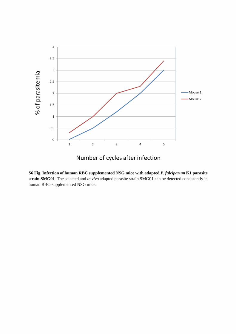
S6 Fig. Infection of human RBC supplemented NSG mice with adapted P. falciparum K1 parasite strain SMG01. The selected and in vivo adapted parasite strain SMG01 can be detected consistently in human RBC-supplemented NSG mice.

S1 Table. Ex vivo infection of human RBCs from humanized mice by P. falciparum (3D7). Total human RBCs and parasitemia in 14 mice are shown. Parasitemia ranged between 0.02% (mouse 4) and 1.6% (mouse 6) or 1% to 59.3% when normalized to human RBCs.

![RESEARCHARTICLE Comparative DeNovo Transcriptomeor.nsfc.gov.cn/bitstream/00001903-5/239624/1/1000013919268.pdf · domainandanintracellularserine/threoninekinasedomain[13–14]](https://img.dokumen.tips/doc/110x75/5a72fef57f8b9ac0538e42a1/researcharticle-comparative-denovo-transcriptomeornsfcgovcnbitstream00001903-523962411000013919268pdf.jpg)