Embed Size (px)
Citation preview

Research Priorities for Chagas Disease, H
AT and Leishmaniasis
975 W H O T e c h n i c a l R e p o r t S e r i e s
975
Technical Report of the TDR Disease Reference Group on Chagas Disease, Human African Trypanosomiasis and Leishmaniasis
Research Priorities for Chagas Disease, Human African Trypanosomiasis and Leishmaniasis
WH
O Technical Report Series
Research Priorities for Chagas Disease, HAT and Leishmaniasis
This report provides a review and analysis of the research landscape for three diseases – Chagas disease, human African trypanosomiasis and leishmaniasis – that disproportionately afflict poor and remote populations with limited access to health services. It represents the work of the disease reference group on Chagas Disease, Human African Trypanosomiasis and Leishmaniasis (DRG3) which was established to identify key research priorities through review of research evidence and input from stakeholders' consultations.The diseases, which are caused by related protozoan parasites, are described in terms of their epidemiology and disease burden, clinical forms and pathogenesis, HIV coinfection, diagnosis, drugs and drug resistance, vaccines, vector control, and health-care interventions. Priority areas for research are identified based on criteria such as public health relevance, benefit and impact on poor populations and equity, and feasibility.The priorities are found in the areas of diagnostics, drugs, vector control, asymptomatic infection, economic analysis of treatment and vector control methods, and in some specific issues such as surveillance methods or transmission-blocking vaccines for particular diseases.This report will be useful to researchers, policy and decision-makers, funding bodies, implementation organizations, and civil society.This is one of ten disease and thematic reference group reports that have come out of the TDR Think Tank, all of which have contributed to the development of the Global Report for Research on Infectious Diseases of Poverty, available at: www.who.int/tdr/capacity/global_report.

Global Report for Research on Infectious Diseases of PovertyAvailable online at: www.who.int/tdr/capacity/global_report
Research Priorities for Zoonoses and Marginalized InfectionsWHO Technical Report Series, No. 971 (120 pages)Available online at: www.who.int/tdr/publications/zoonoses
Research Priorities for Helminth InfectionsWHO Technical Report Series, No. 972 (196 pages)Available online at: www.who.int/tdr/publications/helminth_infections
Research Priorities for the Environment, Agriculture and Infectious Diseases of PovertyWHO Technical Report Series, No. 976 (140 pages)Available online at: www.who.int/tdr/publications/environment
SELECTED WHO PUBLICATIONS OF RELATED INTEREST
Further information on these and other WHO publications can be obtained fromWHO Press, World Health Organization, 1211 Geneva 27, Switzerland
(tel.: +41 22 791 3264; fax: +41 22 791 4857; e-mail: [email protected];order on line: http://www.who.int/bookorders)
The World Health Organization was established in 1948 as a specialized agency of the United Nations serving as the directing and coordinating authority for international health matters and public health. One of WHO’s constitutional functions is to provide objective and reliable information and advice in the field of human health, a responsibility that it fulfils in part through its extensive programme of publications. The Organization seeks through its publications to support national health strategies and address the most pressing public health concerns of populations around the world. To respond to the needs of Member States at all levels of development, WHO publishes practical manuals, handbooks and training material for specific categories of health workers; internationally applicable guidelines and standards; reviews and analyses of health policies, programmes and research; and state-of-the-art consensus reports that offer technical advice and recommendations for decision-makers. These books are closely tied to the Organization’s priority activities, encompassing disease prevention and control, the development of equitable health systems based on primary health care, and health promotion for individuals and communities. Progress towards better health for all also demands the global dissemination and exchange of information that draws on the knowledge and experience of all WHO’s Member Countries and the collaboration of world leaders in public health and the biomedical sciences. To ensure the widest possible availability of authoritative information and guidance on health matters, WHO secures the broad international distribution of its publications and encourages their translation and adaptation. By helping to promote and protect health and prevent and control disease throughout the world, WHO’s books contribute to achieving the Organization’s principal objective – the attainment by all people of the highest possible level of health.
The WHO Technical Report Series makes available the findings of various international groups of experts that provide WHO with the latest scientific and technical advice on a broad range of medical and public health subjects. Members of such expert groups serve without remuneration in their personal capacities rather than as representatives of governments or other bodies; their views do not necessarily reflect the decisions or the stated policy of WHO. An annual subscription to this series, comprising four to six such reports, costs CHF 150.00/US$ 180.00 (CHF 105.00/US$ 126.00 in developing countries). For further information, please contact: WHO Press, World Health Organization, 20 Avenue Appia, 1211 Geneva 27, Switzerland (tel. +41 22 791 3264; fax: +41 22 791 4857; e-mail: [email protected]; order on line: http://www.who.int/bookorders).

Research Priorities for Chagas Disease, Human African Trypanosomiasis and Leishmaniasis
W H O T e c h n i c a l R e p o r t S e r i e s
This report contains the collective views of an international group of experts and does not necessarily represent the decisions or the stated policy of the World Health Organization
Technical Report of the TDR Disease Reference Group on Chagas Disease, Human African Trypanosomiasis and Leishmaniasis
© World Health Organization 2012
All rights reserved. Publications of the World Health Organization are available on the WHO web site (www.who.int) or can be purchased from WHO Press, World Health Organization, 20 Avenue Appia, 1211 Geneva 27, Switzerland (tel.: +41 22 791 3264; fax: +41 22 791 4857; e-mail: [email protected]).
Requests for permission to reproduce or translate WHO publications – whether for sale or for noncommercial distribution – should be addressed to WHO Press through the WHO web site (http://www.who.int/about/licensing/copyright_form/en/index.html).
The designations employed and the presentation of the material in this publication do not imply the expression of any opinion whatsoever on the part of the World Health Organization concerning the legal status of any country, territory, city or area or of its authorities, or concerning the delimitation of its frontiers or boundaries. Dotted lines on maps represent approximate border lines for which there may not yet be full agreement.
The mention of specific companies or of certain manufacturers’ products does not imply that they are endorsed or recommended by the World Health Organization in preference to others of a similar nature that are not mentioned. Errors and omissions excepted, the names of proprietary products are distinguished by initial capital letters.
All reasonable precautions have been taken by the World Health Organization to verify the information contained in this publication. However, the published material is being distributed without warranty of any kind, either expressed or implied. The responsibility for the interpretation and use of the material lies with the reader. In no event shall the World Health Organization be liable for damages arising from its use.
This publication contains the collective views of an international group of experts and does not necessarily represent the decisions or the policies of the World Health Organization.
Printed in Italy
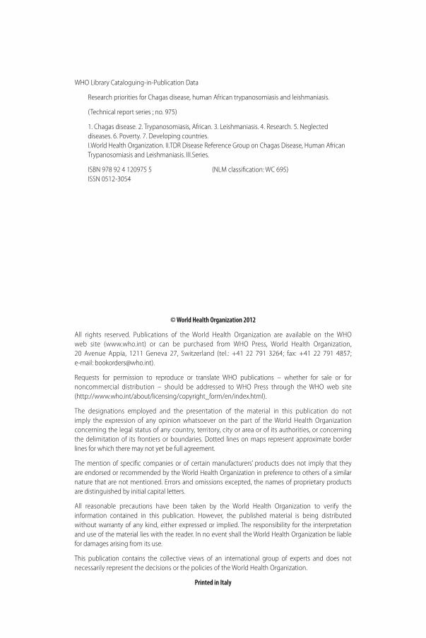
WHO Library Cataloguing-in-Publication Data
Research priorities for Chagas disease, human African trypanosomiasis and leishmaniasis.
(Technical report series ; no. 975)
1. Chagas disease. 2. Trypanosomiasis, African. 3. Leishmaniasis. 4. Research. 5. Neglected diseases. 6. Poverty. 7. Developing countries. I.World Health Organization. II.TDR Disease Reference Group on Chagas Disease, Human African Trypanosomiasis and Leishmaniasis. III.Series.
ISBN 978 92 4 120975 5 (NLM classification: WC 695) ISSN 0512-3054
iii
Contents
WHO/TDR Disease Reference Group on Chagas Disease, Human African Trypanosomiasis and Leishmaniasis v
Abbreviations and acronyms vii
Executive summary xi
1. Introduction 11.1 Reasons for setting research priorities 41.2 TDR stewardship mandate 51.3 Goal of this report /strategic objectives 61.4 Overview 6
2. Methodology and prioritization 92.1 Authoritative evidence review to define thematic areas 92.2 Stakeholders' consultation and second round of criteria-based ranking 92.3 Final round of ranking 10
3. Epidemiology and burden of disease 113.1 Chagas disease 113.2 Human African trypanosomiasis 143.3 Leishmaniasis 17
4. Clinical forms, pathogenesis and HIV coinfection 214.1 Chagas disease 21
4.1.1 Clinical presentations 214.1.2 Chagas heart disease 214.1.3 Gastrointestinal manifestations 234.1.4 Congenital Chagas disease 234.1.5 Chagas disease and HIV coinfection 23
4.2 Human African trypanosomiasis 244.2.1 Pathogenesis 244.2.2 Clinical presentation 24
4.3 Leishmaniasis 264.3.1 Clinical presentations 26
5. Diagnosis 315.1 Chagas disease 31
5.1.1 Parasitological diagnosis 315.1.2 Serology 315.1.3 Quantitative and qualitative detection of parasite DNA 315.1.4 Diagnosis in newborns 345.1.5 Diagnosis of therapy efficacy and cure 345.1.6 Diagnosis of drug resistant Chagas disease 35
5.2 Human African trypanosomiasis 355.2.1 Demonstration of parasite 355.2.2 Serology 355.2.3 Quantitative and qualitative detection of parasite DNA 385.2.4 Staging of the disease 38

iv
5.3 Leishmaniasis 385.3.1 Parasitological diagnosis 385.3.2 Serology 405.3.3 Detection of parasite products 415.3.4 Quantitative and qualitative detection of parasite DNA 41
6. Drugs and drug resistance 436.1 Current treatment and development of new drugs 47
6.1.1 Chagas disease 476.1.2 Human African trypanosomiasis 496.1.3 Leishmaniasis 50
7. Vaccines against Chagas disease, human African trypanosomiasis and leishmaniasis 55
7.1 Overview 557.2 Chagas disease 557.3 Human African trypanosomiasis 587.4 Leishmaniasis 59
8. Vector control 638.1 Chagas disease: triatomine bug control 638.2 Human African trypanosomiasis: tsetse fly control 64
8.2.1 Tsetse species of highest priority 658.2.2 Control activities at all levels 668.2.3 Costs of vector control 67
8.3 Leishmaniasis: sandfly control 678.3.1 Source reduction 678.3.2 Targeting sugar-feeding adult sandflies 728.3.3 Extermination of sandflies feeding on domestic animals 728.3.4 Flight barriers for preventing sandflies from reaching houses 738.3.5 Protection of homes – indoor residual insecticide spraying 748.3.6 Protection of rooms/porches – insecticide diffusers and coils 748.3.7 Personal protection – insecticide-treated nets and repellents 748.3.8 Integrated disease-control campaigns 75
9. Economic evaluation of health-care interventions 77
10. Research priority recommendations 81
Acknowledgements 83
References 85
Appendices 99

v
WHO/TDR Disease Reference Group on Chagas Disease, Human African Trypanosomiasis and Leishmaniasis (DRG3) 2009–2010
MembersProfessor M. Barrett, University of Glasgow, Glasgow Scotland
Professor M. Boelaert, Institute of Tropical Medicine, Epidemiology and Disease Control Unit, Antwerp, Belgium
Ms M. Castillo-Riquelme, Health Economics Consultant, Ministerio de Salud, Departamento de Economía de la Salud, Ministry of Health, Chile
Professor M.J. Lehane, Liverpool School of Tropical Medicine, Liverpool, England
Professor P. Lutumba, Institut National de Recherche Bio-Médicale, Kinshasa University, Kinshasa, Democratic Republic of Congo
Professor E. Matovu, College of Veterinary Medicine, Animal Resources and Biosecurity, Makerere University, Kampala, Uganda
Professor M.M. Mukhtar, Institute of Endemic Diseases, University of Khartoum, Khartoum, Sudan (co-Chair)
Dr D. Sacks, Head, Intracellular Parasite Biology Section, Laboratory of Parasitic Diseases, National Institutes of Health, National Institute of Allergy and Infectious Diseases, Bethesda MD, USA
Professor C. Schofield, Coordinator, Latin American Network for Research on the Biology and Control of Triatominae (ECLAT), Faculty of Infectious and Tropical Diseases, London School of Hygiene and Tropical Medicine, London, England
Dr S.A. Sosa-Estani, Director, National Institute of Parasitology, ANLIS Malbrán, Ministerio de Salud, Argentina
Professor K. Stuart, Founder, Seattle Biomedical Research Institute, Seattle, WA, USA (Chair)
Professor S. Sundar, Institute of Medical Sciences, Banaras Hindu University, Varanasi, India
Professor A. Warburg, Faculty of Medicine, Hebrew University, Einkerem, Israel
Professor B. Zingales, Instituto de Quimica, Universidade de São Paulo, Sao Paulo, Brazil (co-Chair)
Career Development FellowDr S. Bakhiet, Institute of Endemic Diseases, University of Khartoum, Sudan
SecretariatDr D. Kioy, Special Programme for Research and Training in Tropical Diseases, World
Health Organization, Geneva, Switzerland (Secretary)

vi
WH
O T
echn
ical
Rep
ort S
erie
s, N
o. 9
75, 2
012
Research Priorities for Chagas Disease, HAT and Leishmaniasis Report of the TDR Disease Reference Group
Dr A.M.J Oduola, Coordinator, Special Programme for Research and Training in Tropical Diseases, World Health Organization, Geneva, Switzerland
Dr Michael Wilson, Scientist, Special Programme for Research and Training in Tropical Diseases, WHO, Geneva, Switzerland

vii
Abbreviations and acronyms
ADCL Anergic diffuse cutaneous leishmaniasis
AIDS Acquired immune deficiency syndrome
BCG Bacille Calmette-Guérin
BDCL Borderline disseminated cutaneous leishmaniasis
CATT Card agglutination test for trypanosomiasis
CD Cluster of differentiation
CL Cutaneous leishmaniasis
CNS Central nervous system
CpG ODN CpG oligodeoxynucleotides
CSA Crude soluble antigen
CSF Cerebrospinal fluid
CTC Capillary tube centrifugation
DALYs Disability adjusted life years
DAT Direct agglutination test
DDT Dichlorodiphenyltrichloroethane
DEET N, N-diethyl-meta-toluamide
DNDi Drugs for Neglected Diseases Initiative
DRG Disease Reference Group
DTH Delayed-type hypersensitivity test
DTU Discrete typing unit
ELISA Enzyme-linked immunosorbent assay
FIND Foundation for Innovative New Diagnostics
GDP Gross domestic product
GIS Geographic information system
GP63 Glycoprotein 63
HAT Human African trypanosomiasis
HIV Human immunodeficiency virus
HLA Human leukocyte antigen

viii
WH
O T
echn
ical
Rep
ort S
erie
s, N
o. 9
75, 2
012
Research Priorities for Chagas Disease, HAT and Leishmaniasis Report of the TDR Disease Reference Group
ICAM-1 Intercellular adhesion molecule 1
ICT Immunochromatography test
IFN-γ Interferon-gamma
IG Immunoglobulin
IHA Indirect haemaglutination
IIF Indirect immunofluorescence
ITN Insecticide treated nets
LAMP Loop mediated isothermal amplification
LLINs Long-lasting insecticidal mosquito nets
LNP Lymph node puncture
LPG Lipophosphoglycan
LRC Leishmania recidiva cutis
LST Leishmanin skin test
mAECT Miniature anion exchange centrifugation technique
MASP Mannan-binding lectin-associated serine protease
MCL Mucocutaneous leishmaniasis
MH Microhaematocrit concentration method
ML Mucosal leishmaniasis
MPL-SE Monophosphoryl lipid A plus squalene
NECT Nifurtimox-eflornithine combination therapy
NTDs Neglected tropical diseases
PATTEC Pan African Tsetse and Trypanosomiasis Eradication Campaign
PCR Polymerase chain reaction
PKDL Post kala-azar dermal leishmaniasis
POC Point of care
QALY Quality adjusted life year
QBC Quantitative buffy coat
R&D Research and development
RFLP Restriction fragment length polymorphism

Abbreviations and acronyms
ix
rK39 Recombinant K39
RT-PCR Reverse transcriptase polymerase chain reaction
TBF Thick blood film
TH1 T helper cell type 1
TH2 T helper cell type 2
TDR Special Programme for Research and Training in Tropical Diseases
TRG Thematic reference group
USD United States dollar
VL Visceral leishmaniasis
VSG Variable surface glycoprotein
WHO World Health Organization
YLL Years of life lost


xi
Executive summaryThe Disease Reference Group on Chagas Disease, Human African Trypanosomiasis and Leishmaniasis (DRG3) was part of an independent think-tank of international experts established by the Special Programme for Research and Training in Tropical Diseases (TDR) to identify key research priorities through systematic review of research evidence and input from stakeholders. These three distinct insect-borne diseases, while caused by related kinetoplastid protozoan pathogens, have dissimilar geographical distributions – a reflection of their different insect vectors and range of vector contact with humans. The diseases disproportionately afflict poor and remote populations with limited access to health services; the pathogenic mechanisms are poorly understood but typically entail immunological processes.
A team of experts identified research gaps by critically reviewing the disease landscape and state of research – from discovery through to implementation research – and incorporating stakeholder input; they also identified research opportunities for developing the knowledge and tools needed for treatment and prevention of the three diseases. The stakeholders included laboratory, clinical and field researchers, and personnel from national or international health organizations from 16 countries and 4 continents; they presented their research priorities and the reasons for their selection. A Delphic approach was used to integrate the input and prioritize areas for future research. Criteria were identified on which to base the selection of gaps and opportunities and ranking of priorities; they included critical needs, public health relevance, the benefit and magnitude of the impact on populations, the ratio of feasibility and scientific difficulty to the associated cost/benefit, cure versus prevention, impact on poor populations and equity implications, short and long-term impact, and sustainability. Consensus was achieved in identifying priority areas for future research and investment, including areas common to the three diseases as well as disease-specific priorities. These priorities are intended to be useful to researchers, policy and decision-makers, funding bodies, implementation organizations, and civil society.
The priorities fall into the general areas of diagnostics, drugs, vaccines, vector control and health systems. The diagnostics priorities include improved means to identify: specific disease states – from asymptomatic and chronic to cured conditions, and drug resistant and other types of parasite variants. Specific diagnostics are needed for: infants of T. cruzi-infected mothers, second-stage human African trypanosomiais, and visceral leishmaniasis in different global regions. Besides case finding and management, these diagnostic tools are also of value to epidemiologic and disease control studies, and drug and vaccine trials.
A robust pipeline of drugs for the three diseases, especially for the chronic and more lethal disease stages, was recognized as a priority area. There is pressing need to replace the few drugs that are currently available and which have
xii
WH
O T
echn
ical
Rep
ort S
erie
s, N
o. 9
75, 2
012
Research Priorities for Chagas Disease, HAT and Leishmaniasis Report of the TDR Disease Reference Group
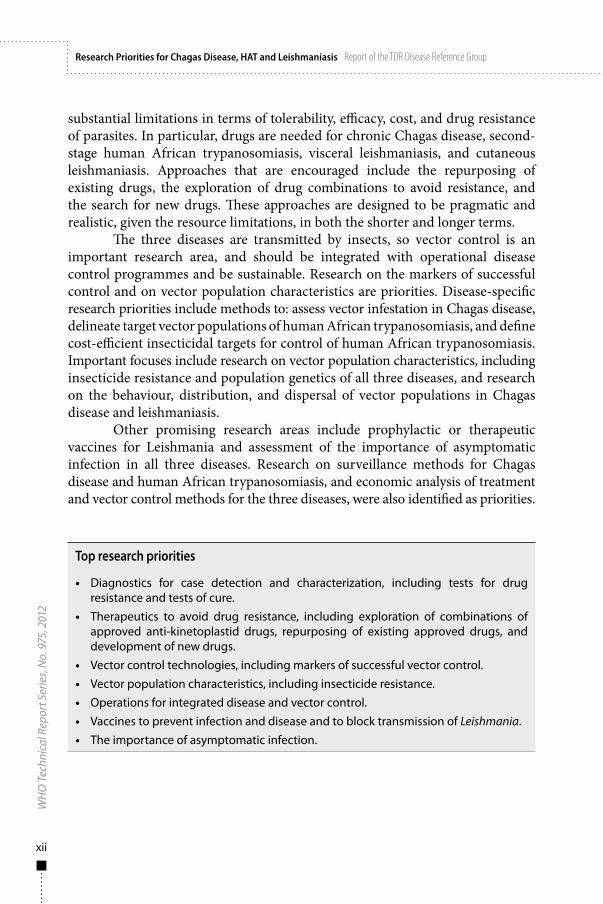
substantial limitations in terms of tolerability, efficacy, cost, and drug resistance of parasites. In particular, drugs are needed for chronic Chagas disease, second-stage human African trypanosomiasis, visceral leishmaniasis, and cutaneous leishmaniasis. Approaches that are encouraged include the repurposing of existing drugs, the exploration of drug combinations to avoid resistance, and the search for new drugs. These approaches are designed to be pragmatic and realistic, given the resource limitations, in both the shorter and longer terms.
The three diseases are transmitted by insects, so vector control is an important research area, and should be integrated with operational disease control programmes and be sustainable. Research on the markers of successful control and on vector population characteristics are priorities. Disease-specific research priorities include methods to: assess vector infestation in Chagas disease, delineate target vector populations of human African trypanosomiasis, and define cost-efficient insecticidal targets for control of human African trypanosomiasis. Important focuses include research on vector population characteristics, including insecticide resistance and population genetics of all three diseases, and research on the behaviour, distribution, and dispersal of vector populations in Chagas disease and leishmaniasis.
Other promising research areas include prophylactic or therapeutic vaccines for Leishmania and assessment of the importance of asymptomatic infection in all three diseases. Research on surveillance methods for Chagas disease and human African trypanosomiasis, and economic analysis of treatment and vector control methods for the three diseases, were also identified as priorities.
Top research priorities
• Diagnostics for case detection and characterization, including tests for drug resistance and tests of cure.
• Therapeutics to avoid drug resistance, including exploration of combinations of approved anti-kinetoplastid drugs, repurposing of existing approved drugs, and development of new drugs.
• Vector control technologies, including markers of successful vector control.• Vector population characteristics, including insecticide resistance.• Operations for integrated disease and vector control.• Vaccines to prevent infection and disease and to block transmission of Leishmania.• The importance of asymptomatic infection.

1
1 Details on TDR's strategy can be found at: http://www.who.int/tdr/about2 Details of TDR's research priority reports can be found at: www.who.int/tdr/capacity/gap_analysis
1. IntroductionAs part of its ten-year strategy1 to foster “an effective global research effort on infectious diseases of poverty in which disease-endemic countries play a pivotal role”, the Special Programme for Research and Training in Tropical Diseases (TDR) established a global research Think Tank of 125 international experts to continually and systematically review evidence, assess research needs and, following periodic national and regional stakeholder consultations, to set research priorities for accelerating the control of infectious diseases of poverty. Working in ten disease-specific and thematic reference groups (DRGs/TRGs), the experts are crucial contributors to TDR's stewardship mandate for the acquisition and analysis of information on infectious diseases of poverty2. Their work is ultimately intended to promote control-relevant research, achieve research innovation, and enhance the capacity of disease-endemic countries to resolve public health problems related to the disproportionate burden of infectious diseases among the poor.
The Think Tank was designed to draw on the best expertise internationally, and to maximize partnerships with countries most affected by diseases of poverty. The ten reference groups making up the Think Tank include researchers and public health experts from the most affected countries; these countries also host the groups. WHO country and regional offices support both the reference groups and broad-based stakeholder consultations (see Appendix 1).
DRG3 consisted of 14 experts recognized as academic or public health leaders in the area of Chagas disease, human African trypanosomiasis (HAT) and/or leishmaniasis; they came from research institutions, international organizations, bilateral institutions, health and medical organizations, governmental and inter-governmental organizations worldwide. Particular attention was paid to the geographical distribution – to ensure disease-endemic country and regional input as well as technical input – and gender balance of the membership. Members were formally appointed by the Director of TDR for an initial period of two years. The chair and co-chairs of the group were selected on the basis of their internationally-recognized research, their long-term experience in research and control related to the three diseases, and their experience in disease-endemic countries (see Appendix 2).
Regional and national stakeholder consultations enabled validation, endorsement and setting of final research priorities, and ensured that the work of the group is authoritative, scientifically credible, and relevant for policy. In

2
WH
O T
echn
ical
Rep
ort S
erie
s, N
o. 9
75, 2
012
Research Priorities for Chagas Disease, HAT and Leishmaniasis Report of the TDR Disease Reference Group
addition to email consultations during the initial phase of gap and opportunity identification, a stakeholders' consultation meeting on Chagas disease, HAT and leishmaniasis was hosted by the Instituto Oswaldo Cruz (FIOCRUZ), 29–31 March 2010, in Rio de Janeiro, Brazil. The meeting was organized by WHO/TDR in collaboration with Instituto Oswaldo Cruz and the WHO Country Office, Brazil, with the goal of discussing and contributing to the setting of research priorities for the three diseases.
To ensure that the countries most affected by diseases of poverty contributed to, and shared ownership of, the research agenda emerging from this initiative, the reference groups were hosted by disease-endemic countries in partnership with WHO country and regional offices (see Appendix 1).
The three distinct diseases, Chagas disease, HAT and leishmaniasis, disproportionately affect low and lower-middle income countries, typically afflicting their poorest and most isolated populations. The low visibility and lack of perceived exposure to the diseases as well as the paucity of data contribute to the low priority and limited control programmes afforded by national health programmes. In addition, the afflicted lack a political voice due to their poverty and the social stigmatization and discrimination resulting from the disabling and disfiguring consequences of the diseases. Nevertheless, as illustrated by Millennium Development Goals 4, 5, and 6 that relate to maternal and child health and mortality and infectious diseases, these diseases illustrate a right to health issue. While health interventions and research and development (R&D) for the three diseases have long been inadequate and underfunded, hopefully the increased awareness of the diseases will enable critical research.
Although Chagas disease, HAT and leishmaniasis are substantially different vector-borne diseases, they are caused by related kinetoplastid protozoan pathogens. The diseases have characteristic geographic and socioeconomic distributions that reflect the ranges and environments of the insect vectors that transmit them. Despite the large numbers of people at risk of infection and the substantial burden of disease, which includes lethal, horrific, and/or debilitating consequences, with few exceptions no major interventions have been developed for generations. The pathogens are each transmitted by a different haematophagous insect, within which the parasite has a complex life-cycle. The infective and invasive forms of Trypanosoma cruzi, the causal agent of Chagas disease, develop in the hindgut of triatomine bugs and are transmitted in the bug faeces deposited during feeding; the other major routes of parasite transmission are congenital, transfusion of infected blood, and ingestion of contaminated food or beverage. The infective forms of the Trypanosoma brucei group, which includes animal infective trypanosomes of substantial economic importance, develop in the midgut and salivary glands of tsetse flies, and are transmitted through the proboscis when the fly takes a blood meal; T. brucei is also occasionally transmitted mechanically

Introduction
3
by various biting flies, without an intervening developmental cycle, for example in (stock) animals that have extensive insect exposure. The infective forms of Leishmania develop in the region of the stomodeal valve of sandflies, between the foregut and proboscis, and are transmitted within a gel-like substance that is extruded through the proboscis during feeding. Such vector diversity means that insect biological, ecological, and behavioural factors must be considered in vector control programmes. The vectors' characteristic ranges are primarily tropical and sub-tropical, and the parasites also have animal reservoirs. Although the diseases largely afflict populations in low resource settings, the vectors – even infected vectors – also occur in more highly resourced areas. However, there is substantial overlap between the ranges of the insect vectors and the geographic areas with low resources.
Thus the biodiversity of both insect vector and pathogen will necessarily affect the utility of diagnostics, the efficacy of drugs and diagnostics, and any disease and vector control programmes.
Pathogenesis differs among the three diseases, reflecting the different host target tissues and different host–pathogen interactions. Chagas disease parasites exit the bloodstream and end up in the cytoplasm of host cells; various host cell types are infected during the initial acute phase of infection, but disease is associated mainly with chronic infection of cardiac and enteric tissue cells. The cardiomyopathy entails ventricular hypertrophy, which typically progresses to debilitating arrhythmia and requires a pacemaker; it can ultimately progress to severe cardiac damage and result in sudden death. HAT parasites rapidly proliferate extracellularly within the blood and lymphatic systems during the initial acute phase, evading the host immune response by an elaborate process of surface antigen variation. The parasites subsequently invade the central nervous system (CNS), which may provide an environment that is less susceptible to immune clearance; CNS HAT has multiple clinical consequences as well as extreme behavioural sequellae and is lethal if untreated. Leishmania parasites infect macrophages of the human host where they reside within the phagolysosomes; they cause a spectrum of diseases ranging from cutaneous lesions, which vary in severity and duration, to visceral disease, which is often lethal. The type of disease is generally correlated with the infecting species of Leishmania. Cutaneous disease is associated with L. major, tropica, mexicana, amazonensis and donovani; visceral disease with L. donovani, infantum and chagasi; and mucocutaneous disease, which is horribly disfiguring, with the L. viannia complex (e.g. L. braziliensis and guyanensis).
Thus the strains and species of all three pathogenic trypanosomatids manifest considerable genetic and biological diversity; nevertheless, host factors also likely contribute to the disease characteristics. Details of the pathogenesis of the three diseases are not well understood but it appears that all have a
4
WH
O T
echn
ical
Rep
ort S
erie
s, N
o. 9
75, 2
012
Research Priorities for Chagas Disease, HAT and Leishmaniasis Report of the TDR Disease Reference Group
substantial immunopathogenic component. Elucidating the characteristics of immune responses to infection should aid the understanding of immune control and pathogenesis mechanisms, which should aid the development of diagnostics, potential immunotherapeutics, and the possible development of vaccines.
The parasites that are responsible for Chagas disease, HAT and leishmaniasis have been the subject of considerable molecular and immunological analyses. This may be because they can be readily cultivated in the laboratory and can infect mouse model systems. They are also reasonably amenable to laboratory experimentation, which has led to many novel fundamental discoveries. However, there are substantial gaps in knowledge especially with respect to disease specific characteristics that are the foundation for needed interventions.
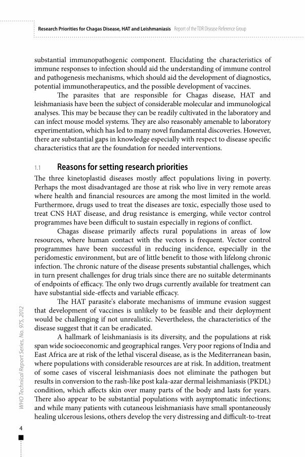
1.1 Reasons for setting research prioritiesThe three kinetoplastid diseases mostly affect populations living in poverty. Perhaps the most disadvantaged are those at risk who live in very remote areas where health and financial resources are among the most limited in the world. Furthermore, drugs used to treat the diseases are toxic, especially those used to treat CNS HAT disease, and drug resistance is emerging, while vector control programmes have been difficult to sustain especially in regions of conflict.
Chagas disease primarily affects rural populations in areas of low resources, where human contact with the vectors is frequent. Vector control programmes have been successful in reducing incidence, especially in the peridomestic environment, but are of little benefit to those with lifelong chronic infection. The chronic nature of the disease presents substantial challenges, which in turn present challenges for drug trials since there are no suitable determinants of endpoints of efficacy. The only two drugs currently available for treatment can have substantial side-effects and variable efficacy.
The HAT parasite's elaborate mechanisms of immune evasion suggest that development of vaccines is unlikely to be feasible and their deployment would be challenging if not unrealistic. Nevertheless, the characteristics of the disease suggest that it can be eradicated.
A hallmark of leishmaniasis is its diversity, and the populations at risk span wide socioeconomic and geographical ranges. Very poor regions of India and East Africa are at risk of the lethal visceral disease, as is the Mediterranean basin, where populations with considerable resources are at risk. In addition, treatment of some cases of visceral leishmaniasis does not eliminate the pathogen but results in conversion to the rash-like post kala-azar dermal leishmaniasis (PKDL) condition, which affects skin over many parts of the body and lasts for years. There also appear to be substantial populations with asymptomatic infections; and while many patients with cutaneous leishmaniasis have small spontaneously healing ulcerous lesions, others develop the very distressing and difficult-to-treat

Introduction
5
diffuse cutaneous disease or the horribly disfiguring mucocutaneous disease which erodes the oral and nasal mucosae.
All three diseases have animal reservoirs, typically wild animal populations that are often asymptomatic. However, domestic animals such as pigs and dogs can also be reservoirs and these may have greater impact on human populations because of their physical proximity. Ironically, while leishmaniasis is a disease that afflicts some of the poorest populations, a commercial market may exist for a vaccine or treatment of leishmaniasis for companion dogs in developed countries. Similarly, species of African trypanosomes afflict cattle (bovines, camels, horses), and some populations at risk for the human disease feel that it is almost as important to develop interventions for the cattle disease, reflecting their economic dependence on these animals.
The kinetoplastid diseases are classic diseases of poverty. The motivation for development of the existing drugs was commercial, reflecting the historical colonial relationship; commercial research and development fell off sharply as this era waned. Recognition of the global nature of many diseases and their direct and indirect effects on economies, conflict, and health equity has resulted in a resurgence of motivation, albeit primarily by the public sector. While the HIV pandemic and periodic emergence of other diseases have raised awareness of the global impact of many diseases, this has also complicated efforts to combat kinetoplastid diseases since Leishmania and T. cruzi are opportunistic pathogens and any immune compromise can convert a chronic infection to an acute life-threatening condition. Similarly, HIV complicates Chagas disease. Nevertheless, the ability to develop diagnostic and therapeutic tools, and in some cases perhaps even vaccines, is within current technical capabilities for leishmaniasis and Chagas disease. Combined with integrated and sustainable disease and vector control programmes, many of the diseases could be eliminated with consequent human benefits.
1.2 Stewardship mandateResearch on the three kinetoplastid diseases has been a priority area for WHO/TDR since its inception and is one of the ten emphasis areas selected by WHO/TDR in its Stewardship strategy. This strategy designated four Thematic Reference Groups (TRGs) and six Disease Reference Groups (DRGs). The Chagas Disease, HAT and Leishmaniasis Reference Group (DRG3) has, among its members, relevant expertise and experience with the three diseases, spanning basic research, implementation research and health systems research. The Group was charged with providing an evidence-based analysis of the status of research in the field, identifying gaps and opportunities, and engaging stakeholders from disease endemic and developed countries including basic and translational researchers from the public, private, not-for-profit and for-profit sectors as well as funders

6
WH
O T
echn
ical
Rep
ort S
erie
s, N
o. 9
75, 2
012
Research Priorities for Chagas Disease, HAT and Leishmaniasis Report of the TDR Disease Reference Group
and decision-makers who might influence policy and action. The intent is to identify priority research areas that would stimulate innovative and actionable research.
1.3 Goal of this report /strategic objectivesThe aim of DRG3 for Chagas disease, HAT and leishmaniasis, is to:
■ Provide expert analysis and perspective by research experts, integrating the current status of research on these diseases and incorporating input from relevant stakeholders.
■ Identify research gaps and opportunities where research activities can provide knowledge and tools that can lead to interventions to alleviate or prevent disease.
■ Identify priority areas on which to focus research activity and investment to advance the understanding of these diseases and contribute to health improvement.
■ Build a consensus on priority areas that are common to the three diseases, are disease specific, and/or cut across the other DRGs and TRGs.
The intended audience for the results of the deliberations and conclusions includes policy-makers, especially in resource poor and endemic countries; national, regional and local decision-makers; funding organizations; UN agencies; civil society; and researchers and trainees.
1.4 OverviewIn reports of analyses and priorities, issues of research on disease characteristics (basic studies), detection and characterization (diagnostics), prevention (vaccines and operational), and treatment (drugs), are bound to be addressed. In this report, an attempt has been made to integrate prior reports and the priorities they identified. Both disease-specific issues and priorities, and themes that span the three diseases, are identified. It is hoped that some of the themes will also fit in with the other DRGs and TRGs and generate wide-ranging synergy. Overall it is clear that the current therapies used for treatment of Chagas disease, HAT and leishmaniasis are limited due to a combination of toxicity, cost, limited or undetermined efficacy, and the progressive development of drug resistance. Thus drug R&D is needed. Current diagnostic procedures and tools are not effective enough and there is a need to develop new and/or improved diagnostic tools for case finding and management, for epidemiological studies, and for disease surveillance and associated control programmes. Diagnostics are also needed along with definitions of disease and cure in order to monitor clinical trials of

Introduction
7
drugs. Realistic financial and capacity assessment indicates that the search for needed new therapies will require practical and innovative approaches both at the bench and in the organization of research in order to take advantage of existing opportunities while looking to the long term (e.g. avoiding drug resistance). Research on prevention strategies is unlikely to rely on vaccines, except perhaps for Leishmania, but rather on vector control, which has shown positive impact.


9
2. Methodology and prioritizationDRG3 consisted of 14 academic or public health leaders in the areas of Chagas disease, human African trypanosomiasis (HAT) and/or leishmaniasis, as mentioned in the introduction. The members came from research institutions, international organizations, health and medical organizations, governmental and inter-governmental organizations worldwide. The chair and co-chairs were selected on the basis of their internationally-recognized research and long-term experience in research and control related to these diseases, and their experience working in disease endemic countries (see Appendix 2). The reference group was hosted by Sudan and Brazil, in partnership with the WHO country and regional offices (Appendix 1).
A multi-stage interactive process was used to identify promising areas for research; this entailed assembling, evaluating, ranking, and reducing the number of priorities identified. The aim was to enable researchers, funding agencies, policy-makers and other public health stakeholders to integrate relevant information and expert views and avoid conflict of interest as they consider various options for making decisions.
2.1 Authoritative evidence review to define thematic areasThe DRG3 team identified thematic areas consistent with the TDR/WHO stewardship mandate. Their considerations spanned basic research through implementation and health systems research for the three diseases, and included: epidemiologic, socioeconomic and clinical perspectives; diagnosis, treatment and drug resistance; drug discovery and development; host/vector and host/pathogen interactions; immunology and vaccine development; vector control and implementation.
An initial report enumerated 27 trans-disease priorities, and an additional two priorities specific to Chagas disease, three to HAT, and nine to leishmaniasis. The priorities were presented and discussed at a combined meeting of the DRG and TRG chairs and co-chairs (2–4 September 2009, TDR/WHO Geneva), along with a tutorial on prioritization methods.
2.2 Stakeholders' consultation and second round of criteria-based ranking
Concept validation and deliberation on the priorities and criteria (values) used to rank the priorities were obtained during a Stakeholders Consultation Meeting (29 March 2010, FIOCRUZ, Rio de Janeiro). The stakeholders included staff from national ministries of health, NGOs and international organizations, and researchers, and represented the three diseases and the thematic areas. They presented their views in individual oral presentations in plenary sessions and

10
WH
O T
echn
ical
Rep
ort S
erie
s, N
o. 9
75, 2
012
Research Priorities for Chagas Disease, HAT and Leishmaniasis Report of the TDR Disease Reference Group
each presentation was followed by discussion. A total of 40 research priorities were identified.
The second round of criteria-based priority-setting took place during the 2nd Annual DRG3 Meeting (30–31 March 2010, FIOCRUZ, Rio de Janeiro), at which input from the preceding stakeholders' consultation meeting was integrated. The top ten priorities for each disease were formulated, employing the Delphi method for prioritization. The ranking of research priorities took into consideration the following values:
■ Curative versus preventive relevance ■ Public health relevance and impact on population health ■ Size of population benefiting from research ■ Pro-poor/poverty alleviation ■ Feasibility/scientific difficulty (cost–benefit) ■ Equity implications ■ Critical need ■ Long-term and hence sustainable benefit.
Consensus was achieved in ranking the priorities, which were fitted to a matrix organized by disease and interventional goals and that, in keeping with the Stewardship mandate, spanned basic to operational research. Themes that were common to two or three of the diseases were also identified.
2.3 Final round of rankingElectronic consultation of DRG3 members was used to generate the final list of seven priorities. This list emphasizes trans-disease priorities from the interventional goal perspective while highlighting disease-specific issues.

11
3. Epidemiology and burden of diseaseTrypanosomatid diseases have diverse epidemiological patterns (Table 1) and clinical outcomes (Table 2). Both Chagas disease and leishmaniasis are globally widely prevalent while human African trypanosomiasis is endemic only on the African continent. Disease occurrence is closely associated with environmental and socioeconomic factors and the three diseases are definitely associated with poverty.
3.1 Chagas diseaseChagas disease is caused by the protozoan Trypanosoma cruzi and the predominant modes of transmission are vectorial, through infected dejections of triatomine bugs (‘kissing bugs’) and by blood transfusion. Additionally, congenital transmission, and oral infection following ingestion of parasite-contaminated food, are important. Twenty to 30 per cent of those infected will suffer irreversible cardiovascular, gastrointestinal, and/or neurological problems. Chagas disease is an important public health problem in Latin America currently affecting an estimated 8 million people in 21 countries and spreading by human migration to a number of non-endemic regions (1). Since 1990, regional control initiatives have substantially reduced the number of new infections in Latin America (2). Transmission of T. cruzi by the main domestic vectors was certified as interrupted in Uruguay in 1997, Chile in 1999, Brazil in 2006 and much of Central America (Guatemala, Honduras, El Salvador, Nicaragua) in 2009–2010. The number of transfusion-related infections has also decreased substantially in all Latin American countries since blood bank screening was made compulsory (3). Therefore, the burden of Chagas disease in Latin America has decreased over recent years. In the 1980s, 100 million people (i.e. 25% of all inhabitants of Latin America) were estimated to be at risk of infection, and 24 million were believed infected in 18 endemic countries in 1980–1985 (4). Estimates of the number of infected people were revised to 9.8 million in 2001 (5). The number of new cases of Chagas disease has also decreased substantially (from 700 000 per year in 1990 to 41 200 per year in 2006), and whereas in the 1980s an estimated 5 million people in the Americas had clinical symptoms of Chagas disease (4), this was revised to 1.2 to 2.8 million in the 1990s. The estimated number of deaths from Chagas disease has decreased from about 50 000 per year to 12 500 per year (6, 7); and the estimated burden of disease in terms of disability-adjusted life years (DALYs) declined from 2.7 million in 1990 (8) to 586 000 in 2001 (9). Estimations of aggregate treatment costs for Chagas disease in several Latin American countries in the 1990s are available (10–13). Still however, the millions of chronically infected persons who are at risk for developing cardiovascular and/or digestive pathology and the high number of cases make Chagas disease one of the leading causes of cardiovascular morbidity and premature death in Latin America.

12
WH
O T
echn
ical
Rep
ort S
erie
s, N
o. 9
75, 2
012
Research Priorities for Chagas Disease, HAT and Leishmaniasis Report of the TDR Disease Reference Group
Tabl
e 1Su
mm
ary o
f epi
dem
iolo
gica
l cha
ract
erist
ics o
f Cha
gas d
iseas
e, h
uman
Afri
can
tryp
anos
omia
sis an
d le
ishm
ania
sis
Dis
ease
Caus
ativ
e ag
ent
Geo
grap
hic
dist
ribut
ion
Vect
or(s
)Re
serv
oir
Tran
smis
sion
Clin
ical
ou
tcom
eIn
cide
nce
Prev
a-le
nce
Chag
as d
iseas
eT.
cruz
iLa
tin A
mer
icaa
Tria
tom
inae
(e
spec
ially
Tr
iato
ma,
Rh
odni
us a
nd
Pans
trong
ylus
)
Smal
l m
amm
als,
mar
supi
als,
and
hum
ans
Zoon
otic
Acut
e ph
ase;
Chro
nic
phas
e:
inde
term
inat
e,
card
iac
and
dige
stiv
e fo
rms
~ 50
000
ne
w c
ases
of
Cha
gas
dise
ase
per y
ear
~ 8
M
T. cr
uzi
infe
ctio
ns
Hum
an A
frica
n tr
ypan
osom
iasis
T. b.
Rh
odes
iens
eT.
b.
gam
bien
se
Afric
aGl
ossin
idae
G. m
orsit
ans s
spp.
G. fu
scip
es ss
pp.
G. p
alpa
lis ss
pp.
Dom
estic
an
d w
ild
anim
als
and
hum
ans
Zoon
otic
and
anth
ropo
notic
Slee
ping
sic
knes
s~1
0 00
0 ne
w c
ases
HA
T pe
r ye
ar
50 0
00–
70 0
00
case
s
Leish
man
iasis
, vi
scer
alL.
don
ovan
i
L. in
fant
um
Asia
and
Eas
t Af
rica
Med
iterr
anea
n re
gion
, Lat
in
Amer
ica
Phle
boto
mus
spp.
Phle
boto
mus
spp.
Lutz
omyi
a sp
p.
Hum
ans
Dog
s
Anth
ropo
notic
Zoon
otic
VL/ P
KDL
VL
~500
000
ne
w c
ases
VL
per
ye
ar
Leish
man
iasis
,cu
tane
ous a
nd
muc
ocut
aneo
us
L. m
ajor
L.
trop
icaL.
aet
hiop
icaL.
bra
zilie
nsis
L. m
exica
na
Wor
ldw
ide
Phle
boto
mus
spp.
Lutz
omyi
a sp
p.Ra
tsGe
rbils
Anth
ro-
pono
otic
; zo
onot
ic
CL DCL
MCL
~1.5
to
2 M
new
CL
cas
es
per y
ear
NA
a In
past
dec
ades
, Cha
gas d
iseas
e ha
s bee
n in
crea
singl
y de
tect
ed in
non
ende
mic
coun
trie
s in
Nor
th A
mer
ica
(Can
ada
and
the
Unite
d St
ates
), th
e W
este
rn P
acifi
c Re
gion
(A
ustr
alia
and
Japa
n) a
nd E
urop
e (m
ainl
y in
Bel
gium
, Fra
nce,
Ital
y, Sp
ain,
Sw
itzer
land
and
the
Unite
d Ki
ngdo
m, a
nd le
ss in
Aus
tria
, Cro
atia
, Den
mar
k, G
erm
any,
Luxe
mbo
urg,
the
Net
herla
nds,
Nor
way
, Por
tuga
l, Ro
man
ia a
nd S
wed
en).
CL, c
utan
eous
leish
man
iasis
; DCL
, diff
use
cuta
neou
s lei
shm
ania
sis; M
CL, m
ucoc
utan
eous
leish
man
iasis
; NA,
no
avai
labl
e da
ta; P
KDL,
pos
t kal
a-az
ar d
erm
al le
ishm
ania
sis;
VL, v
iscer
al le
ishm
ania
sis.
Epidemiology and burden of disease
13
Tabl
e 2Su
mm
ary o
f pat
hoge
nesis
and
clini
cal p
rese
ntat
ions
Dis
ease
Caus
ativ
e ag
ent
Clin
ical
pre
sent
atio
nsPa
thog
enic
mec
hani
sms
Chag
as d
isea
seT.
cruz
iAc
ute
phas
e;Ch
roni
c ph
ase:
inde
term
inat
e, c
ardi
ac,
dige
stiv
e an
d ne
urol
ogic
al fo
rms
Card
iom
yopa
thy:
aut
onom
ic n
ervo
us
syst
em d
eran
gem
ents
, mic
rova
scul
ar
dist
urba
nces
, par
asite
-dep
ende
nt
myo
card
ial d
amag
e, im
mun
e-m
edia
ted
myo
card
ial i
njur
y
Dig
estiv
e fo
rm: e
nter
ic e
nlar
gem
ent a
nd
dysf
unct
ion
due
to c
hron
ic in
flam
mat
ion
and
dest
ruct
ion
of p
aras
ympa
thet
ic a
nd
mot
or n
euro
ns
Hum
an A
fric
an
tryp
anos
omia
sis
T. b
. rho
desi
ense
T. b
. gam
bien
seAc
ute
phas
e: lo
cal l
ymph
aden
opat
hy
and
recu
rren
t fev
ers
CNS
stag
e: s
leep
dis
turb
ance
, men
tal
conf
usio
n an
d ps
ychi
atric
dis
orde
rs
Hae
mol
ymph
atic
sta
ge: i
mm
une
dysr
egul
atio
n
Men
ingo
ence
phal
itic
stag
e: c
ereb
ral
and
men
inge
al o
edem
a, p
unct
ate
haem
orrh
ages
and
myo
card
itis
Leis
hman
iasi
sL.
maj
orL.
don
ovan
i/ L
. inf
antu
mL.
trop
ica
L. a
ethi
opic
aL.
bra
zilie
nsis
L.
mex
ican
a/ L
. am
azon
ensi
s
CL VL/ P
KDL/
ML/
CL
CL CL/L
RCCL
/MCL
/DCL
CL/A
DCL
Para
site
hos
t int
erac
tion,
par
asite
vi
rule
nce
fact
ors
e.g.
LPG
, gp6
3, h
ost
gene
tics
and
TH1/
TH
2 ba
lanc
e.
ADCL
, ane
rgic
diff
use
cuta
neou
s lei
shm
ania
sis; C
L, c
utan
eous
leish
man
iasis
; DCL
, diff
use
cuta
neou
s lei
shm
ania
sis; L
PG, l
ipop
hosp
hogl
ycan
; LRC
, Lei
shm
ania
reci
diva
cut
is;
MCL
, muc
ocut
aneo
us le
ishm
ania
sis; M
L, m
ucos
al le
ishm
ania
sis; P
KDL,
pos
t-ka
la- a
zar d
erm
al le
ishm
ania
sis; V
L, v
iscer
al le
ishm
ania
sis.

14
WH
O T
echn
ical
Rep
ort S
erie
s, N
o. 9
75, 2
012
Research Priorities for Chagas Disease, HAT and Leishmaniasis Report of the TDR Disease Reference Group
Moreover, due to international migration patterns, Chagas disease has now also become an issue in Canada, USA, several countries in Europe, and elsewhere including Japan and Australia (14). It is estimated that over 300 000 T. cruzi infected individuals are living in the USA, mostly immigrants from Mexico and Central America (15). Spain has the second highest number of infected immigrants, an estimated 47 738–67 423, mostly originating from Ecuador, Argentina, Bolivia, and Peru (14).
Chagas disease is poverty-related as the presence of triatomine bugs is associated with the health risk of poor housing. It is considered the parasitic disease with the greatest socioeconomic impact in Latin America. Chagas disease is responsible for lost productivity at an estimated cost of US$ 1.2 billion annually in Latin America. In addition to lost productivity, the medical costs for treating infected individuals who develop severe cardiac or digestive pathology are several times this amount.
3.2 Human African trypanosomiasisHuman African trypanosomiasis (HAT), also known as ‘sleeping sickness’, is a parasitic disease transmitted by tsetse flies. In 2000, WHO estimated that 50 to 60 million people in Africa were exposed to the bite of the tsetse fly. HAT is endemic in 36 sub-Saharan countries: 24 experience T. b. gambiense transmission, leading to West African sleeping sickness, while in 13 countries T. b. rhodesiense is present, causing the more acute East African sleeping sickness syndrome, Uganda being the only country reporting both types (see Figure 1).
T. b. rhodesiense HAT is maintained by an animal reservoir (16), and human infections usually occur only sporadically though the disease is grossly underreported (17). In a localized epidemic in Uganda with 500 reported cases, approximately 300 additional cases died undiagnosed in the community (18). The number of people at risk of T. b. rhodesiense is not known precisely. In 2006, the number of cases reported was 486, i.e. 3% of the worldwide HAT cases (19). West African HAT has a much more protracted course than East African HAT (20) and is responsible for more than 95% of HAT cases worldwide. A possible animal reservoir has been suggested, but its contribution to transmission is unclear. In recent years there have been several major T. b. gambiense epidemics in countries where HAT control had been suddenly interrupted for various reasons (21– 23). HAT control is mainly based on large-scale active case detection campaigns, and depends to a large extent on international aid (23). The number of HAT cases rose substantially in the 1990s, and began to decline in 1998, thanks to an international control effort. A donation scheme coordinated by WHO allowed HAT drugs to be available free of charge from 2002 onwards, and this contributed to a sharp decrease in the number of reported cases.

Epidemiology and burden of disease
15
In 2006, 17 036 new cases of West African sleeping sickness were reported worldwide (19). Fifty-nine per cent of these (10 369/17 500) were reported by the Democratic Republic of Congo (DRC) alone. Sudan and Angola each reported more than 1500 cases, while the Central African Republic, Chad, Côte d'Ivoire, Guinea and Uganda each reported 50–1500 cases. Burkina Faso, Cameroon, Equatorial Guinea, Gabon and Nigeria each reported less than 50 cases.
The underreporting ratio of T. b. gambiense HAT is not well known. Robays et al. (24) estimated that, in 1997–1998, between 40% and 50% of HAT cases in the community were not detected by active population screening due to the refusal by some people to participate in the screening and to the confirmatory tests being of poor sensitivity. Lack of population coverage by screening activities also needs also to be taken into account.
Figure 1Distribution of human African trypanosomiasis cases in Africa
Countries in which the disease was reported in 2011, the black line delineates the T. b. gambiense and T. b. rhodesiense endemic areas.Ref: Data reported by National HAT Control Programs to WHO, 2011.
16
WH
O T
echn
ical
Rep
ort S
erie
s, N
o. 9
75, 2
012
Research Priorities for Chagas Disease, HAT and Leishmaniasis Report of the TDR Disease Reference Group
In 2007, the number of new cases reported worldwide had decreased to 10 769, consistent with the declining trend of the previous ten years and thanks largely to intensified control efforts. Thus the prospect of HAT elimination has arisen (19), and currently fewer than 10 000 cases are reported per year. However, West African HAT affects communities in a prolonged way, as the disease tends to periodically flare up in so-called ‘historic’ foci several years after being brought under control (25). While there is consensus that HAT control should be a sustained and uninterrupted effort, it is unclear today what type of surveillance should be exerted once the incidence rates in a region have been brought to very low levels. When HAT incidence decreases, active case finding by mobile teams becomes less efficient and donors tend to shift their funding to other issues. Repeated outbreaks could occur if historic foci are left unattended for several years. Moreover, there is reason for concern about ‘hidden epidemics’ in war-affected regions in North-East Congo, South Sudan and the Central-African Republic (26).
The global burden caused by HAT has been estimated at 1.6 million disability-adjusted life years (DALYs) per year (Source: WHO, The global burden of disease, 2004 update), but more precise estimations of the population at risk are pending. In DRC, the number of years of life lost (YLL) per death caused by HAT was estimated at 27 (27), but this figure does not address the long-term neurological sequelae that occur in many treated patients, hence a thorough assessment of disability caused by HAT is needed. Based on data from Uganda, Politi et al. (28) estimated the number of DALYs incurred per premature death of T. b. rhodesiense HAT to be 25. These high numbers are due to the fact that HAT mainly affects adolescents and young (economically active) adults and also that it is inevitably fatal in untreated individuals. The use of DALYs for expressing the HAT burden has been criticized as the global burden of disease rankings at country or regional level do not reflect the devastating socioeconomic impact of the disease on the communities affected. HAT is a highly clustered disease, and country or regional averages may therefore be misleading. Within HAT foci, the prevalence can be high, often around 1%, but in the absence of control it can rise relatively rapidly, sometimes to affect over half the population of certain villages. Thus, the burden of the disease falls very heavily on some areas (23).
Sleeping sickness patients require a great deal of care from their relatives. Seeking a diagnosis and obtaining treatment is often costly and time-consuming. There have been few attempts to quantify the full costs (including of care at home and during hospital treatment, seeking a diagnosis, lost income, medical fees, transport, etc.) borne by households with HAT patients. Lutumba et al. (27) investigated the burden of HAT in a rural community of DRC in a retrospective household survey. The burden on households and livelihoods was high, between 1.5 and 10 months of income, even though diagnostics and HAT drugs were
Epidemiology and burden of disease
17
provided free of charge by the national control programme (27). In the Republic of Congo-Brazzaville, the cost to households with HAT patients who were correctly diagnosed and treated came to an amount equivalent to 2.6 to 5 months of household income from agriculture (29). The cost of illness due to HAT borne by households is thus considerable and can compromise the timely uptake of HAT treatment. Households take their time to prepare themselves and mobilize resources, relying on the solidarity of the extended family before presenting themselves for treatment. The high household cost may partly explain the low participation rates at the active population screening session organized by the national HAT control programmes because people fear being identified as a HAT case and having to bear the high costs (24).
3.3 LeishmaniasisLeishmaniasis threatens about 350 million people in 98 countries or territories around the world (Source: World Health Organization 2010). As many as 12 million people are believed to be currently infected, with about 1–2 million estimated new cases occurring each year. Several species of Leishmania lead to specific clinical expression, which may be cutaneous, mucocutaneous or visceral, and of varying severity. Cutaneous leishmaniasis (CL) is the most common form, with an estimated 1.5 million new cases per year. Caused by L. major, L. tropica, L. donovani, L. braziliensis and other species, CL may be a simple, self-limiting skin ulcer, or a highly disfiguring scar and associated stigma. Diffuse cutaneous leishmaniasis, caused by the L. mexicana and L. aethiopica complexes, is a severe form of CL. Mucocutaneous leishmaniasis, caused by L. braziliensis and L. guyanensis in the Americas, is highly disfiguring and mutilating, and can be fatal because of secondary complications.
Visceral leishmaniasis (VL) is the most severe form of leishmaniasis, and is fatal if not treated; it causes 500 000 cases each year with more than 90% of these in India, Bangladesh, Brazil, Nepal and Sudan (Source: World Health Organization 2010). A VL elimination initiative was launched in the WHO South-East Asia Region in 2005 in an attempt to reduce the incidence rate below 1 per 10 000 per year at health district level (30). In other regions, VL seems to be spreading (31, 32). The complexities of assessing the disease burden attributable to leishmaniasis have been discussed elsewhere (34); they relate to clinical and epidemiological diversity, marked geographic clustering, and lack of reliable data on incidence, duration, and impact of the various disease syndromes. At country level, leishmaniasis is often a hidden problem, as patients live in remote areas with poor access to services and case loads are poorly documented (33). In India, ratios of 5 to 8 unreported cases for every officially reported case of visceral leishmaniasis were found, because many VL patients consult in the private sector and are not notified (34–36). Moreover, the country-aggregated figures for VL do

18
WH
O T
echn
ical
Rep
ort S
erie
s, N
o. 9
75, 2
012
Research Priorities for Chagas Disease, HAT and Leishmaniasis Report of the TDR Disease Reference Group
not reflect the real importance of the disease in affected communities because of its geographic clustering. Rijal et al. (37) in Nepal, and Singh et al. (38) in India, described this phenomenon of hyper-clustering, with VL incidence rates ten times higher in endemic villages compared to those computed at the aggregate district level.
VL is a deadly disease if left untreated, and can have a disastrous impact when it strikes a non-immune population. In the Sudan, a VL epidemic caused major disruption of a famine-stricken society when an estimated 100 000 persons died between 1984 and 1994 in Western Upper Nile Province (39). VL also profoundly affects a community in its more ‘endemic’ form. Reported incidence rates of kala-azar in endemic communities vary between 2/1000 person-years in Kenya (40), to 14/1000 person-years in Ethiopia (41). In a community in eastern Sudan, an incidence rate of 4% per person-year was documented (42). In Asia, similar incidence rates were reported (37, 38). VL can unfold without being noticed for years, as was recently documented in Somalia, in the Bakool region, an area from which VL had never previously been reported (43).
The socioeconomic impact of the leishmaniases is not fully recognized though it is obvious they are poverty-related (44). The diseases are generally associated with malnutrition, displacement, poor housing, illiteracy, gender discrimination, weakness of the immune system, and lack of resources; they are also linked to environmental changes, such as deforestation, building of dams, new irrigation schemes and urbanization, and the accompanying migration of non-immune people to endemic areas. Boelaert et al. (45) demonstrated how VL literally affects the ‘poorest of the poor’ as more than 80% of families in VL affected communities in Bihar State in India belonged to the poorest quintiles of wealth distribution of this economically rather weak state. The relationship with poverty cuts two ways: firstly, the peridomestic sandfly vector of VL thrives in the rural environments where people who earn less than US$ 1 per day live, in mud or grass covered houses, in close proximity with their cattle, and humid soils littered with organic waste (46), and secondly, VL drives families deeper into destitution because the disease tends to produce several victims in the same household with huge resultant direct and indirect costs. Several recent studies have shown how devastating the impact of VL can be on households in India (47–49), Nepal (50, 51), and Bangladesh (52, 53). For example, in the latter study, 87 rural households were examined using structured interviews. The median direct expenditure for one VL patient was US$ 87 and the median income lost was US$ 40, and thus the median total expenditure was 1.2 times annual per capita income. The other studies also consistently showed the cost of one episode of VL to exceed the annual per capita income. A study of the socioeconomic costs of cutaneous leishmaniasis in 175 patients in French Guiana in 1979–1980 estimated the direct costs for health services to be approximately US$ 480 per

Epidemiology and burden of disease
19
case, with hospitalization costs accounting for 82% of total costs (54). Households employ multiple coping strategies to cover expenditures, most commonly the sale or rental of assets and taking out of loans (48, 52, 55). These costs are also a deterrent to seeking care, and huge delays between the onset of disease and the seeking of medical care are common (46).


21
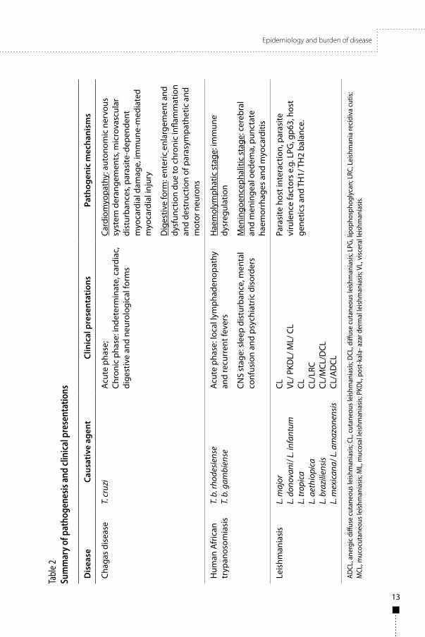
4. Clinical forms, pathogenesis and HIV coinfection4.1 Chagas diseaseT. cruzi is an intracellular parasite that invades different cell types, where it multiplies. The biological, biochemical and genetic diversity of T. cruzi isolates has long been recognized along with the eco-epidemiological complexity. A wide range of genetic markers has been applied to analyse the genetic diversity of the parasite. In 2009, an expert committee reviewed the available knowledge and partitioned T. cruzi isolates into six subgroups or discrete typing units (DTUs) (T. cruzi I-VI) (56). A comprehensive review of phylogeographical and eco-epidemiological features, and the correlation of DTU with natural and experimental infection, has been published (57). In the Southern Cone region, TcII, TcV and TcVI are the main causes of Chagas disease. TcII predominates in eastern and central Brazil, TcV in Argentina, Bolivia, and Paraguay, and TcVI in the Gran Chaco. In the Southern Cone region, chagasic cardiomyopathy can be severe, and a proportion of cases may develop megaoesophagus and megacolon. TcI is implicated with human disease in Amazonia, the Andean countries, Central America, and Mexico, and clinical presentations include chagasic cardiomyopathy. In these regions chagasic megaoesophagus and megacolon are absent or very rare (57, 58). Methods for DTU genotyping are available for widespread use in endemic areas (57, 59). Figure 2 depicts the geographical distribution of T. cruzi DTUs in human infections and in the triatomine vector species of major epidemiological importance. The role of vector species as biological filters for DTU transmission has not been defined.
4.1.1 Clinical presentationsThe acute phase of Chagas disease is recognized only in 1%–2% of infected individuals. In the acute phase, the symptoms are variable and decline spontaneously after 4–8 weeks; appropriate treatment can eliminate the parasite during this phase. In the chronic phase, approximately 70% of seropositive individuals are asymptomatic (indeterminate form), whereas 30% develop serious cardiac and/or digestive pathologies. Circumscribed or necrotizing inflammatory injuries may also occur in the grey matter in the central nervous system (CNS). In addition, each year, 2%–3% of asymptomatic individuals evolve to the above-mentioned symptomatic manifestations; the determinants of this conversion are unknown (60). The outcome of the infection in a particular individual is the result of a set of complex interactions between the genetic make-up of the parasite, the host immunogenetic background, and environmental factors.
4.1.2 Chagas heart diseaseChagas heart disease is the most serious and frequent manifestation of chronic Chagas disease (1); it appears in 20%–30% of infected individuals 10–30 years after the original acute infection. It is the leading cause of infectious myocarditis

22
WH
O T
echn
ical
Rep
ort S
erie
s, N
o. 9
75, 2
012
Research Priorities for Chagas Disease, HAT and Leishmaniasis Report of the TDR Disease Reference Group
worldwide and poses a substantial public health burden due to high morbidity and mortality. Numerous clinical and experimental investigations have shown that a low-grade but incessant parasitism, along with an accompanying immunological response (either parasite driven [most likely] or autoimmune-mediated), plays an important role in producing myocardial damage. At the same time, primary neuronal damage and microvascular dysfunction have been described as ancillary pathogenic mechanisms. Conduction system disturbances, atrial and ventricular arrhythmias, congestive heart failure, systemic and pulmonary thromboembolism and sudden cardiac death are the most common clinical manifestations of chronic Chagas cardiomyopathy (61).
Figure 2Geographical distribution of T. cruzi discrete typing units in human patients
Geographical distribution of T. cruzi discrete typing units (DTUs) in human patients. At present, T. cruzi is partitioned into six discrete typing units (DTUs), T. cruzi I-VI (TcI–TcVI) (ref 56). Data were compiled from a review article (ref 57). Triatomine vector species of major epidemiological importance in Chagas disease are indicated on the map (ref 133). Andean countries: Colombia, Ecuador, Peru, and Venezuela; Southern Cone Countries: Argentina, Bolivia, Brazil, Chile, Paraguay and Uruguay. The line separates the prevalence of Chagas disease clinical manifestations (see ref 57).DTUs, discrete typing units (I to IV); TcI –TcVI, T. cruzi type I-IV.

Clinical forms, pathogenesis and HIV coinfection
23
4.1.3 Gastrointestinal manifestationsThe gastrointestinal manifestations are a progressive enlargement of the oesophagus or the colon and other parts of the intestine, caused by chronic inflammation and destruction of parasympathetic neurons. The main gastro-intestinal symptoms are dysphagia and severe constipation. Oesophageal disease can appear in infections in children, while colonic disease evolves more slowly. Although its clinical significance is not very clear, damage to the nervous system and striated muscle is manifested in the chronic stage as motor cell loss or degeneration in various muscles, with infiltration and demyelination areas in the nerves. Geographical differences in prevalence of the digestive form of Chagas disease have been reported. This clinical presentation predominates in central Brazil and Chile and is essentially absent in Venezuela and Central America (57, 58).
4.1.4 Congenital Chagas diseaseT. cruzi can be transmitted from an infected mother to her child during pregnancy (congenital Chagas disease). Congenital transmission of Chagas disease in humans occurs less frequently than vector-borne and transfusion transmission. The incidence is estimated at more than 15 000 cases annually in the Americas. However, there is considerable uncertainty about congenital transmission rates since it receives no particular attention in endemic countries and perhaps even less in non-endemic countries where transmission does occur. The risk factors that determine the transmission of the parasite to the fetus are unknown. However, it is well established that treatment during the first year of a child's life is close to 100% effective in parasite elimination. The relevant information and recommendations have been issued by an Expert Advisory Group (62).
4.1.5 Chagas disease and HIV coinfectionImmunosuppressant therapies (63) and HIV/AIDS (64) can bring about acute clinical manifestations, which lead to an increased risk of disease reactivation in patients with chronic T. cruzi infection. This suggests the need for routine assessment for the presence of T. cruzi infection in patients whose immune response is suppressed. Whenever a case is detected, it is necessary to monitor reactivation with tools offering fast and simple diagnosis, and prescribe specific treatment as early as possible (65). Although the effectiveness of etiological treatment for the control of reactivation episodes has been demonstrated, it is necessary to gather evidence as to whether preventive treatment is effective in patients with no signs of clinical reactivation who have altered immunological parameters. Some protocols also recommend treating organ donors infected with T. cruzi to reduce the risk of transmission by transplantation.

24
WH
O T
echn
ical
Rep
ort S
erie
s, N
o. 9
75, 2
012
Research Priorities for Chagas Disease, HAT and Leishmaniasis Report of the TDR Disease Reference Group
4.2 Human African trypanosomiasisTwo pathogenic Trypanosoma species cause human African trypanosomiasis (HAT): Trypanosoma brucei rhodesiense causes East African sleeping sickness and T. b. gambiense causes West African sleeping sickness. Once inoculated by the tsetse fly, the parasites first multiply in the dermis at the site of the bite. This multiplication of trypanosomes and infiltration of lymphocytes and macrophages causes local oedema. This lesion, the trypanoma or chancre, is variably clinically detected. Local lymphadenopathy also develops. Waves of trypanosomes then invade the bloodstream from the lymphatic circulation (20).
Successive parasite populations express variants of the surface glycoprotein coat which allows them to escape the human (and animal) immune responses (66). The trypanosome is composed of several thousand invariant antigens surrounded by a coat of about 10 million copies of a variable surface glycoprotein (VSG). At first, the infected person produces a high amount of anti-VSG immunoglobulin M (IgM). This starts with a polyclonal activation of B cells and increases after a period of 3 or 4 days by a transient activation of T lymphocytes. These antibodies destroy most of the circulating parasites except for a small fraction that has switched its VSG. Anti-VSG immunoglobulin G (IgG) is also produced later. The ongoing antigenic variation and repeated exposure of the blood to variant antigens is associated with a profound dysregulation of the immune response and cytokine production that results in immunosuppression. This antigenic variation compromises the development of an effective vaccine (67).
4.2.1 PathogenesisThe disease primarily affects the lymphoid system, brain, heart and lungs. After the initial haemolymphatic stage, the infection progresses to a second or meningoencephalitic stage. The parasites, in association with certain immunological processes, cross the blood-brain barrier (68); they invade perivascular areas after which they first infiltrate the white matter and then later the grey matter. Invasion of the choroid plexus, thalamus, the postrema and median eminence is consistent with the disease symptoms. Signs of cerebral and meningeal oedema and punctate haemorrhages and myocarditis (69) are typically observed in this second stage of disease. Mott cells, plasma cells containing immunoglobulins, are characteristic of the infection. The severity of pathological lesions only partially correlates with parasitaemia (70).
4.2.2 Clinical presentationThe signs and symptoms that characterize sleeping sickness are generally the same for both the acute or rhodesiense form and the chronic or gambiense form of the disease, although they differ in degree and onset (71–73). The primary

Clinical forms, pathogenesis and HIV coinfection
25
lesion (trypanoma or chancre), while characteristic, is rarely observed in practice. Progression from the haemolymphatic stage to the meningoencephalitic second stage will eventually lead to death if untreated (73). There is no CNS involvement in the haemolymphatic first stage in contrast to the advanced meningoencephalitic second stage where CNS involvement is verified. The distinction between the two stages is based on markers in the cerebrospinal fluid but interpretation of these markers is subject to considerable debate.
Symptoms in the early stage are not specific; they include fever, severe headache, joint pains and muscle aches. For T. b. gambiense, the signs and symptoms are sometimes minor and may not sufficiently alert patients to seek health care. For T. b. gambiense, enlargement of the posterior cervical lymph nodes, Winterbottom's sign, typically occurs. Fever is recurrent, lasting from 1 to 3 days in synchrony with waves of parasitaemia. Headache is the most frequent complaint and is typically severe and persistent. Pruritus is frequent, detected through the presence of scratch marks on the skin. Skin rashes known as trypanids occur as ring-like patches with polycyclic contours of 1–10 cm diameter. Anaemia is frequent and may be severe, leading to heart failure. Other signs include oedema, ascites, cardiovascular, endocrinological and renal disorders and superinfections (69). The duration of the first stage varies from a few weeks in T. b. rhodesiense to months or several years in T. b. gambiense; the average duration is not predictable. It is unclear if spontaneous cure occurs or if there are asymptomatic carriers. In the meningoencephalitic second stage, more specific neurological signs appear in addition to the signs of the first stage, which persist during the second stage. The name ‘sleeping sickness’ for the disease stems from the rather typical sleep disturbances, characterized by the disappearance of the circadian rhythm of sleep and wakefulness. Mental confusion and a wide range of psychiatric disorders that include personality disorders, behavioural changes and mood alterations are often the symptoms that alert the patient and surrounding people to the disease. A wide range of other neurological symptoms include abnormal reflexes such as Babinski's sign, exaggerated osteotendinous reflexes and clonus, tone disorders (either hypertonia or hypotonia), abnormal movements, sensory disorders (Kerandel's sign), paresthesia, Hoffmann's sign and loss of sense of position, coordination disorders including ataxia and abnormal gait, convulsions and archaic reflexes (70). Deterioration of consciousness may lead to coma. Neurovegetative disorders may provoke incontinence. Other features include amenorrhea, infertility, abortion and wasting.
The very diverse clinical picture mimics a range of other diseases such as AIDS, schizophrenia and tuberculosis (TB), and clinicians may easily miss the diagnosis if they are not familiar with the disease. This is particularly a problem for clinicians dealing with imported cases in non-endemic areas, e.g. in urban areas e.g. of Kinshasa. Control strategies against HAT in such urban areas are not well defined (24).

26
WH
O T
echn
ical
Rep
ort S
erie
s, N
o. 9
75, 2
012
Research Priorities for Chagas Disease, HAT and Leishmaniasis Report of the TDR Disease Reference Group
4.3 LeishmaniasisMore than 20 Leishmania species are known to be pathogenic for humans. Leishmania parasites have two development stages: the amastigote stage that inhabits the reticulo-endothelial cells of mammals, and the flagellated promastigote stage that develops in the gut of the vector. Leishmania parasites cannot be distinguished by morphology alone and several biochemical and genetic makers are used for characterization of the parasites. Different parasite species can cause similar pathology, e.g. L. donovani and L. major can cause cutaneous ulcers. Analysis of completed Leishmania genome sequence showed high homology between different parasite species and genomic differences have not been identified so far that explain the diverse clinical forms (74).
4.3.1 Clinical presentationsThe clinical outcome of the human infection depends on the parasite species, the environment and the host immune response (Table 3). The majority of infected humans are asymptomatic but a minority develops clinical forms of the disease.
Visceral leishmaniasis. This is the most serious clinical form of leishmaniasis and is fatal if not treated. Visceral leishmaniasis (VL) is primarily a disease of children but also occurs in adults. VL is highly endemic on the Indian subcontinent and in East Africa, where it is caused by L. donovani parasites. The main clinical signs and symptoms of VL include fever (97%), splenomegaly (96.4%), weight loss (95.5%), pallor (93.6%), cough (89.7%), hepatomegaly (87.2%), asthenia (83.3%), anorexia (82.9%) and vomiting (73.9%). Clinical presentations in adults are similar to those in children, except that children are more anaemic. The haematological findings include anaemia, leukopenia and thrombocytopenia. VL in southern Europe and in the New World is caused by L. infantum and presents as non-tender splenomegaly, with or without hepatomegaly, wasting and pallor of mucous membranes. A response is measurable during the course of VL disease with a negative delayed type reaction to Leishmania antigen (negative leishmanin skin test – LST). The antibody response does not correlate with protection and reflects the severe course of the disease.
Post kala-azar dermal leishmaniasis. This is a dermatosis that usually develops following treatment of VL with pentavalent antimonials and less frequently following treatment with other antileishmanial drugs. However, rarely post kala-azar dermal leishmaniasis (PKDL) can develop in patients with no history of VL. PKDL is more prevalent in areas endemic for VL caused by L. donovani; PKDL due to L. infantum is rare but has been reported among HIV/Leishmania coinfected patients in southern Europe who have been treated with antimonials. PKDL lesions range from papules and nodules to a nodulo-papular rash that usually starts on the face and expands to other parts of the body.

Clinical forms, pathogenesis and HIV coinfection
27
Leishmania amastigotes are scanty in PKDL lesions. Histopathology sections of the dermis show various combinations of changes. The dermis is typically infiltrated by lymphocytes and macrophages but plasma cells are scanty or absent. Most cells are CD3 T cells, and there are many CD4 cells. Degenerating basal keratinocytes express HLA-DR, ICAM-1 and Leishmania antigen. Regional lymph nodes show hyperplasia of the B and T-cell zones. Hence the inflammatory reactions in PKDL lesions appear to be in response to Leishmania parasites and/or antigens.
Old world cutaneous leishmaniasis. Cutaneous leishmaniasis (CL) caused by L. tropica develops as a single lesion, which starts as a nodule at the site of inoculation; a crust develops centrally and may fall away exposing an ulcer, which heals gradually. The lupoid form is a unique type of cutaneous
Table 3Leishmania parasites and host immune responses in different leishmaniasis clinical forms
Parasite species Clinical form Dominant immune response
Leishmania donovani VLPKDLMLCL
TH1/TH2TH1TH1Th1
Leishmania infantum VLMLCLLupoid
TH1/TH2TH1Th1NA
Leishmania major CLSporotrichoid
TH1NA
Leishmania tropica CLMCLRecidive
TH1TH1NA
Leishmania aethiopica DCLMCL
TH2TH1
Leishmania braziliensis LCLMCL
TH1NA
Leishmania amazonensis LCLADCL
TH1TH2
NA = no available dataADCL, anergic diffuse cutaneous leishmaniasis; CL, cutaneous leishmaniasis; DCL, diffuse cutaneous leishmaniasis; LCL, localized cutaneous leishmaniasis; MCL, mucocutaneous leishmaniasis; ML, mucosal leishmaniasis; PKDL, post-kala- azar dermal leishmaniasis; VL, visceral leishmaniasis.

28
WH
O T
echn
ical
Rep
ort S
erie
s, N
o. 9
75, 2
012
Research Priorities for Chagas Disease, HAT and Leishmaniasis Report of the TDR Disease Reference Group
leishmaniasis characterized by unusual clinical features and a chronic relapsing course; in this clinical form, new papules and nodules appear at the margin of the remaining scar 1–2 years after healing of the acute lesion. Leishmania recidiva cutis (LRC) is characterized by recidivic lesions that occur at the border of old CL scars and differs from the lupoid form of the disease (75).
CL caused by L. major ranges from simple spontaneously healing ulcers to severely inflamed ulcers that heal in 4–6 months. The sporotrichoid form presents as painless subcutaneous nodules arranged in linear strings and develops after the primary lesion; the nodules may appear before or after treatment of CL with antimonials. Biopsies of the nodules show an inflammatory infiltrate composed of lymphocytes and histiocytes; the infiltrate is particularly dense and rich in plasmocytes at the deep dermis level, and scanty parasites are detected in deep biopsy samples. Outcomes are generally favourable after treatment.
L. aethiopica causes diffuse cutaneous lesions with extensive non-ulcerative nodules and is a very chronic disease. It is sometimes followed by chronic lymphoedema of an affected part of the body. This clinical manifestation is poorly understood.
Mucocutaneous leishmaniasis and other forms of cutaneous leishmaniasis. The lesions of mucocutaneous leishmaniasis (MCL) in the New World range from localized cutaneous leishmaniasis (LCL) with a moderate T-cell hypersensitivity, to MCL or anergic diffuse cutaneous leishmaniasis (ADCL). MCL is mediated by strong T-cell hypersensitivity and is usually caused by L. (V.) braziliensis infection with induction of a prominent Th1-immune response, while (ADCL) is caused by L. (L.) amazonensis with a marked Th2-type immune response. MCL can present as an intermediary form known as borderline disseminated cutaneous leishmaniasis (BDCL). In BDCL, primary skin lesions range from 1–3 in number and have the aspect of an erythematous, infiltrated plaque, variously located on the head, arms or legs. Typically there is lymphatic dissemination of infection with lymph node enlargement and negative delayed hypersensitivity skin test (DTH) prior to treatment, that usually converts to positive DTH after treatment (76).
The major histopathological feature of BDCL is a dermal mononuclear infiltration, with a predominance of heavily parasitized and vacuolated macrophages, together with lymphocytes and plasma cells. For example, one patient with such histopathology had acquired the infection seven years previously and presented with very large numbers of disseminated cutaneous lesions. BDCL shows clinical and histopathological features that are different from those of both LCL and ADCL and there is a good prognosis for cure, which is generally not the case for frank ADCL.
Mucosal leishmaniasis. This form of leishmaniasis is more common in the Old World, where patients present with a primary persistent mucosal ulcer or growth in the oral and/or nasal mucosa. Mucosal leishmaniasis (ML) lesions do

Clinical forms, pathogenesis and HIV coinfection
29
not extend to the skin but are confined to the oral and nasal mucosa. L. donovani isolates from mucosal lesions segregate as a subgroup within the L. donovani complex. In patients, ML is associated with induction of intense TH1 immune response leading to severe inflammation of the affected tissues. The patients are reactive to Leishmania antigens and are positive to leishmanin skin test (LST).
Leishmania/HIV coinfections. Coinfection has emerged as a common and serious opportunistic disease. Coinfection is more prevalent among HIV patients in Mediterranean countries, Brazil, east Africa and the Indian subcontinent. The clinical course and organ involvement of VL are often atypical in HIV positive subjects. Leishmania/HIV coinfections show various clinical presentations, ranging from typical visceral forms to asymptomatic or atypical cases, including CL and MCL. HIV-infected patients have a lower frequency of splenomegaly than HIV-negative individuals (80.8% versus 97.4%; p = 0.02). HIV-infected patients, most of whom were profoundly immunosuppressed (mean CD4+ lymphocyte count, 90 cells/mm³) at the time of leishmaniasis diagnosis, were found to have a greater frequency and degree of leukopenia, lymphocytopenia, and thrombocytopenia (77).


31
5. DiagnosisCurrent diagnostic methods for trypanosomatid diseases are not satisfactory (Table 4). Diagnosis is based on demonstration of the parasites in tissue aspirates or blood, detection of antibody to the parasites, detection of parasite products in the blood or body secretions, or quantitative/qualitative detection of parasite DNA. Most of the diagnostic tests require invasive sampling and lack appropriate sensitivity and specificity. Because the diseases are endemic in communities living in remote areas deprived of effective health services, point-of-care diagnostics are very important.
5.1 Chagas diseaseThe primary methods for diagnosing Chagas disease are serological, and the secondary tests are parasitological (6). Most Chagas cases are currently diagnosed during the chronic phase of the disease. Diagnostic tests for T. cruzi infection are used during epidemiological surveys, surveillance for vectorial transmission, blood screening, screening of pregnant women, and in individual patients, but the tests need to be improved (Table 5).
5.1.1 Parasitological diagnosisAlthough parasitaemia is generally present in the acute phase, the initial infection is seldom detected except in cases where acute symptoms are severe. In such cases, microscopic observation of fresh blood or stained blood smears can reveal parasites (6).
5.1.2 SerologyThree types of conventional test based on detection of parasite-specific antibodies are widely used for immunodiagnosis: indirect haemaglutination (IHA), indirect immunofluorescence (IIF), and enzyme-linked immunosorbent assay (ELISA). The commercially available diagnostic tests employ crude antigenic T. cruzi preparations, semi-purified fractions, or recombinant antigens. Most of the tests have 94%–99.5% sensitivity and 94%–96% specificity (Table 4). Rapid chromatography tests with a mixture of recombinant antigens for screening for anti-T. cruzi antibodies in whole blood and serum, and in umbilical cord blood of infected mothers at the time of delivery, have been assessed in Latin American countries and variable results were obtained (78–80).
5.1.3 Quantitative and qualitative detection of parasite DNADetection of parasite DNA by polymerase chain reaction (PCR) during the chronic phase of T. cruzi infection is less sensitive than serological tests (81).

32
WH
O T
echn
ical
Rep
ort S
erie
s, N
o. 9
75, 2
012
Research Priorities for Chagas Disease, HAT and Leishmaniasis Report of the TDR Disease Reference Group
Tabl
e 4Su
mm
ary o
f cha
ract
erist
ics o
f ava
ilabl
e di
agno
stics
Dis
ease
Para
site
dem
onst
ratio
nPa
rasi
te D
NA
dete
ctio
nSe
rolo
gy
Sour
ceSe
nsiti
vity
Spec
ifici
tyTe
stSe
nsiti
vity
Spec
ifici
tyTe
stSe
nsiti
vity
Spec
ifici
ty
Chag
as
dise
ase
Bloo
d>7
0% w
hen
smea
r per
-fo
rmed
15–
30 d
ays a
fter
onse
t of s
ympt
oms (
only
in
the
acut
e ph
ase)
100%
PCR
RT-P
CR~6
0% (i
n ch
ildre
n)If
prop
erly
do
ne, c
an
be 1
00%
IHA
, IIF,
EL
ISA
(chr
onic
ph
ase)
94%
–99.
5%(k
it-de
pend
ent
diffe
renc
e)
94%
–96%
(kit-
depe
nden
t di
ffere
nce)
Hum
an
Afric
an tr
ypa-
noso
mia
sis
Bloo
d44
.8%
–91%
(c
ombi
natio
n in
clud
ing
mAE
CT)
~100
%PC
RRT
PCR
LAM
P
~95%
~100
%CA
TT68
%–9
9.5%
(regi
onal
di
ffere
nces
)
83.5
%–
98.4
%
Visc
eral
le
ishm
ania
sis
Sple
en
90%
–95%
(sm
ear)
~100
%PC
RRT
-PCR
~95%
~100
%
Bone
m
arro
w60
%–8
5%(s
mea
r)~1
00%
PCR
RT-P
CR~9
5%~1
00%
Lym
ph
node
50%
–60%
(sm
ear)
~100
%N
ot
exte
nsiv
ely
stud
ied
Bloo
dPC
RRT
-PCR
~80%
–95%
~100
%DA
TEL
ISA
rK39
-ICT
91%
–100
%85
%–9
5%90
%–1
00%
72%
–100
%70
%–8
5%93
%–1
00%
Cuta
neou
s le
ishm
ania
sis
Skin
30%
–90%
dep
endi
ng
on th
e pa
rasit
e sp
ecie
s (s
mea
r)
~100
%PC
R~9
0%–9
5%~1
00%
Lym
ph
node
40%
–70%
in
L. b
razil
iens
is in
fect
ions
(a
spira
te c
ultu
re)
~100
%N
ot
exte
nsiv
ely
stud
ied
CATT
, car
d ag
glut
inat
ion
test
for t
rypa
noso
mia
sis;
DAT
, dire
ct a
gglu
tinat
ion
test
; ELI
SA, e
nzym
e-lin
ked
imm
unos
orbe
nt a
ssay
; IC
T, im
mun
ochr
omat
ogra
phy
test
; IH
A, i
ndire
ct h
aem
aglu
tinat
ion;
IIF,
indi
rect
imm
unofl
uore
scen
ce; L
AM
P, lo
op m
edia
ted
isot
herm
al a
mpl
ifica
tion;
mA
ECT,
min
iatu
re a
nion
exc
hang
e ce
ntrif
ugat
ion
tech
niqu
e; P
CR, p
olym
eras
e ch
ain
reac
tion;
RT-
PCR,
reve
rse
tran
scrip
tase
pol
ymer
ase
chai
n re
actio
n.

Diagnosis
33
Tabl
e 5Di
agno
stics
for C
haga
s dise
ase:
kno
wle
dge
gaps
and
rese
arch
prio
ritie
s
Know
ledg
e ga
pRe
sear
ch p
rior
ity
Rese
arch
met
hods
Expe
cted
out
com
eJu
stifi
catio
nSe
lect
ed in
dica
tors
Lack
of a
pro
per
diag
nost
ic to
ol
for i
nfan
ts b
orn
to
infe
cted
mot
hers
Dev
elop
men
t of
a po
int-
of-c
are
(PO
C) d
iagn
ostic
te
st fo
r neo
nate
s
Biom
arke
r dis
cove
ry,
clin
ical
val
idat
ion
and
inco
rpor
atio
n in
a
POC
plat
form
A P
OC
test
that
can
be
use
d to
dia
gnos
e Ch
agas
dis
ease
in
new
born
s in
firs
t-lin
e he
alth
ser
vice
s
Curr
ent p
aras
ite
dete
ctio
n te
sts
are
diffi
cult
to p
erfo
rm
rout
inel
y on
a la
rge-
scal
e ba
sis
Num
ber o
f ful
ly
valid
ated
PO
C te
sts
for n
ewbo
rns
Lack
of a
pro
per
test
to a
sses
s cu
re
Dev
elop
men
t of
a P
OC
test
to
esta
blis
h cu
re
Biom
arke
r dis
cove
ry,
clin
ical
val
idat
ion
and
inco
rpor
atio
n in
a
POC
plat
form
A P
OC
test
that
can
be
use
d to
est
ablis
h cu
re
Curr
ently
, the
re
is n
o te
st, P
OC
or
othe
rwis
e, th
at c
an
be u
sed
as a
relia
ble
mar
ker o
f cur
e
Fully
val
idat
ed
POC
test
s fo
r es
tabl
ishm
ent o
f cu
re o
f Cha
gas
dise
ase
Lack
of a
test
for
asse
ssin
g dr
ug
resi
stan
ce
Dev
elop
men
t of
a te
st s
yste
m fo
r su
rvei
llanc
e of
dr
ug re
sist
ance
Dev
elop
men
t of
mol
ecul
ar m
arke
rs
for d
rug
resi
stan
ce
to a
sses
s dr
ug
effec
tiven
ess
in v
itro
A te
st s
yste
m th
at
can
be u
sed
in a
re
fere
nce
labo
rato
ry
in e
ndem
ic
coun
trie
s
Lack
of a
m
etho
dolo
gy fo
r m
onito
ring
of d
rug
susc
eptib
ility
of T
. cru
zi
stra
ins
at p
ublic
he
alth
labo
rato
ries
Num
ber o
f va
lidat
ed a
ssay
s fo
r m
onito
ring
of d
rug
effec
tiven
ess
Nee
d fo
r PO
C ra
pid
diag
nost
ic
test
Dev
elop
men
t of
PO
C te
sts
for r
apid
cas
e de
tect
ion
Biom
arke
r dis
cove
ry,
clin
ical
val
idat
ion
and
inco
rpor
atio
n in
a
POC
plat
form
A h
ighl
y se
nsiti
ve
and
spec
ific
POC
test
for u
se in
he
alth
clin
ics
and
in
the
field
Rapi
d ch
rom
atog
raph
y te
sts
for s
cree
ning
an
ti-T.
cru
zi a
ntib
odie
s sh
ow v
aria
ble
resu
lts
in L
atin
Am
eric
an
coun
trie
s
Num
ber o
f ful
ly
valid
ated
fiel
d te
sts

34
WH
O T
echn
ical
Rep
ort S
erie
s, N
o. 9
75, 2
012
Research Priorities for Chagas Disease, HAT and Leishmaniasis Report of the TDR Disease Reference Group
Recently an accurate real-time PCR strategy for monitoring clinical reactivation and etiological treatment outcome in Chagas disease patients has been proposed (82). In addition, a multicentric study to standardize PCR methods for detection of T. cruzi DNA in blood samples from Chagas disease patients was carried out under the coordination of the Special Programme for Research and Training in Tropical Diseases (TDR-WHO) (83). Four methods depicted the best performing parameters and await validation through prospective studies in different settings.
5.1.4 Diagnosis in newbornsDirect examination for blood parasites using the microhaematocrit concentration method (MH) in capillary tubes is recommended as a first procedure for diagnosis of congenital T. cruzi transmission. However, this method is not ideal, as samples must be collected in six to eight capillary tubes, each of which must then be examined for at least 20 minutes for accurate parasite detection. The MH has good sensitivity when performed by experts. Under routine conditions the sensitivity of detection is around 20% (84). An alternative diagnostic method is the Strout concentration method (so-called microstrout using Eppendorf tubes). This technique has demonstrated good sensitivity and specificity, but a major problem is reproducibility of the procedure since the ability to detect the parasite is operator-dependent. While parasite detection tests are difficult to perform routinely on a large-scale basis, conventional immunoglobulin G (IgG) serology may allow for the diagnosis of congenital infection in an infant after nine months of age, following the disappearance of maternal antibodies (85).
5.1.5 Diagnosis of therapy efficacy and cureCurrently successful cure of Chagas disease is assessed by the disappearance of anti-T. cruzi antibodies (seroconversion), employing the above-mentioned serological tests, while therapeutic failure is assigned by parasite persistence. However, serological markers may take up to 5 years to disappear. Accordingly, positive serology does not mean active infection, whereas negative serology indicates cure. Parasitological tests have limited sensitivity (xenodiagnosis, 30%–50%; PCR, 60%–90%); consequently a positive parasitological test means treatment failure, whereas a negative result does not indicate absence of parasites. Thus, a diagnostic technique for parasitological cure is urgently needed and necessary for the evaluation of new approaches to Chagas disease treatment. It is anticipated that the assessment of parasitological cure in patients with chronic disease, in whom parasitaemia is extremely low or undetectable, will be very challenging.

Diagnosis
35
5.1.6 Diagnosis of drug resistant Chagas diseaseTherapeutic failures of benznidazole and nifurtimox (see section 6) are documented in Chagas disease. Therapeutic success seems to depend on the interplay among drug susceptibility of T. cruzi strains, drug access and accumulation in different environments, and the host immune response. A test for screening of drug resistance of the infective strain is not available.
5.2 Human African trypanosomiasisThe diagnostic techniques for human African trypanosomiasis (HAT) are discussed below in the context of large-scale population screening programmes for T. b. gambiense, in which serological screening tests were used followed by parasitological confirmation and staging. The serological tests used in this context are ineffective for T. b. rhodesiense and are not suitable for use in primary care centres because of their large kit format.
5.2.1 Demonstration of parasiteNo single parasitological technique is sensitive enough to confirm HAT diagnosis (Table 6). Hence, combinations of microscopic examination of lymph node aspirates, wet blood films, thick blood films and cerebrospinal fluid (CSF) are utilized. However, these complex confirmation algorithms have low overall sensitivities, in the range of 44.8%–75% (86, 87). More sensitive confirmation techniques, such as the capillary tube centrifugation (CTC) technique, miniature anion-exchange centrifugation technique (mAECT), and quantitative buffy coat (QBC) are now adopted by HAT control programmes. Addition of CTC and mAECT to the existing HAT algorithms has been shown to be a cost-effective option that can increase the sensitivity of confirmation to 91% (88).
5.2.2 SerologyThe card agglutination test for trypanosomiasis (CATT) (89) detects antibodies against T. b. gambiense in blood, plasma or serum samples with a sensitivity of 68%–99.5% and a specificity of 83.5%–98.4%, depending on the geographic region. Limited experience is available using CATT on filter paper eluates. Up to 3 million CATT tests per year are now performed as a screening test by active case-finding programmes for HAT in central and West Africa. Other assays proposed for HAT screening are not in routine use mainly because they require sophisticated equipment. Since most HAT drugs are toxic it is necessary to confirm a positive CATT result by demonstration of the parasite; CATT with dilutions of plasma has been proposed as a confirmation test but has not yet been sufficiently validated (90).

36
WH
O T
echn
ical
Rep
ort S
erie
s, N
o. 9
75, 2
012
Research Priorities for Chagas Disease, HAT and Leishmaniasis Report of the TDR Disease Reference Group
Tabl
e 6Di
agno
stics
for h
uman
Afri
can
tryp
anos
omia
sis: k
now
ledg
e ga
ps an
d re
sear
ch p
riorit
ies
Know
ledg
e ga
p Re
sear
ch p
rior
ity
Rese
arch
met
hods
Expe
cted
out
com
eJu
stifi
catio
nSe
lect
ed in
dica
tors
Bett
er m
arke
rs o
f di
seas
e, d
isea
se
stag
e an
d pr
ogno
sis
for
hum
an A
fric
an
tryp
anos
omia
sis
(HAT
)
Dev
elop
men
t of
a no
n-in
vasi
ve
poin
t-of
-car
e (P
OC)
test
to
dete
ct a
nd
disc
rimin
ate
first
an
d se
cond
-sta
ge
HAT
Biom
arke
r di
scov
ery,
and
cl
inic
al v
alid
atio
n an
d in
corp
orat
ion
in a
PO
C pl
atfo
rm
A P
OC
test
fo
r HAT
sta
ge
dete
rmin
atio
n th
at
will
rem
ove
the
need
for l
umba
r pu
nctu
re
Curr
ently
, sta
ge
dete
rmin
atio
n in
H
AT re
lies
on d
irect
ex
amin
atio
n of
ce
rebr
ospi
nal fl
uid.
Se
vera
l lum
bar
punc
ture
s ar
e re
quire
d du
ring
follo
w u
p of
pa
tient
s. Th
is lo
wer
s th
e ac
cept
abili
ty o
f HAT
car
e.
Num
ber o
f ful
ly
valid
ated
PO
C te
sts
for s
tage
de
term
inat
ion
in
HAT
Nee
d fo
r a p
oint
-of
-car
e ra
pid
test
of
cur
e
Dev
elop
men
t of
a P
OC
test
to
mon
itor c
ure
Biom
arke
r di
scov
ery,
and
cl
inic
al v
alid
atio
n an
d in
corp
orat
ion
in a
PO
C pl
atfo
rm
A P
OC
test
for H
AT
that
can
be
used
to
est
ablis
h cu
re
in fi
rst-
line
heal
th
serv
ices
Curr
ently
, HAT
pat
ient
s m
ust b
e fo
llow
ed u
p fo
r tw
o ye
ars
befo
re th
ey
can
be d
ecla
red
cure
d.
This
follo
w u
p in
volv
es
seve
ral i
nvas
ive
wor
k-up
s.
Num
ber o
f ful
ly
valid
ated
PO
C te
sts
for e
stab
lishm
ent
of c
ure
afte
r HAT
tr
eatm
ent
Nee
d fo
r PO
C ra
pid
diag
nost
ic
test
Dev
elop
men
t of a
PO
C te
st fo
r HAT
Bi
omar
ker
disc
over
y, a
nd
clin
ical
val
idat
ion
and
inco
rpor
atio
n in
a P
OC
plat
form
A P
OC
test
that
ca
n be
use
d to
di
agno
se (c
onfir
m)
HAT
in fi
rst-
line
heal
th s
ervi
ces
Curr
ently
, the
re is
no
POC
test
that
can
be
used
at h
ealth
clin
ic
leve
l. D
iagn
ostic
del
ays
are
ther
efor
e co
mm
on.
Num
ber o
f ful
ly
valid
ated
PO
C te
sts
for H
AT d
iagn
osis
; pr
opor
tion
of
elig
ible
cas
es th
at
wer
e di
agno
sed
by
POC.
cont
inue
s

Diagnosis
37
Know
ledg
e ga
p Re
sear
ch p
rior
ity
Rese
arch
met
hods
Expe
cted
out
com
eJu
stifi
catio
nSe
lect
ed in
dica
tors
A ra
pid
diag
nost
ic te
st
that
is n
ot b
ased
on
var
iabl
e su
rfac
e an
tigen
s
Dev
elop
men
t of
sim
ple
confi
rmat
ion
test
fo
r HAT
Biom
arke
r di
scov
ery,
and
cl
inic
al v
alid
atio
n an
d in
corp
orat
ion
in a
PO
C pl
atfo
rm
A h
ighl
y se
nsiti
ve
and
spec
ific
confi
rmat
ion
test
Curr
ent c
onfir
mat
ion
algo
rithm
s in
pop
ulat
ion
scre
enin
g ca
mpa
igns
are
ve
ry la
borio
us
Num
ber o
f ful
ly
valid
ated
HAT
co
nfirm
ator
y te
sts
Lack
of d
ata
on
drug
resi
stan
ce
and
need
for t
ool
to m
onito
r dru
g re
sist
ance
Dev
elop
men
t of
a te
st s
yste
m fo
r su
rvei
llanc
e of
dr
ug re
sist
ance
Dev
elop
men
t of
assa
ys th
at c
an
be re
liabl
y us
ed
to m
onito
r dru
g eff
ectiv
enes
s in
vi
tro
A te
st s
yste
m
that
can
be
used
in
a re
fere
nce
labo
rato
ry in
en
dem
ic c
ount
ries
No
stan
dard
ized
m
etho
dolo
gy th
at a
llow
s m
onito
ring
of H
AT d
rug
effec
tiven
ess
at p
ublic
he
alth
labo
rato
ries
Num
ber o
f va
lidat
ed a
ssay
s fo
r m
onito
ring
of d
rug
effec
tiven
ess
Tabl
e 6 co
ntin
ued

38
WH
O T
echn
ical
Rep
ort S
erie
s, N
o. 9
75, 2
012
Research Priorities for Chagas Disease, HAT and Leishmaniasis Report of the TDR Disease Reference Group
5.2.3 Quantitative and qualitative detection of parasite DNASeveral PCR primers have been developed and recently a standardized commercial PCR kit format has been validated in a case-control design (91). As a global health initiative, the Foundation for Innovative New Diagnostics (FIND) has embarked on a large discovery and development programme for novel HAT diagnostic tests, making use of novel technologies such as loop-mediated isothermal amplification (LAMP) (92), in an attempt to translate the technologies into user-friendly formats for endemic areas. Thus far no data on the clinical benefit of these novel diagnostic approaches are available.
5.2.4 Staging of the diseaseHAT treatment is stage-dependent, and the stage is determined by examining CSF for immune cell count, total protein concentration and the presence of trypanosomes (70). Protein concentration is not applied in the field because it is too complex and reagents are not stable under these conditions. In addition, methods to demonstrate the presence of trypanosomes in CSF are not sufficiently sensitive. Therefore, cell count of CSF is the most commonly used marker for staging. WHO recommends a CSF leukocyte count of ≤ 5 cells/µl (70) as a cut-off point for first stage, although this is still debated. Development of a better and more user-friendly marker for staging was identified by a TDR expert group as number one priority in HAT diagnostics. The need for lumbar puncture is indeed one of the most important constraints to the uptake of HAT screening programmes.
5.3 LeishmaniasisAccurate diagnosis of human leishmaniasis remains a problem for clinicians and coordinators of control programmes (Table 7); the disease covers a spectrum of syndromes, as mentioned above, and each poses its own diagnostic challenges. The clinical features are diverse and depend on the infecting Leishmania species and the host immune response, and medical history is an important factor in diagnosis. Diagnosis is based on clinical presentation confirmed by conventional laboratory diagnosis, such as microscopic demonstration or culturing of Leishmania parasites from biopsies or aspirates from lesions, bone marrow, lymph node or spleen. Other diagnostic methods include serological tests and molecular detection of parasite DNA (93).
5.3.1 Parasitological diagnosisDemonstration of amastigotes in tissue samples from bone marrow, spleen and lymph nodes (for VL), from mucosal lesions (ML), skin lesions (PKDL) and skin ulcers (CL) is the reference standard for diagnosis. The method requires highly-trained technicians, involves invasive sampling, and has variable sensitivity.

Diagnosis
39
Tabl
e 7Di
agno
stics
for l
eish
man
iasis
: kno
wle
dge
gaps
and
rese
arch
prio
ritie
s
Know
ledg
e ga
pRe
sear
ch p
rior
ity
Rese
arch
met
hods
Expe
cted
out
com
eJu
stifi
catio
nSe
lect
ed in
dica
tors
A m
arke
r of a
ctiv
e VL
di
seas
e an
d a
sim
ple
test
and
read
-out
th
at c
an d
etec
t ac
tive
dise
ase
with
su
ffici
ent s
ensi
tivity
an
d sp
ecifi
city
Dev
elop
men
t of
a PO
C te
st fo
r VL
Biom
arke
r di
scov
ery,
and
cl
inic
al v
alid
atio
n an
d in
corp
orat
ion
in a
PO
C pl
atfo
rm
A P
OC
test
that
ca
n be
use
d to
di
agno
se V
L in
fir
st-li
ne h
ealth
se
rvic
es
Curr
ent r
apid
test
s fo
r VL
are
ant
ibod
y ba
sed.
Th
ey a
re n
ot s
peci
fic
for a
cute
VL
dise
ase
and
are
posi
tive
in
asym
ptom
atic
car
riers
an
d in
pas
t cas
es.
Num
ber o
f ful
ly
valid
ated
PO
C te
sts
for a
cute
VL
An
esta
blis
hed
mar
ker o
f cur
e in
VL
Dev
elop
men
t of
a P
OC
test
to
esta
blis
h cu
re
in V
L
Biom
arke
r di
scov
ery,
and
cl
inic
al v
alid
atio
n an
d in
corp
orat
ion
in a
PO
C pl
atfo
rm
A P
OC
test
that
ca
n be
use
d to
es
tabl
ish
cure
in V
L
Curr
ently
, the
re is
no
POC
test
that
can
be
used
as
a m
arke
r of
cure
in V
L
Num
ber o
f ful
ly
valid
ated
PO
C te
sts
for e
stab
lishm
ent
of V
L cu
re
Stan
dard
ized
se
nsiti
ve d
iagn
ostic
s fo
r CL,
ML
and
PKD
L
Dev
elop
men
t of
PO
C te
sts
for
the
diag
nosi
s of
der
mal
le
ishm
ania
sis
(CL,
M
L, P
KDL)
Biom
arke
r di
scov
ery,
and
cl
inic
al v
alid
atio
n an
d in
corp
orat
ion
in a
PO
C pl
atfo
rm
A h
ighl
y se
nsiti
ve
and
spec
ific
POC
test
for u
se in
he
alth
clin
ics
Curr
ent t
ests
for
derm
al le
ishm
ania
sis
are
com
plex
Num
ber o
f fu
lly v
alid
ated
le
ishm
ania
sis
test
s
Valid
ated
in v
itro
test
s to
mon
itor
drug
effe
ctiv
enes
s in
en
dem
ic a
reas
Dev
elop
men
t of
a te
st s
yste
m fo
r su
rvei
llanc
e of
dr
ug re
sist
ance
Dev
elop
men
t of
mol
ecul
ar m
arke
rs
for d
rug
resi
stan
ce
that
can
be
relia
bly
used
to m
onito
r dr
ug e
ffect
iven
ess
in v
itro
A te
st s
yste
m
that
can
be
used
in
a re
fere
nce
labo
rato
ry in
en
dem
ic c
ount
ries
No
stan
dard
ized
m
etho
dolo
gy th
at
allo
ws
mon
itorin
g of
an
ti-le
ishm
ania
l dru
g eff
ectiv
enes
s at
pub
lic
heal
th la
bora
torie
s
Num
ber o
f va
lidat
ed a
ssay
s fo
r m
onito
ring
of d
rug
effec
tiven
ess

40
WH
O T
echn
ical
Rep
ort S
erie
s, N
o. 9
75, 2
012
Research Priorities for Chagas Disease, HAT and Leishmaniasis Report of the TDR Disease Reference Group
The reported sensitivity of direct microscopic examination for VL diagnosis using lymph node aspirates ranges from 52%–58%, and for bone marrow aspirates from 60%–85%. For spleen aspirates the sensitivity is high (93%–98%), however the procedure poses a high risk for life and cannot be performed in remote field settings (93). The sensitivity of parasitological diagnosis can be increased by culture of collected samples in Novy-MacNeal-Nicolle (NNN) media or inoculation in laboratory animals (93). Culturing of promastigotes from infected tissues in NNN agar or other suitable media has been considered a standard for Leishmania diagnosis. However, different Leishmania species show variable growth parameters and contamination is a constant hazard. Immunohistochemical assays have not been extensively validated and specific antibodies are not routinely available.
5.3.2 SerologySeveral serological tests (e.g. immunofluorescence, ELISA) have been evaluated in different endemic settings with variable results. All antibody tests share drawbacks: they remain positive for many months after cure and do not allow the discrimination of current from past infection.
Direct agglutination test. This test is the first antibody detection test for VL used in field settings. Whole, trypsinized, Coomassie-stained promastigotes are used, either as a suspension or in freeze-dried form, facilitating its use in the field (94). The direct agglutination test (DAT) is simple and shows high sensitivity (70%–100%), specificity (90%–100%) (95) and reproducibility. It is easy to perform and does not require specialized equipment; it can be carried out with plasma or serum. Anti-leishmania antibodies may persist for years as a result of previous VL infection, and titres measured by DAT may remain positive for up to 5 years after recovery in >50% VL patients. DAT has been widely used in epidemiology surveys and control programmes. DAT and the rK39 immuno-chromatographic test (rK39 ICT) (see below) were found to have high sensitivity and specificity in a meta-analysis (96) and were recommended for clinical practice. On the other hand, DAT is not helpful in the detection of relapse in VL patients.
Enzyme-linked immunosorbent assay. This is mostly used for diagnosis of VL. Antigens commonly used in the ELISA are: crude soluble antigen (CSA) (obtained by freezing and thawing of live promastigotes), and a recombinant antigen (rk39) (a conserved portion of a kinesin-related protein). The sensitivity of CSA—ELISA is 80%–100%; however cross-reactions with sera from patients with trypanosomiasis, tuberculosis, and toxoplasmosis have been reported (94).
Rapid diagnostic tests. Prototypes of rapid diagnostic tests (RDTs) have been evaluated for diagnosis of VL in different endemic regions. The sensitivity and specificity of the tests vary according to the supplier and the geographic

Diagnosis
41
region, but generally the rK39 antigen-based immunochromatographic test (rK39 ICT) performed extremely well on the Indian subcontinent and is now the recommended test in the VL elimination initiative. RDTs can provide a point-of-care method for diagnosis of VL in remote endemic villages. In a recent multicentre study coordinated by WHO/TDR, new rK39 ICT/RDTs were evaluated in endemic areas of the Indian subcontinent, East Africa and Brazil. The tests performed extremely well in the Indian region with sensitivity ranging from 92.8%–100% and specificity from 96%–100%. However lower sensitivities and specificities (61%–91%) were obtained with sera from East Africa and Brazil.
Leishmanin skin test. The leishmanin skin test (LST) is a marker of cellular immunity and is used for diagnosis of CL and mucocutaneous leishmaniasis (MCL); it is easy to use and highly sensitive and specific. LST is not used for VL diagnosis since patients only develop strong Leishmania-specific cell-mediated immunity several months after cure (97).
5.3.3 Detection of parasite productsOnly one test has so far been developed for detection of Leishmania antigens in urine. This latex agglutination test detects parasite antigens only in patients with active disease, and quickly turns negative after successful treatment. Regrettably, sensitivity of the test is very variable – 47%–87%, it requires boiled urine, and the read-out is quite subjective (98). New formats of the test are under development but no new data or publications are available (93).
5.3.4 Quantitative and qualitative detection of parasite DNASeveral PCR primers have been used for amplification of parasite DNA in samples collected from tissues, blood and body secretions of VL and CL patients. Amplification of parasite kDNA, gp63, hsp70, ITS1, SSU ribosomal RNA and other sequences was evaluated for both diagnosis and typing of the parasites from collected samples (99, 100). In immunocompromised patients, PCR screening for Leishmania infection showed higher sensitivity than microscopical examination and blood culture (101). Sample collection, storage conditions and transportation are some of the challenges with PCR assays; and the clinical benefit of available tests has not yet been clearly demonstrated. For both VL and CL, PCR or other nucleic acid detection methods have been used but ~15% asymptomatic healthy individuals test positive in VL endemic regions. Simplification, standardization and field-friendly modifications are needed for meaningful application of PCR in leishmaniasis. Most of the work with CL patients needs extensive validation and reliability studies, and tests with prognostic value are needed. Overall, the prognostic value of quantitative PCR remains to be proved (102).


43
6. Drugs and drug resistanceNew, effective, safe and affordable drugs, preferably oral, are needed for all the trypanosomiases and leishmaniases. The drugs in current use for treatment of these diseases have well known problems of toxicity, efficacy, administration or length of treatment. Indeed, more than one new drug is needed for each so that combination therapy can be employed to avoid drug resistance and to provide back-up drugs when resistance emerges. There are few new drugs or treatments in clinical trials. Based on prior drug development experience, approximately 120 development projects need to be supported to ensure one new drug for each of the trypanosomatid diseases. Research consortia have organized to provide the multidisciplinary requirements needed for drug development. However, resources are needed to enable the coordination of these researchers' activities and to incorporate the expertise of the for-profit drug development enterprises. Such activities have the ability to lead to the needed drugs. A recent G-FINDER report (103) highlighted the limited amount of funding available for research and development (R&D) for trypanosomatid diseases compared with malaria, tuberculosis (TB) and HIV/AIDS. A highly efficient and collaborative environment is required in order to optimize effort and the use of funding, engaging the academic community, public institutes and the pharmaceutical/biotech sector in a unified effort.
The considerable advances made in identifying, validating and characterizing drug targets represent only one early step in the long and complex process of drug discovery and development. New libraries with novel pharmacophores together with improved screening technologies to identify hits have been brought into alignment. Appropriate models of infection to evaluate leads are also available and have been integrated into the process at several academic centres. Support from public-private partnerships such as the Drugs for Neglected Diseases Initiative (DNDi) and interaction with the pharmaceutical/biotech sector has also been established, bringing in expertise in lead optimization, toxicology and pharmacology. The issue of clinical trials capacity is being addressed in India, Africa and South America.
This is an opportune time for the discovery and development of new drugs for kinetoplastid pathogens (Table 8). The foundation laid by the genome sequencing projects has been used to elucidate gene and protein product functions and metabolic pathways to identify potential drug targets (104), and many targets have been prioritized for drug discovery (www.tdrtargets.org). Indeed, a large number of compounds, as well as drugs that have been approved for use in humans, have been used in whole cell screens against kinetoplastids. In addition, several efforts have been initiated to discover and develop new drugs using target, cell, or animal model system approaches (http://www.drugdiscovery.dundee.ac.uk; http://www.cdipd.org/ [formerly known as the Sandler Center];

44
WH
O T
echn
ical
Rep
ort S
erie
s, N
o. 9
75, 2
012
Research Priorities for Chagas Disease, HAT and Leishmaniasis Report of the TDR Disease Reference Group
Tabl
e 8Dr
ug re
sear
ch p
riorit
ies f
or tr
ypan
osom
atid
s (Ch
agas
, hum
an A
frica
n tr
ypan
osom
iasis
, and
leish
man
iasis
)
Prio
rity
Rese
arch
met
hods
Expe
cted
out
com
eJu
stifi
catio
nIn
dica
tors
Dev
elop
new
th
erap
ies
base
d on
cur
rent
ant
i-ki
neto
plas
tids.
Scre
en li
brar
ies
of
com
poun
ds fr
om th
e ph
arm
aceu
tical
indu
stry
for
anti-
tryp
anos
omat
id a
ctiv
ity
usin
g in
vitr
o an
d an
imal
m
odel
s.
Sens
itivi
ty p
rofil
es
of e
ach
of th
e tr
ypan
osom
atid
s to
ac
tive
com
poun
ds.
Ther
e ar
e en
orm
ous
reso
urce
s of
co
mpo
unds
that
hav
e no
t bee
n ev
alua
ted.
Num
ber o
f scr
eene
d co
mpo
unds
with
pro
ven
anti-
tryp
anos
omat
id
activ
ity.
Dev
elop
new
dr
ugs
with
ant
i-tr
ypan
osom
atid
ac
tivity
.
Det
erm
ine
esse
ntia
l m
etab
olic
pat
hway
s/pr
otei
ns.
Des
ign
spec
ific
inhi
bito
rs
base
d on
str
uctu
re-fu
nctio
n re
latio
nshi
ps.
Det
erm
ine
phar
mac
okin
etic
s of
can
dida
te d
rugs
in a
nim
al
mod
els.
Valid
ated
dru
g ta
rget
s.Co
nfirm
ed s
peci
fic
inhi
bito
rs.
Ther
e is
nee
d to
de
velo
p co
mpl
etel
y ne
w d
rugs
targ
etin
g di
vers
e pa
thw
ays
to p
reve
nt c
ross
re
sist
ance
.
Num
ber o
f val
idat
ed
targ
ets.
Num
ber o
f spe
cific
co
mpo
unds
aga
inst
va
lidat
ed ta
rget
s.
A p
ipel
ine
of c
ompo
unds
al
igne
d fo
r dev
elop
men
t.
cont
inue
s

Drugs and drug resistance
45
Prio
rity
Rese
arch
met
hods
Expe
cted
out
com
eJu
stifi
catio
nIn
dica
tors
Iden
tify
drug
s to
incl
ude
in
com
bina
tion
ther
apy.
Det
erm
ine
drug
sid
e-eff
ects
to
avo
id a
dditi
ve to
xici
ty.
Inve
stig
ate
syne
rgy
of
sele
cted
com
poun
ds in
an
imal
mod
els.
Det
erm
ine
the
mec
hani
sms
of re
sist
ance
to th
e co
mpo
unds
usi
ng la
bora
tory
ge
nera
ted
resi
stan
t cel
l lin
es.
Com
bina
tions
with
m
inim
al s
ide-
effec
ts a
nd
max
imum
effi
cacy
at l
ow
dose
s id
entifi
ed.
Dru
gs a
ffect
ing
diffe
rent
pa
thw
ays
or w
ith
diffe
rent
mec
hani
sms
of
resi
stan
ce s
elec
ted
for
com
bina
tion.
Com
bina
tions
sho
uld
be ra
tiona
lly d
esig
ned
for c
ost e
ffect
iven
ess
(low
dru
g do
ses
appl
ied)
and
to d
elay
re
sist
ance
(diff
eren
t re
sist
ance
mec
hani
sms
or d
rug
targ
ets)
an
d pr
ovid
e fo
r HIV
co
infe
cted
pat
ient
s.
Num
ber o
f saf
ety/
toxi
city
pro
files
of a
ctiv
e co
mpo
unds
.
Num
ber o
f com
bina
tions
pa
ssin
g th
e pr
oof o
f pr
inci
ple.
Dev
elop
di
agno
stic
te
sts
for d
rug
resi
stan
ce.
Det
erm
ine
mec
hani
sms
of
resi
stan
ce.
Gen
omic
com
paris
on o
f se
nsiti
ve a
nd re
sist
ant c
ell
lines
.
Expl
oit d
iffer
ence
s to
des
ign
alle
le s
peci
fic te
sts.
Mec
hani
sms
and
gene
s/pr
otei
ns in
volv
ed in
re
sist
ance
iden
tified
.
Gen
etic
mar
kers
for
resi
stan
ce id
entifi
ed.
Rout
ine
surv
eilla
nce
for v
alid
ated
resi
stan
ce
mar
kers
sho
uld
driv
e tr
eatm
ent p
olic
ies
in
ende
mic
cou
ntrie
s.
Num
ber o
f pub
lishe
d re
sist
ance
mar
kers
.
Tabl
e 8 co
ntin
ued

46
WH
O T
echn
ical
Rep
ort S
erie
s, N
o. 9
75, 2
012
Research Priorities for Chagas Disease, HAT and Leishmaniasis Report of the TDR Disease Reference Group
http://www.griffith.edu.au/science/eskitis-institute-cell-molecular-therapies; http://www.gnf.org; http://www.gsk.com/research/developing-world/index.htm; http//www.iowh.org). Furthermore, consortia (e.g. the Consortium for Parasitic Drug Development, and the Trypanosome Drug Discovery Consortium), public–private partnerships and funding entities (http://www.dndi.org; http://www.gatesfoundation.org) have arisen to stimulate and support these efforts. But while it is encouraging that the span and complexity of drug discovery and development have been addressed collectively by the kinetoplastid R&D community, capacity is heavily biased toward the discovery end of the spectrum. There is insufficient capacity in several areas including the broad area of medicinal chemistry that spans aspects of drug development from compound synthesis and modification, structure/activity relationships, and toxicology, to pharmacokinetics, and absorption, distribution, metabolism and excretion (ADME). Preclinical testing capacity and clinical capabilities are quite limited.
The kinetoplastid genomes encode over 8000 different proteins of which more than 6000 are orthologs and similar among all three species. This similarity and the occurrence of orthologous genes in clusters in the same order (i.e. synteny) and on the same DNA strand reflect the fundamental similarity among the pathogens. It remains to be seen if this similarity can be exploited for the development of similar drugs for orthologous proteins. The organization of genes in clusters on the same DNA strand that are apparently transcribed from a single promoter reflects unusual transcription and RNA processing features. The cells have a minimal transcription initiation apparatus but polyadenylate and add a capped spliced leader to adjacent transcripts in a coordinated fashion. These unusual features may be exploitable for drug discovery.
The genome organization of each kinetoplastid mirrors characteristics of its life style. Leishmania has the fewest genes and the simplest genomic organization. However, it has the ability to amplify genomic regions and also has unique genes which, for example, provide for the synthesis of complex surface glycoconjugates which may enhance its survival in the phagolysosomes of host macrophages. Leishmania may also contain genes that affect the host immune response to the leishmanial infection, and enhance the parasite's survival in the insect vector. T. brucei sub-species genomes contain numerous subtelomeric and minichromosomal variant surface glycoprotein (VSG) genes and pseudogenes, enabling this extracellular parasite to evade the host immune response via antigenic variation that employs processes involving recombination and control of subtelomeric gene transcription. The T. cruzi genome contains large tandemly repeated clusters of trans-sialidase, mucin, and MASP gene families (105). These surface proteins are expressed in the mammalian life-cycle stages and may have roles in immune evasion or adaptation to the intracellular environment. The characteristic features of each pathogen may provide systems that can be targeted for drug development, e.g. Leishmania systems that affect macrophage function,

Drugs and drug resistance
47
T. brucei systems for recombination and telomeric gene transcription, and T. cruzi systems that control expression of surface proteins. The three trypanosomatids have many similar metabolic processes but also have distinct pathways that perhaps reflect processes specific to their functions in the hosts or vectors and which may be exploitable for drug development (106, 107).
New potential drug targets have already emerged from genome analyses as well as phenotypic screens. In an ideal world, these would be common to all three disease pathogens, sufficiently different from the mammalian host or unique to the parasite, and certainly essential for growth or survival of the parasite during the mammalian stages of its life-cycle. Some candidates are to be found in the biosynthetic pathways for fatty acids, glycosylphosphatidylinositol anchors, ergosterol and isoprenoid, as well as folate/pterin and trypanothione metabolism. Other promising areas for intervention include protein farnesyl transferases, cysteine proteases, N-myristoyltransferase, tubulin biosynthesis, S-adenosylmethionine and polyamine metabolism, purine salvage, protein kinases, DNA topoisomerases, and RNA editing enzymes (107). Identification and characterization of molecular targets is merely a first step. Targets need to be validated as essential for parasite growth or survival, using gene knockout or knockdown technologies, and/or using highly specific small molecule inhibitors. Current estimates suggest that about 8%–12% of genes in mammalian, insect, helminth, and fungal genomes are druggable, i.e. able to bind drug-like small molecules. Validating as druggable approximately 800 genes per parasite is a formidable but feasible challenge, even if other targets and pathogenesis determinants are discovered in the approximately 50% of hypothetical proteins in these genomes. However, since drug discovery and development is a risky and expensive undertaking, genetically and/or chemically validated targets also need to be rigorously assessed for chances of success by ranking against additional criteria such as druggability, assay feasibility, toxicity, and potential for the emergence of drug resistance.
6.1 Current treatment and development of new drugs6.1.1 Chagas diseaseThe goal of a specific treatment against T. cruzi infection is to eliminate the parasite from the infected individual and, accordingly, to decrease the probability of developing symptomatic Chagas disease, and hinder parasite transmission. The two registered drugs for Chagas disease treatment were introduced in the 1960's (nifurtimox, Bayer) and 1970's (benznidazole, Roche). Various clinical studies have shown that both drugs are effective in newborns (up to 99% cure) and in the acute phase (up to 80% cure, defined as seroconversion and negative parasitaemia). The major limitation for these compounds when used during the chronic phase is the unsatisfactory ways to assess cure. Positive parasitological

48
WH
O T
echn
ical
Rep
ort S
erie
s, N
o. 9
75, 2
012
Research Priorities for Chagas Disease, HAT and Leishmaniasis Report of the TDR Disease Reference Group
tests are verified in ~20% of treated patients but, as mentioned in section 5.1.5 above, a negative result does not indicate the absence of parasites. On the other hand, the serology can remain reactive in patients for several years after treatment, even in those who are cured (108).
Nifurtimox and benznidazole require prolonged treatment (60 days) and have frequent side-effects that can lead to discontinuation of treatment. In addition, both drugs are genotoxic, which precludes treatment during pregnancy. Interestingly, high tolerance to nifurtimox and benznidazole is verified in infants born with congenital infection and chemotherapy is recommended (109). It has been suggested that a significant reduction in parasite burden may slow or prevent disease progression. In this direction, the BENEFIT project (Benznidazole Evaluation for Interrupting Trypanosomiasis) is an international, multicentre, double-blind, placebo-controlled trial being undertaken to test the hypothesis that benznidazole therapy is beneficial for patients with chronic Chagas heart disease (110).
Available data indicate that success in treatment reflects: the phase of infection in which treatment is administered, the age of the patient at the time he/she receives the treatment, and the region where the patient was infected. In fact, there are regional differences in the decline of antibody levels in treated patients, with reductions apparently occurring more rapidly in the northern region of South America and Central America than in the Southern Cone of South America or in the USA (80), probably due to differences in drug susceptibility among T. cruzi strains (111).
There are many basic limitations in the management of infected patients, including: low drug coverage of those already infected; limitations in demonstrating the efficacy of drugs in adult chronic patients, which is when first contact is made with most infected patients; lack of a proper diagnostic test for infants born to infected mothers; lack of a diagnostic test for treatment follow-up and cure.
Overall, the priorities in Chagas disease research should be to produce new drugs that provide a shorter treatment course with fewer side-effects, and to devise paediatric formulations. Among the most promising approaches are ergosterol biosynthesis inhibitors, such as posaconazole and ravuconazole; inhibitors of cruzipain, the main cysteine protease of T. cruzi; bisphosphonates, that inhibit the parasite's farnesyl-pyrophosphate synthase; and inhibitors of trypanothione synthesis (112). Two proof-of-concept phase II clinical studies with posaconazole for the specific treatment of Chagas disease are currently under way: the first was launched in October 2010 at the Vall d'Hebron Hospital in Barcelona, Spain (http://clinicaltrials.gov/ct2/show/NCT01162967?term=posaconazole,+Chagas+disease&rank=1), and patients began to be recruited into the second trial, sponsored by Merck & Co. (http://clinicaltrials.gov/ct2/show/NCT01377480?term=Chagas&rank=1), in July 2011 in Argentina. Also, the Drugs for

Drugs and drug resistance
49
Neglected Diseases Initiative (DNDi) and Eisai Co. are partnering in a phase II trial of a pro-drug of ravuconazole (E1224), which began in Bolivia in July 2011 (http://clinicaltrials.gov/ct2/show/NCT01489228?term=Chagas&rank=6).
The vinyl sulphone K777 inhibitor of cruzipain is in pre-clinical development.
The pharmacokinetics of a new paediatric formulation of benznidazole are under evaluation for elimination of infection in newborns, and in children with recent chronic infection. Combinations of existing and new drugs are recommended to avoid drug resistance. Associations of compounds with different mechanisms of action have been mentioned as another way to look for new treatment alternatives.
While treatment of patients is still far from ideal, it would be valuable if international consensus could be reached on: who should be treated with the available drugs, the drug regimen, and how patients can be provided with access to the drugs and supportive treatment.
6.1.2 Human African trypanosomiasisHuman African trypanosomiasis (HAT) control is hindered because only a handful of drugs are available, all of which have significant drawbacks including parenteral administration, logistical difficulties in supply, and unacceptable toxicity. Resistance is the inevitable outcome of persistent use of the same compounds for decades without new ones coming onto the market. Only few drugs, melarsoprol, nifurtimox and eflornithine, are efficacious against central nervous system (CNS)-involved stage 2 disease, and this is only applicable to T. b. gambiense infections. There have been approaches to improve the use of currently registered drugs, including a shortened 10-day course (rather than 21–35 days) of melarsoprol that followed pharmacokinetic studies and a clinical trial with a 3-day course of pentamidine. The orally available prodrug parfuramidine that was in clinical trials for 1st stage disease encountered issues of toxicity. Other diamidines have been shown to be active in CNS models of infection and one of them, CPD-0801, is in pre-clinical development. Trypanosomes are highly sensitive to selected nitroheterocyclic compounds and one, fexinidazole, is undergoing clinical trials (phase I evaluation) with DNDi and Sanofi-Aventis following promising results in the stage 2 mouse model of African trypanosomiasis. Representatives of the oxaborole class pursued by DNDi with Scynexis and Anacor, which show activity in the mouse model for stage 2 disease, are also advancing through preclinical studies. There are few other pharmacophores in development. Thus there is need to identify new molecules with trypanocidal activity, with those that can penetrate the blood brain barrier being the candidates of choice.

50
WH
O T
echn
ical
Rep
ort S
erie
s, N
o. 9
75, 2
012
Research Priorities for Chagas Disease, HAT and Leishmaniasis Report of the TDR Disease Reference Group
Also of importance is the identification of compounds to be included in combination therapy. Rationally designed combinations of existing drugs should be the short-term solution while discovery of new drugs is pursued. Nifurtimox-eflornithine combination therapy (NECT) (113) has been successful in reducing the course of treatment for 2nd stage disease. It needs to be advanced and implemented in order to delay resistance that would emerge from eflornithine monotherapy. Conclusive results from phase III evaluation have led to inclusion, in the WHO essential drug list, of nifurtimox in combination with eflornithine to treat HAT.
Drug resistance (treatment failure), especially after melarsoprol treatment, has been reported in The Democratic Republic of the Congo, Sudan, Uganda and Angola. Mutant P2 adenosine transporter is one marker for melarsoprol resistance and an aquaglyceroporin (AQP2) has recently emerged as a second. Certain mutations in the TbAT1 gene that encode the P2 transporter have been diagnosed by polymerase chain reaction/restriction fragment length polymorphism (PCR-RFLP) (114, 115) or allele-specific PCR (116). The tests have primarily been on trypanosome isolates from Uganda. Hence, trypanosome populations from other foci need to be examined to further assess whether they are reliable indicators of resistance and if this is the reason for treatment failures in many regions (117). The urgent need in this area is to carry out more studies on the mechanisms of resistance to the different drugs in current use so that appropriate diagnostic tests for resistance can be developed. The accruing gains from such studies would be valuable. For example, cross resistance between nifurtimox and fexinidazole has already been demonstrated in the laboratory (118). This clearly points to the risk that selection of resistance to the former, which is already under use for HAT albeit in combination, could lead to cross resistance to the new drug, even before it reaches the patient! This observation also suggests that different classes of compounds from those already in use should be considered in drug development. This will obviously provide more choices for treatment, in addition to informing the design for combination therapy.
6.1.3 LeishmaniasisA limited number of drugs are available for the treatment of leishmaniasis and these face challenges including development of drug resistance, limited efficacy for different strains and species, and cost.
For VL, pentavalent antimonials have been the drug of choice for several decades, and lipid amphotericin B formulations have also proved effective. A liposomal formulation, AmBisome, has become a standard with the potential for single-dose treatment and for forming the backbone in combination therapy, but currently there are cost issues (123). Suitable treatment alternatives are provided by miltefosine, the first oral anti-leishmanial to undergo Phase IV clinical trials,

Drugs and drug resistance
51
and a parenteral formulation of paromomycin; more importantly, these two drugs are partners for combination therapy (sequential or concomitant co-administration). There is only one drug in development for visceral leishmaniasis, the oral 8-aminoquinoline sitamaquine, which in clinical trials has proved adequate but probably insufficient for monotherapy. Other lead compounds are in the drug development pipeline with public–private partnerships, academia and industry.
There has been limited progress in the treatment of post kala-azar dermal leishmaniasis (PKDL) with known drugs and vaccine plus antimonial drug in Sudan, but treatment for visceral Leishmania-HIV coinfections remains a considerable problem.
In comparison to visceral leishmaniasis (VL) there are limited verified treatment options for cutaneous leishmaniasis (CL), in which the response to treatment with pentavalent antimonials is unpredictable. Various drugs such as pentamidine, fluconazole, azithromycin, itraconazole have been used as systemic therapy for cutaneous, mucocutaneous, diffuse cutaneous and post kala azar dermal leishmaniasis, and heat therapy, cryotherapy, and intralesional antimony drugs have been used for cutaneous forms of the disease. Lack of well controlled randomised clinical trials for various forms of tegumentary disease is a major gap in formulating guidelines for these diseases (121, 122). The potential of immunomodulators as adjunct therapy remains to be fully exploited.
Pentavalent antimonials and drug resistance. In most parts of the world, 98%–99% of previously untreated visceral leishmaniasis patients respond well to pentavalent antimonials. However, these drugs are almost obsolete in parts of India due to drug resistance; the endemic region of visceral leishmaniasis in North Bihar is the only region in the world where widespread primary failure to pentavalent antimonials has been reported (119, 120). In India, two studies carried out under strictly supervised treatment schedules in the 1990s found that only about one third of patients could be cured with the prevailing regimen. The incidence of primary unresponsiveness was 52%, and 8% of patients relapsed. Over the same period, only 2% of patients from the neighbouring state of (eastern) Uttar Pradesh failed treatment. Thus, it was reconfirmed that a high level of unresponsiveness to pentavalent antimonials exists in Bihar, although the drug continues to be effective in other areas. There are reports of antimony resistance spreading to the Terai regions of Nepal, especially from the adjoining hyperendemic areas of Bihar where up to 30% of patients seem to be unresponsive, while further east, a 90% cure rate has been reported. The unresponsiveness has been confirmed in in vitro and animal studies but no markers that reliably correlate to resistance have yet been identified (119). Due to the anthroponotic nature of transmission on the Indian subcontinent, other anti-leishmanial drugs are likely to meet a fate similar to that of the pentavalent antimonials. Thus, there is an urgent need to find ways to protect these drugs before they lose their utility.
52
WH
O T
echn
ical
Rep
ort S
erie
s, N
o. 9
75, 2
012
Research Priorities for Chagas Disease, HAT and Leishmaniasis Report of the TDR Disease Reference Group
Pentavalent antimonials have proved inconsistent in their effectiveness across the different Leishmania species, and the response in CL patients is not as predictable as in VL patients. There is considerable variation in sensitivity among primary isolates from untreated CL patients, but this correlates with the patient's response to treatment. Primary resistance is quite uncommon but resistance develops in relapsing VL, CL or mucocutaneous leishmaniasis (MCL) patients. The chances of responding to further courses of antimonials diminish once a patient relapses after initial pentavalent antimonial treatment.
Amphotericin B. Virtually all of the reported experience with conventional amphotericin B deoxycholate treatment in visceral leishmaniasis has been derived from India. Patients must be hospitalized for >4 weeks. In addition, amphotericin B infusion leads to severe reactions, and occasionally, to severe toxicity such as myocarditis, anaphylaxis, hypokalaemia and azotemia (120). Although several lipid formulations of amphotericin B are available, and all have been tested in leishmaniasis, liposomal amphotericin B (AmBisome) has the best safety profile. Large bolus doses can be safely given thus reducing the duration of treatment drastically, making it possible to treat several times as many patients over the same time period. With the drug now available at 10% of its original cost, the focus should shift towards using this drug in all endemic regions (120).
Miltefosine. The entry of miltefosine into the therapeutic armamentarium of leishmaniasis is considered a landmark event, it being the first orally effective antileishmanial agent identified. However, miltefosine has limitations including gastrointestinal disturbances, hepatic and renal toxicity. Fortunately, these symptoms are reversible and are not a major cause for concern. As miltefosine is teratogenic it is contraindicated in pregnancy and for women of child-bearing age who are not observing contraception, and thus in practice cannot be used in any woman of child-bearing potential. A potential problem is the prolonged half-life of miltefosine (150–200 hr.); this raises concerns for the emergence of resistance, which arises easily in the laboratory.
Miltefosine cures ~90% patients with visceral leishmaniasis but is not very effective in HIV coinfected patients. In Ethiopia, cure rates were inferior to those with pentavalent antimonials, although the drug is safer. In CL, the response to oral miltefosine is variable, and effectiveness depends on the species. In Colombia, miltefosine cured > 90% of CL patients but not in Guatemala (53%). In Bolivia, cure rates were ~90% and comparable to pentavalent antimonials, while cure rates in MCL were 58%–83%. The drug has also been used in diffuse cutaneous leishmaniasis, but after initially responding, all patients relapsed (120).
Paromomycin. this drug was developed in the 1960s as an anti-leishmanial agent but remained neglected until the 1980s, when topical formulations containing paromomycin (15%) plus methylbenzethonium chloride (12%) were found to be effective in cutaneous leishmaniasis and a parenteral formulation for visceral leishmaniasis was also developed. Unfortunately the clinical development

Drugs and drug resistance
53
of paromomycin came to a halt for several years while production stopped, before a pivotal phase III trial could be undertaken to register this drug for visceral leishmaniasis. The drug's efficacy (94.6%) is comparable to that of miltefosine, and the tolerability is excellent. The drug is likely to cost US$ 10–20 for one adult treatment course, and thus should be considered as the cheapest antileishmanial drug. Ensuring 21 days of intramuscular injections in rural settings is the major challenge with this molecule (120).
In CL, paromomycin in various topical formulations has variable efficacy, and clinical trials are ongoing to identify more effective and less irritant topical creams and gels.
Drug combinations. Combinations of current drugs are currently under consideration. Enhanced overall efficacy and/or reduced treatment duration are major benefits. Other potential advantages of two-drug chemotherapy in kala azar include: (a) less toxicity from the use of lower drug doses and/or shorter treatment courses; (b) convenience, better compliance and lower costs than with more lengthy treatment; and (c) possible reduction in likelihood of resistance developing to either agent (123).


55
7. Vaccines against Chagas disease, human African trypanosomiasis and leishmaniasis
7.1 OverviewThe three diseases are considerably different from the perspective of vaccine development. The differences relate to host–pathogen interactions, especially in the target tissue and means used by the pathogen to evade the host immune response. The initial acute phase infection by T. cruzi is brought under control by the host immune response, except in a small fraction of cases. A multi-year, perhaps lifelong, chronic phase with low parasite load follows during which cardiac and digestive pathology can develop. African trypanosomes proliferate rapidly extracellularly in the blood stream and lymphatics to very high levels until the initial immune response eliminates most of the parasites. Months to years of waves of parasite population expansion and near elimination by the immune system ensue as parasite antigenic variation results in immune evasion. The parasites invade the central nervous system during this second phase and severe and deadly pathogenesis develops. Leishmania infect and proliferate in host macrophages and chronic infections develop that generally remain under immunological control. The infections range from asymptomatic to lethal depending on the parasite species and host and other factors. Infected macrophages may be restricted to the skin and result in cutaneous disease or may accumulate in the liver and spleen resulting in lethal visceral disease. Thus these three diseases present different considerations for the development of prophylactic, therapeutic or transmission-blocking vaccines. Host interactions suggest different potential immune protection mechanisms. In addition, there is biodiversity within each disease group, especially Leishmania, which needs to be considered relative to cross protection between strains and species for each disease.
7.2 Chagas diseaseT. cruzi infection is a lifelong infection controlled by a battery of host immune responses. Life-threatening disease in the early stages of the infection is relatively rare, except in cases of high-dose infections, or in the immunocompromised patient. A more frequent, although not universal, complication is chronic phase disease that emerges many years after the initial infection. Vaccine development efforts have been targeted primarily on the prevention of infection and acute-phase disease and to a lesser extent on the amelioration of disease. For decades, the idea of a vaccine for Chagas disease was not even contemplated for fear that enhancement of anti-T. cruzi immunity by vaccination would only exacerbate the immunopathology that appears to be responsible for clinical disease. With the realization that tissue damage in Chagas disease is tightly linked to the tenacious persistence of T. cruzi in tissues rather than to an over-exuberant immune

56
WH
O T
echn
ical
Rep
ort S
erie
s, N
o. 9
75, 2
012
Research Priorities for Chagas Disease, HAT and Leishmaniasis Report of the TDR Disease Reference Group
response to self antigens (124), the prospects for potentially using vaccines in Chagas disease improved. This potential has been further buoyed by great strides in understanding the immune effector mechanisms responsible for control of infection, as well as discovery of the targets of some of these responses (125).
Naturally induced immunity to T. cruzi generally limits parasite load to very low, almost undetectable, levels in the vast majority of individuals but rarely results in sterile (parasite-free) cure. Several T. cruzi antigens have been tested as prophylactic vaccines in experimental models, but none of them proved to induce the sterile immunity that fully protects animals from becoming infected when challenged with virulent parasite strains. Some vaccines resulted in a milder acute phase disease (126).
The longer-term effects of this enhanced control of the acute infection on subsequent chronic phase disease have not been critically evaluated. For example, does better control of the acute infection translate into less disease despite parasite persistence? This is a crucial question, as deploying a prophylactic vaccine that does not protect from infection, but may or may not protect from disease development, would be highly controversial and almost entirely pointless.
The current understanding of immune responses to T. cruzi does not suggest a clear path for the development of more effective prophylactic vaccines for humans. Protection from reinfection is not evident in hosts with ongoing infections or in those cured of infection by drug treatment (127). This result may in part be due to the fact that immune responses are largely focused on antigens encoded by large gene families that are variable within and between T. cruzi isolates. The high potential for variation in these immune targets does not make them ideal candidates as vaccines (128). Thus, improving upon the relative protection achieved in the natural infection may require retargeting of immune response to less variant members of these gene families or to other non-variant antigens.
An alternative application of a vaccine for Chagas disease would be as a therapeutic for the treatment of already infected individuals who do not appear to be making appropriate and effective immune responses (129). Notably the majority of people infected with T. cruzi live a full, largely symptom-free life, despite being persistently infected. Using therapeutic vaccines to boost disease protective responses could be an alternative or adjunct to the current chemotherapeutics.
Even more practical and achievable in a short timeframe would be a vaccine protocol that could be used to reduce transmission of T. cruzi to humans. While eradication of T. cruzi appears unrealistic, its transmission to humans could be significantly curtailed by reducing the interaction of humans with infected insects. The highly successful Southern Cone initiative validates one method to do this (130). It has been shown that most infections are from bugs that inhabit houses and occur via the preferred feeding of the insects on infected

Vaccines against Chagas disease, human African trypanosomiasis and leishmaniasis
57
companion animals, primarily dogs (131, 132); insects infected in this way then transmit T. cruzi to humans in the household. A vaccine for dogs and other companion or livestock animals that eliminated or reduced the infectiousness of these animals for triatomine insects could dramatically reduce human infections in many areas. Such a vaccine could be based upon live, genetically attenuated parasites or subunit vaccines and would be reasonably easily developed and tested for efficacy. A live vaccine could be delivered orally aiding its distribution to larger groups of animals and might not need to be 100% effective in preventing infection since reducing the level of infectiousness of dogs for insects could impact transmission.
In addition to issues of how to develop an effective vaccine for Chagas disease, a number of other considerations and hurdles must be addressed before the testing and implementation of a vaccine is practical. These include:
■ Testing of a human vaccine. Highly effective prevention of transmission of T. cruzi via vector control is achievable (130, 133) but is laborious, expensive, and will induce insecticide resistance in vector populations. The ability to almost completely prevent transmission using vector control methods presents a challenge to vaccine testing. Placing vaccinated subjects in a setting without vector control and thus where transmission occurs is ethically questionable.
■ Demonstration of efficacy. Diagnostics for T. cruzi infection are currently inadequate as they fail to detect the infection in an undetermined number of subjects. In addition, dependable methodologies for routine early detection of infection, a necessary prerequisite for vaccine trials, have yet to be proven. Transmission of T. cruzi from vector to humans is particularly inefficient. People can live in houses infested with T. cruzi-infected insects for decades and not become infected. Therefore a straightforward vaccine trial would likely require a large number of volunteers and several years of follow-up in order to provide statistically valid results.
■ Integration with other control mechanisms. The development of vaccines for T. cruzi should be part of an integrated effort since they are not likely to be a panacea that makes all other efforts moot. Better diagnostic tools are required in order to accurately estimate the size of the problem and assess the effectiveness of various interventions.
In summary, development of a prophylactic or therapeutic human vaccine for T. cruzi infection may be feasible but may not necessarily be testable or practical to implement. Before substantial efforts to develop such a vaccine are undertaken, serious examination needs to be made of the ethical and practical issues involved. The time is ripe for the development of integrated vector and

58
WH
O T
echn
ical
Rep
ort S
erie
s, N
o. 9
75, 2
012
Research Priorities for Chagas Disease, HAT and Leishmaniasis Report of the TDR Disease Reference Group
transmission control methods that incorporate multiple strategies including insecticide use, surveillance (of insect, animal and human hosts), drug treatment of infected hosts, and vaccines for companion and livestock animals.
7.3 Human African trypanosomiasisThe prospects for a human African trypanosomiasis (HAT) vaccine are bleak due to the parasite's immune evasion process, which is based on its capacity for extensive antigenic variation. Any HAT vaccine is likely to be antibody mediated given that this parasite is exclusively extracellular. Infection results in a strong and effective immune response in which antibodies are directed at the variable surface glycoprotein (VSG) that covers the entire parasite. However, the infection persists because a very small fraction of the parasites switch their antigenic type by changing the VSG they produce and with which they coat the entire cell. The parasites have the capacity for very extensive antigenic variation and are able to generate numerous immunologically distinct antigenic types. A substantial portion of the parasite genome and multiple processes are devoted to this variation. The genome contains hundreds of VSG gene sequences and multiple specific telomeric sites where they are expressed one at a time. Processes including complete and partial replacement, simple and complex gene recombination, and switching transcription among telomeric expression sites combine to enable the switching among perhaps thousands of immunologically distinct VSGs (134).
A vaccine that blocks initial infection is conceivable since the repertoire of surface antigens produced by the metacyclic parasites that are transmitted by the tsetse fly is much more limited than the repertoire of the bloodstream forms (135). Immunization of cattle by infection via tsetse flies followed by trypanocidal drug treatment protected the cattle for as long as five months from infection by homologous but unfortunately not heterologous strains (136). Thus the feasibility of a metacyclic vaccine depends on the diversity of strain and population specific metacyclic antigen repertoires. There is no compelling evidence of protective immune responses against non-variant proteins although this cannot be entirely excluded. There have been studies implying that flagellar pocket associated proteins may confer protection but these have not been studied in detail. The process of entry into the central nervous system (CNS) and the subsequent pathogenesis of this debilitating and lethal second-stage disease are poorly understood. Studies of these critical aspects of the disease might identify targets for vaccine development that would prevent CNS entry or pathogenesis. A transmission-blocking vaccine that would prevent establishment of the parasite in the tsetse vector is also conceivable. In fact, cattle reservoirs of the disease could be useful in developing and deploying such a vaccine, that would protect others rather than the vaccinee. Nevertheless, overall the development of a conventional vaccine against HAT is unlikely in the foreseeable future.

Vaccines against Chagas disease, human African trypanosomiasis and leishmaniasis
59
7.4 LeishmaniasisCutaneous leishmaniasis (CL) is a disease for which the development of a prophylactic vaccine would seem to be justified, based on the strong naturally acquired resistance that develops following a primary infection as well as demonstrated protection seen in a variety of animal models (137, 138). The convalescent immunity conferred by a healed, primary lesion underlies the historical practice, termed ‘leishmanization,’ of inoculation of live, virulent L. major organisms in a selected site, which then protects against more severe lesions, or multiple lesions, especially on the face (139). Thus a safe, perhaps even non-living vaccine against CL would seem feasible if it can be made to elicit all, or at least some of the elements of the immune response that are produced during active infection. In addition, whole cell killed vaccines are known to promote healing in patients with active cutaneous and mucosal disease. Mechanisms controlling naturally acquired resistance to L. major have been extensively detailed using mouse models, and involve induction of antigen-specific CD4+ and CD8+ T cells secreting high levels of interferon-gamma (IFN-γ) (140). Numerous non-living whole cell, sub-unit, protein-based, or DNA-based vaccine candidates have been developed that confer relatively long-term protection against experimental needle challenge in mice (141). These experiences were in large part the rationale for the conduct of killed vaccine trials in Brazil in the 1980's, and the subsequent Special Programme for Research and Training in Tropical Diseases (TDR/WHO)-sponsored clinical trials of killed, whole cell vaccines against CL in Iran, Venezuela, Ecuador, and Columbia, and against visceral leishmaniasis (VL) in Sudan. Unfortunately, these killed vaccines, administered in single or multiple doses, with or without live BCG as adjuvant, in every case failed to significantly reduce the incidence of cutaneous or visceral disease compared to unvaccinated or Bacille Calmette-Guérin (BCG) vaccinated controls (142).
The outcome of these clinical trials raises a number of important questions. The most parsimonious explanation for the failure of the killed vaccines is that the antigen and/or adjuvant and/or dose and route of delivery were inadequate, and that alternative combinations of antigens, adjuvants and delivery systems might overcome these problems. Of more fundamental concern is the possibility that no non-living vaccine will be able to generate, and more importantly to maintain, the level of cell-mediated immunity necessary to protect against sandfly-transmitted infections in humans. It is clearly far too early to draw this conclusion with confidence since the clinical data are so far confined to the limited experience with whole cell, killed vaccines injected without or with BCG. Regrettably, only one defined, second generation vaccine, Leish-111f in MPL-SE, has reached clinical trials in humans (143), and it is important that the prophylactic and therapeutic potential of this vaccine be evaluated through Phase 3 clinical trials.

60
WH
O T
echn
ical
Rep
ort S
erie
s, N
o. 9
75, 2
012
Research Priorities for Chagas Disease, HAT and Leishmaniasis Report of the TDR Disease Reference Group
In the meantime, the contradictions between the human and mouse experiences involving the whole cell, killed vaccines might also suggest that the correlates of vaccine efficacy employing the mouse model, namely the generation of Th1/IFN-γ mediated immunity and the reduction of lesion size and/or parasite number following needle challenge, may not adequately define the requirements for protection against natural transmission in humans. Recent studies suggest that transmission by the bite of infected sandflies imposes more stringent demands on vaccine potency compared to needle challenge, since a killed vaccine that effectively protected mice against needle infection with L. major failed to protect against sandfly challenge (144). Importantly, mice with healed primary lesions, i.e. leishmanized mice, remained well protected against sandfly challenge.
These observations begin to identify the critical factors influencing vaccine efficacy following natural transmission of Leishmania, and raise a number of issues concerning the priorities for vaccine research in the coming years:
■ Animal models, especially mouse models of CL, remain a key early step in the pre-clinical evaluation of vaccine candidates. These models need to be made more predictive of vaccine success against human disease. For example they should employ challenge using infected sandflies and require potency and durability of protection comparable to that conferred by a healed, primary infection.
■ Testing vaccines in dogs should receive greater emphasis, since they can be evaluated using natural exposure. They also provide models of human visceral disease and issues such as their ability to cross-protect against different Leishmania species can be addressed. The finding that immunotherapy using killed L. major vaccine plus BCG successfully treated patients with post kala-azar dermal leishmaniasis (PKDL) in Sudan (145) indicates that L. major contains antigens that cross-protect against L. donovani. Whether or not exposure to live infection with L. major confers cross-protection against canine VL due to L. chagasi/infantum is an important question to address.
■ Given the large number of candidate vaccine antigens still awaiting testing in human trials, and the enormous expense, time, and effort associated with the only second-generation vaccine, Leish-111f, to have entered the clinical development process, antigen discovery should be a lower priority. If the identification of additional target antigens is considered worthwhile, and especially as the completion of the genomic sequences of various Leishmania species may open new opportunities for antigen discovery, then their selection criteria should be based on their expression in, although not necessarily confined to, amastigote stage parasites, and the expression of cross-reactive epitopes present in other species and strains of Leishmania.

Vaccines against Chagas disease, human African trypanosomiasis and leishmaniasis
61
■ Given the extensive experience with, and the established safety profile of, killed whole-cell vaccines, plus their demonstrated relative low cost and ease of production by facilities in disease endemic countries, their prophylactic and therapeutic potentials should be pursued, especially with newer, more powerful adjuvants such as MPL-SE, CpG ODN.


63
8. Vector control8.1 Chagas disease: triatomine bug controlMore than 100 triatomine species (Hemiptera: Reduviidae: Triatominae) transmit T. cruzi to humans. In the simplest terms, there are two main epidemiological scenarios of vector-borne transmission. Triatominae from sylvatic populations enter domestic and/or peridomestic habitats where they may survive and form substantial colonies, or not (146). In the former case, elimination of the domestic triatomine populations is possible by a thorough application of residual insecticide formulations. In the latter case however, where the adventitious bugs fail to colonize the domestic habitat, the role of vector control is limited, and the primary recourse is adequate detection and treatment of any new infections that may result. Hence, a primary research goal is to understand the factors that influence house invasion by sylvatic Triatominae, and also to understand why some bugs may succeed in colonizing a house while others do not.
Since 1990, a series of multinational initiatives against Chagas disease have focused on eliminating domestic populations of Triatominae (together with improving the quality and coverage of blood-donor screening to reduce the likelihood of transfusional transmission). The main targets of these initiatives have been against Triatoma infestans in the Southern Cone countries (133) and Rhodnius prolixus in Central America and the Andean pact countries (147). In the Southern Cone, domestic populations of T. infestans have been eliminated from vast areas, reducing its geographical distribution from over 6 million Km² to under 1 million Km², with residual populations mainly in the Chaco region of North Argentina and South Bolivia (148, 149). In the Chaco, T. infestans is able to maintain substantial peridomestic populations, which are difficult to eliminate using the usual residual insecticide formulations, and represent a continual source of potential reinfestation of the domestic habitats. Similar problems occur in North-east Brazil where T. brasiliensis is able to maintain peridomestic populations providing a source for reinfestation of treated premises (150), and in parts of Central America and Mexico, and the Andean Pact countries, where a range of species (particularly forms of T. dimidiata and species of the T. phyllosoma complex) maintain peridomestic populations which act as sources for reinfestation of domestic habitats (151). As with sylvatic populations, understanding the factors that influence peridomestic survival and the process of reinfestation of domestic habitats is an important research goal, together with improved methods for the control or elimination of peridomestic populations. And to strengthen the case for control of peridomestic Triatominae, an assessment of their potential effects on peridomestic animal productivity would be useful.
The main knowledge gaps for all species of Triatominae relate to their dispersal and colonization behaviour (152). Dispersal is thought to be triggered by ecological events provoking host death or flight, leading to reduced nutritional

64
WH
O T
echn
ical
Rep
ort S
erie
s, N
o. 9
75, 2
012
Research Priorities for Chagas Disease, HAT and Leishmaniasis Report of the TDR Disease Reference Group
status of the bugs and consequent migration, but this idea remains to be fully assessed and included in the context of climatic factors (153, 154). Orientation to a new habitat is very poorly understood; in some cases it may involve perception of microwave radiation, light, or odours, or it may be largely a matter of chance encounter (155). In a new habitat a bug must find a host, involving poorly known orientation cues, and must successfully feed – potentially provoking a host immune reaction to the bug's saliva which may influence its feeding success, and hence success in colonizing the new habitat (156, 157). These aspects, which may differ between genetically defined populations, may all influence the understanding of the domestication process and may also help in development of improved traps which would greatly assist in sampling of bug populations both for research purposes and for operational monitoring of control interventions. In this context however, there is increasing evidence that morphologically similar populations may differ substantially in their biology and behaviour (158–160), making it imperative that populations be adequately defined and mapped.
8.2 Human African trypanosomiasis: tsetse fly controlThe two forms of human African trypanosomiasis (HAT) display distinct epidemiology, which has led to the adoption of separate control approaches. In short, it is believed that the effective reservoir of parasites in the gambiense form of disease resides in humans so that rigorous case detection and treatment can reduce the number of parasites being transmitted and can, in most instances, control outbreaks of the disease (161). While normally effective in a population sense, this approach is less than satisfactory as it leaves individuals open to contraction of a fatal disease as, for a variety of reasons, it is impractical to use the drugs prophylactically (162, 163). It seems improbable that case detection and treatment can lead to eradication because the health infrastructure and personnel are insufficient to achieve complete coverage, and can be expected to remain insufficient in the long term because of the low priority that can be accorded the disease when there is less than a highly significant number of cases. Case detection and treatment is totally reliant on a small number of drugs, and levels of drug resistance that have appeared in the trypanosomes are alarming (164). In addition, the drug melarsoprol which is still commonly used to treat late-stage disease despite the introduction of nifurtimox-eflornithine combination therapy (NECT), causes many fatalities as a direct result of its toxicity. There is no vaccine and no prospect of developing one. No new drugs are likely to reach the market in the next decade following the recent failure of DB289 in phase III trials. Consequently, for all the reasons above, but particularly in case current drugs fail, it is essential that a complementary strategy for disease control is in place. Vector control strategies offer an excellent partner to case detection and treatment because reducing vector density can rapidly halt human
Vector control
65
trypanosomiasis transmission (165). In addition, vector control remains the only available strategy capable of protecting individuals from acquiring infection. Suitable bait technologies for vector control already exist (166) but, for cost and logistical reasons, they are seldom used in practice to control the gambiense form of disease. Thus a major research priority is to produce more cost-effective, target-based control technologies.
In contrast to the gambiense form of disease, the effective reservoir of parasites in the rhodesiense form of disease resides in domestic or wild animals. Humans, who rapidly become ill and so are not available to tsetse flies, form a minor reservoir for further transmission. Consequently case detection and treatment cannot control rhodesiense outbreaks. Control of rhodesiense outbreaks is achieved by either a reduction of trypanosome numbers in animals by drug treatment (possible only when domestic animals are the primary reservoir) and/or vector control (167–169).
Improving the cost-effectiveness of available technologies for tsetse fly control will help to ensure that these control technologies are more widely adopted. Limited resources mean it will be necessary to prioritize research and development efforts if improvements in the cost-effectiveness of tsetse control are to be achieved. Suggestions for research prioritization are given below.
8.2.1 Tsetse species of highest priorityOf the 31 species and sub-species of tsetse currently recognized, only a small number are responsible for the vast majority of HAT transmission. In the period 1997–2006, 92% of the reported ~242 000 cases of HAT caused by T. b. gambiense were in Angola, the Democratic Republic of Congo (DRC), Sudan or Uganda (170). From the available predictive distribution maps of tsetse (171) it seems probable that either Glossina fuscipes fuscipes (northern DRC, Uganda, Sudan), G. f. martini (Tanzania, central DRC) or G. f. quanzensis (northern Angola, southern DRC) are the significant vectors. Over the same period (1997–2006), 51% of the ~6000 reported infections caused by T. b. rhodesiense were in southern Uganda where G. f. fuscipes is the main vector. So it is probable that more than nine out of ten cases of HAT are currently starting with a bite from a subspecies of G. fuscipes. However, there is little hard evidence on transmission patterns in war zones such as the eastern DRC. Nevertheless, it seems clear that research on the subspecies of G. fuscipes should be a high priority. As a second priority, although currently transmission by these species is at a lower level, G. palpalis subspecies are important vectors of HAT in West Africa and should also be targeted for innovations in vector control (172). In comparison to Palpalis group flies (e.g. G. fuscipes sspp. and G. palpalis sspp.), control technologies for Morsitans group flies (e.g. G. morsitans) are already well developed.
Tsetse flies are highly susceptible to synthetic pyrethroid insecticides and currently control of HAT vectors is usually achieved by insecticidal means.

66
WH
O T
echn
ical
Rep
ort S
erie
s, N
o. 9
75, 2
012
Research Priorities for Chagas Disease, HAT and Leishmaniasis Report of the TDR Disease Reference Group
Aerial or ground spraying of insecticides is used in some instances, but most HAT control efforts centre on insecticidal bait technologies using either animals or artificial traps or targets (167, 168, 173, 174). Because wider adoption of vector control specifically designed to deal with HAT is likely to mean local rather than regional control efforts (but see below), bait technology is expected to remain the major technology employed. Game parks and reserves are often highly significant reservoirs of flies. In addition, a large number of current rhodesiense foci are allied to game parks. Interventions in game parks based on target use are required to determine if these can be cost-effectively sustained. Insecticidal control is often difficult in these areas because of environmental concerns and insecticide-treated targets are often the most acceptable approach in these cases. Insecticide-treated cattle offer an efficient means of controlling rhodesiense outbreaks of disease in particular (168, 173). However, the utility of this approach is obviously restricted to areas such as Uganda and Tanzania where cattle are present and form a significant part of the vector's diet.
Unfortunately, in the majority of HAT affected areas cattle are not abundant (e.g. Guinea, southern Côte d'Ivoire, DRC) (175) and/or cattle do not seem to be an important component of the diet of Palpalis group tsetse flies (176). In areas of low cattle density, treatment of pigs may in some cases be a useful alternative strategy. More generally, insecticide-treated targets and artificial traps can be used instead. Target cost is an issue but there is emerging evidence that their cost efficiency can be significantly improved through systematic improvements to the targets (177–179) and this should remain a high priority for research efforts (180). Tsetse fly control relies on insecticides. Insecticide resistance has not been recorded in tsetse, but this requires continuous monitoring.
Regional control programmes need accurate maps of the geographical limits of tsetse fly populations. This needs considerable research investment in tsetse fly population genetics, which at present is in its infancy.
8.2.2 Control activities at all levelsTsetse control activities can be effectively undertaken at a range of levels from regional, involving several countries, to local or village level. Large, regional control programmes such as those run through the Pan African Tsetse and Trypanosomiasis Eradication Campaign (PATTEC) may have considerable impacts on HAT. More local efforts to specifically control HAT can also be highly effective (168). Bait technologies (insecticide-treated cattle or insecticide-treated traps and targets) have the particular advantage that they can be applied by local people, making community control efforts more readily achievable. Clearly, the ideal is for the costs and logistical difficulties of vector control to be brought down to levels where vector control can be carried out directly by the people affected, who can implement it when required. This ideal needs to be kept in mind when researching new control technologies.

Vector control
67
8.2.3 Costs of vector controlCurrently the costs of instituting vector control operations against HAT are rather poorly understood (181). There is need for more accurate data on the costs of vector control from a range of studies to enable effective integration of epidemiological and economic models and facilitate better decision-making.
8.3 Leishmaniasis: sandfly controlOf the more than 800 known species of phlebotomine sandflies (Diptera: Psychodidae) (182, 183), relatively few transmit leishmaniasis (Table 9). The proven vector species belong to two genera, Phlebotomus in the Old World and Lutzomyia in the New World (182, 183).
The following approaches for sandfly control are suggested as priorities for research, not necessarily in order of importance (Table 10). Some examples are offered to illustrate the potential efficacy but citation is by no means comprehensive. The following reviews provide adequate coverage of the literature on sandfly control (184–191).
8.3.1 Source reductionControl of phlebotomine sandflies is very seriously hampered because their breeding places and the habitats of their immature stages are unknown. In the insectary, there is remarkable uniformity in the rearing conditions required for different sandflies. For example, desert-dwelling Phlebotomus papatasi from the Middle East and neotropical Lutzomyia longipalpis are optimally reared under the same conditions (192). Many additional species can also be reared under similar conditions with slight variations (e.g. in temperature, humidity, blood source). From observations of colonized flies as well as field studies, it appears that eggs are deposited individually in small batches in moist, dark, well-protected microhabitats with stable temperatures of 24–28°C and that larvae feed on decaying organic matter. Small numbers of larvae have been recovered from diverse sites including caves, crevices, animal burrows, termite mounds, cracks in the soil, domestic animal shelters, cracked walls, tree-holes, birds' nests and decaying leaf litter on forest floors (193).
In a thorough investigation of domestic structures in Bihar, India, only 59 P. argentipes were recovered from over 255 kg of soil from cattle sheds (194). It is not uncommon to catch 10–100 flies per night in light traps placed in such animal shelters. Hence it is clear that there must be breeding sites that are much more productive. If such habitats were to be identified, it is possible that environmental measures could be taken to render the soil unsuitable for sandfly larvae, and thereby reduce the numbers of emerging sandflies. The validity of this assertion was clearly demonstrated when a significant reduction in adult P. argentipes was noted in homes and cattle sheds where domestic breeding (resting) sites were covered with lime plaster (195).

68
WH
O T
echn
ical
Rep
ort S
erie
s, N
o. 9
75, 2
012
Research Priorities for Chagas Disease, HAT and Leishmaniasis Report of the TDR Disease Reference Group
Tabl
e 9Th
e m
ost i
mpo
rtan
t san
dfly v
ecto
rs o
f lei
shm
ania
sisa
Dis
ease
Caus
ativ
e ag
ent
Vect
ors
Regi
on
Old
Wor
ld v
isce
ral
leis
hman
iasi
sL.
don
ovan
i s.l.
Phle
boto
mus
orie
ntal
isEa
st A
fric
a
Phle
boto
mus
arg
entip
esIn
dian
sub
-con
tinen
t
L. in
fant
umPh
lebo
tom
us (L
arro
usiu
s) s
pp.
(~10
kno
wn
spec
ies)
Med
iterr
anea
n re
gion
, Cen
tral
Asi
a, C
hina
Old
Wor
ld c
utan
eous
le
ishm
ania
sis
L. m
ajor
Ph
lebo
tom
us p
apat
asi
Nor
th A
fric
a, E
ast A
fric
a, M
iddl
e Ea
st, C
entr
al
Asi
a, In
dian
sub
-con
tinen
t
Phle
boto
mus
dub
oscq
uiW
est A
fric
a, E
ast A
fric
a
L. tr
opic
aPh
lebo
tom
us se
rgen
tiN
orth
Afr
ica,
Mid
dle
East
, Tur
key,
Cen
tral
Asi
a
L. a
ethi
opic
aPh
lebo
tom
us lo
ngip
esEt
hiop
ia
New
Wor
ld v
isce
ral
leis
hman
iasi
sL.
infa
ntum
(= c
haga
si)
Lutz
omyi
a lo
ngip
alpi
sM
ost o
f Lat
in A
mer
ica
New
Wor
ld c
utan
eous
le
ishm
ania
sis
L. b
razi
liens
is>1
8 Lu
tzom
yia
spec
ies
(man
y L.
ver
ruca
rum
com
plex
)Br
azil,
Col
ombi
a, V
enez
uela
L. a
maz
onen
sis
Lutz
omyi
a fla
visc
utel
lata
Boliv
ia, B
razi
l, Co
lom
bia,
Ecu
ador
, Guy
ana,
Ve
nezu
ela
a mod
ified
from
Kill
ick-
Kend
rick
(222
)
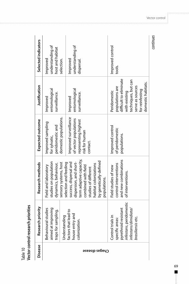
Vector control
69
Tabl
e 10
Vect
or co
ntro
l res
earc
h pr
iorit
ies
Dis
ease
Rese
arch
pri
orit
yRe
sear
ch m
etho
dsEx
pect
ed o
utco
me
Just
ifica
tion
Sele
cted
indi
cato
rs
Chagas disease
Beha
viou
ral s
tudi
es
aim
ed a
t im
prov
ing
trap
s fo
r sam
plin
g.
Fiel
d an
d la
bora
tory
st
udie
s on
pop
ulat
ion
dyna
mic
s, be
havi
our,
sens
ory
syst
ems,
host
se
lect
ion
and
feed
ing
succ
ess,
disp
ersa
l and
di
sper
sion
, and
sho
rt-
term
ada
ptiv
e ca
paci
ty,
com
bine
d w
ith fi
eld
stud
ies
of d
iffer
ent
habi
tat c
olon
izat
ions
by
gen
etic
ally
-defi
ned
popu
latio
ns.
Impr
oved
sam
plin
g fo
r syl
vatic
, pe
ridom
estic
and
do
mes
tic p
opul
atio
ns.
Impr
oved
en
tom
olog
ical
su
rvei
llanc
e.
Impr
oved
un
ders
tand
ing
of
host
and
hab
itat
sele
ctio
n.U
nder
stan
ding
fa
ctor
s th
at le
ad to
ho
use
entr
y an
d co
loni
zatio
n.
Impr
oved
sur
veill
ance
of
vec
tor p
opul
atio
ns
repr
esen
ting
high
est
risk
for h
uman
co
ntac
t.
Impr
oved
en
tom
olog
ical
su
rvei
llanc
e.
Impr
oved
un
ders
tand
ing
of
disp
ersa
l.
Cont
rol t
rials
in
spec
ific
area
s:
pyre
thro
id re
sist
ant
infe
stan
s, pe
ridom
estic
in
fest
ans/
dim
idia
ta/
bras
ilien
sis e
tc.
Fiel
d st
udie
s of
new
co
ntro
l int
erve
ntio
ns
and
new
com
bina
tions
of
inte
rven
tions
.
Impr
oved
con
trol
of
per
idom
estic
po
pula
tions
.
Perid
omes
tic
popu
latio
ns a
re
diffi
cult
to e
limin
ate
with
exi
stin
g te
chni
ques
, but
can
se
rve
as s
ourc
es
for r
einf
estin
g do
mes
tic h
abita
ts.
Impr
oved
con
trol
to
ols.
cont
inue
s

70
WH
O T
echn
ical
Rep
ort S
erie
s, N
o. 9
75, 2
012
Research Priorities for Chagas Disease, HAT and Leishmaniasis Report of the TDR Disease Reference Group
Dis
ease
Rese
arch
pri
orit
yRe
sear
ch m
etho
dsEx
pect
ed o
utco
me
Just
ifica
tion
Sele
cted
indi
cato
rsHuman African Trypanosomiasis
Impr
ovem
ent i
n co
st
effec
tiven
ess
and
ease
of u
se. L
ogis
tics
of e
xist
ing
targ
et
tech
nolo
gy fo
r tse
tse
cont
rol.
Det
erm
inat
ion
of
optim
um v
isua
l and
od
our a
ttra
ctan
t ch
arac
teris
tics
of
targ
ets.
To b
e fo
llow
ed
by fi
eld
valid
atio
n in
va
ried
site
s in
clud
ing
man
grov
e, d
ense
fore
st
and
river
ine
habi
tats
. Co
stin
g of
con
trol
.
Com
mer
cial
ly
avai
labl
e, q
ualit
y co
ntro
lled,
affo
rdab
le
targ
ets
suita
ble
for
smal
l (H
AT fo
ci) a
nd
larg
e (P
ATTE
C) s
cale
co
ntro
l pro
gram
mes
.
Cost
and
logi
stic
al
issu
es li
mit
curr
ent t
sets
e co
ntro
l ope
ratio
ns
requ
iring
co
nsid
erab
le
impr
ovem
ents
in
avai
labl
e co
ntro
l te
chno
logy
.
Rout
ine
use
of
targ
ets
in H
AT
foci
as
a pa
rtne
r to
cas
e de
tect
ion
and
trea
tmen
t pr
ogra
mm
es.
Iden
tifyi
ng
disc
ontin
uitie
s in
ts
etse
fly
popu
latio
ns
in o
rder
to p
riorit
ize,
th
en ta
rget
, HAT
foci
w
here
con
trol
will
be
mos
t sus
tain
able
.
Exte
nsiv
e ts
etse
sa
mpl
ing
at, a
nd a
t va
ryin
g di
stan
ces
from
, H
AT fo
ci fo
llow
ed b
y m
olec
ular
gen
etic
st
udie
s.
PATT
EC w
ill
achi
eve
sequ
entia
l, su
stai
nabl
e el
imin
atio
n of
tset
se
as a
pub
lic h
ealth
pr
oble
m fr
om re
gion
s of
sub
-Sah
aran
Afr
ica.
Curr
ent c
ontr
ol is
po
orly
targ
eted
at
thos
e ts
etse
po
pula
tions
m
ost s
usce
ptib
le
to c
ontr
ol. I
t is
targ
eted
to th
ose
area
s w
here
the
polit
ics
are
mos
t am
enab
le.
PATT
EC's
deve
lopm
ent o
f a
plan
to s
eque
ntia
lly
iden
tify,
targ
et a
nd
elim
inat
e H
AT fo
ci.
Dem
onst
ratio
n co
ntro
l tr
ials
in s
peci
fied
situ
atio
ns.
Tria
ls o
f gro
und-
base
d fo
ggin
g, a
lone
and
in
com
bina
tion
with
ot
her m
etho
ds.
Cont
rol p
ropo
sals
for
diffi
cult
situ
atio
ns
such
as
man
grov
e,
fore
st, a
nd ri
verin
e sp
ecie
s.
Som
e si
tuat
ions
cu
rren
tly d
ifficu
lt w
ith a
vaila
ble
tech
niqu
es
and
avai
labl
e ex
perie
nce.
Impr
oved
con
trol
for
all s
cena
rios.
Tabl
e 10 c
ontin
ued
cont
inue
s
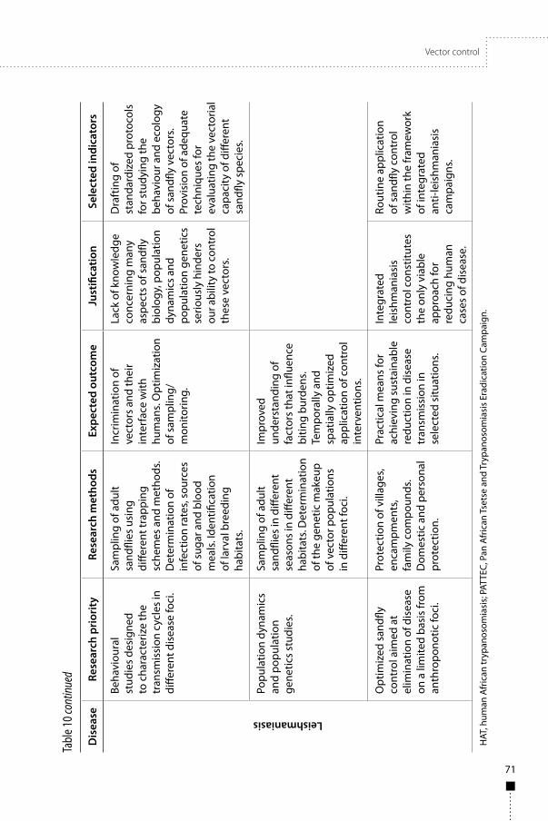
Vector control
71
Dis
ease
Rese
arch
pri
orit
yRe
sear
ch m
etho
dsEx
pect
ed o
utco
me
Just
ifica
tion
Sele
cted
indi
cato
rsLeishmaniasis
Beha
viou
ral
stud
ies
desi
gned
to
cha
ract
eriz
e th
e tr
ansm
issi
on c
ycle
s in
di
ffere
nt d
isea
se fo
ci.
Sam
plin
g of
adu
lt sa
ndfli
es u
sing
di
ffere
nt tr
appi
ng
sche
mes
and
met
hods
. D
eter
min
atio
n of
in
fect
ion
rate
s, so
urce
s of
sug
ar a
nd b
lood
m
eals
. Ide
ntifi
catio
n of
larv
al b
reed
ing
habi
tats
.
Incr
imin
atio
n of
ve
ctor
s an
d th
eir
inte
rfac
e w
ith
hum
ans.
Opt
imiz
atio
n of
sam
plin
g/
mon
itorin
g.
Lack
of k
now
ledg
e co
ncer
ning
man
y as
pect
s of
san
dfly
biol
ogy,
pop
ulat
ion
dyna
mic
s and
po
pula
tion
gene
tics
serio
usly
hin
ders
ou
r abi
lity
to c
ontr
ol
thes
e ve
ctor
s.
Dra
ftin
g of
st
anda
rdiz
ed p
roto
cols
fo
r stu
dyin
g th
e be
havi
our a
nd e
colo
gy
of s
andfl
y ve
ctor
s. Pr
ovis
ion
of a
dequ
ate
tech
niqu
es fo
r ev
alua
ting
the
vect
oria
l ca
paci
ty o
f diff
eren
t sa
ndfly
spe
cies
.
Popu
latio
n dy
nam
ics
and
popu
latio
n ge
netic
s st
udie
s.
Sam
plin
g of
adu
lt sa
ndfli
es in
diff
eren
t se
ason
s in
diff
eren
t ha
bita
ts. D
eter
min
atio
n of
the
gene
tic m
akeu
p of
vec
tor p
opul
atio
ns
in d
iffer
ent f
oci.
Impr
oved
un
ders
tand
ing
of
fact
ors
that
influ
ence
bi
ting
burd
ens.
Tem
pora
lly a
nd
spat
ially
opt
imiz
ed
appl
icat
ion
of c
ontr
ol
inte
rven
tions
.
Opt
imiz
ed s
andfl
y co
ntro
l aim
ed a
t el
imin
atio
n of
dis
ease
on
a li
mite
d ba
sis
from
an
thro
pono
tic fo
ci.
Prot
ectio
n of
vill
ages
, en
cam
pmen
ts,
fam
ily c
ompo
unds
. D
omes
tic a
nd p
erso
nal
prot
ectio
n.
Prac
tical
mea
ns fo
r ac
hiev
ing
sust
aina
ble
redu
ctio
n in
dis
ease
tr
ansm
issi
on in
se
lect
ed s
ituat
ions
.
Inte
grat
ed
leis
hman
iasi
s co
ntro
l con
stitu
tes
the
only
via
ble
appr
oach
for
redu
cing
hum
an
case
s of
dis
ease
.
Rout
ine
appl
icat
ion
of s
andfl
y co
ntro
l w
ithin
the
fram
ewor
k of
inte
grat
ed
anti-
leis
hman
iasi
s ca
mpa
igns
.
HAT
, hum
an A
fric
an tr
ypan
osom
iasi
s; P
ATTE
C, P
an A
fric
an T
sets
e an
d Tr
ypan
osom
iasi
s Er
adic
atio
n Ca
mpa
ign.
Tabl
e 10 c
ontin
ued

72
WH
O T
echn
ical
Rep
ort S
erie
s, N
o. 9
75, 2
012
Research Priorities for Chagas Disease, HAT and Leishmaniasis Report of the TDR Disease Reference Group
Thus, if significant and accessible breeding sites of important vector species (i.e. the VL vectors P. argentipes in India, L. longipalpis in Brazil, and P. orientalis in Sudan) were identified, it might be possible to modify their breeding habitats to render them less suitable for sandfly larvae. Manipulation of soil properties is used in agriculture to alter water conductivity, infiltration rates, aeration, and other physical properties (196). Appropriately used, such practices may help to curtail larval breeding. For example, P. orientalis in Sudan and Ethiopia is frequently associated with cracked black-cotton soil (197). Soil cracking can be significantly and inexpensively reduced by adding gypsum or gypsum with Ca-zeolites (196, 198, 199). Control of larvae in soil habitats may also be achieved by ploughing in organic insecticidal soil fertilizers such as Neem oil-cakes widely used as soil sanitizers. Neem contains the active ingredient azadirachtin, shown to kill sandfly larvae in laboratory experiments (200, 201).
8.3.2 Targeting sugar-feeding adult sandfliesIn nature, both sexes of sandflies (like mosquitoes) seek plants and obtain essential sugar meals from them by piercing stems and leaves (202, 203), ingesting honeydew produced by plant-sucking homopteran insects (204, 205) or nectar (206). Preliminary control studies with mosquitoes demonstrated significant mortality in a desert oasis achieved by spraying of flowering trees with insecticide-laced sugar bait (207). In similar studies conducted in a semi-arid area, spraying of plants with an attractive sugar and fruit juice toxic bait (1.0% w/v boric acid, 0.04% w/v Spinosad) reduced P. papatasi populations significantly (208). Spraying of large tracts of vegetation with a non-specific insecticidal combination is certainly not an environmentally acceptable approach for control. Ideally, bait stations with sandfly/mosquito specific attractive bait need to be developed. Such baits can comprise, for example, commercially available fruit concentrate that can be diluted in water, mixed with yeast and allowed to ferment for a prescribed period before use. In parallel, mixtures of synthetic floral odours from plants that are attractive to sandflies and/or mosquitoes need to be characterized (209). Ideally, such baits will be mixed with an efficient and specific oral toxin (e.g. Bacillus thuringiensis var. israeliensis) (210).
8.3.3 Extermination of sandflies feeding on domestic animalsSeveral sandfly vectors of human leishmaniasis are known to be attracted to and feed on domestic animals. Such zoophilic habits should be explored and potentially exploited for killing sandflies. In laboratory trials, avermectins in very low concentrations were shown to be lethal to sandfly adults (211, 212). In addition, remnants of feed-through insecticides in rodent (hamster) faeces were lethal to sandfly larvae feeding on them (212, 213). This approach may facilitate control of vectors of cutaneous leishmaniasis breeding in rodent burrows and feeding on these animals.

Vector control
73
This approach can be particularly useful when the preferred host is also the reservoir host for the Leishmania parasites. For example, using insecticidal dog collars to control canine VL resulted in reduced disease, through effects that were probably mediated by killing of sandflies and deterring them from feeding on dogs with collars (214–216). One study also demonstrated a reduction in infantile VL in villages where dogs were fitted with deltamethrin-impregnated collars (217). In recent experiments conducted in Bihar, India, tablets containing lipophilic compounds were orally administered to cattle. A significant mortality was demonstrated in P. argentipes females that feed on these animals, and the sandfly populations in treated villages were significantly reduced (R. Poché, personal communication).
P. argentipes, the vector of VL on the Indian sub-continent, frequently aggregates and feeds on cattle (218, 219). Thus, topical insecticidal treatment of domestic cows and buffaloes, which often share living quarters with humans, holds potential for reducing the sandfly populations in and around homes. Some modern insecticidal products on the market are highly lipophilic, permitting spot-on application to a comparatively small area of skin from where the active ingredients dissipate over the entire body. Some products developed for topical use on domestic livestock require no withholding time before milk or meat are consumed by humans (220). Such products hold potential for reducing sandfly numbers, curtailing transmission of visceral leishmaniasis (VL), and increasing cattle productivity by alleviating the burden of helminthic and arthropod parasites.
Similar approaches may also prove efficacious for controlling L. longipalpis, the main vector of VL in Latin America. L. longipalpis is highly ornithophilic and is frequently associated with chicken houses, where it feeds on domestic chickens (221). Methods for safely treating chickens with insecticidal formulations that will kill the flies can be an efficient tool for combating VL in parts of Brazil and other Latin American countries.
8.3.4 Flight barriers for preventing sandflies from reaching housesSandflies seeking blood meals generally advance upwind in short flights, close to the ground, guided by host-derived cues such as CO₂, temperature and humidity (222). When they encounter a vertical obstacle, such as a wall, they proceed upward in short flights and come in repeated contact with the surface of the obstacle. Therefore, spraying of walls or other barriers may be effective against sandflies approaching inhabited areas from the periphery. To test this hypothesis, a 60m long insecticide-impregnated fine-mesh barrier was erected between a natural habitat from where sandflies were approaching and a row of houses. The mesh barrier proved partially efficient (60% reduction) at blocking sandflies (mainly P. sergenti) approaching houses (223). In later studies conducted in a

74
WH
O T
echn
ical
Rep
ort S
erie
s, N
o. 9
75, 2
012
Research Priorities for Chagas Disease, HAT and Leishmaniasis Report of the TDR Disease Reference Group
different location, a longer barrier (400m) was found far more effective (90% reduction, P. papatasi) (Faiman & Warburg, unpublished). The potential of vertical barriers should be explored further in foci where sandflies arrive at human habitations from natural breeding habitats at the periphery of villages.
8.3.5 Protection of homes – indoor residual insecticide sprayingDepending on the application techniques, timing and target species, sandflies can be highly susceptible to insecticides (185). Residual formulations of DDT and pyrethroids have been used to control sandflies both in the Old World and in the neotropics (224–227). Historically the most significant reduction in incidence of VL on the Indian sub-continent was a byproduct of the antimalarial campaign which consisted of house spraying using DDT (187). More recently, focal application of residual formulations of DDT and pyrethroids have been demonstrated effective against endophilic sandflies in Latin America and India (228, 229). Reliable evaluation of environmentally-sound methods for effective use of insecticides in and around human habitation certainly warrant continued efforts. Monitoring of levels of insecticide resistance in sandfly populations should comprise an integral component of campaigns utilizing insecticides in any mode of application (230).
8.3.6 Protection of rooms/porches – insecticide diffusers and coilsDiffusible insecticides hold potential for reducing the biting burden from sandflies in enclosed or partially enclosed spaces. For example, controlled experimentation in inhabited homes demonstrated that prallethrin evaporators were highly efficient at eliminating sandflies from occupied bedrooms in a CL focus in Israel (231). Mosquito coils dispersing insecticide by burning can be used effectively in homes that have no electricity. Indeed mosquito coils were demonstrated to reduce the biting rates of mosquitoes in numerous controlled studies (232). However, reports demonstrating the efficacy of mosquito coils against sandflies (187) are scant. It should be stressed that the possible health implications resulting from prolonged inhalation of smoke produced by coils needs to be investigated thoroughly before any recommendation for use can be made (233).
8.3.7 Personal protection – insecticide-treated nets and repellentsThe use of insecticide-treated materials, in particular insecticide-treated nets (ITNs), is one of the most effective methods for reducing man–vector contact and transmission of malaria (234). In recent years the efficacy of long-lasting insecticidal nets (LLINs) against P. argentipes vectors of kala-azar in India was demonstrated in several studies (191, 229, 235–237). Moreover, VL rates in Sudanese foci were markedly reduced following the widespread distribution of ITNs, despite the fact that the vector P. orientalis is thought to be exophilic (238).

Vector control
75
LLINs should certainly be considered in integrated control campaigns targeting (mainly) endophilic sandflies in anthroponotic foci of CL and VL. Continued efforts should be devoted to developing and testing ITNs in different leishmaniasis foci in different parts of the world.
The active ingredients of currently available insect repellents include N, N-diethyl-3-methylbenzamide (DEET), botanicals, citronella, and picaridin. For a long time DEET has been considered the most efficacious repellent, with a strong safety record and excellent protection against ticks, mosquitoes, sandflies and other arthropods. Newer agents like picaridin and natural products such as oil of lemon eucalyptus are becoming increasingly popular because of their low toxicity and comparable efficacy (239). Since most topical repellents are non-specific it is not necessary to instigate programmes to develop repellents specific for sandflies; new products developed against mosquitoes should be tested to verify their efficacy against sandflies (240).
8.3.8 Integrated disease-control campaignsIntegrated disease-control campaigns comprise different studies and experimental application of a combination of measures for disease control including but not limited to, sandfly control. As a rule, an integrated disease-control campaign should be based upon a profound understanding of the ecology, epidemiology and transmission in a particular disease focus and custom designed to attain optimal results. Thus, baseline studies should be aimed at incriminating the vectors and characterizing their behaviour in relation to the environment (i.e. what the vector species are, where they breed, which plants provide them with sugar meals), potential reservoir hosts (e.g. if the disease is zoonotic or anthroponotic, what the major blood sources are for the vectors), and human population (e.g. where the sandflies arrive from relative to the human living areas, whether they enter houses to bite or are exophilic, whether people sleep inside or outside their houses). The genetic structure of vector populations throughout their distribution should be studied in order to gain knowledge of reproductive isolation that may imply discrete behaviour patterns and vectorial capacities (241, 242). Based on the results of such studies, the most efficacious means for sandfly control should be implemented in conjunction with other disease control strategies such as treatment of affected humans, vaccination and elimination of reservoir hosts (considered elsewhere in this report). Effective collaboration between the professional teams in charge and leaders of the affected populations will promote community participation and should be striven for during control campaigns.


77
9. Economic evaluation of health-care interventionsEconomic evaluation seeks to assess the relative merits of different interventions or health-care technologies. Alternative courses of actions in health care are measured by both their costs and consequences (243). The most used methods for economic evaluation are cost-effectiveness and cost-utility analyses. In cost-effectiveness analyses, outcomes are expressed in disease-specific measures (e.g. clinical outcomes or cases averted). In cost-utility analyses, outcomes are expressed in generic measures (e.g. quality-adjusted life years (QALYs) gained or disability-adjusted life years (DALYs) averted), which combine life expectancy and quality of life. The latter allows for comparison between different conditions, thus contributing to informing policy-makers on resource allocation among health-care priorities.
It has to be borne in mind that the cost-effectiveness of an intervention will depend not only on the costs but also on the existence of an effective intervention, which in the case of treatment for Chagas disease, human African trypanosomiasis (HAT) and leishmaniasis, can be problematical. Thus, despite the potential value of applying economic evaluation for technical efficiency, i.e. determining the most rational way of achieving an objective, prioritizing research on the grounds of economic evaluations is not always possible. Moreover, in the cases of Chagas disease, HAT and leishmaniasis, research priorities should also consider equity considerations, as these diseases disproportionately affect the poor and enhance the poverty cycle.
An assessment of the costs of preventing and treating Chagas disease in Colombia (244) showed that the expected total cost of treatment for a patient with chronic Chagas in this country was US$ 1028 per year while only 2% of the current expenditure for treatment would be needed to adequately spray houses with insecticide. A further analysis modelled the cost-effectiveness of vector control activities in the same setting and showed that the current method of house spraying was cost-effective using an incremental net benefit approach (using a threshold value per DALY averted equivalent to the Colombian per capita GDP) (245). This was true for all the villages analysed in two geographical locations (Boyaca and Antioquia). A recent study in Yucatan, Mexico, suggests that ecosystemic peridomicile management in the control of non-domiciliated T. dimidiata would be an excellent complementary strategy to improve the cost-effectiveness of interventions (246). This is because these strategies would also be effective against other vector-borne diseases, such as malaria and dengue. Another study in northern Argentina estimated retrospectively the cost per case averted for three different modalities of interventions for vector control: vertical, mixed and horizontal approaches (247). The horizontal approach involved community participation and was the observed strategy from 1993 to 2004 in the setting. Results for the other two strategies (vertical, mixed programmes) were
78
WH
O T
echn
ical
Rep
ort S
erie
s, N
o. 9
75, 2
012
Research Priorities for Chagas Disease, HAT and Leishmaniasis Report of the TDR Disease Reference Group
inferred from the observed horizontal programme. Excluding per diem, the cost-effectiveness of vertical, mixed and horizontal strategies were US$ 60, US$ 42 and US$ 32 per averted case, respectively. Community participation as a method of choice for sustained monitoring of infestation by non-domiciliated triatomines has also been justified as a cost-effective method elsewhere (248).
Lutumba et al. (88) used cost-effectiveness analysis to evaluate different algorithms for confirmation of HAT. The study results suggested that a sequence of lymph node puncture, thick blood film, capillary tube centrifugation and mini-anion exchange centrifugation technique (LNP-TBF-CTC-mAECT), was the most cost-effective test algorithm per life saved. A study in Angola (249) found that eflornithine saved more lives than melarsoprol, but that melarsoprol was slightly more cost-effective. The average cost per life saved was US$ 627.6 for melarsoprol and US$ 856.1 for eflornithine. Likewise, the incremental cost–effectiveness ratio was US$ 8169 per additional life saved and US$ 299 per additional life year saved.
Vanlerberghe et al. (250) evaluated the cost-effectiveness of different visceral leishmaniasis (VL) treatment regimens as first-line care in endemic areas. Among four regimens selected, treatment with amphotericin B-deoxycholate was the most effective approach, averting 87.2% of all deaths. However, the least expensive and the most cost-effective treatment was the miltefosine regimen. A study in India (251) concluded that the available treatments for VL were cost-effective, and that combinations were more cost-effective than most monotherapies. The cost-effectiveness ratio ranged from US$ 5–8 per year of lost life averted to US$ 124–213 per death averted.
In Colombia (252), the cost per DALY averted for a patient cured with antimony during an ongoing epidemic of cutaneous leishmaniasis (CL) was estimated to be approximately US$ 15 000. In Kabul (253), the cost-effectiveness of intralesional and intramuscular administration of the pentavalent antimonial drug sodium stibogluconate, the current ‘standard treatment’ for CL, was calculated. The cost per DALY averted per patient cured was estimated to be approximately US$ 1200 dollars (US$ 761–1827); based on these results, the health intervention was considered not cost-effective according to WHO-CHOICE criteria.
Few studies have integrated analyses of disease burden, cost of illness for households, and cost-effectiveness for a particular setting. Lutumba et al. (27) investigated the burden in DALYs, household costs and cost per DALY averted of a HAT outbreak in a rural community of the Democratic Republic of Congo. Data on DALYs and cost of illness were collected through a retrospective household survey. Case-finding activity resulted in 1408 DALYs averted, for a cost saving of US$ 17 per DALY averted. The main challenges related to conducting research in health economics, particularly in economic evaluation, due to the scarcity of trained researchers in this discipline. Economic evaluation of control and
Economic evaluation of health-care interventions
79
prevention strategies for infectious diseases requires competence in mathematical modelling techniques to represent the epidemiology of the disease, and the ability to link this to economic data. Models are usually data poor, as in resource-constrained settings quality data and records are scarce. Nonetheless, this type of research can facilitate proper decision-making and strategic planning in control programmes.

81
10. Research priority recommendationsThe Reference Group identified the following top research priorities for Chagas disease, human African trypanosomiasis and leishmaniasis:
■ Research on new diagnostics for case detection and characterization, including drug resistance and tests of cure.
■ Research on new therapeutics to avoid drug resistance, including exploring combinations of approved anti-kinetoplastid drugs, repurposing of existing approved drugs, and developing new drugs.
■ Research on new vector control technologies, including markers of successful vector control.
■ Research on vector population characteristics, including insecticide resistance.
■ Operational research on integrated disease and vector control. ■ Research on vaccines to prevent Leishmania infection and disease,
and vaccines to block transmission of Leishmania. ■ Research to assess the importance of asymptomatic infection.


83
AcknowledgementsWe wish to express our appreciation to the many people who contributed to this report. We especially thank Professor Simon Croft and Professor Tania Araujo Jorge for hosting meetings at the London School of Hygiene and Tropical Medicine and FIOCRUZ, respectively, as well as for their contributions to the deliberations. We thank Dr. Rick Tarleton (Wake Forest University, Winston-Salem, USA), Professor Felipe Guhl (Universidad de Los Andes, Bogotá, Colombia), Joseph Ndungu (Head, Human African Trypanosomiasis (HAT) and Other Neglected Tropical Diseases, Foundation for Innovative New Diagnostics, Geneva, Switzerland), Professor Alejandro Luquetti (Universidade Federal de Goiás, Goiania, Brasil), and Professor Julio Urbina (Venezuelan Institute for Scientific Research, Caracas, Venezuela) for their extensive and substantive input.
We also thank the many stakeholders who provided input electronically and especially to those who attended the meeting at FIOCRUZ. KDS, BZ, and thank NIH and the Bill and Melinda Gates Foundation, FAPESP and MCT/CNPq/MS-SCTIE-DECIT.
Special acknowledgement to Dr Ayoade Oduola, Dr Michael Wilson, Ms Edith Certain, Dr Deborah Kioy, Dr Fabio Zicker, Dr Julie Reza, Dr Margaret Harris and Elisabetta Dessi (TDR), WHO, Geneva, Switzerland, who were instrumental in creating and managing the DRG, preparing and coordinating the meetings as well as in publishing this report.
Special thanks to Dr Jean Jannin, Dr Pere Simarro and Dr Miguelle Franco, Neglected Tropical Diseases Control (NTD), WHO, for technical contribution and support in the preparation of this report, to the WHO Brazil and Sudan country offices for support in organizing DRG meetings, and to Dr. Zaida Yadon, WHO, Pan American Health Organization (PAHO).
Sincere thanks and appreciation to peer reviewers for their comments and contributions on the technical accuracy of this report. Dr Nina Mattock was responsible for technical editing of the report.
The activities of the DRG, including production of this report, were funded by the Special Programme for Research and Training in Tropical Diseases (TDR) and by the European Commission, under Agreement PP-AP/2008/160-163.


85
References1. Rassi A Jr., Rassi A, Marin-Neto JA. Chagas disease. The Lancet, 2010, 375:1388–1402.
2. Mott KE et al. Parasitic diseases and urban development. Bulletin of the World Health Organization, 1990, 68:691–698.
3. Schmunis GA. Trypanosoma cruzi, the etiologic agent of Chagas' disease: status in the blood supply in endemic and nonendemic countries. Transfusion, 1991, 31:547–557.
4. Control of Chagas' disease: report of a WHO Expert Committee. Geneva, World Health Organization, 1991.
5. Schmunis GA. A tripanossomiase americana e seu impacto na saude publica das Americas. In: Brener Z. AZ, Barral-Neto M., eds. Trypanosoma cruzi e doenca de Chagas. Rio de Janeiro, Guanabara-Koogan, 2000:1–15.
6. Control of Chagas disease: report of a WHO expert committee. Geneva, World Health Organization, 2002.
7. Moncayo A, Silveira AC. Current epidemiological trends for Chagas disease in Latin America and future challenges in epidemiology, surveillance and health policy. Memorias do Instituto Oswaldo Cruz, 2009, 1:17–30.
8. World Development Report 1993: investing in health. New York, World Bank, 1993.
9. Mathers CD, Lopez A, Murray C. The burden of disease and mortality by condition: data, methods, and results for the year 2001. In: Lopez AD et al, eds. Global burden of disease and risk factors. New York, Oxford University Press, 2006.
10. Schofield CJ, Dias JC. A cost-benefit analysis of Chagas disease control. Memorias do Instituto Oswaldo Cruz, 1991, 86:285–295.
11. Schenone H. Human infection by Trypanosoma cruzi in Chile: epidemiology estimates and costs of care and treatment of the chagasic patient. Boletín chileno de parasitología, 1998, 53:23–26.
12. Salvatella AR, Vignolo W. Una aproximación a los costos de internación por cardiopatia Chagasica en Uruguay. Revista da Sociedade Brasileira de Medicina Tropical, 1996, 29:114–118.
13. Disease control priorities in developing countries. 2nd ed. Jamison DT, ed. New York, Oxford University Press, 2006.
14. Gascon J, Bern C, Pinazo MJ. Chagas disease in Spain, the United States and other non-endemic countries. Acta Tropica, 2010, 115:22–27.
15. Bern C, Montgomery SP. An estimate of the burden of Chagas disease in the United States. Clinical Infectious Diseases, 2009, 49:52–54.
16. Fevre EM et al. The burden of human African trypanosomiasis. PLoS Neglected Tropical Diseases, 2008, 2:333.
17. Odiit M et al. Quantifying the level of under-detection of Trypanosoma brucei rhodesiense sleeping sickness cases. Tropical Medicine & International Health, 2005, 10:840–849.
18. Fevre EM, et al. A burgeoning epidemic of sleeping sickness in Uganda. The Lancet, 2005, 366:745–747.
19. Simarro PP, Jannin J, Cattand P. Eliminating human African trypanosomiasis: where do we stand and what comes next? PLoS Medicine, 2008, 5:55.
20. Brun R, et al. Human African trypanosomiasis. The Lancet, 2010, 375:148–159.
21. Moore A et al. Resurgence of sleeping sickness in Tambura County, Sudan. American Journal of Tropical Medicine and Hygiene, 1999, 6:315–318.

86
WH
O T
echn
ical
Rep
ort S
erie
s, N
o. 9
75, 2
012
Research Priorities for Chagas Disease, HAT and Leishmaniasis Report of the TDR Disease Reference Group
22. Stanghellini A, Josenando T. The situation of sleeping sickness in Angola: a calamity. Tropical Medicine & International Health, 2001, 6:330–344.
23. Lutumba P et al. Trypanosomiasis control, Democratic Republic of Congo, 1993–2003. Emerging Infectious Diseases, 2005, 11:1382–1388.
24. Robays J et al. The effectiveness of active population screening and treatment for sleeping sickness control in the Democratic Republic of Congo. Tropical Medicine & International Health, 2004, 9:542–550.
25. Van Nieuwenhove S et al. Sleeping sickness resurgence in the DRC: the past decade. Tropical Medicine & International Health, 2001, 6:335–341.
26. Chappuis F et al. Human African trypanosomiasis in areas without surveillance. Emerging Infectious Diseases, 2010, 16:354–356.
27. Lutumba P et al. Human African trypanosomiasis in a rural community, Democratic Republic of Congo. Emerging Infectious Diseases, 2007, 13:248–254.
28. Politi C et al. Cost-effectiveness analysis of alternative treatments of African gambiense trypanosomiasis in Uganda. Health Economics, 1995, 4:273–287.
29. Gouteux JP et al. Cost of the individual treatment of Trypanosoma brucei gambiense trypanosomiasis in a focus of infection in Niari (Congo). Medecine tropicale (Marseille), 1987, 47:61–63.
30. Sundar S et al. Implementation research to support the initiative on the elimination of kala azar from Bangladesh, India and Nepal – the challenges for diagnosis and treatment. Tropical Medicine & International Health, 2008, 13:2–5.
31. Romero GA, Boelaert M. Control of visceral leishmaniasis in Latin America – a systematic review. PLoS Neglected Tropical Diseases, 2010, 4:584.
32. Dujardin JC et al. Spread of vector-borne diseases and neglect of leishmaniasis, Europe. Emerging Infectious Diseases, 2008, 14:1013–1018.
33. Desjeux P. Leishmaniasis. Public health aspects and control. Clinics in Dermatology, 1996, 14:417–423.
34. Singh SP et al. Serious underreporting of visceral leishmaniasis through passive case reporting in Bihar, India. Tropical Medicine & International Health, 2006, 11:899–905.
35. Mubayi A et al. Transmission dynamics and underreporting of kala-azar in the Indian state of Bihar. Journal of Theoretical Biology, 2010, 262:177–185.
36. Singh VP et al. Estimation of under-reporting of visceral leishmaniasis cases in Bihar, India. American Journal of Tropical Medicine and Hygiene, 2010, 82:9–11.
37. Rijal S et al. Epidemiology of Leishmania donovani infection in high-transmission foci in Nepal. Tropical Medicine & International Health, 2010, 15 Suppl 2:21–28.
38. Singh SP et al. The epidemiology of Leishmania donovani infection in high transmission foci in India. Tropical Medicine & International Health, 2010, 15 Suppl 2:12–20.
39. Griekspoor A, Sondorp E, Vos T. Cost-effectiveness analysis of humanitarian relief interventions: visceral leishmaniasis treatment in the Sudan. Health Policy and Planning, 1999, 14:70–76.
40. Schaefer KU et al. A prospective sero-epidemiological study of visceral leishmaniasis in Baringo District, Rift Valley Province, Kenya. Transactions of the Royal Society of Tropical Medicine and Hygiene, 1995, 89:471–475.
41. Ali A, Ashford RW. Visceral leishmaniasis in Ethiopia. IV. Prevalence, incidence and relation of infection to disease in an endemic area. Annals of Tropical Medicine and Parasitology, 1994, 88:289–293.
References
87
42. Zijlstra EE et al. Endemic kala-azar in eastern Sudan: a longitudinal study on the incidence of clinical and subclinical infection and post-kala-azar dermal leishmaniasis. American Journal of Tropical Medicine and Hygiene, 1994, 51:826–836.
43. Marlet MV et al. A neglected disease of humans: a new focus of visceral leishmaniasis in Bakool, Somalia. Transactions of the Royal Society of Tropical Medicine and Hygiene, 2003, 97:667–671.
44. Alvar J, Yactayo S, Bern C. Leishmaniasis and poverty. Trends in Parasitology, 2006, 22:552–557.
45. Boelaert M et al. The poorest of the poor: a poverty appraisal of households affected by visceral leishmaniasis in Bihar, India. Tropical Medicine & International Health, 2009, 14:639–644.
46. Thakur CP. Socio-economics of visceral leishmaniasis in Bihar (India). Transactions of the Royal Society of Tropical Medicine and Hygiene, 2000, 94:156–157.
47. Meheus F et al. Costs of patient management of visceral leishmaniasis in Muzaffarpur, Bihar, India. Tropical Medicine & International Health, 2006, 11:1715–1724.
48. Sarnoff R et al. The economic impact of visceral leishmaniasis on rural households in one endemic district of Bihar, India. Tropical Medicine & International Health, 2010, 2:42–49.
49. Sundar S et al. Household cost-of-illness of visceral leishmaniasis in Bihar, India. Tropical Medicine & International Health, 2010, 2:50–54.
50. Rijal S et al. The economic burden of visceral leishmaniasis for households in Nepal. Transactions of the Royal Society of Tropical Medicine and Hygiene, 2006, 100:838–841.
51. Adhikari SR, Maskay NM. The economic burden of Kala-azar in households of the Danusha and Mahottari districts of Nepal. Acta Tropica, 2003, 88:1–2.
52. Anoopa Sharma D et al. The economic impact of visceral leishmaniasis on households in Bangladesh. Tropical Medicine & International Health, 2006, 11:757–764.
53. Ahluwalia IB et al. Visceral leishmaniasis: consequences of a neglected disease in a Bangladeshi community. American Journal of Tropical Medicine and Hygiene, 2003, 69:624–628.
54. Dedet JP, Pillot B, Gentilini M. Evaluation of the socioeconomic costs of cutaneous leishmaniasis in French Guiana. Revue d Epidemiologie et de Sante Publique, 1991, 39:129–133.
55. Adhikari SR, Maskay NM, Sharma BP. Paying for hospital-based care of Kala-azar in Nepal: assessing catastrophic, impoverishment and economic consequences. Health Policy and Planning, 2009, 24:129–139.
56. Zingales B et al. A new consensus for Trypanosoma cruzi intraspecific nomenclature: second revision meeting recommends TcI to TcVI. Memorias do Instituto Oswaldo Cruz, 2009, 104:1051–1054.
57. Zingales B et al. The revised Trypanosoma cruzi subspecific nomenclature: Rationale, epidemiological relevance and research applications. Infection, Genetics and Evolution, 2012, 12:240–253.
58. Miles MA, et al. The molecular epidemiology and phylogeography of Trypanosoma cruzi and parallel research on Leishmania: looking back and to the future. Parasitology, 2009, 136:1509–1528.
59. Lewis MD et al. Genotyping of Trypanosoma cruzi: systematic selection of assays allowing rapid and accurate discrimination of all known lineages. American Journal of Tropical Medicine and Hygiene, 2009, 81:1041–1049.
60. Dias JCP. The treatment of Chagas disease (South American trypanosomiasis). Annals of Internal Medicine, 2006, 144:772–774.
61. Rassi Jr A, Rassi A, Marin-Neto JA. Chagas heart disease: pathophysiologic mechanisms, prognostic factors and risk stratification. Memorias do Instituto Oswaldo Cruz, 2009, 104:152–158.

88
WH
O T
echn
ical
Rep
ort S
erie
s, N
o. 9
75, 2
012
Research Priorities for Chagas Disease, HAT and Leishmaniasis Report of the TDR Disease Reference Group
62. PAHO. Consultation on congenital chagas disease, its epidemiology and management. Montevideo, Uruguay, 24–25 June 2004 (http://www.paho.org/english/ad/dpc/cd/dch-chagas-congenita-2004.htm, accessed 28 May 2012).
63. Nishioka SA. Benznidazole in the primary chemoprophylaxis of the reactivation of Chagas' disease in chronic chagasic patients using corticosteroids at immunosuppressive doses: is there sufficient evidence for recommending its use? Revista da Sociedade Brasileira de Medicina Tropical, 2000, 33:83–85.
64. Cordova E et al. Reactivation of Chagas disease with central nervous system involvement in HIV-infected patients in Argentina, 1992–2007. International Journal of Infectious Diseases, 2008, 12:587–592.
65. Diez M et al. Usefulness of PCR strategies for early diagnosis of Chagas' disease reactivation and treatment follow-up in heart transplantation. American Journal of Transplantation, 2007, 7:1633–1640.
66. Sternberg JM. Human African trypanosomiasis: clinical presentation and immune response. Parasite Immunology, 2004, 26:469–476.
67. Barry D, Carrington M. Antigenetic variation. In: Maudlin I, Holmes PH, Miles MA, eds. The trypanosomiases. CABI, 2004:25–37.
68. Lundkvist GB, Kristensson K, Bentivoglio M. Why trypanosomes cause sleeping sickness. Physiology, 2004, 19:198–206.
69. Blum JA et al. Cardiac alterations in human African trypanosomiasis (T. b. gambiense) with respect to the disease stage and antiparasitic treatment. PLoS Neglected Tropical Diseases, 2009, 3:383.
70. Control and surveillance of African trypanosomiasis: report of a WHO Expert Committee. Geneva, World Health Organization, 1998.
71. Clinical manifestations and diagnosis of sleeping sickness. In: Mulligan HW, ed. The African trypanosomiases, 1st ed. London, George Allen and Unwin Ltd., 1970:661–682.
72. Dumas M, Girard PL. Human African trypanosomiasis (sleeping sickness). In: Vinken PJ, ed. Handbook of clinical neurology: infections of the nervous system. Amsterdam, North-Holland Publishing Co., 1978:67–83.
73. Kennedy PG. The continuing problem of human African trypanosomiasis (sleeping sickness). Annals of Neurology, 2008, 64:116–126.
74. Banuls AL, Hide M, Prugnolle F. Leishmania and the leishmaniases: a parasite genetic update and advances in taxonomy, epidemiology and pathogenicity in humans. Advances in Parasitology, 2007, 64:1–109.
75. Bittencourt AL et al. Leishmaniasis recidiva cutis in American cutaneous leishmaniasis. International Journal of Dermatology, 1993, 32:802–805.
76. Silveira FT, Lainson R, Corbett CE. Further observations on clinical, histopathological, and immunological features of borderline disseminated cutaneous leishmaniasis caused by Leishmania (Leishmania) amazonensis. Memorias do Instituto Oswaldo Cruz, 2005, 100:525–534.
77. Pintado V et al. Visceral leishmaniasis in human immunodeficiency virus (HIV)-infected and non-HIV-infected patients. A comparative study. Medicine, 2001, 80:54–73.
78. Umezawa ES et al. Evaluation of recombinant antigens for serodiagnosis of Chagas' disease in South and Central America. Journal of Clinical Microbiology, 1999, 37:1554–1560.
79. Luquetti AO et al. Chagas' disease diagnosis: a multicentric evaluation of Chagas Stat-Pak, a rapid immunochromatographic assay with recombinant proteins of Trypanosoma cruzi. Diagnostic Microbiology and Infectious Disease, 2003, 46:265–271.

References
89
80. Yun O et al. Feasibility, drug safety, and effectiveness of etiological treatment programs for Chagas disease in Honduras, Guatemala, and Bolivia: 10-year experience of Medecins Sans Frontieres. PLoS Neglected Tropical Diseases, 2009, 3:488.
81. Britto CC. Usefulness of PCR-based assays to assess drug efficacy in Chagas disease chemotherapy: value and limitations. Memorias do Instituto Oswaldo Cruz, 2009, 1:122–135.
82. Duffy T et al. Accurate real-time PCR strategy for monitoring bloodstream parasitic loads in Chagas disease patients. PLoS Neglected Tropical Diseases, 2009, 3:419.
83. Schijman AG et al. International study to evaluate PCR methods for detection of Trypanosoma cruzi DNA in blood samples from Chagas disease patients. PLoS Neglected Tropical Diseases, 2011, 5:931.
84. Mora MC et al. Early diagnosis of congenital Trypanosoma cruzi infection using PCR, hemoculture, and capillary concentration, as compared with delayed serology. Journal of Parasitology, 2005, 91:1468–1473.
85. Blanco SB et al. Congenital transmission of Trypanosoma cruzi: an operational outline for detecting and treating infected infants in north-western Argentina. Tropical Medicine & International Health, 2000, 5:293–301.
86. Miezan TW et al. Evaluation of the parasitologic technics used in the diagnosis of human Trypanosoma gambiense trypanosomiasis in the Ivory Coast. Bulletin de la Societe de Pathologie Exotique (Paris), 1994, 87:101–104.
87. Lutumba P et al. Validity, cost and feasibility of the mAECT and CTC confirmation tests after diagnosis of African of sleeping sickness. Tropical Medicine & International Health, 2006, 11:470–478.
88. Lutumba P et al. Cost-effectiveness of algorithms for confirmation test of human African trypanosomiasis. Emerging Infectious Diseases, 2007, 13:1484–1490.
89. Magnus E et al. A card-agglutination test with stained trypanosomes (C.A.T.T.) for the serological diagnosis of T. b. gambiense trypanosomiasis. Annales de la Societe Belge de Medecine Tropicale (Antwerpen), 1978, 58:169–176.
90. Pansaerts R et al. Increased sensitivity of the card agglutination test CATT/Trypanosoma brucei gambiense by inhibition of complement. Acta Tropica, 1998, 70:349–354.
91. Deborggraeve S et al. Molecular dipstick test for diagnosis of sleeping sickness. Journal of Clinical Microbiology, 2006, 44:2884–2889.
92. FIND and partners initiate search for a Test of Cure for sleeping sickness. Geneva, Foundation for Innovative New Diagnostics, 2009 (http://www.finddiagnostics.org/media/news/091217.html, accessed 28 May 2012).
93. Chappuis F et al. Visceral leishmaniasis: what are the needs for diagnosis, treatment and control? Nature Reviews Microbiology, 2007, 5:873–882.
94. Sundar S, Rai M. Laboratory diagnosis of visceral leishmaniasis. Clinical and Diagnostic Laboratory Immunology, 2002, 9:951–958.
95. Singh R et al. Potential of direct agglutination test based on promastigote and amastigote antigens for serodiagnosis of post-kala-azar dermal leishmaniasis. Clinical and Diagnostic Laboratory Immunology, 2005, 12:1191–1194.
96. Chappuis F et al. A meta-analysis of the diagnostic performance of the direct agglutination test and rK39 dipstick for visceral leishmaniasis. British Medical Journal, 2006, 333:723.
97. Sadeghian G et al. Evaluation of leishmanin skin test and its relationship with the clinical form and duration of cutaneous leishmaniasis. Dermatology Online Journal, 2006, 12:3.

90
WH
O T
echn
ical
Rep
ort S
erie
s, N
o. 9
75, 2
012
Research Priorities for Chagas Disease, HAT and Leishmaniasis Report of the TDR Disease Reference Group
98. El-Safi SH et al. Field evaluation of latex agglutination test for detecting urinary antigens in visceral leishmaniasis in Sudan. Eastern Mediterraneam Health Journal, 2003, 9:844–855.
99. Reithinger R, Dujardin JC. Molecular diagnosis of leishmaniasis: current status and future applications. Journal of Clinical Microbiology, 2007, 45:21–25.
100. Tojal da Silva AC et al. Species diversity causing human cutaneous leishmaniasis in Rio Branco, state of Acre, Brazil. Tropical Medicine & International Health, 2006, 11:1388–1398.
101. Campino L et al. Detection of Leishmania in immunocompromised patients using peripheral blood spots on filter paper and the polymerase chain reaction. European Journal of Clinical Microbiology of Infectious Diseases, 2000, 19:396–398.
102. Antinori S et al. Is real-time polymerase chain reaction (PCR) more useful than a conventional PCR for the clinical management of leishmaniasis? American Journal of Tropical Medicine and Hygiene, 2009, 81:46–51.
103. Moran M et al. Neglected disease research and development: how much are we really spending? PLoS Medicine, 2009, 6:e1000030.
104. El-Sayed NM et al. Comparative genomics of trypanosomatid parasitic protozoa. Science, 2005, 309:404–409.
105. Bartholomeu DC et al. Genomic organization and expression profile of the mucin-associated surface protein (masp) family of the human pathogen Trypanosoma cruzi. Nucleic Acids Research, 2009, 37:3407–3417.
106. Berriman M et al. The genome of the African trypanosome Trypanosoma brucei. Science, 2005, 309:416–422.
107. Stuart K et al. Kinetoplastids: related protozoan pathogens, different diseases. Journal of Clinical Investigation, 2008, 118:1301–1310.
108. Sosa-Estani S, Segura EL. Etiological treatment in patients infected by Trypanosoma cruzi: experiences in Argentina. Current Opinion in Infectious Diseases, 2006, 19:583–587.
109. CLAP/PAHO/WHO. Report of the Technical Consultation on Information, Education, and Communication (IEC) on Congenital Chagas Disease. Montevideo, Uruguay, 17–18 May 2007.
110. Marin-Neto JA et al. The BENEFIT trial: testing the hypothesis that trypanocidal therapy is beneficial for patients with chronic Chagas heart disease. Memorias do Instituto Oswaldo Cruz, 2009, 104 Suppl 1:319–324.
111. Filardi LS, Brener Z. Susceptibility and natural resistance of Trypanosoma cruzi strains to drugs used clinically in Chagas disease. Transactions of the Royal Society of Tropical Medicine and Hygiene, 1987, 81:755–759.
112. Urbina J. Specific chemotherapy of Chagas disease: relevance, current limitations and new approaches. Acta Tropica, 2010, 115:55–68.
113. Priotto G et al. Nifurtimox-eflornithine combination therapy for second-stage African Trypanosoma brucei gambiense trypanosomiasis: a multicentre, randomised, phase III, non-inferiority trial. Lancet, 2009, 374:56–64.
114. Maser P et al. A nucleoside transporter from Trypanosoma brucei involved in drug resistance. Science, 1999, 285:242–244.
115. Matovu E et al. Mechanisms of arsenical and diamidine uptake and resistance in Trypanosoma brucei. Eukaryotic Cell, 2003, 2:1003–1008.
116. Nerima B et al. Detection of mutant P2 adenosine transporter (TbAT1) gene in Trypanosoma brucei gambiense isolates from northwest Uganda using allele-specific polymerase chain reaction. Tropical Medicine & International Health, 2007, 12:1361–1368.

References
91
117. Brun R et al. The phenomenon of treatment failures in human African trypanosomiasis. Tropical Medicine & International Health, 2001, 6:906–914.
118. Sokolova AY et al. Cross-resistance to nitro drugs and implications for treatment of human African trypanosomiasis. Antimicrobial Agents and Chemotherapy, 2010, 54:2893–2900.
119. Croft SL, Sundar S, Fairlamb AH. Drug resistance in leishmaniasis. Clinical Microbiology Reviews, 2006, 19:111–126.
120. Sundar S, Rai M. Treatment of visceral leishmaniasis. Expert Opinion on Pharmacotherapy, 2005, 6:2821–2829.
121. Gonzalez U et al. Interventions for Old World cutaneous leishmaniasis. Cochrane Database of Systematic Reviews, 2008, (4):CD005067.
122. Palumbo E. Current treatment for cutaneous leishmaniasis: a review. American Journal of Therapeutics, 2009, 16:178–182.
123. Sundar S et al. New treatment approach in Indian visceral leishmaniasis: single-dose liposomal amphotericin B followed by short-course oral miltefosine. Clinical Infectious Diseases, 2008, 47:1000–1006.
124. Tarleton RL. Chagas disease: a role for autoimmunity? Trends in Parasitology, 2003, 19:447–451.
125. Tarleton RL. Immune system recognition of Trypanosoma cruzi. Current Opinion in Immunology, 2007, 19:430–434.
126. Camargo EP. Perspectives of vaccination in Chagas disease revisited. Memorias do Instituto Oswaldo Cruz, 2009, 104 Suppl 1:275–280.
127. Bustamante JM, Bixby LM, Tarleton RL. Drug-induced cure drives conversion to a stable and protective CD8+ T central memory response in chronic Chagas disease. Nature Medicine, 2008, 14:542–550.
128. Martin DL et al. CD8+ T-Cell responses to Trypanosoma cruzi are highly focused on strain-variant trans-sialidase epitopes. PLoS Pathogens, 2006, 2:e77.
129. Laucella SA et al. Frequency of interferon-gamma-producing T cells specific for Trypanosoma cruzi inversely correlates with disease severity in chronic human Chagas disease. Journal of Infectious Diseases, 2004, 189:909–918.
130. Dias JC. Southern Cone Initiative for the elimination of domestic populations of Triatoma infestans and the interruption of transfusional Chagas disease. Historical aspects, present situation, and perspectives. Memorias do Instituto Oswaldo Cruz, 2007, 102 Suppl 1:11–18.
131. Cohen JE, Gurtler RE. Modeling household transmission of American trypanosomiasis. Science, 2001, 293:694–698.
132. Gurtler RE et al. Domestic dogs and cats as sources of Trypanosoma cruzi infection in rural northwestern Argentina. Parasitology, 2007, 134(Pt 1):69–82.
133. Dias JC, Silveira AC, Schofield CJ. The impact of Chagas disease control in Latin America: a review. Memorias do Instituto Oswaldo Cruz, 2002, 97:603–612.
134. Dubois ME, Demick KP, Mansfield JM. Trypanosomes expressing a mosaic variant surface glycoprotein coat escape early detection by the immune system. Infection and Immunity, 2005, 73:2690–2697.
135. Crowe JS et al. All metacyclic variable antigen types of Trypanosoma congolense identified using monoclonal antibodies. Nature, 1983, 306:389–91.
136. Morrison WI et al. Protective immunity and specificity of antibody responses elicited in cattle by irradiated Trypanosoma brucei. Parasite Immunology, 1982, 4:395–407.

92
WH
O T
echn
ical
Rep
ort S
erie
s, N
o. 9
75, 2
012
Research Priorities for Chagas Disease, HAT and Leishmaniasis Report of the TDR Disease Reference Group
137. Handman E. Leishmaniasis: current status of vaccine development. Clinical Microbiology Reviews, 2001, 14:229–243.
138. Palatnik-de-Sousa CB. Vaccines for leishmaniasis in the fore coming 25 years. Vaccine, 2008, 26(14):1709–24.
139. Nadim A et al. Effectiveness of leishmanization in the control of cutaneous leishmaniasis. Bulletin de la Societe de Pathologie Exotique et de ses Filiales, 1983, 76:377–383.
140. Sacks D, Noben-Trauth N. The immunology of susceptibility and resistance to Leishmania major in mice. Nature Reviews Immunology, 2002, 2:845–858.
141. Kedzierski L, Zhu Y, Handman E. Leishmania vaccines: progress and problems. Parasitology, 2006, 133 Suppl:S87–112.
142. Noazin S et al. Efficacy of killed whole-parasite vaccines in the prevention of leishmaniasis: a meta-analysis. Vaccine, 2009, 27:4747–4753.
143. Coler RN, Reed SG. Second-generation vaccines against leishmaniasis. Trends in Parasitology, 2005, 21:244–249.
144. Peters NC et al. Vector transmission of leishmania abrogates vaccine-induced protective immunity. PLoS Pathogens, 2009, 5:e1000484.
145. Musa AM et al. Immunochemotherapy of persistent post-kala-azar dermal leishmaniasis: a novel approach to treatment. Transactions of the Royal Society of Tropical Medicine and Hygiene, 2008, 102:58–63.
146. Dujardin JP, Schofield CJ, Panzera F. Los Vectores de la Enfermedad de Chagas. Brussels: Academie Royale des Sciences d'Outre Mer; 2002.
147. Yamagata Y, Nakagawa J. Control of Chagas disease. Advances in Parasitology, 2006, 61:129–165.
148. Gurtler RE. Sustainability of vector control strategies in the Gran Chaco Region: current challenges and possible approaches. Memorias do Instituto Oswaldo Cruz, 2009, 104:52–59.
149. Schofield CJ, Jannin J, Salvatella R. The future of Chagas disease control. Trends in Parasitology, 2006, 22:583–588.
150. Borges EC et al. Dynamics between sylvatic, peridomestic and domestic populations of Triatoma brasiliensis (Hemiptera : reduviidae) in Ceara state, northeastern Brazil. Acta Tropica, 2005, 93:119–126.
151. Ibarra-Cerdena CN et al. Ecology of North American Triatominae. Acta Tropica, 2009, 110:178–186.
152. Abad-Franch F, Santos WS, Schofield CJ. Research needs for Chagas disease prevention. Acta Tropica, 2010, 115(1–2, Sp. Iss. SI).
153. Ekkens DB. Nocturnal flights of Triatoma (hemiptera, reduviidae) in Sabino canyon, Arizona. 1: light collections. Journal of Medical Entomology, 1981, 18:211–227.
154. Vazquez-Prokopec GM et al. Seasonal variations in active dispersal of natural populations of Triatoma infestans in rural north-western Argentina. Medical and Veterinary Entomology, 2006, 20:273–279.
155. Lehane MJ et al. The role of temperature and nutritional status in flight initiation by Triatoma infestans. Acta Tropica, 1992, 52:27–38.
156. Kato H et al. A repertoire of the dominant transcripts from the salivary glands of the blood-sucking bug, Triatoma dimidiata, a vector of Chagas disease. Infection, Genetics and Evolution, 2010, 10:184–191.
157. Schwarz A et al. Antibody responses of domestic animals to salivary antigens of Triatoma infestans as biomarkers for low-level infestation of triatomines. International Journal for Parasitology, 2009, 39:1021–1029.
References
93
158. Bargues MD et al. Phylogeography and genetic variation of Triatoma dimidiata, the main chagas disease vector in central America, and its position within the genus Triatoma. PLoS Neglected Tropical Diseases, 2008, 2.
159. Cardozo RM et al. Inheritance of resistance to pyrethroids in Triatoma infestans, the main Chagas disease vector in South America. Infection, Genetics and Evolution, 2010, 10:1174–1178.
160. Panzera F et al. Genomic changes of Chagas disease vector, South America. Emerging Infectious Diseases, 2004, 10:438–446.
161. Simarro PP, Jannin J, Cattand P. Eliminating human African trypanosomiasis: where do we stand and what comes next. PLoS Medicine, 2008, 5:e55.
162. Barrett MP, Coombs GH, Mottram JC. Future prospects in chemotherapy for trypanosomiasis. In: Maudlin I, Holmes PH, Miles MA, eds. The trypanosomiases. CABI, 2004:445–60.
163. Delespaux V, de Koning HP. Drugs and drug resistance in African trypanosomiasis. Drug Resistance Updates, 2007, 10:30–50.
164. Brun R et al. Human African trypanosomiasis. Lancet, 2010, 375:148–159.
165. Gouteux JP, Artzrouni M. Is vector control needed in the fight against sleeping sickness? A biomathematical approach. Bulletin De La Societe De Pathologie Exotique, 1996, 89:299–305.
166. Torr SJ, Hargrove JW, Vale GA. Towards a rational policy for dealing with tsetse. Trends In Parasitology, 2005, 21:537–541.
167. Lancien J. Controlling sleeping sickness in southeastern Uganda with tsetse-fly traps. Annales De La Societe Belge De Medecine Tropicale, 1991, 71:35–47.
168. Welburn SC et al. Crisis, what crisis? Control of Rhodesian sleeping sickness. Trends in Parasitology, 2006, 22:123–128.
169. Welburn SC et al. Sleeping sickness: a tale of two diseases. Trends in Parasitology, 2001, 17:19–24.
170. Simarro PP. Human African trypanosomiasis: an epidemiological update. WHO Weekly Epidemiological Record, 2006, 81:69–80.
171. Rogers DJ, Robinson TP. Tsetse distribution. In: Maudlin I, Holmes PH, Miles MA, eds. The trypanosomiases. CABI, 2004:139–179.
172. Courtin F et al. Sleeping sickness in West Africa (1906-2006): changes in spatial repartition and lessons from the past. Tropical Medicine & International Health, 2008, 13:334–44.
173. Vale GA, Torr S. Development of bait technology to control tsetse. In: Maudlin I, Holmes PH, Miles MA, eds. The trypanosomiases. CABI, 2004:509–524.
174. Laveissière C, Garcia A, Sané B, eds. Lutte contre la maladie du sommeil et soins de santé primaire. IRD Editions, Montpellier, France, 2003.
175. Wint GRW, Robinson TP. Gridded livestock of the world 2007. Rome, Food and Agriculture Organization of the United Nations, 2007:131.
176. Clausen PH et al. Host preferences of tsetse (Diptera: Glossinidae) based on bloodmeal identifications. Medical and Veterinary Entomology, 1998, 12:169–180.
177. Lindh JM et al. Improving the cost-effectiveness of artificial visual baits for controlling the tsetse fly Glossina fuscipes fuscipes. PLoS Neglected Tropical Diseases, 2009, 3.
178. Omolo MO et al. Prospects for developing odour baits to control Glossina fuscipes spp., the major vector of human African trypanosomiasis. PLoS Neglected Tropical Diseases, 2009, 3:e435.
179. Rayaisse JB et al. Prospects for the development of odour baits to control the tsetse flies Glossina tachinoides and G. palpalis s.l., vectors of trypanosomiasis in West Africa. PLoS Neglected Tropical Diseases, 2009, 4:e632.

94
WH
O T
echn
ical
Rep
ort S
erie
s, N
o. 9
75, 2
012
Research Priorities for Chagas Disease, HAT and Leishmaniasis Report of the TDR Disease Reference Group
180. Kuzoe FAS, Schofield CJ. Strategic review of traps and targets for tsetse and African trypanosomiasis control. Geneva, World Health Organization, TDR/IDE/TRY/05.1, 2005.
181. Shaw APM et al. A basis for financial decision-making on strategies for the control of human African trypanosomiasis. TropIKA, 2008, http://www.tropika.net/svc/review/African_Tryps_Financial_decision_making.
182. Seccombe A, Ready P, Huddleston L. A catalogue of Old World phlemotomine sandflies (Diptera: Psychodidae, Phlebotominae) Occasional Papers on Systematic Entomology, 1993, 8:1–57.
183. Young D, Duncan M. Guide to the identification and geographic distribution of Lutzomyia sand-flies in Mexico, West Indies, Central and South America (Diptera: Psychodidae). Gainsville, FL, Associate Publishers, 1994.
184. Amora SS et al. Control of phlebotomine (Diptera: Psychodidae) leishmaniasis vectors. Neotropical Entomology, 2009, 38:303–310.
185. Alexander B, Maroli M. Control of phlebotomine sandflies. Medical and Veterinary Entomology, 2003, 17:1–18.
186. Davies CR et al. Leishmaniasis: new approaches to disease control. British Medical Journal, 2003, 326:377–382.
187. Kishore K et al. Vector control in leishmaniasis. Indian Journal of Medical Research, 2006, 123:467–472.
188. Sharma U, Singh S. Insect vectors of Leishmania: distribution, physiology and their control. Journal of Vector Borne Diseases, 2008, 45:255–272.
189. Maroli M, Khoury C. Prevention and control of leishmaniasis vectors: current approaches. Parassitologia, 2004, 46:211–215.
190. Kassi M et al. Vector control in cutaneous leishmaniasis of the old world: a review of literature. Dermatology Online Journal, 2008, 14:1.
191. Ostyn B et al. Vector control by insecticide-treated nets in the fight against visceral leishmaniasis in the Indian subcontinent, what is the evidence? Tropical Medicine & International Health, 2008, 13:1073–1085.
192. Modi GB, Tesh RB. A simple technique for mass rearing Lutzomyia longipalpis and Phlebotomus papatasi (Diptera: Psychodidae) in the laboratory. Journal of Medical Entomology, 1983, 20:568–569.
193. Feliciangeli MD. Natural breeding places of phlebotomine sandflies. Medical and Veterinary Entomology, 2004, 18:71–80.
194. Singh R, Lal S, Saxena VK. Breeding ecology of visceral leishmaniasis vector sandfly in Bihar state of India. Acta Tropica, 2008, 107:117–120.
195. Kumar V et al. Field trial of an ecological approach for the control of Phlebotomus argentipes using mud & lime plaster. Indian Journal of Medical Research, 1995, 101:154–156.
196. Pal D et al. Significance of soil modifiers (Ca-zeolites and gypsum) in naturally degraded Vertisols of the Peninsular India in redefining the sodic soils. Geoderma, 2006, 136:210–228.
197. Hoogstraal H, Heyneman D. Leishmaniasis in the Sudan Republic: 30. Final epidemiologic report. American Journal of Tropical Medicine and Hygiene, 1969, 18(6_Part_2):1091–1210.
198. Nadler A, Magaritz M. Long-term effects of extensive gypsum amendment applied with sodic water irrigation on the soil properties and soil solution chemical composition. Soil Science Society of America Journal, 1986, 142:196–202.
199. Shainberg I et al. Use of gypsum on soils: A review. In: Stewart B, ed. Advances in soil science. New York, Springer-Verlag, 1989, pp. 1–111.

References
95
200. Andrade Coelho CA et al. Effects of azadirachtin on the development and mortality of Lutzomyia longipalpis larvae (Diptera: Psychodidae: Phlebotominae). Journal of Medical Entomology, 2006, 43:262–266.
201. Andrade-Coelho CA et al. Effect of fruit and leaves of Meliaceae plants (Azadirachta indica and Melia azedarach) on the development of Lutzomyia longipalpis larvae (Diptera: Psychodidae: Phlebotominae) under experimental conditions. Journal of Medical Entomology, 2009, 46:1125–1130.
202. Schlein Y, Muller G. Assessment of plant tissue feeding by sand flies (Diptera: Psychodidae) and mosquitoes (Diptera: Culicidae). Journal of Medical Entomology, 1995, 32:882–887.
203. Schlein Y, Jacobson RL. Sugar meals and longevity of the sandfly Phlebotomus papatasi in an arid focus of Leishmania major in the Jordan Valley. Medical and Veterinary Entomology, 1999, 13:65–71.
204. Moore JS et al. Honeydew sugars in wild-caught Phlebotomus ariasi detected by high performance liquid chromatography (HPLC) and gas chromatography (GC). Medical and Veterinary Entomology, 1987, 1:427–434.
205. Wallbanks KR et al. Aphid derived sugars in the neotropical sandfly – Lutzomyia peruensis. Tropical Medicine and Parasitology, 1991, 42:60–62.
206. Muller G, Schlein Y. Nectar and honeydew feeding of Phlebotomus papatasi in a focus of Leishmania major in Neot Hakikar oasis. Journal of Vector Ecology, 2004, 29:154–158.
207. Muller G, Schlein Y. Sugar questing mosquitoes in arid areas gather on scarce blossoms that can be used for control. International Journal for Parasitology, 2006, 36:1077–1080.
208. Muller GC, Schlein Y. Different methods of using attractive sugar baits (ATSB) for the control of Phlebotomus papatasi. Journal of Vector Ecology, 2011, 36 Suppl 1:S64–70.
209. Jhumur US, Dotterl S, Jurgens A. Floral odors of Silene otites: their variability and attractiveness to mosquitoes. Journal of Chemical Ecology, 2008, 34:14–25.
210. Yuval B, Warburg A. Susceptibility of adult phlebotomine sandflies (Diptera: Psychodidae) to Bacillus thuringiensis var. israeliensis. Annals of Tropical Medicine and Parasitology, 1989, 83:195–196.
211. Kassem HA, Tewfick MK, El Sawaf BM. Evaluation of avermectins as sandfly control agents. Annals of Tropical Medicine and Parasitology, 2001, 95:405–411.
212. Mascari TM et al. Ivermectin as a rodent feed-through insecticide for control of immature sand flies (Diptera: Psychodidae). Journal of the American Mosquito Control Association, 2008, 24:323–326.
213. Mascari TM et al. Evaluation of novaluron as a feed-through insecticide for control of immature sand flies (Diptera: Psychodidae). Journal of Medical Entomology, 2007, 44:714–717.
214. Aoun K et al. Efficacy of Deltamethrine-impregnated collars Scalibor in the prevention of canine leishmaniasis in the area of Tunis. Archives de l Institut Pasteur de Tunis, 2008, 85:63–68.
215. Ferroglio E, Poggi M, Trisciuoglio A. Evaluation of 65% permethrin spot-on and deltamethrin-impregnated collars for canine Leishmania infantum infection prevention. Zoonoses and Public Health, 2008, 55:145–148.
216. Killick-Kendrick R et al. Protection of dogs from bites of phlebotomine sandflies by deltamethrin collars for control of canine leishmaniasis. Medical and Veterinary Entomology, 1997, 11:105–111.
217. Gavgani AS et al. Effect of insecticide-impregnated dog collars on incidence of zoonotic visceral leishmaniasis in Iranian children: a matched-cluster randomised trial. The Lancet, 2002, 360:374–379.
218. Lane RP, Pile MM, Amerasinghe FP. Anthropophagy and aggregation behaviour of the sandfly Phlebotomus argentipes in Sri Lanka. Medical and Veterinary Entomology, 1990, 4:79–88.

96
WH
O T
echn
ical
Rep
ort S
erie
s, N
o. 9
75, 2
012
Research Priorities for Chagas Disease, HAT and Leishmaniasis Report of the TDR Disease Reference Group
219. Addy M et al. Host preference of Phlebotomus argentipes in different biotopes. Tropical and Geographical Medicine, 1983, 35:343–345.
220. Alvinerie M et al. Pharmacokinetics of eprinomectin in plasma and milk following topical administration to lactating dairy cattle. Research in Veterinary Science, 1999;67(3):229–32.
221. Alexander B et al. Role of the domestic chicken (Gallus gallus) in the epidemiology of urban visceral leishmaniasis in Brazil. Emerging Infectious Disease, 2002, 8:1480–1485.
222. Killick-Kendrick R. The biology and control of phlebotomine sand flies. Clinics in Dermatology, 1999, 17:279–289.
223. Faiman R, Cuno R, Warburg A. Control of phlebotomine sand flies with vertical fine-mesh nets. Journal of Medical Entomology, 2009, 46:820–831.
224. Hertig M. Phlebotomus and residual DDT in Greece and Italy. American Journal of Tropical Medicine and Hygiene, 1949, 1:773.
225. Hertig M, Fisher LR. Control of sandflies with DDT. Bulletin of the US Army Medical Department, 1945, 88:97.
226. Le Pont F et al. Impact de pulvérisations de deltaméthrin dans un foyer de leishmaniose de Bolivie. Annales de la Societé Belge de Médicine Tropicale, 1989, 69:223–232.
227. Marcondes CB, Nascimento JA. Evaluation of the effectiveness of deltamethrin (K-othrine CE) in the control of Lutzomyia longipalpis (Diptera: Psychodidae), in the municipality of Santa Rita, Paraiba, Brazil. Revista da Sociedade Brasileira de Medicina Tropical, 1993, 26:15–18.
228. Kelly DW, Mustafa Z, Dye C. Differential application of lambda-cyhalothrin to control the sandfly Lutzomyia longipalpis. Medical and Veterinary Entomology, 1997, 11:13–24.
229. Das ML et al. Comparative study of kala-azar vector control measures in eastern Nepal. Acta Tropica, 2010, 113:162–166.
230. Kelly-Hope L, Ranson H, Hemingway J. Lessons from the past: managing insecticide resistance in malaria control and eradication programmes. The Lancet Infectious Diseases, 2008, 8:387–389.
231. Sirak-Wizeman M et al. Control of phlebotomine sandflies in confined spaces using diffusible repellents and insecticides. Medical and Veterinary Entomology, 2008, 22:405–412.
232. Lawrance CE, Croft AM. Do mosquito coils prevent malaria? A systematic review of trials. Journal of Travel Medicine, 2004, 11:92–96.
233. Liu W et al. Mosquito coil emissions and health implications. Environmental Health Perspectives, 2003, 111:1454–1460.
234. Beier JC et al. Integrated vector management for malaria control. Malaria Journal, 2008, 7 Suppl 1:S4.
235. Dinesh DS et al. Long-lasting insecticidal nets fail at household level to reduce abundance of sandfly vector Phlebotomus argentipes in treated houses in Bihar (India). Tropical Medicine & International Health, 2008, 13:953–958.
236. Picado A et al. Effect of village-wide use of long-lasting insecticidal nets on visceral Leishmaniasis vectors in India and Nepal: a cluster randomized trial. PLoS Neglected Tropical Diseases, 2010, 4:e587.
237. Picado A et al. Effect of untreated bed nets on blood-fed Phlebotomus argentipes in kala-azar endemic foci in Nepal and India. Memorias do Instituto Oswaldo Cruz, 2009, 104:1183–1186.
238. Ritmeijer K et al. Evaluation of a mass distribution programme for fine-mesh impregnated bednets against visceral leishmaniasis in eastern Sudan. Tropical Medicine & International Health, 2007, 12:404–414.

References
97
239. Katz TM, Miller JH, Hebert AA. Insect repellents: historical perspectives and new developments. Journal of the American Academy of Dermatology, 2008, 58:865–871.
240. Katritzky AR et al. Synthesis and bioassay of improved mosquito repellents predicted from chemical structure. Proceedings of the National Academy of Sciences of the United States of America, 2008, 105:7359–7364.
241. Lanzaro GC, Warburg A. Genetic variability in phlebotomine sandflies: possible implications for leishmaniasis epidemiology. Parasitology Today, 1995, 11:151–54.
242. Luckhart S et al. Reframing critical needs in vector biology and management of vector-borne disease. PLoS Neglected Tropical Diseases, 2010, 4:e566.
243. Drummond MF et al. Methods for the economic evaluation of health care programmes. The Journal of Mental Health Policy and Economics, 1999, 2:43.
244. Castillo-Riquelme M et al. The costs of preventing and treating Chagas disease in Colombia. PLoS Neglected Tropical Diseases, 2008, 2:e336.
245. Castillo-Riquelme M et al. Modelling geographic variation in the cost-effectiveness of control policies for infectious vector diseases: the example of Chagas disease. Journal of Health Economics, 2008, 27:405–426.
246. Ferral J et al. Comparative field trial of alternative vector control strategies for non-domiciliated Triatoma dimidiata. American Journal of Tropical Medicine and Hygiene, 2010, 82:60–66.
247. Vazquez-Prokopec GM et al. Cost-effectiveness of Chagas disease vector control strategies in Northwestern Argentina. PLoS Neglected Tropical Diseases, 2009, 3:e363.
248. Dumonteil E et al. Usefulness of community participation for the fine temporal monitoring of house infestation by non-domiciliated triatomines. Journal of Parasitology, 2009, 95:469–471.
249. Robays J et al. Eflornithine is a cost-effective alternative to melarsoprol for the treatment of second-stage human West African trypanosomiasis in Caxito, Angola. Tropical Medicine & International Health, 2008, 13:265–271.
250. Vanlerberghe V et al. Drug policy for visceral leishmaniasis: a cost-effectiveness analysis. Tropical Medicine & International Health, 2007, 12:274–283.
251. Rathi SK, et al. Post-kala-azar dermal leishmaniasis: a histopathological study. Indian Journal of Dermatology, Venereology and Leprology, 2005, 71:250–3.
252. Vega JC et al. Short communication: The cost-effectiveness of cutaneous leishmaniasis patient management during an epidemic in Chaparral, Colombia in 2004. Tropical Medicine & International Health, 2007, 12:1540–1544.
253. Reithinger R, Coleman PG. Treating cutaneous leishmaniasis patients in Kabul, Afghanistan: cost-effectiveness of an operational program in a complex emergency setting. BMC Infectious Diseases, 2007, 7:3.


99
App endices
Appendix 1The Disease-specific and Thematic Reference Groups, the think tank for infectious diseases of poverty, and host countries
Reference group Host institution and country
DRG1 Malaria WHO country office, Cameroon
DRG2 Tuberculosis, leprosy and Buruli ulcer WHO country office, Philippines
DRG3 Chagas disease, human African trypanosomiasis and leishmaniasis
WHO country office, Sudan and Brazil
DRG4 Helminth infections African Programme for Onchocerciasis Control (APOC), Burkina Faso
DRG5 Dengue and other emerging viral diseases of public health importance
WHO country office, Cuba
DRG6 Zoonoses and marginalized infectious diseases of poverty
WHO Regional Office for the Eastern Mediterranean, Egypt
TRG1 Social sciences and gender WHO country office, Ghana
TRG2 Innovation and technology platforms for health interventions in infectious diseases of poverty
WHO country office, Thailand
TRG3Health systems and implementation research
WHO country office, Nigeria
TRG4 Environment, agriculture and infectious diseases of poverty
WHO country office, China

100
WH
O T
echn
ical
Rep
ort S
erie
s, N
o. 9
75, 2
012
Research Priorities for Chagas Disease, HAT and Leishmaniasis Report of the TDR Disease Reference Group
Appendix 2Membership of the Disease Reference Group on Chagas Disease, Human African Trypanosomiasis and Leishmaniasis (DRG3)
Name Country Area Speciality Gender
CHAIR Professor Kenneth Stuart
USA Leishmaniasis molecular biology, informatics
M
CO-CHAIR Professor Maowia Mukhtar
Sudan Leishmaniasis, immunology, pathogenesis
M
Professor Bianca Zingales
Brazil Chagas, molecular biology, informatics
F
MEMBERS Professor Marleen Boelaert
Belgium Leishmaniasis, social sciences, economics, implementation research
F
Ms Marianela Castillo-Riquelme
Chile Chagas, social sciences, economics, implementation research
F
Professor Mike Barret
Scotland Leishmaniasis, drugs, diagnostics, therapeutics, vaccines
M
Professor Michael J Lehane
England HAT, vector issues M
Professor Pascal Lutumba
Democratic Republic of
Congo
HAT, epidemiology, ecology M
Dr Enock Matovu Uganda HAT, drugs, diagnostics, therapeutics, vaccines
M
Dr David Sacks USA Leishmaniasis immunology, pathogenesis
M
Dr Sergio A Sosa-Estani
Argentina Chagas, clinical, health systems
M
Professor Shyam Sundar
India Leishmaniasis, clinical, health systems
M
Professor Rick L Tarleton
USA Chagas, immunology, pathogenesis
M
Dr Alon G Warburg Israel Leishmaniasis, vector issues M
Professor Chris Schofield
England Chagas, Leishmaniasis, HAT M
HAT, human African trypanosomiasis

Global Report for Research on Infectious Diseases of PovertyAvailable online at: www.who.int/tdr/capacity/global_report
Research Priorities for Zoonoses and Marginalized InfectionsWHO Technical Report Series, No. 971 (120 pages)Available online at: www.who.int/tdr/publications/zoonoses
Research Priorities for Helminth InfectionsWHO Technical Report Series, No. 972 (196 pages)Available online at: www.who.int/tdr/publications/helminth_infections
Research Priorities for the Environment, Agriculture and Infectious Diseases of PovertyWHO Technical Report Series, No. 976 (140 pages)Available online at: www.who.int/tdr/publications/environment
SELECTED WHO PUBLICATIONS OF RELATED INTEREST
Further information on these and other WHO publications can be obtained fromWHO Press, World Health Organization, 1211 Geneva 27, Switzerland
(tel.: +41 22 791 3264; fax: +41 22 791 4857; e-mail: [email protected];order on line: http://www.who.int/bookorders)
The World Health Organization was established in 1948 as a specialized agency of the United Nations serving as the directing and coordinating authority for international health matters and public health. One of WHO’s constitutional functions is to provide objective and reliable information and advice in the field of human health, a responsibility that it fulfils in part through its extensive programme of publications. The Organization seeks through its publications to support national health strategies and address the most pressing public health concerns of populations around the world. To respond to the needs of Member States at all levels of development, WHO publishes practical manuals, handbooks and training material for specific categories of health workers; internationally applicable guidelines and standards; reviews and analyses of health policies, programmes and research; and state-of-the-art consensus reports that offer technical advice and recommendations for decision-makers. These books are closely tied to the Organization’s priority activities, encompassing disease prevention and control, the development of equitable health systems based on primary health care, and health promotion for individuals and communities. Progress towards better health for all also demands the global dissemination and exchange of information that draws on the knowledge and experience of all WHO’s Member Countries and the collaboration of world leaders in public health and the biomedical sciences. To ensure the widest possible availability of authoritative information and guidance on health matters, WHO secures the broad international distribution of its publications and encourages their translation and adaptation. By helping to promote and protect health and prevent and control disease throughout the world, WHO’s books contribute to achieving the Organization’s principal objective – the attainment by all people of the highest possible level of health.
The WHO Technical Report Series makes available the findings of various international groups of experts that provide WHO with the latest scientific and technical advice on a broad range of medical and public health subjects. Members of such expert groups serve without remuneration in their personal capacities rather than as representatives of governments or other bodies; their views do not necessarily reflect the decisions or the stated policy of WHO. An annual subscription to this series, comprising four to six such reports, costs CHF 150.00/US$ 180.00 (CHF 105.00/US$ 126.00 in developing countries). For further information, please contact: WHO Press, World Health Organization, 20 Avenue Appia, 1211 Geneva 27, Switzerland (tel. +41 22 791 3264; fax: +41 22 791 4857; e-mail: [email protected]; order on line: http://www.who.int/bookorders).

Research Priorities for Chagas Disease, H
AT and Leishmaniasis
975 W H O T e c h n i c a l R e p o r t S e r i e s
975
Technical Report of the TDR Disease Reference Group on Chagas Disease, Human African Trypanosomiasis and Leishmaniasis
Research Priorities for Chagas Disease, Human African Trypanosomiasis and Leishmaniasis
WH
O Technical Report Series
Research Priorities for Chagas Disease, HAT and Leishmaniasis
This report provides a review and analysis of the research landscape for three diseases – Chagas disease, human African trypanosomiasis and leishmaniasis – that disproportionately afflict poor and remote populations with limited access to health services. It represents the work of the disease reference group on Chagas Disease, Human African Trypanosomiasis and Leishmaniasis (DRG3) which was established to identify key research priorities through review of research evidence and input from stakeholders' consultations.The diseases, which are caused by related protozoan parasites, are described in terms of their epidemiology and disease burden, clinical forms and pathogenesis, HIV coinfection, diagnosis, drugs and drug resistance, vaccines, vector control, and health-care interventions. Priority areas for research are identified based on criteria such as public health relevance, benefit and impact on poor populations and equity, and feasibility.The priorities are found in the areas of diagnostics, drugs, vector control, asymptomatic infection, economic analysis of treatment and vector control methods, and in some specific issues such as surveillance methods or transmission-blocking vaccines for particular diseases.This report will be useful to researchers, policy and decision-makers, funding bodies, implementation organizations, and civil society.This is one of ten disease and thematic reference group reports that have come out of the TDR Think Tank, all of which have contributed to the development of the Global Report for Research on Infectious Diseases of Poverty, available at: www.who.int/tdr/capacity/global_report.