Embed Size (px)
Citation preview
HEAD & FACE MEDICINE
Akyalcin et al. Head & Face Medicine 2013, 9:28http://www.head-face-med.com/content/9/1/28
RESEARCH Open Access
Measurement of skin dose from cone-beamcomputed tomography imagingSercan Akyalcin1*, Jeryl D English1, Kenneth M Abramovitch2 and Xiujiang J Rong3
Abstract
Objective: To measure surface skin dose from various cone-beam computed tomography (CBCT) scanners usingpoint-dosimeters.
Materials & methods: A head anthropomorphic phantom was used with nanoDOT optically stimulatedluminescence (OSL) dosimeters (Landauer Corp., Glenwood, IL) attached to various anatomic landmarks. Thephantom was scanned using multiple exposure protocols for craniofacial evaluations in three different CBCT unitsand a conventional x-ray imaging system. The dosimeters were calibrated for each of the scan protocols on thedifferent imaging systems. Peak skin dose and surface doses at the eye lens, thyroid, submandibular and parotidgland levels were measured.
Results: The measured skin doses ranged from 0.09 to 4.62 mGy depending on dosimeter positions and imagingsystems. The average surface doses to the lens locations were ~4.0 mGy, well below the threshold forcataractogenesis (500 mGy). The results changed accordingly with x-ray tube output (mAs and kV) and also weresensitive to scan field of view (SFOV). As compared to the conventional panoramic and cephalometric imagingsystem, doses from all three CBCT systems were at least an order of magnitude higher.
Conclusions: Peak skin dose and surface doses at the eye lens, thyroid, and salivary gland levels measured fromthe CBCT imaging systems were lower than the thresholds to induce deterministic effects. However, our findingsdo not justify the routine use of CBCT imaging in orthodontics considering the lifetime-attributable risk to theindividual.
Keywords: Skin dose, Cone-beam computed tomography, OSL dosimeters
IntroductionA three-dimensional radiographic examination of thecraniofacial skeleton with cone beam computed tomog-raphy (CBCT) is indicated for a number of clinical con-ditions. However, like any x-ray exposure, CBCT scansalso expose the patient to certain biologic risks of radi-ation. As the indications for CBCT imaging becomemore universal, so does the concern for radiation safetyrelated to dental and orthodontic procedures [1-3].In diagnostic imaging, exposure to x-ray radiation
must be accompanied by a related benefit that outweighsthe associated risks for the use of that radiation. Ortho-dontists, as practicing health-care providers, must re-main cognizant of the risks if CBCT imaging is to
* Correspondence: [email protected] of Orthodontics, The University of Texas Health Science Centerat Houston, School of Dentistry, Houston, TX, USAFull list of author information is available at the end of the article
© 2013 Akyalcin et al.; licensee BioMed CentraCommons Attribution License (http://creativecreproduction in any medium, provided the or
become a more integral part of standard orthodonticpractice. If so, it is important to know what the radiationdoses are for orthodontic-indicated CBCT scans. Thereare already advocates for the universal use of CBCTscans to replace conventional radiographs. Their claim isbased on the premise that the radiation doses fromCBCT are lower than the combined radiation dose of alateral cephalogram, panoramic radiograph and a fullseries of periapical radiographs [2,4]. However, there isno conclusive evidence to fully support these views.In CT radiation dosimetry, CT Dose Index (CTDI)
and its variations such as CTDI100, CTDIW, and CTDIvol[5-7] have been used widely in comparing dose levels ofdifferent scanners and for the purpose of quality assur-ance. As its name states, CTDI is a dose descriptor, nota direct measurement of patient dose. Because it is mea-sured by using a standardized, homogeneous, cylindrical
l Ltd. This is an open access article distributed under the terms of the Creativeommons.org/licenses/by/2.0), which permits unrestricted use, distribution, andiginal work is properly cited.

Akyalcin et al. Head & Face Medicine 2013, 9:28 Page 2 of 7http://www.head-face-med.com/content/9/1/28
phantom, it questionably represents the dose for objectsof substantially different size, shape, or attenuation, likethe human body [7]. Additionally, in the case of cone-beam geometry; the CTDI concept is no longer validbecause of its wide-open beam. An alternative methodhas to be determined for representing radiation dose inCBCT scans. Ideally, in order to determine the dose to apoint within the scan volume, a point (small) dosimeteris required. For such evaluations optically stimulatedluminescence (OSL) ‘dot’ dosimeters, thermolumines-cent dosimeters (TLDs), small solid-state detectors,and metal oxide semiconductor field-effect transistors(MOSFET) have been used. It was recently concludedthat OSL dot dosimeters had good reproducibility andstability in both laboratory and field-tests and met theperformance requirements of standards of the AmericanNational Standards Institute [8].Radiation exposure puts the patient at risk of getting a
radiation-induced cancer or heritable mutation, i.e., sto-chastic effect. To assess the patient radiation risk from aradiation-protection perspective, the effective dose unitof measurement is regarded as the most suitable doseindex [9]. Effective dose takes into account the types oftissues being exposed and the amount of radiation doseto each tissue. It attempts to reflect the equivalentwhole-body dose that results in a stochastic effect, whichis equivalent to stochastic effect from the actualabsorbed dose to those tissues irradiated in a non-uniform, partial body irradiation such as a CT scan [7].Effective dose (Sv) is calculated by a formula that uses
measured absorbed tissue/organ doses exposed during aradiographic procedure and the tissue weighting factorsdetermined based on the radiosensitivity of each organ(ICRP 103). Research studies [10-17] using human phan-toms have overly reported the dose estimation from den-tal CBCT in Sv which is a unit of measure of effectivedose for the estimation of whole-body risk in the contextof stochastic detriment at low doses instead of Gy whichis a hard-physical concept to be used for local radiationabsorbed doses at these localized sites. Moreover, the ef-fective dose from dental CBCT is typically low whencompared to other medical CT scans mainly becausedental CBCT is limited to exposing only the head; andthe weighting factors of the organs in the head are rela-tively small. Calculations of exposure to more radiosen-sitive areas and larger areas such as gonadal tissues,breast, colon, lung and stomach are not considered. Al-though the radiation exposure from a dental CBCT isisolated to a portion of head, this area is usually repeat-edly exposed. There is little or no published data onmeasured skin doses with the use of dental CBCT.The objective of this study was to directly measure
skin dose using OSL nanoDOT dosimeters frommultiple operational scanning modes of three CBCT
scanners and to compare them to similarly measuredskin doses from conventional panoramic and cephalo-metric imaging.
Materials and methodsFour dental x-ray imaging systems were investigated inthis study: three CBCT units and one conventional com-bined panoramic-cephalometric x-ray unit. The CBCTunits were the Kodak 9500 (Kodak Dental Systems,Carestream Health, Rochester, NY, USA), i-CAT NextGeneration (Imaging Sciences International, Hatfield,PA, USA), Galileos Comfort (Sirona Dental Systems,Bensheim, Germany). The conventional unit was theProMax pan/ceph x-ray unit (Planmeca U.S.A. Inc.,Roselle, IL). CBCT scanners were operated under mul-tiple scanning modes. Scan settings are listed in Table 1.Scan protocols were grouped by scan field of view(SFOV) size. Only medium and large SFOVs were in-cluded in the study since they are the most commonlyused ones in orthodontic diagnosis and treatmentplanning. Volume diameters or cylinder heights between10-16 cm were classified as ‘medium’ and those greaterthan 16 cm were placed in the ‘large’ category (Table 1).A head anthropomorphic phantom-- RS-110 (Radiology
Support Devices (RSD) Inc., Long Beach, CA) (Figure 1)was used with nanoDOT dosimeters (Landauer Corp.,Glenwood, IL) (Figure 2) attached to the surface area atthe levels of following anatomic landmarks: eye lens, par-otid, submandibular, and thyroid glands. RSD phantomsare constructed with skeletons that meet radiation inter-action properties of both cortical bone and spongiosa asstandardized by the International Commission on Radi-ation Units and Measurements (ICRU). Moreover, soft-tissue molds and skeleton molds are matched foranatomic fidelity and to simulate attenuation characteris-tics of an average adult human male subject. Specified ra-diosensitive tissues of interest were chosen to providemultiple skin dose readings of the craniofacial area and toalso compare those to the threshold limits of the relatedorgan doses at the investigated regions. The peak skindose was computed by selecting the highest value ofabsorbed dose among the investigated radiosensitive re-gions of interest; eye lens, parotid, submandibular, thyroiddoses were computed by averaging the absorbed dose fornanoDOTS tested within that region.The phantom was positioned with the midsagittal
plane in the center of each image and the occlusal planeparallel to the scan rotation plane (Figure 3). Threeindividual nanoDOTS were used for each of the imagescans at each scan site. This was done to comparethe reliability of the dose readings using intraclass coeffi-cients (ICCs). Exposed nanoDOTS were processedusing a microStar reader (Landauer Corp., Glenwood,IL). The counts read out from the microStar reader were
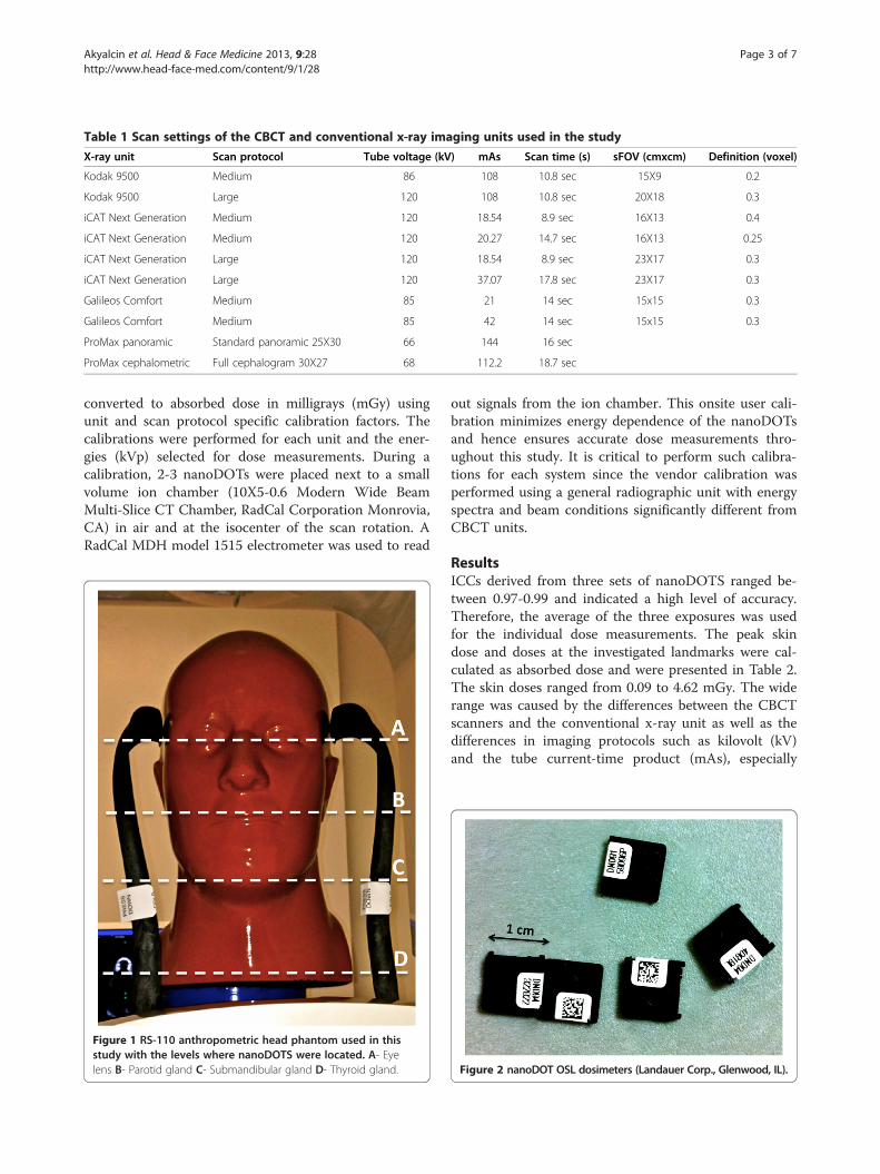
Table 1 Scan settings of the CBCT and conventional x-ray imaging units used in the study
X-ray unit Scan protocol Tube voltage (kV) mAs Scan time (s) sFOV (cmxcm) Definition (voxel)
Kodak 9500 Medium 86 108 10.8 sec 15X9 0.2
Kodak 9500 Large 120 108 10.8 sec 20X18 0.3
iCAT Next Generation Medium 120 18.54 8.9 sec 16X13 0.4
iCAT Next Generation Medium 120 20.27 14.7 sec 16X13 0.25
iCAT Next Generation Large 120 18.54 8.9 sec 23X17 0.3
iCAT Next Generation Large 120 37.07 17.8 sec 23X17 0.3
Galileos Comfort Medium 85 21 14 sec 15x15 0.3
Galileos Comfort Medium 85 42 14 sec 15x15 0.3
ProMax panoramic Standard panoramic 25X30 66 144 16 sec
ProMax cephalometric Full cephalogram 30X27 68 112.2 18.7 sec
Akyalcin et al. Head & Face Medicine 2013, 9:28 Page 3 of 7http://www.head-face-med.com/content/9/1/28
converted to absorbed dose in milligrays (mGy) usingunit and scan protocol specific calibration factors. Thecalibrations were performed for each unit and the ener-gies (kVp) selected for dose measurements. During acalibration, 2-3 nanoDOTs were placed next to a smallvolume ion chamber (10X5-0.6 Modern Wide BeamMulti-Slice CT Chamber, RadCal Corporation Monrovia,CA) in air and at the isocenter of the scan rotation. ARadCal MDH model 1515 electrometer was used to read
Figure 1 RS-110 anthropometric head phantom used in thisstudy with the levels where nanoDOTS were located. A- Eyelens B- Parotid gland C- Submandibular gland D- Thyroid gland.
out signals from the ion chamber. This onsite user cali-bration minimizes energy dependence of the nanoDOTsand hence ensures accurate dose measurements thro-ughout this study. It is critical to perform such calibra-tions for each system since the vendor calibration wasperformed using a general radiographic unit with energyspectra and beam conditions significantly different fromCBCT units.
ResultsICCs derived from three sets of nanoDOTS ranged be-tween 0.97-0.99 and indicated a high level of accuracy.Therefore, the average of the three exposures was usedfor the individual dose measurements. The peak skindose and doses at the investigated landmarks were cal-culated as absorbed dose and were presented in Table 2.The skin doses ranged from 0.09 to 4.62 mGy. The widerange was caused by the differences between the CBCTscanners and the conventional x-ray unit as well as thedifferences in imaging protocols such as kilovolt (kV)and the tube current-time product (mAs), especially
Figure 2 nanoDOT OSL dosimeters (Landauer Corp., Glenwood, IL).

Figure 3 Example of the axial, coronal and sagittal images obtained with KODAK 9500 scanner using the head anthropomorphicphantom: a- Medium FOV b- Large FOV.
Akyalcin et al. Head & Face Medicine 2013, 9:28 Page 4 of 7http://www.head-face-med.com/content/9/1/28
scanning field of view (sFOV). iCAT scanner had con-sistently lower doses for all the variables as compared toother scanners.The surface doses to the locations of the eyes were
~4.0 mGy, well below the 500 mGy threshold for pos-sibly causing cataract development. Relatively higher ra-diation doses were recorded for submandibular andparotid regions in all the CBCT protocols. Lowest doseswere obtained for the thyroid region partly due to thefact that this area is not covered by the sFOV and there-fore is not directly exposed. Similar results wereobtained for the eyes in medium size sFOV protocols.Conventional radiographs had the lowest doses for all ofthe variables tested. However, slight differences between
Table 2 Peak skin and absorbed tissue doses for the investig
X-ray unit kV mAs sFOVSkin
Dose (mGy)
Kodak 9500 86 108 15×9 3.58
Kodak 9500 120 108 20×18 4.15
iCAT Next Generation 120 18.54 16×13 1.44
iCAT Next Generation 120 20.27 16×13 2.51
iCAT Next Generation 120 18.54 23×17 2.08
iCAT Next Generation 120 37.07 23×17 2.60
Galileos Comfort 85 21 15×15 2.49
Galileos Comfort 85 42 15×15 4.62
ProMax panoramic 66 144 0.26
ProMax cephalometric 68 112.2 0.09
the cephalometric and panoramic images were observeddue to their coverage field as expected.
DiscussionDental cone beam computed tomography (CBCT) hasrapidly gained popularity among the dental specialtiesover the last decade. It is a reality that individuals thatrequire the investigation of the maxillofacial structuresin all three dimensions of the space, as usually is thecase in orthodontics and maxillofacial surgery, will bene-fit from CBCT imaging. Although there is a lack of de-finitive data, recent reviews signify the importance ofradiation dose generated by CBCT scanning as a causefor concern [1-4,18,19]. News media is very sensitive on
ated scan protocols
Eye lens Parotid Submandibular Thyroid
Dose (mGy) Dose (mGy) Dose (mGy) Dose (mGy)
0.42 2.88 3.04 0.32
3.55 3.57 3.17 0.45
0.92 1.15 1.21 0.24
1.60 2.05 1.79 0.25
1.41 1.93 1.79 0.25
1.75 2.11 2.23 0.39
0.58 2.45 1.93 0.36
0.94 4.46 3.61 0.46
0.05 0.24 0.14 0.04
0.07 0.06 0.06 0.05

Akyalcin et al. Head & Face Medicine 2013, 9:28 Page 5 of 7http://www.head-face-med.com/content/9/1/28
the unjustified use of CBCT scans on young childrenand adolescents in the orthodontic practice [20]. A re-cent article [21] that reported links between some dentalx-rays and an increased risk of intracranial meningiomahas become a public sensation and has led professionalsto question the applicability of the presented data toother dental diagnostic tools, which depend on more ra-diation dose including CBCT.In reality, it is extremely difficult to determine the
risks of using CBCT scanners in terms of fatal cancerdevelopment because of the confounding factors in doseestimation such as individual differences in patients’physical attributes, biological susceptibility and chal-lenges with dose estimation. However, without dosemeasurements operators lack the objective data neededto approximately adjust mAs or tube potential in orderto avoid excessive patient dose [7].This study mainly focused on the peak skin dose to-
gether with the surface entry doses for various otherradiosensitive tissues/organs. The rationale was to com-pare various operational modes of multiple CBCT scan-ners with each other and more importantly with theconventional radiographs that are routinely used inorthodontic treatment. Based on the investigated CBCTprotocols, peak skin dose at any point did not exceed4.62 mGy, which is well below 1000 mGy. The Inter-national Commission on Radiological Protection (ICRP)guidelines suggest that 2000 mGy may cause transienterythema and temporary epilation [22-24]. In medicalimaging, 2000 mGy is also regarded as the threshold fordeterministic effects [25,26]. However, for most patients,clinically important skin and hair reactions occur onlywhen the skin dose is higher than 5000 mGy [27]. Asevidenced in this paper, CBCT exposure for orthodonticexams is less than 2.5% that of skin threshold dose.Although there is a wide variation in our results for
the skin dose due to different scanning protocols, onecannot claim the superiority of one scanner over anothersolely based on the reported dose information. EachCBCT scanner has different settings and energy levels.Perhaps, the use of pulsed x-ray beam exposure in thisstudy was also a major reason for considerable variationin reported cone-beam unit dosimetry. However, our re-sults in Table 2 clearly demonstrate that when all theother factors were held constant, mAs, kV, and sFOVsettings had effects on the observed radiation doses, withmAs setting being the most effective. Despite the factthat iCAT scanner had a higher kV setting, consistentlylower doses were observed in all the iCAT scanner pro-tocols compared to the others. This may be explained bylower mAs settings of this device. Similarly, when themAs setting of the Galileos was increased from 21 to 42,peak skin dose increased almost 2 times. As higher tubecurrents are often used for larger patients to maintain
image quality, this finding suggests that mAs valueshould be kept minimum wherever possible as long asthere is no significant compromise in the image quality.In computed radiography overexposure will only reduceimage noise and can occur without awareness, as CBCTimages never look overexposed. This is due to thenormalization process of patient attenuation with theCBCT technique. Therefore, operators need to be in-formed about the purpose of the scan before they set thetube current to the default mode or to a higher settingunless otherwise is instructed.Radiation dose at the level of the eyes ranged between
0.42 to 3.55 mGy. Current permissible exposure limitsto eyes are similar to the skin dose [23,24]. However,according to newer studies the threshold for catara-ctogenesis is actually much lower. Recently publisheddata on Chernobyl cleanup workers revealed a signifi-cant increase in cataract rates with increasing radiationdoses, which were, for the most part, less than 500 mGy[28]. Additionally, Chodick et al. [29], argued that likeli-hood of cataract formation increased with increasing ex-posure to ionizing radiation with no apparent thresholdlevel. On the contrary, it was also suggested that therewas no association between computed tomography scansof the head history and cataract [30]. The evidence fromthe literature seems to be inconclusive on the radiationinduced cataract development. However, there is still140 times less risk with the maximum dose obtainedwith the diagnostic CBCT protocols used in this studywhen compared to the recent Chernobyl data [28]. Add-itionally, sFOV may be changed from large to medium,depending on the imaging need, to avoid the exposureof the eyes when monitoring the jaws only. There willstill be scattered radiation as is the case with thyroidgland. Our results demonstrated that radiation dose atthe thyroid level had the least variability since it is notwithin direct exposure field. Even then a dose range of0.24-0.46 mGy was observed at that level that can beconsidered as low. Additionally, Qu et al [31]. recentlydemonstrated that with the use of thyroid collars, doseto thyroid and oesophagus could effectively be reducedto 48.7% and 41.7%, respectively. It was also shown thatthe radiation dose to the eye could be reduced by over60% through the use of leaded glasses during a CBCTexamination [32].One of the unique aspects of the current study was
to utilize the Optically Stimulated Luminescence (OSL)‘dot’ dosimeters. It was already shown that single-irradiation measurements with bleaching after each OSLreadout was found to be associated with a 3.3% reprodu-cibility [33]. In our study, we were also able to observehigh reproducibility for dose measurements with the useof OSL dots. In order to compare conventional radio-graphs to CBCT scanners, absorbed dose was preferred

Akyalcin et al. Head & Face Medicine 2013, 9:28 Page 6 of 7http://www.head-face-med.com/content/9/1/28
to a whole-body effective dose, which would require toomany assumptions since dental CBCT only exposes thehead region. Reported values enable a sound comparisonbetween the procedures for the investigated parameters.Relatively much lower point doses were obtainedwith both the panoramic and cephalometric modes ofProMax unit. It should, however, be remembered thatdose differences are due to the differences in geometryand specific parameters of the CBCT scanners. Whilethe dose range is too wide between the conventional ra-diographs and CBCT scanners, too much attention toradiogenic risk may also distract attention from otherrisks and potential benefits, which may not be in the pa-tient’s best interest [34]. In that sense it is not proper tomisrepresent an individual’s radiation history as part ofthe risk of the proposed procedure. Due to the very re-cent advisory statement by the American Dental Associ-ation Council on Scientific Affairs [35], only a trainedclinician must decide if a procedure can be justified byitself on the basis of radiation and other risks of thatprocedure, the patient’s clinical status, and the benefitsexpected from that procedure. Although doses reportedin this paper may be perceived as very low when com-pared to those in medical imaging, a lifetime-attributablerisk to the individual should also be considered.
ConclusionsWhen planning orthodontic treatment, conventionalpanoramic and cephalometric radiographs are certainlydose sparing when compared to CBCT scans. However,when indicated, CBCT imaging should be consideredwith a radiation conscious approach. As evidenced inthis paper, scan parameters such as mAs and FOV set-tings of the CBCT scanners should be used effectivelydepending on the imaging and individual patient needsfor dose reduction purposes. Hence features of variablekVp stations, mAs selections, and radiation beam colli-mation settings are preferable to a well designed CBCTsystem for dental imaging.
AbbreviationsCBCT: Cone-beam computed tomography; OSL: Optically stimulatedluminescence; SFOV: Scanning field of view; CTDI: CT Dose Index;TLDs: Thermoluminescent dosimeters; MOSFET: Metal oxide semiconductorfield-effect transistors; ICRU: International Commission on Radiation Units andMeasurements; ICRP: The International Commission on RadiologicalProtection.
Competing interestsThe authors declare that they have no competing interests.
Authors’ contributionsSA and XJR were responsible for the design and they carried out theexperimental part of the project. They both oversaw all phases of theproject. JDE performed the dose measurements and helped with the datainterpretation. KMA participated in the design, drafted the paper andprovided feedback on the writing. All authors read and approved the finalmanuscript.
Author details1Department of Orthodontics, The University of Texas Health Science Centerat Houston, School of Dentistry, Houston, TX, USA. 2Oral Diagnosis,Radiology, and Pathology, Loma Linda University, School of Dentistry, LomaLinda, CA, USA. 3Department of Imaging Physics, MD Anderson CancerCenter, Houston, TX, USA.
Received: 22 August 2013 Accepted: 2 October 2013Published: 9 October 2013
References1. Scholz RP: The radiology decision. Semin Orthod 2011, 17:15–19.2. Baumrind S: The road to three-dimensional imaging in orthodontics.
Semin Orthod 2011, 17:2–12.3. Halazonetis DJ: Cone-beam computed tomography is not the imaging
technique of choice for comprehensive orthodontic assessment.Am J Orthod Dentofacial Orthop 2012, 141:403–411.
4. Larson BE: Cone-beam computed tomography is the imaging techniqueof choice for comprehensive orthodontic assessment. Am J OrthodDentofacial Orthop 2012, 141:402–410.
5. Geleijns J, Salvadó Artells M, De Bruin PW, Matter R, Muramatsu Y,McNitt-Gray MF: Computed tomography dose assessment for a 160 mmwide, 320 detector row, cone beam CT scanner. Phys Med Biol 2009,54:3141–3159.
6. McNitt-Gray MF: AAPM/RSNA Physics tutorial for residents: topics in CT.Radiation Dose in CT. Radiographics 2002, 22:1541–1553.
7. Bauhs JA, Vrieze TJ, Primak AN, Bruesewitz MR, McCollough CH: CTDosimetry: Comparison of measurement techniques and devices.Radiographics 2008, 28:245–253.
8. Timilsina B, Gesell TF: Independent evaluation of optically stimulatedluminescence (OSL) ‘dot’ dosemeters for environmental monitoring.Radiat Prot Dosimetry 2011, 143:27–32. Epub 2010 Oct 14.
9. Pauwels R, Beinsberger J, Collaert B, Theodorakou C, Rogers J, Walker A,Cockmartin L, Bosmans H, Jacobs R, Bogaerts R, Horner K: SEDENTEXCTProject Consortium: Effective dose range for dental cone beamcomputed tomography scanners. Eur J Radiol 2012, 81:267–271.
10. Ludlow JB, Davies-Ludlow LE, Brooks SL, Howerton WB: Dosimetry of 3CBCT devices for oral and maxillofacial radiology: CB Mercuray NewTom3G and i-CAT. Dentomaxillofac Radiol 2006, 35:219–226.
11. Ludlow JB, Ivanovic M: Comparative dosimetry of dental CBCT devicesand 64- slice CT for oral and maxillofacial radiology. Oral Surg Oral MedOral Pathol Oral Radiol Endod 2008, 106:106–114.
12. Silva MA, Wolf U, Heinicke F, Bumann A, Visser H, Hirsch E: Cone-beamcomputed tomography for routine orthodontic treatment planning: aradiation dose evaluation. Am J Orthod Dentofacial Orthop 2008, 133:640.e1-5.
13. Hirsch E, Wolf U, Heinicke F, Silva MA: Dosimetry of the cone beam computedtomography Veraviewepocs 3D compared with the 3D Accuitomo indifferent fields of view. Dentomaxillofac Radiol 2008, 37:268–273.
14. Loubele M, Bogaerts R, Van Dijck E, Pauwels R, Vanheusden S, Suetens P,Marchal G, Sanderink G, Jacobs R: Comparison between effective radiationdose of CBCT and MSCT scanners for dentomaxillofacial applications.Eur J Radiol 2009, 71:461–468.
15. Suomalainen A, Kiljunen T, Käser Y, Peltola J, Kortesniemi M: Dosimetry andimage quality of four dental cone beam computed tomographyscanners compared with multislice computed tomography scanners.Dentomaxillofac Radiol 2009, 38:367–378.
16. Roberts JA, Drage NA, Davies J, Thomas DW: Effective dose from conebeam CT examinations in dentistry. Br J Radiol 2009, 82:35–40.
17. Grünheid T, Kolbeck Schieck JR, Pliska BT, Ahmad M, Larson BE: Dosimetryof a cone-beam computed tomography machine compared with adigital x-ray machine in orthodontic imaging. Am J Orthod DentofacialOrthop 2012, 141:436–443.
18. Khelemsky R: The ethics of routine use of advanced diagnostictechnology. J Am Coll Dent 2011, 78:35–39.
19. Van Vlijmen OJ, Kuijpers MA, Bergé SJ, Schols JG, Maal TJ, Breuning H, Kuijpers-Jagtman AM: Evidence supporting the use of cone-beam computedtomography in orthodontics. J Am Dent Assoc 2012, 143:241–252.
20. Bogdanich W, McGinty CJ: Radiation worries for children in dentist’s hair.The New York Times; 2013. http://www.nytimes.com/2010/11/23/us/23scan.html?_r=3 (Accessed July 29, 2013).

Akyalcin et al. Head & Face Medicine 2013, 9:28 Page 7 of 7http://www.head-face-med.com/content/9/1/28
21. Claus EB, Calvocoressi L, Bondy ML, Schildkraut JM, Wiemels JL, Wrensch M:Dental x-rays and risk of meningioma. Cancer 2012, 118:4530–4537.
22. Zhang D, Cagnon CH, Villablanca JP, McCollough CH, Cody DD, StevensDM, Zankl M, Demarco JJ, Turner AC, Khatonabadi M, McNitt-Gray MF: Peakskin and eye lens radiation dose from brain perfusion CT based onMonte Carlo simulation. AJR Am J Roentgenol 2012, 98:412–417.
23. International commission on radiological protection (ICRP):Recommendations of the international commission on radiologicalprotection: ICRP publication 60. Ann ICRP 1991, 21:1–201.
24. International commission on radiological protection (ICRP): Avoidance ofradiation injuries from medical interventional procedures: ICRPpublication 85. Ann ICRP 2000, 30:7–67.
25. Zontar D, Kuhelj D, Skrk D, Zdesar U: Patient peak skin doses from cardiacinterventional procedures. Radiat Prot Dosimetry 2010, 139:262–265.
26. Ying CK, Kandaiya S: Patient skin dose measurements during coronaryinterventional procedures using Gafchromic film. J Radiol Prot 2010,30:585–596.
27. Balter S, Hopewell JW, Miller DL, Wagner LK, Zelefsky MJ: Fluoroscopicallyguided interventional procedures: a review of radiation effects onpatients’ skin and hair. Radiology 2010, 254:326–341.
28. Worgul BV, Kundiyev Y, Sergiyenko N, Chumak VV, Vitte PM, MedvedovskyC, Bakhanova EV, Junk AK, Kyrychenko OY, Musijachenko NV, Shylo SA, VitteOP, Xu S, Xue X, Shore RE: Cataract among Chernobyl cleanup workers:implications regarding permissible eye exposure. Radiat Res 2007,167:233–243.
29. Chodick G, Bekiroglu N, Hauptmann M, Alexander BH, Freedman DM,Doody MM, Cheung LC, Simon SL, Weinstock RM, Bouville A, Sigurdson AJ:Risk of cataract after exposure to low doses of ionizing radiation: a20-year prospective cohort study among US radiologic technologists.Am J Epidemiol 2008, 168:620–631.
30. Hourihan F, Mitchell P, Cumming RG: Possible associations betweencomputed tomography scan and cataract: the blue mountains eyestudy. Am J Public Health 1999, 89:1864–1866.
31. Qu X, Li G, Sanderink G, Zhang Z, Ma X: Dose reduction of cone beam CTscanning for the entire oral and maxillofacial regions with thyroidcollars. Dentomaxillofac Radiol 2012, 41:373–378.
32. Prins R, Dauer LT, Colosi DC, Quinn B, Kleiman NJ, Bohle GC, Holohan B,Al-Najjar A, Fernandez T, Bonvento M, Faber RD, Ching H, Goren AD:Significant reduction in dental cone beam computed tomography(CBCT) eye dose through the use of leaded glasses. Oral Surg Oral MedOral Pathol Oral Radiol Endod 2011, 112:502–507.
33. Al-Senan RM, Hatab MR: Characteristics of an OSLD in the diagnosticenergy range. Med Phys 2011, 38:4396–4405.
34. Balter S, Zanzonico P, Reiss GR, Moses JW: Radiation is not the only risk.AJR Am J Roentgenol 2011, 196:762–767.
35. The American Dental Association Council on Scientific Affairs: The use ofcone-beam computed tomography in dentistry: an advisory statementfrom the American dental association council on scientific affairs.J Am Dent Assoc 2012, 143:899–902.
doi:10.1186/1746-160X-9-28Cite this article as: Akyalcin et al.: Measurement of skin dose from cone-beam computed tomography imaging. Head & Face Medicine 2013 9:28.
Submit your next manuscript to BioMed Centraland take full advantage of:
• Convenient online submission
• Thorough peer review
• No space constraints or color figure charges
• Immediate publication on acceptance
• Inclusion in PubMed, CAS, Scopus and Google Scholar
• Research which is freely available for redistribution
Submit your manuscript at www.biomedcentral.com/submit