Embed Size (px)
Citation preview

Hindawi Publishing CorporationBioMed Research InternationalVolume 2013, Article ID 307602, 6 pageshttp://dx.doi.org/10.1155/2013/307602
Research ArticlePreparation, Modification, and Characterization ofAlginate Hydrogel with Nano-/Microfibers: A New Perspectivefor Tissue Engineering
Bianca Palma Santana,1 Fernanda Nedel,1 Evandro Piva,2 Rodrigo Varella de Carvalho,3
Flávio Fernando Demarco,1 and Neftali Lenin Villarreal Carreño4
1 Nucleus of Cellular and Tecidual Biology (NCTBio), Post-Graduate Program in Dentistry, Federal University of Pelotas,Rua Goncalves Chaves 457, Centro, 96015-560 Pelotas, RS, Brazil
2 Department of Operative Dentistry School of Dentistry, Federal University of Pelotas, Rua Goncalves Chaves 457,Centro, 96015-560 Pelotas, RS, Brazil
3 Department of Operative Dentistry School of Dentistry, University North of Parana (UNOPAR), Rua Marselha,Jardim Piza, 86041-140 Londrina, PR, Brazil
4 Technology Development Center, Federal University of Pelotas, Rua Felix da Cunha 809, Centro, 96010-00 Pelotas, RS, Brazil
Correspondence should be addressed to Bianca Palma Santana; [email protected] and Fernanda Nedel;[email protected]
Received 16 March 2013; Accepted 10 May 2013
Academic Editor: Kacey Gribbin Marra
Copyright © 2013 Bianca Palma Santana et al. This is an open access article distributed under the Creative Commons AttributionLicense, which permits unrestricted use, distribution, and reproduction in any medium, provided the original work is properlycited.
We aimed to develop an alginate hydrogel (AH) modified with nano-/microfibers of titanium dioxide (nfTD) and hydroxyapatite(nfHY) and evaluated its biological and chemical properties. Nano-/microfibers of nfTD and nfHY were combined with AH,and its chemical properties were evaluated by FTIR spectroscopy, X-ray diffraction, energy dispersive X-Ray analysis, and thecytocompatibility by the WST-1 assay. The results demonstrate that the association of nfTD and nfHY nano-/microfibers to AHdid notmodified the chemical characteristics of the scaffold and that the association was not cytotoxic. In the first 3 h of culture withNIH/3T3 cells nfHY AH scaffolds showed a slight increase in cell viability when compared to AH alone or associated with nfTD.However, an increase in cell viability was observed in 24 h when nfTD was associated with AH scaffold. In conclusion our studydemonstrates that the combination of nfHY and nfTD nano-/microfibers in AH scaffold maintains the chemical characteristics ofalginate and that this association is cytocompatible. Additionally the combination of nfHY with AH favored cell viability in a shortterm, and the addition of nfTD increased cell viability in a long term.
1. Introduction
Tissue engineering is a field with potential for designing andconstructing tissues or organs to restore their function oreven completely replace them.The interchange of responsivecells, morphogens, and scaffolds constitutes the three mainelements that grounds tissue engineering [1–6]. Scaffolds arethree-dimensional structures used to support and guide thein-growth of cells, forming the template for cell colonization,proliferation as well as being able to provide different sets ofphysiological signals to the developing tissue [7, 8].Thereforescaffolds perform the structural and biochemical functions
of the native extracellular matrix (ECM) until the cells areable to produce their own ECM [9, 10]. It is well known thatthe native ECM provides a substrate with specific bioactivemolecules that controls cellular process such as cell adhesion,proliferation,migration, differentiation, survival and physicalsupport for cells, characteristics that challenge researchers toelaborate an ideal scaffold [11].
The collagen fibers, which the diameter ranges from 50to 500 nm, are one of the main components of the ECMin tissues that require strength and flexibility (e.g., bone)[10]. Since collagen structure is important for cell attach-ment, proliferation, and differentiation, nano-/microfibers
2 BioMed Research International
have been incorporated to different types of scaffold, such aspoly(l-lactic acid) (PLLA) [9, 10] and alginate hydrogel [3], inorder to recreate collagen fibers functions [12]. Studies havedemonstrated that the incorporation of nano-/microfibersin scaffolds can increase osteoblast viability [13], supportan earlier and enhanced osteoblast phenotype, increase theexpression of genes that are associated with the osteoblastphenotype, and have superior ability to promote mineral-ization; high expression of integrins 𝛼2 and 𝛽1 as well asintegrins 𝛼v and 𝛽3 and activation of FAKs [10]; nano-/microfibers architecture can selectively enhance proteinadsorption (fibronectin and vitronectin) [9].
Alginate is a naturally derived polysaccharide that hasbeen widely used in drug delivery [14–16], cell encapsulationmaterial [17], and injectable cell transplantation vehicle [18].Alginate is composed of (1–4) 𝛽-d-mannuronic acid and𝛼-l-guluronic acid residues linked either randomly or ashomopolymeric blocks [19]. The ratio of the two sugars(mannuronic/guluronic acids) is generally 1.5, with somedeviation depending on the source [20].
The crosslinking and gelation of the polymers are mainlyachieved by the exchange of sodium ions from the guluronicacids with the divalent cations and the stacking of theseguluronic groups to form the characteristic egg-box structure[20]. A simple method to increase the ionic crosslinkingdensity is adding sufficient amounts of divalent cations,which is gradually diffuse out from the gel and slowlydegrades and is excreted in the urine [18]; in this processes weobtain the propermechanical properties of alginate hydrogels(AH) [21].
Alginate has been frequently used in tissue engineeringdue to the following properties: biocompatibility, low toxicity,nonimmunogenicity, relatively low cost, and mild gelationbehavior with divalent cations [21]. In addition, AH can beused like beads or gel that absorbs water and swell readilywithout dissolving [22]. In recent years, AH have foundapplications in medicine [18], pharmacology [15], biologicalscience [23], and dentistry [24] and have been used in largescale in tissue engineering [25].
In a previous work we developed a new method to syn-thesize nano-/microfibers of titanium dioxide (nfTD) andhydroxyapatite (nfHY) and showed that both fibers were notcytotoxic and resembled the structure of natural collagen[3]. In the present study, we developed an alginate hydrogelmodified with nano-/microfibers of titanium dioxide (nfTD)and hydroxyapatite (nfHY) and evaluated its biological andchemical properties.
2. Materials and Methods
2.1. Alginate Hydrogel Combined with Nano-/Microfibers. So-dium alginate (NaC
6H7O6—Vetec Quımica Fina LTDA)
was dissolved in deionized water and mixed with calciumsulfate (CaSO
4⋅2H2O—Vetec Quımica Fina LTDA) forming
the ionic crosslinking or the hydrogel (2%wt). The nano-/microfibers titanium dioxide and hydroxyapatite kept aconstant concentration of 0.07% g/mL; they were added onthe hydrogel during the magnetically stirred.
2.2. FTIR Spectroscopy. The chemical structure characteri-zation of alginate hydrogel with and without nano-/microfibers was conducted by infrared spectroscopy. The infraredspectra of alginate hydrogel were measured with an FTIRspectrophotometer (Fourier Transform Infrared Spectropho-tometer, IRPrestige-21, Shimadzu). Each spectrumof sampleswas acquired via accumulation of 96 scans with a resolutionof 4 cm−1.
2.3. X-Ray Diffraction (XRD). XRD patterns of dry AH withand without the nano-/microfibers were obtained by diffractmeter (XRD, Shimadzu, model XRD-6000). The equipmentuses the diffraction tube with copper target at a wavelengthapproximately equal to 1.54060 A, with a power of 2 kW,30 kV current of 30mA. The analysis was performed in theangle range from 20∘ to 40∘ for AH combined with nfTD andfrom 20∘ to 80∘ for AH combined with nfHY, at a speed of1 degree/min in continuous scan.
2.4. Energy Dispersive X-Ray Analysis (EDX). The percent-age of nfTD and nfHY nano-/microfibers in the AH wasdetermined by Energy Dispersive X-Ray Analysis (EDX—Ray Ny—EDX 720, Shimadzu). Samples were prepared in asimilar way to those analyzed by XRD.
2.5. Cytocompatibility Test of Alginate Hydrogel Combinedwith Nano-/Microfibers. An immortalized mouse fibroblastcell line (NIH/3T3) was maintained in Dulbecco’s ModifiedEagleMedium (DMEM) supplemented with 10% fetal bovineserum (FBS), 2% L-glutamine, penicillin (100U/mL), andstreptomycin (100mg/mL) (Gibco, Grand Island, NY, USA).Mouse fibroblasts were maintained as a stock culture inDMEM and incubated at 37∘C in a humidified atmosphereof 5% CO
2in air until subconfluency was reached, as de-
scribed previously [26, 27]. AH alone and combined withnfTD and nfHY was sterilized by exposure to germicidalUV (ultraviolet) light for 40min. Subsequently they wereincubated in contact with NIH/3T3 cells at a density of 2 ×10⁴ in 96-well plates for 3, 6, and 24 at 37∘C in a humidifiedatmosphere with 5% of CO
2. An additional control group
was added composed only by cell and medium culture,without the presence of AH scaffolds. At each time point,10 𝜇L of WST-1 (2-(4-iodophenyl)-3-(4-nitrophenyl)-5-(2,4-disulfophenyl)-2H-tetrazolium, monosodium salt) (Roche,Mannheim, Germany) was added to the wells and incubatedfor 2 h. Then, 100𝜇L aliquots were removed from each well,and the optical density at 450 nm was determined in amicroplate reader. All observations were validated by at leastthree independent experiments, and for each experiment theanalyses were performed in triplicate. Data were submitted toone-way ANOVA and Tukey post-hoc tests, with 𝑃 < 0.05.
3. Results
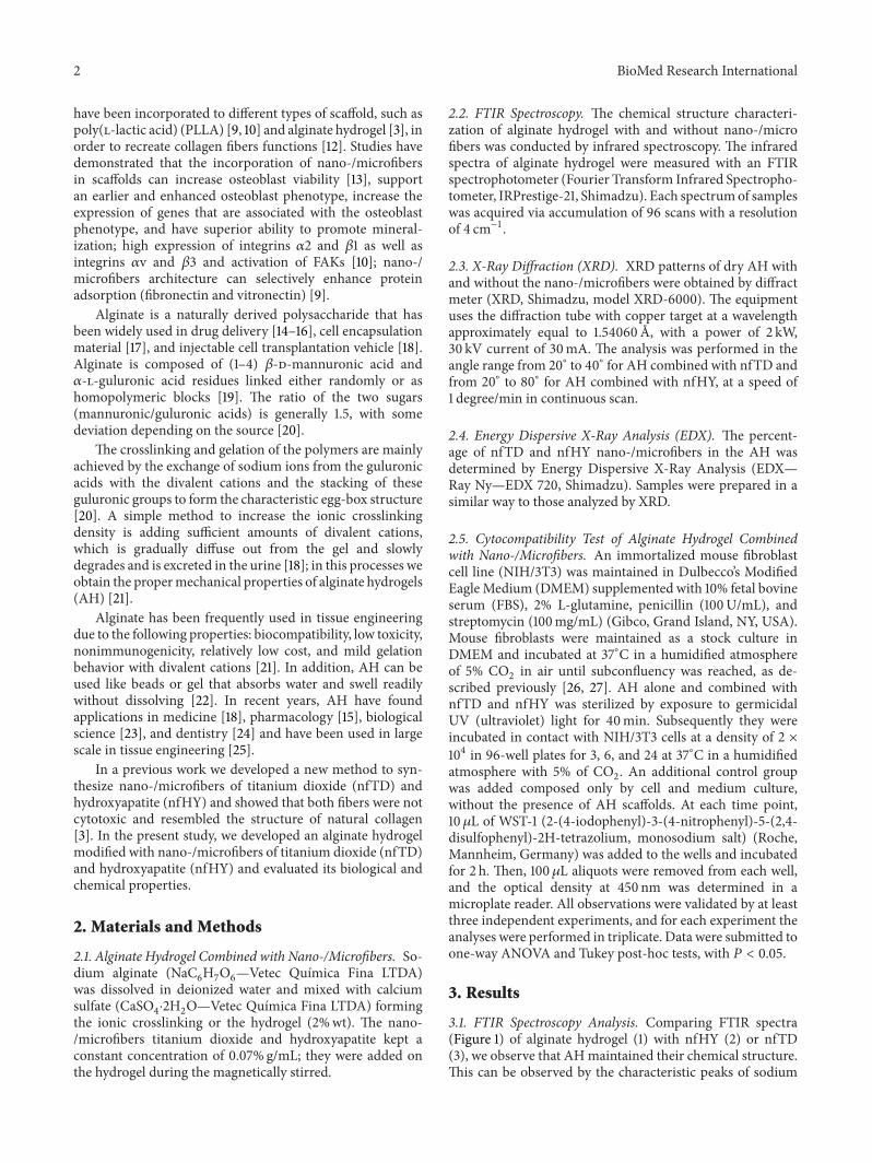
3.1. FTIR Spectroscopy Analysis. Comparing FTIR spectra(Figure 1) of alginate hydrogel (1) with nfHY (2) or nfTD(3), we observe that AHmaintained their chemical structure.This can be observed by the characteristic peaks of sodium

BioMed Research International 3
1622
1419
34041143
1120
4000 3000 2000 1000
(1a)(2a)(3a)
Wavenumbers (cm−1)
Figure 1: FTIR spectra ofHA (1a), AHwith nfHY (2a), andAHwithnfTD (3a).
Table 1: Quantitative analyses of alginate hydrogel.
Sample Results SD Line analysis of X-rayCa 77.09% 0.23 Ca KaS 20.76% 0.10 S KaP 1.21% 0.06 P KaFe 0.57% 0.04 Fe KaCu 0.37% 0.02 Cu Ka
alginate absorption at 2950 cm−1 and 1413 cm−1; due tostretching –CH
2, the carboxylic groups C–O–O show a
broad absorption band as a result of the asymmetric stretchin 1622 cm−1 and the symmetric stretching in 1419 cm−1and –C–OH (O–H stretching vibration is 3404 cm−1, C–Ostretching vibration of secondary alcohol is 1120 cm−1, andC–O stretching vibration of tertiary alcohol is 1143 cm−1).
3.2. X-Ray Diffraction (XRD) Analysis. The presence of tita-nium dioxide and hydroxyapatite crystal phase in theinjectable system was observed by XRD analysis (Figure 2).Results indicated that the nfTD and nfHY preserved thierstructural characteristics during the process, which is favor-able to maintain its bioactivity and biocompatibility.
3.3. EDX. In EDX results we can observe the quantitativeconcentration of AH (Table 1) combined with nfTD (Table 2)and nfHY (Table 3).
3.4. Cytocompatibility Test of Alginate Hydrogel Combinedwith Nano/Micro Fibers. Cell viability was determined by theWST-1 assay, a soluble tetrazolium salt converted to a deep redcolored product by mitochondrial activity [2]. The viabilitydata of NIH/3T3 cells when in contact with AH alone and
Table 2: Quantitative analyses of alginate hydrogel combined withnano-/microfibers of titanium dioxide.
Sample Results SD Line analysis of X-rayCa 62.31% 0.11 Ca KaS 26.05% 0.05 S KaTi 10.44% 0.03 Ti KaP 1.03% 0.02 P KaK 0.17% 0.01 K Ka
Table 3: Quantitative analyses of alginate hydrogel combined withnano-/microfibers of hydroxyapatite.
Sample Results SD Line analysis of X-rayCa 77.55% 0.07 Ca KaS 11.31% 0.02 S KaP 10.44% 0.02 P KaSi 0.35% 0.01 Si KaK 0.25% 0.01 K KaFe 0.11% 0.00 Fe Ka
AH modified with nfTD and nfHY in the period 3, 6, and24 h are present in Figure 3.
The results shows that the addition of nfTD and nfHYto the AH scaffold did not induce cytotoxicity. In the periodof 24 h the AH nfTD provided a higher viability of NIH/3T3cells when compared to the AH nfHY and AH alone.However, in the first 3 h AH nfHY showed a slight increase incell viability when compared to AH alone and associated withnfTD.The exposure time of 3 and 6 h had no significant effecton the cell viability; however, an increase on cell proliferationwas observed with 24 h of exposure.
4. Discussion
Onewell-known limitation of using AH in tissue engineeringis the lack of corresponding binding sites for receptors ofmost cells. Also due to its hydrophilic nature ECM proteinssuch as laminin, fibronectin, and vitronectin do not readilyadsorb to the gel surface [28]. In order to overcome theseproblems, a common approach has been to combine an entireECM protein or peptide sequence capable of binding to cel-lular receptors to the polymer. Combining whole molecules,however, can lead to nonspecific interaction, and the couplingcan be difficult to control.Therefore peptide sequences foundin the ECM can mediate cell adhesion in place of the largermolecules, offering a specific means to control adhesion andresults in a high specificity. The most frequently used is theamino acid sequence arginine-glycine-aspartic acid (Arg-Gly-Asp or RGD) [29].
In this study we attempted to modify AH with nfTDand nfHY nano-/microfibers in order to increase cell adhe-sion. This attempt could favor further improvements of AHproperties enhancing cell adhesion and improving tissueformation. The results demonstrate that the association ofnfTD and nfHY nano-/microfibers to the AHdid notmodifythe chemical characteristics of the scaffold (Figures 1 and 2).

4 BioMed Research International
20 30 40 50 60 70 8011
2301
211
11110
1
110
Inte
nsity
(a.u
.)
AH
TiO2—rutile; card number (jcpds): 21-1276
nfTD
AH + nfTD
2𝜃
(a)
20 30 40
20200
2
300
112
211
211
Hydroxyapatite—card number (jcpds): 09-0432
AH
nfHY
AH + nfHY
Inte
nsity
(a.u
.)
2𝜃
(b)
Figure 2: XRD patterns of AH and AH combined with nfTD (a) and nfHY (b).
0
1
2
3
AbBbABbABb Ab Ab Ab Ab
Aa
Ba Ba
ABa
WST
-1 (a
.u.)
Cel
lA
H ce
llA
H n
fTD
cell
AH
nfH
Y ce
ll
Cel
lA
H ce
llA
H n
fTD
cell
AH
nfH
Y ce
ll
Cel
lA
H ce
llA
H n
fTD
cell
AH
nfH
Y ce
ll
3 h6 h24 h
Figure 3: Mouse fibroblast cell line (NIH/3T3) viability in 3, 6, and24 h when in contact with AH alone and AH combined with nfTDand nfHY. Data are expressed as the mean ± SEM. Uppercase lettersindicate significant differences between the AH alone and the AHnfTD and AH nfHY in the same period of time. Lowercase lettersindicate significant differences in the times tested. A 𝑃 value < 0.05was considered significant (Tukey’s test).
In addition the EDX analysis (Tables 1, 2, and 3) showed thatthe concentration evaluated was favorable to maintain theoriginal properties of AH.
The cytocompatibility assay showed that the addition ofnfTD and nfHY to the AH scaffold did not induce cytotoxic-ity.These results are in agreementwith our recent publication,where an in vitro cytocompatibility assay demonstrated thatthe same nano-/micro fibers alone were not cytotoxic toNIH/3T3 cells [3]. In the first 3 h of culture with NIH/3T3cells AH nfHY showed a slight increase in cell viabilitywhen compared to AH alone and associated with nfTD.Thiscould be partially explained by the higher porosity shown
by the alginate with nfHY in contrast with the alginate withnfTD and AH alone, favoring cell adhesion, proliferation,and migration which could improve initially the cell viability[3]. However, an increase in cell viability was observed in 24 hwhen nfTD was associated with AH scaffold, which could bepartially explained by the flowing characteristics of titaniumand nanofibrous.
Titaniumhas been classified as a cytocompatiblematerial,and it has been extensively used in dentistry [30] and ortho-pedics [31, 32]. It is capable of forming an active oxide layerthat readily interacts with cell-surface proteins and with theECM proteins produced by cells. It is due to this superficialoxide that titanium provides a biocompatibility interfacewith peri-implant tissue [33]. It has been shown that when,mesenchymal stem cells are culturedwith titanium fragmentsthe cell viability improves, and their biology properties aremaintained [34]. Further in vitro studies have demonstratedthat titanium dioxide scaffold can provide a suitable surfacefor osteoblast cell attachment and proliferation [35].
In addition, it is well established that in order to prolif-erate, migrate and differentiate most cells require anchorage.Therefore cellular attachment is an essential step towardsdeveloping a new tissue. It is believed that the adhesion ofcells to surfaces is dependent on the adsorption of highlyadhesive proteins that can be from the serum or secretedby the cells, which links cells to the biomaterial surface[9, 31]. In this context several key attachment proteins(fibronectin, vitronectin, and laminin) have been found toadsorb to the nanofibrous scaffolds at levels of 2.6 to 3.9 timeshigher than solid-walled scaffolds. In addition it has beenshown that nanofibrous scaffolds adsorb a different profileof proteins in comparison to solid-walled scaffolds from thesame material [9]. Nanofibrous scaffolds also have shown toincrease in neonatal mouse osteoblasts the expression of inte-grins associated with collagen (𝛼2𝛽1), fibronectin (𝛼V𝛽3),
BioMed Research International 5
and vitronectin (𝛼V𝛽3) when compared with solid-walledscaffolds [10]. In addition nanofibrous scaffolds have shownto increase cell attachment with several cell lines includingosteoblastic cells [13], fibroblasts, normal rat kidney cells[36, 37], smooth muscle cells [38], and neural stem cells [39].
5. Conclusion
In our study we demonstrated that the combination of nfHYand nfTD nano-/microfibers in alginate hydrogel scaffoldmaintains the chemical characteristics of alginate, and thatthis association is cytocompatible. Additionally the combina-tion of nfHY with AH favored cell viability in a short term,and the addition of nfTD increased cell viability in a longterm.
Acknowledgments
The authors would like to thank the Brazilian Governmentagencies (Conselho Nacional de Desenvolvimento Cientıficoe Tecnologico (CNPq), Coordenacao de Aperfeicoamentode Pessoal de Nıvel Superior (CAPES), Financiadora deEstudos e Projetos (FINEP), and Fundacao de Apoio aPesquisa do Estado doRioGrande do Sul (FAPERGS)) for thefinancial support (CNPq—404693/2012-1) and scholarships(DOCFIX—FAPERGS/CAPES 09/2012).
References
[1] F. Nedel, D. D. A. Andre, I. O. de Oliveira et al., “Stem cells:therapeutic potential in dentistry,”The journal of contemporarydental practice, vol. 10, no. 4, pp. 90–96, 2009.
[2] F. Nedel, F. N. Soki, M. C. M. Conde et al., “Comparativeanalysis of two colorimetric assays in dental pulp cell density,”International Endodontic Journal, vol. 44, no. 1, pp. 59–64, 2011.
[3] B. P. Santana, G. F. D. R. Paganotto, F. Nedel et al., “Nano-/microfiber scaffold for tissue engineering: physical and biologi-cal properties,” Journal of Biomedical Materials Research Part A,vol. 100, no. 11, pp. 3051–3058, 2012.
[4] R. Langer and J. P. Vacanti, “Tissue engineering,” Science, vol.260, no. 5110, pp. 920–926, 1993.
[5] F. F. Demarco,M. C.M. Conde, B. N. Cavalcanti, L. Casagrande,V. T. Sakai, and J. E. Nor, “Dental pulp tissue engineering,”Brazilian Dental Journal, vol. 22, no. 1, pp. 3–14, 2011.
[6] F. P. Hartwig, F. Nedel, T. V. Collares, S. B. Tarquinio, J. E.Nor, and F. F. Demarco, “Telomeres and tissue engineering: thepotential roles of TERT in VEGF-mediated angiogenesis,” StemCell Reviews and Reports, vol. 8, no. 4, pp. 1275–1281, 2012.
[7] J. Zhu, “Bioactive modification of poly(ethylene glycol) hydro-gels for tissue engineering,” Biomaterials, vol. 31, no. 17, pp.4639–4656, 2010.
[8] F. F. Demarco, L. Casagrande, Z. Zhang et al., “Effects of mor-phogen and scaffold porogen on the differentiation of dentalpulp stem cells,” Journal of Endodontics, vol. 36, no. 11, pp. 1805–1811, 2010.
[9] K. M. Woo, V. J. Chen, and P. X. Ma, “Nano-fibrous scaffoldingarchitecture selectively enhances protein adsorption contribut-ing to cell attachment,” Journal of Biomedical Materials ResearchA, vol. 67, no. 2, pp. 531–537, 2003.
[10] K. M. Woo, J. Jun, V. J. Chen et al., “Nano-fibrous scaffoldingpromotes osteoblast differentiation and biomineralization,”Bio-materials, vol. 28, no. 2, pp. 335–343, 2007.
[11] B.-H. Choi, Y. S. Choi, D. G. Kang, B. J. Kim, Y. H. Song, and H.J. Cha, “Cell behavior on extracellular matrix mimic materialsbased on mussel adhesive protein fused with functional pep-tides,” Biomaterials, vol. 31, no. 34, pp. 8980–8988, 2010.
[12] L. A. Smith and P. X. Ma, “Nano-fibrous scaffolds for tissueengineering,” Colloids and Surfaces B, vol. 39, no. 3, pp. 125–131,2004.
[13] K. Tuzlakoglu, N. Bolgen, A. J. Salgado, M. E. Gomes, E. Piskin,and R. L. Reis, “Nano- and micro-fiber combined scaffolds:a new architecture for bone tissue engineering,” Journal ofMaterials Science, vol. 16, no. 12, pp. 1099–1104, 2005.
[14] C.-Y. Yu, X.-C. Zhang, F.-Z. Zhou, X.-Z. Zhang, S.-X. Cheng,and R.-X. Zhuo, “Sustained release of antineoplastic drugsfrom chitosan-reinforced alginate microparticle drug deliverysystems,” International Journal of Pharmaceutics, vol. 357, no. 1-2, pp. 15–21, 2008.
[15] H. H. Tønnesen and J. Karlsen, “Alginate in drug deliverysystems,” Drug Development and Industrial Pharmacy, vol. 28,no. 6, pp. 621–630, 2002.
[16] C. Juliano, M. Cossu, P. Pigozzi, G. Rassu, and P. Giunchedi,“Preparation, in vitro characterization and preliminary in vivoevaluation of buccal polymeric films containing chlorhexidine,”AAPS PharmSciTech, vol. 9, no. 4, pp. 1153–1158, 2008.
[17] G. D. Nicodemus and S. J. Bryant, “Cell encapsulation inbiodegradable hydrogels for tissue engineering applications,”Tissue Engineering B, vol. 14, no. 2, pp. 149–165, 2008.
[18] L. N. Novikova, A. Mosahebi, M. Wiberg, G. Terenghi, J. O.Kellerth, and L. N. Novikov, “Alginate hydrogel and matrigelas potential cell carriers for neurotransplantation,” Journal ofBiomedical Materials Research A, vol. 77, no. 2, pp. 242–252,2006.
[19] J. P. Frampton, M. R. Hynd, M. L. Shuler, and W. Shain,“Fabrication and optimization of alginate hydrogel constructsfor use in 3D neural cell culture,” Biomedical Materials, vol. 6,no. 1, Article ID 015002, 2011.
[20] W. R. Gombotz and S. F. Wee, “Protein release from alginatematrices,” Advanced Drug Delivery Reviews, vol. 31, no. 3, pp.267–285, 1998.
[21] H. Park, S.W. Kang, B. Kim, D. J.Mooney, andK. Y. Lee, “Shear-reversibly crosslinked alginate hydrogels for tissue engineering,”Macromolecular Bioscience, vol. 9, no. 9, pp. 895–901, 2009.
[22] A. C. Jen, M. C. Wake, and A. G. Mikos, “Review: hydrogels forcell immobilization,” Biotechnology and Bioengineering, vol. 50,no. 4, pp. 357–364, 1996.
[23] T. Sone, E. Nagamori, T. Ikeuchi et al., “A novel gene deliverysystem in plants with calcium alginate micro-beads,” Journal ofBioscience and Bioengineering, vol. 94, no. 1, pp. 87–91, 2002.
[24] K. Dobie, G. Smith, A. J. Sloan, and A. J. Smith, “Effects ofalginate hydrogels and TGF-𝛽1 on human dental pulp repair invitro,” Connective Tissue Research, vol. 43, no. 2-3, pp. 387–390,2002.
[25] J.W. Lee, Y. J. Park, S. J. Lee, S. K. Lee, andK.Y. Lee, “The effect ofspacer arm length of an adhesion ligand coupled to an alginategel on the control of fibroblast phenotype,” Biomaterials, vol. 31,no. 21, pp. 5545–5551, 2010.
[26] S. Henn, F. Nedel, R. V. de Carvalho et al., “Characterizationof an antimicrobial dental resin adhesive containing zincmethacrylate,” Journal of Materials Science, vol. 22, no. 8, pp.1797–1802, 2011.
6 BioMed Research International
[27] F. Nedel, K. Begnini, P. H. Carvalho, R. G. Lund, F. T. Beira,and F. A. del Pino, “Antiproliferative activity of flower hexaneextract obtained from mentha spicata associated with mentharotundifolia against the MCF7, KB, and NIH/3T3 Cell Lines,”Journal of Medicinal Food, vol. 15, no. 11, pp. 955–958, 2012.
[28] S. I. Jeong, M. D. Krebs, C. A. Bonino, S. A. Khan, and E.Alsberg, “Electrospun alginate nanofibers with controlled celladhesion for tissue engineering,” Macromolecular Bioscience,vol. 10, no. 8, pp. 934–943, 2010.
[29] J. L. Drury and D. J. Mooney, “Hydrogels for tissue engineering:scaffold design variables and applications,” Biomaterials, vol. 24,no. 24, pp. 4337–4351, 2003.
[30] K. Cai, A. Rechtenbach, J. Hao, J. Bossert, and K. D. Jandt,“Polysaccharide-protein surface modification of titanium viaa layer-by-layer technique: characterization and cell behaviouraspects,” Biomaterials, vol. 26, no. 30, pp. 5960–5971, 2005.
[31] J. Choi, T. Konno, R. Matsuno, M. Takai, and K. Ishihara, “Sur-face immobilization of biocompatible phospholipid polymermultilayered hydrogel on titanium alloy,” Colloids and SurfacesB, vol. 67, no. 2, pp. 216–223, 2008.
[32] S. Oh, K. S. Brammer, Y. S. J. Li et al., “Stem cell fatedictated solely by altered nanotube dimension,” Proceedings ofthe National Academy of Sciences of the United States of America,vol. 106, no. 7, pp. 2130–2135, 2009.
[33] B. E. Rapuano, J. J. E. Lee, and D. E. Macdonald, “Tita-nium alloy surface oxide modulates the conformation ofadsorbed fibronectin to enhance its binding to 𝛼5𝛽1 integrinsin osteoblasts,” European Journal of Oral Sciences, vol. 120, no. 3,pp. 185–194, 2012.
[34] J. F. Blanco, F. M. Sanchez-Guijo, S. Carrancio, S. Muntion, J.Garcıa-Brinon, and M. del Canizo, “Titanium and tantalumas mesenchymal stem cell scaffolds for spinal fusion: an invitro comparative study,” European Spine Journal, vol. 20, no. 3,Supplemnt, pp. 353–360, 2011.
[35] M. Gomez-Florit, M. Rubert, J. M. Ramis et al., “TiO2Scaffolds
Sustain Differentiation of MC3T3-E1 Cells,” Journal of Biomate-rials and Tissue Engineering, vol. 2, no. 4, pp. 336–344, 2012.
[36] M. Schindler, I. Ahmed, J. Kamal et al., “A synthetic nanofibrillarmatrix promotes in vivo-like organization and morphogenesisfor cells in culture,” Biomaterials, vol. 26, no. 28, pp. 5624–5631,2005.
[37] K. Park, Y. M. Ju, J. S. Son, K. Ahn, and D. K. Han, “Surfacemodification of biodegradable electrospun nanofiber scaffoldsand their interaction with fibroblasts,” Journal of BiomaterialsScience, vol. 18, no. 4, pp. 369–382, 2007.
[38] C. Y. Xu, R. Inai, M. Kotaki, and S. Ramakrishna, “Alignedbiodegradable nanofibrous structure: a potential scaffold forblood vessel engineering,” Biomaterials, vol. 25, no. 5, pp. 877–886, 2004.
[39] F. Yang, R. Murugan, S. Wang, and S. Ramakrishna, “Electro-spinning of nano/micro scale poly(l-lactic acid) aligned fibersand their potential in neural tissue engineering,” Biomaterials,vol. 26, no. 15, pp. 2603–2610, 2005.

Submit your manuscripts athttp://www.hindawi.com
Stem CellsInternational
Hindawi Publishing Corporationhttp://www.hindawi.com Volume 2014
Hindawi Publishing Corporationhttp://www.hindawi.com Volume 2014
MEDIATORSINFLAMMATION
of
Hindawi Publishing Corporationhttp://www.hindawi.com Volume 2014
Behavioural Neurology
EndocrinologyInternational Journal of
Hindawi Publishing Corporationhttp://www.hindawi.com Volume 2014
Hindawi Publishing Corporationhttp://www.hindawi.com Volume 2014
Disease Markers
Hindawi Publishing Corporationhttp://www.hindawi.com Volume 2014
BioMed Research International
OncologyJournal of
Hindawi Publishing Corporationhttp://www.hindawi.com Volume 2014
Hindawi Publishing Corporationhttp://www.hindawi.com Volume 2014
Oxidative Medicine and Cellular Longevity
Hindawi Publishing Corporationhttp://www.hindawi.com Volume 2014
PPAR Research
The Scientific World JournalHindawi Publishing Corporation http://www.hindawi.com Volume 2014
Immunology ResearchHindawi Publishing Corporationhttp://www.hindawi.com Volume 2014
Journal of
ObesityJournal of
Hindawi Publishing Corporationhttp://www.hindawi.com Volume 2014
Hindawi Publishing Corporationhttp://www.hindawi.com Volume 2014
Computational and Mathematical Methods in Medicine
OphthalmologyJournal of
Hindawi Publishing Corporationhttp://www.hindawi.com Volume 2014
Diabetes ResearchJournal of
Hindawi Publishing Corporationhttp://www.hindawi.com Volume 2014
Hindawi Publishing Corporationhttp://www.hindawi.com Volume 2014
Research and TreatmentAIDS
Hindawi Publishing Corporationhttp://www.hindawi.com Volume 2014
Gastroenterology Research and Practice
Hindawi Publishing Corporationhttp://www.hindawi.com Volume 2014
Parkinson’s Disease
Evidence-Based Complementary and Alternative Medicine
Volume 2014Hindawi Publishing Corporationhttp://www.hindawi.com