Embed Size (px)
Citation preview

Research ArticlePredictors of Diabetic Foot Reulceration beneath the Hallux
R. J. Molines-Barroso, J. L. Lázaro-Martínez , J. V. Beneit-Montesinos, F. J. Álvaro-Afonso,E. García-Morales, and Y. García-Álvarez
Diabetic Foot Unit, Facultad de Medicina, Universidad Complutense de Madrid, Instituto de Investigación Sanitaria del HospitalClínico San Carlos (IdISSC), Madrid, Spain
Correspondence should be addressed to J. L. Lázaro-Martínez; [email protected]
Received 6 August 2018; Revised 20 October 2018; Accepted 12 December 2018; Published 8 January 2019
Guest Editor: Claudia Giacomozzi
Copyright © 2019 R. J. Molines-Barroso et al. This is an open access article distributed under the Creative Commons AttributionLicense, which permits unrestricted use, distribution, and reproduction in any medium, provided the original work isproperly cited.
Aims. To evaluate the factors that predict reulceration beneath the hallux in people with a history of diabetic foot ulceration.Methods. A prospective study conducted between January 2012 and December 2014 was performed in a diabetic foot unit toassess the risk factors associated with hallux reulceration. Sixty patients with diabetic neuropathy and a history of previous ulcerwere consecutively included. Sociodemographic factors and comorbidities plus the biomechanical and radiographic factors wereobtained. Follow-up on participants was conducted every month, and they wore offloading therapeutic footwear andcustom-made insoles. Hallux reulceration during the follow-up period was assessed as the main outcome measure in the study.Results. Patients were followed up during 29 (14.2-64.4) months. Twenty-nine patients (52%) developed a new ulceration: 9patients (31%) in the hallux and 20 (69%) in other locations. Functional hallux limitus (p = 0 005, 95% CI (2.097–73.128), HR12.384) and increased body mass index (p = 0 044, 95% CI (1.003-1.272), HR 1.129) were associated with the halluxulceration-free survival time in the multivariate Cox model. Conclusions. Obesity and the presence of functional hallux limitusincrease the probability of developing hallux reulceration in patients with diabetic neuropathy and a history of ulcers.
1. Introduction
The lifetime incidence of foot ulcers in people with diabeteshas been recently estimated to be between 19% and 34%[1]. At least 85% of lower-extremity amputations are pre-ceded by a diabetic foot ulcer (DFU), which severelyincreases the economic costs of health care and decreaseslife expectancy [2]. The primary prevention of DFUbecomes critical, since death in the first year followingdiagnosis of the first DFU has been reported in as manyas 12% of patients [3].
Diabetic neuropathy, preulcerative lesion, peripheralarterial disease, foot deformity, and increased plantar pres-sure have been identified as the main risk factors for DFU[4, 5]. Forty percent of patients will have a recurrence within1 year following healing of the ulcer. The precipitating factorsthat initially led to the ulcer are generally not resolved afterhealing [1].
The forefoot is the area of higher prevalence of DFU.In particular, the hallux constitutes one-third of all areasaffected by DFUs [6]. Hallux reulceration can lead to hal-lux amputation which has devastating effects on foot bio-mechanics and increases the risk of new ulcers andlower-extremity amputation [7]. Research on the risk fac-tors of hallux reulceration can lead to the reduction ofthe incidence of ulcers and help avoid hallux amputationand its devastating consequences.
The normal range of motion (ROM) of the first meta-tarsophalangeal joint (first MTPJ) is defined as being morethan 65° dorsiflexion, while hallux rigidus is the severelimitation of hallux dorsiflexion (<30°) [8, 9]. The ROM ofthe first MTPJ is routinely evaluated in a non-weight-bearingposition in patients with diabetes at high risk of ulcer[10, 11]. However, mobility of the first MTPJ in the rest-ing position has proven to be a poor predictor of abnor-mal first ray function during gait [12]. Foot examination
HindawiJournal of Diabetes ResearchVolume 2019, Article ID 9038171, 7 pageshttps://doi.org/10.1155/2019/9038171

in a non-weight-bearing position can be normal, but dorsi-flexion of the first MTPJ can be blocked during gait, whichmeans that this risk factor may remain unrecognized. Thiscondition whereby range of motion is reduced when the fore-foot is loaded is referred to as functional hallux limitus [13].
Research conducted by Nubé et al. [14] and Cowleyet al. [15] evaluated the limited mobility of the first MTPJin relation to ulcer location on the hallux, but they wereunable to find an association. ElMakki et al. [16] reporteda relationship between hallux ulceration and a group ofdeformities which included limited mobility of the firstMTPJ. These research studies [14–16] evaluated the factorsassociated with hallux ulcers in patients with diabetes andmeasured the range of motion of the first MTPJ in theresting position.
Boffeli et al. [9] explored the ROM of the first MTPJ bothin the resting position and in the weight-bearing position in agroup of patients with hallux ulceration. These authors [9]demonstrated that almost all patients exhibited limited firstMTPJ mobility, and almost half of the patients presentedfunctional hallux limitus.
A higher prevalence of limited mobility of the firstMTPJ has been reported in patients with previous halluxulceration [9]; however, to date, the association betweenhallux reulceration and limited mobility of the first MTPJhas not yet been explored by means of a prospectivefollow-up study.
Our study aim was to evaluate the factors that predictreulceration beneath the hallux in people with a history ofdiabetic foot ulceration.
2. Materials and Methods
A prospective study was conducted between January 2012and December 2014 involving patients admitted to a dia-betic foot unit at the Complutense University of Madridin Spain, which is an outpatient center. Sixty individualswere consecutively evaluated according to the followingcriteria: aged over 18 years, diagnosed with type 1 or type2 diabetes mellitus (DM) according to the criteria of theAmerican Diabetes Association, presence of peripheralneuropathy, presence of a first event of a recently healedulcer, and location of the ulcer on the forefoot. Baselineclinical data are shown in Table 1.
Patients who met the following criteria were excluded:those with peripheral arterial disease (PAD), active ulcer,diabetic neuropathic osteoarthropathy, history of previousamputation, ulcer caused by trauma, history of rheumatoidarthritis, or disease causing peripheral neuropathy otherthan DM.
PAD was considered when both distal pulses were absentand/or the ankle brachial index (ABI) was <0.9. In patientswhose ABI was >1.4 or in those with diagnostic uncertainty,a toe pressure of <55mmHg or a toe brachial index of <0.7was used to diagnose PAD [17].
The local ethics committee approved this study, and allpatients signed their informed written consent, in accordancewith the principles of the Declaration of Helsinki.
2.1. Peripheral Sensory Neuropathy. Neuropathy was diag-nosed by using the Semmes-Weinstein 5.07/10 g monofila-ment and a biothesiometer (both from Novalab Ibérica,Madrid, Spain) [18]. The presence of peripheral sensory neu-ropathy was confirmed by a lack of feeling detected in eitherone or both of the tests.
2.2. Biomechanical Assessment. Foot type was classified usingthe validated protocol of the Foot Posture Index (FPI-6) [19].A total FPI-6 score between 0 and +5 indicates a neutral foot,a score of above +6 indicates a pronated or highly pronatedfoot, and a score between -1 and -12 indicates a supinatedor highly supinated foot.
Hallux deformities were considered when the halluxpresented one of the following: hallux valgus, bony prom-inence of the first metatarsal head, or hallux hammertoedeformity [7, 9, 20].
The range of mobility of the following joints was mea-sured by using a two-armed goniometer: the ankle joint, thesubtalar joint, and the first MTPJ.
Ankle dorsiflexion was examined with the patient in thesupine position, keeping the subtalar joint position neutralwhile forcefully dorsiflexing at the ankle joint and measuringthe angle formed between the bisections of the fibula and lat-eral foot, which had been previously marked on the patient’sskin [20].
The ROM of the subtalar joint was examined with thepatient in the prone position and holding the calcaneus withone hand and the talar head/neck with the thumb and indexfinger of the other hand. The adduction (inversion) andabduction (eversion) ROM were assessed with the hand onthe calcaneus [20].
Finally, the degree of dorsiflexion of the first MTPJ wasrecorded with the patient sitting in the resting position(first MTPJ ROM) and with the patient standing in aweight-bearing position (first MTPJ ROMw-b) (Figure 1).The center of the goniometer was placed on the centerof the metatarsal head. The proximal arm was placed par-allel to the floor, and the foot on the ground was held
Table 1: Demographic data of the sample.
(N = 60) Patients
Male/female 51 (85)/9 (15)
Mean age (years) 62 ± 8 3Diabetes mellitus (years) 17 ± 13Diabetes mellitus type 1/type 2 14 (23)/46 (77)
Ankle brachial index 1 18 ± 0 28Retinopathy 31 (52%)
Nephropathy 15 (25%)
Body mass index (kg/m2) 29.4 (±5.4)
Location of the previous ulceration
Hallux 21 (35)
Lesser toes 13 (22)
Metatarsals 26 (43)
Glycated haemoglobin (mmol/mol) (%) 58± 9 (7.4± 1.2)n (%) for categorical variables; mean ± SD for continuous variables.
2 Journal of Diabetes Research
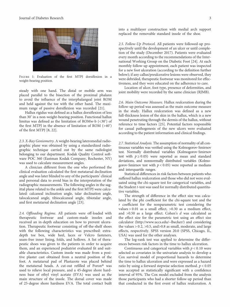
steady with one hand. The distal or mobile arm wasplaced parallel to the bisection of the proximal phalanxto avoid the influence of the interphalangeal joint ROMand held against the toe with the other hand. The maxi-mum range of passive dorsiflexion was recorded [21].
Hallux rigidus was defined as a hallux dorsiflexion of lessthan 30° in a non-weight-bearing position. Functional halluxlimitus was defined as the limitation of ROMw-b (<30°) ofthe first MTPJ in the absence of limitation of ROM (>40°)of the first MTPJ [8, 22].
2.3. X-Ray Goniometry.Aweight-bearing lateromedial radio-graphic plane was obtained by using a standardized radio-graphic technique carried out by the same radiologistbelonging to our department. Kodak Quality Control soft-ware POC 360 (Eastman Kodak Company, Rochester, NY)was used to calculate measurement angles.
A clinician different from the one who performed theclinical evaluation calculated the first metatarsal declinationangle and was later blinded to any of the participants’ clinicaland personal data to avoid bias in the interpretation of theradiographic measurements. The following angles in the sag-ittal plane related to the ankle and the first MTPJ were calcu-lated: calcaneal inclination angle, talar declination angle,talocalcaneal angle, tibiocalcaneal angle, tibiotalar angle,and first metatarsal declination angle [23].
2.4. Offloading Regime. All patients were off-loaded withtherapeutic footwear and custom-made insoles andreceived an in-depth education on how to prevent ulcera-tion. Therapeutic footwear consisting of off-the-shelf shoeswith the following characteristics was prescribed: extra-depth toe box, wide heel, laces or Velcro fasteners,seam-free inner lining, folds, and hollows. A list of thera-peutic shoes was given to the patients in order to acquirethem, and an experienced podiatrist evaluated fit and suit-able characteristics. Custom insole was made from a posi-tive plaster cast obtained from a neutral position of thefoot. A metatarsal pad of Plastazote was placed behindthe metatarsal heads. A cut-out refilled of Poron® wasused to relieve local pressure, and a 45-degree shore hard-ness base of ethyl vinyl acetate (EVA) was used as themain structure of the insole. The dorsal cover was madeof 25-degree shore hardness EVA. The total contact built
into a multilayer construction with medial arch supportreplaced the removable standard insole of the shoe.
2.5. Follow-Up Protocol. All patients were followed-up pro-spectively until the development of an ulcer or until comple-tion of the study (December 2017). Patients were evaluatedevery month according to the recommendations of the Inter-national Working Group on the Diabetic Foot [24]. At eachmonthly follow-up appointment, each patient was inspectedfor a new foot ulceration (according to the definition furtherbelow); if any callus/preulcerative lesions were observed, theywere debrided, therapeutic footwear was monitored for effec-tiveness, and they were educated on the adherence to care.
Location of ulcer, foot type, presence of deformities, andjoint mobility were recorded by the same clinician (RJMB).
2.6. Main Outcome Measure. Hallux reulceration during thefollow-up period was assessed as the main outcome measurein the study. Hallux reulceration was defined as a newfull-thickness lesion of the skin in the hallux, which is a newwound penetrating through the dermis of the hallux, withoutreference to time factors [25]. Potential factors responsiblefor casual pathogenesis of the new ulcers were evaluatedaccording to the patient information and clinical findings.
2.7. Statistical Analysis.The assumption of normality of all con-tinuous variables was verified using the Kolmogorov-Smirnovtest. Normally distributed variables (Kolmogorov-Smirnovtest with p ≥ 0 05) were reported as mean and standarddeviations, and nonnormally distributed variables (Kolmo-gorov-Smirnov test with p < 0 05) were reported as mediansand interquartile ranges.
Statistical differences in risk factors between patients whosuffered hallux reulceration and those who did not were eval-uated using the chi-square test for categorical variables, andthe Student t-test was used for normally distributed quantita-tive variables.
The strength of difference in the effect size was calcu-lated by the phi coefficient for the chi-square test and ther coefficient for the nonparametric test considering thevalues > 0 01 as a small effect, >0.30 as a medium effect,and >0.50 as a large effect. Cohen’s d was calculated asthe effect size for the parametric test using an effect sizecalculator (http://www.uccs.edu/~lbecker/) and consideringthe values > 0 2, >0.5, and>0.8 as small, moderate, and largeeffects, respectively. SPSS version 20.0 (SPSS, Chicago, IL,USA) was used for the other analyses.
The log-rank test was applied to determine the differ-ences between risk factors in the time to hallux ulceration.
Continuous and categorical variables with p < 0 10 wereselected as covariates in the univariate analysis to develop aCox survival model of proportional hazards to determinethe time to hallux ulceration and were expressed as a hazardratio by using a forward stepwise selection method. p < 0 05was accepted as statistically significant with a confidenceinterval of 95%. The Cox model excluded from the analysisthose participants who had a shorter follow-up period thanthat conducted in the first event of hallux reulceration. A
Figure 1: Evaluation of the first MTPJ dorsiflexion in aweight-bearing position.
3Journal of Diabetes Research
collinearity analysis was performed with the explanatoryvariables of the Cox model.
3. Results
Four patients dropped out of the study because of theirrefusal to wear protective therapeutic footwear and/or cus-tom insole and were thus removed from the study. They pre-sented no ulcers at the time of abandoning the study. Finally,56 patients were included in the analysis. Patients werefollowed up prospectively for a median time period of 29months (interquartile ranges (IR) 14.2-64.4 months).
Twenty-nine patients (52%) developed reulceration dur-ing follow-up and showed a median time to ulceration of 19months (IR, 6.0-30.0 months). Different locations of reul-ceration were as follows: 9 patients (31%) developed a newulcer in the hallux, 6 (21%) in the minor toes, 13 (45%)
beneath the metatarsals, and 1 patient (3%) in the heel. Allulcers were classified as neuropathic reulceration, and therewere no ulcers caused by trauma.
Several variables related to demographic and foot charac-teristics revealed differences between patients who developedhallux ulceration and patients who developed ulceration inother locations or without reulceration (Table 2). Partici-pants who developed hallux ulceration were more likely topresent a higher BMI (p = 0 030, Cohen’s d = 0 404), areduced dorsiflexion of the first MTFJ in a weight-bearingposition (p = 0 041, Cohen’s d = −0 422), and functional hal-lux limitus (p = 0 001, r = 0 428) as baseline characteristics.Hallux rigidus showed no association with hallux ulceration(p = 0 729, r = −0 046).
In the radiographic analysis (Table 2), lower first meta-tarsal inclination was associated with hallux ulceration(p = 0 024, Cohen’s d = −0 350).
Table 2: Differences between the risk factors for hallux reulceration.
(N = 56 patients) Hallux reulceration (n = 9) Nonhallux reulceration (n = 47) p value Effect size
Male/female 8 (88.9)/1 (11) 39 (83)/8 (17) 0.658 −0.059a
Mean age (years) 63 ± 9 7 62 ± 7 7 0.712 0.057b
Duration of DM (years) 9 ± 10 8 17 ± 13 3 0.072 −0.314b
Type 1/type 2 DM 0 (0)/9 (100) 13 (28)/34 (72) 0.072 0.241a
Body mass index (kg/m2) 33 6 ± 5 0 29 1 ± 5 2 0.030∗ 0.404b
Nephropathy 3 (33) 12 (25) 0.628 0.065a
Retinopathy 4 (44) 25 (53) 0.630 −0.064a
HbA1c (mmol/mol) (%) 54 ± 6 7 1 ± 0 8 57 ± 8 7 3 ± 1 1 0.599 −0.207b
Ankle brachial index 1 43 ± 0 46 1 24 ± 0 28 0.254 0.242b
Hallux deformity 3 (33) 11 (23) 0.529 0.084a
Hallux valgus 1 10
Metatarsal prominence 0 8
Hallux hammertoe 2 2
First MTPJ ROM (degrees) 47 33 ± 19 36 49 79 ± 19 15 0.734 −0.064b
First MTPJ ROMw-b (degrees) 21 11 ± 7 15 33 26 ± 16 99 0.041∗ −0.422b
Functional hallux limitus 7 (78) 11 (23) 0.001∗ 0.428a
Hallux rigidus 4 (44) 18 (38) 0.729 0.046a
Ankle ROM (degrees) 90 89 ± 5 49 87 60 ± 5 83 0.129 0.279b
Inversion ROM (degrees) 17 56 ± 4 87 16 85 ± 4 64 0.697 0.074b
Eversion ROM (degrees) 9 33 ± 1 41 9 74 ± 3 16 0.411 −0.083b
FPI 2 33 ± 2 50 0 66 ± 4 30 0.125 0.230b
Tibiotalar angle X-ray 112 01 ± 5 11 111 77 ± 6 21 0.902 0.021b
Tibiocalcaneal angle X-ray 67 96 ± 6 94 65 88 ± 7 44 0.433 0.143b
Talocalcaneal angle X-ray 45 11 ± 3 25 46 31 ± 6 13 0.403 −0.121b
Talar declination angle X-ray 26 64 ± 4 02 25 35 ± 4 16 0.397 0.156b
Calcaneal inclination angle X-ray 17 91 ± 4 51 20 91 ± 6 70 0.114 −0.254b
First metatarsal declination angle X-ray 22 81 ± 2 14 24 98 ± 3 50 0.024∗ −0.350b
Abbreviations: HbA1c: glycated hemoglobin; ROM: range of motion; first MTPJ ROM: range of dorsiflexion of the first metatarsophalangeal joint in the restingposition; first MTPJ ROMw-b: range of dorsiflexion of the first metatarsophalangeal joints in the weight-bearing position; FPI: Foot Posture Index. The“nonhallux reulceration” group of patients included other locations of reulceration and patients who did not develop a new ulcer during the follow-up. an(%) for categorical variables; the phi coefficient was used for the chi-square test: representing effect size values of 0.01 as small effect, 0.30 as medium effect,and 0.50 as large effect. bMean ± SD for normally distributed variables; for independent samples, Student’s t-test; effect size as the Cohen’s d: representingeffect size values > 0 2 as small effect, >0.5 as moderate effect, and >0.8 as large effect; d is positive if the mean difference is in the predicted direction.
4 Journal of Diabetes Research
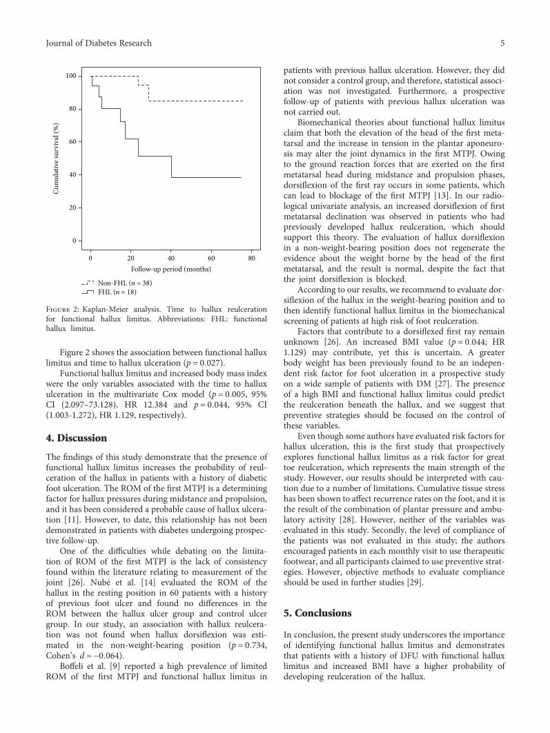
Figure 2 shows the association between functional halluxlimitus and time to hallux ulceration (p = 0 027).
Functional hallux limitus and increased body mass indexwere the only variables associated with the time to halluxulceration in the multivariate Cox model (p = 0 005, 95%CI (2.097–73.128), HR 12.384 and p = 0 044, 95% CI(1.003-1.272), HR 1.129, respectively).
4. Discussion
The findings of this study demonstrate that the presence offunctional hallux limitus increases the probability of reul-ceration of the hallux in patients with a history of diabeticfoot ulceration. The ROM of the first MTPJ is a determiningfactor for hallux pressures during midstance and propulsion,and it has been considered a probable cause of hallux ulcera-tion [11]. However, to date, this relationship has not beendemonstrated in patients with diabetes undergoing prospec-tive follow-up.
One of the difficulties while debating on the limita-tion of ROM of the first MTPJ is the lack of consistencyfound within the literature relating to measurement of thejoint [26]. Nubé et al. [14] evaluated the ROM of thehallux in the resting position in 60 patients with a historyof previous foot ulcer and found no differences in theROM between the hallux ulcer group and control ulcergroup. In our study, an association with hallux reulcera-tion was not found when hallux dorsiflexion was esti-mated in the non-weight-bearing position (p = 0 734,Cohen’s d = −0 064).
Boffeli et al. [9] reported a high prevalence of limitedROM of the first MTPJ and functional hallux limitus in
patients with previous hallux ulceration. However, they didnot consider a control group, and therefore, statistical associ-ation was not investigated. Furthermore, a prospectivefollow-up of patients with previous hallux ulceration wasnot carried out.
Biomechanical theories about functional hallux limitusclaim that both the elevation of the head of the first meta-tarsal and the increase in tension in the plantar aponeuro-sis may alter the joint dynamics in the first MTPJ. Owingto the ground reaction forces that are exerted on the firstmetatarsal head during midstance and propulsion phases,dorsiflexion of the first ray occurs in some patients, whichcan lead to blockage of the first MTPJ [13]. In our radio-logical univariate analysis, an increased dorsiflexion of firstmetatarsal declination was observed in patients who hadpreviously developed hallux reulceration, which shouldsupport this theory. The evaluation of hallux dorsiflexionin a non-weight-bearing position does not regenerate theevidence about the weight borne by the head of the firstmetatarsal, and the result is normal, despite the fact thatthe joint dorsiflexion is blocked.
According to our results, we recommend to evaluate dor-siflexion of the hallux in the weight-bearing position and tothen identify functional hallux limitus in the biomechanicalscreening of patients at high risk of foot reulceration.
Factors that contribute to a dorsiflexed first ray remainunknown [26]. An increased BMI value (p = 0 044; HR1.129) may contribute, yet this is uncertain. A greaterbody weight has been previously found to be an indepen-dent risk factor for foot ulceration in a prospective studyon a wide sample of patients with DM [27]. The presenceof a high BMI and functional hallux limitus could predictthe reulceration beneath the hallux, and we suggest thatpreventive strategies should be focused on the control ofthese variables.
Even though some authors have evaluated risk factors forhallux ulceration, this is the first study that prospectivelyexplores functional hallux limitus as a risk factor for greattoe reulceration, which represents the main strength of thestudy. However, our results should be interpreted with cau-tion due to a number of limitations. Cumulative tissue stresshas been shown to affect recurrence rates on the foot, and it isthe result of the combination of plantar pressure and ambu-latory activity [28]. However, neither of the variables wasevaluated in this study. Secondly, the level of compliance ofthe patients was not evaluated in this study; the authorsencouraged patients in each monthly visit to use therapeuticfootwear, and all participants claimed to use preventive strat-egies. However, objective methods to evaluate complianceshould be used in further studies [29].
5. Conclusions
In conclusion, the present study underscores the importanceof identifying functional hallux limitus and demonstratesthat patients with a history of DFU with functional halluxlimitus and increased BMI have a higher probability ofdeveloping reulceration of the hallux.
Cum
ulat
ive s
urvi
val (
%)
0
0
20
20
40
40
Follow-up period (months)60
60
80
80
Non-FHL (n = 38)FHL (n = 18)
100
Figure 2: Kaplan-Meier analysis. Time to hallux reulcerationfor functional hallux limitus. Abbreviations: FHL: functionalhallux limitus.
5Journal of Diabetes Research
Data Availability
The datasets used to support this study are not freely avail-able in view of participants’ privacy protection.
Conflicts of Interest
The authors declare that they have no conflicts of interest.
Authors’ Contributions
RJMB and JLLM contributed to the conception and designof the study. RJMB and FJAA performed the analysis andinterpretation of the results. RJMB wrote the manuscript.All authors were responsible for the critical revision ofthe manuscript for important intellectual content. Allauthors approved the final version of the article. RJMB isthe guarantor of the work.
Acknowledgments
The authors thank the study participants.
References
[1] D. G. Armstrong, A. J. M. Boulton, and S. A. Bus, “Diabeticfoot ulcers and their recurrence,” The New England Journalof Medicine, vol. 376, no. 24, pp. 2367–2375, 2017.
[2] A. J. Boulton, L. Vileikyte, G. Ragnarson-Tennvall, andJ. Apelqvist, “The global burden of diabetic foot disease,” TheLancet, vol. 366, no. 9498, pp. 1719–1724, 2005.
[3] J. W. Walsh, O. J. Hoffstad, M. O. Sullivan, and D. J. Margolis,“Association of diabetic foot ulcer and death in apopulation-based cohort from the United Kingdom,” DiabeticMedicine, vol. 33, no. 11, pp. 1493–1498, 2016.
[4] C. A. Abbott, A. L. Carrington, H. Ashe et al., “TheNorth-West Diabetes Foot Care Study: incidence of, and riskfactors for, new diabetic foot ulceration in acommunity-based patient cohort,” Diabetic Medicine, vol. 19,no. 5, pp. 377–384, 2002.
[5] R. Waaijman, M. de Haart, M. L. J. Arts et al., “Risk factors forplantar foot ulcer recurrence in neuropathic diabetic patients,”Diabetes Care, vol. 37, no. 6, pp. 1697–1705, 2014.
[6] D. G. Armstrong, L. A. Lavery, and L. B. Harkless, “Validationof a diabetic wound classification system. The contribution ofdepth, infection, and ischemia to risk of amputation,” DiabetesCare, vol. 21, no. 5, pp. 855–859, 1998.
[7] R. J. Molines-Barroso, J. L. Lazaro-Martinez, F. J. Alvaro-Afonso, I. Sanz-Corbalan, J. L. Garcia-Klepzig, andJ. Aragon-Sanchez, “Validation of an algorithm to predictreulceration in amputation patients with diabetes,” Interna-tional Wound Journal, vol. 14, no. 3, pp. 523–528, 2017.
[8] C. Formosa, A. Gatt, and N. Chockalingam, “The importanceof clinical biomechanical assessment of foot deformity andjoint mobility in people living with type-2 diabetes within aprimary care setting,” Primary Care Diabetes, vol. 7, no. 1,pp. 45–50, 2013.
[9] T. J. Boffeli, J. K. Bean, and J. R. Natwick, “Biomechanicalabnormalities and ulcers of the great toe in patients with diabe-tes,” The Journal of Foot and Ankle Surgery, vol. 41, no. 6,pp. 359–364, 2002.
[10] W. R. Ledoux, J. B. Shofer, D. G. Smith et al., “Relationshipbetween foot type, foot deformity, and ulcer occurrence inthe high-risk diabetic foot,” Journal of Rehabilitation Researchand Development, vol. 42, no. 5, pp. 665–672, 2005.
[11] C. Payne, D. Turner, and K. Miller, “Determinants of plantarpressures in the diabetic foot,” Journal of Diabetes and itsComplications, vol. 16, no. 4, pp. 277–283, 2002.
[12] M. K. Allen, T. J. Cuddeford, W. M. Glasoe et al., “Relationshipbetween static mobility of the first ray and first ray, midfoot,and hindfoot motion during gait,” Foot & Ankle International,vol. 25, no. 6, pp. 391–396, 2004.
[13] E. Maceira and M. Monteagudo, “Functional hallux rigidusand the Achilles-calcaneus-plantar system,” Foot and AnkleClinics, vol. 19, no. 4, pp. 669–699, 2014.
[14] V. L. Nubé, L. Molyneaux, and D. K. Yue, “Biomechanical riskfactors associated with neuropathic ulceration of the hallux inpeople with diabetes mellitus,” Journal of the AmericanPodiatricMedical Association, vol. 96, no. 3, pp. 189–197, 2006.
[15] M. S. Cowley, E. J. Boyko, J. B. Shofer, J. H. Ahroni, and W. R.Ledoux, “Foot ulcer risk and location in relation to prospectiveclinical assessment of foot shape and mobility among personswith diabetes,” Diabetes Research and Clinical Practice, vol. 82,no. 2, pp. 226–232, 2008.
[16] M. ElMakki Ahmed, A. O. Tamimi, S. I. Mahadi, A. H.Widatalla, and M. A. Shawer, “Hallux ulceration in diabeticpatients,” The Journal of Foot & Ankle Surgery, vol. 49,no. 1, pp. 2–7, 2010.
[17] N. C. Schaper, G. Andros, J. Apelqvist et al., “Diagnosis andtreatment of peripheral arterial disease in diabetic patientswith a foot ulcer. A progress report of the International Work-ing Group on the Diabetic Foot,” Diabetes/MetabolismResearch and Reviews, vol. 28, Supplement 1, pp. 218–224,2012.
[18] A. J. Boulton, D. G. Armstrong, S. F. Albert et al., “Compre-hensive foot examination and risk assessment: a report of thetask force of the foot care interest group of the American Dia-betes Association, with endorsement by the American Associ-ation of Clinical Endocrinologists,” Diabetes Care, vol. 31,no. 8, pp. 1679–1685, 2008.
[19] A. C. Redmond, J. Crosbie, and R. A. Ouvrier, “Developmentand validation of a novel rating system for scoring standingfoot posture: the Foot Posture Index,” Clinical Biomechanics,vol. 21, no. 1, pp. 89–98, 2006.
[20] J. L. Lazaro-Martinez, F. J. Aragon-Sanchez, J. V. Beneit-Montesinos, M. A. Gonzalez-Jurado, E. Garcia Morales,and D. Martinez Hernandez, “Foot biomechanics inpatients with diabetes mellitus: doubts regarding the relation-ship between neuropathy, foot motion, and deformities,” Jour-nal of the American Podiatric Medical Association, vol. 101,no. 3, pp. 208–214, 2011.
[21] T. S. Roukis, P. R. Scherer, and C. F. Anderson, “Position of thefirst ray and motion of the first metatarsophalangeal joint,”Journal of the American Podiatric Medical Association,vol. 86, no. 11, pp. 538–546, 1996.
[22] D. A. Nawoczenski, J. F. Baumhauer, and B. R. Umberger,“Relationship between clinical measurements and motion ofthe first metatarsophalangeal joint during gait,” The Journalof Bone and Joint Surgery. American Volume, vol. 81, no. 3,pp. 370–376, 1999.
[23] R. Christman, Foot and Ankle Radiology, Wolters KluwerHealth, Philadelphia, PA, USA, 2nd edition, 2015.
6 Journal of Diabetes Research

[24] K. Bakker, J. Apelqvist, N. C. Schaper, and InternationalWorking Group on the Diabetic Foot Editorial Board, “Practi-cal guidelines on the management and prevention of the dia-betic foot 2011,” Diabetes/Metabolism Research and Reviews,vol. 28, Supplement 1, pp. 225S–231S, 2012.
[25] R. J.Molines-Barroso, J. L. Lazaro-Martinez, J.Aragon-Sanchez,E.Garcia-Morales, J. V. Beneit-Montesinos, and F. J. Alvaro-Afonso, “Analysis of transfer lesions in patients who under-went surgery for diabetic foot ulcers located on the plantaraspect of the metatarsal heads,” Diabetic Medicine, vol. 30,no. 8, pp. 973–976, 2013.
[26] B. Durrant and N. Chockalingam, “Functional hallux limitus: areview,” Journal of the American Podiatric Medical Associa-tion, vol. 99, no. 3, pp. 236–243, 2009.
[27] E. J. Boyko, J. H. Ahroni, V. Stensel, R. C. Forsberg, D. R.Davignon, and D. G. Smith, “A prospective study of risk fac-tors for diabetic foot ulcer. The Seattle Diabetic Foot Study,”Diabetes Care, vol. 22, no. 7, pp. 1036–1042, 1999.
[28] J. J. Van Netten, J. G. van Baal, A. Bril, M. Wissink, and S. A.Bus, “An exploratory study on differences in cumulative plan-tar tissue stress between healing and non-healing plantar neu-ropathic diabetic foot ulcers,” Clinical Biomechanics, vol. 53,pp. 86–92, 2018.
[29] R. Waaijman, R. Keukenkamp, M. de Haart, W. P. Polomski,F. Nollet, and S. A. Bus, “Adherence to wearing prescriptioncustom-made footwear in patients with diabetes at high riskfor plantar foot ulceration,” Diabetes Care, vol. 36, no. 6,pp. 1613–1618, 2013.
7Journal of Diabetes Research

Stem Cells International
Hindawiwww.hindawi.com Volume 2018
Hindawiwww.hindawi.com Volume 2018
MEDIATORSINFLAMMATION
of
EndocrinologyInternational Journal of
Hindawiwww.hindawi.com Volume 2018
Hindawiwww.hindawi.com Volume 2018
Disease Markers
Hindawiwww.hindawi.com Volume 2018
BioMed Research International
OncologyJournal of
Hindawiwww.hindawi.com Volume 2013
Hindawiwww.hindawi.com Volume 2018
Oxidative Medicine and Cellular Longevity
Hindawiwww.hindawi.com Volume 2018
PPAR Research
Hindawi Publishing Corporation http://www.hindawi.com Volume 2013Hindawiwww.hindawi.com
The Scientific World Journal
Volume 2018
Immunology ResearchHindawiwww.hindawi.com Volume 2018
Journal of
ObesityJournal of
Hindawiwww.hindawi.com Volume 2018
Hindawiwww.hindawi.com Volume 2018
Computational and Mathematical Methods in Medicine
Hindawiwww.hindawi.com Volume 2018
Behavioural Neurology
OphthalmologyJournal of
Hindawiwww.hindawi.com Volume 2018
Diabetes ResearchJournal of
Hindawiwww.hindawi.com Volume 2018
Hindawiwww.hindawi.com Volume 2018
Research and TreatmentAIDS
Hindawiwww.hindawi.com Volume 2018
Gastroenterology Research and Practice
Hindawiwww.hindawi.com Volume 2018
Parkinson’s Disease
Evidence-Based Complementary andAlternative Medicine
Volume 2018Hindawiwww.hindawi.com
Submit your manuscripts atwww.hindawi.com