Embed Size (px)
Citation preview

Nel et al. BMC Microbiology 2014, 14:9http://www.biomedcentral.com/1471-2180/14/9
RESEARCH ARTICLE Open Access
Mycobacterium bovis BCG infection severelydelays Trichuris muris expulsion and co-infectionsuppresses immune responsiveness to bothpathogensHendrik J Nel1,2*, Nelita du Plessis1*, Leanie Kleynhans1, André G Loxton1, Paul D van Helden1 and Gerhard Walzl1
Abstract
Background: The global epidemiology of parasitic helminths and mycobacterial infections display extensive geographicaloverlap, especially in the rural and urban communities of developing countries. We investigated whether co-infection withthe gastrointestinal tract-restricted helminth, Trichuris muris, and the intracellular bacterium, Mycobacterium bovis (M. bovis)BCG, would alter host immune responses to, or the pathological effect of, either infection.
Results: We demonstrate that both pathogens are capable of negatively affecting local and systemic immune responsestowards each other by modifying cytokine phenotypes and by inducing general immune suppression. T. muris infectioninfluenced non-specific and pathogen-specific immunity to M. bovis BCG by down-regulating pulmonary TH1 and Tregresponses and inducing systemic TH2 responses. However, co-infection did not alter mycobacterial multiplication ordissemination and host pulmonary histopathology remained unaffected compared to BCG-only infected mice.Interestingly, prior M. bovis BCG infection significantly delayed helminth clearance and increased intestinal crypt cellproliferation in BALB/c mice. This was accompanied by a significant reduction in systemic helminth-specific TH1 and TH2cytokine responses and significantly reduced local TH1 and TH2 responses in comparison to T. muris-only infected mice.
Conclusion: Our data demonstrate that co-infection with pathogens inducing opposing immune phenotypes, can havedifferential effects on compartmentalized host immune protection to either pathogen. In spite of local andsystemic decreases in TH1 and increases in TH2 responses co-infected mice clear M. bovis BCG at the same rate asBCG only infected animals, whereas prior mycobacterial infection initiates prolonged worm infestation in parallelto decreased pathogen-specific TH2 cytokine production.
Keywords: Helminth, Co-infection, Mycobacteria, Tuberculosis
BackgroundTuberculosis (TB) is most prevalent in resource-poorcountries and factors such as genetic susceptibility,malnutrition and circulating strain differences have beenimplicated as determinants of TB disease developmentin these regions [1,2]. Compelling evidence demonstrates
* Correspondence: [email protected]; [email protected] of Molecular Biology and Human Genetics, MRC Centre forMolecular and Cellular Biology, NRF/DST Centre of Excellence in BiomedicalTB Research, Faculty Medicine and Health Sciences, Stellenbosch University,Tygerberg, South Africa2University of Queensland Diamantina Institute, Brisbane, QLD, AustraliaFull list of author information is available at the end of the article
© 2014 Nel et al.; licensee BioMed Central LtdCommons Attribution License (http://creativecreproduction in any medium, provided the or
that many of these factors increase disease risk partlythough the induction of host immune dysregulation andultimately affect host control of Mycobacterium tubercu-losis (M. tb) proliferation [3]. The high prevalence ofparasitic helminth infections in TB affected communi-ties, has highlighted co-infection as another risk factorcompromising host immunity and thus a potential deter-minant for development of TB [4,5]. In support of thistheory, several reports indicated that TB patients arecommonly found to be co-infected with helminth spe-cies such as Trichuris trichiura and Ascaris lumbricoides[6] and present with increased total and helminth-specific serum immunoglobulin E (IgE) [7].
. This is an open access article distributed under the terms of the Creativeommons.org/licenses/by/2.0), which permits unrestricted use, distribution, andiginal work is properly cited.

Nel et al. BMC Microbiology 2014, 14:9 Page 2 of 13http://www.biomedcentral.com/1471-2180/14/9
Host control of mycobacterial or helminth infectionslargely rely on the induction of appropriately polarizedimmune responses. Protective immune responses to M. tbinfection are associated with enhanced T helper 1 (TH1)type cellular immunity and the production of characte-ristic TH1 cytokines such as tumor necrosis factor alpha(TNF-α), interferon-gamma (IFN-γ) and interleukin-12(IL-12) [8]. Conversely, protection against most helminthsrequires a T helper 2 (TH2) type cellular immune re-sponse with production of distinct TH2 cytokines such asIL-4, IL-5, IL-13 and IL-9 [9,10]. Since TH1 and TH2immune responses have the ability to concurrently down-regulate each other, a state of co-infection could resultin inappropriate protective host immune responses toeither infections [11]. Furthermore, both pathogenshave the potential to induce regulatory T cell (Treg)responses associated with production of immune sup-pressive cytokines such as IL-10 and transforming growthfactor beta (TGF-β) [10-13].In line with the TH1/TH2 dichotomy, hypotheses con-
cerning helminth-mycobacterial co-infection postulatethat a helminth-induced TH2 immune bias could inhibitdevelopment of protective cellular immune responses toM. tb, increase mycobacterial proliferation or lead to thefailure of vaccine strategies against TB [14,15]. This theoryis supported by numerous studies which have reported areduction in TH1 responses to be associated with pooroutcomes in TB patients [16] and latently infected individ-uals [17] with concurrent helminth infection. Helminth-induced regulatory (Treg) responses such as TGF-β andIL-10 production have also been implicated in S. mansoni-induced progression to active TB of HIV-1 infectedUgandans [18]. It was also established that deworming ofhelminth-infected individuals restores cellular immuneresponses to mycobacterial purified protein derivatives(PPD) [19-21]. Similarly, deworming of helminth-infectedEthiopians immigrants in Israel resulted in increasedcellular immune responses against HIV- andM. tb-specificantigens compared to untreated individuals [22], suggest-ing deteriorating immune responses and poor clinicaloutcomes in helminth-infected individuals might not be aresult of inadequate nutrition or sanitation. Several reportshave also indicated helminth-mediated modulation ofvaccine responses. Children with prenatal sensitizationto filariae and schistosomes were reported to display adown-regulation in TH1 responsiveness to BCG vaccin-ation [23] and animal co-infection models have furtherdemonstrated that a pre-existing infection with a lung-migrating helminth, can inhibit development of protectiveinnate anti-TB responses by inducing the IL-4 receptorpathway and accumulation of alternatively activatedmacrophages [24]. In summary, most reports indicatethat helminth infection significantly affects TB suscepti-bility. In contrast, very little data addressing the effect
of mycobacterial infection on host immunity to hel-minth infections are available.In the current study, we assessed the influence of co-
infection on immune responses against the individualpathogens. We established a BALB/c co-infection modelusing Mycobacterium bovis (M. bovis) BCG and the gas-trointestinal tract-restricted rodent helminth, Trichurismuris (T. muris) as TH1 and TH2 pathogenic assaults, re-spectively. The M. bovis BCG murine infection model isroutinely used for studying anti-mycobacterial responsesduring latency as the associated immune response is simi-lar to that induced during human M. tb infection [25],whereas T. muris infection serves as a well describedmodel for gastrointestinal tract restricted human soil-transmitted helminth (STH) infection [26]. We exploredthe possibility that concurrent infection with two patho-gens, normally cleared by mice during single pathogeninfection, might lead to mutually inhibitory immunedynamics and subsequent uncontrolled infection.
MethodsAnimalsSpecified pathogen free (SPF) female BALB/c mice (WTand IL-4 knock-out strains) between 6–8 weeks of age,were kept at the Faculty of Medicine and Health SciencesAnimal Unit, Stellenbosch University (SU; South Africa)under conditions compatible with the SU guidelines forthe care of animals. All procedures were approved by theSU Animal Ethics Board [Project license: 2003/186/p].
Parasite enumeration and antigen preparationT. muris eggs were donated by Allison Bancroft (Univer-sity of Manchester, UK). Egg propagation in BALB/c IL-4knock-out mice (gift from Frank Brombacher, Universityof Cape Town, South Africa), helminth collection, andexcretory/secretory (E/S) antigen preparations, wereperformed as described previously [27,28]. Helminthburdens were determined by quantification of intestinaladult worms by examining faecal matter under a dissec-tion microscope. Mycobacterium bovis BCG Pasteur(donated by Robin Warren, SU, South Africa) was prop-agated to logarithmic growth phase in Middlebrook7H9 (Difco) liquid culture, supplemented with 0.2% gly-cerol, 0.05% Tween 80 and 10% albumin-dextrose-catalase(ADC, Merck) at 37°C. Bacterial proliferation was assessedby manual counting of colony forming units (CFU) fromserial dilutions of homogenized lungs and spleens, platedon Middelbrook 7H11 (Difco) agar plates supplementedwith 0.2% glycerol and 10% oleic acid-albumin-dextrose-catalase (OADC, BD Biosciences).
Co-infection protocolTwo infection protocols were used during this study.Each experiment consisted of 3 groups of 5–10 animals

Nel et al. BMC Microbiology 2014, 14:9 Page 3 of 13http://www.biomedcentral.com/1471-2180/14/9
per group. Groups included M. bovis BCG-T. muris co-infected, BCG-only infected and T. muris-only infectedmice. The first protocol (Figure 1A) was intended toestablish a chronic, low grade M. bovis BCG infectionthat was subsequently followed by a TH2-inducingT. muris infection. Mice were infected intranasally (i.n.)with 1–5 × 10⁵ CFU BCG bacilli per mouse or an equalvolume of PBS. Briefly, mice were lightly anesthetized byintraperitoneal (i.p.) injection of a 200 μl mixture consist-ing of Ketamine (12 mg/ml Anaket-V, Centaur Labs) andXylazine (1.6 mg/ml, Rompun, Bayer). Mice were gentlylifted by the loose skin at the throat, and kept upright withits head tilted back and the nose pointed up. Using a pip-ette with a sterile tip, 40 μl of the declumped mycobacter-ial suspension was applied to the nostrils. Animals weremaintained upright for another 30 seconds to ensurecomplete delivery to the respiratory system. Six weeks(day 42) later, mice were infected under light anaesthesiaintragastrically (i.g.) with 200–250 (low dose) or 500–600(high dose) embryonated T. muris eggs or an equal vol-ume of PBS. At week 9 (day 63), mice were culled and the
M. bovis BCGi.n.
M. bovis BCG i.n
PBS
T. muris eggsi.g.
PBST. muris eggs
i.g.
Kill
Day 1
Day 42
Day 63
T. muris eggs i.g.
T. muris eggs i.g.
PBS
M. bovis BCG i.n
T. Muris eggs i.g.
PBSM. bovis BCG
i.n
T. muris eggs i.g.
PBS
Kill
Day 1,10
Day 10
Day 22,32,42
Day 52
A
B
Figure 1 Experimental design. (A) BALB/c mice were infected i.n.with M. bovis BCG on day 1, followed by i.g. T. muris infection onday 42. Mice were killed on day 63 and the relevant tissues collectedfor further analysis. (B) BALB/c mice were infected i.g. with T. murisevery 10 days starting on day 1. Animals were co-infected i.n. withM. bovis BCG on day 10. Mice were killed on day 52 and the relevanttissues collected. Appropriate single infections and PBS controls wereincluded in parallel for both protocols. Experiments were performedwith 5 to 10 animals per group. P values <0.05 were consideredstatistically significant. (ns = non significant).
relevant organs removed for investigation. The secondprotocol (Figure 1B) was designed to first establish aTH2-inducing T. muris infection prior to challenge withM. bovis BCG infection. Animals were infected i.g.with 200–250 embryonated T. muris eggs or an equalamount of PBS on day 1 and every 10 days thereafteruntil experimental completion. On day 10, animals wereinfected i.n. with 1–5 × 10⁵ CFU BCG bacilli or an equalvolume of PBS. After 6 weeks (day 52), all mice werehumanely euthanized and the relevant organs removedfor investigation. Experiments were completed in tripli-cate at three separate times.
Immune phenotyping and intracellular cytokine analysisImmune phenotyping was performed using single cellsuspensions from spleens and mesenteric lymph nodes(MLNs). Intracellular cytokine expression was deter-mined following stimulation with 50 ng/ml Phorbol12-myristate 13-acetate (PMA) (Sigma), 1 μg/ml Ionomy-cin (Sigma) and 10 μg/ml Brefeldin A (BFA) (Sigma) for 4hours at 37°C and 5% CO2. Cells were resuspended in PBScontaining 1% BSA and 0.1% Sodium Azide (wash buffer)and stained for 30 minutes with fluorochrome conjugatedanti-mouse antibodies against CD3, CD4, CD8, CD25,B220, Foxp3, IFN-γ and IL-4 (BD Biosciences, Caltagor Biolegend). Cells were fixed with 1% formaldehyde,washed and resuspended in wash buffer. Lymphocytepopulations were determined based on their Forward/Side scatter profile and gates set with the help of appro-priate FMOs and Isotype controls. Acquisitions wereperformed on a FACSCalibur (BD Biosciences) usingappropriate instrument settings, color compensationand isotype controls for all antibodies. At least 5 × 10⁴
lymphocyte events were acquired and data analysis per-formed using CellQuest software (BD Bioscience).
In vitro pathogen-specific cytokine analysisSpleen (1 × 10⁷ cells/ml) single cell suspensions werestimulated for 24 hours with live BCG cultures (MOI 5:1),50 μg/ml E/S antigen or culture media as control at 37°C,5% CO2. Culture supernatants were used for cytokineconcentration analyses using the luminex bead-array tech-nology (LINCO Research) to test for the soluble cytokinesIFN-γ, TNF-α, IL-4, IL-10, IL-13 and IL-17 using a Bio-Plex platform (Bio-Rad Laboratories). Background read-ings were controlled by subtraction of unstimulatedcontrol sample measurements. Values were checkedagainst internal quality controls to monitor analysis ac-curacy within specified concentration ranges.
Nucleic acid extraction and relative quantitative real timePCRTotal RNA was extracted from the upper right lobe ofmouse lungs and spleen tips using Trizol (Gibco BRL)

Nel et al. BMC Microbiology 2014, 14:9 Page 4 of 13http://www.biomedcentral.com/1471-2180/14/9
and subsequently treated with a DNA-free kit (Ambion)to remove contaminating DNA. First strand cDNA wastranscribed using the QuantiTect Reverse Transcriptionkit (Qiagen) according to the manufacturer’s protocols.Relative quantification of IFN-γ, IL-4, IL-10, TGF-β andFoxp3 were performed using SYBR Green PCR MasterMix kit (Roche), cDNA (500 μg) and primers (0.5 μM)on the LightCycler system v3.5 (Roche). All primers weredesigned to span intron-exon boundaries (Table 1). Thedelta-delta Ct method was used to calculate relative geneexpression levels between two samples. Gene expressionwas assayed quantitatively and normalized to that of ahousekeeping gene (GAPDH, HPRT, 18S-RNA) to obtaina RNA ratio in order to establish the relevant change inRNA expression [29].
HistologyLeft upper lung lobes were fixed in 10% buffered forma-lin, embedded in paraffin blocks and sections (3-5 μm)stained with Haematoxylin and Eosin (H&E) for lightmicroscopy. Pulmonary histopathological scoring wasperformed in a blinded fashion and calculated separatelyfor each lung section as previously described [32]. Inbrief, a scale of 0 to 4 was used to individually score thelevel of peribronchiolitis, perivasculitis, interstitial pneu-monitis and alveolitis of each section in order to obtainan average score for each lung. A score of 0 was basedupon observation of normal, uninfected mouse lungsamples and a score of 4 on previous studies of greatestinflammatory change and pathology brought about by i.nM. bovis BCG infection in BALB/c mice. Scoring ofgastrointestinal histopathology was achieved by measuringmucus production, presence of mast cells and mitoticbody enumeration in fixed caecum tips imbedded in paraf-fin blocks. Sections (3-5 μm) were used for Periodic AcidSchiff (PAS) staining to score goblet cell-mucus produc-tion within caecal crypts as the percentage PAS positive
Table 1 List of primer sequences used for relative quantitativ
Target Forward
HPRT GACTGTAGATTTTATCAGACT
GPDH GGTGGCAGAGGCCTTTG*18S [30] GTCTGTGATGCCCTTAGATG*TGF-β [30] CCGCAACAACGCCATCTATG*IFN-γ [31] AAGTTCTGGGCTTCTCCTCCT*IL-10 [30] CTGGACAACATACTGCTAAC*IL-4 [31] TCAACCCCCAGCTAGTTGTC
GATA3 CTGGAGGAGGAACGCTAATG
Tbet AGCAAGGACGGCGAATGTT*Foxp3 [30] CACAATATGCGACCCCCTTTC*Primer sequences adapted from reference.
stain in the crypt epithelium and lamina propria. Acidifiedtoluidine blue staining was used for the quantification ofmast cells in caecum tip samples and enumeration of mi-totic bodies within caecum crypts. Scoring was conductedfrom two sets (cross sectional and longitudinal) of 20caecal crypt units per animal. All slides were evaluatedusing the ZS300 Imaging system v.3.0 (Carl Zeiss Vision).
Statistical analysisData was analyzed using STATISTCA v.7 (StatSoft) soft-ware. Nonparametric analysis and Mann–Whitney U testswere performed for comparison between groups and thedata presented as median values. Multiple group analysisincluded the multiple comparison correction (Bonferroni).Statistically significant differences were judged as p ≤ 0.05.
ResultsM. bovis BCG clearance and lung pathology is notinfluenced by an established or successive T. murisinfectionThe influence of T. muris infection on host ability tocontrol a chronic, low grade M. bovis BCG infection inBALB/c mice was investigated for both experimentalprotocols (Figure 1A and B). Results demonstrated thatan ongoing helminth-induced TH2 immune background,pre-established by T. muris trickle infection, failed toalter mycobacterial proliferation and dissemination whencompared to M. bovis BCG-only infected mice in thelungs (Figure 2A) and spleen (data not shown). Similarly,initiation of a TH2 immune environment subsequent toBCG infection, resulted in equivalent pulmonary bacter-ial burdens between co-infected and BCG-only infectedgroups (Figure 2B). These end point CFU findings wereconfirmed by growth curve data demonstrating no signifi-cant difference in pulmonary mycobacterial burden be-tween co-infected and M. bovis BCG-only infected mice atseveral time points postM. bovis BCG infection (Figure 2C).
e real-time PCR
Reverse
GTCTGGCCTGTATCCAACACTTC
TGCCGATTTAGCATCTCCTT
AGCTTATGACCCGCACTTAC
CTCTGCACGGGACAGCAAT
G GCCAGTTCCTCCAGATATCCAAGA
CG GGGCATCACTTCTACCAGGTAA
TTCAAGCATGGAGTTTTCCC
GGTTGAAGGAGCTGCTCTTG
GGGTGGACATATAAGCGGTTC
AACATGCGAGTAAACCAATGGTA

A B
0
1.0 104
2.0 104
3.0 104
4.0 104
BCG/T.murisBCG
ns
CF
U/L
un
g
0
2.0 104
4.0 104
6.0 104
8.0 104
T.muris/BCGBCG
ns
CF
U/L
un
g
C
14 24 35
Days post i.n. BCG infection
CF
U/L
un
g
1.0x10⁴
2.0x10⁴
3.0x10⁴
4.0x10⁴
T. muris /BCG
BCG
0.0
ns
ns
D
BCG/T. muris BCG/PBS
Uninfected PBS/T. muris
b = Peribronchiolitisv = Perivasculitisi = Interstitial pneumonitisa = Alveolitis
E
b v
i
a
20x 20x 20x
0
1
2
3
4
T.murisBCG
BCG/T.muris
*ns
*
ns
Naive
Lung
pat
holo
gy s
core
Figure 2 (See legend on next page.)
Nel et al. BMC Microbiology 2014, 14:9 Page 5 of 13http://www.biomedcentral.com/1471-2180/14/9
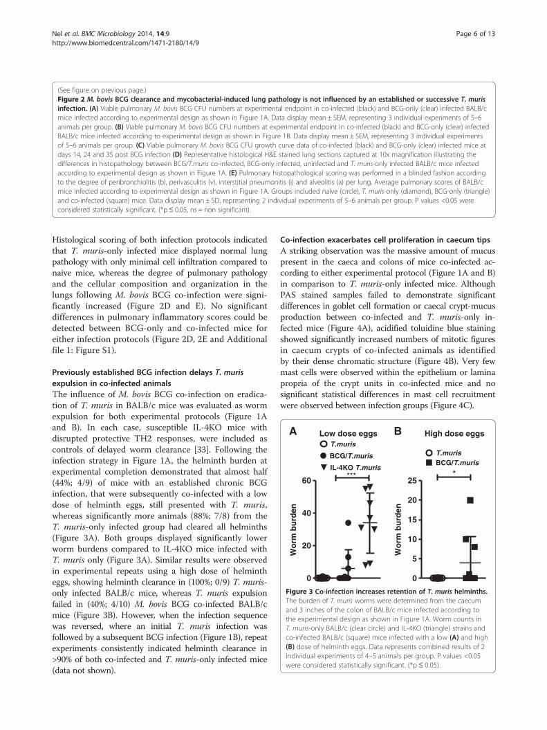
(See figure on previous page.)Figure 2 M. bovis BCG clearance and mycobacterial-induced lung pathology is not influenced by an established or successive T. murisinfection. (A) Viable pulmonary M. bovis BCG CFU numbers at experimental endpoint in co-infected (black) and BCG-only (clear) infected BALB/cmice infected according to experimental design as shown in Figure 1A. Data display mean ± SEM, representing 3 individual experiments of 5–6animals per group. (B) Viable pulmonary M. bovis BCG CFU numbers at experimental endpoint in co-infected (black) and BCG-only (clear) infectedBALB/c mice infected according to experimental design as shown in Figure 1B. Data display mean ± SEM, representing 3 individual experimentsof 5–6 animals per group. (C) Viable pulmonary M. bovis BCG CFU growth curve data of co-infected (black) and BCG-only (clear) infected mice atdays 14, 24 and 35 post BCG infection (D) Representative histological H&E stained lung sections captured at 10x magnification illustrating thedifferences in histopathology between BCG/T.muris co-infected, BCG-only infected, uninfected and T. muris-only infected BALB/c mice infectedaccording to experimental design as shown in Figure 1A. (E) Pulmonary histopathological scoring was performed in a blinded fashion accordingto the degree of peribronchiolitis (b), perivasculitis (v), interstitial pneumonitis (i) and alveolitis (a) per lung. Average pulmonary scores of BALB/cmice infected according to experimental design as shown in Figure 1A. Groups included naive (circle), T. muris-only (diamond), BCG-only (triangle)and co-infected (square) mice. Data display mean ± SD, representing 2 individual experiments of 5–6 animals per group. P values <0.05 wereconsidered statistically significant. (*p≤ 0.05, ns = non significant).
A BLow dose eggs
0
20
40
60
BCG/T.muris
T.muris
IL-4KO T.muris***
Wo
rm b
urd
en
High dose eggs
0
5
10
15
20
25
BCG/T.murisT.muris
*
Wo
rm b
urd
en
Figure 3 Co-infection increases retention of T. muris helminths.The burden of T. muris worms were determined from the caecumand 3 inches of the colon of BALB/c mice infected according tothe experimental design as shown in Figure 1A. Worm counts inT. muris-only BALB/c (clear circle) and IL-4KO (triangle) strains andco-infected BALB/c (square) mice infected with a low (A) and high(B) dose of helminth eggs. Data represents combined results of 2individual experiments of 4–5 animals per group. P values <0.05were considered statistically significant. (*p ≤ 0.05).
Nel et al. BMC Microbiology 2014, 14:9 Page 6 of 13http://www.biomedcentral.com/1471-2180/14/9
Histological scoring of both infection protocols indicatedthat T. muris-only infected mice displayed normal lungpathology with only minimal cell infiltration compared tonaive mice, whereas the degree of pulmonary pathologyand the cellular composition and organization in thelungs following M. bovis BCG co-infection were signi-ficantly increased (Figure 2D and E). No significantdifferences in pulmonary inflammatory scores could bedetected between BCG-only and co-infected mice foreither infection protocols (Figure 2D, 2E and Additionalfile 1: Figure S1).
Previously established BCG infection delays T. murisexpulsion in co-infected animalsThe influence of M. bovis BCG co-infection on eradica-tion of T. muris in BALB/c mice was evaluated as wormexpulsion for both experimental protocols (Figure 1Aand B). In each case, susceptible IL-4KO mice withdisrupted protective TH2 responses, were included ascontrols of delayed worm clearance [33]. Following theinfection strategy in Figure 1A, the helminth burden atexperimental completion demonstrated that almost half(44%; 4/9) of mice with an established chronic BCGinfection, that were subsequently co-infected with a lowdose of helminth eggs, still presented with T. muris,whereas significantly more animals (88%; 7/8) from theT. muris-only infected group had cleared all helminths(Figure 3A). Both groups displayed significantly lowerworm burdens compared to IL-4KO mice infected withT. muris only (Figure 3A). Similar results were observedin experimental repeats using a high dose of helmintheggs, showing helminth clearance in (100%; 0/9) T. muris-only infected BALB/c mice, whereas T. muris expulsionfailed in (40%; 4/10) M. bovis BCG co-infected BALB/cmice (Figure 3B). However, when the infection sequencewas reversed, where an initial T. muris infection wasfollowed by a subsequent BCG infection (Figure 1B), repeatexperiments consistently indicated helminth clearance in>90% of both co-infected and T. muris-only infected mice(data not shown).
Co-infection exacerbates cell proliferation in caecum tipsA striking observation was the massive amount of mucuspresent in the caeca and colons of mice co-infected ac-cording to either experimental protocol (Figure 1A and B)in comparison to T. muris-only infected mice. AlthoughPAS stained samples failed to demonstrate significantdifferences in goblet cell formation or caecal crypt-mucusproduction between co-infected and T. muris-only in-fected mice (Figure 4A), acidified toluidine blue stainingshowed significantly increased numbers of mitotic figuresin caecum crypts of co-infected animals as identifiedby their dense chromatic structure (Figure 4B). Very fewmast cells were observed within the epithelium or laminapropria of the crypt units in co-infected mice and nosignificant statistical differences in mast cell recruitmentwere observed between infection groups (Figure 4C).

40x
20x
A
B
C
0
5
10
15
20
25
BCG/T.muris
T.muris
ns
% P
AS
per
cry
pt
area
0
5
10
15
20
25
BCG/T.muris
T.muris
*
No
. of
Mit
oti
c b
od
ies
0
20
40
60
80
100
BCG/T. murisT. muris
ns
Mas
t ce
ll co
un
ts(4
0 ca
ecu
m c
ryp
ts)
Figure 4 Co-infection increases mitotic figures in the caecum crypts. (A) Histological analysis of goblet cell numbers as determined by thepercentage PAS+ cells (indicated by arrow) per 2 x 20 cross sectional crypt units in T. muris-only (clear) and co-infected (black) BALB/c miceinfected according to the experimental design as shown in Figure 1A. Data display median ±min-max, representing 2–3 individual experimentsof 5 animals per group. (B) Toluidine blue stained mitotic bodies (indicated by the arrows) were counted in 2 x 20 crypts/slide. Numbers ofmitotic bodies as determined from cross-sectional and longitudinal crypt units in co-infected (black) and T. muris-only (clear) infected BALB/c miceinfected according to Figure 1A. Data display median ±min-max, representing 2–3 individual experiments of 5 animals per group (C) Toluidineblue staining for the assessment of mast cells (indicated by arrows) in cross sectional and longitudinal crypt units demonstrated few mast cellswithin the lamina propria and crypt epithelium of the caecum tissue with most mast cells residing within the submucosa surrounding the caecum. Bargraph indicating the numbers of mast cells measured in co-infected (black) and T. muris-only (clear) infected groups per 40 caecum crypts. Data displaymedian ± SD of 5 animals per group. P values <0.05 were considered statistically significant. (ns = non significant).
Nel et al. BMC Microbiology 2014, 14:9 Page 7 of 13http://www.biomedcentral.com/1471-2180/14/9

Nel et al. BMC Microbiology 2014, 14:9 Page 8 of 13http://www.biomedcentral.com/1471-2180/14/9
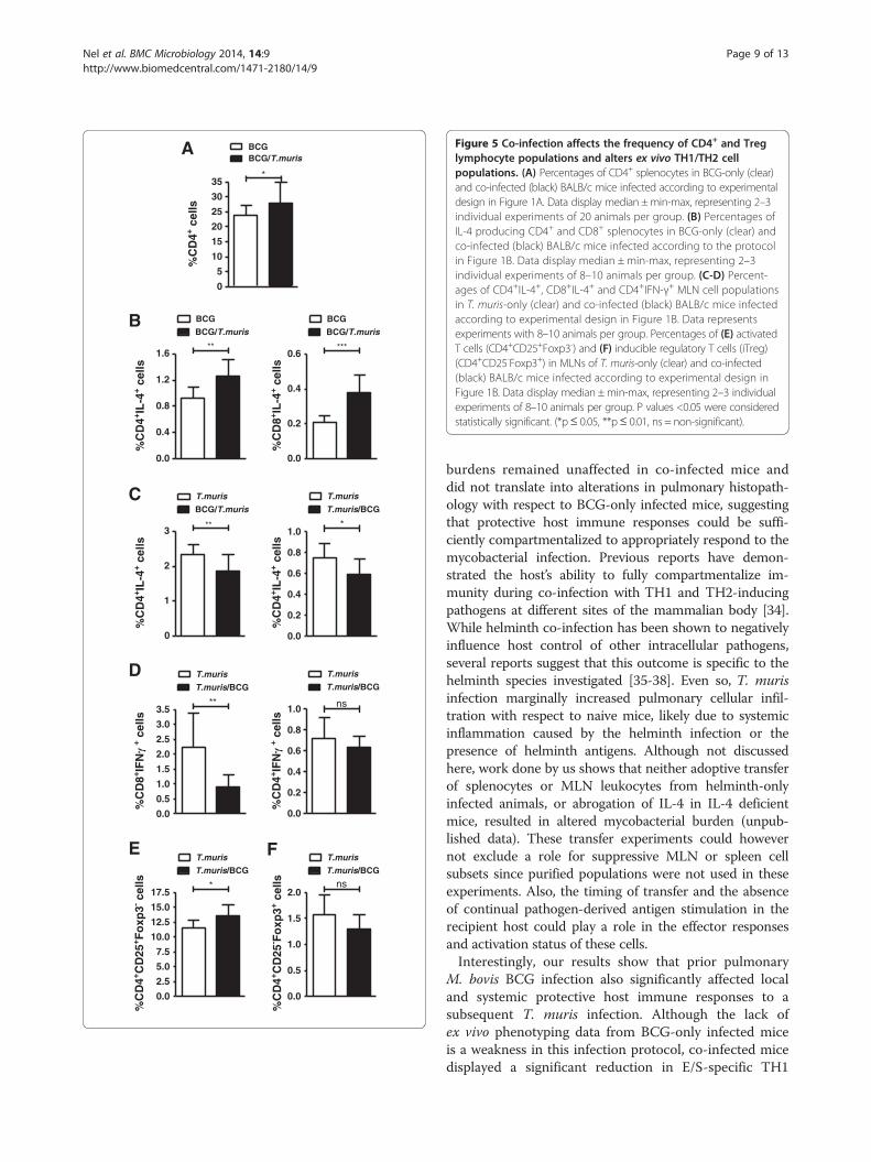
Co-infection increases CD4+ splenocyte frequencies andmodifies the TH1/TH2 immune balanceFlow cytometric analysis demonstrated that co-infectionaccording to either infection protocol (Figure 1A and B)did not impact lymphocyte composition in the spleen orMLN, since no significant differences between infectiongroups were observed for populations of CD3+ T cellsor B220+ B cells (data not shown). However, analysisof ex-vivo lymphocyte subpopulations in BALB/c miceinfected according to Figure 1A, revealed an increase inCD4+ T helper cell population in the spleens of miceco-infected according to the protocol in Figure 1A,when compared to BCG-only infected mice (Figure 5A).Although no differences in the percentages of naturalregulatory T cell (CD4+CD25+Foxp3+) populations wereobserved between infection groups in either the spleenor MLN (data not shown), co-infection significantlyincreased the percentage of IL-4-producing CD4+ andCD8+ splenocytes in comparison to M. bovis BCG-onlyinfected controls (Figure 5B). IL-4-producing CD4+ andCD8+ MLN cells from co-infected mice were howeversignificantly reduced in comparison to T. muris-only in-fected mice (Figure 5C). A marked decrease in CD8+IFNγ+
MLN cells was also observed in co-infected mice in com-parison to mice infected only with T. muris, whereasfrequencies of CD4+ IFNγ+ MLN cells were measuredat similar levels between co-infected and T.muris-onlyinfected mice (Figure 5D).When the infection order was reversed during trickle in-
fection to address the effect of introduction of co-infectionwith M. bovis BCG into an established helminth-inducedTH2 environment (Figure 1B), a significant increase in acti-vated effector T cell (CD4+CD25+Foxp3-) percentages inMLNs of co-infected animals was observed in comparisonto T. muris-only infected controls (Figure 5E). A trendtowards decreased frequencies of inducible regulatory Tcells (iTreg) (CD4+CD25-Foxp3+) was also observed in theMLNs of co-infected compared to T. muris-only infectedmice (Figure 5F). No significant differences in ex vivo cyto-kine production between infection groups were observedfor CD4+ and CD8+ lymphocytes in the spleen or MLNs(data not shown).
Co-infection reduces pathogen-specific TH1 and TH2immune responsesPathogen-specific TH1/TH2/TH17/Treg cytokine immuneresponses in the spleen were analyzed only in BALB/c miceinfected according to the protocol in Figure 1A, sinceno significant differences in ex vivo T cell cytokine pro-duction between infection groups were observed in thespleens or lungs of mice infected according to theprotocol in Figure 1B.E/S stimulated splenocytes from both co-infected and
BCG-only infected mice displayed a prominent reduction
in TH2/Treg (IL-4, IL-13 and IL-10) cytokine productionwhen compared to T. muris-only infected animals, al-though IL-4 levels were significantly increased in co-infected compared to BCG-only infected mice (Figure 6A).Similarly, E/S-specific TH1 cytokines (TNF-α and IFN-γ)were reduced in both the co-infected and BCG-onlyinfected groups with respect to T. muris-only infectedanimals (Figure 6A). No notable differences between theinfection groups were observed for helminth-specificIL-17 production (data not shown).BCG-stimulated splenocytes displayed notably low
concentrations of TH2 (IL-4 and IL-13) cytokines inall infection groups. Although no significant differencesin concentrations of the cytokines, IFN-γ and IL-17(Figure 6B) were measured between infection groups,co-infection significantly decreased production of the cy-tokines TNF-α, IL-10 and IL-4 in comparison to T. muris-only and/or BCG-only infected mice (Figure 6B).
Co-infection reduces the pulmonary cytokine geneexpression profile relative to BCG-only infected animalsTo assess whether the immunological changes observedin mice infected according to the infection protocol indi-cated in Figure 1A, also extends to alterations in pul-monary and splenic gene expression levels, the relativegene expression of co-infected mice and BCG-only in-fected mice was determined. At week 9, the relative geneexpression ratios from co-infected mice demonstratedsignificantly decreased RNA levels in the lungs for TGF-β (p = 0.034), Foxp3 (p = 0.042) and IFN-γ (p = 0.012)relative to BCG-only infected mice (Figure 7). The levelsof IL-10 (p = 0.072) also showed a trend towards de-creased expression across these two groups (Figure 7).Analysis of RNA profiles in the spleen failed to showsignificant variations in expression levels for any of thegenes measured, between co-infected and BCG-onlyinfected groups (data not shown).
DiscussionIn this study, we demonstrate the capability of the gastro-intestinal tract restricted helminth,T. muris, to induce localand systemic TH2 immune responses that affect immunityto M. bovis BCG. Of particular interest was the sig-nificant reduction in BCG-specific TNF-α and IL-10cytokine concentrations and significant increase in IL-4-producing CD4+ and CD8+ T cells in the spleens ofco-infected mice, in comparison to BCG-only infectedmice. In addition, we show that co-infection signifi-cantly reduced pulmonary IFN-γ, TGF-β and Foxp3gene expression, relative to BCG-only infected mice.Collectively, our data show a down-regulation in pul-monary TH1 and Treg-associated responses and theinduction of systemic TH2 responsiveness followingco-infection. Nevertheless, lung and systemic bacterial
A
B
C
0
5
10
15
20
25
30
35
BCG/T.murisBCG
*
%C
D4+ c
ells
0.0
0.2
0.4
0.6
0.8
1.0
T.muris
T.muris/BCG
*
%C
D4+ IL
-4+ c
ells
0.0
0.4
0.8
1.2
1.6
BCG
BCG/T.muris
**
%C
D4+ IL
-4+ c
ells
0.0
0.2
0.4
0.6
BCG
BCG/T.muris
***
%C
D8+ IL
-4+ c
ells
0
1
2
3
T.muris
BCG/T.muris
**
%C
D4+ IL
-4+ c
ells
D
0.0
0.5
1.0
1.5
2.0
2.5
3.0
3.5
T.muris
T.muris/BCG
**
0.0
0.2
0.4
0.6
0.8
1.0
T.muris
T.muris/BCG
ns
E F
0.0
2.5
5.0
7.5
10.0
12.5
15.0
17.5
T.muris
T.muris/BCG
*
%C
D4+ C
D25
+ Fo
xp3- c
ells
%C
D4+ C
D25
- Fo
xp3+ c
ells
0.0
0.5
1.0
1.5
2.0
T.muris
T.muris/BCG
ns
Figure 5 Co-infection affects the frequency of CD4+ and Treglymphocyte populations and alters ex vivo TH1/TH2 cellpopulations. (A) Percentages of CD4+ splenocytes in BCG-only (clear)and co-infected (black) BALB/c mice infected according to experimentaldesign in Figure 1A. Data display median ±min-max, representing 2–3individual experiments of 20 animals per group. (B) Percentages ofIL-4 producing CD4+ and CD8+ splenocytes in BCG-only (clear) andco-infected (black) BALB/c mice infected according to the protocolin Figure 1B. Data display median ± min-max, representing 2–3individual experiments of 8–10 animals per group. (C-D) Percent-ages of CD4+IL-4+, CD8+IL-4+ and CD4+IFN-γ+ MLN cell populationsin T. muris-only (clear) and co-infected (black) BALB/c mice infectedaccording to experimental design in Figure 1B. Data representsexperiments with 8–10 animals per group. Percentages of (E) activatedT cells (CD4+CD25+Foxp3-) and (F) inducible regulatory T cells (iTreg)(CD4+CD25-Foxp3+) in MLNs of T. muris-only (clear) and co-infected(black) BALB/c mice infected according to experimental design inFigure 1B. Data display median ±min-max, representing 2–3 individualexperiments of 8–10 animals per group. P values <0.05 were consideredstatistically significant. (*p≤ 0.05, **p≤ 0.01, ns = non-significant).
Nel et al. BMC Microbiology 2014, 14:9 Page 9 of 13http://www.biomedcentral.com/1471-2180/14/9
burdens remained unaffected in co-infected mice anddid not translate into alterations in pulmonary histopath-ology with respect to BCG-only infected mice, suggestingthat protective host immune responses could be suffi-ciently compartmentalized to appropriately respond to themycobacterial infection. Previous reports have demon-strated the host’s ability to fully compartmentalize im-munity during co-infection with TH1 and TH2-inducingpathogens at different sites of the mammalian body [34].While helminth co-infection has been shown to negativelyinfluence host control of other intracellular pathogens,several reports suggest that this outcome is specific to thehelminth species investigated [35-38]. Even so, T. murisinfection marginally increased pulmonary cellular infil-tration with respect to naive mice, likely due to systemicinflammation caused by the helminth infection or thepresence of helminth antigens. Although not discussedhere, work done by us shows that neither adoptive transferof splenocytes or MLN leukocytes from helminth-onlyinfected animals, or abrogation of IL-4 in IL-4 deficientmice, resulted in altered mycobacterial burden (unpub-lished data). These transfer experiments could howevernot exclude a role for suppressive MLN or spleen cellsubsets since purified populations were not used in theseexperiments. Also, the timing of transfer and the absenceof continual pathogen-derived antigen stimulation in therecipient host could play a role in the effector responsesand activation status of these cells.Interestingly, our results show that prior pulmonary
M. bovis BCG infection also significantly affected localand systemic protective host immune responses to asubsequent T. muris infection. Although the lack ofex vivo phenotyping data from BCG-only infected miceis a weakness in this infection protocol, co-infected micedisplayed a significant reduction in E/S-specific TH1

A
0
50
100
150
200
250
BCGT.muris
BCG/T.muris
*
*
**
*IL
-4 p
g/m
l
0
20
40
60
BCGT.muris
BCG/T.muris
**
ns
0
2000
4000
6000
8000
BCGT.muris
BCG/T.muris
**
ns
0
50
100
150
200
BCGT.muris
BCG/T.muris
**
ns
IL-1
3 p
g/m
l
0
100
200
300
400
500
600
700
BCGT.muris
BCG/T.muris
**
ns
IL-1
0 p
g/m
lB
0
50
100
150
200
250
BCGT.muris
BCG/T.muris
*
*
**
0
100
200
300
400
BCGT.muris
BCG/T.muris
*
IL-1
0 p
g/m
l
0.0
2.5
5.0
7.5
10.0
BCGT.muris
BCG/T.muris
*
IL-4
pg
/ml
0
100
200
300
400
500
600
BCGT.muris
BCG/T.murisns
ns
ns
0
100
200
300
400
BCGT.muris
BCG/T.murisns
ns ns
IL-1
7 p
g/m
l
Figure 6 Co-infection leads to altered pathogen-specific TH1 and TH2 immune responses. TH1 and TH2 cytokine concentrations weremeasured from 24 hour (A) E/S stimulated and (B) BCG-stimulated splenocyte cultures of co-infected (grey), T. muris-only (clear) and BCG-only(black) BALB/c mice infected according to the protocol illustrated in Figure 1A. Results from stimulated values were corrected for backgroundunstimulated controls. Data display median ±min-max, representing 2–3 individual experiments of 5 animals per group. P values <0.05 wereconsidered statistically significant. (*p≤ 0.05, **p ≤ 0.01, ns = non-significant).
Nel et al. BMC Microbiology 2014, 14:9 Page 10 of 13http://www.biomedcentral.com/1471-2180/14/9

Lung RNA BCG vs BCG/T.muris
IL-10 Foxp3 GATA3 Tbet IL-40
1.5 10-5
1.0 10-5
5.0 10-5
0.001
0.002
0.005
0.010
0.015 BCG/T.murisBCG
*
**
mR
NA
Exp
ress
ion
(No
rmal
ised
c to
18S
)
Figure 7 Co-infection decreases the expression ratio ofpulmonary RNA cytokine transcripts relative to those of BCG-onlyinfected BALB/c mice. BALB/c mice were co-infected (black)according to the protocol illustrated in Figure 1A with BCG-only(clear) infected mice included as controls. At week 9, total RNAwas extracted from the right upper lung lobe, cDNA producedand the relative gene expression ratio in co-infected mice relativeto that of BCG-only infected mice, determined by real-time PCR.Following HKG normalization and delta-delta Ct analysis, theexpression ratio of the genes TGF-β, IL-10, Foxp3, GATA3, T-bet,IFN-γ were calculated. Data display median ± SE, representing 8–10animals per group. P values <0.05 were considered statistically significantin comparison to BCG-only infected. (*= p < 0.05).
Nel et al. BMC Microbiology 2014, 14:9 Page 11 of 13http://www.biomedcentral.com/1471-2180/14/9
and TH2 cytokine responses in the spleen, and signifi-cantly reduced IL-4 producing CD4+ and CD8+ T cellsand IFN-γ-producing CD8+ T cells in the mesentericlymph nodes when compared to T. muris-only infectedmice. In support of a functional role for this reductionin T. muris-specific immunity, we demonstrated an associ-ated delay in helminth clearance and increased helminth-related intestinal pathology in co-infected mice, whencompared to T. muris-only infected mice. These intestinalpathological changes were characterized by increased cellturnover, suggesting increased apoptosis or cell damage,necessitating cell replacement [39]. Intestinal crypt cellapoptosis was previously reported to occur followingT. muris infection and subsequently shown to be reducedfollowing neutralization of IFN-γ and TNF-α [40]. In par-allel with this we observed an increase in intestinal mucusproduction, which likely operates as a compensatorymechanism to aide expulsion of persisting parasites. Ourresults verify reports illustrating that M. bovis co-infectionincrease helminth parasite burden and correlates withdecreased IL-4 and IL-13 cytokine production [41]. Ourfindings also agree with early reports demonstrating areduction in protective immune responses and a delay inT. muris expulsion during other co-infections with Nema-tospiroides dubius, Plasmodium berghei or Trypanosomabrucei [42-44]. It is well established that resolution ofT. muris infection is characterized by the production ofTH2 cytokines, resulting in intestinal goblet cell hyperpla-sia and increased intestinal epithelial cell turnover [45,46].On the other hand, mast cells, γδ T cells and eosinophilsare suggested as dispensable for T. muris expulsion [45,47]
and the contribution of B cells and antibody responses re-mains controversial [48-50]. Previous reports convincinglyshow that T. muris infection is delayed following depletionof CD4 T cells [51], inhibition/down-regulation of TH2cytokines [33,45] and increased TH1 polarization [52].It is therefore likely that our observation of reducedhelminth-specific TH2 responses in this co-infectionmodel could, at least in part, explain the delay in T.muris expulsion, although induction of TH1 immuneresponses to M. bovis BCG following T. muris infectionwould also influence parasite expulsion. Interestingly,altering the infection sequence to elucidate the effect of asubsequent mycobacterial infection on an establishedhelminth-induced TH2 immune response did not haveany negative influence on mycobacterial or helminth clear-ance by the host. This is most likely to be due to the rapidclearance of the helminth infection and development ofresistance to re-infection, or due to the presence of anestablished TH1 immune response for altering helminthclearance [53].These modified pathogen-specific and non-specific
immune responses following co-infection provide clearevidence that both pathogens have the ability to recip-rocally modulate immune responses towards each otherat their individual infection foci. More importantly, thedown-regulation of overall immune responsiveness inthe context of both infections suggests co-infection-induced immune suppression as a possible mechanism.Several reports confirm that chronic immune activationduring helminth infections could initiate immune sup-pression or anergy [22]. Here, we show significant in-creases in the frequency of systemic CD4+ T cells andeffector T cells in MLN of co-infected animals, suggest-ing increased immune activation following co-infection.Although the presence of immune suppressive regula-tory cell populations was investigated, no differences inthe frequencies of Treg populations could be detectedbetween infection groups in either of the BALB/c co-infection models. As Treg cells exert their suppressivefunction in a cytokine dependent manner and also interactwith other T cells and APC directly, the implications ofco-infection on regulatory immune mechanisms are notclear. Changes in IL-10, Foxp3 and TGF-β gene expres-sion reveal that the role of Tregs cannot be excluded. Ourresults could point towards a role for other immune re-gulatory cell populations, and current research efforts arefocused towards the involvement of innate nuocytes andmyeloid derived suppressor cells (MDSCs) [54,55].
ConclusionIn summary, the work presented here supports the hy-pothesis that co-infection by two unrelated and anatom-ically separated pathogens can reciprocally alter thehost’s immune response to either infection. Co-infection

Nel et al. BMC Microbiology 2014, 14:9 Page 12 of 13http://www.biomedcentral.com/1471-2180/14/9
altered host pathology and the host’s ability to expel invad-ing helminth parasites; however the magnitude of theimpact was dependent on the sequence of co-infection.These phenotypic changes were associated with alterationsin organ-restricted TH1/TH2/Treg immune balance, im-mune suppression and pathogen-specific and non-specificcytokine responses. It is likely that multiple mechanismsmay operate concurrently and further research is neededto identify the critical factors involved, although ourresults strongly support a mechanism whereby chronicimmune activation leads to hyporesponsiveness resultingin reduced pathogenic control during co-infection. Thesefindings demonstrate the complexity of immune responseregulation and systemic interaction between innate andadaptive immunity and thereby hightlights the need forgreater understanding of the role of infection history onthe evolution of host immunity.
Additional file
Additional file 1: Figure S1. Representative histological H & E stainedlung sections captured at 10x magnification illustrating the differences inhistopathology between T. muris/BCG co-infected, BCG-only infected,uninfected and T. muris - only infected BALB/c mice infected accordingto experimental design as shown in Figure 1B.
Competing interestsThe authors declare that they have no competing interests.
Authors’ contributionsStudy concept & design – GW, HJN. Acquisition of data – HJN, LK. Statisticalanalysis – HJN, NDP. Analysis and interpretation of data – GW, HJN, NDP.Drafting of the manuscript – HJN, NDP. Critical revisions to the manuscript –GW, AGL, NDP, PVH. Obtained Funding – GW, HJN. Study Supervision – GW.All authors read and approved the final manuscript.
Authors’ informationHendrik J Nel and Nelita du Plessis co-first author.
AcknowledgementsThis work was supported by the South African National Research Foundationand the South African Medical Research Council (MRC) through financialcontributions to this project. We thank N. Brown for her technical assistance.
Author details1Division of Molecular Biology and Human Genetics, MRC Centre forMolecular and Cellular Biology, NRF/DST Centre of Excellence in BiomedicalTB Research, Faculty Medicine and Health Sciences, Stellenbosch University,Tygerberg, South Africa. 2University of Queensland Diamantina Institute,Brisbane, QLD, Australia.
Received: 11 July 2013 Accepted: 10 January 2014Published: 17 January 2014
References1. Bellamy R: Genetic susceptibility to tuberculosis. Clin Chest Med 2005,
26:233–246. vi.2. Hanekom M, van Pittius NC G, McEvoy C, Victor TC, Van Helden PD, Warren
RM: Mycobacterium tuberculosis Beijing genotype: a template forsuccess. Tuberculosis 2011, 91:510–523.
3. Schluger NW, Rom WN: The host immune response to tuberculosis.Am J Respir Crit Care Med 1998, 157:679–691.
4. WHO | The world health report 1999 - making a difference. http://www.who.int/whr/1999/en/index.html.
5. Elias D, Mengistu G, Akuffo H, Britton S: Are intestinal helminths riskfactors for developing active tuberculosis? Trop Med Int Health 2006,11:551–558.
6. Hotez PJ, Molyneux DH, Fenwick A, Ottesen E, Ehrlich Sachs S, Sachs JD:Incorporating a rapid-impact package for neglected tropical diseases withprograms for HIV/AIDS, tuberculosis, and malaria. PLoS Med 2006, 3:e102.
7. Adams JF, Schölvinck EH, Gie RP, Potter PC, Beyers N, Beyers AD: Decline intotal serum IgE after treatment for tuberculosis. Lancet 1999,353:2030–2033.
8. Flynn JL, Chan J: Immunology of tuberculosis. Annu Rev Immunol 2001,19:93–129.
9. Dixon H, Blanchard C, Deschoolmeester ML, Yuill NC, Christie JW,Rothenberg ME, Else KJ: The role of Th2 cytokines, chemokines andparasite products in eosinophil recruitment to the gastrointestinalmucosa during helminth infection. Eur J Immunol 2006, 36:1753–1763.
10. Yazdanbakhsh M, van den Biggelaar A, Maizels RM: Th2 responses withoutatopy: immunoregulation in chronic helminth infections and reducedallergic disease. Trends Immunol 2001, 22:372–377.
11. Maizels RM, Balic A, Gomez-Escobar N, Nair M, Taylor MD, Allen JE: Helminthparasites–masters of regulation. Immunol Rev 2004, 201:89–116.
12. McKee AS, Pearce EJ: CD25 + CD4+ Cells contribute to Th2 polarizationduring helminth infection by suppressing Th1 response development.J Immunol 2004, 173:1224–1231.
13. Hesse M, Piccirillo CA, Belkaid Y, Prufer J, Mentink-Kane M, Leusink M,Cheever AW, Shevach EM, Wynn TA: The pathogenesis of schistosomiasis iscontrolled by cooperating IL-10-producing innate effector and regulatoryT cells. J Immunol 2004, 172:3157–3166.
14. Borkow G, Weisman Z, Leng Q, Stein M, Kalinkovich A, Wolday D, BentwichZ: Helminths, human immunodeficiency virus and tuberculosis. Scand JInfect Dis 2001, 33:568–571.
15. Bentwich Z, Kalinkovich A, Weisman Z, Borkow G, Beyers N, Beyers AD:Can eradication of helminthic infections change the face of AIDS andtuberculosis? Immunol Today 1999, 20:485–487.
16. Resende Co T, Hirsch CS, Toossi Z, Dietze R, Ribeiro-Rodrigues R: Intestinalhelminth co-infection has a negative impact on both anti-mycobacteriumtuberculosis immunity and clinical response to tuberculosis therapy. Clin ExpImmunol 2007, 147:45–52.
17. Babu S, Bhat SQ, Kumar NP, Jayantasri S, Rukmani S, Kumaran P, Gopi PG,Kolappan C, Kumaraswami V, Nutman TB: Human type 1 and 17 responsesin latent tuberculosis are modulated by coincident filarial infection throughcytotoxic T lymphocyte antigen–4 and programmed death–1. J Infect Dis2009, 200:288–298.
18. Brown M, Mawa PA, Joseph S, Bukusuba J, Watera C, Whitworth JAG, DunneDW, Elliott AM: Treatment of schistosoma mansoni infection increaseshelminth-specific type 2 cytokine responses and HIV-1 loads in coinfectedUgandan adults. J Infect Dis 2005, 191:1648–1657.
19. Elias D, Britton S, Kassu A, Akuffo H: Chronic helminth infections maynegatively influence immunity against tuberculosis and other diseasesof public health importance. Expert Rev Anti-Infect Ther 2007, 5:475–484.
20. Stewart GR, Boussinesq M, Coulson T, Elson L, Nutman T, Bradley JE:Onchocerciasis modulates the immune response to mycobacterialantigens. Clin Exp Immunol 1999, 117:517–523.
21. Elias D, Wolday D, Akuffo H, Petros B, Bronner U, Britton S: Effect ofdeworming on human T cell responses to mycobacterial antigens inhelminth‐exposed individuals before and after bacille calmette–guérin(BCG) vaccination. Clin Exp Immunol 2001, 123:219–225.
22. Borkow G, Bentwich Z: Chronic immune activation associated withchronic helminthic and human immunodeficiency virus infections: roleof hyporesponsiveness and anergy. Clin Microbiol Rev 2004,17:1012–1030.
23. Malhotra I, Mungai P, Wamachi A, Kioko J, Ouma JH, Kazura JW, King CL:Helminth- and bacillus calmette-guérin-induced immunity in childrensensitized in utero to filariasis and schistosomiasis. J Immunol 1999,162:6843–6848.
24. Potian JA, Rafi W, Bhatt K, McBride A, Gause WC, Salgame P: Preexistinghelminth infection induces inhibition of innate pulmonary anti-tuberculosisdefense by engaging the IL-4 receptor pathway. J Exp Med 2011,208:1863–1874.
25. Fulton SA, Martin TD, Redline RW, Henry Boom W: Pulmonary immuneresponses during primary mycobacterium bovis- calmette-guerin bacillusinfection in C57Bl/6 mice. Am J Respir Cell Mol Biol 2000, 22:333–343.

Nel et al. BMC Microbiology 2014, 14:9 Page 13 of 13http://www.biomedcentral.com/1471-2180/14/9
26. Klementowicz JE, Travis MA, Grencis RK: Trichuris muris: a model ofgastrointestinal parasite infection. Semin Immunopathol 2012, 34:815–828.
27. Wakelin D: Acquired immunity to trichuris muris in the albino laboratorymouse. Parasitology 1967, 57:515–524.
28. Else KJ, Wakelin D, Roach TI: Host predisposition to trichuriasis: themouse–T. muris model. Parasitology 1989, 98(Pt 2):275–282.
29. Arocho A, Chen B, Ladanyi M, Pan Q: Validation of the 2-DeltaDeltaCtcalculation as an alternate method of data analysis for quantitative PCRof BCR-ABL P210 transcripts. Diagn Mol Pathol 2006, 15:56–61.
30. Wang X, Seed B: A PCR primer bank for quantitative gene expressionanalysis. Nucleic Acids Res 2003, 31:e154.
31. Aubin E, Lemieux R, Bazin R: Absence of cytokine modulation followingtherapeutic infusion of intravenous immunoglobulin or anti-red bloodcell antibodies in a mouse model of immune thrombocytopenic purpura.Br J Haematol 2007, 136:837–843.
32. Hamelin M-È, Yim K, Kuhn KH, Cragin RP, Boukhvalova M, Blanco JCG, PrinceGA, Boivin G: Pathogenesis of human metapneumovirus lung infection inBALB/c mice and cotton rats. J Virol 2005, 79:8894–8903.
33. Bancroft AJ, Artis D, Donaldson DD, Sypek JP, Grencis RK: Gastrointestinalnematode expulsion in IL-4 knockout mice is IL-13 dependent. Eur JImmunol 2000, 30:2083–2091.
34. Lamb TJ, Graham AL, Le Goff L, Allen JE: Co-infected C57BL/6 mice mountappropriately polarized and compartmentalized cytokine responses tolitomosoides sigmodontis and leishmania major but disease progressionis altered. Parasite Immunol 2005, 27:317–324.
35. Sangaré LR, Herrin BR, Herrin BR, John-Stewart G, Walson JL: Species-specifictreatment effects of helminth/HIV-1 co-infection: a systematic review andmeta-analysis. Parasitology 2011, 138:1546–1558.
36. Erb KJ, Trujillo C, Fugate M, Moll H: Infection with the helminthnippostrongylus brasiliensis does not interfere with efficient eliminationof mycobacterium bovis BCG from the lungs of mice. Clin Diagn LabImmunol 2002, 9:727–730.
37. Frantz FG, Rosada RS, Turato WM, Peres CM, Coelho-Castelo AAM, RamosSG, Aronoff DM, Silva CL, Faccioli LH: The immune response to toxocariasisdoes not modify susceptibility to mycobacterium tuberculosis infectionin BALB/C mice. Am J Trop Med Hyg 2007, 77:691–698.
38. Elias D, Akuffo H, Thors C, Pawlowski A, Britton S: Low dose chronicSchistosoma mansoni infection increases susceptibility to mycobacteriumbovis BCG infection in mice. Clin Exp Immunol 2005, 139:398–404.
39. Artis D, Potten CS, Else KJ, Finkelman FD, Grencis RK: Trichuris muris: hostintestinal epithelial cell hyperproliferation during chronic infection isregulated by interferon-γ. Exp Parasitol 1999, 92:144–153.
40. Cliffe LJ, Potten CS, Booth CE, Grencis RK: An increase in epithelial cellapoptosis is associated with chronic intestinal nematode infection. InfectImmun 2007, 75:1556–1564.
41. Carmo AM, Vicentini MA, Dias AT, Alves LL, Alves CCS, Brandi JS, De PaulaML, Fernandes A, Barsante MM, Souza MA, Teixeira HC, Negrão-Corrêa D,Ferreira AP: Increased susceptibility to strongyloides venezuelensis in micedue to mycobacterium bovis co-infection which modulates production ofTh2 cytokines. Parasitology 2009, 136:1357–1365.
42. Jenkins SN, Behnke JM: Impairment of primary expulsion of Trichurismuris in mice concurrently infected with nematospiroides dubius.Parasitology 1977, 75:71–78.
43. Legesse M, Erko B, Balcha F: Increased parasitaemia and delayed parasiteclearance in Schistosoma mansoni and plasmodium berghei co-infectedmice. Acta Trop 2004, 91:161–166.
44. Phillips RS, Selby GR, Wakelin D: The effect of plasmodum berghei andtrypanosoma brucei infections on the immune expulsion of thenematode Trichuris muris from mice. Int J Parasitol 1974, 4:409–415.
45. Cliffe LJ, Humphreys NE, Lane TE, Potten CS, Booth C, Grencis RK:Accelerated intestinal epithelial cell turnover: a new mechanism ofparasite expulsion. Science 2005, 308:1463–1465.
46. Khan WI, Abe T, Ishikawa N, Nawa Y, Yoshimura K: Reduced amount ofintestinal mucus by treatment with anti‐CD4 antibody interferes withthe spontaneous cure of Nippostrongylus brasiliensis‐infection in mice.Parasite Immunol 1995, 17:485–491.
47. Else KJ, Hültner L, Grencis RK: Cellular immune responses to the murinenematode parasite Trichuris muris: II differential induction of TH-cellsubsets in resistant versus susceptible mice. Immunology 1992,75:232–237.
48. Else KJ, Grencis RK: Antibody-independent effector mechanisms inresistance to the intestinal nematode parasite Trichuris muris. Infect Immun1996, 64:2950–2954.
49. Wakelin D, Selby GR: Immune expulsion of Trichuris muris from resistantmice: suppression by irradiation and restoration by transfer of lymphoidcells. Parasitology 1976, 72:41–50.
50. Lee TD, Wakelin D, Grencis RK: Cellular mechanisms of immunity to thenematode Trichuris muris. Int J Parasitol 1983, 13:349–353.
51. Koyama K, Tamauchi H, Ito Y: The role of CD4+ and CD8+ T cells inprotective immunity to the murine nematode parasite Trichuris muris.Parasite Immunol 1995, 17:161–165.
52. Else KJ, Entwistle GM, Grencis RK: Correlations between worm burden andmarkers of Th1 and Th2 cell subset induction in an inbred strain ofmouse infected with Trichuris muris. Parasite Immunol 1993, 15:595–600.
53. Bancroft AJ, Else KJ, Humphreys NE, Grencis RK: The effect of challengeand trickle Trichuris muris infections on the polarisation of the immuneresponse. Int J Parasitol 2001, 31:1627–1637.
54. Nagaraj S, Collazo M, Corzo CA, Youn J-I, Ortiz M, Quiceno D, Gabrilovich DI:Regulatory myeloid suppressor cells in health and disease. Cancer Res2009, 69:7503–7506.
55. Neill DR, Wong SH, Bellosi A, Flynn RJ, Daly M, Langford TKA, Bucks C, KaneCM, Fallon PG, Pannell R, Jolin HE, McKenzie ANJ: Nuocytes represent anew innate effector leukocyte that mediates type-2 immunity. Nature2010, 464:1367–1370.
doi:10.1186/1471-2180-14-9Cite this article as: Nel et al.: Mycobacterium bovis BCG infectionseverely delays Trichuris muris expulsion and co-infection suppressesimmune responsiveness to both pathogens. BMC Microbiology 2014 14:9.
Submit your next manuscript to BioMed Centraland take full advantage of:
• Convenient online submission
• Thorough peer review
• No space constraints or color figure charges
• Immediate publication on acceptance
• Inclusion in PubMed, CAS, Scopus and Google Scholar
• Research which is freely available for redistribution
Submit your manuscript at www.biomedcentral.com/submit


![RESEARCH ARTICLE Open Access Comparative analysis of ... · Mycobacterium tuberculosis F11 (ExPEC) Y Y Causes TB; isolated from TB patient in S. Africa [5] Mycobacterium bovis BCG](https://img.dokumen.tips/doc/110x75/5fbd310e8dceeb5e767f265e/research-article-open-access-comparative-analysis-of-mycobacterium-tuberculosis.jpg)