Embed Size (px)
Citation preview
RESEARCH ARTICLE Open Access
EGFR, KRAS, BRAF, ALK, and cMET geneticalterations in 1440 Sardinian patients withlung adenocarcinomaMaria Colombino1†, Panagiotis Paliogiannis2*† , Antonio Cossu2†, Davide Adriano Santeufemia3, Sardinian LungCancer (SLC) Study Group, Maria Cristina Sini1, Milena Casula1, Grazia Palomba1, Antonella Manca1, Marina Pisano1,Valentina Doneddu2 and Giuseppe Palmieri1
Abstract
Background: Lung cancer is one of the most incident neoplastic diseases, and a leading cause of death for cancerworldwide. Knowledge of the incidence of druggable genetic alterations, their correlation with clinical andpathological features of the disease, and their interplay in cases of co-occurrence is crucial for selecting the besttherapeutic strategies of patients with non-small cell lung cancer. In this real-life study, we describe the molecularepidemiology of genetic alterations in five driver genes and their correlations with the demographic and clinicalcharacteristics of Sardinian patients with lung adenocarcinoma.
Methods: Data from 1440 consecutive Sardinian patients with a histologically proven diagnosis of lungadenocarcinoma from January 2011 through July 2016 were prospectively investigated. EGFR mutation analysis wasperformed for all of them, while KRAS and BRAF mutations were searched in 1047 cases; ALK alterations weredetermined with fluorescence in situ hybridization in 899 cases, and cMET amplifications in 788 cases.
Results: KRAS mutations were the most common genetic alterations involving 22.1% of the cases and beingmutually exclusive with the EGFR mutations, which were found in 12.6% of them. BRAF mutations, ALKrearrangements, and cMET amplifications were detected in 3.2, 5.3, and 2.1% of the cases, respectively. Concomitantmutations were detected only in a few cases.
Conclusions: Almost all the genetic alterations studied showed a similar incidence in comparison with otherCaucasian populations. Concomitant mutations were rare, and they probably have a scarce impact on the clinicalmanagement of Sardinians with lung adenocarcinoma. The low incidence of concomitant cMET amplifications atdiagnosis suggests that these alterations are acquired in subsequent phases of the disease, often during treatmentwith TKIs.
Keywords: Lung cancer, Adenocarcinoma, Targeted therapies, EGFR, KRAS, BRAF, ALK, cMET
BackgroundLung cancer is one of the most incident neoplastic dis-eases and a leading cause of death for cancer worldwide[1, 2]. Its incidence has been increasing in developingcountries and in women in the last decade, while itbegan to decline in males in most developed countries
[2]. Mortality rates remain high, despite recent advancesin the prevention, screening, surgical and medical man-agement of patients with lung cancer. Surgery is an ef-fective treatment in the early stages of the non-small celllung cancer (NSCLC) subtypes; unfortunately, approxi-mately 80% of the sufferers are at an advanced stage atthe time of diagnosis, and approximately 20% of themare affected by small cell lung cancer (SCLC), which hasno substantial benefits from surgery [3]. Chemotherapyhas been the main treatment available for advancedstage patients for years. Last-generation chemotherapy
© The Author(s). 2019 Open Access This article is distributed under the terms of the Creative Commons Attribution 4.0International License (http://creativecommons.org/licenses/by/4.0/), which permits unrestricted use, distribution, andreproduction in any medium, provided you give appropriate credit to the original author(s) and the source, provide a link tothe Creative Commons license, and indicate if changes were made. The Creative Commons Public Domain Dedication waiver(http://creativecommons.org/publicdomain/zero/1.0/) applies to the data made available in this article, unless otherwise stated.
* Correspondence: [email protected]†Maria Colombino, Panagiotis Paliogiannis and Antonio Cossu contributedequally to this work.2Department of Medical, Surgical, and Experimental Sciences, University ofSassari, Viale San Pietro 43, 07100 Sassari, ItalyFull list of author information is available at the end of the article
Colombino et al. BMC Pulmonary Medicine (2019) 19:209 https://doi.org/10.1186/s12890-019-0964-x
drugs combined with a platinum regimen showed a 5-year survival improvement of 11%, but with a mediansurvival time of only 8–10 months [4, 5]. In addition,chemotherapy drugs cannot differentiate tumour cellsand normal cells, leading to dramatically strong adversereactions that compromise the effectiveness and com-pleteness of therapies.Efforts to improve the results of the oncological treat-
ments for NSCLC, together with the technological advancesin DNA sequencing, led to the development of new thera-peutic strategies based on the knowledge and classificationof specific molecular features of the disease. Subsets of pa-tients with adenocarcinoma and activating mutations withinthe kinase domain of the epidermal growth factor receptor(EGFR) gene have been successfully treated with selectivetyrosine kinase inhibitors (TKIs), such as erlotinib, gefitinib,afatinib, and osimertinib, which are also characterized byreduced adverse events in comparison with traditionalchemotherapy [6, 7]. In addition, anaplastic lymphoma kin-ase (ALK) and ROS proto-oncogene 1 (ROS1) fusions havebeen demonstrated to be effectively druggable with targetedinhibitors such as crizotinib, alectinib, and ceritinib and arecurrently recommended for the treatment of advanced stageadenocarcinoma harbouring that kind of genetic alteration[8–10]. Furthermore, active research is ongoing for theevaluation of the clinical impact of additional draggable gen-etic alterations, such as Kirsten rat sarcoma viral oncogenehomologue (KRAS) and v-raf murine sarcoma viral onco-gene homologue B (BRAF) mutations or proto-oncogenecMET amplifications involved in the pathogenesis of lungcancer, and have proven effective in treating other malignan-cies [11, 12]. In addition, the coexistence of driver mutationsin the same tumours has been demonstrated to consistentlyimpact the therapeutic outcomes and survival rates of pa-tients undergoing chemotherapy or targeted therapies forNSCLC, as they can alter the responses to target therapies[13]. For these reasons, recent guidelines suggest that 9genes related to targeted therapies should be detected, in-cluding EGFR, KRAS, HER2, ALK, ROS1, cMET, BRAF,RET, and NTRK [14].Knowledge of the incidence of such genetic alterations,
their correlation with clinical and pathological featuresof the disease, and their interplay in cases of co-occurrence is crucial for selecting the best therapeuticstrategies of patients with NSCLC. In the present study,we describe the molecular epidemiology of EGFR, KRAS,BRAF, ALK and MET genetic alterations and their corre-lations with the demographic and clinical characteristicsof 1440 Sardinian patients with lung adenocarcinoma.
MethodsPatients and samplesA total of 1440 consecutive Sardinian patients with ahistologically proven diagnosis of adenocarcinoma of the
lung from January 2011 through July 2016 were pro-spectively enrolled and investigated. For all the patientsenrolled, medical records and pathology reports wereused to retrieve the demographic and clinical data at thetime of diagnosis; sex, age, smoking habits, type of sam-ple (primary tumour or metastasis) and the origin of thesample (biopsy or surgery) were assessed. To avoid anybias, the patients were consecutively enrolled regardlessof age at diagnosis and disease characteristics of the pri-mary tumour. Sardinian origin was ascertained throughverification of the place of birth for all patients. All pa-tients were informed about the aims of this study and,before the tissue sample was collected, provided writteninformed consent. The study was performed in accord-ance with the principles of the Declaration of Helsinkiand was approved by the Committee for the Ethics ofthe Research and Bioethics of the National ResearchCouncil (CNR).
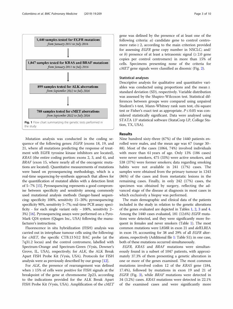
Molecular analysesFormalin-fixed, paraffin-embedded lung adenocarcinomatissue samples from each patient were obtained from theInstitutes of Pathology participating in the study. Tissuesections were estimated by light microscopy to containat least 80% of neoplastic cells. In cases with lower neo-plastic cell content, tissue sections (placed on slides)underwent tumour macro-dissection (using a single edgerazor blade and a marked haematoxylin/eosin slide as aguide) to remove unwanted tissue parts and enrich thespecimen with malignant cells. All tumour tissues wereprocessed at the Institute of Biomolecular Chemistry(CNR, Sassari, Italy), which performed molecular ana-lyses for all the Sardinian hospitals in the period of thestudy. EGFR mutation analysis was performed in allcases, as it was the first to be introduced in clinical prac-tice. KRAS and BRAF mutation analysis was started sub-sequently and was carried out globally in 1047 caseswith available biopsy tissue. The study of the genetic al-terations of ALK started in September 2012 with theintroduction of the test in clinical practice and involved899 patients. Finally, cMET amplification testing wascarried out in 778 cases with available tissue samples(Fig. 1).Genomic DNA was isolated from tissue sections using
a standard protocol, and DNA quality was assessed foreach specimen, as previously reported [6]. Briefly, paraf-fin was removed from formalin-fixed paraffin-embedded(FFPE) samples by treatment with Bio-Clear (Bio-Optica,Milan, Italy), and DNA was purified using the QIAampDNA FFPE Tissue Kit (Qiagen Inc., Valencia, CA, USA)following the manufacturer’s instructions. Yields of puri-fied DNA were assessed by the Qubit dsDNA High-Sensitivity Assay Kit on the Qubit 2.0 Fluorometer (LifeThermofisher, Waltham, MA USA).
Colombino et al. BMC Pulmonary Medicine (2019) 19:209 Page 2 of 10
Mutation analysis was conducted in the coding se-quence of the following genes: EGFR (exons 18, 19, and21, where all mutations predicting the response of treat-ment with EGFR tyrosine kinase inhibitors are located),KRAS (the entire coding portion: exons 2, 3, and 4), andBRAF (exon 15, where nearly all of the oncogenic muta-tions are located). Quantitative measurements of mutationswere based on pyrosequencing methodology, which is areal-time sequencing-by-synthesis approach that allows forthe quantification of mutated alleles with a detection limitof 5–7% [15]. Pyrosequencing represents a good comprom-ise between specificity and sensitivity among commonlyused mutational analysis methods (Sanger-based sequen-cing: specificity 100%, sensitivity 15–20%; pyrosequencing:specificity 90%, sensitivity 5–7%; real-time PCR assay: speci-ficity - for each single variant only − 100%, sensitivity 2–3%) [16]. Pyrosequencing assays were performed on a Pyro-Mark Q24 system (Qiagen Inc., USA) following the manu-facturer’s instructions.Fluorescence in situ hybridization (FISH) analysis was
carried out in interphase tumour cells using the following:for cMET, the specific CTB.13 N12 BAC probe (at the7q31.2 locus) and the control centromere, labelled withSpectrum-Orange and Spectrum-Green (Vysis, Downer’sGrove, IL, USA), respectively; for ALK, the ALK BreakApart FISH Probe Kit (Vysis, USA). Protocols for FISHanalysis were as previously described by our group [12].For ALK, the presence of rearrangement was defined
when ≥ 15% of cells were positive for FISH signals at thebreakpoint of the gene at chromosome 2p23, accordingto the indications provided for the ALK Break ApartFISH Probe Kit (Vysis, USA). Amplification of the cMET
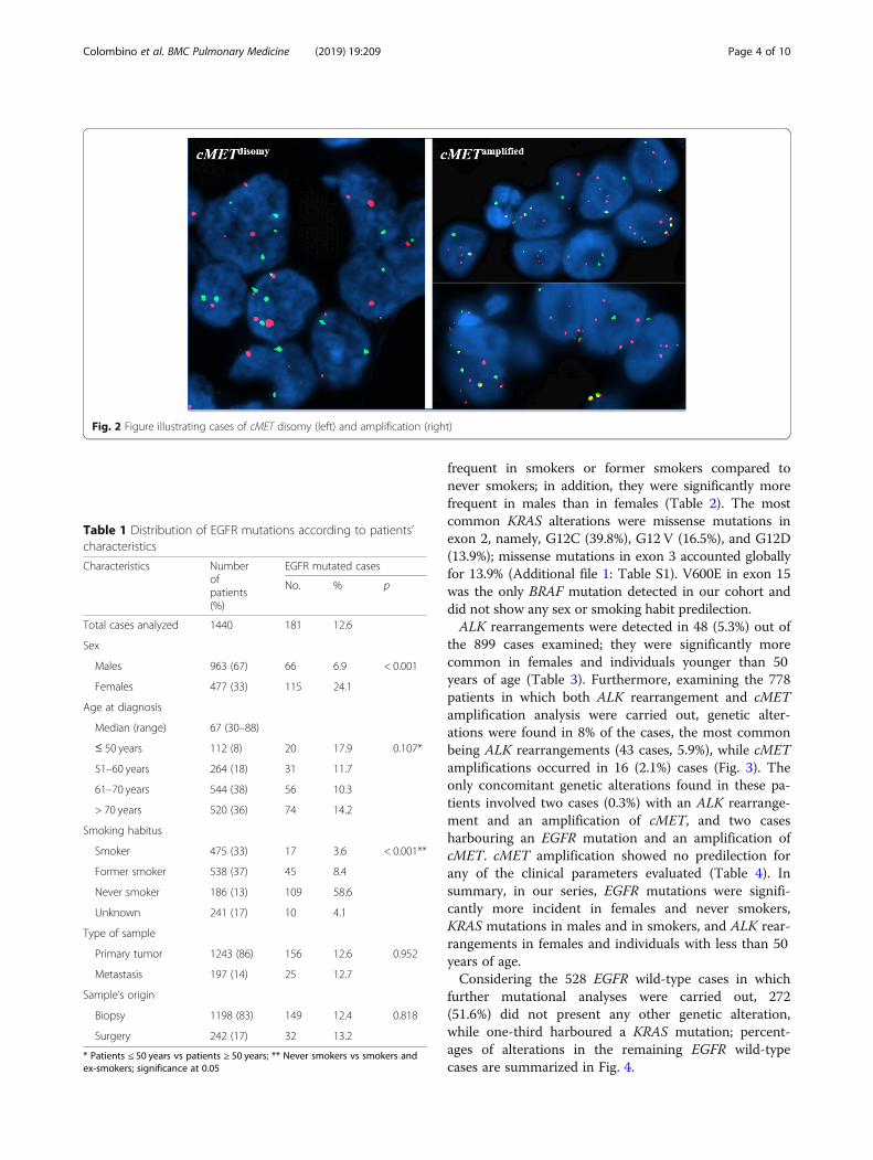
gene was defined by the presence of at least one of thefollowing criteria: a) candidate gene to control centro-mere ratio ≥ 2, according to the main criterion providedfor assessing EGFR gene copy number in NSCLC; and/or b) presence of at least a tetrasomic signal (≥ 2.0 genecopies per control centromere) in more than 15% ofcells. Specimens presenting none of the criteria forcMET gene signals were classified as disomic (Fig. 2).
Statistical analysesDescriptive analysis for qualitative and quantitative vari-ables was conducted using proportions and the mean ±standard deviation (SD), respectively. Variable distributionwas assessed by the Shapiro-Wilcoxon test. Statistical dif-ferences between groups were compared using unpairedStudent’s t-test, Mann-Whitney rank sum test, chi-squaretest or Fisher’s exact test as appropriate. P ≤ 0.05 was con-sidered statistically significant. Data were analysed usingSTATA 13® statistical software (StataCorp LP, College Sta-tion, TX, USA).
ResultsNine hundred sixty-three (67%) of the 1440 patients en-rolled were males, and the mean age was 67 (range 30–88). Most of the cases (1064, 74%) involved individualswith more than 61 years of age. Only 13% (186 cases)were never smokers, 475 (33%) were active smokers, and538 (37%) were former smokers; data regarding smokinghabits were not available in 241 (17%) cases. Thesamples were obtained from the primary tumour in 1243(86%) of the cases and from metastatic lesions in theremaining cases. Finally, in only 242 (17%) cases, thespecimen was obtained by surgery, reflecting the ad-vanced stage of the disease at diagnosis in most cases inwhich exclusively a biopsy was performed.The main demographic and clinical data of the patients
included in the study in relation to the genetic alterationsof the genes evaluated are depicted in Tables 1, 2, 3 and 4.Among the 1440 cases evaluated, 181 (12.6%) EGFR muta-tions were detected, and they were significantly more fre-quent in females and never smokers (Table 1). The mostcommon mutations were L858R in exon 21 and delELREAin exon 19, accounting for 38 and 29% of all EGFR alter-ations, respectively (Additional file 1: Table S1); in one case,both of these mutations occurred simultaneously.EGFR, KRAS and BRAF mutations were simultan-
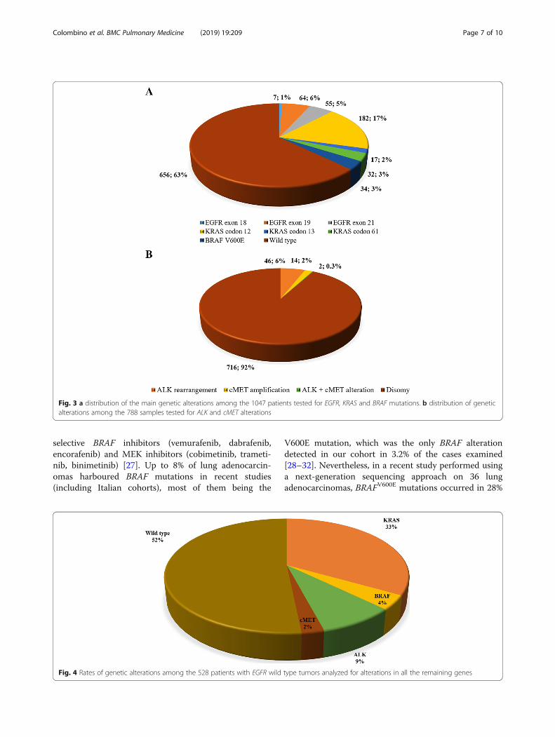
eously found in a subset of 1047 patients, with approxi-mately 37.3% of them presenting a genetic alteration inone or more of the genes examined. The most commonmutations involved codon 12 of the KRAS gene (184,17.4%), followed by mutations in exon 19 and 21 ofEGFR (Fig. 3), while BRAF mutations were detected in34 (3.2%) cases. KRAS mutations were detected in 22.1%of the examined cases and were significantly more
Fig. 1 Flow chart summarizing the genetic tests performed inthe study
Colombino et al. BMC Pulmonary Medicine (2019) 19:209 Page 3 of 10
frequent in smokers or former smokers compared tonever smokers; in addition, they were significantly morefrequent in males than in females (Table 2). The mostcommon KRAS alterations were missense mutations inexon 2, namely, G12C (39.8%), G12 V (16.5%), and G12D(13.9%); missense mutations in exon 3 accounted globallyfor 13.9% (Additional file 1: Table S1). V600E in exon 15was the only BRAF mutation detected in our cohort anddid not show any sex or smoking habit predilection.ALK rearrangements were detected in 48 (5.3%) out of
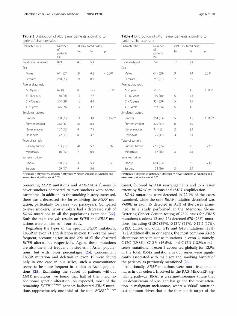
the 899 cases examined; they were significantly morecommon in females and individuals younger than 50years of age (Table 3). Furthermore, examining the 778patients in which both ALK rearrangement and cMETamplification analysis were carried out, genetic alter-ations were found in 8% of the cases, the most commonbeing ALK rearrangements (43 cases, 5.9%), while cMETamplifications occurred in 16 (2.1%) cases (Fig. 3). Theonly concomitant genetic alterations found in these pa-tients involved two cases (0.3%) with an ALK rearrange-ment and an amplification of cMET, and two casesharbouring an EGFR mutation and an amplification ofcMET. cMET amplification showed no predilection forany of the clinical parameters evaluated (Table 4). Insummary, in our series, EGFR mutations were signifi-cantly more incident in females and never smokers,KRAS mutations in males and in smokers, and ALK rear-rangements in females and individuals with less than 50years of age.Considering the 528 EGFR wild-type cases in which
further mutational analyses were carried out, 272(51.6%) did not present any other genetic alteration,while one-third harboured a KRAS mutation; percent-ages of alterations in the remaining EGFR wild-typecases are summarized in Fig. 4.
Fig. 2 Figure illustrating cases of cMET disomy (left) and amplification (right)
Table 1 Distribution of EGFR mutations according to patients’characteristics
Characteristics Numberofpatients(%)
EGFR mutated cases
No. % p
Total cases analyzed 1440 181 12.6
Sex
Males 963 (67) 66 6.9 < 0.001
Females 477 (33) 115 24.1
Age at diagnosis
Median (range) 67 (30–88)
≤ 50 years 112 (8) 20 17.9 0.107*
51–60 years 264 (18) 31 11.7
61–70 years 544 (38) 56 10.3
> 70 years 520 (36) 74 14.2
Smoking habitus
Smoker 475 (33) 17 3.6 < 0.001**
Former smoker 538 (37) 45 8.4
Never smoker 186 (13) 109 58.6
Unknown 241 (17) 10 4.1
Type of sample
Primary tumor 1243 (86) 156 12.6 0.952
Metastasis 197 (14) 25 12.7
Sample’s origin
Biopsy 1198 (83) 149 12.4 0.818
Surgery 242 (17) 32 13.2
* Patients ≤ 50 years vs patients ≥ 50 years; ** Never smokers vs smokers andex-smokers; significance at 0.05
Colombino et al. BMC Pulmonary Medicine (2019) 19:209 Page 4 of 10
DiscussionThe study of the genetic alterations in patients withNSCLC has profoundly changed the therapeutic land-scape of the disease. Considering the 1047 patients inwhom EGFR, KRAS, and BRAF mutation analysis wassimultaneously carried out in our study, approximately37% were found to have a genetic alteration in one ofthe examined genes. This percentage is slightly lowerthan those of previous studies, which reported approxi-mately half of the patients with lung adenocarcinomaharbouring an EGFR or KRAS mutation [17]. This maydepend on the genetic characteristics of the target popu-lation in our study, characterized by high levels of gen-etic homogeneity due to geographical reasons. In anycase, the concept that mutations in driver genes occur ina consistent percentage of lung adenocarcinomas re-mains, but its impact seems to be different in differentpopulations; indeed, the incidence of EGFR mutations issignificantly higher in Asian populations (even higherthan 50%) compared to western countries [18].In our series, EGFR mutations were searched in 1440
patients and were found in 12.6% of them, which is con-sistent with the partial results published in a previous re-port involving patients from the same population [6].
This figure is slightly lower than that described in recentprospective studies performed in other Caucasian popu-lations [19, 20]. In addition, EGFR mutations signifi-cantly more frequent in females (24.1%) and neversmokers (58.6%), a finding that has been extensively re-ported in previous studies and from different geograph-ical areas [6, 19, 21]. The incidence of EGFR mutationshas been reported as low as 28% in American neversmokers and as high as 68% in Asian never smokers[22]; the rate found in our series is closer to those re-ported in Asian populations. As mere speculation, it isinteresting that Sardinians, who have long been recog-nized as forming a distinct outlier within contemporaryEuropean genetic diversity, experienced an immigrationof individuals belonging to the initial wave of migrationfrom the Asian areas (mainly the Middle East) intosoutheastern Europe during the early Neolithic transi-tion, leading to the observed genetic affinity of the an-cients descending from these migrants to present-daySardinians [23, 24].A recent meta-analysis evaluated the EGFR, ALK-
EML4 and KRAS mutational patterns in smokers andnon-smokers of various ethnicities [20]. The authorsconfirmed that there was a significantly increased risk of
Table 2 Distribution of KRAS and BRAF mutations according to patients’ characteristics
Characteristics Numberofpatients(%)
KRAS mutated cases BRAF mutated cases
No. % p No. % p
Total cases analyzed 1047 231 22.1 34 3.2
Sex
Males 693 (66) 169 24.4 0.018 25 3.6 0.462
Females 354 (34) 61 17.2 9 2.5
Age at diagnosis
≤ 50 years 94 (9) 24 25.5 0.472* 3 3.2 1.000*
51–60 years 189 (18) 46 24.3 7 3.7
61–70 years 394 (38) 78 19.9 10 2.5
> 70 years 370 (35) 83 22.4 14 3.6
Smoking habitus
Smoker 336 (32) 74 22.0 0.001** 14 4.2 0.411**
Former smoker 367 (35) 89 24.3 10 2.7
Never smoker 139 (13) 14 10.1 2 1.4
Unknown 205 (20) 54 26.3 8 3.9
Type of sample
Primary tumor 889 (85) 197 22.2 0.940 29 3.3 1.000
Metastasis 158 (15) 34 21.5 5 3.2
Sample’s origin
Biopsy 848 (81) 193 22.8 0.304 28 3.3 0.987
Surgery 199 (19) 38 19.1 6 3.0
* Patients ≤ 50 years vs patients ≥ 50 years; ** Never smokers vs smokers and ex-smokers; significance at 0.05
Colombino et al. BMC Pulmonary Medicine (2019) 19:209 Page 5 of 10
presenting EGFR mutations and ALK-EML4 fusions innever smokers compared to ever smokers with adeno-carcinoma. In addition, as the smoking history increased,there was a decreased risk for exhibiting the EGFR mu-tation, particularly for cases > 30 pack-years. Comparedto ever smokers, never smokers had a decreased risk ofKRAS mutations in all the populations examined [22].Both the meta-analysis results on EGFR and KRAS mu-tations were confirmed in our study.Regarding the types of the specific EGFR mutations,
L858R in exon 21 and deletion in exon 19 were the mostfrequent, accounting for 38 and 29% of all the observedEGFR alterations, respectively. Again, these mutationsare also the most frequent in studies in Asian popula-tions, but with lower percentages [25]. ConcomitantL858R mutation and deletion in exon 19 were foundonly in one case in our series; such a concomitanceseems to be more frequent in studies in Asian popula-tions [25]. Examining the subset of patients withoutEGFR mutations, we found that half of them had noadditional genetic alteration. As expected, most of theremaining EGFRwild-type patients harboured KRAS muta-tions (approximately one-third of the total EGFRwild-type
cases), followed by ALK rearrangements and to a lesserextent by BRAF mutations and cMET amplification.KRAS mutations were detected in 22.1% of the cases
examined, while the only BRAF mutation described wasV600E in exon 15 detected in 3.2% of the cases exam-ined. In a study performed at the Memorial Sloan-Kettering Cancer Center, testing of 2529 cases for KRASmutations (codons 12 and 13) detected 670 (26%) muta-tions, including G12C (39%), G12 V (21%), G12D (17%),G12A (11%), and other G12 and G13 mutations (12%)[17]. Additionally, in our series, the most common KRASalterations were missense mutations in exon 2, namely,G12C (39.8%), G12 V (16.5%), and G12D (13.9%); mis-sense mutations in exon 3 accounted globally for 13.9%of the total. KRAS mutations in our series were signifi-cantly associated with male sex and smoking history ofthe patients, as previously mentioned [26].Additionally, BRAF mutations were more frequent in
males in our cohort. Involved in the RAS-MEK-ERK sig-nalling pathway, BRAF is a serine/threonine kinase thatlies downstream of RAS and has gained the most atten-tion in malignant melanomas, where a V600E mutationis a common driver that is the therapeutic target of the
Table 3 Distribution of ALK rearrangements according topatients’ characteristics
Characteristics Numberofpatients(%)
ALK mutated cases
No. % p
Total cases analyzed 899 48 5.3
Sex
Males 641 (67) 27 4.2 < 0.001
Females 258 (33) 21 8.1
Age at diagnosis
≤ 50 years 62 (8) 8 12.9 0.014*
51–60 years 168 (18) 13 7.7
61–70 years 344 (38) 15 4.4
> 70 years 325 (36) 12 3.7
Smoking habitus
Smoker 288 (33) 11 3.8 0.459**
Former smoker 332 (37) 21 6.3
Never smoker 107 (13) 8 7.5
Unknown 172 (17) 8 4.7
Type of sample
Primary tumor 783 (87) 41 5.2 0.892
Metastasis 116 (13) 7 6.0
Sample’s origin
Biopsy 730 (83) 39 5.3 0.850
Surgery 169 (17) 9 5.6
* Patients ≤ 50 years vs patients ≥ 50 years; ** Never smokers vs smokers andex-smokers; significance at 0.05
Table 4 Distribution of cMET rearrangements according topatients’ characteristics
Characteristics Numberofpatients(%)
cMET mutated cases
No. % p
Total analyzed 778 16 2.1
Sex
Males 641 (69) 9 1.4 0.231
Females 242 (31) 7 2.9
Age at diagnosis
≤ 50 years 55 (7) 1 1.8 1.000*
51–60 years 139 (18) 5 2.6
61–70 years 301 (39) 5 1.7
> 70 years 283 (36) 5 1.8
Smoking habitus
Smoker 264 (33) 5 1.9 1.000**
Former smoker 295 (37) 6 2.0
Never smoker 94 (13) 2 2.1
Unknown 125 (17) 3 2.4
Type of sample
Primary tumor 661 (85) 13 2.0 0.720
Metastasis 117 (15) 3 2.6
Sample’s origin
Biopsy 654 (84) 13 2.0 0.730
Surgery 124 (16) 3 2.4
* Patients ≤ 50 years vs patients ≥ 50 years; ** Never smokers vs smokers andex-smokers; significance at 0.05
Colombino et al. BMC Pulmonary Medicine (2019) 19:209 Page 6 of 10
selective BRAF inhibitors (vemurafenib, dabrafenib,encorafenib) and MEK inhibitors (cobimetinib, trameti-nib, binimetinib) [27]. Up to 8% of lung adenocarcin-omas harboured BRAF mutations in recent studies(including Italian cohorts), most of them being the
V600E mutation, which was the only BRAF alterationdetected in our cohort in 3.2% of the cases examined[28–32]. Nevertheless, in a recent study performed usinga next-generation sequencing approach on 36 lungadenocarcinomas, BRAFV600E mutations occurred in 28%
Fig. 3 a distribution of the main genetic alterations among the 1047 patients tested for EGFR, KRAS and BRAF mutations. b distribution of geneticalterations among the 788 samples tested for ALK and cMET alterations
Fig. 4 Rates of genetic alterations among the 528 patients with EGFR wild type tumors analyzed for alterations in all the remaining genes
Colombino et al. BMC Pulmonary Medicine (2019) 19:209 Page 7 of 10
of the cases, mostly in smokers (90%), and in concomi-tance with AKT or PIK3CA mutations, non-V600E muta-tions occurred in 72% of the cases and in concomitancewith KRAS mutations in four cases [33]. These findingssuggest that the epidemiological landscape of BRAF andother genetic alterations in NSCLC will be further clearedas new technologies for genetic testing become availablefor routine diagnostic purposes.The ALK rearrangements are druggable targets in
NSCLC patients with specific inhibitors. Considering the778 patients examined for both ALK rearrangementsand cMET amplifications, we found that 8% of them har-boured ALK or cMET genetic alterations. The rates ofALK rearrangements (5.3%) and cMET amplifications(2.1%) found in our cohort were similar to those re-ported in the scientific literature [34, 35]. ALK transloca-tions are common in young patients with non-smokinghistory and with no apparent ethnic differences [36]; inour study, they were more frequent in young females,without any association with smoking status. cMET geneamplification causes 1st generation EGFR-TKI resistanceby activating EGFR-independent phosphorylation ofERBB3 and downstream activation of the PI3K/AKTpathway, providing a bypass mechanism. This redundantactivation of ERBB3 permits cells to transmit the samedownstream signalling in the presence of EGFR-TKIs.This mechanism involves 5–22% of resistant adenocar-cinomas and is not related to that dependent on theEGFRT790M mutation on exon 20 (not searched in thisstudy), which represents approximately 60% of resistancecases [37, 38]. Considering that the incidence of cMETamplifications in our cohort was 2.1%, most of themseem to occur in subsequent phases of the disease andduring treatment with TKIs. This dictates the need for adouble inhibition of both EGFR and cMET to overcomethe development of drug resistance.cMET was amplified in all four cases in which two
concomitant driver genetic alterations were found. Twoof them harboured an EGFR mutation and a cMET amp-lification, while the remaining two cases presented anALK rearrangement with a simultaneous cMET amplifi-cation. Indication for a starting therapy combining in-hibitors of both altered pathways may be necessary inthose cases. No coexistence of EGFR, KRAS, or BRAFmutations was detected in our cohort, confirming thewidely described mutually exclusive mutational pattern.The concomitant EGFR-KRAS mutations are describedmainly in case reports; in a large cohort Chinese studyon 5125 patients, 153 cases harbouring concomitant ab-errations were found, and among them, 30 carried con-comitant EGFR-KRAS mutations [39]. Nevertheless,recent large cohort studies report a higher grade of theconcomitance of ALK mutations in NSCLC patients, es-pecially those harbouring EGFR mutations [40–43]. ALK
mutations are reported to occur in concomitance withEGFR mutations in 0–6% of cases [40–43]; in our co-hort, no such cases were found. Lee et al. analysed theclinical features of six patients harbouring EGFR-KRASmutations and six patients with EGFR-ALK mutations,evidencing different morphological features of the tu-mours and behaviour to treatments [44]. Most EGFR-KRAS mutation patients showed papillary and acinarhistologic patterns with hobnail cells, while all EGFR-ALK mutation patients showed solid or cribriform pat-terns, and three had signet ring cells. Responses to treat-ment in patients with genetic co-alterations wererecently evaluated in a large cohort Chinese study in-cluding 3774 cases [45]. The authors reported 63 (1.7%)samples with more than one driver gene mutation;among these, 43 were co-alterations with an EGFR mu-tation, and 20 had an ALK rearrangement. In this study,1st-line EGFR-TKI treatment did not significantly im-prove the progression-free survival (PFS) of patientsharbouring concomitant EGFR mutations compared topatients harbouring a single EGFR mutation. However,for concomitant EGFR mutation patients, TKI therapywas more effective than chemotherapy (median PFS of10.8 vs 5.2 months, P = 0.023) [43]. In any case, the inter-action of concomitant genetic alterations in terms of syn-ergism versus the possible dominance of one rather thanthe other oncogene and the subsequent impact on tar-geted therapies are currently not completely clarified.Our study has some limitations, mainly the non-
homogeneous distribution of the genetic analysesperformed; this simply depended on the gradual intro-duction of such analyses in clinical practice and theavailability of sample tissues for testing. Furthermore,analyses did not include the T790 M mutation on exon18 or the histological subtypes of the tumours exam-ined. Nevertheless, the consistent number of the glo-bal cases analysed taken from real-life clinical practice,the genetic homogeneity of the population examined,and the quality of the methods employed for the testsrepresent the strengths of our work.
ConclusionsOur data showed that KRAS mutations are the mostcommon genetic alterations in Sardinian patients withlung adenocarcinoma, involving 22.1% of the cases ex-amined and being mutually exclusive with the EGFRmutations, which were found in 12.6% of the casesstudied. BRAF mutations, ALK rearrangements, andcMET amplifications were detected in 3.2, 5.3, and 2.1%of them, respectively; these figures are relatively low incomparison with most studies in other Caucasian popu-lations. Concomitant mutations were detected only in afew cases, suggesting that they rarely may represent afactor of drug resistance in Sardinians with lung
Colombino et al. BMC Pulmonary Medicine (2019) 19:209 Page 8 of 10
adenocarcinoma, as opposed to other populations inwhich such concomitance is more common. The lowincidence of concomitant cMET amplifications at diag-nosis suggests that these alterations are acquired insubsequent phases of the disease, often during treat-ment with TKIs.
Supplementary informationSupplementary information accompanies this paper at https://doi.org/10.1186/s12890-019-0964-x.
Additional file 1: Table S1. Sequence variations in candidate genes.
AbbreviationsALK: Anaplastic lymphoma kinase; BRAF: v-raf murine sarcoma viraloncogene homolog B; EGFR: Epidermal growth factor receptor;FFPE: Formalin-fixed paraffin-embedded; FISH: Fluorescence in situhybridization; KRAS: Kirsten rat sarcoma viral oncogene homolog;NSCLC: Non-small cell lung cancer; PFS: Progression-free survival; ROS1: ROSproto-oncogene 1; SCLC: Small cell lung cancer; SD: Standard deviation;TKIs: Tyrosine kinase inhibitors
AcknowledgmentsThe Sardinian Lung Cancer (SLC) Study Group includes the followingmembers who participated as investigators in this study and should beconsidered as co-authors: Antonio Pazzola, Giovanni Maria Fadda, PietroPirina, Alessandro Fois, Carlo Putzu, Giorgio Ginesu, Alberto Porcu (AziendaOspedaliera Universitaria, Sassari, Italy); Giorgio Astara, Mario Scartozzi(Azienda Ospedaliera Universitaria, Cagliari, Italy); Anna Maria Carta, EfisioDefraia, Daniela Guerzoni, Giuseppe Porcu (Azienda Ospedaliera Brotzu,Cagliari, Italy); Gianfranco Bardino, Claudio Sini (Ospedale Civile, Olbia, Italy);Francesca Capelli, Maria Giuseppina Sarobba (Ospedale Zonchello, Nuoro,Italy).
Authors’ contributionsMC, PP, AC and GP made substantial contributions in the conception anddesign of the study, as well as in data analysis and interpretation. MC, PP,DAS and GP contributed in drafting the manuscript. All the members of SLCStudy Group substantially contributed in clinical data collection andinterpretation, and performed critical revisions of the manuscript. MC, MCS,MiC, GrP, AM, MP, VD contributed in performing and interpreting molecularanalyses. AC and VD interpreted pathological data. PP and GP performed thefinal revision of the manuscript. All authors read and approved the finalmanuscript.
FundingThe mutational analyses performed were partially funded by the SardinianRegional Government (Regione Autonoma della Sardegna). The fundingsource had no role in study design, data collection, decision to publish, orpreparation of the manuscript.
Availability of data and materialsThe datasets used and/or analysed during the current study are availablefrom the corresponding author on reasonable request.
Ethics approval and consent to participateAll study participants provided written consent. The study was approved bythe Committee for the Ethics of the Research and Bioethics of the NationalResearch Council (CNR).
Consent for publicationAll the patients provided written informed consent for the anonymous useof their clinical data for the purposes of the study.
Competing interestsGiuseppe Palmieri has/had an advisory role for Bristol Myers Squibb, Incyte,Merck Sharp & Dohme, Novartis, Pierre Fabre, and Roche-Genetech. All theremaining authors declare no conflict of interest.
Author details1Unit of Cancer Genetics, Institute Biomolecular Chemistry, CNR, Traversa LaCrucca 3, 07100 Sassari, Italy. 2Department of Medical, Surgical, andExperimental Sciences, University of Sassari, Viale San Pietro 43, 07100 Sassari,Italy. 3Medical Oncology Unit, Civil Hospital, Via Don Minzoni, 07041 Alghero,Italy.
Received: 21 May 2019 Accepted: 18 October 2019
References1. Siegel RL, Miller KD, Jemal A. Cancer statistics, 2018. CA Cancer J Clin. 2018;
68:7–30.2. Paliogiannis P, Attene F, Cossu A, Budroni M, Cesaraccio R, Tanda F, et al.
Lung cancer epidemiology in North Sardinia, Italy. Multidiscip Respir Med.2013;8:45.
3. Rivera MP, Mehta AC, Wahidi MM. Establishing the diagnosis of lung cancer:diagnosis and management of lung cancer, 3rd ed: American College ofChest Physicians evidence-based clinical practice guidelines. Chest. 2013;143(Suppl 5):142–65.
4. Nagasaka M, Gadgeel SM. Role of chemotherapy and targeted therapyin early-stage non-small cell lung cancer. Expert Rev Anticancer Ther.2018;18:63–70.
5. Li HD, Liu SL. Molecular targeted therapy for non-small cell lung cancer: thereality in China and coping strategy. Prac J Med Pharm. 2018;35:373–9.
6. Paliogiannis P, Attene F, Cossu A, Defraia E, Porcu G, Carta A, et al. Impact oftissue type and content of neoplastic cells of samples on the quality ofepidermal growth factor receptor mutation analysis among patients withlung adenocarcinoma. Mol Med Rep. 2015;12:187–91.
7. Dong J, Li B, Lin D, Zhou Q, Huang D. Advances in targeted therapy andimmunotherapy for non-small cell lung cancer based on accurate moleculartyping. Front Pharmacol. 2019;10:230.
8. Shaw AT, Kim DW, Nakagawa K, Seto T, Crinó L, Ahn MJ, et al. Crizotinibversus chemotherapy in advanced ALK-positive lung cancer. N Engl J Med.2013;368:2385–94.
9. Bergethon K, Shaw AT, Ou SH, Katayama R, Lovly CM, McDonald NT, et al.ROS1 rearrangements define a unique molecular class of lung cancers. JClin Oncol. 2012;30:863–70.
10. Lindeman NI, Cagle PT, Aisner DL, Arcila ME, Beasley MB, Bernicker EH, et al.Updated molecular testing guideline for the selection of lung cancerpatients for treatment with targeted tyrosine kinase inhibitors: guidelinefrom the College of American Pathologists, the International Association forthe Study of Lung Cancer, and the Association for Molecular Pathology. JMol Diagn. 2018;20:129–59.
11. Palomba G, Doneddu V, Cossu A, Paliogiannis P, Manca A, Casula M, et al.Prognostic impact of KRAS, NRAS, BRAF, and PIK3CA mutations in primarycolorectal carcinomas: a population-based study. J Transl Med. 2016;14:292.
12. Sini MC, Doneddu V, Paliogiannis P, Casula M, Colombino M, Manca A, et al.Genetic alterations in main candidate genes during melanoma progression.Oncotarget. 2018;9:8531–41.
13. Zito Marino F, Ronchi A, Accardo M, Franco R. Concomitant ALK/KRAS andALK/EGFR mutations in non-small cell lung cancer: different profile ofresponse to target therapies. Transl Cancer Res. 2017;6(Suppl 3):457–60.
14. NCCN. Clinical practice guidelines in oncology. Non-small cell lungcancer. https://www.nccn.org/professionals/physician_gls/default.aspx.Accessed 10 May 2019.
15. Ihle MA, Fassunke J, König K, Grünewald I, Schlaak M, Kreuzberg N, et al.Comparison of high resolution melting analysis, pyrosequencing, nextgeneration sequencing and immunohistochemistry to conventional Sangersequencing for the detection of p.V600E and non-p.V600E BRAF mutations.BMC Cancer. 2014;14:13.
16. Gao J, Wu H, Shi X, Huo Z, Zhang J, Liang Z. Comparison of next-generation sequencing, quantitative PCR, and Sanger sequencing formutation profiling of EGFR, KRAS, PIK3CA and BRAF in clinical lungtumors. Clin Lab. 2016;62:689–96.
17. Dogan S, Shen R, Ang DC, Johnson ML, D'Angelo SP, Paik PK, et al.Molecular epidemiology of EGFR and KRAS mutations in 3,026 lungadenocarcinomas: higher susceptibility of women to smoking-related KRAS-mutant cancers. Clin Cancer Res. 2012;18:6169–77.
18. Ulivi P, Chiadini E, Dazzi C, Dubini A, Costantini M, Medri L, et al.Nonsquamous, non-small-cell lung cancer patients who carry a double
Colombino et al. BMC Pulmonary Medicine (2019) 19:209 Page 9 of 10
mutation of EGFR, EML4-ALK or KRAS: frequency, clinical-pathologicalcharacteristics, and response to therapy. Clin Lung Cancer. 2016;17:384–90.
19. Rosell R, Moran T, Queralt C, Porta R, Cardenal F, Camps C, et al. Screeningfor epidermal growth factor receptor mutations in lung cancer. N Engl JMed. 2009;361:958–67.
20. Nana-Sinkam SP, Powell CA. Molecular biology of lung cancer: diagnosisand management of lung cancer, 3rd ed: American College of ChestPhysicians evidence-based clinical practice guidelines. Chest. 2013;143(Suppl 5):30–9.
21. Pao W, Miller V, Zakowski M, Doherty J, Politi K, Sarkaria I, et al. EGF receptorgene mutations are common in lung cancers from “never smokers” and areassociated with sensitivity of tumors to gefitinib and erlotinib. Proc NatlAcad Sci U S A. 2004;101:13306–11.
22. Chapman AM, Sun KY, Ruestow P, Cowan DM, Madl AK. Lung cancermutation profile of EGFR, ALK, and KRAS: meta-analysis and comparison ofnever and ever smokers. Lung Cancer. 2016;102:122–34.
23. Sikora M, Carpenter ML, Moreno-Estrada A, Henn BM, Underhill PA, Sánchez-Quinto F, et al. Population genomic analysis of ancient and moderngenomes yields new insights into the genetic ancestry of the TyroleanIceman and the genetic structure of Europe. PLoS Genet. 2014;10:1004353.
24. Chiang CWK, Marcus JH, Sidore C, Biddanda A, Al-Asadi H,Zoledziewska M, et al. Genomic history of the Sardinian population.Nat Genet. 2018;50:1426–34.
25. Shi Y, Au JS, Thongprasert S, Srinivasan S, Tsai CM, Khoa MT, et al. Aprospective, molecular epidemiology study of EGFR mutations in Asianpatients. J Thorac Oncol. 2014;9:154–62.
26. Kosaka T, Yatabe Y, Endoh H, Kuwano H, Takahashi T, Mitsudomi T.Mutations of the epidermal growth factor receptor gene in lung cancer:biological and clinical implications. Cancer Res. 2004;64:8919–23.
27. Palmieri G, Ombra M, Colombino M, Casula M, Sini M, Manca A, et al.Multiple molecular pathways in melanomagenesis: characterization oftherapeutic targets. Front Oncol. 2015;5:183.
28. Litvak AM, Paik PK, Woo KM, Sima CS, Hellmann MD, Arcila ME, et al. Clinicalcharacteristics and course of 63 patients with BRAF mutant lung cancers. JThorac Oncol. 2014;9:1669–74.
29. Kinno T, Tsuta K, Shiraishi K, Mizukami T, Suzuki M, Yoshida A, et al.Clinicopathological features of nonsmall cell lung carcinomas with BRAFmutations. Ann Oncol. 2014;25:138–42.
30. Cardarella S, Ogino A, Nishino M, Butaney M, Shen J, Lydon C, et al.Clinical, pathologic, and biologic features associated with BRAFmutations in non-small cell lung cancer. Clin Cancer Res Off J AmAssoc Cancer Res. 2013;19:4532–40.
31. Paik PK, Arcila ME, Fara M, Sima CS, Miller VA, Kris MG, et al. Clinicalcharacteristics of patients with lung adenocarcinomas harboring BRAFmutations. J Clin Oncol Off J Am Soc Clin Oncol. 2011;29:2046–51.
32. Marchetti A, Felicioni L, Malatesta S, Grazia Sciarrotta M, Guetti L, Chella A,et al. Clinical features and outcome of patients with non-small-cell lungcancer harboring BRAF mutations. J Clin Oncol Off J Am Soc Clin Oncol.2011;29:3574–9.
33. Salimian KJ, Fazeli R, Zheng G, Ettinger D, Maleki Z. V600E BRAF versus non-V600E BRAF mutated lung adenocarcinomas: cytomorphology, histology,coexistence of other driver mutations and patient characteristics. Acta Cytol.2018;62:79–84.
34. Williams AS, Greer W, Bethune D, Craddock KJ, Flowerdew G, Xu Z. ALK+lung adenocarcinoma in never smokers and long-term ex-smokers:prevalence and detection by immunohistochemistry and fluorescence insitu hybridization. Virchows Arch. 2016;469:533–40.
35. Salgia R. MET in lung cancer: biomarker selection based on scientificrationale. Mol Cancer Ther. 2017;16:555–65.
36. Sasaki T, Rodig SJ, Chirieac LR, Jänne PA. The biology and treatment ofEML4-ALK non-small cell lung cancer. Eur J Cancer. 2010;46:1773–80.
37. Shi P, Oh YT, Zhang G, Yao W, Yue P, Li Y, et al. Met geneamplification and protein hyperactivation is a mechanism of resistanceto both first and third generation EGFR inhibitors in lung cancertreatment. Cancer Lett. 2016;380:494–504.
38. Suda K, Murakami I, Katayama T, Tomizawa K, Osada H, Sekido Y, et al.Reciprocal and complementary role of MET amplification and EGFR T790Mmutation in acquired resistance to kinase inhibitors in lung cancer. ClinCancer Res. 2010;16:5489–98.
39. Li S, Li L, Zhu Y, Huang C, Qin Y, Liu H, et al. Coexistence of EGFR withKRAS, or BRAF, or PIK3CA somatic mutations in lung cancer: a
comprehensive mutation profiling from 5125 Chinese cohorts. Br J Cancer.2014;110:2812–20.
40. Hu W, Liu Y, Chen J. Concurrent gene alterations with EGFR mutation andtreatment efficacy of EGFR-TKIs in Chinese patients with non-small cell lungcancer. Oncotarget. 2017;8:25046–54.
41. Yang JJ, Zhang XC, Su J, Xu CR, Zhou Q, Tian HX, et al. Lung cancers withconcomitant EGFR mutations and ALK rearrangements: diverse responses toEGFR-TKI and crizotinib in relation to diverse receptors phosphorylation. ClinCancer Res. 2014;20:1383–92.
42. Schmid S, Gautschi O, Rothschild S, Mark M, Froesch P, Klingbiel D, et al.Clinical outcome of ALK-positive non-small cell lung cancer (NSCLC)patients with de novo EGFR or KRAS co-mutations receiving tyrosine kinaseinhibitors (TKIs). J Thorac Oncol. 2017;12:681–8.
43. Zito Marino F, Liguori G, Aquino G, La Mantia E, Bosari S, Ferrero S,et al. Intratumor heterogeneity of ALK-rearrangements andhomogeneity of EGFR-mutations in mixed lung adenocarcinoma. PLoSOne. 2015;10:0139264.
44. Lee T, Lee B, Choi YL, Han J, Ahn MJ, Um SW. Non-small cell lung cancerwith concomitant EGFR, KRAS, and ALK mutation: clinicopathologic featuresof 12 cases. J Pathol Transl Med. 2016;50:197–203.
45. Zhuang X, Zhao C, Li J, Su C, Chen X, Ren S, et al. Clinical features andtherapeutic options in non-small cell lung cancer patients with concomitantmutations of EGFR, ALK, ROS1, KRAS or BRAF. Cancer Med. 2019. https://doi.org/10.1002/cam4.2183.
Publisher’s NoteSpringer Nature remains neutral with regard to jurisdictional claims inpublished maps and institutional affiliations.
Colombino et al. BMC Pulmonary Medicine (2019) 19:209 Page 10 of 10