Embed Size (px)
Citation preview
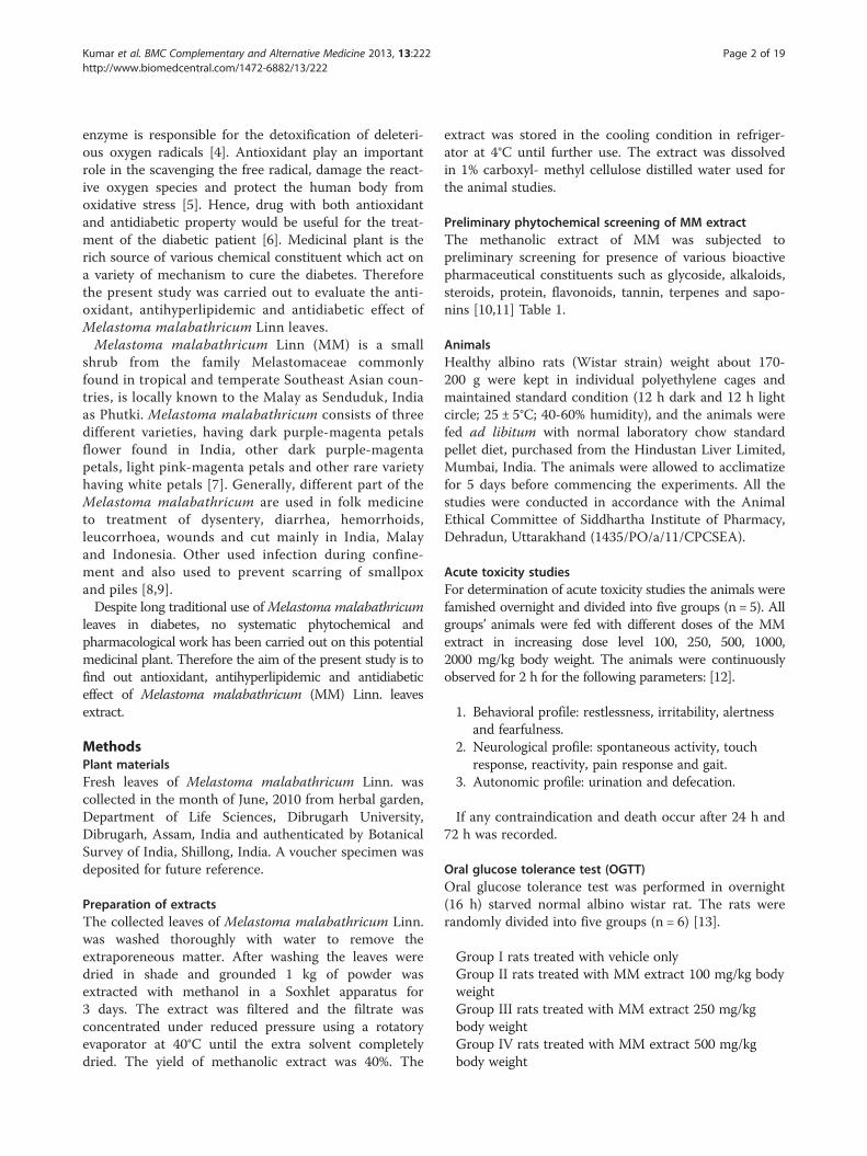
Kumar et al. BMC Complementary and Alternative Medicine 2013, 13:222http://www.biomedcentral.com/1472-6882/13/222
RESEARCH ARTICLE Open Access
Anti-diabetic, anti-oxidant and anti-hyperlipidemicactivities of Melastoma malabathricum Linn. leavesin streptozotocin induced diabetic ratsVikas Kumar1*, Danish Ahmed1, Pushpraj S Gupta1, Firoz Anwar2 and Mohd Mujeeb3
Abstract
Background: Melastoma malabathricum (MM) Linn leaves traditionally use in the treatment of diabetic conditions.The aim of the present investigation was to evaluate the antioxidant, antihyperlipidemic and antidiabetic activity ofmethanolic extract taken from Melastoma malabathricum Linn (Melastomaceae).
Methods: The methanolic leaves extract of MM Linn leaves used for the study. Chemical test of different extract,acute toxicity study and oral glucose test was performed. Diabetes was induced in rat by single intra-peritonealinjection of streptozotocin (55 mg/kg). The rats were divided into following groups: Group I – normal control,Group II (Vehicle) – diabetic control, Group III (STZ-toxic) – MM I (100 mg/kg, p.o.), Group IV – MM II (250 mg/kg, p.o.), Group V – MM III (500 mg/kg, p.o.), Group VI – glibenclamide (10 mg/kg, p.o.). Bodyweight of each rat in thedifferent groups was recorded daily. Biochemical and antioxidant enzyme parameters were determined on day 28.Histology of different organ (heart, liver, kidney, and pancreas) was performed after sacrificing the rats witheuthanasia.
Results: The methanolic extract of MM did not show any acute toxicity up-to the dose of 2000 mg/kg and shownbetter glucose utilization in oral glucose tolerance test. Orally treatment of different doses of MM leaves extractdecreased the level of serum glucose, glycated hemoglobin, glucose-6-phosphatase, fructose-1-6-biphosphate andincreased the level of plasma insulin, hexokinase. MM treatment decreased liver malondialdehyde but increased thelevel of superoxide dismutase, catalase and glutathione peroxidase. In oral glucose tolerance test observedincreased utilization of glucose. Streptozotocin induced diabetes groups rat treated with different doses of MMleaves extract and glibenclamide significantly increased the body weight. Histopathology analysis on different organof STZ (streptozotocin) induced diabetic rat show there regenerative effect on the liver, kidney, heart and pancreas.
Conclusion: The antioxidant, antihyperlipidemic and antidiabetic effect of methanolic extract from Melastomamalabathricum Linn suggests a potential therapeutic treatment to antidiabetic conditions.
Keywords: Melastoma malabathricum, Streptozotocin, Antidiabetic, Antihyperlipidemic, Glibenclamide
BackgroundDiabetes mellitus (DM) is a chronic complication of de-rangement of protein carbohydrate and lipid metabolismcharacterized by increased blood glucose level resultingfrom the defects in insulin secretion insulin action orboth [1]. DM is the worldwide problem to leading microvascular and macrovascular complications [2]. DM is a
* Correspondence: [email protected] of Pharmaceutical Sciences, Faculty of Health Sciences, SamHigginbottom Institute of Agriculture, Technology & Sciences(SHIATS)-Deemed University, Allahabad, Uttar Pradesh 211007, IndiaFull list of author information is available at the end of the article
© 2013 Kumar et al.; licensee BioMed CentralCommons Attribution License (http://creativecreproduction in any medium, provided the orwaiver (http://creativecommons.org/publicdomstated.
chronic complication that affected an estimated 135 mil-lion people in 1995, 285 million people worldwide in2010 and data reached approx 500 million people in2025 mainly increasing in rural and poor populationthroughout the world [3].In hyperglycemic condition continuous generation of
reactive oxygen species (ROS) occurred. Reactive oxygenspecies increased the oxidative stress mainly due to overproduction of oxygen free radicals, as oxidative stressplay an important role in development of diabetes. Oxi-dative stress effect the endogenous antioxidant, which
Ltd. This is an Open Access article distributed under the terms of the Creativeommons.org/licenses/by/2.0), which permits unrestricted use, distribution, andiginal work is properly cited. The Creative Commons Public Domain Dedicationain/zero/1.0/) applies to the data made available in this article, unless otherwise
Kumar et al. BMC Complementary and Alternative Medicine 2013, 13:222 Page 2 of 19http://www.biomedcentral.com/1472-6882/13/222
enzyme is responsible for the detoxification of deleteri-ous oxygen radicals [4]. Antioxidant play an importantrole in the scavenging the free radical, damage the react-ive oxygen species and protect the human body fromoxidative stress [5]. Hence, drug with both antioxidantand antidiabetic property would be useful for the treat-ment of the diabetic patient [6]. Medicinal plant is therich source of various chemical constituent which act ona variety of mechanism to cure the diabetes. Thereforethe present study was carried out to evaluate the anti-oxidant, antihyperlipidemic and antidiabetic effect ofMelastoma malabathricum Linn leaves.Melastoma malabathricum Linn (MM) is a small
shrub from the family Melastomaceae commonlyfound in tropical and temperate Southeast Asian coun-tries, is locally known to the Malay as Senduduk, Indiaas Phutki. Melastoma malabathricum consists of threedifferent varieties, having dark purple-magenta petalsflower found in India, other dark purple-magentapetals, light pink-magenta petals and other rare varietyhaving white petals [7]. Generally, different part of theMelastoma malabathricum are used in folk medicineto treatment of dysentery, diarrhea, hemorrhoids,leucorrhoea, wounds and cut mainly in India, Malayand Indonesia. Other used infection during confine-ment and also used to prevent scarring of smallpoxand piles [8,9].Despite long traditional use ofMelastoma malabathricum
leaves in diabetes, no systematic phytochemical andpharmacological work has been carried out on this potentialmedicinal plant. Therefore the aim of the present study is tofind out antioxidant, antihyperlipidemic and antidiabeticeffect of Melastoma malabathricum (MM) Linn. leavesextract.
MethodsPlant materialsFresh leaves of Melastoma malabathricum Linn. wascollected in the month of June, 2010 from herbal garden,Department of Life Sciences, Dibrugarh University,Dibrugarh, Assam, India and authenticated by BotanicalSurvey of India, Shillong, India. A voucher specimen wasdeposited for future reference.
Preparation of extractsThe collected leaves of Melastoma malabathricum Linn.was washed thoroughly with water to remove theextraporeneous matter. After washing the leaves weredried in shade and grounded 1 kg of powder wasextracted with methanol in a Soxhlet apparatus for3 days. The extract was filtered and the filtrate wasconcentrated under reduced pressure using a rotatoryevaporator at 40°C until the extra solvent completelydried. The yield of methanolic extract was 40%. The
extract was stored in the cooling condition in refriger-ator at 4°C until further use. The extract was dissolvedin 1% carboxyl- methyl cellulose distilled water used forthe animal studies.
Preliminary phytochemical screening of MM extractThe methanolic extract of MM was subjected topreliminary screening for presence of various bioactivepharmaceutical constituents such as glycoside, alkaloids,steroids, protein, flavonoids, tannin, terpenes and sapo-nins [10,11] Table 1.
AnimalsHealthy albino rats (Wistar strain) weight about 170-200 g were kept in individual polyethylene cages andmaintained standard condition (12 h dark and 12 h lightcircle; 25 ± 5°C; 40-60% humidity), and the animals werefed ad libitum with normal laboratory chow standardpellet diet, purchased from the Hindustan Liver Limited,Mumbai, India. The animals were allowed to acclimatizefor 5 days before commencing the experiments. All thestudies were conducted in accordance with the AnimalEthical Committee of Siddhartha Institute of Pharmacy,Dehradun, Uttarakhand (1435/PO/a/11/CPCSEA).
Acute toxicity studiesFor determination of acute toxicity studies the animals werefamished overnight and divided into five groups (n = 5). Allgroups’ animals were fed with different doses of the MMextract in increasing dose level 100, 250, 500, 1000,2000 mg/kg body weight. The animals were continuouslyobserved for 2 h for the following parameters: [12].
1. Behavioral profile: restlessness, irritability, alertnessand fearfulness.
2. Neurological profile: spontaneous activity, touchresponse, reactivity, pain response and gait.
3. Autonomic profile: urination and defecation.
If any contraindication and death occur after 24 h and72 h was recorded.
Oral glucose tolerance test (OGTT)Oral glucose tolerance test was performed in overnight(16 h) starved normal albino wistar rat. The rats wererandomly divided into five groups (n = 6) [13].
Group I rats treated with vehicle onlyGroup II rats treated with MM extract 100 mg/kg bodyweightGroup III rats treated with MM extract 250 mg/kgbody weightGroup IV rats treated with MM extract 500 mg/kgbody weight
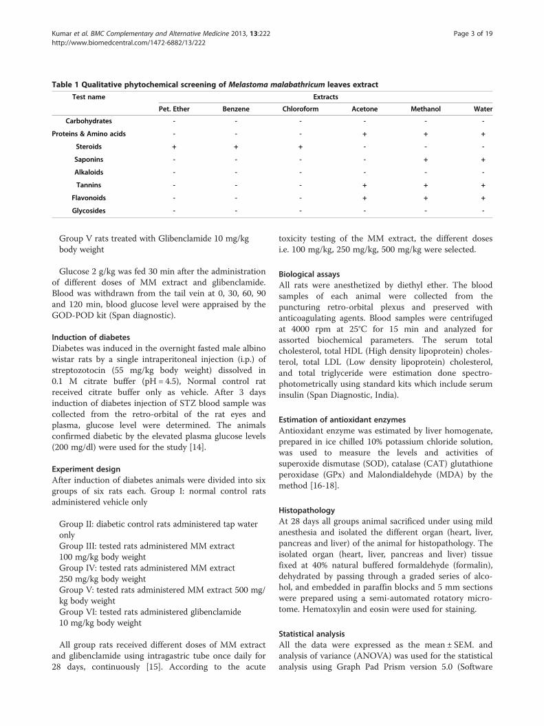
Table 1 Qualitative phytochemical screening of Melastoma malabathricum leaves extract
Test name Extracts
Pet. Ether Benzene Chloroform Acetone Methanol Water
Carbohydrates - - - - - -
Proteins & Amino acids - - - + + +
Steroids + + + - - -
Saponins - - - - + +
Alkaloids - - - - - -
Tannins - - - + + +
Flavonoids - - - + + +
Glycosides - - - - - -
Kumar et al. BMC Complementary and Alternative Medicine 2013, 13:222 Page 3 of 19http://www.biomedcentral.com/1472-6882/13/222
Group V rats treated with Glibenclamide 10 mg/kgbody weight
Glucose 2 g/kg was fed 30 min after the administrationof different doses of MM extract and glibenclamide.Blood was withdrawn from the tail vein at 0, 30, 60, 90and 120 min, blood glucose level were appraised by theGOD-POD kit (Span diagnostic).
Induction of diabetesDiabetes was induced in the overnight fasted male albinowistar rats by a single intraperitoneal injection (i.p.) ofstreptozotocin (55 mg/kg body weight) dissolved in0.1 M citrate buffer (pH = 4.5), Normal control ratreceived citrate buffer only as vehicle. After 3 daysinduction of diabetes injection of STZ blood sample wascollected from the retro-orbital of the rat eyes andplasma, glucose level were determined. The animalsconfirmed diabetic by the elevated plasma glucose levels(200 mg/dl) were used for the study [14].
Experiment designAfter induction of diabetes animals were divided into sixgroups of six rats each. Group I: normal control ratsadministered vehicle only
Group II: diabetic control rats administered tap wateronlyGroup III: tested rats administered MM extract100 mg/kg body weightGroup IV: tested rats administered MM extract250 mg/kg body weightGroup V: tested rats administered MM extract 500 mg/kg body weightGroup VI: tested rats administered glibenclamide10 mg/kg body weight
All group rats received different doses of MM extractand glibenclamide using intragastric tube once daily for28 days, continuously [15]. According to the acute
toxicity testing of the MM extract, the different dosesi.e. 100 mg/kg, 250 mg/kg, 500 mg/kg were selected.
Biological assaysAll rats were anesthetized by diethyl ether. The bloodsamples of each animal were collected from thepuncturing retro-orbital plexus and preserved withanticoagulating agents. Blood samples were centrifugedat 4000 rpm at 25°C for 15 min and analyzed forassorted biochemical parameters. The serum totalcholesterol, total HDL (High density lipoprotein) choles-terol, total LDL (Low density lipoprotein) cholesterol,and total triglyceride were estimation done spectro-photometrically using standard kits which include seruminsulin (Span Diagnostic, India).
Estimation of antioxidant enzymesAntioxidant enzyme was estimated by liver homogenate,prepared in ice chilled 10% potassium chloride solution,was used to measure the levels and activities ofsuperoxide dismutase (SOD), catalase (CAT) glutathioneperoxidase (GPx) and Malondialdehyde (MDA) by themethod [16-18].
HistopathologyAt 28 days all groups animal sacrificed under using mildanesthesia and isolated the different organ (heart, liver,pancreas and liver) of the animal for histopathology. Theisolated organ (heart, liver, pancreas and liver) tissuefixed at 40% natural buffered formaldehyde (formalin),dehydrated by passing through a graded series of alco-hol, and embedded in paraffin blocks and 5 mm sectionswere prepared using a semi-automated rotatory micro-tome. Hematoxylin and eosin were used for staining.
Statistical analysisAll the data were expressed as the mean ± SEM. andanalysis of variance (ANOVA) was used for the statisticalanalysis using Graph Pad Prism version 5.0 (Software
Kumar et al. BMC Complementary and Alternative Medicine 2013, 13:222 Page 4 of 19http://www.biomedcentral.com/1472-6882/13/222
Name is mentioned). The values were considered to besignificant when the P value was p < 0.05.
ResultsPreliminary phytochemical screeningPreliminary phytochemical screening of the methanolicextract of Melastoma malabathricum showed terpenoids,flavonoids, phenolic compound, tannins, Saponins, andtriterpenes. But the content of flavonoids and phenoliccompound were found to be more prominent in the extract(Table 1).
Acute toxicity studyAn acute toxicity study of the Melastoma malabathricumLinn leaves extract were publicized the non-toxic natureof the drug. The different doses of the Melastomamalabathricum Linn leaves extract were not showing anytoxic reaction or lethality at any of the doses selected untilthe end of the study period. Acute toxicity of themethanolic extract of Melastoma malabathricum revealedthe non-toxic nature of the different doses. There were nolethality or toxic reactions found in the selected groupwhich received the different doses of the extract until theend of the experimental period.
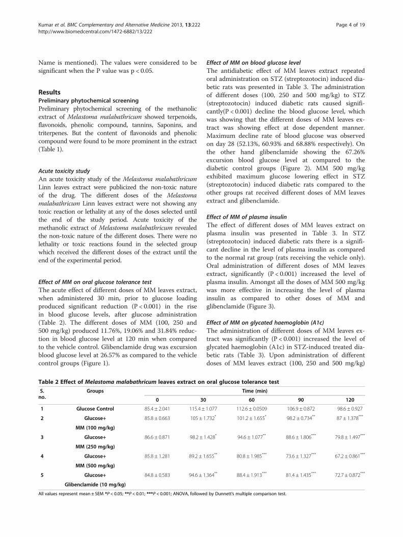
Effect of MM on oral glucose tolerance testThe acute effect of different doses of MM leaves extract,when administered 30 min, prior to glucose loadingproduced significant reduction (P < 0.001) in the risein blood glucose levels, after glucose administration(Table 2). The different doses of MM (100, 250 and500 mg/kg) produced 11.76%, 19.06% and 31.84% reduc-tion in blood glucose level at 120 min when comparedto the vehicle control. Glibenclamide drug was excursionblood glucose level at 26.57% as compared to the vehiclecontrol groups (Figure 1).
Table 2 Effect of Melastoma malabathricum leaves extract on
S.no.
Groups
0 30
1 Glucose Control 85.4 ± 2.041 115.4 ±
2 Glucose+ 85.8 ± 0.663 105 ± 1
MM (100 mg/kg)
3 Glucose+ 86.6 ± 0.871 98.2 ± 1
MM (250 mg/kg)
4 Glucose+ 85.8 ± 1.281 89.2 ± 1
MM (500 mg/kg)
5 Glucose+ 84.8 ± 0.583 94.6 ± 1
Glibenclamide (10 mg/kg)
All values represent mean ± SEM *P < 0.05; **P < 0.01; ***P < 0.001; ANOVA, followe
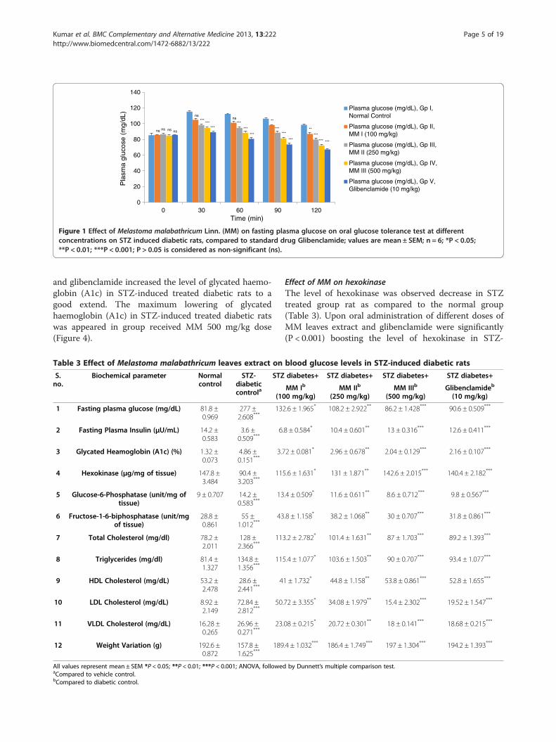
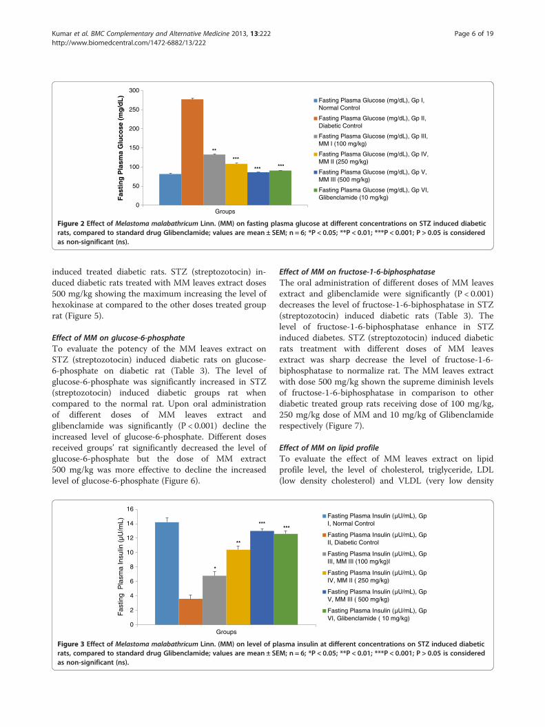
Effect of MM on blood glucose levelThe antidiabetic effect of MM leaves extract repeatedoral administration on STZ (streptozotocin) induced dia-betic rats was presented in Table 3. The administrationof different doses (100, 250 and 500 mg/kg) to STZ(streptozotocin) induced diabetic rats caused signifi-cantly(P < 0.001) decline the blood glucose level, whichwas showing that the different doses of MM leaves ex-tract was showing effect at dose dependent manner.Maximum decline rate of blood glucose was observedon day 28 (52.13%, 60.93% and 68.88% respectively). Onthe other hand glibenclamide showing the 67.26%excursion blood glucose level at compared to thediabetic control groups (Figure 2). MM 500 mg/kgexhibited maximum glucose lowering effect in STZ(streptozotocin) induced diabetic rats compared to theother groups rat received different doses of MM leavesextract and glibenclamide.
Effect of MM of plasma insulinThe effect of different doses of MM leaves extract onplasma insulin was presented in Table 3. In STZ(streptozotocin) induced diabetic rats there is a signifi-cant decline in the level of plasma insulin as comparedto the normal rat group (rats receiving the vehicle only).Oral administration of different doses of MM leavesextract, significantly (P < 0.001) increased the level ofplasma insulin. Amongst all the doses of MM 500 mg/kgwas more effective in increasing the level of plasmainsulin as compared to other doses of MM andglibenclamide (Figure 3).
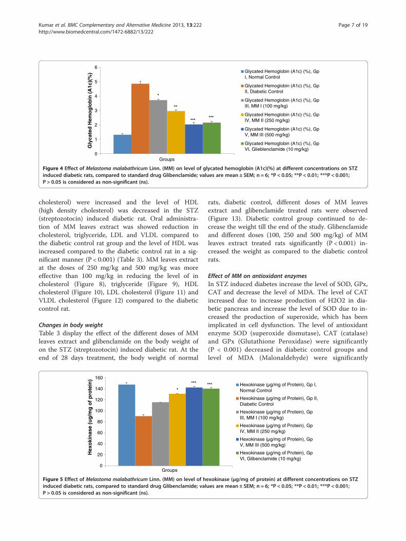
Effect of MM on glycated haemoglobin (A1c)The administration of different doses of MM leaves ex-tract was significantly (P < 0.001) increased the level ofglycated haemoglobin (A1c) in STZ-induced treated dia-betic rats (Table 3). Upon administration of differentdoses of MM leaves extract (100, 250 and 500 mg/kg)
oral glucose tolerance test
Time (min)
60 90 120
1.077 112.6 ± 0.0509 106.9 ± 0.872 98.6 ± 0.927
.732* 101.2 ± 1.655* 98.2 ± 0.734** 87 ± 1.378***
.428* 94.6 ± 1.077** 88.6 ± 1.806*** 79.8 ± 1.497***
.655** 80.8 ± 1.985*** 73.6 ± 1.327*** 67.2 ± 0.861***
.364** 88.4 ± 1.913*** 81.4 ± 1.435*** 72.7 ± 0.872***
d by Dunnett’s multiple comparison test.
0
20
40
60
80
100
120
140
0 30 60 90 120
Plasma glucose (mg/dL), Gp I,Normal Control
Plasma glucose (mg/dL), Gp II,MM I (100 mg/kg)
Plasma glucose (mg/dL), Gp III,MM II (250 mg/kg)
Plasma glucose (mg/dL), Gp IV,MM III (500 mg/kg)
Plasma glucose (mg/dL), Gp V,Glibenclamide (10 mg/kg)
Pla
sma
gluc
ose
(mg/
dL)
Time (min)
***
***
******
******
***
******
******
***
ns
ns ns ns
ns
ns **
**
Figure 1 Effect of Melastoma malabathricum Linn. (MM) on fasting plasma glucose on oral glucose tolerance test at differentconcentrations on STZ induced diabetic rats, compared to standard drug Glibenclamide; values are mean ± SEM; n = 6; *P < 0.05;**P < 0.01; ***P < 0.001; P > 0.05 is considered as non-significant (ns).
Kumar et al. BMC Complementary and Alternative Medicine 2013, 13:222 Page 5 of 19http://www.biomedcentral.com/1472-6882/13/222
and glibenclamide increased the level of glycated haemo-globin (A1c) in STZ-induced treated diabetic rats to agood extend. The maximum lowering of glycatedhaemoglobin (A1c) in STZ-induced treated diabetic ratswas appeared in group received MM 500 mg/kg dose(Figure 4).
Table 3 Effect of Melastoma malabathricum leaves extract on
S.no.
Biochemical parameter Normalcontrol
STZ-diabeticcontrola
STZ
(10
1 Fasting plasma glucose (mg/dL) 81.8 ±0.969
277 ±2.608***
13
2 Fasting Plasma Insulin (μU/mL) 14.2 ±0.583
3.6 ±0.509***
6
3 Glycated Heamoglobin (A1c) (%) 1.32 ±0.073
4.86 ±0.151***
3.7
4 Hexokinase (μg/mg of tissue) 147.8 ±3.484
90.4 ±3.203***
11
5 Glucose-6-Phosphatase (unit/mg oftissue)
9 ± 0.707 14.2 ±0.583***
13
6 Fructose-1-6-biphosphatase (unit/mgof tissue)
28.8 ±0.861
55 ±1.012***
43
7 Total Cholesterol (mg/dl) 78.2 ±2.011
128 ±2.366***
11
8 Triglycerides (mg/dl) 81.4 ±1.327
134.8 ±1.356***
11
9 HDL Cholesterol (mg/dL) 53.2 ±2.478
28.6 ±2.441***
4
10 LDL Cholesterol (mg/dL) 8.92 ±2.149
72.84 ±2.812***
50
11 VLDL Cholesterol (mg/dL) 16.28 ±0.265
26.96 ±0.271***
23
12 Weight Variation (g) 192.6 ±0.872
157.8 ±1.625***
189
All values represent mean ± SEM *P < 0.05; **P < 0.01; ***P < 0.001; ANOVA, followeaCompared to vehicle control.bCompared to diabetic control.
Effect of MM on hexokinaseThe level of hexokinase was observed decrease in STZtreated group rat as compared to the normal group(Table 3). Upon oral administration of different doses ofMM leaves extract and glibenclamide were significantly(P < 0.001) boosting the level of hexokinase in STZ-
blood glucose levels in STZ-induced diabetic rats
diabetes+ STZ diabetes+ STZ diabetes+ STZ diabetes+
MM Ib
0 mg/kg)MM IIb
(250 mg/kg)MM IIIb
(500 mg/kg)Glibenclamideb
(10 mg/kg)
2.6 ± 1.965* 108.2 ± 2.922** 86.2 ± 1.428*** 90.6 ± 0.509***
.8 ± 0.584* 10.4 ± 0.601** 13 ± 0.316*** 12.6 ± 0.411***
2 ± 0.081* 2.96 ± 0.678** 2.04 ± 0.129*** 2.16 ± 0.107***
5.6 ± 1.631* 131 ± 1.871** 142.6 ± 2.015*** 140.4 ± 2.182***
.4 ± 0.509* 11.6 ± 0.611** 8.6 ± 0.712*** 9.8 ± 0.567***
.8 ± 1.158* 38.2 ± 1.068** 30 ± 0.707*** 31.8 ± 0.861***
3.2 ± 2.782* 101.4 ± 1.631** 87 ± 1.703*** 89.2 ± 1.393***
5.4 ± 1.077* 103.6 ± 1.503** 90 ± 0.707*** 93.4 ± 1.077***
1 ± 1.732* 44.8 ± 1.158** 53.8 ± 0.861*** 52.8 ± 1.655***
.72 ± 3.355* 34.08 ± 1.979** 15.4 ± 2.302*** 19.52 ± 1.547***
.08 ± 0.215* 20.72 ± 0.301** 18 ± 0.141*** 18.68 ± 0.215***
.4 ± 1.032*** 186.4 ± 1.749*** 197 ± 1.304*** 194.2 ± 1.393***
d by Dunnett’s multiple comparison test.
0
50
100
150
200
250
300
Groups
Fasting Plasma Glucose (mg/dL), Gp I,Normal Control
Fasting Plasma Glucose (mg/dL), Gp II,Diabetic Control
Fasting Plasma Glucose (mg/dL), Gp III,MM I (100 mg/kg)
Fasting Plasma Glucose (mg/dL), Gp IV,MM II (250 mg/kg)
Fasting Plasma Glucose (mg/dL), Gp V,MM III (500 mg/kg)
Fasting Plasma Glucose (mg/dL), Gp VI,Glibenclamide (10 mg/kg)F
asti
ng
Pla
sma
Glu
cose
(m
g/d
L)
*** ***
***
**
Figure 2 Effect of Melastoma malabathricum Linn. (MM) on fasting plasma glucose at different concentrations on STZ induced diabeticrats, compared to standard drug Glibenclamide; values are mean ± SEM; n = 6; *P < 0.05; **P < 0.01; ***P < 0.001; P > 0.05 is consideredas non-significant (ns).
Kumar et al. BMC Complementary and Alternative Medicine 2013, 13:222 Page 6 of 19http://www.biomedcentral.com/1472-6882/13/222
induced treated diabetic rats. STZ (streptozotocin) in-duced diabetic rats treated with MM leaves extract doses500 mg/kg showing the maximum increasing the level ofhexokinase at compared to the other doses treated grouprat (Figure 5).
Effect of MM on glucose-6-phosphateTo evaluate the potency of the MM leaves extract onSTZ (streptozotocin) induced diabetic rats on glucose-6-phosphate on diabetic rat (Table 3). The level ofglucose-6-phosphate was significantly increased in STZ(streptozotocin) induced diabetic groups rat whencompared to the normal rat. Upon oral administrationof different doses of MM leaves extract andglibenclamide was significantly (P < 0.001) decline theincreased level of glucose-6-phosphate. Different dosesreceived groups’ rat significantly decreased the level ofglucose-6-phosphate but the dose of MM extract500 mg/kg was more effective to decline the increasedlevel of glucose-6-phosphate (Figure 6).
0
2
4
6
8
10
12
14
16
Groups
Fas
ting
Pla
sma
Insu
lin (
µU/m
L)
*
**
***
Figure 3 Effect of Melastoma malabathricum Linn. (MM) on level of plrats, compared to standard drug Glibenclamide; values are mean ± SEas non-significant (ns).
Effect of MM on fructose-1-6-biphosphataseThe oral administration of different doses of MM leavesextract and glibenclamide were significantly (P < 0.001)decreases the level of fructose-1-6-biphosphatase in STZ(streptozotocin) induced diabetic rats (Table 3). Thelevel of fructose-1-6-biphosphatase enhance in STZinduced diabetes. STZ (streptozotocin) induced diabeticrats treatment with different doses of MM leavesextract was sharp decrease the level of fructose-1-6-biphosphatase to normalize rat. The MM leaves extractwith dose 500 mg/kg shown the supreme diminish levelsof fructose-1-6-biphosphatase in comparison to otherdiabetic treated group rats receiving dose of 100 mg/kg,250 mg/kg dose of MM and 10 mg/kg of Glibenclamiderespectively (Figure 7).
Effect of MM on lipid profileTo evaluate the effect of MM leaves extract on lipidprofile level, the level of cholesterol, triglyceride, LDL(low density cholesterol) and VLDL (very low density
Fasting Plasma Insulin (µU/mL), Gp I, Normal Control
Fasting Plasma Insulin (µU/mL), Gp II, Diabetic Control
Fasting Plasma Insulin (µU/mL), Gp III, MM III (100 mg/kg)l
Fasting Plasma Insulin (µU/mL), Gp IV, MM II ( 250 mg/kg)
Fasting Plasma Insulin (µU/mL), Gp V, MM III ( 500 mg/kg)
Fasting Plasma Insulin (µU/mL), Gp VI, Glibenclamide ( 10 mg/kg)
***
asma insulin at different concentrations on STZ induced diabeticM; n = 6; *P < 0.05; **P < 0.01; ***P < 0.001; P > 0.05 is considered
0
1
2
3
4
5
6
Groups
Glycated Hemoglobin (A1c) (%), GpI, Normal Control
Glycated Hemoglobin (A1c) (%), GpII, Diabetic Control
Glycated Hemoglobin (A1c) (%), GpIII, MM I (100 mg/kg)
Glycated Hemoglobin (A1c) (%), GpIV, MM II (250 mg/kg)
Glycated Hemoglobin (A1c) (%), GpV, MM III (500 mg/kg)
Glycated Hemoglobin (A1c) (%), GpVI, Glieblenclamide (10 mg/kg)G
lyca
ted
Hem
og
lob
in (
A1c
)(%
)*
**
******
Figure 4 Effect of Melastoma malabathricum Linn. (MM) on level of glycated hemoglobin (A1c)(%) at different concentrations on STZinduced diabetic rats, compared to standard drug Glibenclamide; values are mean ± SEM; n = 6; *P < 0.05; **P < 0.01; ***P < 0.001;P > 0.05 is considered as non-significant (ns).
Kumar et al. BMC Complementary and Alternative Medicine 2013, 13:222 Page 7 of 19http://www.biomedcentral.com/1472-6882/13/222
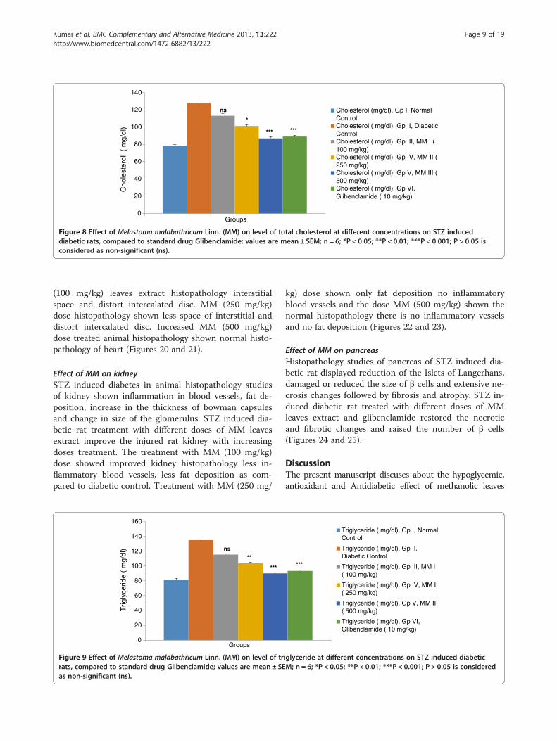
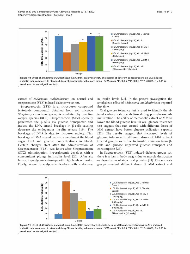
cholesterol) were increased and the level of HDL(high density cholesterol) was decreased in the STZ(streptozotocin) induced diabetic rat. Oral administra-tion of MM leaves extract was showed reduction incholesterol, triglyceride, LDL and VLDL compared tothe diabetic control rat group and the level of HDL wasincreased compared to the diabetic control rat in a sig-nificant manner (P < 0.001) (Table 3). MM leaves extractat the doses of 250 mg/kg and 500 mg/kg was moreeffective than 100 mg/kg in reducing the level of incholesterol (Figure 8), triglyceride (Figure 9), HDLcholesterol (Figure 10), LDL cholesterol (Figure 11) andVLDL cholesterol (Figure 12) compared to the diabeticcontrol rat.
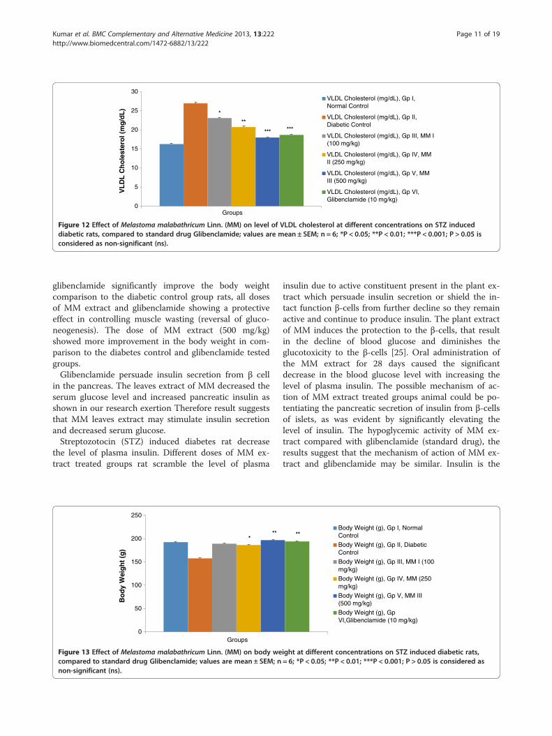
Changes in body weightTable 3 display the effect of the different doses of MMleaves extract and glibenclamide on the body weight ofon the STZ (streptozotocin) induced diabetic rat. At theend of 28 days treatment, the body weight of normal
0
20
40
60
80
100
120
140
160
Groups
Hex
oki
nas
e (u
g/m
g o
f p
rote
in)
*
***
Figure 5 Effect of Melastoma malabathricum Linn. (MM) on level of heinduced diabetic rats, compared to standard drug Glibenclamide; valP > 0.05 is considered as non-significant (ns).
rats, diabetic control, different doses of MM leavesextract and glibenclamide treated rats were observed(Figure 13). Diabetic control group continued to de-crease the weight till the end of the study. Glibenclamideand different doses (100, 250 and 500 mg/kg) of MMleaves extract treated rats significantly (P < 0.001) in-creased the weight as compared to the diabetic controlrats.
Effect of MM on antioxidant enzymesIn STZ induced diabetes increase the level of SOD, GPx,CAT and decrease the level of MDA. The level of CATincreased due to increase production of H2O2 in dia-betic pancreas and increase the level of SOD due to in-creased the production of superoxide, which has beenimplicated in cell dysfunction. The level of antioxidantenzyme SOD (superoxide dismutase), CAT (catalase)and GPx (Glutathione Peroxidase) were significantly(P < 0.001) decreased in diabetic control groups andlevel of MDA (Malonaldehyde) were significantly
Hexokinase (µg/mg of Protein), Gp I, Normal Control
Hexokinase (µg/mg of Protein), Gp II, Diabetic Control
Hexokinase (µg/mg of Protein), Gp III, MM I (100 mg/kg)
Hexokinase (µg/mg of Protein), Gp IV, MM II (250 mg/kg)
Hexokinase (µg/mg of Protein), Gp V, MM III (500 mg/kg)
Hexokinase (µg/mg of Protein), Gp VI, Glibenclamide (10 mg/kg)
***
xokinase (μg/mg of protein) at different concentrations on STZues are mean ± SEM; n = 6; *P < 0.05; **P < 0.01; ***P < 0.001;

0
2
4
6
8
10
12
14
16
Groups
Glucose-6-phosphate (unit/mg oftissue), Gp I, Normal Control
Glucose-6-phosphate (unit/mg oftissue), Gp II, Diabetic Control
Glucose-6-phosphate (unit/mg oftissue), Gp III, MM I (100 mg/kg)
Glucose-6-phosphate (unit/mg oftissue), Gp IV, MM II (250 mg/kg)
Glucose-6-phosphate (unit/mg oftissue), Gp V, MM III (500 mg/kg)
Glucose-6-phosphate (unit/mg oftissue), Gp VI, Glebenclamide (10mg/kg)
Glu
cose
-6-p
has
ph
ate
(Un
it/m
g o
f ti
ssu
e)*
***
***
ns
Figure 6 Effect of Melastoma malabathricum Linn. (MM) on level of glucose-6-phosphate at different concentrations on STZ induceddiabetic rats, compared to standard drug Glibenclamide; values are mean ± SEM; n = 6; *P < 0.05; **P < 0.01; ***P < 0.001; P > 0.05 isconsidered as non-significant (ns).
Kumar et al. BMC Complementary and Alternative Medicine 2013, 13:222 Page 8 of 19http://www.biomedcentral.com/1472-6882/13/222
increased (Table 4). Glibenclamide (5 mg/kg) and dif-ferent doses of MM leaves extract (100, 250 and 500mg/kg) received groups rat signification (p < 0.001) in-crease the level of SOD (Figure 14), CAT (Figure 15),GPx (Figure 16) and decreased the level of MDA(Figure 17). The result suggests that glibenclamide andall the doses of MM leaves extract increase the level ofSOD, CAT and GPx, but MM leaves extract doses 500mg/kg was more effective in increase the level of SOD,CAT and GPx in diabetic rat as compared with differ-ent doses of MM leaves extract and glibenclamide.
Effect of MM on liverLiver histopathology studies of STZ induced diabetic rat,the accumulation of fat was increased in the diabetesand large area of hepatocytes taken over by macro drop-let of fat. Treatment start with different doses of MM
0
10
20
30
40
50
60
Groups
Fru
cto
se-1
-6-b
isp
has
ph
ate
(un
it/m
g o
f p
rote
in)
*
**
***
Figure 7 Effect of Melastoma malabathricum Linn. (MM) on level of frinduced diabetic rats, compared to standard drug Glibenclamide; valP > 0.05 is considered as non-significant (ns).
leaves extract as the dose of MM leaves extract in-creased, the histological condition was improved(Figure 18). The treatment with MM (100 mg/kg) doseshown micro droplet of fat accumulation on rathistopathology, other dose MM (250 mg/kg) dose hasshown some micro droplet of fat accumulation on ratliver histopathology as compared to the diabetic con-trol and MM (100 mg/kg) dose. The treatment MMdose (500 mg/kg) shown the rat liver histopathologysimilar to the glibenclamide drugs (Figure 19).
Effect of MM on heartEffect of Melastoma malabathricum Linn. (MM) onSTZ induced diabetic rat heart. In STZ induced diabetesgroup rat histopathology shown increase the interstitialspace increased intercalated disc and increased the levelof fat deposition. The treatment starts with dose of MM
Fructose-1-6-bisphasphate (unit/mg oftissue), Gp I, Normal Contrrol
Fructose-1-6-bisphasphate (unit/mg oftissue), Gp II, Diabetic Contrrol
Fructose-1-6-bisphasphate (unit/mg oftissue), Gp III, MM I (100 mg/kg)
Fructose-1-6-bisphasphate (unit/mg oftissue), Gp IV, MM II (250 mg/kg)
Fructose-1-6-bisphasphate (unit/mg oftissue), Gp V, MM III (500 mg/kg)
Fructose-1-6-bisphasphate (unit/mg oftissue), Gp VI, Glibenclamide (10mg/kg)
***
uctose-1-6-bisphasphate at different concentrations on STZues are mean ± SEM; n = 6; *P < 0.05; **P < 0.01; ***P < 0.001;
0
20
40
60
80
100
120
140
Groups
Cholesterol (mg/dl), Gp I, NormalControlCholesterol ( mg/dl), Gp II, DiabeticControlCholesterol ( mg/dl), Gp III, MM I (100 mg/kg)Cholesterol ( mg/dl), Gp IV, MM II (250 mg/kg)Cholesterol ( mg/dl), Gp V, MM III (500 mg/kg)Cholesterol ( mg/dl), Gp VI,Glibenclamide ( 10 mg/kg)
Cho
lest
erol
( m
g/dl
)*
*** ***
ns
Figure 8 Effect of Melastoma malabathricum Linn. (MM) on level of total cholesterol at different concentrations on STZ induceddiabetic rats, compared to standard drug Glibenclamide; values are mean ± SEM; n = 6; *P < 0.05; **P < 0.01; ***P < 0.001; P > 0.05 isconsidered as non-significant (ns).
Kumar et al. BMC Complementary and Alternative Medicine 2013, 13:222 Page 9 of 19http://www.biomedcentral.com/1472-6882/13/222
(100 mg/kg) leaves extract histopathology interstitialspace and distort intercalated disc. MM (250 mg/kg)dose histopathology shown less space of interstitial anddistort intercalated disc. Increased MM (500 mg/kg)dose treated animal histopathology shown normal histo-pathology of heart (Figures 20 and 21).
Effect of MM on kidneySTZ induced diabetes in animal histopathology studiesof kidney shown inflammation in blood vessels, fat de-position, increase in the thickness of bowman capsulesand change in size of the glomerulus. STZ induced dia-betic rat treatment with different doses of MM leavesextract improve the injured rat kidney with increasingdoses treatment. The treatment with MM (100 mg/kg)dose showed improved kidney histopathology less in-flammatory blood vessels, less fat deposition as com-pared to diabetic control. Treatment with MM (250 mg/
0
20
40
60
80
100
120
140
160
Groups
Trig
lyce
ride
( m
g/dl
) ns
**
***
Figure 9 Effect of Melastoma malabathricum Linn. (MM) on level of trrats, compared to standard drug Glibenclamide; values are mean ± SEas non-significant (ns).
kg) dose shown only fat deposition no inflammatoryblood vessels and the dose MM (500 mg/kg) shown thenormal histopathology there is no inflammatory vesselsand no fat deposition (Figures 22 and 23).
Effect of MM on pancreasHistopathology studies of pancreas of STZ induced dia-betic rat displayed reduction of the Islets of Langerhans,damaged or reduced the size of β cells and extensive ne-crosis changes followed by fibrosis and atrophy. STZ in-duced diabetic rat treated with different doses of MMleaves extract and glibenclamide restored the necroticand fibrotic changes and raised the number of β cells(Figures 24 and 25).
DiscussionThe present manuscript discuses about the hypoglycemic,antioxidant and Antidiabetic effect of methanolic leaves
Triglyceride ( mg/dl), Gp I, NormalControl
Triglyceride ( mg/dl), Gp II,Diabetic Control
Triglyceride ( mg/dl), Gp III, MM I( 100 mg/kg)
Triglyceride ( mg/dl), Gp IV, MM II( 250 mg/kg)
Triglyceride ( mg/dl), Gp V, MM III( 500 mg/kg)
Triglyceride ( mg/dl), Gp VI,Glibenclamide ( 10 mg/kg)
***
iglyceride at different concentrations on STZ induced diabeticM; n = 6; *P < 0.05; **P < 0.01; ***P < 0.001; P > 0.05 is considered
0
10
20
30
40
50
60
Groups
HDL Cholesterol (mg/dL), Gp I, NormalControl
HDL Cholesterol (mg/dL), Gp II,Diabetic Control
HDL Cholesterol (mg/dL), Gp III, MM I(100 mg/kg)
HDL Cholesterol (mg/dL), Gp VI, MM II(250 mg/kg)
HDL Cholesterol (mg/dL), Gp V, MM III(500 mg/kg)
HDL Cholesterol (mg/dL), Gp VI,Glibenclamide (10 mg/kg)
HD
L C
ho
lest
ero
l (m
g/d
L)
***
*** ***
Figure 10 Effect of Melastoma malabathricum Linn. (MM) on level of HDL cholesterol at different concentrations on STZ induceddiabetic rats, compared to standard drug Glibenclamide; values are mean ± SEM; n = 6; *P < 0.05; **P < 0.01; ***P < 0.001; P > 0.05 isconsidered as non-significant (ns).
Kumar et al. BMC Complementary and Alternative Medicine 2013, 13:222 Page 10 of 19http://www.biomedcentral.com/1472-6882/13/222
extract of Melastoma malabathricum on normal andstreptozotocin (STZ) induced diabetic wistar rats.Streptozotocin (STZ) is a nitrosourea compound
(cytotoxic compound) obtained from soil microbeStreptomyces achromogenes, is mediated by reactiveoxygen species (ROS). Streptozotocin (STZ) speciallypenetrates the β-cells via glucose transporter andinduce the DNA strand breakage in β-cells causingdecrease the endogenous insulin release [19]. Thebreakage of DNA is due to nitrourea moiety. Thisbreakage of DNA strand leads to amendment the bloodsugar level and glucose concentrations in blood.Certain changes start after the administration ofStreptozotocin (STZ), two hours after Streptozotocin(STZ) administration, hyperglycemia develops with aconcomitant plunge in insulin level [20]. After sixhours, hyperglycemia develops with high levels of insulin.Finally, severe hyperglycemia develops with a decrease
0
10
20
30
40
50
60
70
80
Groups
LD
L C
ho
lest
ero
l (m
g/d
L)
**
***
***
Figure 11 Effect of Melastoma malabathricum Linn. (MM) on level of Ldiabetic rats, compared to standard drug Glibenclamide; values are mconsidered as non-significant (ns).
in insulin levels [21]. In the present investigation theantidiabetic effect of Melastoma malabathricum reportedfirst time.Oral glucose tolerance test is used to identify the al-
tered carbohydrate metabolism during post glucose ad-ministration. The ability of methanolic extract of MM tolower the blood glucose level in oral glucose tolerancetest suggest that rats treated with different doses ofMM extract have better glucose utilization capacity[22]. The results suggest that increased levels ofglucose tolerance in different doses of MM extracttreated groups were due to insulin emission from β-cells and glucose improved glucose transport andconsumption [23].In Streptozotocin (STZ) induced diabetes groups rat,
there is a loss in body weight due to muscle destructionor degradation of structural proteins [24]. Diabetic ratsgroups received different doses of MM extract and
LDL Cholesterol (mg/dL), Gp I, NormalControlLDL Cholesterol (mg/dL), Gp II,DiabeticControlLDL Cholesterol (mg/dL), Gp III, MM I(100 mg/kg)LDL Cholesterol (mg/dL), Gp IV, MM II(250 mg/kg)LDL Cholesterol (mg/dL), Gp V, MM III(500 mg/kg)LDL Cholesterol (mg/dL), Gp VI,Glibenclamide (10 mg/kg)
***
DL cholesterol at different concentrations on STZ inducedean ± SEM; n = 6; *P < 0.05; **P < 0.01; ***P < 0.001; P > 0.05 is
0
5
10
15
20
25
30
Groups
VLDL Cholesterol (mg/dL), Gp I,Normal Control
VLDL Cholesterol (mg/dL), Gp II,Diabetic Control
VLDL Cholesterol (mg/dL), Gp III, MM I(100 mg/kg)
VLDL Cholesterol (mg/dL), Gp IV, MMII (250 mg/kg)
VLDL Cholesterol (mg/dL), Gp V, MMIII (500 mg/kg)
VLDL Cholesterol (mg/dL), Gp VI,Glibenclamide (10 mg/kg)
VL
DL
Ch
ole
ster
ol (
mg
/dL
)*
**
*** ***
Figure 12 Effect of Melastoma malabathricum Linn. (MM) on level of VLDL cholesterol at different concentrations on STZ induceddiabetic rats, compared to standard drug Glibenclamide; values are mean ± SEM; n = 6; *P < 0.05; **P < 0.01; ***P < 0.001; P > 0.05 isconsidered as non-significant (ns).
Kumar et al. BMC Complementary and Alternative Medicine 2013, 13:222 Page 11 of 19http://www.biomedcentral.com/1472-6882/13/222
glibenclamide significantly improve the body weightcomparison to the diabetic control group rats, all dosesof MM extract and glibenclamide showing a protectiveeffect in controlling muscle wasting (reversal of gluco-neogenesis). The dose of MM extract (500 mg/kg)showed more improvement in the body weight in com-parison to the diabetes control and glibenclamide testedgroups.Glibenclamide persuade insulin secretion from β cell
in the pancreas. The leaves extract of MM decreased theserum glucose level and increased pancreatic insulin asshown in our research exertion Therefore result suggeststhat MM leaves extract may stimulate insulin secretionand decreased serum glucose.Streptozotocin (STZ) induced diabetes rat decrease
the level of plasma insulin. Different doses of MM ex-tract treated groups rat scramble the level of plasma
0
50
100
150
200
250
Groups
Bo
dy
Wei
gh
t(g
)
***
Figure 13 Effect of Melastoma malabathricum Linn. (MM) on body wecompared to standard drug Glibenclamide; values are mean ± SEM; nnon-significant (ns).
insulin due to active constituent present in the plant ex-tract which persuade insulin secretion or shield the in-tact function β-cells from further decline so they remainactive and continue to produce insulin. The plant extractof MM induces the protection to the β-cells, that resultin the decline of blood glucose and diminishes theglucotoxicity to the β-cells [25]. Oral administration ofthe MM extract for 28 days caused the significantdecrease in the blood glucose level with increasing thelevel of plasma insulin. The possible mechanism of ac-tion of MM extract treated groups animal could be po-tentiating the pancreatic secretion of insulin from β-cellsof islets, as was evident by significantly elevating thelevel of insulin. The hypoglycemic activity of MM ex-tract compared with glibenclamide (standard drug), theresults suggest that the mechanism of action of MM ex-tract and glibenclamide may be similar. Insulin is the
Body Weight (g), Gp I, NormalControl
Body Weight (g), Gp II, DiabeticControl
Body Weight (g), Gp III, MM I (100mg/kg)
Body Weight (g), Gp IV, MM (250mg/kg)
Body Weight (g), Gp V, MM III(500 mg/kg)
Body Weight (g), GpVI,Glibenclamide (10 mg/kg)
**
ight at different concentrations on STZ induced diabetic rats,= 6; *P < 0.05; **P < 0.01; ***P < 0.001; P > 0.05 is considered as
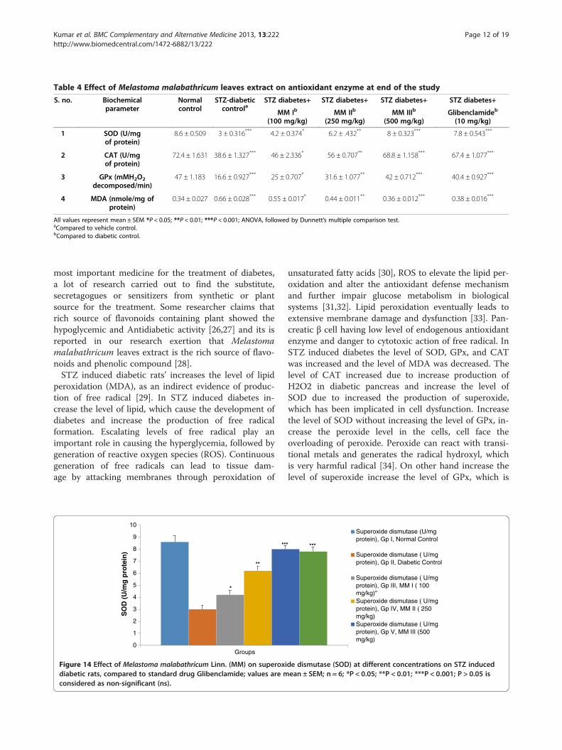
Table 4 Effect of Melastoma malabathricum leaves extract on antioxidant enzyme at end of the study
S. no. Biochemicalparameter
Normalcontrol
STZ-diabeticcontrola
STZ diabetes+ STZ diabetes+ STZ diabetes+ STZ diabetes+
MM Ib
(100 mg/kg)MM IIb
(250 mg/kg)MM IIIb
(500 mg/kg)Glibenclamideb
(10 mg/kg)
1 SOD (U/mgof protein)
8.6 ± 0.509 3 ± 0.316*** 4.2 ± 0.374* 6.2 ± .432** 8 ± 0.323*** 7.8 ± 0.543***
2 CAT (U/mgof protein)
72.4 ± 1.631 38.6 ± 1.327*** 46 ± 2.336* 56 ± 0.707** 68.8 ± 1.158*** 67.4 ± 1.077***
3 GPx (mMH2O2
decomposed/min)47 ± 1.183 16.6 ± 0.927*** 25 ± 0.707* 31.6 ± 1.077** 42 ± 0.712*** 40.4 ± 0.927***
4 MDA (nmole/mg ofprotein)
0.34 ± 0.027 0.66 ± 0.028*** 0.55 ± 0.017* 0.44 ± 0.011** 0.36 ± 0.012*** 0.38 ± 0.016***
All values represent mean ± SEM *P < 0.05; **P < 0.01; ***P < 0.001; ANOVA, followed by Dunnett’s multiple comparison test.aCompared to vehicle control.bCompared to diabetic control.
Kumar et al. BMC Complementary and Alternative Medicine 2013, 13:222 Page 12 of 19http://www.biomedcentral.com/1472-6882/13/222
most important medicine for the treatment of diabetes,a lot of research carried out to find the substitute,secretagogues or sensitizers from synthetic or plantsource for the treatment. Some researcher claims thatrich source of flavonoids containing plant showed thehypoglycemic and Antidiabetic activity [26,27] and its isreported in our research exertion that Melastomamalabathricum leaves extract is the rich source of flavo-noids and phenolic compound [28].STZ induced diabetic rats’ increases the level of lipid
peroxidation (MDA), as an indirect evidence of produc-tion of free radical [29]. In STZ induced diabetes in-crease the level of lipid, which cause the development ofdiabetes and increase the production of free radicalformation. Escalating levels of free radical play animportant role in causing the hyperglycemia, followed bygeneration of reactive oxygen species (ROS). Continuousgeneration of free radicals can lead to tissue dam-age by attacking membranes through peroxidation of
0
1
2
3
4
5
6
7
8
9
10
Groups
SO
D (
U/m
g p
rote
in)
*
**
**
Figure 14 Effect of Melastoma malabathricum Linn. (MM) on superoxidiabetic rats, compared to standard drug Glibenclamide; values are mconsidered as non-significant (ns).
unsaturated fatty acids [30], ROS to elevate the lipid per-oxidation and alter the antioxidant defense mechanismand further impair glucose metabolism in biologicalsystems [31,32]. Lipid peroxidation eventually leads toextensive membrane damage and dysfunction [33]. Pan-creatic β cell having low level of endogenous antioxidantenzyme and danger to cytotoxic action of free radical. InSTZ induced diabetes the level of SOD, GPx, and CATwas increased and the level of MDA was decreased. Thelevel of CAT increased due to increase production ofH2O2 in diabetic pancreas and increase the level ofSOD due to increased the production of superoxide,which has been implicated in cell dysfunction. Increasethe level of SOD without increasing the level of GPx, in-crease the peroxide level in the cells, cell face theoverloading of peroxide. Peroxide can react with transi-tional metals and generates the radical hydroxyl, whichis very harmful radical [34]. On other hand increase thelevel of superoxide increase the level of GPx, which is
Superoxide dismutase (U/mgprotein), Gp I, Normal Control
Superoxide dismutase ( U/mgprotein), Gp II, Diabetic Control
Superoxide dismutase ( U/mgprotein), Gp III, MM I ( 100mg/kg)"Superoxide dismutase ( U/mgprotein), Gp IV, MM II ( 250mg/kg)Superoxide dismutase ( U/mgprotein), Gp V, MM III (500mg/kg)
* ***
de dismutase (SOD) at different concentrations on STZ inducedean ± SEM; n = 6; *P < 0.05; **P < 0.01; ***P < 0.001; P > 0.05 is
0
10
20
30
40
50
60
70
80
Groups
Catalase (U/mg protein), Gp I,Normal ControlCatalse (U/mg protein), Gp II,Diabetic ControlCatalase ( U/mg protein), Gp III,MM I ( 100 mg/kg)Catalase ( U/mg protein), Gp IV,MM II ( 250 mg/kg)Catalase ( U/mg protein), Gp V,MM III ( 500 mg/kg)Catalase ( U/mg protein), Gp VI,Glibenclamide (10 mg/kg)
Cat
alas
e(
U/m
g p
rote
in)
*
**
*** ***
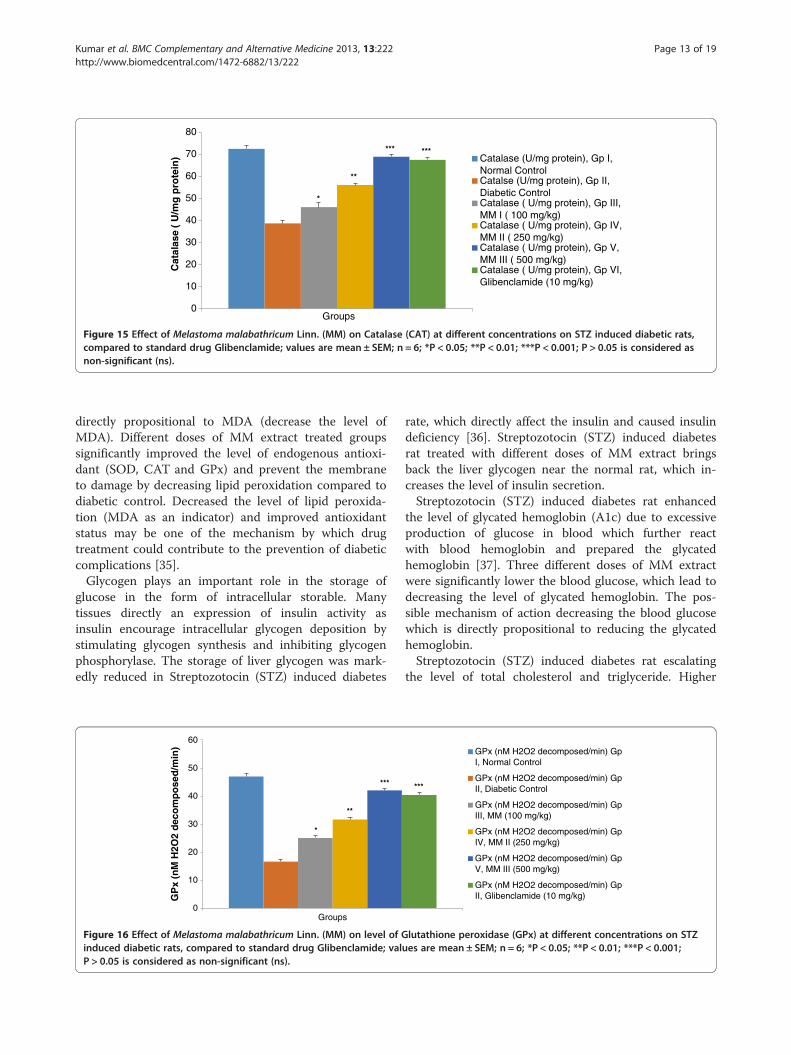
Figure 15 Effect of Melastoma malabathricum Linn. (MM) on Catalase (CAT) at different concentrations on STZ induced diabetic rats,compared to standard drug Glibenclamide; values are mean ± SEM; n = 6; *P < 0.05; **P < 0.01; ***P < 0.001; P > 0.05 is considered asnon-significant (ns).
Kumar et al. BMC Complementary and Alternative Medicine 2013, 13:222 Page 13 of 19http://www.biomedcentral.com/1472-6882/13/222
directly propositional to MDA (decrease the level ofMDA). Different doses of MM extract treated groupssignificantly improved the level of endogenous antioxi-dant (SOD, CAT and GPx) and prevent the membraneto damage by decreasing lipid peroxidation compared todiabetic control. Decreased the level of lipid peroxida-tion (MDA as an indicator) and improved antioxidantstatus may be one of the mechanism by which drugtreatment could contribute to the prevention of diabeticcomplications [35].Glycogen plays an important role in the storage of
glucose in the form of intracellular storable. Manytissues directly an expression of insulin activity asinsulin encourage intracellular glycogen deposition bystimulating glycogen synthesis and inhibiting glycogenphosphorylase. The storage of liver glycogen was mark-edly reduced in Streptozotocin (STZ) induced diabetes
0
10
20
30
40
50
60
Groups
GP
x (n
M H
2O2
dec
om
po
sed
/min
)
*
**
***
Figure 16 Effect of Melastoma malabathricum Linn. (MM) on level of Ginduced diabetic rats, compared to standard drug Glibenclamide; valP > 0.05 is considered as non-significant (ns).
rate, which directly affect the insulin and caused insulindeficiency [36]. Streptozotocin (STZ) induced diabetesrat treated with different doses of MM extract bringsback the liver glycogen near the normal rat, which in-creases the level of insulin secretion.Streptozotocin (STZ) induced diabetes rat enhanced
the level of glycated hemoglobin (A1c) due to excessiveproduction of glucose in blood which further reactwith blood hemoglobin and prepared the glycatedhemoglobin [37]. Three different doses of MM extractwere significantly lower the blood glucose, which lead todecreasing the level of glycated hemoglobin. The pos-sible mechanism of action decreasing the blood glucosewhich is directly propositional to reducing the glycatedhemoglobin.Streptozotocin (STZ) induced diabetes rat escalating
the level of total cholesterol and triglyceride. Higher
GPx (nM H2O2 decomposed/min) GpI, Normal Control
GPx (nM H2O2 decomposed/min) GpII, Diabetic Control
GPx (nM H2O2 decomposed/min) GpIII, MM (100 mg/kg)
GPx (nM H2O2 decomposed/min) GpIV, MM II (250 mg/kg)
GPx (nM H2O2 decomposed/min) GpV, MM III (500 mg/kg)
GPx (nM H2O2 decomposed/min) GpII, Glibenclamide (10 mg/kg)
***
lutathione peroxidase (GPx) at different concentrations on STZues are mean ± SEM; n = 6; *P < 0.05; **P < 0.01; ***P < 0.001;

0
0.1
0.2
0.3
0.4
0.5
0.6
0.7
0.8
Groups
MDA (nmole/mg of protein), Gp I,Normal Control
MDA (nmole/mg of protein), Gp II,Diabetic Control
MDA (nmole/mg of protein), Gp III,MM I (100 mg/kg)
MDA (nmole/mg of protein), Gp IV,MM II (250 mg/kg)
MDA (nmole/mg of protein), Gp V,MM III (500 mg/kg)
MDA (nmole/mg of protein), Gp VI,Glibenclamide (10 mg/kg)
MD
A (
nm
ole
/mg
of
pro
tein
)*
**
******
Figure 17 Effect of Melastoma malabathricum Linn. (MM) on level of Malondialdehyde (MDA) at different concentrations on STZinduced diabetic rats, compared to standard drug Glibenclamide; values are mean ± SEM; n = 6; *P < 0.05; **P < 0.01; ***P < 0.001;P > 0.05 is considered as non-significant (ns).
Kumar et al. BMC Complementary and Alternative Medicine 2013, 13:222 Page 14 of 19http://www.biomedcentral.com/1472-6882/13/222
levels of cholesterol (Hypercholesteremia) and higherlevel of triglyceride (Hypertriglyceridemia) are the pri-mary factor involved in the escalation of coronary heartdisease and atherosclerosis, the secondary complicationsoccurring in the diabetes [38]. STZ induced diabeticgroups treated with glibenclamide and different doses ofMM leaves extract brought back the increased level oftotal cholesterol and triglyceride near to the normallevels, which could be due to that all drug treated group
Figure 18 Effect of Melastoma malabathricum Linn. (MM) on liver fatDiabetic control: Diabetic control histopathology of rat liver shown thkg): Tested drug histopathology shown granularity and quantity of fadiabetic control. (D) MM II (250 mg/kg): Tested drug histopathology there(500 mg/kg): The amount of tested drug increased and the histopathologykg): Standard drug treated group shown histopathology similar to the normanatomical regions. For each group, 6 rats were examined and 50 picturesfrom the 80 pictures in this group. Original magnification, 10 ×.
start the increased level of insulin secretion, which inturn, inhibit hormones sensitive lipase and increase theutilization of glucose and decrease the mobilization offree fatty acids from the fat depositions. STZ induceddiabetic groups increased the level of LDL (low densitylipoprotein) cholesterol increased the coronary risk fac-tor and decreased level of HDL (high density lipopro-tein) cholesterol shown cardiovascular risk factor. Indiabetic condition increased the level of TC and TG is
accumulation in different groups of rats: (A) Normal control: (B)e micro fat droplet deposition (yellow arrow) (C) MM I (100 mg/t droplet (yellow arrow) were much less compared to thewas only few micro fat droplet was present (yellow arrow). (E) MM IIIsimilar to the glibenclamide treated group. (F) Glibenclamide (10 mg/al control groups. The samples were obtained from the same liverwere taken. The above picture for each group was chosen randomly
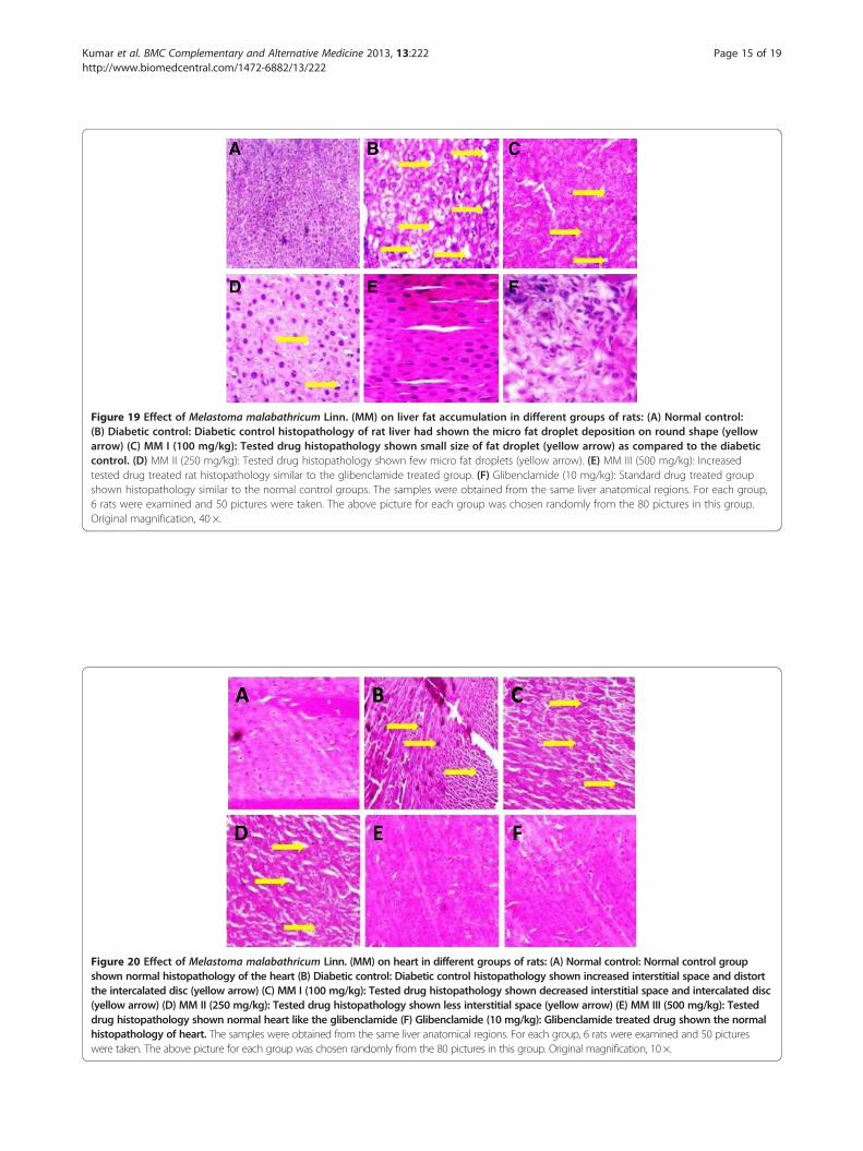
Figure 19 Effect of Melastoma malabathricum Linn. (MM) on liver fat accumulation in different groups of rats: (A) Normal control:(B) Diabetic control: Diabetic control histopathology of rat liver had shown the micro fat droplet deposition on round shape (yellowarrow) (C) MM I (100 mg/kg): Tested drug histopathology shown small size of fat droplet (yellow arrow) as compared to the diabeticcontrol. (D) MM II (250 mg/kg): Tested drug histopathology shown few micro fat droplets (yellow arrow). (E) MM III (500 mg/kg): Increasedtested drug treated rat histopathology similar to the glibenclamide treated group. (F) Glibenclamide (10 mg/kg): Standard drug treated groupshown histopathology similar to the normal control groups. The samples were obtained from the same liver anatomical regions. For each group,6 rats were examined and 50 pictures were taken. The above picture for each group was chosen randomly from the 80 pictures in this group.Original magnification, 40 ×.
Figure 20 Effect of Melastoma malabathricum Linn. (MM) on heart in different groups of rats: (A) Normal control: Normal control groupshown normal histopathology of the heart (B) Diabetic control: Diabetic control histopathology shown increased interstitial space and distortthe intercalated disc (yellow arrow) (C) MM I (100 mg/kg): Tested drug histopathology shown decreased interstitial space and intercalated disc(yellow arrow) (D) MM II (250 mg/kg): Tested drug histopathology shown less interstitial space (yellow arrow) (E) MM III (500 mg/kg): Testeddrug histopathology shown normal heart like the glibenclamide (F) Glibenclamide (10 mg/kg): Glibenclamide treated drug shown the normalhistopathology of heart. The samples were obtained from the same liver anatomical regions. For each group, 6 rats were examined and 50 pictureswere taken. The above picture for each group was chosen randomly from the 80 pictures in this group. Original magnification, 10 ×.
Kumar et al. BMC Complementary and Alternative Medicine 2013, 13:222 Page 15 of 19http://www.biomedcentral.com/1472-6882/13/222
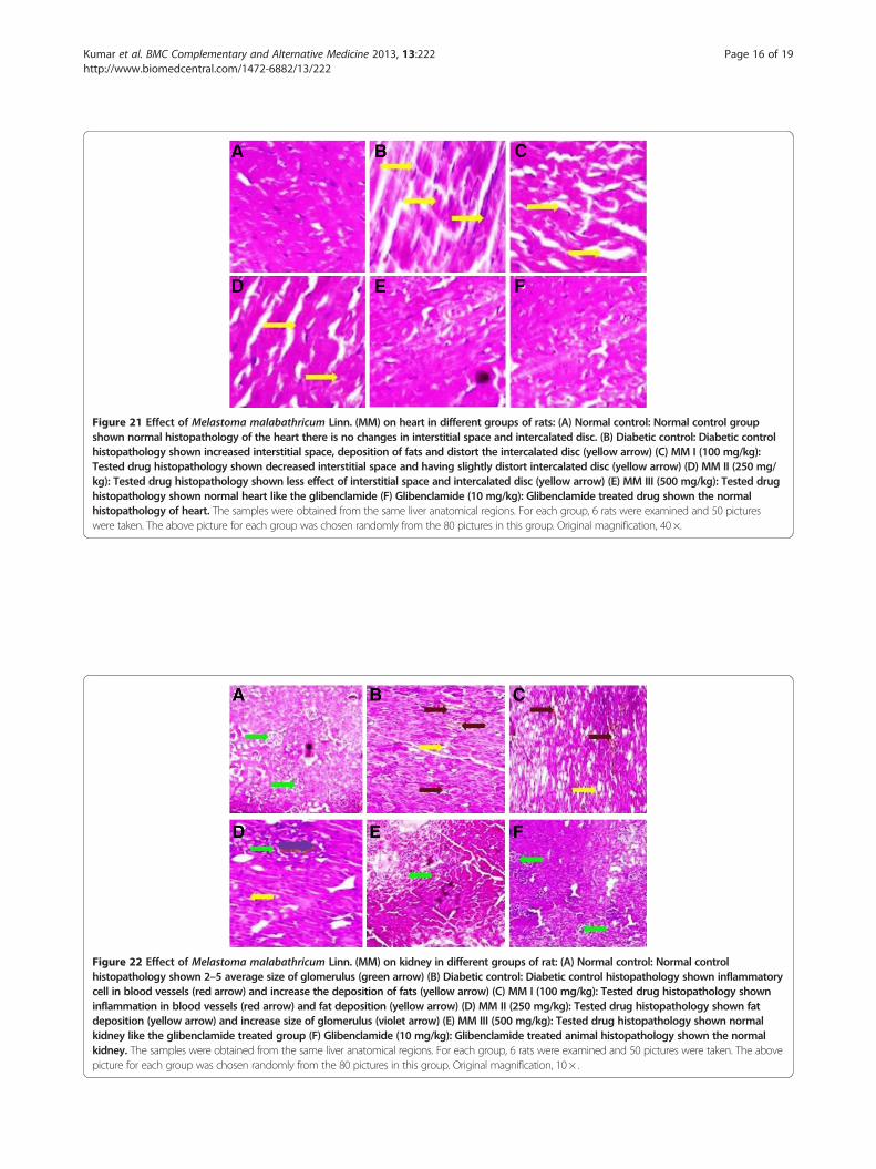
Figure 21 Effect of Melastoma malabathricum Linn. (MM) on heart in different groups of rats: (A) Normal control: Normal control groupshown normal histopathology of the heart there is no changes in interstitial space and intercalated disc. (B) Diabetic control: Diabetic controlhistopathology shown increased interstitial space, deposition of fats and distort the intercalated disc (yellow arrow) (C) MM I (100 mg/kg):Tested drug histopathology shown decreased interstitial space and having slightly distort intercalated disc (yellow arrow) (D) MM II (250 mg/kg): Tested drug histopathology shown less effect of interstitial space and intercalated disc (yellow arrow) (E) MM III (500 mg/kg): Tested drughistopathology shown normal heart like the glibenclamide (F) Glibenclamide (10 mg/kg): Glibenclamide treated drug shown the normalhistopathology of heart. The samples were obtained from the same liver anatomical regions. For each group, 6 rats were examined and 50 pictureswere taken. The above picture for each group was chosen randomly from the 80 pictures in this group. Original magnification, 40 ×.
Figure 22 Effect of Melastoma malabathricum Linn. (MM) on kidney in different groups of rat: (A) Normal control: Normal controlhistopathology shown 2–5 average size of glomerulus (green arrow) (B) Diabetic control: Diabetic control histopathology shown inflammatorycell in blood vessels (red arrow) and increase the deposition of fats (yellow arrow) (C) MM I (100 mg/kg): Tested drug histopathology showninflammation in blood vessels (red arrow) and fat deposition (yellow arrow) (D) MM II (250 mg/kg): Tested drug histopathology shown fatdeposition (yellow arrow) and increase size of glomerulus (violet arrow) (E) MM III (500 mg/kg): Tested drug histopathology shown normalkidney like the glibenclamide treated group (F) Glibenclamide (10 mg/kg): Glibenclamide treated animal histopathology shown the normalkidney. The samples were obtained from the same liver anatomical regions. For each group, 6 rats were examined and 50 pictures were taken. The abovepicture for each group was chosen randomly from the 80 pictures in this group. Original magnification, 10 × .
Kumar et al. BMC Complementary and Alternative Medicine 2013, 13:222 Page 16 of 19http://www.biomedcentral.com/1472-6882/13/222

Figure 23 Effect of Melastoma malabathricum Linn. (MM) on kidney in different groups of rat: (A) Normal control: Normal controlhistopathology shown average size of glomerulus (green arrow) (B) Diabetic control: Diabetic control histopathology showninflammatory cell in blood vessels (red arrow) and increase the deposition of fats (yellow arrow) (C) MM I (100 mg/kg): Testeddrug histopathology shown inflammation in blood vessels (red arrow) and fat deposition (yellow arrow) (D) MM II (250 mg/kg):Tested drug histopathology shown fat deposition (yellow arrow) and increase size of glomerulus (violet arrow) (E) MM III(500 mg/kg): Tested drug histopathology shown normal glomerulus but slightly bigger in size (F) Glibenclamide (10 mg/kg):Glibenclamide treated animal histopathology shown the normal kidney. The samples were obtained from the same liver anatomicalregions. For each group, 6 rats were examined and 50 pictures were taken. The above picture for each group was chosen randomly fromthe 80 pictures in this group. Original magnification, 40 ×.
Figure 24 Photomicrographs of histological changes of rat pancreas of isletes of Langerhans: (A) Normal control: normalhistological structure of rat pancreas showing normal islet (green arrow) (B) Diabetic control: diabetic control rat showing dilatedacini (blue arrow) and focal necrosis (yellow arrow) (C) MM I (100 mg/kg): tested drug rat showing dilated acini (blue arrow) andfocal necrosis (yellow arrow) (D) MM II (250 mg/kg): tested drug rat showing and focal necrosis (yellow arrow) (E) MM III (500 mg/kg): tested drug rat showing and focal necrosis (yellow arrow) (F) Glibenclamide (10 mg/kg): glibenclamide treated rat pancreasshowing normal islet (green arrow). For each group 6 rats were examined and 50 pictures were taken. The above picture for each groupwas chosen randomly from the 80 pictures in this group. Original magnification, 10 ×.
Kumar et al. BMC Complementary and Alternative Medicine 2013, 13:222 Page 17 of 19http://www.biomedcentral.com/1472-6882/13/222
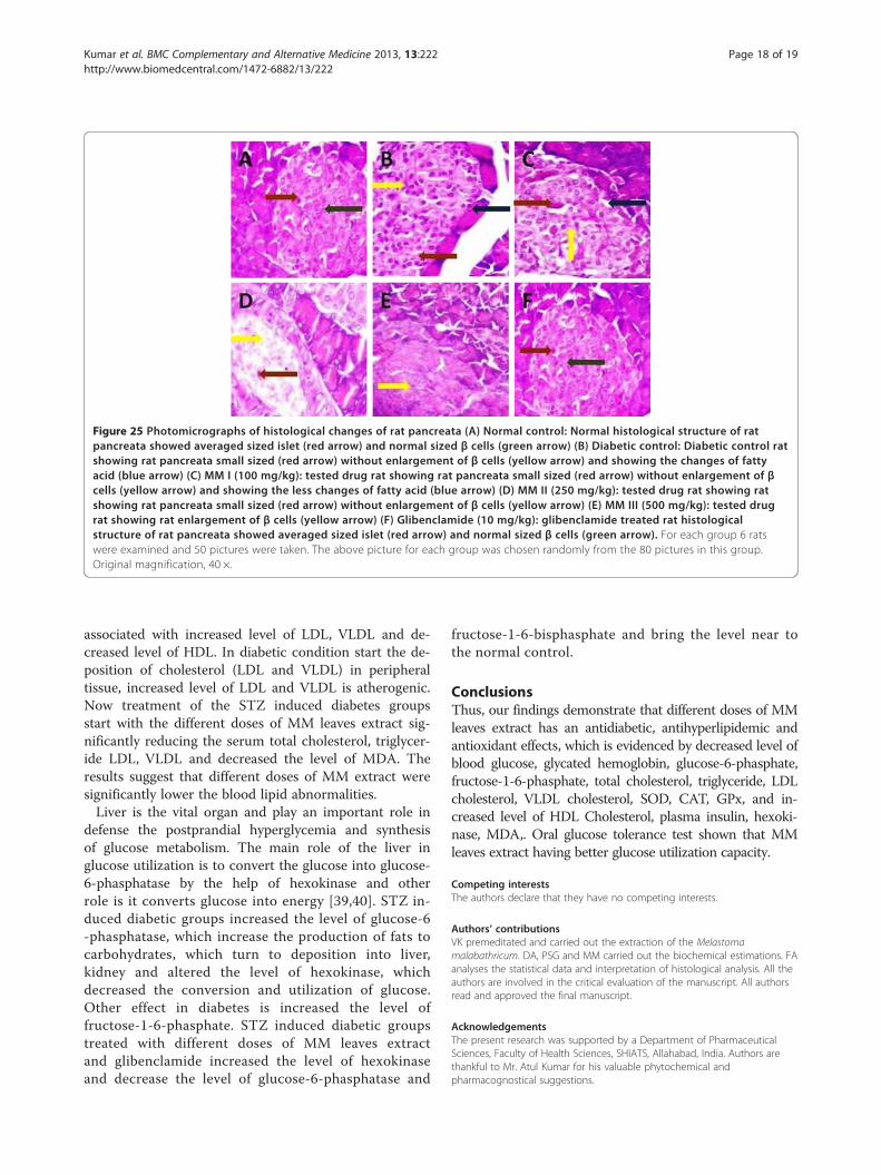
Figure 25 Photomicrographs of histological changes of rat pancreata (A) Normal control: Normal histological structure of ratpancreata showed averaged sized islet (red arrow) and normal sized β cells (green arrow) (B) Diabetic control: Diabetic control ratshowing rat pancreata small sized (red arrow) without enlargement of β cells (yellow arrow) and showing the changes of fattyacid (blue arrow) (C) MM I (100 mg/kg): tested drug rat showing rat pancreata small sized (red arrow) without enlargement of βcells (yellow arrow) and showing the less changes of fatty acid (blue arrow) (D) MM II (250 mg/kg): tested drug rat showing ratshowing rat pancreata small sized (red arrow) without enlargement of β cells (yellow arrow) (E) MM III (500 mg/kg): tested drugrat showing rat enlargement of β cells (yellow arrow) (F) Glibenclamide (10 mg/kg): glibenclamide treated rat histologicalstructure of rat pancreata showed averaged sized islet (red arrow) and normal sized β cells (green arrow). For each group 6 ratswere examined and 50 pictures were taken. The above picture for each group was chosen randomly from the 80 pictures in this group.Original magnification, 40 ×.
Kumar et al. BMC Complementary and Alternative Medicine 2013, 13:222 Page 18 of 19http://www.biomedcentral.com/1472-6882/13/222
associated with increased level of LDL, VLDL and de-creased level of HDL. In diabetic condition start the de-position of cholesterol (LDL and VLDL) in peripheraltissue, increased level of LDL and VLDL is atherogenic.Now treatment of the STZ induced diabetes groupsstart with the different doses of MM leaves extract sig-nificantly reducing the serum total cholesterol, triglycer-ide LDL, VLDL and decreased the level of MDA. Theresults suggest that different doses of MM extract weresignificantly lower the blood lipid abnormalities.Liver is the vital organ and play an important role in
defense the postprandial hyperglycemia and synthesisof glucose metabolism. The main role of the liver inglucose utilization is to convert the glucose into glucose-6-phasphatase by the help of hexokinase and otherrole is it converts glucose into energy [39,40]. STZ in-duced diabetic groups increased the level of glucose-6-phasphatase, which increase the production of fats tocarbohydrates, which turn to deposition into liver,kidney and altered the level of hexokinase, whichdecreased the conversion and utilization of glucose.Other effect in diabetes is increased the level offructose-1-6-phasphate. STZ induced diabetic groupstreated with different doses of MM leaves extractand glibenclamide increased the level of hexokinaseand decrease the level of glucose-6-phasphatase and
fructose-1-6-bisphasphate and bring the level near tothe normal control.
ConclusionsThus, our findings demonstrate that different doses of MMleaves extract has an antidiabetic, antihyperlipidemic andantioxidant effects, which is evidenced by decreased level ofblood glucose, glycated hemoglobin, glucose-6-phasphate,fructose-1-6-phasphate, total cholesterol, triglyceride, LDLcholesterol, VLDL cholesterol, SOD, CAT, GPx, and in-creased level of HDL Cholesterol, plasma insulin, hexoki-nase, MDA,. Oral glucose tolerance test shown that MMleaves extract having better glucose utilization capacity.
Competing interestsThe authors declare that they have no competing interests.
Authors’ contributionsVK premeditated and carried out the extraction of the Melastomamalabathricum. DA, PSG and MM carried out the biochemical estimations. FAanalyses the statistical data and interpretation of histological analysis. All theauthors are involved in the critical evaluation of the manuscript. All authorsread and approved the final manuscript.
AcknowledgementsThe present research was supported by a Department of PharmaceuticalSciences, Faculty of Health Sciences, SHIATS, Allahabad, India. Authors arethankful to Mr. Atul Kumar for his valuable phytochemical andpharmacognostical suggestions.
Kumar et al. BMC Complementary and Alternative Medicine 2013, 13:222 Page 19 of 19http://www.biomedcentral.com/1472-6882/13/222
Author details1Department of Pharmaceutical Sciences, Faculty of Health Sciences, SamHigginbottom Institute of Agriculture, Technology & Sciences(SHIATS)-Deemed University, Allahabad, Uttar Pradesh 211007, India.2Sidharatha Institute of Pharmacy, Dehradun, Uttarakhand 248001, India.3Department of Photochemistry & Pharmacognosy, Faculty of Pharmacy,Jamia Hamdard, New Delhi 110062, India.
Received: 15 May 2013 Accepted: 4 September 2013Published: 9 September 2013
References1. Dewanjee S, Das AK, Sahu R, Gangopadhyay M: Antidiabetic activity of
Diospyros peregrina fruit: effect on hyperglycemia, hyperlipidemia anaugmented oxidative stress in experimental type 2 diabetes. Food ChemToxicol 2009, 47:2679–2685.
2. Umar A, Ahmed QU, Muhammad BY, Dogarai BB, Soad SZ: Anti-hyperglycemic activity of the leaves of Tetracera scandens Linn. Merr.(Dille-niaceae) in alloxan induced diabetic rats. J Ethnopharmacol 2010,1:140–145.
3. Liu H, Liu X, Lee J, Liu Y, Yang H, Wang G: Insulin therapy restores impairedfunction and expression of P-glycoprotein in blood–brain barrier ofexperimental diabetes. Biochem Pharmacol 2008, 2008(75):1649–1658.
4. Oberley LW: Free radicals and diabetes. Free Radic Biol Med 1988, 5:113–124.5. Baynes JW: Role of oxidative stress in development of complications in
diabetes. Diab 1991, 40:405–412.6. Baynes JW: Reactive oxygen in the etiology and complications of
diabetes. In Drug, Diet and Disease: Mechanistic Approach to Diabetes, vol. 2.Edited by Ioannides C, Flatt PR. Hertfordshire: Ellis Horwood Limited;1995:203–231.
7. Joffry SM, Yob NJ, Rofiee MS, Affandi MMRMM, Suhaili Z, Othman F, AkimAM, Desa MNM, Zakaria ZA: Melastoma malabathricum (L.) SmithEthnomedicinal Uses, Chemical Constituents, and PharmacologicalProperties: A Review. Evid Based Complement Altern Med 2012, 48:258434.
8. Begum D, Nath SC: Ethnobotanical review of medicinal plants used forskin diseases and related problems in Northeastern India. J Herbs SpicesMed Plants 2000, 7(3):55–93.
9. Ong HC, Norzalina J: Malay herbal medicine in Gemencheh, NegriSembilan Malaysia. Fitoterapia 1999, 70(1):10–14.
10. Kumar V, Yadav PKS, Singh UP, Bhat HR, Rana A, Zaman K:Pharmacognostical evaluation of Cuscuta reflexa Roxb. Pharmcog J 2011,2(6):74–82.
11. Kumar V, Yadav PKS, Singh UP, Bhat HR, Zaman K: Pharmacognostical andphytochemical studies on the leaves of Paederia foetida Linn. Int J PharmTech Res 2009, 3:918–920.
12. Turner MA: Screening Methods in Pharmacology. New York: Academic Press;1965:26.
13. Bonner-weir S: Morphological evidence of pancreatic polarity of betacells within islets of Langerhans. Diab 1988, 37:616–621.
14. Arunachalam K, Parimelazhagan T: Antidiabetic activity of Ficus amplissimaSmith. Bark extract in streptozotocin induced diabetic rats. J Ethnopharmacol2013, 147:302–310.
15. Ahmed D, Sharma M, Mukerjee A, Ramteke PW, Kumar V: Antidiabetic,Anti-hyperlipidemic & Hepatoprotective effect of a Polyherbal Unaniformulation “Qurs Tabasheer” in STZ-diabetic wistar rats. ComplemenAltern Med 2013, 13:10.
16. Methi B, Aggarwal R, Chakrabarti: Neuroprotective effect of pioglitazoneon acute phase changes induced by partial global cerebral ischemia inmice. Indian J Exp Biol 2010, 48:793–799.
17. Hissin PJ, Hilf R: A fluorometric method for determination of oxidized andreduced glutathione in tissues. Anal Biochem 1976, 74:214–216.
18. Carroll NV, Longley RW, Roe JH: The determination of glycogen in liverand muscle by use of anthrone reagent. J Biol Chem 1956, 220:583–593.
19. Irudayaraj SS, Sunil C, Duraipandiyan V, Ignacimuthu S: Antidiabetic andantioxidant activities of Toddalia asiatica (L.) Lam. leaves in Streptozotocininduced diabetic rats. J Ethnopharmacol 2012, 143:515–523.
20. Lenzen S: The mechanisms of alloxan- and streptozotocin induceddiabetes. Diabetologia 2007, 51(2):216–226.
21. Arulmozhi S, Mazumderb PM, Lohidasan S, Thakurdesai P: Antidiabetic andantihyperlipidemic activity of leaves of Alstonia scholaris Linn. R.Br. Eur JIntegr Med 2010, 2:23–32.
22. Ceriello A: Postprandial hyperglycemia and diabetes complications: is ittime to treat? Diab 2005, 54:1–7.
23. Santiagu SI, Christudas S, Veeramuthu D, Savarimuthu I: Antidiabetic andantioxidant activities of Toddalia asiatica (L.) Lam. Leaves in Streptozotocininduced diabetic rats. J Ethnopharmacol 2012, 143:515–523.
24. Salahuddin M, Jalalpure SS: Antidiabetic activity of aqueous fruit extractof Cucumis trigonus Roxb. In streptozotocin-induced diabetic rats.J Ethnopharmacol 2010, 127:565–567.
25. Pushparaj NP, Tan HKB, Tan HC: The mechanism of hypoglycemic actionof the semi-purified fractions of Averrhoa bilimbi in streptozotocindiabetic rats. Life Sci 2001, 70:535–547.
26. Eidi M, Eidi A, Zamanizadeh H: Effect of Salvia officinalis L. leaves onserum glucose and insulin in healthy and streptozotocin-induceddiabetic rats. J Ethnopharmacol 2005, 100:310–313.
27. Hosseinzaded H, Ramezani M, Danaei AR: Antihyperglycaemic effect ofSecurigera securidaca L. seed extract in mice. Phytother Res 2002, 16:745–747.
28. Sharma HK, Kumar A: Evaluation of total phenol, flavonoids and invitroantioxidant activity of methanolic extract of Melastoma malabathricum.Asian J Chem 2011, 23(1):434–438.
29. Maritim AC, Sanders RA, Watkins JB: Diabetes, Oxidative Stress andantioxidants. J Biochem Mol Toxicol 2003, 17(1):24–38.
30. Ravi K, Rajasekeran S, Subramanian S: Hypoglycemic activity of Eugeniajambolana seed kernels on streptozotocin-induced diabetes in rats.Pharm Biol 2004, 42(8):598–603.
31. Ravi K, Ramachandran B, Subramanian S: Effect of Eugenia jambolana seedkernel on antioxidant defense system in streptozotocin-induceddiabetes in rats. Life Sci 2004, 75:2717–2731.
32. Balasubashini MS, Rukkumani R, Viswanathan P, Venugopal PM: Ferulic acidalleviates lipid peroxidation in diabetic rats. Phytother Res 2004, 18:310–314.
33. Alfy A, Ahmed A, Fatani A: Protective effect of red grape seedsproanthocyanidins against induction of diabetes by alloxan in rats.Pharmacol Res 2005, 52:264–270.
34. Halliwell B, Gutteridge JM: Free Radical in Biology and Medicine. UK: OxfordUniversity Press; 1999.
35. Kamalakkannan N, Prince P: Antihyperglycaemic and antioxidant effect ofrutin, a polyphenolic flavonoid, in streptozotocin-induced diabetic wistarrats. Basic Clin Pharmacol Toxicol 2006, 98:97–103.
36. Chandramohan G, Ignacimuthu S, Pugalendi KV: A novel compound fromCasearia esculenta (Roxb.) root and its effect on carbohydratemetabolism in streptozotocin diabetic rats. Eur J Pharmacol 2008,590:437–443.
37. Pari L, Saravanan R: Antidiabetic effect of diasulin, an herbal drug, onblood glucose, plasma insulin and hepatic enzymes of glucosemetabolism in hyperglycaemic rats. Diabetes Obes Metab 2004, 6:286–292.
38. Ananthan R, Latha M, Ramkumar K, Pari L, Baskar C, Bai V: Effect ofGymnema montanum leaves on serum and tissue lipids in alloxandiabetic rats. Exp Diabetes Res 2003, 4:183–189.
39. Gupta BL, Baquer NZ: Hexokinase, glucose-6-phosphate dehydrogenaseand antioxidant enzymes in diabetic reticulocytes: effects of insulin andvanadate. Biochem Mol Biol Int 1998, 46(6):1145–1152.
40. Gold AH: The effect of diabetes and insulin on liver glycogen synthetaseactivation. J Biol Chem 1970, 245:903–905.
doi:10.1186/1472-6882-13-222Cite this article as: Kumar et al.: Anti-diabetic, anti-oxidant and anti-hyperlipidemic activities of Melastoma malabathricum Linn. leaves instreptozotocin induced diabetic rats. BMC Complementary and AlternativeMedicine 2013 13:222.