Embed Size (px)
Citation preview

Chakraborty et al. BMC Genomics 2014, 15:659http://www.biomedcentral.com/1471-2164/15/659
RESEARCH ARTICLE Open Access
An integrated omics analysis: impact ofmicrogravity on host response tolipopolysaccharide in vitroNabarun Chakraborty†, Aarti Gautam†, Seid Muhie, Stacy-Ann Miller, Marti Jett and Rasha Hammamieh*
Abstract
Background: Microgravity facilitates the opportunistic infections by augmenting the pathogenic virulence andsuppressing the host resistance. Hence the extraterrestrial infections may activate potentially novel bionetworksdifferent from the terrestrial equivalent, which could only be probed by investigating the host-pathogenrelationship with a minimum of terrestrial bias.
Results: We customized a cell culture module to expose human endothelial cells to lipopolysaccharide (LPS). Theassay was carried out onboard the STS-135 spaceflight, and a concurrent ground study constituted the baseline.Transcriptomic investigation revealed a possible immune blunting in microgravity suppressing in particular Lbp,MyD88 and MD-2, which encode proteins responsible for early LPS uptake. Certain cytokines, such as IL-6 and IL-8,surged in response to LPS insult in microgravity, as suggested by the proteomics study. Contrasting proteomicexpressions of B2M, TIMP-1 and VEGRs suggested impaired pro-survival adaptation and healing mechanisms.Differential expression of miR-200a and miR-146b suggested the susceptibility of hosts in spaceflight to oxidativestress and further underscored the influence of microgravity on the immunity.
Conclusions: A molecular interpretation explaining the etiology of the microgravitational impact on the host-pathogenrelationship elucidated comprehensive immune blunting of the host cells responding to LPS challenges. Longer LPSexposure prompted a delayed host response, potentially ineffectual in preventing pathogens from opportunisticinvasion. Significant consequences include the subsequent failure in recruiting the growth factors and adebilitated apoptosis. Follow up studies with larger sample size are warranted.
BackgroundDuring spaceflight, astronauts experience a unique setof stressors comprised of microgravity (μG), suboptimalnutrition, social isolation, atypical work environment,solar radiation, and alteration of circadian rhythm. Detri-mental consequences were observed in the astronauts’immune [1-3] and musculoskeletal systems [4], andmany of the astronauts’ physiological [5] and cognitionphenotypes [6,7] were persistently altered long after themissions’terminations. The immunological investigationsof the astronauts recorded several dysregulations, such asthe altered production of cytokines [2], enhanced sympa-thetic neuroimmune responses [8], compromised functions
* Correspondence: [email protected]†Equal contributorsUS Army Center for Environmental Health Research Fort Detrick, 568Doughten Drive, Fort Detrick, MD 21702-5010, USA
© 2014 Chakraborty et al.; licensee BioMed CeCreative Commons Attribution License (http:/distribution, and reproduction in any mediumDomain Dedication waiver (http://creativecomarticle, unless otherwise stated.
of monocytes [9,10], suppressed cytotoxicity of T-cells andNatural Killer (NK) cells [11], and reduced phagocytic cap-abilities of neutrophils [12]. A few aspects of immunologicaldisorders such as the alteration of glucocorticoid-mediatedimmune response [8] were observed only after the long-term deployment for the space missions, which could beattributed to the accumulated effect of μG.It has long been known that microorganisms such as
Escherichia coli proliferate more rapidly in reduced gravity[13], thereby multiplying the risk of onboard cross-contamination, colonization, and infection. Worse, μGcan potentially alter microbial physiology and augmentpathogenesis as demonstrated by the studies using simu-lated μG [14,15]. Together, host defenses under extraterres-trial stress could be highly susceptible to the opportunisticpathogens armed with their aggressive virulence and rapidproliferative aptitude [16].
ntral Ltd. This is an Open Access article distributed under the terms of the/creativecommons.org/licenses/by/4.0), which permits unrestricted use,, provided the original work is properly credited. The Creative Commons Publicmons.org/publicdomain/zero/1.0/) applies to the data made available in this
Chakraborty et al. BMC Genomics 2014, 15:659 Page 2 of 14http://www.biomedcentral.com/1471-2164/15/659
A number of in vitro, in vivo and ex vivo studies probingspace-flown biomaterials suggested a potential bluntingof the immune response to pathogens and their variousderivatives [16]. Of particular interest are the studiesthat investigated the impact of reduced gravity on theimmunological responses to LPS shock. LPS, a commonouter membrane component of typical gram-negativebacteria, can elicit strong immune responses in the hostcells that may lead to sepsis [17,18]. The serological re-sponses of the astronauts were governed by the durationof the terrestrial LPS exposure to the whole blood samplesin vitro. The shorter LPS exposure caused a prolongedelevation of interleukin-1ra (IL-1ra) accompanied by tem-porary elevations of IL-8 and LPS binding protein (LBP),and suppression of IL-6 and IL-1β [19]; while the longerLPS exposure suppressed the phagocytic activity andreduced the expressions of IL-6, IL-8, IL-1β and TNF-αthat persisted for 7 d after the mission terminated [20].The in vitro LPS exposure of spleen cells derived fromspace-flown C57BL/6j mice resulted in elevated IL-6 andIL-10, but not TNF-α [21]. A rapid onset of LPS-inducedapoptosis was observed in squids subjected to simulatedμG [22].To date, μG-induced immunological perturbations
were investigated either by measuring the expressions ofinflammatory markers in the space-flown biomaterials[3,8,9,12,1,2,10] or by presenting the space-flown hostcells to the terrestrial endotoxic shock in vitro [19-21].The expression analysis of astronauts’ serological markers[3,8,9,12] was limited by the delay time between themission termination and the assay initiation, which canpotentially be an important time window for the stressedcells to get readjusted to the terrestrial environment. Like-wise, inducing the pathogenic shock to the space-flownsamples on ground [19-21] risked the host-pathogenrelationship being critically influenced by the terrestrialbias. These limitations were systematically minimized aswe attempted to probe the extraterrestrial impact onthe host-pathogen relationships with minimum terrestrialbias.The purpose of the present study was to understand
the impact of μG on the in vitro host immune responseto LPS assault. Towards this objective, the endothelialcells primed in the bioreactors were exposed to LPS for4 h and 8 h at two gravitational limits. The project wasintegrated and flown under the direction of DoD's SpaceTest Program. Associated signatures at the genomic andproteomic levels were analyzed. To our knowledge,this is a novel molecular-level approach to assess thehost cells infected in spaceflight; although previousstudies probed the molecular makeup of pathogens in-oculated in the spaceflight [23,24]. Recent efforts usedmodeled μG to investigate the molecular makeups ofthe host [22]. The complexity of spaceflight could
never be captured, however, by any of such simulatedparadigms [25-27].
MethodsReagents, cells, aseptic conditions and hardwareThis in vitro study used commercially available cell lines,so ethics is not needed.Human dermal microvascular endothelial cells (HMVEC-
dBL; Lonza, Walkersville, MD) were maintained in EGM-2MV growth medium (Lonza, MD) containing growthfactors, antimicrobials, cytokines and 5% FBS (all purchasedfrom Lonza, MD) at 37°C in a humidified atmospherecontaining 5% CO2. To avoid phenotypic drift associatedwith decreasing expression of surface receptor molecules,HMVEC-dBL was not used beyond passage 7. Fibronectinand LPS from E. coli 055: B5 were obtained from SigmaChemical Co. (St. Louis, MO).We selected a human micro-vascular endothelial cell
line because of the manifold involvement of this cell typein the wound healing cascade. At the onset of wound repair,these endothelial cells typically coordinate the recruitmentof cytokines and growth factors at the site of injury andsubsequently initiate communication with the leukocytesand other tissues to trigger the healing cascade [28,29].The Cell Culture Module (CCM) from Tissue Genesis,
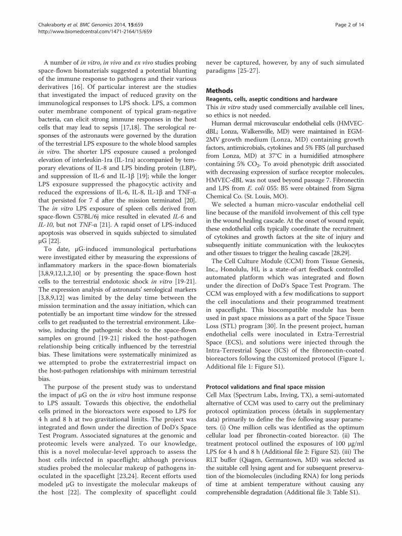
Inc., Honolulu, HI, is a state-of-art feedback controlledautomated platform which was integrated and flownunder the direction of DoD's Space Test Program. TheCCM was employed with a few modifications to supportthe cell inoculations and their programmed treatmentin spaceflight. This biocompatible module has beenused in past space missions as a part of the Space TissueLoss (STL) program [30]. In the present project, humanendothelial cells were inoculated in Extra-TerrestrialSpace (ECS), and solutions were injected through theIntra-Terrestrial Space (ICS) of the fibronectin-coatedbioreactors following the customized protocol (Figure 1,Additional file 1: Figure S1).
Protocol validations and final space missionCell Max (Spectrum Labs, Inving, TX), a semi-automatedalternative of CCM was used to carry out the preliminaryprotocol optimization process (details in supplementarydata) primarily to define the five following assay parame-ters. (i) One million cells was identified as the optimumcellular load per fibronectin-coated bioreactor. (ii) Thetreatment protocol outlined the exposures of 100 μg/mlLPS for 4 h and 8 h (Additional file 2: Figure S2). (iii) TheRLT buffer (Qiagen, Germantown, MD) was selected asthe suitable cell lysing agent and for subsequent preserva-tion of the biomolecules (including RNA) for long periodsof time at ambient temperature without causing anycomprehensible degradation (Additional file 3: Table S1).
Figure 1 Experiment scheme followed in the spaceflight and the concurrent ground study. Briefly, each of the two CCMs were loaded withsix bioreactors. One CCM was operated in the spaceflight and the other one on ground, concurrently. Both CCMs followed same protocoldepicted in the figure; where the top row shows the overall assay sequence segregated into three time zones: (i) the first 10 days, (ii) 11th dayand (iii) the last 5 days of the space mission. The protocol carried out on the 11th day is elaborated inside the box bordered by the broken lines.The cells received typical nourishments during first 10 days of space mission. At the beginning of the 11th day, 100 μg/ml LPS were injected tothe flowpaths of bioreactors 5 and 6 in both CCMs (arrowhead with LPS 0 h). Four hours later, the same amount of LPS was injected intobioreactor 3 and 4 (arrowhead with LPS 4 h); and after the subsequent four hours, RLT solution was discharged, replacing the media in theflowpaths of all bioreactors [1-6] (arrowhead with RLT 8 h). The biosamples in the bioreactors [1-6] in both gravity limits were preserved in theRLT solution till the space shuttle landed 5 days after the RLT discharge. We collected the bioreactors 3 hours after the landing.
Chakraborty et al. BMC Genomics 2014, 15:659 Page 3 of 14http://www.biomedcentral.com/1471-2164/15/659
Figure 1 depicts the final experiment scheme, and theflowpaths of the CCM are shown in Additional file 1:Figure S1.
Nucleic acid extraction from the bioreactorsThe RLT solution was drawn from the the bioreactorsusing syringes, followed by washing with PBS. The bio-reactors were inoculated with Trizol™ (Invitrogen, Inc.,Grand Island, NY) to lyse any remaining non-denaturedcells. We extracted mRNA and miRNA from the RLTsolution using the AllPrep DNA/RNA extraction kit(Qiagen, MD) and from the Trizol™ portion followingthe manufacturer’s protocols. The nucleic acids werequantified and qualified by a NanoDrop ND-1000 spec-trophotometer (NanoDrop Technologies, Wilmington,DE). The mRNA and miRNA (8.5 ng-12.5 ng) amplifica-tion was carried out using NuGEN Ovation Pico WTAsystem V2 kit (NuGEN Technologies, San Carlos, CA)and a Global miRNA amplification system (SystemsBioSciences, Mountain View, CA) following the respectivemanufacturer’s protocol.
mRNA whole genome oligonucleotide microarrayDual dye microarray using the Whole Human GenomeMicroarray Kit (Agilent Technologies, Inc., Santa Clara,CA) was carried out following the vendor’s protocol. Welabeled 12.5 ng amplified RNA with biotin and the sameamount of reference RNA with fluorescein. Sampleswere hybridized to Agilent 4 × 44 k slides and incubatedfor 16 h at 55°C. A protocol with a series of washes wascarried out for tagging Cy-3 and Cy-5 dyes to reference and
sample RNA, respectively. The slides were scanned usingAgilent Technologies Scanner G2505C US09493743 andfeature extracted using the software v. 10.7 (Agilent,Inc., CA). The assay quality was verified by assessingno alteration of a set of housekeeping genes across theexperimental parameters discussed in the supplementarysection.We have submitted the microarray data to the Gene
Expression Omnibus (GEO) and this can be searched forusing the GEO accession number: GSE54213.
Statistical analysis and biological annotationsWe used GeneSpring v.7.1 (Silicon Genetics, RedwoodCity, CA) to perform gene expression studies and cluster-ing analysis. GraphPad Prism (GraphPad Software, Inc. LaJolla, CA) and R platform (www.r-project.org) were usedfor the statistical analysis. Unless otherwise mentioned,the analysis was performed using pair-wise moderate t-testwith cut-off p < 0.05. It is a recommended routine forsmall sample populations to ensure maximum confidencescores [31].Using the IPA platform, we mapped the regulatory
networks significantly enriched with Genes of Interest(GoI). To construct the network module, IPA was usedto mine the manually curated literature searches(Ingenuity® ExpertAssist Findings) and interaction datafrom third-party databases, such as IntAct, BIND. Themulti-stage heuristic approach integrated the highly in-terconnected GoIs, and in the process, selected a moredensely populated network over a sparsely populated alter-native [32].
Chakraborty et al. BMC Genomics 2014, 15:659 Page 4 of 14http://www.biomedcentral.com/1471-2164/15/659
Characterization of GoIsThe transcriptomic regulations responding to LPS treat-ments on the ground for 4 h and 8 h (defined as G-4 hand G-8 h, respectively) were normalized by the groundcontrols (G-C), potentially emphasizing the host responseexclusively induced by LPS. Similarly, the transcriptomicregulations explaining the LPS-induced response mediatedby μG for 4 h and 8 h LPS assault (defined as S-4 h andS-8 h, respectively) were normalized by their spaceflightbaseline (S-C). Theoretically, the exclusive impacts of μGwere suppressed only to highlight the gene’s interactiveeffects with the host responses to LPS (μG × LPS-inducedhost responses).Nine pairs of biologically meaningful combinations
were tested. The pairs, namely G-4 h vs. S-4 h and G-8 hand S-8 h were contrasted to understand the effect of μGon LPS assault. The pairs namely G-C vs. G-4 h, G-C vs.G-8 h, S-C vs. S-4 h and S-C vs. S-8 h were contrasted tounderstand the effect of LPS assault at two gravitationlimits. The pairs namely G-4 h vs. G-8 h and S-4 h vs.S-8 h might illustrate the effect of durations at twogravitational limits. And, finally G-C vs. S-C might helpin understanding the exclusive effect of μG.The comparative analysis between the ground and
space controls (G-C vs. S-C) revealed 2,517 transcripts(Set A; 6.3% of global gene set presented in the Agilentarray). These genes are potentially the exclusive markersof μG.A four hour LPS exposure carried out in two gravita-
tional limits (G-4 h vs. S-4 h) altered 7,832 genes (Set B;19.58% of global gene set presented in the Agilent array).This resulted in a potential set of markers explaining theextraterrestrial effects on the LPS-induced host response.Among the other pairs of interest, 1,302 genes (Set C,
3.3% of global gene set presented in the Agilent array)emerged significantly different between 8 h LPS exposurein ground versus the controls inoculated in ground (G-8 hvs. G-C), a potential set of markers of the host responseindependent of the gravitational alteration.All other pairs representing biologically meaningful
combinations of interest including G-4 h vs. G-C, S-4 hvs. S-C, S-8 h vs. S-C, G-8 h vs. S-8 h, G-4 h vs. G-8 hand S-4 h vs. S-8 h failed to identify any genes significantlyaltered beyond the cut-off.A Venn diagram (Additional file 4: Figure S3) illustrated
the transcriptomic profile distributed among the three setsof genes defined hereby. Set A and Set B shared 1,534genes likely to be manipulated by μG exclusively inde-pendent of any other factors including toxic shock. Simi-larly, there were 1,186 genes shared between Set B andSet C that could have shifted exclusively due to the hostresponse independent of the gravitational shift. Screeningoff these potential false positive genes identified 5,379genes (13.4% of global gene set presented in the Agilent
array); defined as GoI-LPSμG, highlighting the signaturesof microgravitational impacts on LPS-induced host response(μG × LPS induced host response).Although the present study was primarily focused on
understanding the impact of gravitational shifts onLPS-induced host responses, a parallel comparativeinvestigation has been carried out to understand theexclusive impact of μG [1-3,8-10,12,19-21]. Associatedset molecular markers was constituted by 2,517 genes(Set A, defined as GoI-μG); 901 (36% of GoI-μG) and1,616 (64% of GoI-μG) transcripts were up- and down-regulated in S-C in comparison to G-C.
microRNA PCR assaySelective real-time PCR assays were carried out usingSABioscience kit (Qiagen, Inc.) using the amplified miRNAsamples. As per the vendor’s protocol, we loaded 100 ngmiRNA to 384-well plates containing anchored miRNAprobes (50–75 bp; including miRNA sequence, tailing, andthe universal primer) and the hybridization outcomeswere quantified by the ABI HT 7900 real-time PCRsystem (Life technologies, Inc., Grand Island, NY). Thevendor-recommended algorithm computed the relativemiRNA expression level using the change of thresholdcycle (Ct) i.e. 2 ^ (−Δ Ct), where Δ Ct = Ct (GoI) – avg.(Ct (HKG)). GoI represents the gene-of-interest, andHKG is the housekeeping gene. In order to eliminatethe false positive candidates, the selected miRNA readswere screened by the following dual criteria appliedtogether: (i) the control threshold cycle should be >30and sample cycle <30 (or vice versa); and (ii) the p-valuefor the fold-change should be either unavailable or rela-tively high (p > 0.05) from the assay background.From the pool of screened miRNA reads, we identified
the probes that had different expressions (p < 0.05) be-tween G-C vs. S-C. This cluster of miRNAs was alteredexclusively by μG. Likewise, comparing G-4 h and S-4 h,the miRNA signatures of LPS assault mediated by μGwere identified.
miRNA-mRNA target miningWe used the IPA platform (Ingenuity® Systems, www.ingenuity.com) to predict the mRNA targets of the se-lected miRNA modulators. The microRNA Target Filterpredicted the mRNA targets by mining four databases,namely TargetScan, TarBase, miRecords, and the Ingenuity®Knowledge Base. The list was screened further to identifynegatively correlated (r < −0.5) mRNA-miRNA pairs asdescribed elsewhere [33].
ImmunoassayThe sump bags, dedicated to store the spent media at theend of mission, returned from space with 75 ml solution.Aliquots collected from the sump bags were centrifuged,

Chakraborty et al. BMC Genomics 2014, 15:659 Page 5 of 14http://www.biomedcentral.com/1471-2164/15/659
and the cell-free supernatants were sent to Myriad RBM(Myriad RBM, Inc., Austin, TX) for Human InflammationMulti-Analyte Profiling immunoassay. Assays were run onan automated Luminex MAP™ platform at the MyriadRBM CLIA certified lab and validated for the fundamentalassay parameters of least detectable dose (LDD), lowerlimit of quantitation (LLOQ), spike recovery, linearity,precision and sample stability. Full assay validation docu-ments are retrievable upon request from Myriad RBM(www.myriadrbm.com).Complementary immunoassays were carried out in-
house using either the high throughput multiplex theBioPlex immunoassay (BioRad, Hercules, CA) or usingthe 96-well format based sandwich ELISA. Both assaytypes were primarily carried out for validation purposes.For the BioPlex assay, all of the reagents were purchasedfrom Millipore or Panomics (Affymetrix, Santa Clara,CA), and the results were analyzed using BioPlex man-ager software v4.1 (BioRad,. For the 96-well ELISA assay,we purchased the antibodies and other reagents fromQIAGEN, Inc., R & D systems, and BD Biosciences. Eachsample was assayed in triplicate, accompanied by appro-priate quality controls.
ResultsIdentification of genes of interest (GoI) from the wholegenome oligonucleotide assaysThe host-pathogen relationship in μG could be (i) exclu-sively mediated by LPS, independent of any other foreignstimulators including the gravitational shift or (ii) exclu-sively mediated by μG, independent of any other factorsincluding the endotoxic shock or (iii) mediated by theinterplay of the two above mentioned factors (μG × LPS-induced host responses). Present interpretation of thestressor landscape is technically a methodological decisionmade by the authors; thereby other interpretational possi-bilities cannot be ruled out. Furthermore, the role of fourthmediator explained by the factors beyond the control ofexperimental regulations could not be trivialized. We pre-sume however that the carefully supervised experimentalsetup and the appropriately placed control studies poten-tially limited the roles of the uncontrolled influences.The aim of the present study was to find the molecular
signatures associated with the host immune responsespecific to LPS assault mediated by μG (μG × LPS-induced host responses). Meeting this objective, weprocessed the microarray mRNA data set by heuristicstatistical manipulations accompanied by a systematicselection and rejection scheme described in supple-mentary data. The final outcome was 5,379 transcrip-tomic signatures associated with the host response toLPS assault mediated by μG (13.4% of global gene setpresented in the Agilent array) defined as GoI-LPSμG(Additional file 3: Table S2).
A parallel comparative investigation identified 2,517transcriptomic signatures of μG exclusively (6.3% ofglobal gene set presented in the Agilent array), which wenamed as GoI-μG. This list is comprised of 901 (36%)and 1,616 (64%) transcripts over- and under-expressed inthe spaceflight control (S-C), respectively, as comparedto transcripts expressed in the ground control (G-C)(Additional file 3: Table S2). GoI-μG and GoI-LPSμGwere designed as two orthogonal gene sets.
Characteristics of GoI-LPSμGThe transcriptomic perturbations caused by 4 h and 8 hLPS exposures in ground control (defined by G-4 hand G-8 h, respectively) were nearly identical. Of theGoI-LPSμG, there were 50% and 44% transcripts ofGoI-LPSμG elevated, 40% and 38% transcripts suppressed,and 8% and 17% transcripts unchanged in G-4 h and G-8 h, respectively. We considered the ground control (G-C)as the baseline for analyzing G-4 h and G-8 h.Comparing G-4 h and G-8 h, there were more tran-
scripts left unperturbed by the LPS exposures for 4 h (85%of GoI-LPSμG) and 8 h (61% of GoI-LPSμG) carried outin the spaceflight (defined by S-4 h and S-8 h, respect-ively). We considered the spaceflight control (S-C) as thebaseline for analyzing S-4 h and S-8 h. Merely 15% ofGoI-LPSμG were regulated in S-4 h; and among them 3%were up- and 12% down-regulated from S-C. In S-8 h,there were 21% up- and 18% down-regulated in compari-son to S-C.
Principal component analysis (PCA)The PCA (Figure 2) using GoI-LPSμG further explainedthe impact of μG on LPS-induced host responses. Prin-cipal components (PC) 1 and 2 explained 80.11% and10.13% of the total variance, respectively, and together,they explained more than 90% of total variance.
Significantly enriched immunologically relevant networksEarly signs of endothelial host response to LPS assaultinclude activation of inflammatory signals, which resultsfrom the enrollment of growth factors and the modula-tion of apoptosis activities [34-36]. In this context, wetrained the IPA platform to curate a set of pathwaysprimarily focused on three families associated with (i)cytokines/chemokines, (ii) apoptosis, and (iii) growthfactors (Additional file 3: Table S3). To note, severalgenes co-enriched more than one pathway. Likewise,several of the pathways were listed under more thanone of the families; for instance, the GM-CSF pathwaywas listed under both cytokines/chemokines andgrowth factors, and here we documented it under thegrowth factors.

Figure 2 Principal component analysis (PCA) of 5,379 transcriptomic signatures of host responses to LPS assault mediated by differentgravitational forces. The arrows indicate the shifts of the transcriptomic expressions due to LPS insults at two gravitational limits. The geneswere mined by pair-wise comparison between the LPS-treated samples normalized by the untreated samples inoculated on ground and inspaceflight respectively; for instance, G-4 h and G-8 h were normalized by G-C and S-4 h and S-8 h were normalized by S-C. The close Euclidianproximity of the self-normalized G-C and S-C implies that variance profile of this PCA does not represent the exclusive impact of microgravity. Adistant clustering between the LPS assault carried out on ground (G-4 h and G-8 h) and that carried out in spaceflight, S-4 h in particular, ismarked by PC 1 explaining 80% of total variance. Thereby the impact of microgravity on LPS assault emerges as the most significant factor inexplaining the genomic perturbation among these 5,379 genes. Modest Euclidian proximity between S-4 h and S-C (r = 0.16) suggests animmune blunting in spaceflight. Clearly, the host in spaceflight failed to respond to LPS assault. The position of S-8 h equidistant from the twoclusters formed by (a) S-C, G-C and S-4 h and to (b) G-4 h and G-8 h suggests a delayed host response in spaceflight.
Chakraborty et al. BMC Genomics 2014, 15:659 Page 6 of 14http://www.biomedcentral.com/1471-2164/15/659
Cytokine signaling networks associated with GoI-LPSμGThere were 160 genes identified from GoI-LPSμGsignificantly enriching 11 networks associated with thecytokine signaling (Figure 3A). These genes are docu-mented in Additional file 3: Table S2 and their hier-archical clustering is shown in the Additional file 5:Figure S4B.Interpreting the 160 genes, G-4 h and G-8 h shared
high correlation (r = 0.96) enlisting 61% up- and 33%down-regulated transcripts. S-4 h with maximum num-ber of transcripts (77% of 160 genes) unchanged fromthe S-C level was significantly different from rest of thethree experimental conditions, namely S-8 h, G-4 h, andG-8 h (p < 0.0001, all three conditions). Demonstrating amodest correlation with G-4 h and G-8 h (r = 0.6, bothconditions), S-8 h contained 41% of 160 transcriptsshowing S-C-like regulation.
Cytokine signaling networks associated with GoI-μGThere were 31 transcripts (34% up and 64% down-regulated) identified from GoI-μG significantly enriching3 cytokine/chemokine signaling networks (Additionalfile 6: Figure S5). These genes are documented inAdditional file 3: Table S2, and their hierarchicalclustering is shown in the Additional file 5: FigureS4A. The IL-1 signaling network was significantlyco-enriched by GoI-LPSμG and –μG (Additional file 7:Figure S6A).
Apoptosis signaling networks associated with GoI-LPSμGThere were 95 transcripts mined from GoI-LPSμGsignificantly enriching 5 apoptosis signaling networks(Figure 4). These genes are documented in Additional file3: Table S2 and their hierarchical clustering is shown inthe Additional file 5: Figure S4C.
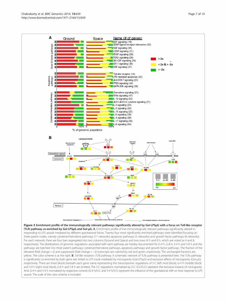
Figure 3 Enrichment profile of the immunologically relevant pathways significantly altered by GoI-LPSμG with a focus on Toll-like receptor(TLR) pathway co-enriched by GoI-LPSμG and GoI-μG. A. Enrichment profile of the immunologically relevant pathways significantly altered inresponding to LPS assault mediated by different gravitational forces. Twenty four most significantly enriched pathways were identified focusing onthree parent nodes, namely cytokine/chemokine pathways (11 networks) apoptosis pathways (5 networks) and growth factor pathways (8 networks).For each network, there are four bars segregated into two columns (Ground and Space) and two rows (4 h and 8 h, which are noted as 4 and 8,respectively). The distributions of genomic regulations associated with each pathway are hereby documented for G-4 h, G-8 h, S-4 h and S-8 h and thepathways are batched into three parent pathways: cytokine/chemokine pathways, apoptosis pathways and growth factor pathways. The fraction of theelevated (fold change > 2) and suppressed (fold change < −2) transcripts are colored by red and green, respectively. The unchanged fractions areyellow. The color scheme is at the right. B. Toll-like receptors (TLR) pathway. A schematic network of TLRs pathway is presented here. The TLRs pathwayis significantly co-enriched by both gene sets linked to LPS insult mediated by microgravity (GoI-LPSμG) and exclusive effects of microgravity (GoI-μG),respectively. There are three blocks beneath each gene name representing the transcriptomic regulations of S-C (left most block), G-4 h (middle block)and S-4 h (right most block). G-8 h and S-8 h are omitted. The S-C regulations normalized by G-C (S-C/G-C) represent the exclusive impacts of microgravity.And, G-4 h and S-4 h normalized by respective controls (G-4 h/G-C and S-4 h/S-C) represent the influence of the gravitational shift on host response to LPSassault. The scale of the color scheme is included.
Chakraborty et al. BMC Genomics 2014, 15:659 Page 7 of 14http://www.biomedcentral.com/1471-2164/15/659
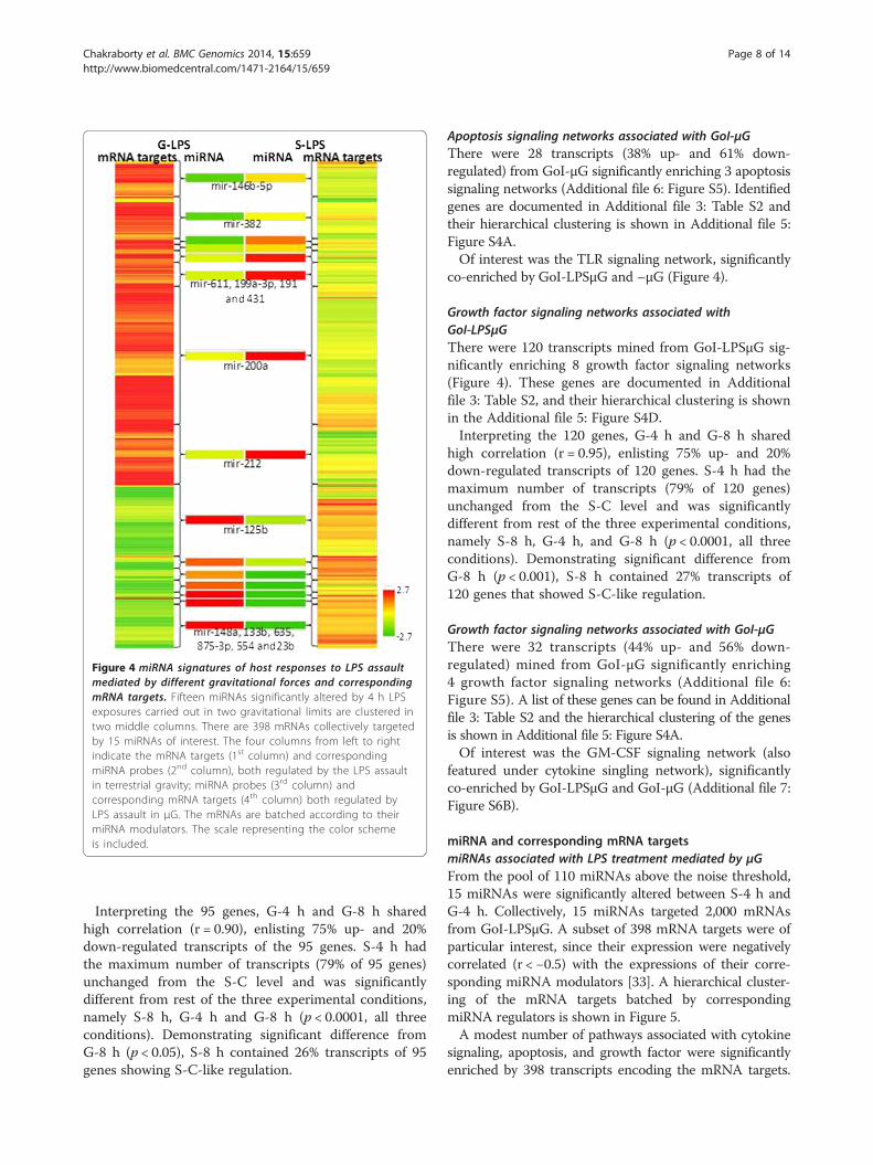
Figure 4 miRNA signatures of host responses to LPS assaultmediated by different gravitational forces and correspondingmRNA targets. Fifteen miRNAs significantly altered by 4 h LPSexposures carried out in two gravitational limits are clustered intwo middle columns. There are 398 mRNAs collectively targetedby 15 miRNAs of interest. The four columns from left to rightindicate the mRNA targets (1st column) and correspondingmiRNA probes (2nd column), both regulated by the LPS assaultin terrestrial gravity; miRNA probes (3rd column) andcorresponding mRNA targets (4th column) both regulated byLPS assault in μG. The mRNAs are batched according to theirmiRNA modulators. The scale representing the color schemeis included.
Chakraborty et al. BMC Genomics 2014, 15:659 Page 8 of 14http://www.biomedcentral.com/1471-2164/15/659
Interpreting the 95 genes, G-4 h and G-8 h sharedhigh correlation (r = 0.90), enlisting 75% up- and 20%down-regulated transcripts of the 95 genes. S-4 h hadthe maximum number of transcripts (79% of 95 genes)unchanged from the S-C level and was significantlydifferent from rest of the three experimental conditions,namely S-8 h, G-4 h and G-8 h (p < 0.0001, all threeconditions). Demonstrating significant difference fromG-8 h (p < 0.05), S-8 h contained 26% transcripts of 95genes showing S-C-like regulation.
Apoptosis signaling networks associated with GoI-μGThere were 28 transcripts (38% up- and 61% down-regulated) from GoI-μG significantly enriching 3 apoptosissignaling networks (Additional file 6: Figure S5). Identifiedgenes are documented in Additional file 3: Table S2 andtheir hierarchical clustering is shown in Additional file 5:Figure S4A.Of interest was the TLR signaling network, significantly
co-enriched by GoI-LPSμG and –μG (Figure 4).
Growth factor signaling networks associated withGoI-LPSμGThere were 120 transcripts mined from GoI-LPSμG sig-nificantly enriching 8 growth factor signaling networks(Figure 4). These genes are documented in Additionalfile 3: Table S2, and their hierarchical clustering is shownin the Additional file 5: Figure S4D.Interpreting the 120 genes, G-4 h and G-8 h shared
high correlation (r = 0.95), enlisting 75% up- and 20%down-regulated transcripts of 120 genes. S-4 h had themaximum number of transcripts (79% of 120 genes)unchanged from the S-C level and was significantlydifferent from rest of the three experimental conditions,namely S-8 h, G-4 h, and G-8 h (p < 0.0001, all threeconditions). Demonstrating significant difference fromG-8 h (p < 0.001), S-8 h contained 27% transcripts of120 genes that showed S-C-like regulation.
Growth factor signaling networks associated with GoI-μGThere were 32 transcripts (44% up- and 56% down-regulated) mined from GoI-μG significantly enriching4 growth factor signaling networks (Additional file 6:Figure S5). A list of these genes can be found in Additionalfile 3: Table S2 and the hierarchical clustering of the genesis shown in Additional file 5: Figure S4A.Of interest was the GM-CSF signaling network (also
featured under cytokine singling network), significantlyco-enriched by GoI-LPSμG and GoI-μG (Additional file 7:Figure S6B).
miRNA and corresponding mRNA targetsmiRNAs associated with LPS treatment mediated by μGFrom the pool of 110 miRNAs above the noise threshold,15 miRNAs were significantly altered between S-4 h andG-4 h. Collectively, 15 miRNAs targeted 2,000 mRNAsfrom GoI-LPSμG. A subset of 398 mRNA targets were ofparticular interest, since their expression were negativelycorrelated (r < −0.5) with the expressions of their corre-sponding miRNA modulators [33]. A hierarchical cluster-ing of the mRNA targets batched by correspondingmiRNA regulators is shown in Figure 5.A modest number of pathways associated with cytokine
signaling, apoptosis, and growth factor were significantlyenriched by 398 transcripts encoding the mRNA targets.

Figure 5 Proteomics profile illustrating the impact of microgravityon the immune responses to LPS assault. The hierarchical clusteringdepicts the proteomic expression of the focused cytokine profilesampled from the 4 h LPS exposures carried out in two gravitationallimits. The columns from left to right indicate the proteomicexpression observed (i) in spaceflight compared to ground control(S-C/G-C)- marked as S-C, (ii) LPS exposure for 4 h compared to groundcontrol (G-4 h/G-C)- marked as G-4 h and (iii) LPS exposure for 4 hcompared to space control (S-4 h/S-C)- marked as S-4 h. The scalerepresenting the color scheme is included.
Chakraborty et al. BMC Genomics 2014, 15:659 Page 9 of 14http://www.biomedcentral.com/1471-2164/15/659
More interestingly, the core of the mined networks stillshared the same family trees enriched by the global geneset of GoI-LPSμG. The list included IL-15 signaling, HGFsignaling, interferon signaling, IL-10 signaling, IL-6 signal-ing, TGF-beta signaling and TLR signaling.miR-200a (fold change: G 4 h: −0.03; S-4 h: 1.13;
r = − 0.67; p =0.09) regulated the highest number oftranscripts (119), which was 30% of all the mRNAscollectively targeted by 15 miRNA.
miRNAs associated with μGFrom the pool of 92 miRNA reads expressed above thenoise threshold, nine were significantly different between
the two gravitational limits. Collectively, these ninemiRNAs targeted 600 mRNAs from GoI-μG. A subsetof 169 mRNA targets (46 up- and 123 down-regulated)were of particular interest, since their expressions werenegatively correlated (r < − 0.5) with the expression oftheir corresponding miRNA modulators [33]. A hierarch-ical clustering of the mRNA targets batched by corre-sponding miRNA regulators is shown in Additional file 8:Figure S7.Although the results were likely compromised by seeding
a small sample size (169 mRNA targets), we were able tomap a small set of pathways. Interestingly, the core of themined networks still shared the same family trees enrichedby the global gene set of GoI-μG. The list includedNGF signaling, FGF signaling, p38 MAPK signaling,HGF signaling, GM-CSF signaling, chemokine signaling,IL-4 signaling and NF-kB signaling.miR-4651 (fold change: 2.21) regulated the largest num-
ber of mRNAs [37], which was 35% of the 169 significantmRNA targets.
Pathway focused proteomic assaysPathway-focused proteomic assays were performed toillustrate the impact of μG on LPS-induced host response(Figure 5). A more than two fold change from the back-ground level was considered significant. Some of the pro-teins relevant to the μG studies, such as IL-2, IL-10 andIFN- were assayed, but failed to meet the quality controlthreshold, and therefore, are not reported here.The protein profile associated with cytokine signaling
included IL-8, IL-6, IL6-ra, and B2M that were all ele-vated, and IL-17 and IL-1ra that were suppressed in S-C.Likewise, IL-8, IL-6, IL1-ra, and B2M were elevated inG-4 h in comparison to G-C. In contrast, there was amodest elevation in IL-8 and IL-6, and suppression inIL-6ra and B2M in S-4 h as compared to S-C.In addition, we investigated the protein profile associ-
ated with growth factors, which showed elevated HGF,PDGF, MCP-1, TIMP-1 and VEGFR-1/-2; and unchangedG-CSF and VEGF in S-C comparing G-C. In comparisonto same base like, i.e. G-C, G-4 h showed elevated HGF,PDGF, TIMP-1, MCP-1, VEGFR-1; a modestly elevatedVEGFR-2; and unchanged VEGF and G-CSF. In contrast,S-4 h in comparison to S-C depicted modestly elevatedMCP-1, VEGF, PDGF, G-CSF; unchanged VEGFR-2; andsuppressed HGF, TIMP-1 and VEGFR-1.Individual proteomic assays using a 96-well plate based
sandwich ELISA validated the expressions of IL-6, IL-8,VEGF, TIMP1 and MCP1/CCl2.
DiscussionThe focus of the present study is to find the molecularsignatures associated with the host response to LPSmediated by μG. Our primary motivation was attributed

Chakraborty et al. BMC Genomics 2014, 15:659 Page 10 of 14http://www.biomedcentral.com/1471-2164/15/659
to many evidences alleging the pathogenic interference asthe major challenge to the space missions [38,39]. Accom-panying evidence supported the extraterrestrial conditionas the potent habitat and stimulator of aggressive virulence[13-15,38]. The perennial pathogenic threat may seriouslyintimidate the future of space missions [16]; a solution tothe problem is therefore exigent.The host in μG is likely to counter a dual challenge.
The intrinsic challenge is defined by its own immune-compromised state stressed by μG, while pathogens withaugmented proliferation and virulence constitute theadditional challenge. As a consequence, this atypicalopportunistic infection may activate novel bionetworksdifferent from its terrestrial counterparts. The presentstudy was designed to investigate this hypothesis andto our knowledge, this is the first of this kind of attempt,although similar studies have been performed in simulatedreduced gravitational environment [22]. Comprehensivegenomics and transcriptomic studies were purposed toidentify key networks involved in extraterrestrial immuneresponse. Putative markers were suggested, althoughrobust validation was beyond the scope of the presentproject since we probed a small sample size. Furtherinvestigations with larger sample sizes are essential.In the present study, human microvascular endothelial
cells were exposed to LPS for 4 h and 8 h in spaceflightand on ground. The untreated bioreactors processed inboth gravitational limits defined the controls for respectivegravitational limits. All the assays were performed in du-plicate, and concurrently in spaceflight (STS-135, Atlantislaunched on July 11th, 2011) and on ground (KennedySpace Center, FL). The ground study mimicked the otherelements of the environment of the spaceflight, such astemperature and humidity (Figure 1). We were limited touse of only six bioreactors in the spaceflight, considerablycompromising the power of the study. To overcome thestudy limitations, we took stringent measures to optimizeevery aspect of the assay designs detailed in the supple-mentary section.
Transcriptomics and biological annotationsTwo orthogonal gene sets were identified from the ana-lyses of the transcriptome microarrays. We carried out apair-wise moderated t-test routine with cut-off p < 0.05,a recommended variance modeling strategy for such asmall sample size [31]. The larger gene set consisting of5,379 transcripts (GoI-LPSμG) was the potential signaturesof the host response to LPS assault mediated by μG. Asmaller set of 2,517 transcripts (GoI-μG) exclusively alteredby μG suggested that the reduced gravity was potentially aless significant contributor in molecular perturbations ascompared to the LPS assault. This methodological decisionproposed herein was guided by the hypothesis that μG, LPSassault and their interaction play major roles in perturbing
the molecular makeups. The role of the supplementaryfactors, such as those beyond the experimental controlmight have been limited by designing a well-regulatedexperimental strategy.The durations of LPS exposure (4 h vs. 8 h) emerged
as the less significant factor on ground. G-4 h and G-8 hwere positively correlated, but clustered distinctly fromthe ground control (G-C) depicted by a large Euclidianseparation along PC 1 between them (Figure 2). Of note,the controls on the ground and in spaceflight were spatiallycloser since they were self-normalized to highlight theimpact of gravitational forces on the host responses. Asignificant Euclidian separation between S-4 h and G-4 hdepicted the distinctive characteristics of the host-pathogenrelationship in μG. A close proximity between the S-C andS-4 h suggested a compromised early response to the endo-toxic shock, a potential state of immune blunting. Clearly,the host cells subjected to μG induced stress failed to buildresistance against the LPS assault.Longer exposure to LPS, however, triggered a delayed
response, which possibly allowed the pathogen a criticaltime window to evade the host resistance. As supportingevidence, immune-compromised mice (Lbp −/− andCD14 −/−) succumbed to fatality caused by gram-negativebacterial infection, despite a delayed response 4 h after thebacterial invasion [40]. It is important to note that thediminished host response to LPS was only observed in thespace-flown samples. Distinct clustering of G-C from G-4 hand G-8 h (Figure 2) conferred μG as a more dominantfactor in mediating the immunological response, at leastwithin the time scale probed herein. A pair of data pointsassociated with longer LPS incubation could validate thispotential explanation with higher confidence.Using the available sample size, a comprehensive an-
notation pathways (Figure 3, Additional file 6: Figure S5and Additional file 7: Figure S6, and Additional file 3:Table S3) was carried out to map the genomic membersof GoI-μG and GoI-LPSμG.Pathways co-enriched by GoI-μG and GoI-LPSμG
are of the particular interest; such as the TLR pathway(Figure 3B) that plays a key role in early pathogenicrecognition. In accordance to some previous reports[41] we observed an exclusive impact of μG in elevatingTLR4, which potentially triggered the stress-induceddownstream apoptosis [42]. In contrast, Lbp remainedsuppressed after both 4 h and 8 h exposure in space-flight and on ground, which could be attributed to thehigh concentration of LPS administered in this studythat typically inhibits TLR4 from recruiting LBP forLPS uptake [43]. The subsequent delayed elevation ofMyD88 (2.5 log2 fold change in G-4 h and G-8 h incomparison to G-C; −0.20 in S-4 h and 1.20 in S-8 h incomparison to S-C; Additional file 3: Table S2) andsuppression of MD-2 (2.6 fold change in G-4 h and G-8 h
Chakraborty et al. BMC Genomics 2014, 15:659 Page 11 of 14http://www.biomedcentral.com/1471-2164/15/659
in comparison to G-C; 1.40 in S-4 h and −0.26 in S-8 h incomparison to S-C; Additional file 3: Table S2) furthersuggested a defect in recognizing LPS in spaceflight.Hereby, μG potentially inhibited the dimerization ofTLR-4/MyD88 [44] and TLR4/MD-2 [45], two criticalcomplexes tasked for early pathogenic recognition.Synergistic suppression of the LPS receptor complexes inspaceflight might have induced temporary LPS tolerance,a characteristic reminiscent of septic shock [44]. The im-pact cascaded towards impairing downstream apoptosisand the adaptive immune system as the transcripts encod-ing mitogen-activated protein kinase, TNF receptors andnecrosis factors showed temporary suppression, followedby delayed activation induced by LPS insult in spaceflight(Additional file 3: Table S2).Two other pathways of interest, namely IL-10 (Additional
file 7: Figure S6A) and GM-CSF (Additional file 7:Figure S6B), were co-enriched by GoI-μG and GoI-LPSμG. The exclusive impact of μG was silencing ofIL-10 and its receptor (IL10-Ra), the anti-inflammatoryimmune regulators. In contrast, past observations re-ported elevated IL-10 in space-flown murine T-cells [46];while terrestrial UV radiation inhibited human epidermalIL-10 secretion [47].The GM-CSF network primarily controls the produc-
tion and function of blood cells and regulates a number ofkey apoptotic networks like PI3K/Akt pathway [48]. Thetranscripts encoding PI3K and AKT were elevated by μGand terrestrial LPS assault. The LPS assault in spaceflightdid not enhance their activities after 4 h exposure, possiblyleading to compromised cell survival and proliferationconsequences.
Novel miRNA targetsThe microRNAs of the miR-200 family are classified asthe redox regulators [49], and increased miR-200a andensuing p38α depletion were reported as the consequencesof oxidative stress [50]. In consideration of μG as a po-tential stimulant of oxidative stress primarily induced bydepletion of the energy [51], miR-200a could be a putativesignature of the host response to LPS assault mediatedby μG. Of note, miR-200a targeted the largest pool ofmRNAs from GoI-μG; in coherence, we also observedseveral of p38α encoding transcripts (MAPK1, MAPRE2and MAPK14) inhibited in S-4 h.We observed a negative correlation between miR-
146b-5p expression and the gravitational shift (expressionshifted from modestly down-regulated to up-regulatedas terrestrial gravity shifted to μG). Past observationssuggested that there is cross talk between the sup-pressed miR-146a/b with the onset of cytokine signalingand TLR-mediated LPS recognition [52]. Other majormicroRNAs of the present study included miR-212, asignature of alcoholic stress [53] and miR-125b that
was altered by in vitro endotoxic assault on murinecells [54].
Pathway focused proteomicsPathway focused proteomic investigation of a selectedpanel of cytokines and growth factors (Figure 5) foundoverexpressed IL-6 [10,55] and IL-8 [10], and suppressedIL-17 [1] in S-C supporting various past reports.Suppressed PDGF-β receptor in rat osteoblast cells wasobserved in spaceflight [56], while we found increasedexpression of PDGF in epithelial cells. LPS assault surgedthe expression of IL-6 and IL-8; while others such asB2M, TIMP-1 MCP-1 and VEGFR failed to respond tothe extraterrestrial LPS assault, potentially indicating astate of diminished immunity. Overexpression of VEGFR-1/-2 in μG could be interpreted as a protective measureagainst UV-induced photo-damage [57]. Terrestrial LPSshock induced the pro-survival mechanisms promoted byoverexpressed VEGFRs, while the LPS shock in spaceflightfailed to stimulate the same.In the context of the alleged link of μG to bone loss
[58], the synchronized activities of TIMP-1 and MMP-9were of particular interest. Typically, TIMP-1 operatescounteractively with MMPs [59]. Overexpressed TIMP-1in spaceflight (MMP9 registered no change) could feed-back control the bone-resorbing activity [37]. LPS shockaugmented TIMP-1 on ground but inhibited it in space-flight, thereby possibly restricting the regeneration of theepidermis and angiogenesis of the wound. The imbal-ance between TIMPs and MMPs could delay woundhealing [60].
ConclusionsThe present study, although compromised by the smallsample size, was able to illustrate several aspects ofimmunological responses to the endotoxic assault medi-ated by μG. It was beyond the scope of present paper toprobe the pathways with non-immunological implications;although such proposition could have many scientificinterests. We created the critical distinguishing features ofthe present study by implementing the host-pathogeninteractions in spaceflight and subsequently lysing the cellsonboard. Thereby the present study is set characteristicallyapart from the past reports. In principal, this customizedtechnological maneuver helped us to separate the host-pathogen relationships from the terrestrial bias. The deliv-erables were the molecular markers specifically associatedwith host responses mediated by μG.A synergistic immune blunting was evident in space-
flight. The early pathogenic detection in spaceflight wasfeeble and delayed which could confer a critical timeadvantage to the pathogen, facilitating an opportunisticinvasion. An in vivo study reported fatality caused byvery similar time delay in responding to endotoxic
Chakraborty et al. BMC Genomics 2014, 15:659 Page 12 of 14http://www.biomedcentral.com/1471-2164/15/659
shock [40]. Downstream apoptotic response and growthfactor recruitment processes were negatively affected, aswell. Impaired healing and defective angiogenesis wereother major proteomic findings.MicroRNA candidates such as miR-200a and miR-
146b are typically associated with oxidative stress andimmune regulation, respectively, and were found alteredby the LPS assault in spaceflight. A multitude of evidencesuggesting μG as a compelling modulator of oxidativestress [51] and immune suppression [2,12,16] further in-trigues our interest in these miRNA families identifiedherein. The significant relationships of these miRNAs tothe phenotypic signatures of the extraterrestrial conditionsstill stands out; despite of the compromised power of thestudy due to the small sample size. Systematic investiga-tion of these genomic regulators could potentially throwvaluable insights into the very young and dynamic field ofastrobiology. Taken together, a more comprehensivefollow up assessment is necessary to validate the observa-tions presented here. In particular, the elaborate in vitroassays with strategic time points beyond the time frameintroduced herein and the coherent in vivo studies arerecommended.
Additional files
Additional file 1: Figure S1. The flowpaths of the CCM operated in thespaceflight and on ground. The flowpath A was designed to support thebioreactors 1 and 2; and flowpath B was designed to support to the restof the bioreactors [3-6] operated in spaceflight and on ground,concurrently. Each flowpath consisted of one bioreactor, one pump, oneoxygen chamber (O2), one 70 ml media bag and one sump bag. An LPSbag was attached to the flowpath B, depicting the major differencebetween the two flowpaths, A and B. During the first 10 days of thespace mission, the media from the media bags was circulated throughthe path marked by “Common” (C) and “Normally Open” (NO). On the11th day, the unidirectional “Normally Closed” (NC) paths connecting LPSbags to the bioreactors 5 and 6, respectively, were switched on. Fourhours later, LPS was injected to the flowpaths integrated to bioreactors 3and 4. After the next 4 hours, the media bags were disconnected fromthe flowpaths by shutting off the associated “Normally Open” (NO) paths.The LPS bags were disconnected, too, by shutting off the respective“Normally Closed” (NC) paths. At the same time, the “Normally Closed”paths linked to the RLT bags and sump bags were switched open in allflowpaths integrated to bioreactors 1 to 6. The consequent arrangementwas maintained for the remaining 5 days of the space mission. Weobtained nucleic acids and proteins from the bioreactors and mediabags, respectively. Sump bags collected proteins, too, but not in anoptimum amount to run meaningful studies.
Additional file 2: Figure S2. Optimization of LPS concentration andincubation duration. The cell viability was measured from a series ofassays probing cells exposed to a dynamic range of LPS concentrations(0.01-1000 ng/ul) for various durations (2–24 h). The x-axis corresponds tothe range of LPS exposure duration and y-axis to the percentage cellviability comparing the time matched control cells. The arrows point outthe parameters selected for final experiments (100 μg/ml LPS treatmentfor 4 h and 8 h). Hereby, two conditions showing little to modest loss ofcell viability are selected with the presumption that microgravity wouldcause further cell damage. Optimum numbers of viable cells are requiredfor carrying out the downstream multi-omics assays.
Additional file 3: Table S1. Identification of most suitable cell lysis andnucleic acid fixing buffer. Six commercially available lysis buffers purchasedfrom three vendors were tested. The integrity and concentration of RNAand DNA extracted from HUVEC-dBL cells were investigated for 15 dayssuspending the nucleic acids in respective buffer at room temperature.The performances of the buffer candidates were measured compared toRLT solution and ranked. The * marked entries were top 90 percentilecandidates. The RNA integrities were probed in the scale of 260/280 and260/230 ratios and the yields (μg) were used to rank their performances.Table S2. Gene lists of interest. Two gene lists are recorded herein,enriched by genes (i) altered by microgravity exclusively (GoI- μG) and (ii)altered by LPS assault mediated by microgravity (GoI-LPSμG). For respectivegenes the regulation of S-C, G-4 h, G-8 h (all normalized by G-C) and S-4 hand S-8 h (both normalized by S-C) were reported. Table S3. Pathways ofinterest. The pathways were significantly enriched by genes (i) altered bymicrogravity exclusively and (ii) altered by LPS assault mediated bymicrogravity. For respective functions the number of participating genesand percentage of enrichment were reported. The pathways werebatched under three parent terms (i) cytokine signaling, (ii) apoptosisand (ii) growth factor signaling. Table S4. The identified list of miRNAand corresponding mRNA targets. The miRNA-mRNA pairs are curatedherein mapping from the gene sets (i) altered by microgravity exclusively(GoI-μG) and (ii) altered by LPS assault mediated by microgravity (GoI-LPSμG).The regulations of S-C (normalized by G-C) were reported for the genomicsubsets of GoI- μG. Likewise, the regulations of G-4 h (normalized by G-C)and S-4 h (normalized by S-C) were reported for the genomic subsets ofGoI-LPSμG.
Additional file 4: Figure S3. The Venn diagram of the gene profilesillustrating the characteristics of the gene of interest. The populationoverlaps of the three gene sets mined in the present study are depicted(not in scale by size). A pair wise moderated t-test identified 2,517transcripts altered between ground and space control (G-C vs. S-C), whichis depicted in the red circle in the upper left (Set A). We consider thissubset of 2,517 transcripts as the markers of microgravity, marked bybroken yellow line and defined as GoI-μG. Similar routine identified 1,302genes significantly altered between G-C and G-8 h assays (Set C). Thisgene set is reported in the blue circle in the upper right. The intrinsichost response independent of the gravitational shift could be the primaryfactor in altering these transcriptomic expressions. Likewise, 7,879 genesaltered between G-4 h and S-4 h (Set B). The green circle in the lower middledepicts this transcript set. Systematically rejecting the transcripts possiblyassociated with intrinsic host response and microgravity exclusively, wecurated 5,379 molecular signatures of host response to LPS assault mediatedby μG (μG x host response to LPS insult) and defined as GoI-LPSμG.
Additional file 5: Figure S4. Hierarchical clustering of the genessignificantly enriching the pathways of interest. The Euclidian algorithmclustered the transcripts listed in three focused groups of pathways linked tocytokine signaling, apoptosis and growth factor signaling. (A) The right-mostcolumn represents the transcripts altered by the gravitational change (GoI-μG),segregated from top to bottom in the order of cytokine signaling, apoptosisand growth factor signaling. (B-D) The Euclidean clustering represents theregulation of the transcripts (B) encoding cytokine signaling (C) apoptosis and(D) growth factor signaling altered by LPS insult mediated by gravitationalalteration (GoI-LPSμG). The assays were carried out by exposing endothelialcells to LPS for 4 and 8 h on ground (two left-most columns of B-D) and inthe spaceflight (two right-most columns of B-D).
Additional file 6: Figure S5. Enrichment profile of the immunologicallyrelevant pathways significantly altered by microgravity. Ten mostsignificantly enriched (p < 0.1) pathways are identified focusing on threeparent nodes, namely cytokine signaling (3 networks) apoptosis (3networks) and growth factor signaling (4 networks). The fractional sharingof elevated (fold change > 2) and suppressed (fold change < −2) genomicmembers are colored by red and green, respectively. The unchangedfractions are shaded yellow. The color scheme is in the right.
Additional file 7: Figure S6. A. IL-10 signaling pathway. A schematicnetwork of IL-10 pathway is presented. The IL-10 pathway is co-enriched byboth gene sets linked to LPS insult mediated by microgravity (GoI-LPSμG)and exclusive effects of microgravity (GoI-μG), respectively. There are threeblocks beneath each gene name representing the transcriptomic regulation

Chakraborty et al. BMC Genomics 2014, 15:659 Page 13 of 14http://www.biomedcentral.com/1471-2164/15/659
of S-C (left most block), G-4 h (middle block) and S-4 h (right most block).Both G-8 h and S-8 h are not shown in the figure. The S-C regulationsnormalized by G-C (S-C/G-C) represent the exclusive impacts of microgravity.And, G-4 h and S-4 h normalized by respective controls (G-4 h/G-C andS-4 h/S-C) represents the influence of the gravitational shift on host responseto LPS assault. The scale representing the color scheme is included. B. GM-CSFsignaling pathway. A schematic network of the GM-CSF pathway is presented.The GM-CSF pathway is co-enriched by both gene sets linked to LPS insultmediated by microgravity (GoI-LPSμG) and exclusive effects of microgravity(GoI-μG), respectively. There are three blocks beneath each gene namerepresenting the transcriptomic regulations of S-C (left most block), G-4 h(middle block) and S-4 h (right most block). Both G-8 h and S-8 h are notshown in the figure. The S-C regulations normalized by G-C (S-C/G-C)represent the exclusive impacts of microgravity. And, G-4 h and S-4 hnormalized by respective controls (G-4 h/G-C and S-4 h/S-C) representthe influence of gravitational shift on host response to LPS assault. Thescale representing the color scheme is included.
Additional file 8: Figure S7. miRNA and corresponding mRNA targetsregulated by microgravity. Nine miRNAs significantly altered by gravitationallimits are clustered in the left column. There are 169 mRNAs clustered in theright column collectively targeted by these 9 miRNAs of interest. ThemRNAs are batched according to their miRNA modulators. The scalerepresenting the color scheme is included.
Competing interestsThe authors declare that they have no competing interests.Financial competing interests• In the past five years have you received reimbursements, fees, funding, orsalary from an organization that may in any way gain or lose financially fromthe publication of this manuscript, either now or in the future? Is such anorganization financing this manuscript (including the article-processingcharge)? If so, please specify.NO• Do you hold any stocks or shares in an organization that may in any waygain or lose financially from the publication of this manuscript, either now orin the future? If so, please specify.NO• Do you hold or are you currently applying for any patents relating to thecontent of the manuscript? Have you received reimbursements, fees,funding, or salary from an organization that holds or has applied for patentsrelating to the content of the manuscript? If so, please specify.NO• Do you have any other financial competing interests? If so, please specify.NONon-financial competing interests• Are there any non-financial competing interests (political, personal,religious, ideological, academic, intellectual, commercial or any other)to declare in relation to this manuscript? If so, please specify.NO
Authors’ contributionsNC, AG, MJ and RH conceived and designed the assay. AG, NC and S-AM carriedout the experiment. NC, SM and RH analyzed the data. NC and RH preparedthe manuscript. All authors read and approved the document.
AcknowledgementsThis experiment was integrated and flown under the management anddirection of DoD’s Space Test Program. We are in debt to many colleaguesfrom TGI, Inc., and the NASA support team to complete our work. Ourparticular thanks go to Mr. Tom Canon and Ms. Cris Kosnik of TGI for theirrelentless support and thoughtful advice. Our team of technicians includingMr. Edward Waddy, Mr. William Santos and Ms. Allison Hoke achieved anoteworthy task at KSC, FL to complete the ground based works. We arethankful to MRMC for the financial support. Our team of editors includesDr. Julia Scheerer, Mr. William Santos, and Ms. Linda Brennan (USACEHR),who deserve special acknowledgement. We also like to extend our thanks toDr. Joseph M. Palma, Director, Systems Biology Enterprise, US Army MedicalResearch and Materiel Command, Fort Detrick, MD.
DisclaimerThe views, opinions, and/or findings contained in this report are those ofthe authors and should not be construed as official Department of theArmy position, policy, or decision, unless so designated by other officialdocumentation.Citations of commercial organizations or trade names in this report do notconstitute an official Department of the Army endorsement or approval ofthe products or services of these organizations.
Received: 6 March 2014 Accepted: 30 July 2014Published: 7 August 2014
References1. Crucian B, Stowe R, Mehta S, Uchakin P, Quiriarte H, Pierson D, Sams C:
Immune system dysregulation occurs during short duration spaceflighton board the space shuttle. J Clin Immunol 2013, 33(2):456–465.
2. Crucian BE, Stowe RP, Pierson DL, Sams CF: Immune system dysregulationfollowing short- vs long-duration spaceflight. Aviat Space Environ Med2008, 79(9):835–843.
3. Stowe RP, Sams CF, Pierson DL: Adrenocortical and immune responsesfollowing short- and long-duration spaceflight. Aviat Space Environ Med2011, 82(6):627–634.
4. Sibonga JD: Spaceflight-induced bone loss: is there an osteoporosis risk?Curr Osteoporos Rep 2013, 11(2):92–98.
5. Williams D, Kuipers A, Mukai C, Thirsk R: Acclimation during space flight:effects on human physiology. CMAJ 2009, 180(13):1317–1323.
6. Blaber E, Marcal H, Burns BP: Bioastronautics: the influence of microgravityon astronaut health. Astrobiology 2010, 10(5):463–473.
7. Grabherr L, Mast FW: Effects of microgravity on cognition: The case ofmental imagery. J Vestib Res 2010, 20(1):53–60.
8. Stowe RP, Sams CF, Pierson DL: Effects of mission duration onneuroimmune responses in astronauts. Aviat Space Environ Med 2003,74(12):1281–1284.
9. Kaur I, Simons ER, Castro VA, Ott CM, Pierson DL: Changes in monocytefunctions of astronauts. Brain Behav Immun 2005, 19(6):547–554.
10. Crucian B, Stowe R, Quiriarte H, Pierson D, Sams C: Monocyte phenotypeand cytokine production profiles are dysregulated by short-durationspaceflight. Aviat Space Environ Med 2011, 82(9):857–862.
11. Konstantinova IV, Antropova EN, Rykova MP, Guseva OA, Lesniak AT:Cellular and humoral immunity in cosmonauts as affected by spaceflight factors. Vestn Akad Med Nauk SSSR 1985, 8:51–58.
12. Kaur I, Simons ER, Castro VA, Mark Ott C, Pierson DL: Changes in neutrophilfunctions in astronauts. Brain Behav Immun 2004, 18(5):443–450.
13. Taylor GR: Space microbiology. Annu Rev Microbiol 1974, 28:121–137.14. Arunasri K, Adil M, Venu Charan K, Suvro C, Himabindu Reddy S, Shivaji S:
Effect of simulated microgravity on E. coli K12 MG1655 growth andgene expression. PLoS One 2013, 8(3):e57860.
15. Xiang L, Qi F, Dai D, Li C, Jiang Y: Simulated microgravity affects growthof Escherichia coli and recombinant beta-D-glucuronidase production.Appl Biochem Biotechnol 2010, 162(3):654–661.
16. Gueguinou N, Huin-Schohn C, Bascove M, Bueb JL, Tschirhart E, Legrand-FrossiC, Frippiat JP: Could spaceflight-associated immune system weakeningpreclude the expansion of human presence beyond Earth's orbit? J Leukoc Biol2009, 86(5):1027–1038.
17. Wang HL, Akinci IO, Baker CM, Urich D, Bellmeyer A, Jain M, Chandel NS,Mutlu GM, Budinger GR: The intrinsic apoptotic pathway is required forlipopolysaccharide-induced lung endothelial cell death. J Immunol 2007,179(3):1834–1841.
18. Sumi N, Nishioku T, Takata F, Matsumoto J, Watanabe T, Shuto H,Yamauchi A, Dohgu S, Kataoka Y: Lipopolysaccharide-activatedmicroglia induce dysfunction of the blood-brain barrier in ratmicrovascular endothelial cells co-cultured with microglia.Cell Mol Neurobiol 2010, 30(2):247–253.
19. Kaur I, Simons ER, Kapadia AS, Ott CM, Pierson DL: Effect of spaceflight onability of monocytes to respond to endotoxins of gram-negativebacteria. Clin Vaccine Immunol 2008, 15(10):1523–1528.
20. Morukov BV, Rykova MP, Antropova EN, Berendeeva TA, Ponomaryov SA,Larina IM: Parameters of the innate and adaptive immunity incosmonauts after long-term space flight on board the internationalspace station. Hum Physiol 2010, 36(3):264–273.

Chakraborty et al. BMC Genomics 2014, 15:659 Page 14 of 14http://www.biomedcentral.com/1471-2164/15/659
21. Baqai FP, Gridley DS, Slater JM, Luo-Owen X, Stodieck LS, Ferguson V,Chapes SK, Pecaut MJ: Effects of spaceflight on innate immune functionand antioxidant gene expression. J Appl Physiol 2009, 106(6):1935–1942.
22. Foster JS, Khodadad CL, Ahrendt SR, Parrish ML: Impact of simulatedmicrogravity on the normal developmental time line of an animal-bacteria symbiosis. Sci rep 2013, 3:1340.
23. Li T, Chen J, Chang D, Fang X, Wang J, Guo Y, Su L, Xu G, Wang Y, Chen Z,Liu C: Draft Genome Sequence of Escherichia coli Strain LCT-EC59.Genome Announc 2013, 1(1):e00242.
24. Su L, Zhou L, Liu J, Cen Z, Wu C, Wang T, Zhou T, Chang D, Guo Y, Fang X,Wang J, Li T, Yin S, Dai W, Zhou Y, Zhao J, Fang C, Yang R, Liu C:Phenotypic, genomic, transcriptomic and proteomic changes in Bacilluscereus after a short-term space flight. Adv Space Res 2014, 53(1):12.
25. Kotovskaya AR: The problem of artificial gravity: The current state andprospects. Hum Physiol 2010, 36(7):780–787.
26. Christensen NJ, Heer M, Ivanova K, Norsk P: Sympathetic nervous activitydecreases during head-down bed rest but not during microgravity.J Appl Physiol 2005, 99(4):1552–1557.
27. Hammond TG, Lewis FC, Goodwin TJ, Linnehan RM, Wolf DA, Hire KP,Campbell WC, Benes E, O’Reilly KC, Globus RK, Kaysen JH: Gene expressionin space. Nat Med 1999, 5(4):359.
28. Berger AC, Feldman AL, Gnant MF, Kruger EA, Sim BK, Hewitt S, Figg WD,Alexander HR, Libutti SK: The angiogenesis inhibitor, endostatin, does notaffect murine cutaneous wound healing. J Surg Res 2000, 91(1):26–31.
29. Bauer SM, Bauer RJ, Velazquez OC: Angiogenesis, vasculogenesis, andinduction of healing in chronic wounds. Vasc Endovascular Surg 2005,39(4):293–306.
30. Freed LE, Vunjak-Novakovic G: Spaceflight bioreactor studies of cells andtissues. Adv Space Biol Med 2002, 8:177–195.
31. Jeanmougin M, de Reynies A, Marisa L, Paccard C, Nuel G, Guedj M: Shouldwe abandon the t-test in the analysis of gene expression microarraydata: a comparison of variance modeling strategies. PLoS One 2010,5(9):e12336.
32. Thomas S, Bonchev D: A survey of current software for network analysisin molecular biology. Hum Genomics 2010, 4(5):353–360.
33. Liu T, Papagiannakopoulos T, Puskar K, Qi S, Santiago F, Clay W, Lao K, LeeY, Nelson SF, Kornblum HI, Doyle F, Petzold L, Shraiman B, Kosik KS:Detection of a microRNA signal in an in vivo expression set of mRNAs.PLoS One 2007, 2(8):e804.
34. Bierhaus A, Chen J, Liliensiek B, Nawroth PP: LPS and cytokine-activatedendothelium. Semin Thromb Hemost 2000, 26(5):571–587.
35. Werner S, Grose R: Regulation of wound healing by growth factors andcytokines. Physiol Rev 2003, 83(3):835–870.
36. Gregory CD, Pound JD: Microenvironmental influences of apoptosisin vivo and in vitro. Apoptosis 2010, 15(9):1029–1049.
37. Schiwon K, Arends K, Rogowski KM, Furch S, Prescha K, Sakinc T, Van HoudtR, Werner G, Grohmann E: Comparison of antibiotic resistance, biofilmformation and conjugative transfer of Staphylococcus and Enterococcusisolates from International Space Station and Antarctic Research StationConcordia. Microb Ecol 2013, 65(3):638–651.
38. Kim W, Tengra FK, Young Z, Shong J, Marchand N, Chan HK, Pangule RC,Parra M, Dordick JS, Plawsky JL, Collins CH: Spaceflight promotes biofilmformation by Pseudomonas aeruginosa. PLoS One 2013, 8(4):e62437.
39. Yang KK, Dorner BG, Merkel U, Ryffel B, Schutt C, Golenbock D, FreemanMW, Jack RS: Neutrophil influx in response to a peritoneal infection withSalmonella is delayed in lipopolysaccharide-binding protein orCD14-deficient mice. J Immunol 2002, 169(8):4475–4480.
40. Zhou Y, Ni H, Li M, Sanzari JK, Diffenderfer ES, Lin L, Kennedy AR, WeissmanD: Effect of solar particle event radiation and hindlimb suspension ongastrointestinal tract bacterial translocation and immune activation.PLoS One 2012, 7(9):e44329.
41. Rakoff-Nahoum S, Medzhitov R: Role of toll-like receptors in tissue repairand tumorigenesis. Biochemistry (Mosc) 2008, 73(5):555–561.
42. Seki E, Brenner DA: Toll-like receptors and adaptor molecules in liverdisease: update. Hepatology 2008, 48(1):322–335.
43. Medvedev AE, Lentschat A, Wahl LM, Golenbock DT, Vogel SN: Dysregulationof LPS-induced Toll-like receptor 4-MyD88 complex formation and IL-1receptor-associated kinase 1 activation in endotoxin-tolerant cells.J Immunol 2002, 169(9):5209–5216.
44. Mazzoni A, Segal DM: Controlling the Toll road to dendritic cellpolarization. J Leukoc Biol 2004, 75(5):721–730.
45. Gridley DS, Slater JM, Luo-Owen X, Rizvi A, Chapes SK, Stodieck LS, FergusonVL, Pecaut MJ: Spaceflight effects on T lymphocyte distribution, functionand gene expression. J Appl Physiol 2009, 106(1):194–202.
46. Schmitt DA, Walterscheid JP, Ullrich SE: Reversal of ultraviolet radiation-inducedimmune suppression by recombinant interleukin-12: suppression of cytokineproduction. Immunology 2000, 101(1):90–96.
47. Brauer H, Strauss J, Wegner W, Muller-Tidow C, Horstmann M, Jucker M:Leukemia-associated mutations in SHIP1 inhibit its enzymatic activity,interaction with the GM-CSF receptor and Grb2, and its ability toinactivate PI3K/AKT signaling. Cell Signal 2012, 24(11):2095–2101.
48. Magenta A, Cencioni C, Fasanaro P, Zaccagnini G, Greco S, Sarra-Ferraris G,Antonini A, Martelli F, Capogrossi MC: miR-200c is upregulated byoxidative stress and induces endothelial cell apoptosis and senescencevia ZEB1 inhibition. Cell Death Differ 2011, 18(10):1628–1639.
49. Mateescu B, Batista L, Cardon M, Gruosso T, de Feraudy Y, Mariani O, NicolasA, Meyniel JP, Cottu P, Sastre-Garau X, Mechta-Grigoriou F: miR-141 andmiR-200a act on ovarian tumorigenesis by controlling oxidative stressresponse. Nat Med 2011, 17(12):1627–1635.
50. Stein TP: Space flight and oxidative stress. Nutrition 2002, 18(10):867–871.51. Taganov KD, Boldin MP, Chang KJ, Baltimore D: NF-kappaB-dependent
induction of microRNA miR-146, an inhibitor targeted to signalingproteins of innate immune responses. Proc Natl Acad Sci U S A 2006,103(33):12481–12486.
52. Tang Y, Banan A, Forsyth CB, Fields JZ, Lau CK, Zhang LJ, Keshavarzian A:Effect of alcohol on miR-212 expression in intestinal epithelial cells andits potential role in alcoholic liver disease. Alcohol Clin Exp Res 2008,32(2):355–364.
53. Tili E, Michaille JJ, Cimino A, Costinean S, Dumitru CD, Adair B, Fabbri M, AlderH, Liu CG, Calin GA, Croce CM: Modulation of miR-155 and miR-125b levelsfollowing lipopolysaccharide/TNF-alpha stimulation and their possible rolesin regulating the response to endotoxin shock. J Immunol 2007,179(8):5082–5089.
54. Stein TP, Schluter MD: Excretion of IL-6 by astronauts during spaceflight.Am J Physiol 1994, 266(3 Pt 1):E448–E452.
55. Akiyama H, Kanai S, Hirano M, Shimokawa H, Katano H, Mukai C, Nagaoka S,Morita S, Kumei Y: Expression of PDGF-beta receptor, EGF receptor, andreceptor adaptor protein Shc in rat osteoblasts during spaceflight.Mol Cell Biochem 1999, 202(1–2):63–71.
56. Zhu JW, Wu XJ, Lu ZF, Luo D, Cai SQ, Zheng M: Role of VEGF receptors innormal and psoriatic human keratinocytes: evidence from irradiationwith different UV sources. PLoS One 2013, 8(1):e55463.
57. Nagaraja MP, Risin D: The current state of bone loss research: data fromspaceflight and microgravity simulators. J Cell Biochem 2013,114(5):1001–1008.
58. Deshmane SL, Kremlev S, Amini S, Sawaya BE: Monocyte chemoattractantprotein-1 (MCP-1): an overview. J Interferon Cytokine Res 2009,29(6):313–326.
59. Sobue T, Hakeda Y, Kobayashi Y, Hayakawa H, Yamashita K, Aoki T,Kumegawa M, Noguchi T, Hayakawa T: Tissue inhibitor ofmetalloproteinases 1 and 2 directly stimulate the bone-resorbing activityof isolated mature osteoclasts. J Bone Miner Res 2001, 16(12):2205–2214.
60. Vaalamo M, Leivo T, Saarialho-Kere U: Differential expression of tissueinhibitors of metalloproteinases (TIMP-1, -2, -3, and -4) in normal andaberrant wound healing. Hum Pathol 1999, 30(7):795–802.
doi:10.1186/1471-2164-15-659Cite this article as: Chakraborty et al.: An integrated omics analysis:impact of microgravity on host response to lipopolysaccharidein vitro. BMC Genomics 2014 15:659.


![Transplantation - OMICS Publishing Group · endothelial keratoplasty (DSAEK) has become a standard procedure for corneal transplantation in patients with endothelial dysfunction [20-23]](https://img.dokumen.tips/doc/110x75/5f025baf7e708231d403e06d/transplantation-omics-publishing-group-endothelial-keratoplasty-dsaek-has-become.jpg)