Embed Size (px)
Citation preview

Hindawi Publishing CorporationJournal of MaterialsVolume 2013, Article ID 613717, 7 pageshttp://dx.doi.org/10.1155/2013/613717
Research ArticleInvestigations of Self-Healing Property of Chitosan-ReinforcedEpoxy Dye Composite Coatings
Hüsnügül Y𝚤lmaz Atay,1,2 Leyla Eral DoLan,3 and Erdal Çelik1,2
1 Department of Metallurgical and Materials Engineering, Dokuz Eylul University, T𝚤naztepe Campus, 35160 Izmir, Turkey2 Center for Fabrication and Applications of Electronic Materials (EMUM), Dokuz Eylul University, T𝚤naztepe Campus,35160 Izmir, Turkey
3 Department of Chemistry, Izmir Institute of Technology, Urla, 35430 Izmir, Turkey
Correspondence should be addressed to Husnugul Y𝚤lmaz Atay; [email protected]
Received 20 November 2012; Revised 9 January 2013; Accepted 13 January 2013
Academic Editor: Mohindar S. Seehra
Copyright © 2013 Husnugul Y𝚤lmaz Atay et al. This is an open access article distributed under the Creative Commons AttributionLicense, which permits unrestricted use, distribution, and reproduction in any medium, provided the original work is properlycited.
Chitosan has a very wide application range in different parts of life such as in biomedical and antimicrobial areas. In recent yearsthe self-healing property of chitosan becomes more of an issue. In the study chitosan was used to obtain a self-healing compositematerial. An epoxy dye was converted to a self-healing coating. Different types of samples were prepared by coating the glasssubstrates with a polymermatrix reinforced with various amounts of chitosan.The samples were characterized by fourier transformInfrared (FTIR) and scanning electron microscope-energy dispersive spectroscopy (SEM-EDS). In addition, self-healing test wasapplied as a primary objective of this research. In this respect, the samples were scratched with a very thin pin, and they wereanalyzed by SEM periodically. It was observed that chitosan-reinforced dyes showed self-healing property. Mechanism of the self-healing process was also scrutinized.
1. Introduction
A self-healing material is a material that has the built-inability to partially repair damage occurring during its servicelife time. Self-healing is the ability of a material to restoremechanical properties, which were earlier perturbed by aplastic deformation or failure involving cracks and voids.Usually, certain properties of any engineering material suchas metals, polymers, ceramics, cementitious, and elastomericand fibre-reinforced composite materials degrade over timedue to environmental conditions or fatigue or due to dam-age incurred during operation. This damage is often on amicroscopic scale, requiring periodic inspection and repairto avoid them growing and causing failure [1]. Self-healingmaterials address this degradation through the inclusionof an “active” phase that responds to the microdamage byinitiating a repair mechanism, and several mechanisms havebeen proposed for the engineering materials. Investigation ofpossible self-healing mechanisms is an important emerging
field of nanotechnology [2]. The inspiration of self-healingmaterials comes from biological systems, which have theability to heal after being wounded. Initiation of cracksor other types of damage on a microscopic level changedthermal, electrical, and acoustical properties and eventuallyled to whole scale failure of the material. Usually, crackscannot be mended by hand because it is hard/difficult todetected. Amaterial that can intrinsically correct damage canlower production costs of a number of different industrialprocesses, can reduce the inefficiency over time caused bydegradation, and can prevent costs incurred by materialfailure [3].
Different strategies and approaches to devise self-healingmaterials in metals, ceramics, and polymers have been inves-tigated.These materials possess different intrinsic properties,and self-healing is based on the same common generalprinciple in all of them. Self-healing can take place on themicroscopic to macroscopic level. The prerequisite for aself-healing of a (mechanical) damage is the generation of

2 Journal of Materials
a mobile phase, and the mobile phase can close the crack(Figure 1). If a material is damaged (Figures 1(a) and 1(b)),a crack can occur. Then subsequently a “mobile phase”(Figure 1(c)) which is triggered either by the occurrenceof damage (in the ideal case) or by external stimuli wasgenerated. Following the mobile phase generation, the dam-age can be removed due to the directed mass transporttowards the damage site and the subsequent local mendingreaction (Figure 1(d)). The latter assures the (re)connectionof the crack planes by physical interactions and/or chemicalbonds. After the healing of the damage the previouslymobile material is immobilised again, resulting in the bestcase in fully restored mechanical properties (Figure 1(e))[4].
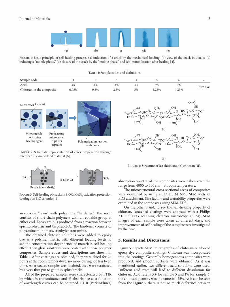
The first report of a completely autonomous man-madeself-healing material belongs to a group [5]. They reportedan epoxy system containing microcapsules, and the micro-capsules were filled with a (liquid-thermosetting) monomer.If a microcrack occurs in this system, the microcapsule willrupture, and the monomer will fill the crack (Figure 2) [6].Subsequently it will polymerise by the initiation of catalystparticles dispersed through the system. This model systemof a self-healing particle proved to work very well in purepolymers and polymer coatings [7].
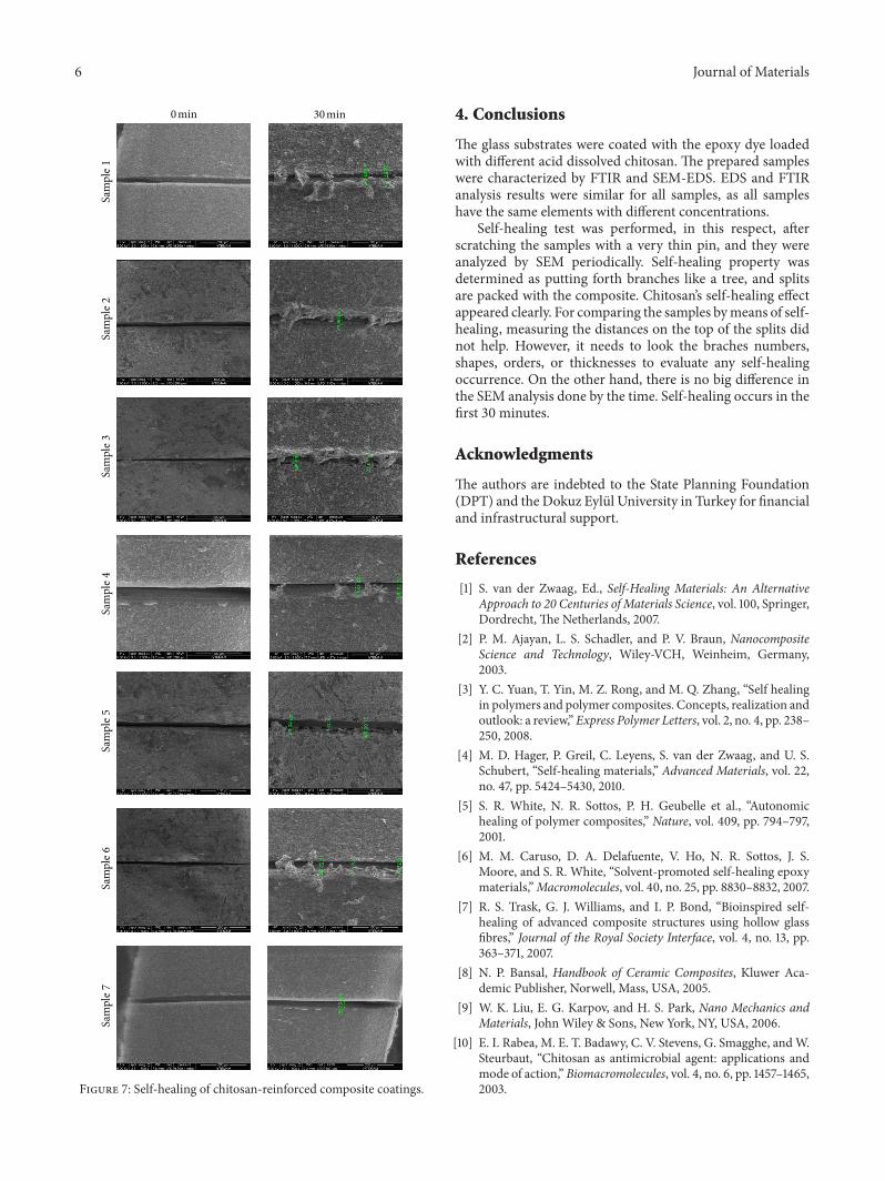
SiC/C/SiC model composites and Si-Ti-C-O fiber-bonded ceramics are the examples that show self-healingfeatures under heating at high temperatures [8]. In addition,semiorganic and ceramic protective coatings, silicatenanocomposites, and SiC/SiC fibrous composites may revealchemical mechanisms of self-healing on exposure to oxygen[9]. Generally speaking, very high local temperatures arerequired to repair most current ceramics, due to the highactivation energies of the diffusive mass transport in thecovalent or ionic structures of ceramics (e.g., SiC/Al
6Si2O13
composite: 1300∘C). In Figure 3, the healing reaction isinitiated by the heat of combustion, and a self-healingoxidation and corrosion protection coating on SiC ceramic(burner nozzle) occur. A reactive filler (MOSi
2) embedded
in a SiOC matrix is oxidized by permeating oxygen, and theformed SiO
2closes the crack completely (Figure 3) [4].
Although metals and ceramics have been investigatedfor self-healing studies, polymers (and composites) are byfar the most studied material class in the context of self-healing behaviour. This may be due to easy achievement offunctionalisation and modification of the polymeric systemsthat require low temperatures to induce mobility and needthe large volume of mobile molecules in comparison to thevolume of mobile atoms [4].
Chitosan is one of self-healing materials which hasbeen investigated in polymer technology. It is a linearpolysaccharide obtained by extensive deacetylation of chitin(Figure 4(a)). It is mainly composed of two kinds of (1→ 4) linked structural units, namely, 2-amino-2-deoxy-d-glucose and N-acetyl-2-amino-2-deoxy-d-glucose. Thechemical structure of a completely deacetylated chitosan isrepresented in Figure 4(b). Nonetheless, as much as it isvirtually impossible to completely deacetylate chitin, what isusually known as chitosan is a family of chitins with differentbut always lowdegrees of acetylation.The capacity of chitosan
to dissolve in dilute aqueous solutions is the commonlyaccepted criterion to differentiate it from chitin [10, 11].
Chitosan has a very wide application range everywherein life. It is a very useful polymer for biomedical applicationsin terms of its biocompatibility, biodegradability, and lowtoxicity [12]. The well-established antimicrobial propertiesof chitosan and chitin against a wide spectrum of bacteria,fungi, and viruses can lead to a potentially large reductionin the amount of synthetic food preservatives currently used.Despite of this, the precise mechanisms of antimicrobialaction of chitosan and chitin are yet to be elucidated [10].
Recently the self-healing properties of chitosan have beeninvestigated. A group of scientists completed a research ona self-healing coating which repairs small scratches afterbeing exposed to sunlight. The sunlight causes the chitosanmolecules to react with split oxetane rings (a combinationof polyurethane paint, chitosanmolecules, and a ring-shapedmolecule) which in turn closes the split, essentially healingthe surface [13].
Ghosh and Urban [14] reported the development ofpolyurethane networks that exhibit self-repairing character-istics upon exposure to ultraviolet light.The network consistsof an oxetane-substituted chitosan precursor incorporatedinto two-component polyurethane. Upon mechanical dam-age of the network, four-member oxetane rings open tocreate two reactive ends. When exposed to ultraviolet light,chitosan chain scission occurs, which forms cross-links withthe reactive oxetane ends, thus repairing the network. Thesematerials are capable of repairing themselves and can be usedin many coatings applications, ranging from transportationto packaging or fashion and biomedical industries [14].
In our study, an epoxy dye was converted to a self-healing coating by using chitosan. First homogeneus chitosancolloids were obtained within acid solutions, and sundryamounts of those were loaded to the polymer matrix. Theglass substrates were coated with the polymer matrix, anddifferent coated glasses were obtained. Characterizations ofthe samples were performed by Fourier Transform Infrared(FTIR) and scanning electron microscope (SEM). In addi-tion, self-healing tests were performed for the samplesscratched with a very thin pin, and the samples were analyzedby SEM periodically.
2. Experimental Procedures
2.1. Preprocessing. Chitosan (MW 150 kDa, degree of deacet-ylation 84.5%) was purchased from Sigma (St. Louis, USA).Different amounts of chitosan were dissolved completely intwo different acetic acid solutions (1% and 3% by v/v) understirring. Then, chitosan solutions were stirred in ultrasoniccleaner. Finally the solutions were stirred for 20min in airby using magnetic balls, and homogeneus solutions wereobtained. The percentages of chitosan and acid solution arepresented in Table 1.
2.2. Fabrication of Composite Materials. Epoxy dye has beenused as a composite matrix that is known as polyepox-ide, a thermosetting copolymer formed from reaction of
Journal of Materials 3
(a) (b) (c) (d) (e)
Figure 1: Basic principle of self-healing process. (a) induction of a crack by the mechanical loading, (b) view of the crack in details, (c)inducing a “mobile phase,” (d) closure of the crack by the “mobile phase,” and (e) immobilisation after healing [4].
Table 1: Sample codes and definitions.
Sample code 1 2 3 4 5 6 7Acid 3% 3% 3% 3% 3% 1% Pure dyeChitosan in the composite 0.05% 0.5% 2.5% 5% 1.25% 1.25%
Microcrack Catalyst
Microcapsule containing
healing agent
Propagatingmicrocrack
rupturescapsules
Polymerization reactionseals crack
Figure 2: Schematic representation of crack propagation throughmicrocapsule-imbedded material [6].
SiO2
Repair filler (MoSi2)
O2
Si-O-C(>1200
∘C)
Figure 3: Self-healing of cracks in SiOC/MoSi2oxidation protection
coatings on SiC ceramics [4].
an epoxide “resin” with polyamine “hardener.” The resinconsists of short-chain polymers with an epoxide group ateither end. Epoxy resin is produced from a reaction betweenepichlorohydrin and bisphenol-A. The hardener consists ofpolyamine monomers, triethylenetetramine.
The obtained chitosan solutions were added to epoxydye as a polymer matrix with different loading levels tosee the concentration dependence of material’s self-healingeffect. Then glass substrates were coated with those polymercomposites. Sample codes and descriptions are shown inTable 1. After coatings are obtained, they were dried for 24hours at the room temperature; no more curing job has beendone. After coated samples are obtained, they were scratchedby a very thin pin to get thin splits/cracks.
All of the prepared samples were characterized by FTIRby which % transmittance and % absorbance as a functionof wavelength curves can be obtained. FTIR (PerkinElmer)
OH OH
OHOH
O
O
OO
O
OOOO
O
OOHO
HOHO
HONH
NH
NH
NH2
(a)
OH
OH
OH
OH
OO
OO
OO
O
O
OOHO
HOHO
HO NH
NH2
NH2
NH2
(b)
Figure 4: Structure of (a) chitin and (b) chitosan [11].
absorption spectra of the composites were taken over therange from 4000 to 400 cm−1 at room temperature.
The microstructural cross-sectional areas of compositeswere examined by using a JEOL JJM 6060 SEM with anEDS attachment. Size factors and wettability properties wereexamined in the composites using SEM-EDS.
On the other hand, to see the self-healing property ofchitosan, scratched coatings were analyzed with a PhilipsXL 30S FEG scanning electron microscope (SEM). SEMimages of each sample were taken at different days, andimprovements of self healing of the samples were investigatedby the time.
3. Results and Discussions
Figure 5 depicts SEM micrographs of chitosan-reinforcedepoxy dye composite coatings. Chitosan was incorporatedinto the coatings. Generally homogeneous composites wereproduced, and smooth surfaces were obtained. As it wasmentioned earlier, two different acid solutions were used.Different acid rates will lead to different dissolution forchitosan. Acid rate is 3% for sample 5 and 1% for sample 6;the chitosan quantity was the same as 1.25%. As it can be seenfrom the Figure 5, there is not so much difference between

4 Journal of Materials
1
(a)
2
(b)
3
(c)
4
(d)
5
(e)
6
(f)
7
(g)
Figure 5: SEM micrographs of chitosan-reinforced composite coatings.
samples 5 and 6 in terms of surfaces. So it can be said thatusing different concentration acid solutions do not affect themorphology of the coatings.
Figure 6 shows FTIR analysis results of the samples. It canbe seen from the figure that chitosan exhibits main charac-teristic bands of carbonyl (C=O-NHR) and amine group (–NH2) at 1450 cm−1. However, this band can correspond to C-
H bending from epoxy matrix. In the same way, CH3COH
group band can be observed at 748 cm−1 related to thechitosan, but this may come from the epoxy also, because thecharacteristic peaks of epoxy indicate the formation of Si-O,Si-O-Si, and Si-C at 751, 1084, and 1270 cm−1, respectively.Thebroad band due to the stretching vibration of –NH
2and –OH
group from chitosan can be observed at 2950 cm−1, but it maybe related with epoxy alkyl groups (–CH
3, –CH
2) stretching
[16, 17].Both chitosan and epoxy are polymers, and they have
organic bands. As epoxy dye is formed by different materialsand having different bands, some of its bands can overlapwithchitosan bands in the FTIR analysis. Therefore, sometimes itcan be difficult to separate the peaks and their correspondingbands.
Figure 7 depicts SEM micrographs of scratched coatings.As mentioned earlier, the micrographs were taken from thesamples in 0min, 30min, 7 days, 20 days, and 35 days afterthe scratching to watch the improvement on healing. Fromall chitosan-reinforced samples self-healing properties wereobtained just after 30min, and no more improvement hasbeen found after that time. Therefore there were not anydifferences between the SEMmicrographs taken 7, 20, and 35days after scratching. Due to this they are not displayed in thispaper.
Generally from all figures, self-healing property can bedetermined as branches of the tree and fill and seal the open
cracks. As, there is not any healing behaviour in sample 7:chitosan rate is 0.00% and it can be said that chitosan’s self-healing effect appeared clearly with spanning the gap of athin scratch. Epoxy polymer composites at room temperaturefar below the glass transition temperature tried to diffuseacross those gaps. It is clear if this can be a cross-linkednetwork of epoxy composites working by the agency ofchitosan. Otherwise sample-7 also should have that kind ofhealing property. This is important for the possible diffusionprocesses. On the other hand, the distinctions on the cracktips were measured and written on the micrographs, and theself-healingmaynot be determined according to those values.Because as mentioned previously, branches were developedlike the arms for a wound heal. That is to say that self-healingmechanism might be operated with those branches.
The self-healing results are in agreement with Kessleraet al. When they allowed the specimens to heal, par-tially cured poly (DCPD-dicyclopentadiene), strands wereobserved bridging the crack behind the crack tip. Present onthe surface are several strands of poly (DCPD) that bridgedthe two surfaces of the delamination before ultimately rup-turing and collapsing in a folded film on the fracture surface[18]. On the other hand, as it is explained in detail in [19],the healing mechanism is similar to molecular interdiffusionmethod. This method is that when two pieces of the samepolymer are brought into contact at a temperature, theinterface gradually disappears and the mechanical strengthat the polymer-polymer interface increases as the crack healsdue tomolecular diffusion across the interface. Visual healingof the fracture surfaceswas found to occur before a significantrecovery in strength was achieved, with the interdiffusion ofnumerous chain segments being reported as the most likelyhealing mechanism [19]. This agrees with our results that thethin craks were healed better than the wide ones. Such that,

Journal of Materials 5
0 2000 40000
0.2
0.4Ab
sorb
ance
(%)
7
Wavenumber (cm−1)
(a)
0 2000 4000
0
0.1
0.2
Abso
rban
ce (%
)
6
Wavenumber (cm−1)
(b)
0 2000 40000
0.10.20.30.4
Abso
rban
ce (%
)
5
Wavenumber (cm−1)
(c)
0 2000 4000
0.3
00.10.2
Abso
rban
ce (%
)
4
Wavenumber (cm−1)
(d)
0 2000 40000
0.2
0.4
Abso
rban
ce (%
)
3
Wavenumber (cm−1)
(e)
0 2000 40000
0.2
0.4Ab
sorb
ance
(%)
2
Wavenumber (cm−1)
(f)
0 2000 40000
0.2
0.4
Abso
rban
ce (%
)
1
Wavenumber (cm−1)
(g)
Figure 6: FTIR analysis results of chitosan-reinforced composite coatings.
there are much more branches in the samples of 3 and 6 thansample 4 (Figure 7).
Additionally, the mechanism of the self-healing isexplained by Wool [20] as the healing of polymer-polymerinterfaces.This can occur due to softening of the surface layerbelow glass transition temperature. They treated the surfacelayer softening as a gradient rigidity percolation issue. Theyobtained the healing after cracks are formed in the coatingrevealing the presence of excess healing fluid on the coatingsurface. The optical image of self-healing structure shown intheir article is very similar to our study [20].
Wool and O’Connor [21] suggested a five-stage modelto explain the crack healing process in terms of surfacerearrangement, surface approach, wetting, diffusion, andrandomization [19]. When it is compared with the SEMphotographs, the similarity can be seen. Some researches [15,22] also presented a microscopic theory for the diffusion and
randomization stages.They observed that the development ofthe mechanical strength during the crack healing process ofglassy polymers is related to interdiffusion of the molecularchains and subsequent formation of molecular entangle-ments. The research carried out by Wool and O’Connor [23]and McGarel and Wool [24] confirmed that the phenomenaof crack healing in the thermoplastics occur most effectivelyat or above the T
𝑔of these materials [19]. However, the
self-healing study in this article was obtained in the roomtemperature.
As no differences obtained from the morphological anal-ysis of 30min, 7 days, 20 days, and 35 days after scratching, itcan be said that the self-healing occurs in the first 30 minutesin those coatings.This agreeswith the literature of [18, 25] thatthe self-healing behaviours were ontained in the first 30–60minutes. However, in [26] self-healing was investigated in 1day and 2 days.
6 Journal of Materials
0 min 30 minSa
mpl
e 1Sa
mpl
e 2Sa
mpl
e 3Sa
mpl
e 4Sa
mpl
e 5Sa
mpl
e 6Sa
mpl
e 7
Figure 7: Self-healing of chitosan-reinforced composite coatings.
4. Conclusions
The glass substrates were coated with the epoxy dye loadedwith different acid dissolved chitosan. The prepared sampleswere characterized by FTIR and SEM-EDS. EDS and FTIRanalysis results were similar for all samples, as all sampleshave the same elements with different concentrations.
Self-healing test was performed, in this respect, afterscratching the samples with a very thin pin, and they wereanalyzed by SEM periodically. Self-healing property wasdetermined as putting forth branches like a tree, and splitsare packed with the composite. Chitosan’s self-healing effectappeared clearly. For comparing the samples bymeans of self-healing, measuring the distances on the top of the splits didnot help. However, it needs to look the braches numbers,shapes, orders, or thicknesses to evaluate any self-healingoccurrence. On the other hand, there is no big difference inthe SEM analysis done by the time. Self-healing occurs in thefirst 30 minutes.
Acknowledgments
The authors are indebted to the State Planning Foundation(DPT) and the Dokuz Eylul University in Turkey for financialand infrastructural support.
References
[1] S. van der Zwaag, Ed., Self-Healing Materials: An AlternativeApproach to 20 Centuries of Materials Science, vol. 100, Springer,Dordrecht, The Netherlands, 2007.
[2] P. M. Ajayan, L. S. Schadler, and P. V. Braun, NanocompositeScience and Technology, Wiley-VCH, Weinheim, Germany,2003.
[3] Y. C. Yuan, T. Yin, M. Z. Rong, and M. Q. Zhang, “Self healingin polymers and polymer composites. Concepts, realization andoutlook: a review,” Express Polymer Letters, vol. 2, no. 4, pp. 238–250, 2008.
[4] M. D. Hager, P. Greil, C. Leyens, S. van der Zwaag, and U. S.Schubert, “Self-healing materials,” Advanced Materials, vol. 22,no. 47, pp. 5424–5430, 2010.
[5] S. R. White, N. R. Sottos, P. H. Geubelle et al., “Autonomichealing of polymer composites,” Nature, vol. 409, pp. 794–797,2001.
[6] M. M. Caruso, D. A. Delafuente, V. Ho, N. R. Sottos, J. S.Moore, and S. R. White, “Solvent-promoted self-healing epoxymaterials,”Macromolecules, vol. 40, no. 25, pp. 8830–8832, 2007.
[7] R. S. Trask, G. J. Williams, and I. P. Bond, “Bioinspired self-healing of advanced composite structures using hollow glassfibres,” Journal of the Royal Society Interface, vol. 4, no. 13, pp.363–371, 2007.
[8] N. P. Bansal, Handbook of Ceramic Composites, Kluwer Aca-demic Publisher, Norwell, Mass, USA, 2005.
[9] W. K. Liu, E. G. Karpov, and H. S. Park, Nano Mechanics andMaterials, John Wiley & Sons, New York, NY, USA, 2006.
[10] E. I. Rabea, M. E. T. Badawy, C. V. Stevens, G. Smagghe, andW.Steurbaut, “Chitosan as antimicrobial agent: applications andmode of action,” Biomacromolecules, vol. 4, no. 6, pp. 1457–1465,2003.

Journal of Materials 7
[11] V. K. Mourya and N. N. Inamdar, “Chitosan-modificationsand applications: opportunities galore,” Reactive and FunctionalPolymers, vol. 68, no. 6, pp. 1013–1051, 2008.
[12] C. Peniche, W. Arguelles-Monal, and F. M. Goycoolea, “Chitinand Chitosan: major sources, properties and applications,” inMonomers, Polymers and Composites fromRenewable Resources,pp. 517–542, Elsevier, Amsterdam, The Netherlands, 2008.
[13] R. S. Trask, H. R. Williams, and I. P. Bond, “Self-healing poly-mer composites: mimicking nature to enhance performance,”Bioinspiration and Biomimetics, vol. 2, no. 1, pp. P1–P9, 2007.
[14] B. Ghosh andM.W. Urban, “Self-repairing oxetane-substitutedchitosan polyurethane networks,” Science, vol. 323, no. 5920, pp.1458–1460, 2009.
[15] Y. H. Kim and R. P. Wool, “A theory of healing at a polymer-polymer interface,”Macromolecules, vol. 16, no. 7, pp. 1115–1120,1983.
[16] Y. Boonsongrit, B. W. Mueller, and A. Mitrevej, “Character-ization of drug-chitosan interaction by 1H NMR, FTIR andisothermal titration calorimetry,” European Journal of Pharma-ceutics and Biopharmaceutics, vol. 69, no. 1, pp. 388–395, 2008.
[17] Z. Osman and A. K. Arof, “FTIR studies of chitosan acetatebased polymer electrolytes,” Electrochimica Acta, vol. 48, no. 8,pp. 993–999, 2003.
[18] M. R. Kesslera, N. R. Sottosc, and S. R. Whiteb, “Self-healingstructural composite materials,” Composites A, no. 34, pp. 743–753, 2003.
[19] D. Y. Wu, S. Meure, and D. Solomon, “Self-healing polymericmaterials: a review of recent developments,” Progress in PolymerScience, vol. 33, no. 5, pp. 479–522, 2008.
[20] R. P. Wool, “Self-healing materials: a review,” SoftMatter, vol. 4,no. 3, pp. 400–418, 2008.
[21] R. P. Wool and K. M. O’Connor, “A theory of crack healing inpolymers,” Journal of Applied Physics, vol. 52, pp. 5953–5963,1981.
[22] H. H. Kausch and K. Jud, “Molecular aspects of crack formationand healing in glassy polymers,” Plastics and Rubber Processingand Applications, vol. 2, pp. 265–268, 1982.
[23] P. R. Wool and K. M. O’Connor, “Craze healing in polymerglasses,” Polymer Engineering & Science, vol. 21, pp. 970–977,1981.
[24] O. J. McGarel and R. P. Wool, “Craze growth and healing inpolystyrene,” Journal of Polymer Science B: Polymer Physics, vol.25, no. 12, pp. 2541–2560, 1987.
[25] H. Jin, C. L.Mangun,D. S. Stradley, J. S.Moore,N. R. Sottos, andS. R. White, “Self-healing thermoset using encapsulated epoxy-amine healing chemistry,” Polymer, no. 53, pp. 581–587, 2012.
[26] M. L. Zheludkevich, J. Freire TCSR, S. C. M. Fernandes et al.,“Self-healing protective coatings with “green” chitosan basedpre-layer reservoir of corrosion inhibitor,” Journal of MaterialsChemistry, vol. 21, pp. 4805–4812, 2011.

Submit your manuscripts athttp://www.hindawi.com
ScientificaHindawi Publishing Corporationhttp://www.hindawi.com Volume 2014
CorrosionInternational Journal of
Hindawi Publishing Corporationhttp://www.hindawi.com Volume 2014
Polymer ScienceInternational Journal of
Hindawi Publishing Corporationhttp://www.hindawi.com Volume 2014
Hindawi Publishing Corporationhttp://www.hindawi.com Volume 2014
CeramicsJournal of
Hindawi Publishing Corporationhttp://www.hindawi.com Volume 2014
CompositesJournal of
NanoparticlesJournal of
Hindawi Publishing Corporationhttp://www.hindawi.com Volume 2014
Hindawi Publishing Corporationhttp://www.hindawi.com Volume 2014
International Journal of
Biomaterials
Hindawi Publishing Corporationhttp://www.hindawi.com Volume 2014
NanoscienceJournal of
TextilesHindawi Publishing Corporation http://www.hindawi.com Volume 2014
Journal of
NanotechnologyHindawi Publishing Corporationhttp://www.hindawi.com Volume 2014
Journal of
CrystallographyJournal of
Hindawi Publishing Corporationhttp://www.hindawi.com Volume 2014
The Scientific World JournalHindawi Publishing Corporation http://www.hindawi.com Volume 2014
Hindawi Publishing Corporationhttp://www.hindawi.com Volume 2014
CoatingsJournal of
Advances in
Materials Science and EngineeringHindawi Publishing Corporationhttp://www.hindawi.com Volume 2014
Smart Materials Research
Hindawi Publishing Corporationhttp://www.hindawi.com Volume 2014
Hindawi Publishing Corporationhttp://www.hindawi.com Volume 2014
MetallurgyJournal of
Hindawi Publishing Corporationhttp://www.hindawi.com Volume 2014
BioMed Research International
MaterialsJournal of
Hindawi Publishing Corporationhttp://www.hindawi.com Volume 2014
Nano
materials
Hindawi Publishing Corporationhttp://www.hindawi.com Volume 2014
Journal ofNanomaterials