Embed Size (px)
Citation preview
Research ArticleIn Vitro Antifungal Activity against Oral Candida Species Usinga Denture Base Coated with Silver Nanoparticles
Yoshiaki Kamikawa, Daisuke Hirabayashi, Tomohiro Nagayama, Jyunichi Fujisaki,Tomofumi Hamada, Ryoichi Sakamoto, Yasuko Kamikawa, and Kazumasa Sugihara
Department of Maxillofacial Diagnostic and Surgical Science, Graduate School of Medical and Dental Science,Kagoshima University, Sakuragaoka 8-35-1, Kagoshima 890-8544, Japan
Correspondence should be addressed to Yoshiaki Kamikawa; [email protected]
Received 15 December 2013; Accepted 9 April 2014; Published 30 April 2014
Academic Editor: Renyun Zhang
Copyright © 2014 Yoshiaki Kamikawa et al. This is an open access article distributed under the Creative Commons AttributionLicense, which permits unrestricted use, distribution, and reproduction in any medium, provided the original work is properlycited.
Although oral Candida easily adheres to denture base materials, many denture detergents are effective only against bacteria but notagainstCandida. Silver nanoparticles (AgNPs), which are known to have potent antibacterial and antifungal activity, have been usedin the prevention of oral candidiasis (OC). We evaluated the adherence of Candida albicans and Candida glabrata on a heat-curedAcron resin piece supported by AgNPs by low-vacuum scanning electron microscopy (SEM) andmeasuring colony-forming units.C. albicans and C. glabrata increasingly adhered to the resin surface of the control piece over time, but the adhesion AgNP of bothCandida species to the AgNP-coated surface was significantly inhibited (𝑃 < 0.001). Low-vacuum SEM revealed that C. albicansand C. glabrata on the resin surface of control pieces appeared as oval colonies, with a major axis of 3-4 𝜇m and a smooth cell wall,but those on the AgNP-coated resin surface were less abundant than the control and showed swollen yeast features, with a majoraxis of more than 5 𝜇m and a corrugated cell wall. Our results suggest a way to prevent denture-associated OC by using denturebase materials processed by AgNPs.
1. Introduction
Because oral care decreases the disease prevalence of respi-ratory tract infections of perioperative and elderly patients,as well as persons requiring nursing care [1], the importanceof oral care is widely recognized. Progress in social aginghas led to an increased population of denture-wearers, butdenture cleaning is not easy for the elderly. Although thetedious care required renders denture wearing cumbersome,to such a patient or a person requiring social care, denturewearing is indispensable for food intake, aswell as tomaintainnutritional status. Candida, which easily adheres to denturebase material, serves as a hotbed for mycotic stomatitis[2]. However, there are very few commercially availabledenture-cleaning agents effective againstCandida. Metal ionssuch as silver, copper, and zinc have bactericidal action.The antibacterial activities and safety of silver have becomeapparent in recent years, resulting in its growing use in thefields of food and medicine. Silver nanoparticles (AgNPs)have high antimicrobial activity and have shown potent
inhibition of Candida, equivalent to that of amphotericin B[3–5]. Studies on the targets and mechanisms of AgNPs haveindicated that reactive oxygen species (ROS) and hydroxylradicals produced by AgNPs disrupt the mitochondria andcell membrane of a fungus, leading to apoptosis [4, 6].However, although AgNP-containing denture resin preventsthe adhesion of Candida albicans to the resin [7], it does notinfluence the adhesion of other fungi or biofilm formation,despite antifungal activity [8]. In order to demonstrate theanticandidiasis action of AgNPs, we evaluated the applicationof AgNP-processed denture base AgNP for the prevention oforal candidiasis (OC).
2. Materials and Methods
2.1. Preparation of Silver-Incorporated Heat-Cured Resin. Aheat-cured acrylic resin for denture base, Acron, was used inthe study. A paraffin wax sheet (146 × 74mm), with a 1.0mmthickness (BiteWax,Quest Corp., Japan), was embeddedwith
Hindawi Publishing CorporationJournal of NanomaterialsVolume 2014, Article ID 780410, 6 pageshttp://dx.doi.org/10.1155/2014/780410
2 Journal of Nanomaterials
Ag-coated
Control (uncoated)
1 3 8 12
Incubation time (h)
1 3 8 12
Incubation time (h)
(a)
Ag-coated
Control (uncoated)
1 3 8 12
Incubation time (h)
1 3 8 12
Incubation time (h)
(b)
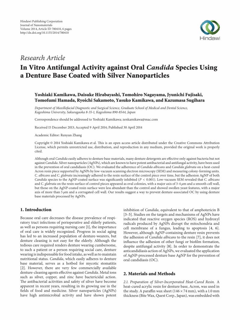
Figure 1: Macroscopic observation of (a) Candida albicans (C. albicans) and (b) Candida glabrata (C. glabrata) growth on CHROMagarCandidamedium, in which the Acron pieces (AgNP-coated and uncoated) were rinsed with 10mL PBS solution and incubated with the yeastsuspension for 1, 3, 8, and 12 h.
plaster and washed with hot water after curing. A mixtureof poly(methyl methacrylate) powder and solution (PMMA;Acron, GC Co. Ltd., Japan), at ratio of 100 g : 46 mL, was usedto infill the gap in the plaster at 100∘C and 15MPa for 15min.Theplasterwas carefully removed to obtain a heat-cured resinsheet with a 1.0mm thickness, and the Acron piece was cutinto 10mm × 10mm pieces to be used in the study. AgNPswere obtained bymicrowave irradiation of a solutionmixturecontaining silver nitrate, polyethylene glycol, ethanol, water,and 24%/5% w/w aqueous ammonia in the presence of theAcron piece, yielding an Acron-AgNP composite [9]. AnAcron piece without AgNP coating was used as the control.
2.2. Preparation of Yeast Culture Solution. The Candida stan-dard strains used in the study were C. albicans (ATCC18804)andC. glabrata (ATCC90030).The suspension solution of theCandida standard strains was prepared in RPMI 1640 culturemedium adjusted to a McFarland factor of 0.5 (absorbance at530 nm).
2.3. Adhesion Assay and Analysis. Candidal adhesion toAcron with (processed) or without (control) silver incorpo-ration was assayed in sterile microplates (12 wells/plates).Each piece was placed in a well containing 1mL of the yeastsuspension (5 × 10⁵ cells/mL), in order to cover the piececompletely, and then incubated at 36∘C for 1, 3, 8, and 12 h.Five processed pieces and 5 control pieces were used foreach incubation time. Colony-forming capability, assessed bycolony-forming units (CFU)/mL, was determined as follows:the test piece was removed from the cell suspension, rinsed3 times with phosphate-buffered saline (PBS) to remove non-adherentCandida, and transmitted to a new sterile petri dish.Adherent Candida was then removed by shaking for 10minwith 1mL 0.1% TritonX-100 PBS solution. A 10𝜇L aliquot
of the solution was inoculated on CHROMagar Candidamedium (Nippon Becton and Dickinson Company, Ltd.,Japan) and incubated for 48 h to determine CFU/mL. Eachpiece was rinsed 3 times with PBS solution, fixed with 5%glutaraldehyde solution for 60min, and incubated with 2%osmic acid for 30min. The surface of the test pieces wereobserved with a low-vacuum scanning electron microscope(SEM, TM-1000, Hitachi, Japan) with magnification ratios of1,000x and 10,000x.
2.4. Statistical Analysis. The numbers of C. albicans and C.glabrata adherent to the surface of Acron pieces with andwithout silver coating were evaluated by two-way analysis ofvariance, followed by Tukey’s test. A significance level of 0.05was used for all analytical tests.
3. Results
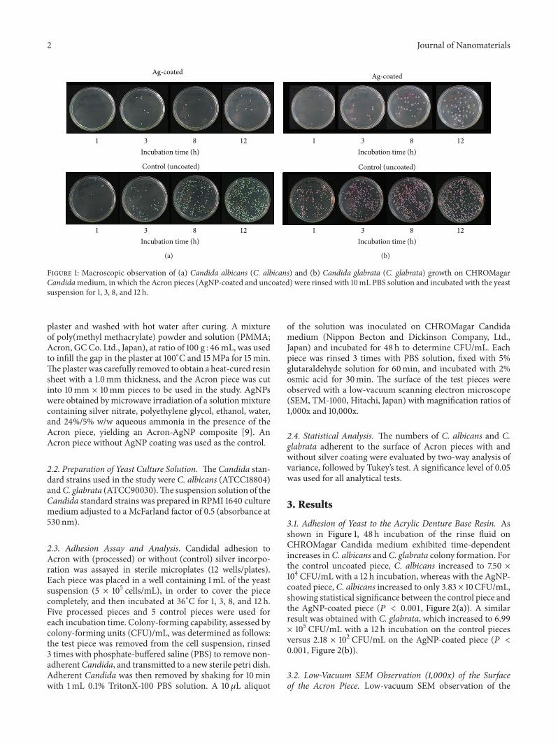
3.1. Adhesion of Yeast to the Acrylic Denture Base Resin. Asshown in Figure 1, 48 h incubation of the rinse fluid onCHROMagar Candida medium exhibited time-dependentincreases in C. albicans and C. glabrata colony formation. Forthe control uncoated piece, C. albicans increased to 7.50 ×10⁴ CFU/mL with a 12 h incubation, whereas with the AgNP-coated piece, C. albicans increased to only 3.83 × 10 CFU/mL,showing statistical significance between the control piece andthe AgNP-coated piece (𝑃 < 0.001, Figure 2(a)). A similarresult was obtained with C. glabrata, which increased to 6.99× 10⁵ CFU/mL with a 12 h incubation on the control piecesversus 2.18 × 10² CFU/mL on the AgNP-coated piece (𝑃 <0.001, Figure 2(b)).
3.2. Low-Vacuum SEM Observation (1,000x) of the Surfaceof the Acron Piece. Low-vacuum SEM observation of the
Journal of Nanomaterials 3
1
10
100
1000
10000
100000
1000000
1 3 8 12
Adhe
red
cells
(CFU
/mL)
Incubation time (hr)
ControlAg-coated
∗
∗
∗ ∗
(a)
ControlAg-coated
1
10
100
1000
10000
100000
1000000
10000000
1 3 8 12
Adhe
red
cells
(CFU
/mL)
Incubation time (hr)
∗∗
∗ ∗
(b)
Figure 2: Number of adherent cells on an Acron piece coated with AgNPs (Ag-coated, 𝑛 = 5) or without AgNPs (control, 𝑛 = 5), as measuredby counting colony forming units (CFU) on CHROMagar Candidamedium, where 10 mL of PBS rinse solution from the Acron pieces (Ag-coated or uncoated) were incubated with (a)C. albicans and (b)C. glabrata for 1, 3, 8, or 12 h. ∗Significantly different from control (𝑃 < 0.001).
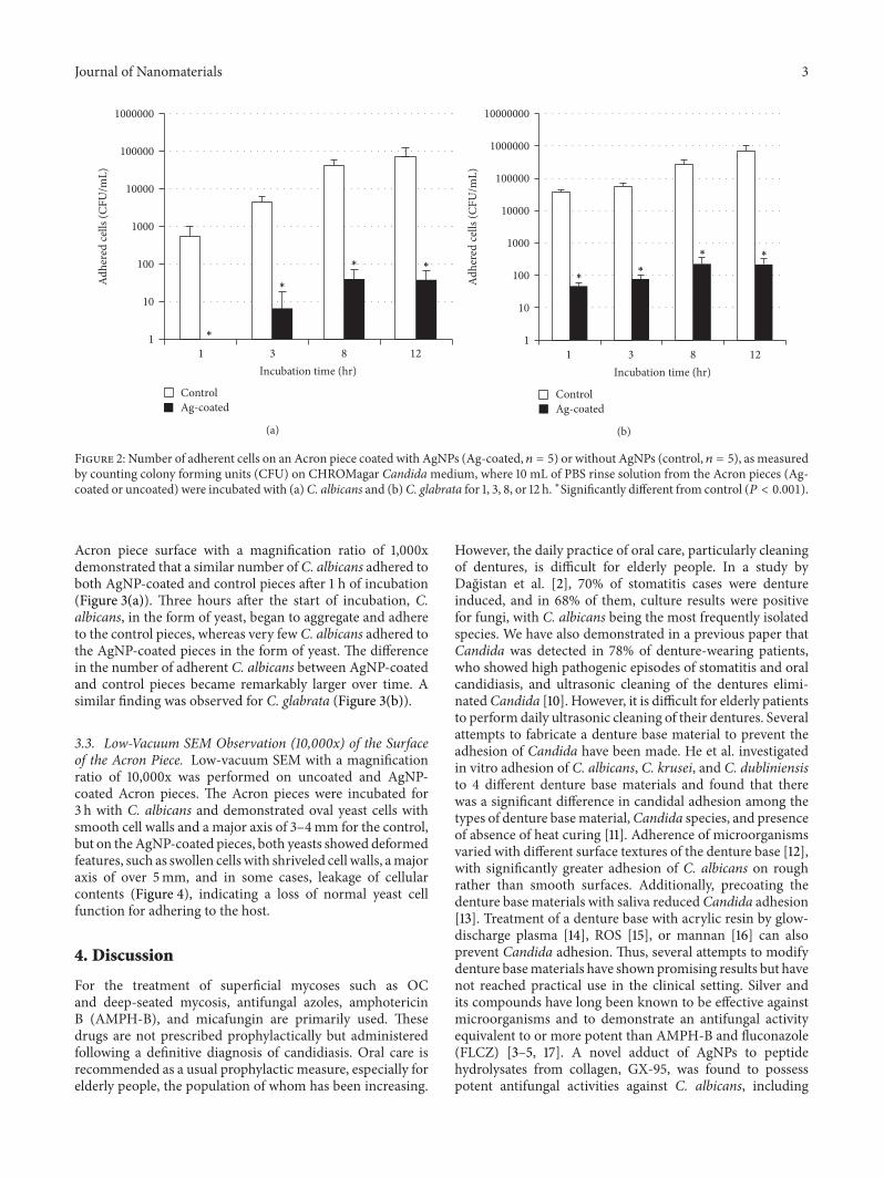
Acron piece surface with a magnification ratio of 1,000xdemonstrated that a similar number ofC. albicans adhered toboth AgNP-coated and control pieces after 1 h of incubation(Figure 3(a)). Three hours after the start of incubation, C.albicans, in the form of yeast, began to aggregate and adhereto the control pieces, whereas very few C. albicans adhered tothe AgNP-coated pieces in the form of yeast. The differencein the number of adherent C. albicans between AgNP-coatedand control pieces became remarkably larger over time. Asimilar finding was observed for C. glabrata (Figure 3(b)).
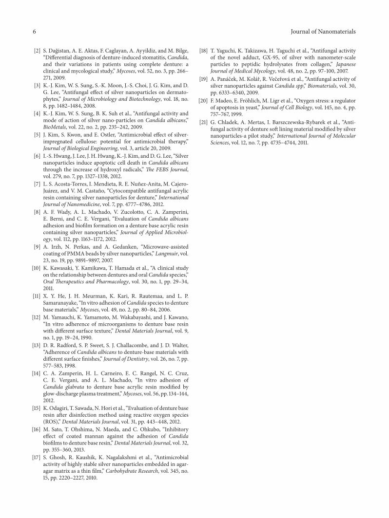
3.3. Low-Vacuum SEM Observation (10,000x) of the Surfaceof the Acron Piece. Low-vacuum SEM with a magnificationratio of 10,000x was performed on uncoated and AgNP-coated Acron pieces. The Acron pieces were incubated for3 h with C. albicans and demonstrated oval yeast cells withsmooth cell walls and a major axis of 3–4mm for the control,but on theAgNP-coated pieces, both yeasts showed deformedfeatures, such as swollen cells with shriveled cell walls, amajoraxis of over 5mm, and in some cases, leakage of cellularcontents (Figure 4), indicating a loss of normal yeast cellfunction for adhering to the host.
4. Discussion
For the treatment of superficial mycoses such as OCand deep-seated mycosis, antifungal azoles, amphotericinB (AMPH-B), and micafungin are primarily used. Thesedrugs are not prescribed prophylactically but administeredfollowing a definitive diagnosis of candidiasis. Oral care isrecommended as a usual prophylactic measure, especially forelderly people, the population of whom has been increasing.
However, the daily practice of oral care, particularly cleaningof dentures, is difficult for elderly people. In a study byDagistan et al. [2], 70% of stomatitis cases were dentureinduced, and in 68% of them, culture results were positivefor fungi, with C. albicans being the most frequently isolatedspecies. We have also demonstrated in a previous paper thatCandida was detected in 78% of denture-wearing patients,who showed high pathogenic episodes of stomatitis and oralcandidiasis, and ultrasonic cleaning of the dentures elimi-natedCandida [10]. However, it is difficult for elderly patientsto perform daily ultrasonic cleaning of their dentures. Severalattempts to fabricate a denture base material to prevent theadhesion of Candida have been made. He et al. investigatedin vitro adhesion of C. albicans, C. krusei, and C. dubliniensisto 4 different denture base materials and found that therewas a significant difference in candidal adhesion among thetypes of denture basematerial,Candida species, and presenceof absence of heat curing [11]. Adherence of microorganismsvaried with different surface textures of the denture base [12],with significantly greater adhesion of C. albicans on roughrather than smooth surfaces. Additionally, precoating thedenture base materials with saliva reduced Candida adhesion[13]. Treatment of a denture base with acrylic resin by glow-discharge plasma [14], ROS [15], or mannan [16] can alsoprevent Candida adhesion. Thus, several attempts to modifydenture basematerials have shownpromising results but havenot reached practical use in the clinical setting. Silver andits compounds have long been known to be effective againstmicroorganisms and to demonstrate an antifungal activityequivalent to or more potent than AMPH-B and fluconazole(FLCZ) [3–5, 17]. A novel adduct of AgNPs to peptidehydrolysates from collagen, GX-95, was found to possesspotent antifungal activities against C. albicans, including

4 Journal of Nanomaterials
Ag-coated
Control (uncoated)
1 3 8 12
Incubation time (h)
1 3 8 12
Incubation time (h)
(a)
Ag-coated
Control (uncoated)
1 3 8 12
Incubation time (h)
1 3 8 12
Incubation time (h)
(b)
Figure 3: Low-vacuum scanning electronmicroscopic observation (1,000x) of the surface of an AgNP-coated and uncoated Acron piece.TheAcron piece was incubated with (a) C. albicans or (b) C. glabrata for 1, 3, 8, or 12 h. (a) Although similar numbers of C. albicans adhered toboth AgNP-coated and control pieces at 1 h, C. albicans began to aggregate and adhere to the control pieces in the form of yeast at 3 h, whereasonly a few C. albicans adhered to AgNP-coated pieces in the form of yeast. The difference in the number of adherent C. albicans increasedbetween the AgNP-coated and control pieces with incubation time. (b) A similar observation was observed with C. glabrata.
strains resistant to FLCZ, itraconazole, and flucytosine [18].In addition, the cytotoxicity of silver, especially of AgNPs, tohuman cells was found negligible in comparison with thatto fungal cells [5, 19]. The antifungal effects by silver areattributable to the disruption of the structure of the fungalcell membrane due to the destruction of membrane integrity,
resulting in leakage of intracellular ions and other materialsand to the inhibition of the normal budding process byaffecting the cell cycle at the G2/M phase [4]. C. albicansexposed to AgNPs exhibited increased ROS and hydroxylradical production, resulting in mitochondrial dysfunctionand apoptosis [6]. Because intracellular accumulation of ROS

Journal of Nanomaterials 5
Control (uncoated)
5𝜇m
C. albicans
(a)
Ag-coated
5𝜇m
(b)
5𝜇m
C. glabrata
(c)
5𝜇m
(d)
Figure 4: Low-vacuum scanning electron microscopy (10,000x) of the surface of Acron pieces coated with (Ag-coated) and without(uncoated) silver nanoparticles. Acron pieces were incubated with (a) C. albicans and (b) C. glabrata for 3 h. C. albicans and C. glabratahad oval yeast cells with smooth cell walls and a major axis of 3-4mm in the control, but the Ag-coated piece showed swollen yeast cells withshriveled cell walls and a major axis of over 5mm, with leakage of cellular contents in some cases.
is accompanied by a mutation in CDC48 or by expression ofmammalian bax, which are triggers of apoptosis, the genera-tion of oxygen radicals is a key event in the ancestral apoptoticpathway of yeast [20]. Application of silver, especially AgNPs,to denture base materials inhibits the adhesion of Candidato the denture surface and hence prevents OC. Acosta-Torres et al. reported that PMMA-AgNP discs significantlyreduced the adherence of C. albicans in vitro, presenting anew biocompatible antifungal PMMA denture base material[7]. Chladek et al. found in vitro antifungal efficacy of16.3–52.5% by modifying the soft lining of dentures withAgNPs [21]. Wady et al. demonstrated that an AgNP solutionwith antifungal activity against C. albicans showed no effecton C. albicans adherence and biofilm formation after itsincorporation into a denture base resin [8]. Thus, potentantifungal activity of AgNPs has been commonly observedbut inhibitory activity against Candida adhesion to a denturebase has not. In the present study, adherence of C. albicansandC. glabrata to the surface of heat-curedAcron resin piecesmodified by AgNPs was significantly lower than that of thecontrol Acron piece, based on CFU. By SEM observation, C.albicans and C. glabrata on the surface of the control Acronpiece showed oval yeast cells with smooth cell walls and amajor axis of 3-4 𝜇m, but they were remarkably deformedby the AgNP coating, as evidenced by swollen cells withshriveled cell walls, a major axis of over 5𝜇m, and leakageof cellular components. The latter effect resulted in a loss inability of yeast cells to adhere to the host.Thus, AgNPs-coated
denture base material has several advantages in inhibitingcandidal adhesion on the denture surface comparing AgNPs-contained denture basematerial, such as easy preparation andhigh exposure of silver ions on the denture surface.
In conclusion, potent antifungal effects of AgNP coatingof denture base material were demonstrated in vitro, asshown by inhibition of Candida adherence to the denturematerial surface and deformation of the normal morphologyof Candida. Further attempts to apply AgNP-coated denturebase materials for clinical use are expected.
Conflict of Interests
The authors declare that there is no conflict of interestsregarding the publication of this paper.
Acknowledgment
The authors express special thanks to Akihiro Matsuura,Ph.D., Medi-Phi Co., Ltd., for his great contribution to dataanalysis and preparation of the paper.
References
[1] T. Yoneyama, M. Yoshida, T. Matsui, H. Sasaki, and The OralCare Working Group, “Oral care and pneumonia,” The Lancet,vol. 354, no. 9177, p. 515, 1999.
6 Journal of Nanomaterials
[2] S. Dagistan, A. E. Aktas, F. Caglayan, A. Ayyildiz, and M. Bilge,“Differential diagnosis of denture-induced stomatitis, Candida,and their variations in patients using complete denture: aclinical and mycological study,”Mycoses, vol. 52, no. 3, pp. 266–271, 2009.
[3] K.-J. Kim, W. S. Sung, S.-K. Moon, J.-S. Choi, J. G. Kim, and D.G. Lee, “Antifungal effect of silver nanoparticles on dermato-phytes,” Journal of Microbiology and Biotechnology, vol. 18, no.8, pp. 1482–1484, 2008.
[4] K.-J. Kim, W. S. Sung, B. K. Suh et al., “Antifungal activity andmode of action of silver nano-particles on Candida albicans,”BioMetals, vol. 22, no. 2, pp. 235–242, 2009.
[5] J. Kim, S. Kwon, and E. Ostler, “Antimicrobial effect of silver-impregnated cellulose: potential for antimicrobial therapy,”Journal of Biological Engineering, vol. 3, article 20, 2009.
[6] I.-S. Hwang, J. Lee, J. H.Hwang, K.-J. Kim, andD.G. Lee, “Silvernanoparticles induce apoptotic cell death in Candida albicansthrough the increase of hydroxyl radicals,” The FEBS Journal,vol. 279, no. 7, pp. 1327–1338, 2012.
[7] L. S. Acosta-Torres, I. Mendieta, R. E. Nunez-Anita, M. Cajero-Juarez, and V. M. Castano, “Cytocompatible antifungal acrylicresin containing silver nanoparticles for denture,” InternationalJournal of Nanomedicine, vol. 7, pp. 4777–4786, 2012.
[8] A. F. Wady, A. L. Machado, V. Zucolotto, C. A. Zamperini,E. Berni, and C. E. Vergani, “Evaluation of Candida albicansadhesion and biofilm formation on a denture base acrylic resincontaining silver nanoparticles,” Journal of Applied Microbiol-ogy, vol. 112, pp. 1163–1172, 2012.
[9] A. Irzh, N. Perkas, and A. Gedanken, “Microwave-assistedcoating of PMMAbeads by silver nanoparticles,” Langmuir, vol.23, no. 19, pp. 9891–9897, 2007.
[10] K. Kawasaki, Y. Kamikawa, T. Hamada et al., “A clinical studyon the relationship between dentures and oralCandida species,”Oral Therapeutics and Pharmacology, vol. 30, no. 1, pp. 29–34,2011.
[11] X. Y. He, J. H. Meurman, K. Kari, R. Rautemaa, and L. P.Samaranayake, “In vitro adhesion ofCandida species to denturebase materials,”Mycoses, vol. 49, no. 2, pp. 80–84, 2006.
[12] M. Yamauchi, K. Yamamoto, M. Wakabayashi, and J. Kawano,“In vitro adherence of microorganisms to denture base resinwith different surface texture,” Dental Materials Journal, vol. 9,no. 1, pp. 19–24, 1990.
[13] D. R. Radford, S. P. Sweet, S. J. Challacombe, and J. D. Walter,“Adherence of Candida albicans to denture-base materials withdifferent surface finishes,” Journal of Dentistry, vol. 26, no. 7, pp.577–583, 1998.
[14] C. A. Zamperin, H. L. Carneiro, E. C. Rangel, N. C. Cruz,C. E. Vergani, and A. L. Machado, “In vitro adhesion ofCandida glabrata to denture base acrylic resin modified byglow-discharge plasma treatment,”Mycoses, vol. 56, pp. 134–144,2012.
[15] K.Odagiri, T. Sawada,N.Hori et al., “Evaluation of denture baseresin after disinfection method using reactive oxygen species(ROS),” Dental Materials Journal, vol. 31, pp. 443–448, 2012.
[16] M. Sato, T. Ohshima, N. Maeda, and C. Ohkubo, “Inhibitoryeffect of coated mannan against the adhesion of Candidabiofilms to denture base resin,”DentalMaterials Journal, vol. 32,pp. 355–360, 2013.
[17] S. Ghosh, R. Kaushik, K. Nagalakshmi et al., “Antimicrobialactivity of highly stable silver nanoparticles embedded in agar-agar matrix as a thin film,” Carbohydrate Research, vol. 345, no.15, pp. 2220–2227, 2010.
[18] T. Yaguchi, K. Takizawa, H. Taguchi et al., “Antifungal activityof the novel adduct, GX-95, of silver with nanometer-scaleparticles to peptidic hydrolysates from collagen,” JapaneseJournal of Medical Mycology, vol. 48, no. 2, pp. 97–100, 2007.
[19] A. Panacek, M. Kolar, R. Vecerova et al., “Antifungal activity ofsilver nanoparticles against Candida spp,” Biomaterials, vol. 30,pp. 6333–6340, 2009.
[20] F. Madeo, E. Frohlich, M. Ligr et al., “Oxygen stress: a regulatorof apoptosis in yeast,” Journal of Cell Biology, vol. 145, no. 4, pp.757–767, 1999.
[21] G. Chladek, A. Mertas, I. Barszczewska-Rybarek et al., “Anti-fungal activity of denture soft lining material modified by silvernanoparticles-a pilot study,” International Journal of MolecularSciences, vol. 12, no. 7, pp. 4735–4744, 2011.

Submit your manuscripts athttp://www.hindawi.com
ScientificaHindawi Publishing Corporationhttp://www.hindawi.com Volume 2014
CorrosionInternational Journal of
Hindawi Publishing Corporationhttp://www.hindawi.com Volume 2014
Polymer ScienceInternational Journal of
Hindawi Publishing Corporationhttp://www.hindawi.com Volume 2014
Hindawi Publishing Corporationhttp://www.hindawi.com Volume 2014
CeramicsJournal of
Hindawi Publishing Corporationhttp://www.hindawi.com Volume 2014
CompositesJournal of
NanoparticlesJournal of
Hindawi Publishing Corporationhttp://www.hindawi.com Volume 2014
Hindawi Publishing Corporationhttp://www.hindawi.com Volume 2014
International Journal of
Biomaterials
Hindawi Publishing Corporationhttp://www.hindawi.com Volume 2014
NanoscienceJournal of
TextilesHindawi Publishing Corporation http://www.hindawi.com Volume 2014
Journal of
NanotechnologyHindawi Publishing Corporationhttp://www.hindawi.com Volume 2014
Journal of
CrystallographyJournal of
Hindawi Publishing Corporationhttp://www.hindawi.com Volume 2014
The Scientific World JournalHindawi Publishing Corporation http://www.hindawi.com Volume 2014
Hindawi Publishing Corporationhttp://www.hindawi.com Volume 2014
CoatingsJournal of
Advances in
Materials Science and EngineeringHindawi Publishing Corporationhttp://www.hindawi.com Volume 2014
Smart Materials Research
Hindawi Publishing Corporationhttp://www.hindawi.com Volume 2014
Hindawi Publishing Corporationhttp://www.hindawi.com Volume 2014
MetallurgyJournal of
Hindawi Publishing Corporationhttp://www.hindawi.com Volume 2014
BioMed Research International
MaterialsJournal of
Hindawi Publishing Corporationhttp://www.hindawi.com Volume 2014
Nano
materials
Hindawi Publishing Corporationhttp://www.hindawi.com Volume 2014
Journal ofNanomaterials


![1110 Zapf [Read-Only] - Orthopaedic Research and Education5/8/2017 3 Treatment • Rx topical antifungal 2x per day for 2-4 weeks • Rx oral antifungal • • Those who do not respond](https://img.dokumen.tips/doc/110x75/5e27b054054e0f11ba65418c/1110-zapf-read-only-orthopaedic-research-and-582017-3-treatment-a-rx-topical.jpg)