Embed Size (px)
Citation preview
Research ArticleHPV Infection, but Not EBV or HHV-8 Infection,Is Associated with Salivary Gland Tumours
Maja Hühns,1 Georg Simm,1 Andreas Erbersdobler,1 and Annette Zimpfer1,2
1 Institute of Pathology, University of Rostock, Strempelstrasse 14, 18055 Rostock, Germany2Institute of Pathology, University Medical Centre Jena, Ziegelmuhlenweg 1, 07743 Jena, Germany
Correspondence should be addressed to Maja Huhns; [email protected]
Received 1 July 2015; Accepted 21 October 2015
Academic Editor: Monica Cantile
Copyright © 2015 Maja Huhns et al. This is an open access article distributed under the Creative Commons Attribution License,which permits unrestricted use, distribution, and reproduction in any medium, provided the original work is properly cited.
Benign and malignant salivary gland tumours are clinically heterogeneous and show different histology. Little is known about therole of human herpes virus 8 (HHV-8), Epstein-Barr virus (EBV), and human papillomavirus (HPV) infection in salivary glandneoplasms. We investigated the presence of the three viruses in formalin-fixed, paraffin-embedded tissue samples in a cohort of200 different salivary gland tumours.We performed EBV-LMP-1 and HHV-8 and p16 immunohistochemistry, a specific chip basedhybridization assay for detection and typing of HPV and a chromogenic in situ hybridization for EBV analysis. Only one case, apolymorphic low-grade carcinoma, showed HHV-8 expression and one lymphoepithelial carcinoma was infected by EBV. In 17cases (9%) moderate or strong nuclear and cytoplasmic p16 expression was detected. The HPV type was investigated in all of thesecases and additionally in 8 Warthin’s tumours. In 19 cases HPV type 16 was detected, mostly in Warthin’s tumour, adenoid cysticcarcinoma, and adenocarcinoma NOS. We concluded that HHV-8 infection and EBV infection are not associated with salivarygland cancer, but HPV infection may play a role in these tumour entities.
1. Introduction
Benign and malignant salivary gland tumours belong to rarehead and neck tumours. Most cases are benign and only 20%are malignant [1]. The majority of the diseases arise in thesixth decade and the sex distribution is equal [2]. Salivarygland tumours show a wide range of phenotypic, biological,and clinical heterogeneity [3]. They occur in the major andminor salivary glands, whereof 80% of major salivary glandtumours are present in the parotid glands, and less than halfof these tumours are malignant [3].
In general, several viruses have been demonstrated tobe the trigger of neoplastic diseases of the head and neck,like human papillomavirus (HPV) [4] and different humanherpes viruses, like human herpes virus-4/Epstein-Barr virus(EBV) [5], cytomegalovirus (CMV) [6], and human herpesvirus 8 (HHV-8) [7]. However, the role of viruses in the gen-esis of salivary gland tumours remains still debatable.
The aim of the present study was to determinethe prevalence of different pathogens like HHV-8, EBV,and HPV in a large cohort of salivary gland tumours. The
involvement of these three pathogens was analyzed byimmunohistochemistry, a specific chip based hybridizationassay, or chromogenic in situ hybridization.
2. Material and Methods
2.1. Patients. A total of 200 patients with salivary glandtumours were included, diagnosed between 1990 and 2014(Table 1). Among those there were 93 malignant and 107benign tumours of different entities (Table 2). The formalin-fixed, paraffin embedded specimens were retrieved fromthe archive of the Institute of Pathology at the UniversityMedicine of Rostock.
The study was performed with internal review boardapproval and patients’ data were anonymized in accordancewith German laws concerning safety data.
2.2. Clinical Data. Clinical data were obtained by reviewingthe charts of the Clinical Cancer Registry, University ofRostock. These data were anonymized and included sex, ageat diagnosis, and stage (Tables 1 and 2).
Hindawi Publishing CorporationBioMed Research InternationalVolume 2015, Article ID 829349, 7 pageshttp://dx.doi.org/10.1155/2015/829349
2 BioMed Research International
Table 1: Patient characteristics of salivary gland tumours.
Clinical characteristics 𝑛 = 200
Median age, years 59.9Range 11–95Sex
Male 107 (53.5%)Female 93 (46.5%)
Tumour locationParotid gland 175 (87.5%)Left side 84 (48%)Right side 69 (39.5%)
Submandibular gland 14 (7%)Left side 7 (3.5%)Right side 7 (3.5%)
Minor salivary glands 11 (5.5%)Stage at presentation (malignant)
I 36 (38.7%)II 18 (19.4%)III 20 (21.5%)IV 17 (18.3%)
No stadium determinable 2 (2.2%)
2.3. Construction of Tissue Microarrays (TMA). For TMAconstruction, a hematoxylin and eosin (H&E) stained slidefrom each block was used to define the representative tumourand normal region by an experienced pathologist. Tissuecylinders with a diameter of 1.0mm were punched from thespecimen block and brought into an empty paraffin block[8] by using a precision instrument (Beecher Instruments,Silver Spring, MD, USA). Three tissue cylinders of tumourand one cylinder with normal tissue from every specimenwere prepared. Taken together, 10 different TMA blocks withmalignant and benign specimens were constructed.
2.4. Immunohistochemistry. For each TMA block, four-mi-crometer sections were transferred to an adhesive-coatedglass slide system (Instrumedics Inc, Hackensack, NJ, USA)and stained with H&E. Only cases containing at least 10%tumour tissue were further analyzed.
Immunohistochemical staining was performed with anautostainer (EnVision FLEX, High pH, (Link), DAKO, Ham-burg, Germany) according to the manufacturer’s standardprotocol with primary antibodies against Cytokeratin AE1/AE3 (monoclonal mouse, reference number: C1702C01, titre1 : 500, DCS, Hamburg, Germany), EBV-latent membraneprotein-1 (LMP-1) (monoclonal mouse, clones CS.1–4, “readyto use,” Dako, Hamburg, Germany), HHV-8 (monoclonalmouse, 1 : 50, reference number 6011336, Leica, Wetzlar,Germany), and p16 (monoclonal mouse, clone G175-405,1 : 20, BD Biosciences, Heidelberg, Germany).
Cytokeratin AE1/AE3 immunohistochemistry served asa positive control for the tissue studied and proved thestainability of the archival material.
For LMP1 of EBV the presence of unequivocal mem-branous and cytoplasmatic staining in >2% of tumour cells
was considered positive. For HHV-8 the presence of nuclearimmunoreactivity in >2% of tumour cells was consideredpositive. Positivity for p16 was considered when a moderateto strong staining was observed in the cytoplasm and inthe nucleus. The staining intensities were graded as negative,weak, moderate, and strong by visual inspection by anexperienced pathologist. Weak and patchy p16 signalling wasjudged as negative. For positive controls, (tumour) tissueswith known marker expression were carried along. The pos-itive controls were as follows: HHV8: Kaposi sarcoma in anAIDS patient; EBV-LMP-1: tonsil with a known infectiousmononucleosis (EBV positive); EBER-CISH: tonsil with aknown infectious mononucleosis (EBV positive); P16: carci-noma of the tonsil with a known HPV infection.
In the negative control experiments, the primary antibod-ies were omitted.
2.5. Detection of EBV. Chromogenic in situ hybridization(CISH) staining for EBV encoded RNA (EBER) transcriptswas performed on 4𝜇m deparaffinized tissue sections,mounted on adhesive coated glass slides according to manu-facturer’s instructions (ZytoVision, Bremerhaven, Germany).Slides were digested with pepsin solution for 10 minutes,incubated with biotin-labelled ZytoFast EBV probe for 60minutes at 55∘C, followed by incubationwith AP-streptavidinfor 30 minutes at 37∘C, and colouring was performed withNBT/BZIP at 37∘C for maximal 40 minutes. Cells exhibitingnuclear staining were considered positive. Positive and nega-tive controls were included in each run.
2.6. Identification ofHPVTypes. Thecases selected formolec-ular HPV analysis were all tumours with moderate or strongcytoplasmic and nuclear p16 expression and additionallyin 8 Warthin’s tumours. Tumour DNA was extracted fromformalin-fixed, paraffin embedded sections in 25 salivarygland cancers with the ReliaPrep FFPE gDNA Miniprepsystem (Promega, Mannheim, Germany) according to man-ufacturers’ instruction. For identification of HPV types theHPV Type 3.5 LCD-Array Kit (Chipron, Berlin, Germany)was used according to manufacturer’s instructions. Briefly,two PCR reactions were performed using supplied My11/09(product size 450 bp) and “125” (product size 125 bp) primermixes and analyzed by agarose gel electrophoresis. BothPCR products were mixed and hybridized on the 3.5 LCDchip slide. The slide was subsequently scanned on the SlideReader Scanner and evaluated with the Slide Reader Software(Chipron, Berlin, Germany).
3. Results
3.1. Patient and Tumour Characteristics. We analyzed 200patients with malignant or benign salivary gland tumours,diagnosed between 1990 and 2014 (Table 1). The mean ageat diagnosis was 58.9 years (range 11–95 years), 107 weremale (53.5%), and 93 were female (46.5%). Different tumourentities were included, whereas in malignant tumours mainlymucoepidermoid carcinoma (18.3%), adenoid cystic carci-noma (17.2%), and adenocarcinoma NOS (10.8%) occurred.
BioMed Research International 3
Table 2: Tumour characteristics of malignant and benign tumours (𝑛 = 200).
Type of tumour Number of cases Frequency in %
Malignant
Mucoepidermoid carcinoma 17 18.3Adenoid cystic carcinoma 16 17.2Adenocarcinoma NOS 10 10.8Salivary duct carcinoma 9 9.7Acinus cell carcinoma 7 7.5Adenoid basal-cell carcinoma 5 5.4Squamous cell carcinoma 5 5.4Nonkeratinized squamous cell carcinoma 4 4.3Keratinized squamous cell carcinoma 4 4.3Oncocytic carcinoma 2 2.2Lymphoepithelial carcinoma 2 2.2Micropapillary carcinoma 2 2.2Myoepithelial carcinoma 5 5.4Pseudo sarcomatoid carcinoma 1 1.1Polymorphic low-grade carcinoma 1 1.1Undifferentiated carcinoma 1 1.1Cystadenocarcinoma 1 1.1Malignant melanoma 1 1.1
Benign
Cystadenolymphoma (Warthin’s tumour) 46 43Pleomorphic adenoma 33 30.8Basal-cell adenoma 15 14.0Oncocytoma 7 6.5Myoepithelioma 3 2.9Cystadenoma 2 1.9Adenolyphoma 1 0.9
NOS, not otherwise specified.
In benign tumours basically Warthin’s tumours (43%) andpleomorphic adenoma (33%) were diagnosed.
3.2. Morphological Evaluation of the Specimens. 190/200(95%) of the specimens contained >10% tumour tissue. Tencases (5%) had limited tumour tissue andwere excluded fromthe study.
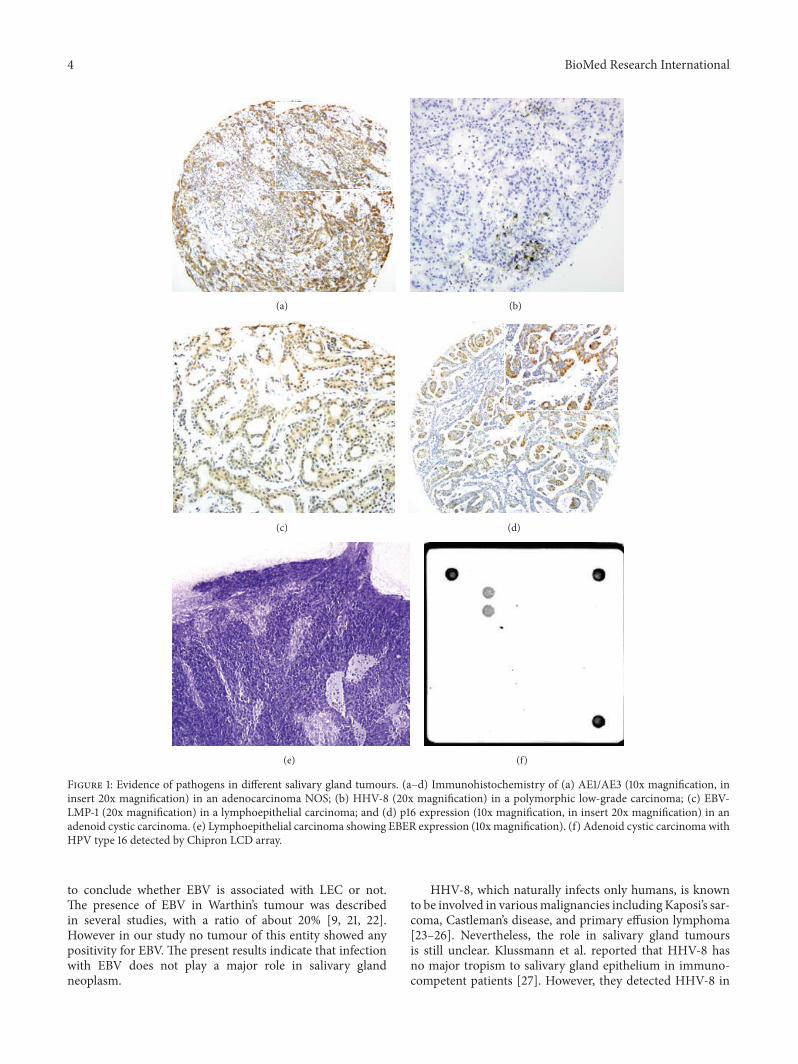
3.3. Expression and Detection of HHV-8, EBV, and HPVby Immunohistochemistry. AE1/AE3 positivity was found in183/190 (96.3%) cases, indicating that most tumours were ofepithelial origin and were suitable for further investigations(Figure 1(a)).
Only one case, a polymorphic low-grade carcinomaexhibited HHV-8 expression, located in the nucleus(Figure 1(b)).
A small subfraction of only 4 cases expressed the EBV-LPM-1 protein and just one case, a lymphoepithelial carci-noma, was positive in EBER-CISH analyses (Figures 1(c) and1(e)).
Moderate or strong nuclear and cytoplasmic p16 wasseen in 17/190 (9%) cases (Figure 1(d)). The most frequenttumour type with positive nuclear expression was adenoidcystic carcinoma, followed by adenocarcinoma and acinuscell carcinoma.
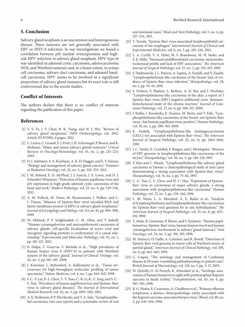
3.4. Detection of HPV. Detection of HPV types was per-formed in all 17 p16 nuclear positive cases and also in 8 War-thin’s tumours. In 19/25 cases (76%) HPV type was classified(Table 3); 6 cases were negative or DNA was not amplifiable.In all 19 positive cases HPV type 16 was detected (Figure 1(f))in different tumour entities (Table 4), mostly in malignantcancer types (12/25) compared to benign tumours (7/25).
4. Discussion
The participation of viruses in salivary gland tumours isreceiving increasing interest. The role of EBV in lymphoep-ithelial carcinomas (LEC), nasopharyngeal carcinomas, andalso benignWarthin’s tumourswas reported in several studies[9, 10]. In our study, we investigated different salivary glandtumours, including 46 Warthin’s tumours and two casesof LEC. But EBV was detected only in one LEC (Figures1(c) and 1(e)). LEC is a rare malignancy and only fewcases with EBV infection were described in the literature.It occurs mainly in East Asia population and only rarelyin western countries (reviewed in [11]). However, there aremany controversies about the association of EBV infectionwith LEC. Some studies described negative results [12–17],while other authors found a positive association [11, 18–20].On the basis of our data of only two cases, we are not able
4 BioMed Research International
(a) (b)
(c) (d)
(e) (f)
Figure 1: Evidence of pathogens in different salivary gland tumours. (a–d) Immunohistochemistry of (a) AE1/AE3 (10x magnification, ininsert 20x magnification) in an adenocarcinoma NOS; (b) HHV-8 (20x magnification) in a polymorphic low-grade carcinoma; (c) EBV-LMP-1 (20x magnification) in a lymphoepithelial carcinoma; and (d) p16 expression (10x magnification, in insert 20x magnification) in anadenoid cystic carcinoma. (e) Lymphoepithelial carcinoma showing EBER expression (10xmagnification). (f) Adenoid cystic carcinoma withHPV type 16 detected by Chipron LCD array.
to conclude whether EBV is associated with LEC or not.The presence of EBV in Warthin’s tumour was describedin several studies, with a ratio of about 20% [9, 21, 22].However in our study no tumour of this entity showed anypositivity for EBV. The present results indicate that infectionwith EBV does not play a major role in salivary glandneoplasm.
HHV-8, which naturally infects only humans, is knownto be involved in variousmalignancies including Kaposi’s sar-coma, Castleman’s disease, and primary effusion lymphoma[23–26]. Nevertheless, the role in salivary gland tumoursis still unclear. Klussmann et al. reported that HHV-8 hasno major tropism to salivary gland epithelium in immuno-competent patients [27]. However, they detected HHV-8 in
BioMed Research International 5
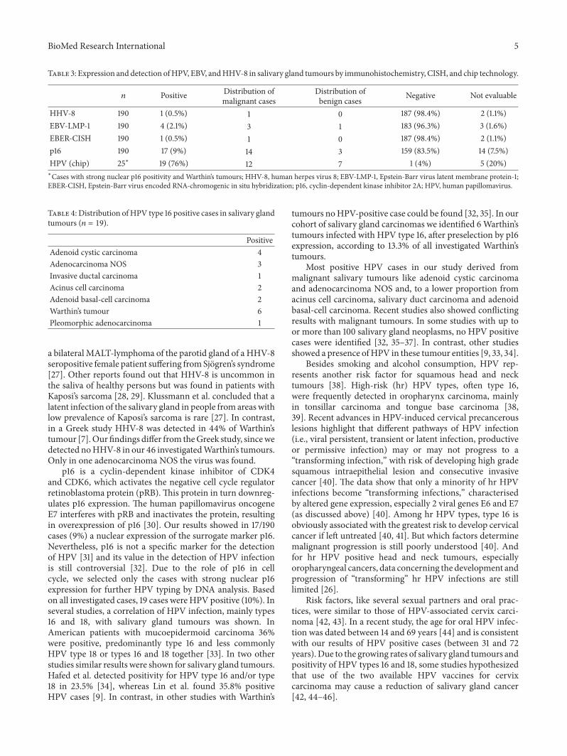
Table 3: Expression and detection ofHPV, EBV, andHHV-8 in salivary gland tumours by immunohistochemistry, CISH, and chip technology.
𝑛 Positive Distribution ofmalignant cases
Distribution ofbenign cases Negative Not evaluable
HHV-8 190 1 (0.5%) 1 0 187 (98.4%) 2 (1.1%)EBV-LMP-1 190 4 (2.1%) 3 1 183 (96.3%) 3 (1.6%)EBER-CISH 190 1 (0.5%) 1 0 187 (98.4%) 2 (1.1%)p16 190 17 (9%) 14 3 159 (83.5%) 14 (7.5%)HPV (chip) 25∗ 19 (76%) 12 7 1 (4%) 5 (20%)∗Cases with strong nuclear p16 positivity and Warthin’s tumours; HHV-8, human herpes virus 8; EBV-LMP-1, Epstein-Barr virus latent membrane protein-1;EBER-CISH, Epstein-Barr virus encoded RNA-chromogenic in situ hybridization; p16, cyclin-dependent kinase inhibitor 2A; HPV, human papillomavirus.
Table 4: Distribution ofHPV type 16 positive cases in salivary glandtumours (𝑛 = 19).
PositiveAdenoid cystic carcinoma 4Adenocarcinoma NOS 3Invasive ductal carcinoma 1Acinus cell carcinoma 2Adenoid basal-cell carcinoma 2Warthin’s tumour 6Pleomorphic adenocarcinoma 1
a bilateral MALT-lymphoma of the parotid gland of a HHV-8seropositive female patient suffering fromSjogren’s syndrome[27]. Other reports found out that HHV-8 is uncommon inthe saliva of healthy persons but was found in patients withKaposi’s sarcoma [28, 29]. Klussmann et al. concluded that alatent infection of the salivary gland in people from areas withlow prevalence of Kaposi’s sarcoma is rare [27]. In contrast,in a Greek study HHV-8 was detected in 44% of Warthin’stumour [7].Our findings differ from theGreek study, sincewedetected noHHV-8 in our 46 investigatedWarthin’s tumours.Only in one adenocarcinoma NOS the virus was found.
p16 is a cyclin-dependent kinase inhibitor of CDK4and CDK6, which activates the negative cell cycle regulatorretinoblastoma protein (pRB). This protein in turn downreg-ulates p16 expression. The human papillomavirus oncogeneE7 interferes with pRB and inactivates the protein, resultingin overexpression of p16 [30]. Our results showed in 17/190cases (9%) a nuclear expression of the surrogate marker p16.Nevertheless, p16 is not a specific marker for the detectionof HPV [31] and its value in the detection of HPV infectionis still controversial [32]. Due to the role of p16 in cellcycle, we selected only the cases with strong nuclear p16expression for further HPV typing by DNA analysis. Basedon all investigated cases, 19 cases were HPV positive (10%). Inseveral studies, a correlation of HPV infection, mainly types16 and 18, with salivary gland tumours was shown. InAmerican patients with mucoepidermoid carcinoma 36%were positive, predominantly type 16 and less commonlyHPV type 18 or types 16 and 18 together [33]. In two otherstudies similar results were shown for salivary gland tumours.Hafed et al. detected positivity for HPV type 16 and/or type18 in 23.5% [34], whereas Lin et al. found 35.8% positiveHPV cases [9]. In contrast, in other studies with Warthin’s
tumours noHPV-positive case could be found [32, 35]. In ourcohort of salivary gland carcinomas we identified 6Warthin’stumours infected with HPV type 16, after preselection by p16expression, according to 13.3% of all investigated Warthin’stumours.
Most positive HPV cases in our study derived frommalignant salivary tumours like adenoid cystic carcinomaand adenocarcinoma NOS and, to a lower proportion fromacinus cell carcinoma, salivary duct carcinoma and adenoidbasal-cell carcinoma. Recent studies also showed conflictingresults with malignant tumours. In some studies with up toor more than 100 salivary gland neoplasms, no HPV positivecases were identified [32, 35–37]. In contrast, other studiesshowed a presence ofHPV in these tumour entities [9, 33, 34].
Besides smoking and alcohol consumption, HPV rep-resents another risk factor for squamous head and necktumours [38]. High-risk (hr) HPV types, often type 16,were frequently detected in oropharynx carcinoma, mainlyin tonsillar carcinoma and tongue base carcinoma [38,39]. Recent advances in HPV-induced cervical precancerouslesions highlight that different pathways of HPV infection(i.e., viral persistent, transient or latent infection, productiveor permissive infection) may or may not progress to a“transforming infection,” with risk of developing high gradesquamous intraepithelial lesion and consecutive invasivecancer [40]. The data show that only a minority of hr HPVinfections become “transforming infections,” characterisedby altered gene expression, especially 2 viral genes E6 and E7(as discussed above) [40]. Among hr HPV types, type 16 isobviously associated with the greatest risk to develop cervicalcancer if left untreated [40, 41]. But which factors determinemalignant progression is still poorly understood [40]. Andfor hr HPV positive head and neck tumours, especiallyoropharyngeal cancers, data concerning the development andprogression of “transforming” hr HPV infections are stilllimited [26].
Risk factors, like several sexual partners and oral prac-tices, were similar to those of HPV-associated cervix carci-noma [42, 43]. In a recent study, the age for oral HPV infec-tion was dated between 14 and 69 years [44] and is consistentwith our results of HPV positive cases (between 31 and 72years).Due to the growing rates of salivary gland tumours andpositivity of HPV types 16 and 18, some studies hypothesizedthat use of the two available HPV vaccines for cervixcarcinoma may cause a reduction of salivary gland cancer[42, 44–46].
6 BioMed Research International
5. Conclusion
Salivary gland neoplasm is an uncommon and heterogeneousdisease. These tumours are not generally associated withEBV or HHV-8 infection. In our investigations we found acorrelation between p16 nuclear overexpression and high-risk HPV infection in salivary gland neoplasm. HPV type 16was identified in adenoid cystic carcinoma, adenocarcinomaNOS, andWarthin’s tumours and, to a lesser extent, in acinuscell carcinoma, salivary duct carcinoma, and adenoid basal-cell carcinoma. HPV seems to be involved in a significantproportion of salivary gland tumours but its exact role is stillcontroversial due to the recent studies.
Conflict of Interests
The authors declare that there is no conflict of interestsregarding the publication of this paper.
References
[1] V. S. To, J. Y. Chan, R. K. Tsang, and W. I. Wei, “Review ofsalivary gland neoplasms,” ISRN Otolaryngology, vol. 2012,Article ID 872982, 6 pages, 2012.
[2] L. Licitra, C.Grandi, F. J. Prott, J. H. Schornagel, P. Bruzzi, andR.Molinari, “Major and minor salivary glands tumours,” CriticalReviews in Oncology/Hematology, vol. 45, no. 2, pp. 215–225,2003.
[3] D. J. Adelstein, S. A. Koyfman, A. K. El-Naggar, and E. Y.Hanna,“Biology and management of salivary gland cancers,” Seminarsin Radiation Oncology, vol. 22, no. 3, pp. 245–253, 2012.
[4] J. M. Boland, E. D. McPhail, J. J. Garcıa, J. E. Lewis, and D. J.Schembri-Wismayer, “Detection of human papilloma virus andp16 expression in high-grade adenoid cystic carcinoma of thehead and neck,” Modern Pathology, vol. 25, no. 4, pp. 529–536,2012.
[5] A. M. Pollock, M. Toner, M. Mcmenamin, J. Walker, and C.I. Timon, “Absence of Epstein-Barr virus encoded RNA andlatent membrane protein (LMP1) in salivary gland neoplasms,”Journal of Laryngology andOtology, vol. 113, no. 10, pp. 906–908,1999.
[6] M. Melnick, P. P. Sedghizadeh, C. M. Allen, and T. Jaskoll,“Human cytomegalovirus and mucoepidermoid carcinoma ofsalivary glands: cell-specific localization of active viral andoncogenic signaling proteins is confirmatory of a causal rela-tionship,” Experimental and Molecular Pathology, vol. 92, no. 1,pp. 118–125, 2012.
[7] E. Dalpa, V. Gourvas, S. Baritaki et al., “High prevalence ofhuman herpes virus 8 (HHV-8) in patients with Warthin’stumors of the salivary gland,” Journal of Clinical Virology, vol.42, no. 2, pp. 182–185, 2008.
[8] J. Kononen, L. Bubendorf, A. Kallioniemi et al., “Tissue mi-croarrays for high-throughput molecular profiling of tumorspecimens,” Nature Medicine, vol. 4, no. 7, pp. 844–847, 1998.
[9] F. C.-F. Lin, P.-L. Chen, T.-Y. Tsao, C.-R. Li, K.-C. Jeng, and S. C.-S. Tsai, “Prevalence of human papillomavirus and Epstein–Barrvirus in salivary gland diseases,” The Journal of InternationalMedical Research, vol. 42, no. 5, pp. 1093–1101, 2014.
[10] A. E. Rytkonen, P. P. Hirvikoski, and T. A. Salo, “Lymphoepithe-lial carcinoma: two case reports and a systematic review of oral
and sinonasal cases,”Head and Neck Pathology, vol. 5, no. 4, pp.327–334, 2011.
[11] T. Terada, “Epstein-Barr virus associated lymphoepithelial car-cinoma of the esophagus,” International Journal of Clinical andExperimental Medicine, vol. 6, no. 3, pp. 219–226, 2013.
[12] L. A. Cerilli, V. A. Holst, M. S. Brandwein, M. H. Stoler, andS. E. Mills, “Sinonasal undifferentiated carcinoma: immunohis-tochemical profile and lack of EBV association,” The AmericanJournal of Surgical Pathology, vol. 25, no. 2, pp. 156–163, 2001.
[13] F. Dadmanesh, J. L. Peterse, A. Sapino, A. Fonelli, and V. Eusebi,“Lymphoepithelioma-like carcinoma of the breast: lack of evi-dence of Epstein-Barr virus infection,” Histopathology, vol. 38,no. 1, pp. 54–61, 2001.
[14] S. Ferlicot, F. Plantier, L. Rethers, A. D. Bui, and J. Wechsler,“Lymphoepithelioma-like carcinoma of the skin: a report of 3Epstein-Barr virus (EBV)-negative additional cases. Immuno-histochemical study of the stroma reaction,” Journal of Cuta-neous Pathology, vol. 27, no. 6, pp. 306–311, 2000.
[15] J. Kulka, I. Kovalszky, E. Svastics, M. Berta, and T. Fule, “Lym-phoepithelioma-like carcinoma of the breast: not Epstein-Barrvirus-, but human papilloma virus-positive,”Human Pathology,vol. 39, no. 2, pp. 298–301, 2008.
[16] E. Szekely, “Lymphoepithelioma-like cholangiocarcinoma(LELC) not associated with Epstein-Barr virus,” The AmericanJournal of Surgical Pathology, vol. 25, no. 11, pp. 1464–1466,2001.
[17] J. C. Tardıo, E. Cristobal, F. Burgos, and J. Menarguez, “Absenceof EBV genome in lymphoepithelioma-like carcinomas of thelarynx,” Histopathology, vol. 30, no. 2, pp. 126–128, 1997.
[18] T. Kuo and C. Hsueh, “Lymphoepithelioma-like salivary glandcarcinoma in Taiwan: a clinicopathological study of nine casesdemonstrating a strong association with Epstein-Barr virus,”Histopathology, vol. 31, no. 1, pp. 75–82, 1997.
[19] C.-C. Tsai, C.-L. Chen, and H.-C. Hsu, “Expression of Epstein-Barr virus in carcinomas of major salivary glands: a strongassociation with lymphoepithelioma-like carcinoma,” HumanPathology, vol. 27, no. 3, pp. 258–262, 1996.
[20] L. M. Weiss, L. A. Movahed, A. E. Butler et al., “Analysisof lymphoepithelioma and lymphoepithelioma-like carcinomasfor Epstein-Barr viral genomes by in situ hybridization,” TheAmerican Journal of Surgical Pathology, vol. 13, no. 8, pp. 625–631, 1989.
[21] T. Atula, R. Grenman, P. Klemi, and S. Syrjanen, “Human papil-lomavirus, Epstein-Barr virus, humanherpesvirus 8 and humancytomegalovirus involvement in salivary gland tumours,” OralOncology, vol. 34, no. 5, pp. 391–395, 1998.
[22] M. Santucci, O. Gallo, A. Calzolari, and R. Bondi, “Detection ofEpstein-Barr viral genome in tumor cells of Warthin’s tumor ofparotid gland,” American Journal of Clinical Pathology, vol. 100,no. 6, pp. 662–665, 1993.
[23] C. Casper, “The aetiology and management of Castlemandisease at 50 years: translating pathophysiology to patient care,”British Journal of Haematology, vol. 129, no. 1, pp. 3–17, 2005.
[24] W. Quinibi, O. Al-Furayh, K. Almeshari et al., “Serologic asso-ciation of human herpesvirus eight with posttransplant Kaposi’ssarcoma in Saudi Arabia,” Transplantation, vol. 65, no. 4, pp.583–585, 1998.
[25] R.G.Nador, E. Cesarman,A. Chadburn et al., “Primary effusionlymphoma: a distinct clinicopathologic entity associated withtheKaposi’s sarcoma-associated herpes virus,”Blood, vol. 88, no.2, pp. 645–656, 1996.
BioMed Research International 7
[26] T. Rampias, C. Sasaki, P. Weinberger, and A. Psyrri, “E6 and E7gene silencing and transformed phenotype of human papillo-mavirus 16-positive oropharyngeal cancer cells,” Journal of theNational Cancer Institute, vol. 101, no. 6, pp. 412–423, 2009.
[27] J. P. Klussmann, A. Muller, M. Wagner et al., “Human her-pesvirus type 8 in salivary gland tumors,” Journal of ClinicalVirology, vol. 16, no. 3, pp. 239–246, 2000.
[28] D. J. Blackbourn, E. T. Lennette, J. Ambroziak, D. V. Mourich,and J. A. Levy, “Human herpesvirus 8 detection in nasalsecretions and saliva,” Journal of Infectious Diseases, vol. 177, no.1, pp. 213–216, 1998.
[29] P. Cattani, M. Capuano, F. Cerimele et al., “Human herpesvirus8 seroprevalence and evaluation of nonsexual transmissionroutes by detection of DNA in clinical specimens from humanimmunodeficiency virus-seronegative patients fromcentral andsouthern Italy, with and without Kaposi’s sarcoma,” Journal ofClinical Microbiology, vol. 37, no. 4, pp. 1150–1153, 1999.
[30] M. E. McLaughlin-Drubin, C. P. Crum, and K. Munger,“Human papillomavirus E7 oncoprotein induces KDM6A andKDM6B histone demethylase expression and causes epigeneticreprogramming,” Proceedings of the National Academy of Sci-ences of the United States of America, vol. 108, no. 5, pp. 2130–2135, 2011.
[31] R. L. Cantley, E. Gabrielli, F. Montebelli, D. Cimbaluk, P. Gat-tuso, and G. Petruzzelli, “Ancillary studies in determininghuman papillomavirus status of squamous cell carcinoma of theoropharynx: a review,” Pathology Research International, vol.2011, Article ID 138469, 7 pages, 2011.
[32] A. Skalova, J. Kapırkova, P. Andrle, L. Hosticka, and T. Vanecek,“Human papillomaviruses are not involved in the etiopathogen-esis of salivary gland tumors,” Czecho-Slovak Pathology, vol. 49,no. 2, pp. 72–75, 2013.
[33] T. Isayeva, N. Said-Al-Naief, Z. Ren, R. Li, D. Gnepp, andM. Brandwein-Gensler, “Salivary mucoepidermoid carcinoma:demonstration of transcriptionally active human papillo-mavirus 16/18,” Head and Neck Pathology, vol. 7, no. 2, pp. 135–148, 2013.
[34] L. Hafed, H. Farag, O. Shaker, and D. El-Rouby, “Is humanpapilloma virus associated with salivary gland neoplasms? Anin situ-hybridization study,”Archives of Oral Biology, vol. 57, no.9, pp. 1194–1199, 2012.
[35] E. Senft, J. Lemound, A. Stucki-Koch, N. Gellrich, H. Kreipe,and K. Hussein, “Expression of cyclin-dependent kinaseinhibitor 2A 16, tumour protein 53 and epidermal growth factorreceptor in salivary gland carcinomas is not associated withoncogenic virus infection,” International Journal of Oral Science,vol. 7, no. 1, pp. 18–22, 2015.
[36] J. A. Bishop, R. Yonescu, D. Batista, A. Yemelyanova, P. K.Ha, and W. H. Westra, “Mucoepidermoid carcinoma does notharbor transcriptionally active high risk human papillomaviruseven in the absence of the MAML2 translocation,” Head andNeck Pathology, vol. 8, no. 3, pp. 298–302, 2014.
[37] G. Jour, K. West, V. Ghali, D. Shank, G. Ephrem, and B. M.Wenig, “Differential expression of p16INK4A and cyclin D1 inbenign and malignant salivary gland tumors: a study of 44cases,”Head and Neck Pathology, vol. 7, no. 3, pp. 224–231, 2013.
[38] R. Herrero, X. Castellsague, M. Pawlita et al., “Human papillo-mavirus and oral cancer: the international agency for researchon cancer multicenter study,” Journal of the National CancerInstitute, vol. 95, no. 23, pp. 1772–1783, 2003.
[39] J. P. Klussmann, S. J. Weissenborn, U. Wieland et al., “Preva-lence, distribution, and viral load of human papillomavirus 16
DNA in tonsillar carcinomas,” Cancer, vol. 92, no. 11, pp. 2875–2884, 2001.
[40] R. D. M. Steenbergen, P. J. F. Snijders, D. A. M. Heideman, andC. J. L. M. Meijer, “Clinical implications of (epi)genetic changesin HPV-induced cervical precancerous lesions,”Nature ReviewsCancer, vol. 14, no. 6, pp. 395–405, 2014.
[41] M. Schiffman, S. Boyle, T. Raine-Bennett et al., “The role ofhuman papillomavirus genotyping in cervical cancer screening:a large-scale evaluation of the cobas HPV test,”Cancer Epidemi-ology, Biomarkers & Prevention, vol. 24, no. 9, pp. 1304–1310,2015.
[42] G. Campisi and L. Giovannelli, “Controversies surroundinghuman papilloma virus infection, head & neck vs oral cancer,implications for prophylaxis and treatment,” Head & NeckOncology, vol. 1, article 8, 2009.
[43] H. Riechelmann, “Humane papillomaviren bei Kopf-Hals-Karzinomen,” Laryngo-Rhino-Otologie, no. 89, pp. 43–48, 2010.
[44] E. Daley, V. Dodd, R. DeBate et al., “Prevention of HPV-relatedoral cancer: assessing dentists’ readiness,”PublicHealth, vol. 128,no. 3, pp. 231–238, 2014.
[45] M. L. Gillison, A. K. Chaturvedi, and D. R. Lowy, “HPV pro-phylactic vaccines and the potential prevention of noncervicalcancers in both men and women,” Cancer, vol. 113, no. 10, pp.3036–3046, 2008.
[46] A. B. Ryerson, E. S. Peters, S. S. Coughlin et al., “Burdenof potentially human papillomavirus-associated cancers of theoropharynx and oral cavity in the US, 1998–2003,” Cancer, vol.113, no. 10, pp. 2901–2909, 2008.

Submit your manuscripts athttp://www.hindawi.com
Stem CellsInternational
Hindawi Publishing Corporationhttp://www.hindawi.com Volume 2014
Hindawi Publishing Corporationhttp://www.hindawi.com Volume 2014
MEDIATORSINFLAMMATION
of
Hindawi Publishing Corporationhttp://www.hindawi.com Volume 2014
Behavioural Neurology
EndocrinologyInternational Journal of
Hindawi Publishing Corporationhttp://www.hindawi.com Volume 2014
Hindawi Publishing Corporationhttp://www.hindawi.com Volume 2014
Disease Markers
Hindawi Publishing Corporationhttp://www.hindawi.com Volume 2014
BioMed Research International
OncologyJournal of
Hindawi Publishing Corporationhttp://www.hindawi.com Volume 2014
Hindawi Publishing Corporationhttp://www.hindawi.com Volume 2014
Oxidative Medicine and Cellular Longevity
Hindawi Publishing Corporationhttp://www.hindawi.com Volume 2014
PPAR Research
The Scientific World JournalHindawi Publishing Corporation http://www.hindawi.com Volume 2014
Immunology ResearchHindawi Publishing Corporationhttp://www.hindawi.com Volume 2014
Journal of
ObesityJournal of
Hindawi Publishing Corporationhttp://www.hindawi.com Volume 2014
Hindawi Publishing Corporationhttp://www.hindawi.com Volume 2014
Computational and Mathematical Methods in Medicine
OphthalmologyJournal of
Hindawi Publishing Corporationhttp://www.hindawi.com Volume 2014
Diabetes ResearchJournal of
Hindawi Publishing Corporationhttp://www.hindawi.com Volume 2014
Hindawi Publishing Corporationhttp://www.hindawi.com Volume 2014
Research and TreatmentAIDS
Hindawi Publishing Corporationhttp://www.hindawi.com Volume 2014
Gastroenterology Research and Practice
Hindawi Publishing Corporationhttp://www.hindawi.com Volume 2014
Parkinson’s Disease
Evidence-Based Complementary and Alternative Medicine
Volume 2014Hindawi Publishing Corporationhttp://www.hindawi.com


![Nasopharynx€¦ · The Nasopharyngeal-Carcinoma (NPC) arises from the mucosal epithelium of the nasopharynx and is associated with an Epstein-Barr virus (EBV) infection [1]. EBV](https://img.dokumen.tips/doc/110x75/5f1d813d96302222034407ff/nasopharynx-the-nasopharyngeal-carcinoma-npc-arises-from-the-mucosal-epithelium.jpg)