Embed Size (px)
Citation preview
RESEARCH ARTICLE
Shape and position of the node and notochord along the bilateralplane of symmetry are regulated by cell–extracellular matrixinteractions
Maria Pulina*, Dong Liang and Sophie Astrof`
ABSTRACT
The node and notochord (and their equivalents in other species) are
essential signaling centers, positioned along the plane of bilateral
symmetry in developing vertebrate embryos. However, genes and
mechanisms regulating morphogenesis of these structures and their
placement along the embryonic midline are not well understood. In
this work, we provide the first evidence that the position of the node
and the notochord along the bilateral plane of symmetry are under
genetic control and are regulated by integrin a5b1 and fibronectin in
mice. We found that the shape of the node is often inverted in
integrin a5-null and fibronectin-null mutants, and that the positioning
of node and the notochord is often skewed away from the perceived
plane of embryonic bilateral of symmetry. Our studies also show that
the shape and position of the notochord are dependent on the
shape and embryonic placement of the node. Our studies suggest
that fibronectin regulates the shape of the node by affecting apico-
basal polarity of the nodal cells. Taken together, our data indicate
that cell–extracellular matrix interactions mediated by integrin a5b1
and fibronectin regulate the geometry of the node as well as the
placement of the node and notochord along the plane of bilateral
symmetry in the mammalian embryo.
KEY WORDS: Fibronectin, Integrin alpha 5, Extracellular matrix,
Node, Notochord, Bilateral symmetry
INTRODUCTIONDevelopment of the embryonic midline structures is an essentialprocess during embryogenesis, yet it remains poorly understood(Mikawa et al., 2004; Stemple, 2005). One of the earliest known
events leading to the establishment of the embryonic midline isinduction of the primitive streak through the expression of Vg1by cells of the posterior marginal zone in chick embryos (Wei and
Mikawa, 2000). The primitive streak in the chick and mouse isthe first morphological structure marking the plane of bilateralsymmetry in the developing embryo. The anterior part of theprimitive streak is molecularly distinct from the posterior
primitive streak and gives rise to two other essentialmidline structures, the node and the notochord (Lee and
Anderson, 2008).
Proper development of the node and the notochord are requisitefor the ensuing vertebrate development (Meno et al., 1998;
Stemple, 2005; Shiratori and Hamada, 2006). These structures arerequired for the establishment and maintenance of the left–rightembryonic axis, which, in turn, is essential for the developmentand proper function of all visceral organs (Burdine and Schier,
2000; Tabin, 2006; Yashiro et al., 2007; Lee and Anderson,2008). In addition, the notochord provides essential structural andorganizing functions during development; factors synthesized by
the notochord are essential for the establishment of the dorso-ventral polarity of the neural tube, patterning of the definitiveendoderm, and development of the dorsal aorta and the heart,
among others (Fouquet et al., 1997; Sumoy et al., 1997; Goldsteinand Fishman, 1998; Stemple, 2005; Bressan et al., 2009).Defective development and differentiation of the node and thenotochord give rise to severe embryonic defects in model
organisms and cause birth defects in humans (Almog et al.,2001; Ramsdell, 2005; Turgut et al., 2006).
Embryonic development involves finely orchestrated
interactions among cells and tissues derived from every germlayer. Thus the position, shape and function of each organ andstructure are important and ultimately depend on interactions of
cells with their extracellular microenvironment. For example,deletion of laminin c1, a component of 10 laminin heterotrimers(Yurchenco, 2011), leads to embryonic demise beforegastrulation due to defective formation of the Reichert’s
membrane (Miner and Yurchenco, 2004); deletion of integrinb1 chain, a component of the twelve known integrin heterodimersis lethal before implantation (Wickstrom et al., 2011). Similarly,
absence of cytoplasmic mediators propagating signals from theECM such as talins or kindlins, also lead to early embryoniclethality (Wickstrom et al., 2011). Interestingly, in the examples
described above, the main causes of aberrant embryonicmorphogenesis, even at the earliest stages, are thought to bedefective establishment and/or maintenance of cell polarity.
Integrins are a major class of transmembrane proteinsconnecting the extracellular matrix (ECM) proteins with thecytoskeletal machinery regulating cellular responses to ECM(Hynes, 2002; Whittaker and Hynes, 2002; Whittaker et al.,
2006). Integrins are heterodimers composed of one alpha and onebeta subunits, both chains are type-I transmembrane proteins.There are 18 known integrin alpha chains and 8 known integrin
beta chains in mammals giving rise to twenty-four differentintegrin heterodimers with unique and overlapping specificitiesfor their extracellular ligands (Huttenlocher and Horwitz, 2011).
Integrin a5 is known to exclusively heterodimerize with integrin
Department of Medicine, Center for Translational Medicine, Thomas JeffersonUniversity, Philadelphia, PA 19107, USA.*Present address: Laboratory of Molecular and Cellular Neuroscience,Rockefeller University, New York, NY 10065, USA.
`Author for correspondence ([email protected])
This is an Open Access article distributed under the terms of the Creative Commons AttributionLicense (http://creativecommons.org/licenses/by/3.0), which permits unrestricted use, distributionand reproduction in any medium provided that the original work is properly attributed.
Received 4 March 2014; Accepted 1 May 2014
� 2014. Published by The Company of Biologists Ltd | Biology Open (2014) 3, 583–590 doi:10.1242/bio.20148243
583
BiologyOpen
by guest on July 13, 2018http://bio.biologists.org/Downloaded from
b1 (Hynes, 2002); the integrin a5b1 heterodimer is a majorreceptor for fibronectin (FN1), a large ECM protein essential for
embryonic development and adult homeostasis (Hynes, 2009;Hynes, 2012). In vitro experiments demonstrated that the bindingof a5b1 to FN1 leads to activation of extracellular signal-regulated kinases (Erks) and other intracellular signal mediators
regulating cell migration, survival and proliferation (Schwartz,2010; Geiger and Yamada, 2011; Huttenlocher and Horwitz,2011; Wickstrom et al., 2011). Other cell-surface integrins can
also function as FN1 receptors, and include av-containingintegrin heterodimers, as well as integrins a4b1, a8b1, anda9b1 (Wickstrom et al., 2011). However, when compared with
the deletion of any other integrin alpha chain, genetic ablation ofintegrin a5 leads to the most severe embryonic phenotype, whichis also similar to the phenotype of FN1-null embryos (George
et al., 1993; Yang et al., 1993; George and Hynes, 1994; Georgeet al., 1997; Takahashi et al., 2007). Consistent with in vitrostudies, mouse mutants expressing a defective form of FN1, inwhich a5b1 binding motif Arg–Gly–Asp is mutated, develop
phenotypes comparable with those of integrin a5-null embryos(Takahashi et al., 2007). Studies from our own lab demonstratedthat integrin a5b1 and FN1 play similar roles in the development
of the cardiac neural crest, formation of the heart and inestablishment and maintenance of the left–right body plan (Mittalet al., 2010; Pulina et al., 2011; Mittal et al., 2013). Taken
together, these data indicate that in early embryogenesis, integrina5b1 is a major modulator of signaling by FN1 and that cell–ECM interactions mediated by the binding of integrin a5b1 to
FN1 play essential roles in embryonic development.In this paper we report our novel findings that integrin a5b1 and
its ligand FN1 are essential for the development and the placementof the node and the notochord along the embryonic plane of
bilateral symmetry. We also show that the proper geometry of thenode is essential for the proper shape and placement of thenotochord relative to the embryonic plane of bilateral symmetry.
MATERIALS AND METHODSAnimal modelsFN1 and integrin a5 null mutations were generated by the Richard Hynes
lab (Yang et al., 1993; George et al., 1997) and are available from the
Jackson labs. FN1-null or integrin a5-null embryos were obtained by
mating heterozygous adult mice of C57BL/6J genetic background. All
experiments involving vertebrate animals were approved by the
Institutional Animal Care and Use Committee of Weill Cornell
Medical School and Thomas Jefferson University, and were performed
in accordance with federal guidelines for humane care of animals.
MicroscopyScanning electron microscopy, in situ hybridization and confocal
fluorescence microscopy were performed as described (Pulina et al.,
2011). For scanning electron microscopy, embryos were collected in the
evening of embryonic day (E) 7.5. For immunofluorescence and in situ
hybridization experiments, embryos were collected at E8.0 and ranged
from the late headfold stage, as defined by Downs and Davies to the 4
somite stage (Downs and Davies, 1993). We used Imaris software
(Bitplane) to perform 3D reconstructions of confocal data and for data
analysis. Controls and mutants were stained simultaneously using the
same solutions, in the same Eppendorf tubes or glass vials, and imaged
on the same days using identical microscope settings. The images were
then manipulated utilizing Imaris software, using identical settings for
manipulation of image brightness and background. When integrin a5-null
mutants were analyzed, their wild-type or heterozygous littermates
served as controls. Similarly, when FN1-null mutants were analyzed,
their wild-type or heterozygous littermates were used as controls. All
embryos were genotyped by using yolk sacs as described (Goh et al.,
1997; Astrof et al., 2007).
RESULTS AND DISCUSSIONIntegrin a5b1 regulates the shape and position of the nodeand notochordOur earlier studies demonstrated that integrin a5 regulates thedevelopment of the left–right axis of asymmetry (Pulina et al.,2011). In order to determine the function of integrin a5 during
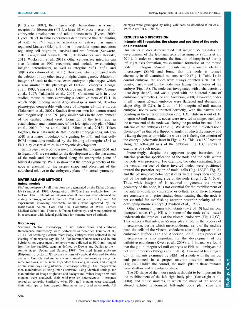
left–right axis formation, we examined formation of the mousenode in integrin a5-null mutants using scanning electronmicroscopy (SEM) and found that the node is formedaberrantly in all examined mutants, n510 (Fig. 1; Table 1). In
control embryos, the nodes were always oriented such that thepointy, narrow end of the node was facing the anterior of theembryo (Fig. 1A). The node was invaginated with a characteristic
‘‘tear-drop shape’’, and was aligned with the bilateral plane ofembryonic symmetry (Lee and Anderson, 2008). However, nodesin all integrin a5-null embryos were flattened and aberrant in
shape (Fig. 1B,C,G). In 2 out of 10 integrin a5-null mutantembryos, nodes were oriented correctly, with the narrow endpointing in the anterior direction (Fig. 1D), while in 6 out of 10
integrin a5-null mutants, nodes were inverted in shape, such thatthe narrow end of the node was facing the posterior instead of theanterior of the embryo (Table 1). We define the ‘‘inverted nodephenotype’’ as that of a flipped triangle, in which the narrow end
is facing the posterior, while the wide side is facing the anterior ofthe embryo (schematic inset in Figs 4, 5). This is a 180˚ rotationalong the left–right axis of the embryos. Fig. 1B,C shows 2
examples of such nodes.Interestingly, despite the apparent shape inversion, the
anterior–posterior specification of the node and the cells within
the node was preserved. For example, the cilia emanating fromthe ventral surface of these inverted nodes were positionedtoward the posterior region of nodal cells (Fig. 1A9,B9, Fig. 2),and the presumptive notochordal cells were always seen coming
out of the anterior-facing side of the node (Figs 1, 2, 4, 5, 6).Thus, while integrin a5 is required to generate the propergeometry of the node, it is not essential for the establishment of
the anterior–posterior embryonic or cellular axis. These findingsare consistent with prior studies demonstrating that the node isnot essential for establishing anterior–posterior polarity of the
developing mouse embryo (Davidson et al., 1999).Other examined integrin a5-mutants (n52 of 10) had narrow,
disrupted nodes (Fig. 1G) with some of the node cells located
underneath the large cells of the visceral endoderm (Fig. 1G,G9).This suggests that integrin a5 may play a role in the process ofintercalation, during which mesendodermal cells of the midlinepush the cells of the visceral endoderm apart and appear on the
embryonic surface (Lee and Anderson, 2008). This process ofintercalation is also important for the development of thedefinitive endoderm (Kwon et al., 2008), and indeed, we found
that the gut in integrin a5-null embryos or FN1-null embryos didnot form properly (Villegas et al., 2013). Two out of ten integrina5-null mutants examined by SEM had a node with the narrow
end positioned in a proper anterior–posterior orientation(Fig. 1D). Unlike the control, the nodal pits in these mutantswere shallow and irregular in shape.
The 3D shape of the mouse node is thought to be important forthe establishment of the left–right body plan (Cartwright et al.,2004), and mouse mutants, in which the shape of the node isaltered exhibit randomized left–right body plan (Lee and
RESEARCH ARTICLE Biology Open (2014) 3, 583–590 doi:10.1242/bio.20148243
584
BiologyOpen
by guest on July 13, 2018http://bio.biologists.org/Downloaded from
Anderson, 2008; Lee et al., 2010). Thus defective establishmentof the left–right axis in integrin a5-null mutants (Pulina et al.,
2011) can be explained by aberrant morphogenesis of the node.Taken together, our studies indicate that integrin a5b1 plays animportant role in embryo development by regulating nodemorphogenesis and the establishment of the left–right body plan.
Fibronectin regulates the geometry and midline placementof the node and notochordFN1 is a major extracellular ligand for the integrin a5b1 (Hynes,2012). If integrin a5b1 regulates formation of the stereotypicalshape of the mouse node by binding FN1, we would expect to
find shallow, inverted and disrupted nodes in FN1-null mutants aswell. Indeed, our SEM studies showed that in 5 out of 13 FN1-null mutants had inverted nodes and 8 out of 13 FN1-null mutants
had narrow and discontinuous nodes (Fig. 1E,F,H,H9), consistentwith our previous study (Pulina et al., 2011). We found that, likein integrin a5-null mutants, the presumptive notochordal cellsoriginated from either corner of the anterior-facing mutant node
(Fig. 1E,F, two different examples of mutant nodes are shown).Similar to integrin a5-null mutants, the primary cilia on theventral cells of FN1-null nodes were properly positioned toward
the cells’ posterior (Fig. 2C,C9). These findings suggest that FN1and its major integrin receptor a5b1 regulate geometry of thenode but not the overall anterior–posterior polarity of the embryo
or of the cells composing the node.
Fibronectin regulates apico-basal polarity of nodal cellsWe hypothesized that defective node morphogenesis in FN1-nullmutants was caused by defective apico-basal polarity of the cellscomposing the node. To test this idea, we stained control andmutant embryos using rhodamine-conjugated phalloidin to detect
F-actin. In addition, control and FN1-null embryos were co-stained using antibodies to acetylated a-tubulin (components ofstable microtubules, including those inside the primary cilia) or
antibodies to the ciliary protein Arl13b, which localizes toprimary cilia (Caspary et al., 2007). The ventral-most cells of thewild-type node are apically constricted and assemble F-actin
rings at the constricted, ventral-most surfaces of nodal cells(Fig. 3A,A9) (Lee and Anderson, 2008). Another notable featureof the properly apically polarized cells of the wild-type node isthe presence of stable microtubules and the primary cilia at the
apical side of the ventral-most cells of the node (Lee andAnderson, 2008). These two features of apico-basal polarity weredisrupted in FN1-null mutants (Fig. 3B,B9,D). Remarkably,
primary cilia marked by the expression of Arl13B were foundboth at apical and basal surfaces of nodal cells in FN1-nullmutants (Fig. 3B9, arrows). Future experiments utilizing
additional cell polarity markers and earlier time points duringnodal morphogenesis are necessary to further confirm the polaritystatus of nodal cells in the mutants and to determine whether FN1
regulates the establishment or the maintenance of nodal cellpolarity. Upon examination of integrin a5-null nodes, we foundectopical distribution of F-actin in one out of three mutantembryos (data not shown), suggesting that additional FN1-
binding integrins regulate apico-basal polarity of the nodal cells.
Integrin a5b1 and fibronectin are required to position thenode and notochord along the plane of mirror symmetry indeveloping mouse embryosAt E7.5–E8.0, wild-type embryos appear bilaterally symmetrical
and have no external, gross morphological features that
Fig. 1. Integrin a5 and fibronectin are required for morphogenesisof the mouse node. (A,A9) Shape of the node in the control mouseembryo at E7.5. (A) The dotted line outlines the entire shape of the node.(A9) Primary cilia (arrows) are positioned at the posterior of nodal cells(some examples are outlined). (B,B9,C,D,G,G9) Node shapes areaberrant in integrin a5-null E7.5 embryos. (B) Notice the inverted shapeof the node in this integrin a5-null mutant. Nodes were inverted in 6 outof 10 integrin a5-null mutants. (B,C) Examples of such inverted nodes.(B9) Magnified view of cilia in the node shown in panel B. Each primarycilium (arrows) is positioned at the posterior of each cell (outlined).(C) Note two notochords emanating from each corner of a mutant node(dotted line, arrows). (D) The pointy end of the node was positionedin a correct orientation in 2/10 integrin a5-null mutant nodes (dotted lineoutlines the node and the notochord). (E,F,H,H9) Aberrant nodes inE7.5 FN1-null mutants. (E,F) Two examples of inverted nodes inFN1-null mutants. Nodes were inverted in 5 out of 13 FN1-null mutants.(G,H) In some a5-null and FN1-null mutants, the nodes were narrowand disrupted. (G9,H9) Magnified views of panels G and H. Areascontaining node cells are outlined. Arrows point to cilia protruding frombeneath the cells of the primitive endoderm. A–anterior, P–posterior,L–left and R–right axes are marked and their directions are the same inall panels. Scale bars: 10 mm.
RESEARCH ARTICLE Biology Open (2014) 3, 583–590 doi:10.1242/bio.20148243
585
BiologyOpen
by guest on July 13, 2018http://bio.biologists.org/Downloaded from

distinguish the left side from the right. The primitive streak and
the node are bilaterally symmetrical and lie along the bilateralplane of symmetry. Cells of the notochord arise from the node(Yamanaka et al., 2007), and the narrow part of the node and thenotochordal plate align along the plane of bilateral symmetry in
wild-type embryos, suggesting that the shape of the node and thepositioning of the narrow part of the node along the midline couldbe important for the placement of the narrow strip of notochordal
cells along the embryonic plane of bilateral symmetry.In order to observe the shape and placement of the node and the
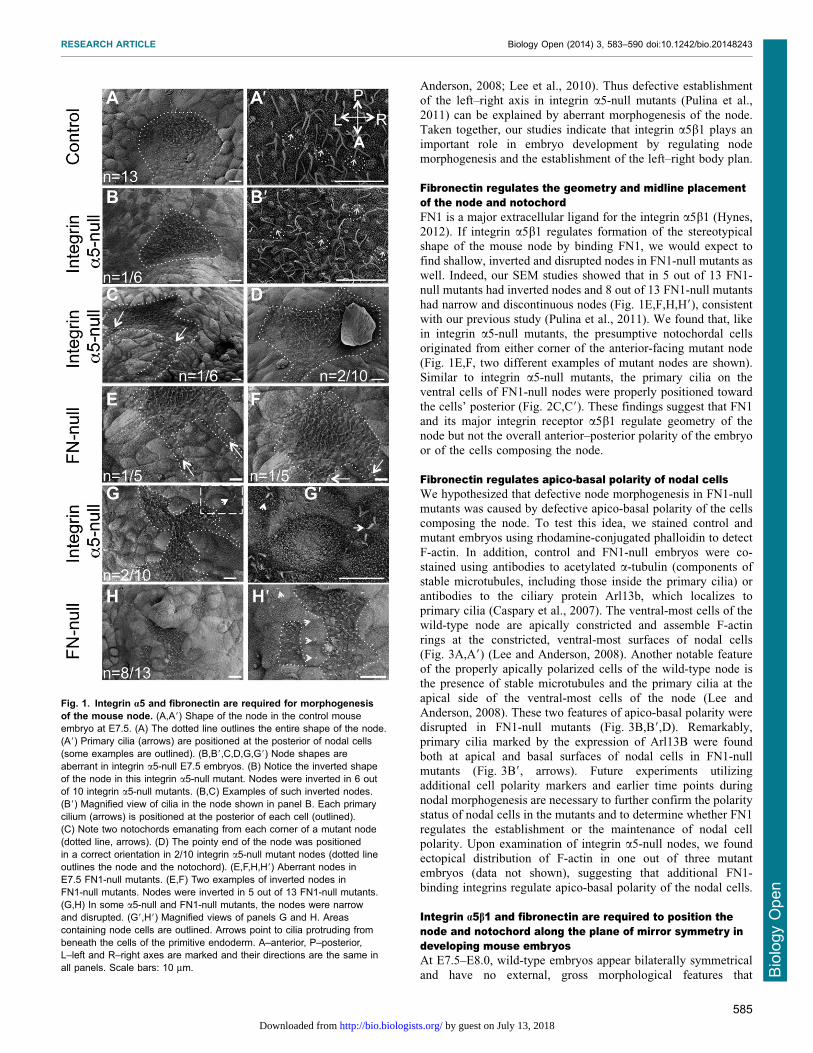
notochord in more detail, we stained embryos to detect expression
of the Shh growth factor and the T transcription factor in the nodeand the notochord using whole mount in situ hybridization (ISH).In accord with our SEM studies (Fig. 1), the expression of Shh and
T in the node collectively demonstrated that a large proportion ofintegrin a5-null (n57 out of 9) and FN1-null embryos (n59 out of19) had inverted nodes with the wide side of the node orientedtoward the embryonic anterior (Figs 4, 5). Expression of Shh
marked inverted nodes in 4 out of 5 integrin a5-null embryos(Fig. 4) and expression of T demonstrated node inversion in 3 out 4
integrin a5-null embryos (Fig. 5C,D). In addition, expression of
FoxA2 indicated inverted nodes in all of the four examined integrina5-null embryos (Fig. 4F,G). Expression of Shh showed that 4 outof 12 nodes in FN1-null mutants were inverted (Fig. 4D), andexpression of T demonstrated the presence of inverted nodes in 5
out of 7 FN1-nulls (Fig. 5F).Our ISH and SEM experiments also demonstrated that the
notochords in integrin a5-null and FN1-null mutants formed
aberrantly, and that the proper positioning and shape of thenotochord are correlated with the shape of the node (Figs 4–6). Incontrols, notochordal cells were arrayed in a narrow line contiguous
with the narrow portion of the node (Fig. 4A,F, Fig. 5A, Fig. 6A,A9),as though the narrowing of the node focused the position ofnotochordal cells along the midline. Accordingly, in those integrin
Table 1. Summary of node shapes seen with SEM
Node shape
Genotype Narrow/disrupted Narrow end pointing toward the posterior Narrow end pointing toward the anterior
Control (wt or het) 0 0 13Integrin a5-null 2 6 2Fibronectin-null 8 5 0
SEM-scanning electron microscopy; wt-wild type; het: FN+/2 or integrin a5+/2.
Fig. 2. Anterior–posterior polarity of the nodal cells does not dependon integrin a5b1 or FN1. Red: F-actin is detected using rhodamine-phalloidin; Green: acetylated (stable) a-tubulin. Positions of the notochordsare marked by long arrows in panels A–C. Note three notochords originatefrom the middle and the two corners of the inverted node in this FN1-nullembryo (arrows, C). Protruding cilia (small arrows, A9–C9) are located at theposterior of each nodal cell. Axes are marked as in Fig. 1. All embryos werecollected in the morning of E8.0. Scale bars: 10 mm.
Fig. 3. FN1 is required for the apico-basal polarity of the nodal cells.Transverse optical sections through the node (dotted lines in panels A and Bindicate the plane of optical section shown in panels A9 and B9). 3Dreconstructions of views through the transverse edge of each section arepresented. In each view (A9,B9), multiple layers of cells posterior to the dottedlines in panels A and B are collapsed into one plane. F-actin (red) andacetylated a-tubulin and cilia (green) are only present at the apical (ventral)surface of the ventral-most nodal cells in controls (A9,C). The dotted lines inpanels A9,B9,C and D mark 10 mm from the apical surface of the ventralnode, and indicate about one cell layer. In FN1-null mutants (B,B9,D), F-actinis distributed apically and basally and its presence is not restricted to theventral cell surface. In FN1-null embryos, cilia, visualized using anti-Arl13bantibody (B9) or anti-acetylated a-tubulin antibody (D), are present on ventraland internal cells of the node, and point apically and basally (B9, arrows).(C,D) Embryos were stained to detect F-actin and acetylated a-tubulin. Theviews are 3D reconstructions through multiple cell layers of the nodes asdescribed in panels A9 and B9. The absence of nuclear stain allowsvisualizing the distribution of F-actin and a-tubulin through the nodes,internally. F-actin is localized apically in all cells of the node, while in mutantsF-actin is re-distributed from the apical surface throughout the cells in themutants nodes. Axes are as in Fig. 2. All embryos were collected in themorning of E8.0. Scale bars: 20 mm.
RESEARCH ARTICLE Biology Open (2014) 3, 583–590 doi:10.1242/bio.20148243
586
BiologyOpen
by guest on July 13, 2018http://bio.biologists.org/Downloaded from
a5-null or FN1-null mutants, in which the narrow ends of the nodeswere oriented toward the anterior, the notochords formed a narrowline coincident with the embryonic bilateral plain of symmetry
(Fig. 4E, Fig. 5B,E). However, this was not the case in mutants withinverted nodes, and we observed two scenarios in mutants withinverted nodes: 1) the notochordal cells originated from the entire
anterior-facing, wide side of the node, giving rise to wide notochords(Fig. 4B, Fig. 5C,F); or 2) the notochordal cells originated mainlyfrom one of the corners of the node, giving rise to narrow notochords
positioned to the left side or the right of the presumptive bilateralplane of symmetry (Fig. 4D, Fig. 6B,B9). In some cases, notochordalcells originating from both corners of the inverted node (Fig. 4D,Fig. 5D) or from the wide face of the inverted node (Fig. 4B) gave
rise to initially wide regions of the notochord that became ‘‘re-focused’’ along the midline in a more anterior position, suggestingthe presence of a trophic factor(s) located along the embryonic
midline. This factor(s) may be produced by the neural ectoderm,since we observed notochordal cells following the bends of the neuraltube in one of the mutants (Fig. 4C, red arrows). SEM further
demonstrated the striking ‘‘off-midline’’ positioning of the nodes andnotochords in FN1-null mutants (Fig. 6).
One notable feature of our findings is variability in thegeometry and position of the nodes and notochords in the
mutants. The genetic background was not a factor modulating thevariability of the observed phenotypes, since all embryosanalyzed were obtained from mutant mice crossed into C57BL/
6J background for over 10 generations, and then intercrossed forover 10 years. The embryonic stage was also not a factor, sincemutant embryos with different node geometries contained well-
developed anterior intestinal portals (e.g. Fig. 6B,D,E), indicating
that mutants have at least reached the late headfold stage (Downs
and Davies, 1993). At this stage, the nodes and notochords in allcontrols are well-formed. Another indicator of embryonic age atearly time points of development is the length of nodal cilia (Lee
and Anderson, 2008). However, we found that the lengths ofnodal cilia in mutants did not differ from similarly-stagedcontrols (Pulina et al., 2011). Therefore, the variability in thegeometry and position of the nodes and notochords implies the
loss of a critical regulatory mechanism in FN-null and integrina5-null mutants, leading to stochastic organization of the nodeand ‘‘off-midline’’ placement of the notochord.
Relationship between the expression of integrin a5 and FN1matrix assemblyThe form, shape and stereotypical features of the E8.0 embryos,including somites and the midline structures, are easily visualizedfollowing whole mount staining of embryos using rhodamine-
conjugated phalloidin (Fig. 7A,B) or FN1 (Fig. 7C). However,these structures are no longer recognizable following the samestaining of integrin a5-null embryos (Fig. 7E–G). Absence ofrecognizable embryonic patterns in integrin a5-null mutants
stained with rhodamine-conjugated phalloidin indicates aberrantdistribution of F-actin bundles in mutant cells, consistent with anotion that cell polarity is disrupted in entire integrin a5-null
embryos. Interestingly, FN1 protein distribution is disorganizedin integrin a5-null mutants as well. In control embryos, FN1
Fig. 5. Integrin a5 and FN1 regulate the geometry of the node and thedevelopment of the notochord as shown by the expression of T. Whole-mount in situ hybridizations using an anti-sense T RNA probe. (A) Control.(B–D) Integrin a5-null mutants. (E,F) FN1-null mutants. In those mutants inwhich the node is oriented with the pointy end toward the anterior (bluearrows, B,E) and along the midline, the notochords are positioned at theembryonic midline and are narrow (B). In mutants with inverted nodes,notochordal cells emanate from the entire wide face of the node (blue arrow),giving rise to the wide notochord (C,F), or from two corners of the node (D).(D) Notochordal cells are refocused along the midline anteriorly (dottedarrow). Embryos were collected at E8.0. Littermate control embryos rangedfrom 2–5 somites. Mutants do not develop somites. Axes are as in Fig. 4.Scale bar: 500 mm.
Fig. 4. Integrin a5 and FN1 are required the proper geometry of thenode and the morphogenesis of the notochord as shown by theexpression of Shh and FoxA2. (A–E) Whole-mount in situ hybridizationsusing a mouse Shh anti-sense RNA probe. In controls (A) the narrow tipof the node points anteriorly and is contiguous with the narrow strip of thenotochord. In mutants with inverted nodes, notochordal cells emanatefrom both corners of the node (B), giving rise to a dispersed notochord(red arrows). (C) Notochordal cells in this mutant ‘‘follow’’ the bends of theneural tube (red arrows). (D) When most notochordal cells emanate from onecorner or (E) from a narrow tip of the node (black arrows), the notochords arenarrow in the mutants. Triangles indicate properly oriented (orange) orinverted nodes (blue). (F,G) FoxA2 protein expression (green). 3Dreconstructions of immunofluorescence confocal images of control (F) andintegrin a5-null embryos (G), demonstrating inverted nodes in the mutants.Embryos were collected at E8.0. Littermate control embryos ranged from 2–5somites. Mutants do not develop somites. Axes are labeled. Scale bars:500 mm.
RESEARCH ARTICLE Biology Open (2014) 3, 583–590 doi:10.1242/bio.20148243
587
BiologyOpen
by guest on July 13, 2018http://bio.biologists.org/Downloaded from

distinctly localizes around the node and the notochord
(Fig. 7C,D). This localization is mediated by integrin a5b1 andis disrupted in its absence (Fig. 7G,H).
Integrin heterodimers containing av subunits aredispensable for the establishment of the left–right axis in themouseIn the absence of integrin a5b1, cell–ECM interactions could be
potentially mediated by av-containing integrin heterodimers(Yang and Hynes, 1996; van der Flier et al., 2010). Studies inzebrafish indicated that additional FN1 receptors containing
integrin av subunits, are important for the formation of theKupffer’s vesicle, the organ of asymmetry in zebrafish, and forthe establishment of the left–right axis of asymmetry in fish(Ablooglu et al., 2010). In order to test the role of integrin av in
the establishment of the left–right axis, we analyzed cardiaclooping in mouse embryos lacking integrin av (Bader et al.,1998). The bending of the heart tube to the right is the first
morphological manifestation of the left–right asymmetry in theembryo (Ramsdell, 2005). Our analyses indicated that cardiaclooping occurred correctly in integrin av-null mutants, n512
(Fig. 8), demonstrating that av-containing integrins are notrequired for left–right axis formation or for its maintenance.Moreover, we observed normal, rightward cardiac looping in all
compound integrin av2/2; a52/+ mutant embryos (n56),indicating that a single copy of integrin a5 is sufficient for thenormal formation of the node and the notochord, for the midlinebarrier function, and for the development of the left–right axis.
Normal heart looping strongly implies that the formation of thenode and the notochord are not affected in integrin av-null orav2/2; a52/+ mutant embryos. Our studies indicate that unlike in
zebrafish, av-containing integrin heterodimers are not essentialfor the left–right axis development in mammals, and that amongFN1 receptors, integrin a5b1 plays an essential, major role in
morphogenesis of the node and notochord, and in theestablishment of the left–right axis of asymmetry duringembryogenesis (Pulina et al., 2011).
Fig. 7. Integrin a5 plays a major role in assembly of FN1 matrix,localization of F-actin, and organization of embryonic tissues. Wholemount immunofluorescence staining of E8.0 embryos to detect F-actin (red)and FN1 (green). (A–D) Controls. (E–H) Integrin a5-null mutants. In controls,F-actin and FN1, outline the node, notochord (arrowhead) and somites(stars). In mutants, these structures are not distinguishable. Note that in themutant (F,G) the node and the notochord are positioned to the left of thebilateral plane of symmetry. FN1 matrix is assembled as long squiggly fibrilsin controls (C,D). In integrin a5-null mutants, FN1 protein is mostly present asdots (notched arrowhead, H). Filled arrowheads point at notochord, rectanglein panel G is expanded in panel H. Control and mutant embryos wereisolated at E8.0. Staining for FN1 was performed on 13 control embryosranging from 0–4 somites, and in none of the cases, FN was found in dots(notched arrowhead, H) as observed in integrin a5-null mutants.
Fig. 6. FN1 is required for midline positioning of the node andnotochord demonstrated by scanning electron microscopy. Ventralsurfaces of control (A–A0) or mutant (B–E0) embryos are shown. Dashedlines outline approximate, perceived bilateral planes of symmetry in themutants. Red arrows point to notochords. Red stars mark nodes. Panelsmarked by 9 and 0 are magnified views of panels A–E. (A–A0) In controls, theposition of the node and the notochord coincides with the bilateral plane ofsymmetry. (B–B0) In mutant, notochord emanates from the left corner of thenode and is positioned to the left of the bilateral plane of symmetry.(C–C0) The mutant node and notochord are positioned to the right of thebilateral plane of symmetry. (D–D0) The node (red star) is positioned far to theright of the bilateral plane of symmetry and there is no identifiable notochord.(E–E0) Portions of disrupted node are located to either side of theapproximate plane of bilateral symmetry. Note the presence of endodermalinvagination in this mutant (white arrow) as well as in the control and all othermutants shown, indicating that mutants are at least at the late headfold stageof development. Embryos were collected at E8.0 and ranged from the lateheadfold stage up to 3 somites (controls). Mutants do not develop somites.Axes are as in Fig. 1. Scale bars: 30 mm.
Fig. 8. Integrin av is not required for the early left–right patterning.(A) Normal, right-sided heart looping. Control (A) and integrin av-null (B) mouseembryos at E9.5. Red arrows point at the right ventricle in control and mutant.Magnification is the same in both panels. Scale bar: 500 mm.
RESEARCH ARTICLE Biology Open (2014) 3, 583–590 doi:10.1242/bio.20148243
588
BiologyOpen
by guest on July 13, 2018http://bio.biologists.org/Downloaded from

ConclusionsOur work highlights complex mechanisms involved in the
morphogenesis of the midline structures, the node and thenotochord. And while convergent extension is an importantmechanism regulating notochord morphogenesis (Yamanakaet al., 2007), our studies indicate that additional mechanisms are
at play, namely, that the geometry and the position of the node inthe developing embryo determine the midline positioning and thewidth of the notochord. Importantly, our studies indicate that the
node and the notochord do not develop along the bilateral planeof embryonic symmetry by default. Instead, there exist activemechanisms regulating the midline placement of these structures.
These mechanisms critically depend on the engagement of cellularintegrin a5b1 with extracellular matrix protein FN1.
Our studies demonstrate that absence of integrin a5 or FN1
proteins leads to similar defects in formation of the midlinestructures, suggesting that integrin a5b1 transduces signaling byFN1. However, a number of other integrins bind FN1 in vitro, andintegrins containing av chain are known to compensate for the
absence of integrin a5b1 in vitro during FN1 fibril assembly(Yang and Hynes, 1996; van der Flier et al., 2010). In vivo, FN1-binding integrin heterodimers containing a4 or a3 chains do
not appear to cooperate with integrin a5b1 during earlyembryogenesis (Yang et al., 1999). av-containing integrinscooperate with integrin a5b1 in early mouse embryogenesis to
facilitate gastrulation as well as in midgestation, duringremodeling of the pharyngeal arch arteries (Yang et al., 1999;van der Flier et al., 2010). In zebrafish, av-containing integrins
were shown to be important for the establishment of the left–rightasymmetry by regulating morphogenesis of the Kupffer’s vesicle.However, we did not find defects in establishment or maintenanceof the left–right asymmetry in mouse embryos with global
deletion of integrin av. Moreover, decreasing the dosage ofintegrin a5b1 did not induce left–right defects in integrin av-nullmutants. These studies suggest that in mice, integrin a5b1 is the
main FN1-binding integrin heterodimer transducing FN1 signalsduring morphogenesis of the node and the notochord andregulating the establishment of the left–right axis. Consistent
with this, we found that FN1 matrix was not well-assembledin integrin a5-null embryos. Our experiments suggest thatassembly of FN1-containing ECM mediated by integrin a5b1and FN1-integrin a5b1 signaling are important for polarized
distribution of actin stress fibers, apico-basal cell polarity andmorphogenesis of the node and the notochord. Disruptedlocalization of F-actin in integrin a5 mutants implies defective
tissue tension and/or aberrant distribution of mechanical forceswithin the developing mutant embryos (Schwartz, 2010). Asanisotropic tissue tension is important for gastrulation (Brunet
et al., 2013) and for the development of proper geometricalcellular patterns during embryogenesis (Guillot and Lecuit, 2013;LeGoff et al., 2013), we hypothesize that the binding of cellular
integrin a5b1 to FN1 is important for generating and/ormaintaining mechanical forces within the developing embryo,facilitating correct packing of the cells within the node andenabling the placement of the node and the notochord along the
bilateral plane of symmetry.
AcknowledgementsWe thank Nina Lampen at the Electron Microscopy Core Facility at the MemorialSloan–Kettering Cancer Center for help with SEM and ShivaprasadBhuvanendran in the Bio-Imaging Resource Center at the Rockefeller Universityfor help with confocal imaging. We thank Drs Takashi Mikawa, Ann Foley, NathanAstrof, Karl Degenhardt and Alvin Chin for advice and careful reading of the
manuscript, and we thank Rachel DeRita and Sonam Dhiman for careful editing ofthe manuscript. We are grateful to Dr Kathryn Anderson for Arl13b antibody andthe plasmids used for preparation of in situ hybridization probes.
Competing interestsThe authors have no competing interests to declare.
Author contributionsS.A. conceived and designed the project, performed imaging, data analyses,prepared the figures and wrote the paper. M.P. and D.L. carried out the research.
FundingThis work was supported by the National Institutes of Health NHLBI [grant numberHL103920], American Heart Association scientist development grant [0835556D],American Heart Association Innovative Research Grant [12IRG9130012] and theW. W. Smith Charitable Trust (to S.A.).
ReferencesAblooglu, A. J., Tkachenko, E., Kang, J. and Shattil, S. J. (2010). IntegrinalphaV is necessary for gastrulation movements that regulate vertebrate bodyasymmetry. Development 137, 3449-3458.
Almog, B., Leibovitch, L. and Achiron, R. (2001). Split notochord syndrome –prenatal ultrasonographic diagnosis. Prenat. Diagn. 21, 1159-1162.
Astrof, S., Kirby, A., Lindblad-Toh, K., Daly, M. and Hynes, R. O. (2007). Heartdevelopment in fibronectin-null mice is governed by a genetic modifier onchromosome four. Mech. Dev. 124, 551-558.
Bader, B. L., Rayburn, H., Crowley, D. and Hynes, R. O. (1998). Extensivevasculogenesis, angiogenesis, and organogenesis precede lethality in micelacking all alpha v integrins. Cell 95, 507-519.
Bressan, M., Davis, P., Timmer, J., Herzlinger, D. and Mikawa, T. (2009).Notochord-derived BMP antagonists inhibit endothelial cell generation andnetwork formation. Dev. Biol. 326, 101-111.
Brunet, T., Bouclet, A., Ahmadi, P., Mitrossilis, D., Driquez, B., Brunet, A. C.,Henry, L., Serman, F., Bealle, G., Menager, C. et al. (2013). Evolutionaryconservation of early mesoderm specification by mechanotransduction inBilateria. Nat. Commun. 4, 2821.
Burdine, R. D. and Schier, A. F. (2000). Conserved and divergent mechanisms inleft-right axis formation. Genes Dev. 14, 763-776.
Cartwright, J. H., Piro, O. and Tuval, I. (2004). Fluid-dynamical basis of theembryonic development of left-right asymmetry in vertebrates. Proc. Natl. Acad.Sci. USA 101, 7234-7239.
Caspary, T., Larkins, C. E. and Anderson, K. V. (2007). The graded response toSonic Hedgehog depends on cilia architecture. Dev. Cell 12, 767-778.
Davidson, B. P., Kinder, S. J., Steiner, K., Schoenwolf, G. C. and Tam, P. P.(1999). Impact of node ablation on the morphogenesis of the body axis and thelateral asymmetry of the mouse embryo during early organogenesis. Dev. Biol.211, 11-26.
Downs, K. M. and Davies, T. (1993). Staging of gastrulating mouse embryos bymorphological landmarks in the dissecting microscope. Development 118, 1255-1266.
Fouquet, B., Weinstein, B. M., Serluca, F. C. and Fishman, M. C. (1997). Vesselpatterning in the embryo of the zebrafish: guidance by notochord. Dev. Biol.183, 37-48.
Geiger, B. and Yamada, K. M. (2011). Molecular architecture and function ofmatrix adhesions. Cold Spring Harb. Perspect. Biol. 3, a005033.
George, E. L. and Hynes, R. O. (1994). Gene targeting and generation of mutantmice for studies of cell–extracellular matrix interactions. Methods Enzymol. 245,386-420.
George, E. L., Georges-Labouesse, E. N., Patel-King, R. S., Rayburn, H. andHynes, R. O. (1993). Defects in mesoderm, neural tube and vasculardevelopment in mouse embryos lacking fibronectin. Development 119, 1079-1091.
George, E. L., Baldwin, H. S. and Hynes, R. O. (1997). Fibronectins are essentialfor heart and blood vessel morphogenesis but are dispensable for initialspecification of precursor cells. Blood 90, 3073-3081.
Goh, K. L., Yang, J. T. and Hynes, R. O. (1997). Mesodermal defects and cranialneural crest apoptosis in alpha5 integrin-null embryos. Development 124, 4309-4319.
Goldstein, A. M. and Fishman, M. C. (1998). Notochord regulates cardiaclineage in zebrafish embryos. Dev. Biol. 201, 247-252.
Guillot, C. and Lecuit, T. (2013). Mechanics of epithelial tissue homeostasis andmorphogenesis. Science 340, 1185-1189.
Huttenlocher, A. and Horwitz, A. R. (2011). Integrins in cell migration. ColdSpring Harb. Perspect. Biol. 3, a005074.
Hynes, R. O. (2002). Integrins: bidirectional, allosteric signaling machines. Cell110, 673-687.
Hynes, R. O. (2009). The extracellular matrix: not just pretty fibrils. Science 326,1216-1219.
Hynes, R. O. (2012). The evolution of metazoan extracellular matrix. J. Cell Biol.196, 671-679.
Kwon, G. S., Viotti, M. and Hadjantonakis, A. K. (2008). The endoderm of themouse embryo arises by dynamic widespread intercalation of embryonic andextraembryonic lineages. Dev. Cell 15, 509-520.
RESEARCH ARTICLE Biology Open (2014) 3, 583–590 doi:10.1242/bio.20148243
589
BiologyOpen
by guest on July 13, 2018http://bio.biologists.org/Downloaded from
Lee, J. D. and Anderson, K. V. (2008). Morphogenesis of the node andnotochord: the cellular basis for the establishment and maintenance of left-rightasymmetry in the mouse. Dev. Dyn. 237, 3464-3476.
Lee, J. D., Migeotte, I. and Anderson, K. V. (2010). Left-right patterning in themouse requires Epb4.1l5-dependent morphogenesis of the node and midline.Dev. Biol. 346, 237-246.
LeGoff, L., Rouault, H. and Lecuit, T. (2013). A global pattern of mechanicalstress polarizes cell divisions and cell shape in the growing Drosophila wingdisc. Development 140, 4051-4059.
Meno, C., Shimono, A., Saijoh, Y., Yashiro, K., Mochida, K., Ohishi, S., Noji,S., Kondoh, H. and Hamada, H. (1998). lefty-1 is required for left-rightdetermination as a regulator of lefty-2 and nodal. Cell 94, 287-297.
Mikawa, T., Poh, A. M., Kelly, K. A., Ishii, Y. and Reese, D. E. (2004). Inductionand patterning of the primitive streak, an organizing center of gastrulation in theamniote. Dev. Dyn. 229, 422-432.
Miner, J. H. and Yurchenco, P. D. (2004). Laminin functions in tissuemorphogenesis. Annu. Rev. Cell Dev. Biol. 20, 255-284.
Mittal, A., Pulina, M., Hou, S. Y. and Astrof, S. (2010). Fibronectin and integrinalpha 5 play essential roles in the development of the cardiac neural crest.Mech. Dev. 127, 472-484.
Mittal, A., Pulina, M., Hou, S. Y. and Astrof, S. (2013). Fibronectin and integrinalpha 5 play requisite roles in cardiac morphogenesis. Dev. Biol. 381, 73-82.
Pulina, M. V., Hou, S. Y., Mittal, A., Julich, D., Whittaker, C. A., Holley, S. A.,Hynes, R. O. and Astrof, S. (2011). Essential roles of fibronectin in thedevelopment of the left-right embryonic body plan. Dev. Biol. 354, 208-220.
Ramsdell, A. F. (2005). Left-right asymmetry and congenital cardiac defects:getting to the heart of the matter in vertebrate left-right axis determination. Dev.Biol. 288, 1-20.
Schwartz , M. A. (2010) . In tegr ins and extracel lu lar mat r ix inmechanotransduction. Cold Spring Harb. Perspect. Biol. 2, a005066.
Shiratori, H. and Hamada, H. (2006). The left-right axis in the mouse: from originto morphology. Development 133, 2095-2104.
Stemple, D. L. (2005). Structure and function of the notochord: an essential organfor chordate development. Development 132, 2503-2512.
Sumoy, L., Keasey, J. B., Dittman, T. D. and Kimelman, D. (1997). A role fornotochord in axial vascular development revealed by analysis of phenotype and theexpressionofVEGR-2 in zebrafish flhandntlmutant embryos.Mech.Dev.63, 15-27.
Tabin, C. J. (2006). The key to left-right asymmetry. Cell 127, 27-32.Takahashi, S., Leiss, M., Moser, M., Ohashi, T., Kitao, T., Heckmann, D.,Pfeifer, A., Kessler, H., Takagi, J., Erickson, H. P. et al. (2007). The RGD motif
in fibronectin is essential for development but dispensable for fibril assembly.J. Cell Biol. 178, 167-178.
Turgut, M., Cullu, E. and Ulucan, H. (2006). Incomplete Currarino triad as anembryological variant. Case report and review of the literature. J. Neurosurg.105 Suppl., 504-507.
van der Flier, A., Badu-Nkansah, K., Whittaker, C. A., Crowley, D., Bronson,R. T., Lacy-Hulbert, A. and Hynes, R. O. (2010). Endothelial alpha5 and alphavintegrins cooperate in remodeling of the vasculature during development.Development 137, 2439-2449.
Villegas, S. N., Barrios-Llerena, M. E., Pulina, M., Hadjantonakis, A. K., LeBihan, T., Astrof, S. and Brickman, J. M. (2013). PI3K/Akt1 signalling specifiesforegut precursors by generating regionalized extra-cellular matrix. eLife 2,e00806.
Wei, Y. and Mikawa, T. (2000). Formation of the avian primitive streak fromspatially restricted blastoderm: evidence for polarized cell division in theelongating streak. Development 127, 87-96.
Whittaker, C. A. and Hynes, R. O. (2002). Distribution and evolution of vonWillebrand/integrin A domains: widely dispersed domains with roles in celladhesion and elsewhere. Mol. Biol. Cell 13, 3369-3387.
Whittaker, C. A., Bergeron, K. F., Whittle, J., Brandhorst, B. P., Burke, R. D.and Hynes, R. O. (2006). The echinoderm adhesome. Dev. Biol. 300, 252-266.
Wickstrom, S. A., Radovanac, K. and Fassler, R. (2011). Genetic analyses ofintegrin signaling. Cold Spring Harb. Perspect. Biol. 3, a005116.
Yamanaka, Y., Tamplin, O. J., Beckers, A., Gossler, A. and Rossant, J. (2007).Live imaging and genetic analysis of mouse notochord formation revealsregional morphogenetic mechanisms. Dev. Cell 13, 884-896.
Yang, J. T. and Hynes, R. O. (1996). Fibronectin receptor functions in embryoniccells deficient in alpha 5 beta 1 integrin can be replaced by alpha V integrins.Mol. Biol. Cell 7, 1737-1748.
Yang, J. T., Rayburn, H. and Hynes, R. O. (1993). Embryonic mesodermaldefects in alpha 5 integrin-deficient mice. Development 119, 1093-1105.
Yang, J. T., Bader, B. L., Kreidberg, J. A., Ullman-Cullere, M., Trevithick, J. E.and Hynes, R. O. (1999). Overlapping and independent functions of fibronectinreceptor integrins in early mesodermal development. Dev. Biol. 215, 264-277.
Yashiro, K., Shiratori, H. and Hamada, H. (2007). Haemodynamics determinedby a genetic programme govern asymmetric development of the aortic arch.Nature 450, 285-288.
Yurchenco, P. D. (2011). Basement membranes: cell scaffoldings and signalingplatforms. Cold Spring Harb. Perspect. Biol. 3, a004911.
RESEARCH ARTICLE Biology Open (2014) 3, 583–590 doi:10.1242/bio.20148243
590
BiologyOpen
by guest on July 13, 2018http://bio.biologists.org/Downloaded from