Embed Size (px)
Citation preview

RESEARCH ARTICLE
Evaluation of Blind Nasotracheal Suctioning and Non bronchoscopic Mini-Bronchoalveolar
Lavage in Critically Ill Patients with Infectious Pneumonia:
A Preliminary Study
Pascal Meyer, MD; Hélène Rousseau, MSc; Jean-Michel Maillet, MD; Stéphane Thierry,
MD; Oumar Sy, MD; Eric Vicaut, MD, PhD ; Fabrice Thiolliere, MD; Gerald Choukroun,
MD; Karim Chergui, MD; Guillaume Chevrel, MD and Eric Maury, MD, PhD.
Dr Meyer, Dr Thiolliere, DR Choukroun; Dr chergui and Dr Chevrel are affiliated with the
intensive care unit, centre hospitalier sud-francilien, Corbeil-Essonnes, France
Ms Rousseau, Pr Vicaut are affiliated with the Clinical Research Unit Lariboisière, Paris,
France
Dr Maillet and Dr Thierry are affiliated with the Intensive Care Unit, Centre Cardiologique du
Nord, Saint-Denis, France
Dr Sy is affiliated with the Intensive Care Unit, Hôpital Marc Jacquet, Melun, France
Pr Maury is affiliated with the Intensive Care Unit, Hôpital Saint-Antoine, Paris, France
Running title: Evaluation of nasotracheal suctioning and mini-BAL
Sources of financial support. The authors received no financial support for this study. The
authors have no involvement with organization(s) with a financial interest in the subject
matter. The promoter Assitance Publique–Hôpitaux de Paris received funding from Prodimed
to conduct this study. The authors, coordinated by Dr. Meyer, analyzed the data and wrote
this paper independently, with no input from Prodimed.
List of abbreviations used in the text: NTS = nasotracheal suctioning. ICU = intensive care
unit. BAL = bronchoalveolar lavage. CI = confidence interval. CAP = community-acquired
RESPIRATORY CARE Paper in Press. Published on July 23, 2013 as DOI: 10.4187/respcare.02356
Copyright (C) 2013 Daedalus Enterprises Epub ahead of print papers have been peer-reviewed and accepted for publication but are posted before being copy edited
and proofread, and as a result, may differ substantially when published in final version in the online and print editions of RESPIRATORY CARE.

pneumonia. HAP = hospital-acquired pneumonia. NIV = non-invasive ventilation.
Abstract: 267
Text: 2779
Dr Pascal Meyer is the guarantor of the paper, he contributed to the study design and
organization of the study, the literature review, manuscript writing and was the principal
investigator.
Hélène Rousseau contributed to statistical analyses and manuscript writing.
Dr Jean-Michel Maillet and Dr Stéphane Thierry were investigators and contributed to data
collection and manuscript writing.
Dr Oumar Sy was an investigator and contributed to data collection and revision of the
manuscript.
Prof Eric Vicaut contributed to statistical analyses and manuscript writing.
Dr Fabrice Thiolliere was an investigator and contributed to data collection and revision of
the manuscript.
Dr Gerald Choukroun was an investigator and contributed to data collection and revision of
the manuscript.
Dr Karim Chergui was an investigator and contributed to data collection and revision of the
manuscript.
Dr Guillaume Chevrel was an investigator and contributed to data collection and revision of
the manuscript.
Prof Eric Maury contributed to the study design, data analysis and manuscript writing.
ABSTRACT
BACKGROUND: We evaluated the diagnostic performance and safety of combined
RESPIRATORY CARE Paper in Press. Published on July 23, 2013 as DOI: 10.4187/respcare.02356
Copyright (C) 2013 Daedalus Enterprises Epub ahead of print papers have been peer-reviewed and accepted for publication but are posted before being copy edited
and proofread, and as a result, may differ substantially when published in final version in the online and print editions of RESPIRATORY CARE.
blind nasotracheal suctioning (NTS) and non-bronchoscopic mini-bronchoalveolar
lavage (BAL) to obtain respiratory secretion specimens from spontaneously breathing,
non-intubated patients with infectious pneumonia in intensive care. METHODS:
Patients suspected of having infectious pneumonia were included prospectively. Three
samples were obtained: expectorated sputum, and NTS and mini-BAL with a double
telescopic catheter (Combicath®). After administering local anesthesia, NTS was done
according to standard recommendations. Then, mini-BAL was performed; the bronchial
aspiration tube serves as a guide for the mini-BAL catheter, with tracheal position being
verified by colorimetric capnography. RESULTS: Thirty-six patients (29 men, median
age 69 years, median Simplified Acute Physiology Score II 32) were included. Thirty-two
(89%) underwent NTS and mini-BAL; 13 (36%) had expectorated sputum. Based on
colorimetric capnography confirmation of the tracheal position, 75% (24 /32) of the
successful combined procedures were achieved on the first attempt. The median
duration of the combined procedure was 7 min. Bacterial pneumonia was diagnosed in
24/36 (67%) patients, among whom, 21 (88%) had undergone NTS and mini-BAL,
successful respectively, for 8/21 (38% [confidence interval 0.17–0.58] and 14/21 (67%
[confidence interval 0.46–0.86]). Mini-BAL diagnosed a significantly higher percentage
of bacterial pneumonias than NTS. Expectorated sputum yielded no diagnoses.
CONCLUSIONS: Blind NTS associated with colorimetric capnography allows
microbiological diagnosis that can be enhanced by non-bronchoscopic mini-BAL. It is
probably useful to consider colorimetric capnography when the anatomical bronchial
tube position is clinically doubtful. Non-bronchoscopic mini-BAL is a new and
innovative way to collect bronchial secretions without fibroscopy. The results of our
preliminary study demonstrated the feasibility of this procedure. Key words: pneumonia,
intensive care, blind nasotracheal suctioning, mini-bronchoalveolar lavage, colorimetric
RESPIRATORY CARE Paper in Press. Published on July 23, 2013 as DOI: 10.4187/respcare.02356
Copyright (C) 2013 Daedalus Enterprises Epub ahead of print papers have been peer-reviewed and accepted for publication but are posted before being copy edited
and proofread, and as a result, may differ substantially when published in final version in the online and print editions of RESPIRATORY CARE.
capnography, expectorated sputum.
This study was registered at ClinicalTrials.org/gov under the number NCT00763620.
INTRODUCTION
When a spontaneously breathing, non intubated patient suffering from HAP or CAP
develops acute respiratory insufficiency, the search for pathogens is problematic. In this
setting, the most commonly conducted laboratory analyses are expectorated sputum culture,
blood cultures and urinalysis, with the search for pneumococcal and Legionella antigens in
urine sediment.1-4 Unfortunately, these tests have variable diagnostic yields.5-10
Flexible bronchoscopic techniques (bronchoalveolar lavage (BAL), protected distal
specimen, brush sampling) are expensive and specialized methods that cannot be widely used,
despite their probably having the best diagnostic yields.1,2,5,11 Transtracheal aspiration and
transthoracic puncture of the pneumonia locus can obtain uncontaminated specimens and give
satisfactory microbiological diagnosis results, but they require needle puncture, which is
responsible for numerous side effects that limit their use during respiratory insufficiency.12,13
Blind nasotracheal suctioning (NTS) can obtain a sputum sample for microbiological
analyses.14,15 NTS has been found to be more sensitive for the diagnosis of pneumocystis
pneumonia than sputum induction and an alternative to flexible bronchoscopy.16 In the
specific setting of bacterial identification in severe pneumonia, no recent study, to the best of
our knowledge, evaluated NTS in adults. NTS in children failed to obtain specific
microbiological diagnosis because of frequent contamination by upper airway secretions17.
Nonbronchoscopic mini-BAL is currently used for the microbiological diagnosis of
pneumonia acquired under mechanical ventilation (MV) with satisfactory sensitivity and
specificity.18-20 It represents an alternative to flexible bronchoscopy, when the latter is not
RESPIRATORY CARE Paper in Press. Published on July 23, 2013 as DOI: 10.4187/respcare.02356
Copyright (C) 2013 Daedalus Enterprises Epub ahead of print papers have been peer-reviewed and accepted for publication but are posted before being copy edited
and proofread, and as a result, may differ substantially when published in final version in the online and print editions of RESPIRATORY CARE.

available or considered too dangerous.18–22 It is also useful for the microbiological diagnosis
of CAP in intubated patients on MV.23
This preliminary study was undertaken to evaluate the feasibility, safety and diagnostic
performance of 2 mini-invasive methods, NTS and mini-BAL, in spontaneously breathing,
non intubated patients hospitalized for pneumonia in an ICU.
MATERIAL AND METHODS
This prospective study, conducted from 2008 to 2010 in 3 nonuniversity centers, included
ICU patients with suspected HAP- or CAP and not requiring intubation. Assistance Public–
Hôpitaux de Paris was the study promoter. The protocol was approved by our local Ethics
Committee (Comité de Protection des Personnes Hôtel-Dieu, Paris, France n° 0811817) and
French Agency for the Safety of Health Products. All patients included or their surrogate gave
written informed consent to participate.
Materials and the combined Procedure
We combined 2 techniques: blind NTS and mini-BAL with a telescopic catheter. Because the
latter is too flexible to be directly introduced nasotracheally, it was designed to be inserted
through a bronchial aspiration tube (40 cm long × 16-Fr diameter; Sondjet SM21 C, Peters
Surgical, Bobigny, France), wich serves as the guide for the catheter that can then access the
bronchial tree. The double telescopic mini-BAL catheter (Combicath, Prodimed, Saint-Leu
La Forêt, France) (Fig.1) consists of an external polyethylene tube (60-cm long × 2.7 mm
diameter) and an internal Teflon tube (65-cm long × 1.7-mm diameter).
Our procedure consists of first blindly inserting an aspiration tube into the airways via
the nostril, under systematic local xylocaine-spray anesthesia, and verifying its correct
positioning in the trachea with a colorimetric CO2 detector (Easycap II, Tyco®, Plaisir,
France) that changes color in several seconds (respectively, from purple to brown to yellow
RESPIRATORY CARE Paper in Press. Published on July 23, 2013 as DOI: 10.4187/respcare.02356
Copyright (C) 2013 Daedalus Enterprises Epub ahead of print papers have been peer-reviewed and accepted for publication but are posted before being copy edited
and proofread, and as a result, may differ substantially when published in final version in the online and print editions of RESPIRATORY CARE.
for <4, 4–15 and >15 mm Hg).24,25 The telescopic catheter is then inserted through the
aspiration tube to perform mini-BAL.
Experienced physicians performed all procedures. No sedation was used. Tracheal anesthesia,
was left to the decision of the investigator. When needed, it was performed as follows:
insertion of a very fine catheter (like that used for oxygen therapy) into the nostril; injection
of 5 cc of xylocaine and asking the patient to gargle.
After waiting for local anesthesia to become effective, insertion of the suction catheter
into the nostril started the clock. First, NTS was performed, according to standard
recommendations.15 Only 3 attempts within 10 min were possible. When correctly placed, the
tracheal aspirate is obtained by simple suction. Then, the prelubricated mini-BAL catheter is
introduced into the airways through the aspiration tube. Once in the bronchus, the internal
catheter, protected in the external sheath, is advanced as needed. The mini-BAL consists of
injecting 20 mL of physiological saline that are immediately recovered, and 2–3 mL of this
fluid are used for bacterial analyses.
To evaluate safety, the following information was collected before and 15 min after
the procedure: respiratory rate, heart rate, systolic and diastolic blood pressures, and SpO2.
Patients
All non intubated, spontaneously breathing (including under intermittent non invasive
ventilation (NIV)) patients >18 years old suspected of having CAP or HAP whose severity
necessitated ICU admission, were eligible for inclusion. The criteria for noninclusion were
bronchospasm, severe coagulopathy (platelet count <100,000/mm3, prothrombin time <50%,
activated partial thromboplastin time ratio >2), patient intubated on MV, microbiological
diagnosis made before hospitalization and/or pregnancy.
Study Design
RESPIRATORY CARE Paper in Press. Published on July 23, 2013 as DOI: 10.4187/respcare.02356
Copyright (C) 2013 Daedalus Enterprises Epub ahead of print papers have been peer-reviewed and accepted for publication but are posted before being copy edited
and proofread, and as a result, may differ substantially when published in final version in the online and print editions of RESPIRATORY CARE.
Only 1 study group was constituted and 3 specimens were obtained from each patient:
expectorated sputum, tracheal aspirate and mini-BAL fluid. Blood cultures and the search for
pneumococcal and Legionella antigens in urine sediment were also performed.
In this preliminary study, we focused our search on the most common bacteria causing
infectious pneumonia. Only Gram-staining and culture were performed. If other micro
organisms were suspected, the patient underwent a conventional microbiological work-up (eg,
flexible fibroscopy with BAL). For suspected CAP with negative culture, serology for
Mycoplasmia pneumoniae, Chlamydia pneumoniae and Legionella was done.
Standard positivity thresholds for each analysis were applied. For sputum, good quality
specimens, each field had >25 neutrophils and <10 epithelial cells. 6,7 Because the litterature
concerning non intubated patients is sparse, no well-established thresholds for the 2 other
techniques were available. Therefore, we chose to apply known thresholds for intubated
patients: quantitative culture positivity thresholds of 106 CFU/mL for the tracheal aspirate and
103 CFU/mL for mini-BAL. 19,20,26
The ICU of 1 cardiac surgery center and 2 general hospitals participated in this study.
Because the majority of included patients had been referred for management of severe
pneumonia, the diagnoses and indications for transfer were not established by doctors in the
participating centers. To remedy this situation and the absence of a gold standard, an Expert
Committee, composed of an intensivist, a pneumologist and a bacteriologist, was established
to determine the final diagnosis of infectious or noninfectious pneumopathy and the causative
pathogen when possible.
The primary judgment criterion was the frequency of a microbiological pneumonia
diagnosis. The secondary objective was to evaluate the safety of the 2 mini-invasive
procedures.
RESPIRATORY CARE Paper in Press. Published on July 23, 2013 as DOI: 10.4187/respcare.02356
Copyright (C) 2013 Daedalus Enterprises Epub ahead of print papers have been peer-reviewed and accepted for publication but are posted before being copy edited
and proofread, and as a result, may differ substantially when published in final version in the online and print editions of RESPIRATORY CARE.
Statistical Analyses
All data were monitored by the Lariboisière–Saint-Louis Hospital independent Clinical
Research Unit. Descriptive statistics for quantitative parameters are means ± standard
deviation or medians (interquartile range, IQR), according to the Gaussian or nonGaussian
distribution of the data (Shapiro–Wilk test), and frequencies [95% confidence intervals (CI)]
for qualitative parameters. Sensitivity, specificity and positive (PPV)- and negative-predictive
values (NPV) were calculated using standard formulas. Results of diagnostic tests conducted
on the same patient were compared with the McNemar test. Physiological parameters were
compared with Wilcoxon’s signed-rank test for the P-value, and the Hodges–Lehmann test
for the CI and medians for variables with nonGaussian distributions, and with Student’s t-test
for those with Gaussian distributions. SAS software v9.2 (SAS Institute, Cary, NC) was used
to compute all analyses.
RESULTS
The characteristics of the 36 patients at ICU admission are given in Tables 1 and 2. Only
immunocompetent patients were included. Nine patients received tracheal anesthesia. NTS
and mini-BAL were possible for 32 (89%), and expectorated sputum was obtained from 13
(36%). The 4 failures were attributed to 1 epistaxis, 2 interventions were stopped in
uncooperative patients and 1 had choanal obstruction. Among the 32 successful procedures,
defined as colorimetric capnography confirmation of the correct tracheal position, 24 (75%)
were achieved on the first attempt, 4 (13%) on the second and 4 (13%) on the third. The
median duration of the combined procedure was 7 [4–9] minutes. Four patients experienced
adverse events: 1 mild epistaxis, 1 acute respiratory distress requiring MV, and 2 moderate
respiratory distress episodes, 1 resolved with increased oxygen therapy and the other
requiring transient NIV.
Physiological variables (respiratory rate, cardiac rate, blood pressure and oxygen flow)
RESPIRATORY CARE Paper in Press. Published on July 23, 2013 as DOI: 10.4187/respcare.02356
Copyright (C) 2013 Daedalus Enterprises Epub ahead of print papers have been peer-reviewed and accepted for publication but are posted before being copy edited
and proofread, and as a result, may differ substantially when published in final version in the online and print editions of RESPIRATORY CARE.

did not change significantly between before and 15 minutes after the procedure (Table 3).
For the included patients, the Expert Committee retained the diagnosis of bacterial
pneumonia for 24/36 (67%) (Table 4) with 100% concordance: 15 CAP and 9 HAP. Twenty-
five (69%) of the included patients and 17/24 (71%) of the pneumonias had been treated with
antibiotics before inclusion. Microbiological documentation was obtained for 15/24 (63%)
pneumonia patients, among whom 14 had undergone mini-BAL and had blood cultures; NTS
yielded 8 microbiological diagnoses that were also obtained with the corresponding mini-
BAL samples. The Expert Committee classified 4 cultures as contaminations, 2 positive mini-
BAL (1 with Neisseria and Streptococcus alpha haemolitycus and 1 with methicillin-resistant
Staphylococcus aureus below the diagnostic threshold) and 2 NTS (1with Streptoccocus mitis
and 1 with Streptococcus alpha haemoliticus), which were not held responsible for the
infectious pneumonias. They were classified as negative for the statistical analysis (Table 5).
Among the 24 patients with bacterial pneumonia, 21 (88%) had NTS and mini-BAL
specimens (Table 5). Microbiological diagnoses were obtained with mini-BAL [14/21
(67%)], with NTS [8/21(38%)] and blood culture results [1/21 (5%)]. Sensitivity, Specificity,
PPV and NPV based on the 32 patients with a successful combined-procedure are reported in
Table 6. The McNemar test (T = 0.03) showed that mini-BAL diagnosed a significantly
higher percentage of pneumonias than NTS. The searches for Legionella antigens in urine and
blood serology for Legionella, Mycoplasma and Chlamydiae were always negative.
Among the 7 pneumococcal pneumonias, urine antigen levels were negative for 3
patients and positive for 2, and had not been done for 2 patients with post-operative
polymicrobial HAP. Pneumococcal pneumonia was diagnosed on blood-culture results alone
for 1 patient; mini-BAL and tracheal aspirate for 2; mini-BAL alone for 2; and mini-BAL,
aspirate and urine antigens for 2, including 1 of the polymicrobial episodes.
RESPIRATORY CARE Paper in Press. Published on July 23, 2013 as DOI: 10.4187/respcare.02356
Copyright (C) 2013 Daedalus Enterprises Epub ahead of print papers have been peer-reviewed and accepted for publication but are posted before being copy edited
and proofread, and as a result, may differ substantially when published in final version in the online and print editions of RESPIRATORY CARE.
DISCUSSION
The diagnosis of infectious pneumonia remains difficult.27 By analyzing the patients' medical
charts a posteriori, the Expert Committee, recognized infectious bacterial pneumonia in only
two-thirds of the cases (after excluding infections not managed by the protocol, like
tuberculosis and pneumocystosis). Other authors reported similar findings.28
That unclear setting reinforces the need for a simple method to obtain respiratory
specimens for the management of acute respiratory insufficiency.
Blind NTS is frequently used in routine clinical practice to withdraw retained secretions from
the trachea.15,16 Despite its common use, precise data on its safety are lacking. Our study also
provides technical data concerning NTS that are not described in the literature (duration of the
procedure, rate of correct positioning in the trachea, evolution of physiological data).
Our findings showed that the combined procedure was easy to perform; it was successful for
89% of the patients within a median time of 2 minutes for NTS and 7 minutes for the double
sampling.
The tracheal or esophageal position of the probe is reliably determined by colorimetric
capnography. 24,25
In our study, colorimetric capnography was performed in all patients with 75% of the
first tube-placement attempts being successful, leading us to conclude that clinical parameters
(eg, cough) are often insufficient to ensure correct positioning in the trachea. Therefore
colorimetric capnography would probably be useful to consider for all blind nasal insertions
of medical devices regardless of the final destination, esophagus or bronchus (eg, nasogastric
feeding tube, blind NTS, gastric lavage for diagnosis of tuberculosis), especially when the
anatomical position is clinically doubtful.
The physiological variables evaluated did not vary significantly between before and 15
RESPIRATORY CARE Paper in Press. Published on July 23, 2013 as DOI: 10.4187/respcare.02356
Copyright (C) 2013 Daedalus Enterprises Epub ahead of print papers have been peer-reviewed and accepted for publication but are posted before being copy edited
and proofread, and as a result, may differ substantially when published in final version in the online and print editions of RESPIRATORY CARE.

minutes after the procedure, in agreement with a previous study.16
We compared the diagnostic performances of expectorated sputum, NTS and mini-
BAL. NTS was an effective alternative to flexible bronchoscopy for diagnosing Pneumocystis
pneumonia in one study. 16 In contrast, for bacterial pneumonia, available data indicated it
performed poorly.17 We combined mini-BAL with NTS to enhance diagnostic sensitivity.
Nonbronchoscopic mini-BAL in spontaneously breathing non intubated patients is an
innovative way to collect bronchial secretions. The telescopic catheter is minimally invasive
and was designed to replace flexible bronchoscopy for the microbiological diagnosis of
intubated patients on MV.2,18–23 Coupling the catheter with a bronchial aspiration tube allows
its use during spontaneous breathing.
About 70% of patients included had been admitted to the ICU after failure of first-line
antibiotics prescribed by their primary-care physicians. Microbiological diagnoses identified a
broad panel of microorganisms in NTS and mini-BAL samples respectively 38% and 67% of
the pneumonias recognized by the Expert Committee.
Concerning the bacteriological identification rate with classical methods,29 NTS seems to
perform comparably. In contrast, compared to the bacteriological identification rate with
nonbronchoscopic mini-BAL performed on intubated patients (around 80%),19,23 our rate was
slightly lower. Our microbiological diagnosis rate remains high, given the high frequency of
previous antibiotic administrations, but conforms to known data. 20,21,30
Statistical comparisons among laboratory tests showed the superiority of mini-BAL over
expectorated sputum and NTS. Notably, the latter 2 share the same limitation of high
variability. Indeed, the success of sputum and NTS cultures depends on the presence of
secretions in the trachea. Only 36% of our patients’ sputum samples were contributive, which
corresponds to previously reported findings,6,7 but none of them was positive. That
observation probably reflects the small number of patients but also supports the variable
RESPIRATORY CARE Paper in Press. Published on July 23, 2013 as DOI: 10.4187/respcare.02356
Copyright (C) 2013 Daedalus Enterprises Epub ahead of print papers have been peer-reviewed and accepted for publication but are posted before being copy edited
and proofread, and as a result, may differ substantially when published in final version in the online and print editions of RESPIRATORY CARE.

aspect of this examination. On the other hand, mini-BAL enabled systematic collection of
secretions from the tracheobronchial tree. This constant nonvariable sampling significantly
increased the positive microbiological diagnosis rate.
Limitations
The small patient number is an important limitation of our study. The aim of this preliminary
study was to ascertain the tolerance and feasibility of mini-BAL compared to NTS (eg,
excluding an unacceptable microbiological identification failure rate or respiratory distress).
In this sense, our aim was achieved as we demonstrated the feasibility of mini-BAL.
However, numerous questions remain unresolved.
It remains unclear if the search for a pathogen is useful for all kinds of pneumonia. It is
recommended for nosocomial pneumonia but the contribution of this search remains debated
for CAP.
The mini-BAL is a blind procedure that cannot replace mandatory flexible bronchoscopy, eg,
to guide BAL in affected lobes, search for underlying malignancy in prolonged cases...
Another general limitation is the lack of a gold standard to diagnose bacterial pneumonia. In
the particular population we studied, it was difficult to establish strong diagnostic criteria, so
no real gold standard was applied. We tried to remedy this situation by creating an Expert
Committee.
Other than immediate tolerance, we did not examine patient comfort. An analysis of post-
procedural patient comfort (eg, asking the patient to evaluate his experience on a comfort
scale) would indeed have been valuable, and will be pursued in future investigations.
Three (9%) of our 32 patients experienced adverse events: respiratory distress caused by
worsening of dyspnea with desaturation without bronchospasm or local complications such as
RESPIRATORY CARE Paper in Press. Published on July 23, 2013 as DOI: 10.4187/respcare.02356
Copyright (C) 2013 Daedalus Enterprises Epub ahead of print papers have been peer-reviewed and accepted for publication but are posted before being copy edited
and proofread, and as a result, may differ substantially when published in final version in the online and print editions of RESPIRATORY CARE.

stridor or laryngospasm.
Two mild episodes resolved quickly, 1 with increased oxygen therapy, the other with NIV.
These events are similar to those described by Larson RP et al. who reported 2 transient
episodes of critical hypoxemia after NTS in 43 Pneumocystis carinii pneumonia patients and
no serious adverse event16.
The third patient required intubation several hours after the combined procedure.
At admission, this patient, suspected of having nosocomial pneumonia, had dyspnea with
SaO2 85% under 15 L/mn O2 with a high concentration mask. Symptoms regressed under
NIV and he was enrolled in the study. After the combined procedure, dyspnea worsened with
desaturation. NIV initially controlled his respiratory failure but respiratory exhaustion
occurred and he was intubated and then mechanically ventilated for19 days; he was
discharged from the ICU 8 days later without sequelae. NTS and mini-BAL were positive and
identified Klebsiella pneumonia and Pseudomonas aeruginosa.
His disease severity at admission makes it difficult to directly attribute the accelerated illness
evolution to the procedure. However, the episode represents a bias in our study, because we
did not foresee a threshold of respiratory distress severity (eg, minimal SpO2, radiological
extension…) in our noninclusion criteria. This situation should be rectified in a larger study to
confirm our findings. In light of this event, we recommend using mini-BAL for patients with
pneumonia of intermediate severity, exclusively hospitalized in an ICU and excluding
markedly hypoxic subjects susceptible of requiring rapid intubation.
CONCLUSIONS
The results of this preliminary study demonstrated the feasibility of combined NTS
and nonbronchoscopic mini-BAL wich represents a potential tool for the microbiological
diagnosis of nonintubated patients with intermediate severity pneumopathy. Blind NTS
RESPIRATORY CARE Paper in Press. Published on July 23, 2013 as DOI: 10.4187/respcare.02356
Copyright (C) 2013 Daedalus Enterprises Epub ahead of print papers have been peer-reviewed and accepted for publication but are posted before being copy edited
and proofread, and as a result, may differ substantially when published in final version in the online and print editions of RESPIRATORY CARE.

allows microbiological diagnosis in this specific population of patients. It is probably useful
to consider colorimetric capnography for NTS when correct anatomical position is clinically
doubtful. Its diagnostic performance can be improved by non-bronchoscopic mini-BAL.
However, the limitations and contraindications of this examination remain to be specified by
complementary investigations on larger populations.
RESPIRATORY CARE Paper in Press. Published on July 23, 2013 as DOI: 10.4187/respcare.02356
Copyright (C) 2013 Daedalus Enterprises Epub ahead of print papers have been peer-reviewed and accepted for publication but are posted before being copy edited
and proofread, and as a result, may differ substantially when published in final version in the online and print editions of RESPIRATORY CARE.
Aknowledgments
Dr Belmekki and Dr Malbrunot centre hospitalier sud-francilien, Corbeil-Essonnes, France
helped with data analyses.
Dr Fiette, centre hospitalier Marc Jacquet, Melun, France helped with data collection.
Dr Brodaty, centre cardiologique du Nord, Saint-Denis, France, was an investigator.
Mrs Pare and Mrs Neculaita, clinical research unit Lariboisière, Paris France, performed the
data monitoring.
RESPIRATORY CARE Paper in Press. Published on July 23, 2013 as DOI: 10.4187/respcare.02356
Copyright (C) 2013 Daedalus Enterprises Epub ahead of print papers have been peer-reviewed and accepted for publication but are posted before being copy edited
and proofread, and as a result, may differ substantially when published in final version in the online and print editions of RESPIRATORY CARE.
REFERENCES
1. Mandell LA, Wunderink RG, Anzueto A, Bartlett JG, Campbell GD, Dean NC, et al.
Infectious Diseases Society of America/American Thoracic Society consensus guidelines
on the management of community acquired pneumonia in adults. Clin Infect Dis 2007;44
(Suppl):S27-S72.
2. ATS Board of Directors and IDSA Guideline Committee. Guidelines for the management
of adults with hospital-acquired, ventilator-associated, and healthcare-associated
pneumonia. Am J Respir Crit Care Med 2005;171(4):388-416.
3. Waterer GW, Rello J, Wunderink RG. Management of community-acquired pneumonia.
Am J Respir Crit Care Med 2011;183(2):157-164.
4. Garcia-Vidal C, Fernadez-Sabe N, Carratala J, Diaz V, Verdaguer R, Dorca J, et al. Early
mortality in patients with community-acquired pneumonia: causes and risk factors. Eur
Respir J 2008;32(3):733-739.
5. Bartlett JG. Diagnostic tests for community-acquired pneumonia. Clin Infect Dis
2011;52(Suppl):S296-S304.
6. Roson B, Carratala J, Verdaguer R, Dorca J, Manresa F, Gudiol F. Prospective study of
the usefulness of sputum Gram stain in the initial approach to community-acquired
pneumonia requiring hospitalization. Clin Infect Dis 2000;31(4):869-874.
7. Garcia-Vazquez E, Marcos MA, Mensa J, de Roux A, Puig J, Font C, et al. Assessment
of the usefulness of sputum culture for diagnosis of community-acquired pneumonia
using the PORT predictive scoring system. Arch Intern Med 2004;164(16):1807-1811.
8. Campbell SG, Marrie TJ, Anstey R, Dickinson G, Ackroyd-Stolarz S. The contribution of
blood cultures to the clinical management of adult patients admitted to the hospital with
community-acquired pneumonia: a prospective observational study. Chest
2003;123(4):1142-1150.
RESPIRATORY CARE Paper in Press. Published on July 23, 2013 as DOI: 10.4187/respcare.02356
Copyright (C) 2013 Daedalus Enterprises Epub ahead of print papers have been peer-reviewed and accepted for publication but are posted before being copy edited
and proofread, and as a result, may differ substantially when published in final version in the online and print editions of RESPIRATORY CARE.

9. Smith MD, Sheppard CL, Hogan A, Harrison TG, Dance DA, Derrington P, et al.
Diagnosis of Streptococcus pneumoniae infection in adults with bacteriemia and
community-acquired pneumonia: clinical comparison of pneumococcal PCR and urinary
antigen detection. J Clin Microbiol 2009;47(4):1046-1049.
10. Murdoch DR. Diagnosis of Legionella infection. Clin Infect Dis 2003;36(1):64-69.
11. Örtqvist A, Kalin M, Lejdeborn L, Lundberg B. Diagnostic fiberoptic bronchoscopy and
protected brush culture in patients with community-acquired pneumonia. Chest
1990;97(3):576-582.
12. Ostergaard L, Andersen PL. Etiology of community-acquired pneumonia. Evaluation by
transtracheal aspiration, blood culture or serology. Chest 1993;104(5):1400-1407.
13. Scott JAG, Hall AJ. The value and complications of percutaneous transthoracic lung
aspiration for the etiologic diagnosis of community-acquired pneumonia. Chest
1999;116(6):1716-1732.
14. Burton GG, Hodgkin JE, Ward JJ, editors. Respiratory care: a guide to clinical practice,
4th edition. Philadelphia: JB Lippincott; 1997:600-607.
15. AARC Clinical Practice Guideline. Nasotracheal suctioning 2004 revision and update.
Respir Care 2004;49(9):1080-1084.
16. Larson RP, Ingalls-Severn KJ, Wright JR, Kiviat NC, Maunder RJ. Diagnosis of
Pneumocystis carinii pneumonia by respiratory care practitioners: advantages of a
nasotracheal suctioning method over sputum induction. Respir Care 1989;34(4):249-253.
17. Zang T, Black S, Hao C, Ding Y, Ji W, Chen R, et al. The blind nasotracheal aspiration
method is not a useful tool for pathogen detection of pneumonia in children. PloS ONE
2010;5(12):e15885. doi:10.1371/journal.pone.0015885
18. Rouby JJ, Rossignon MD, Nicolas MH, Martin de Lassale E, Cristin S, Grosset J, et al. A
prospective study of protected bronchoalveolar lavage in the diagnosis of nosocomial
RESPIRATORY CARE Paper in Press. Published on July 23, 2013 as DOI: 10.4187/respcare.02356
Copyright (C) 2013 Daedalus Enterprises Epub ahead of print papers have been peer-reviewed and accepted for publication but are posted before being copy edited
and proofread, and as a result, may differ substantially when published in final version in the online and print editions of RESPIRATORY CARE.

pneumonia. Anesthesiology 1989;71(5):679-685.
19. Rouby JJ, Martin de Lassalle E, Poete P, Nicolas MH, Bodin L, Jarlier V, et al.
Nosocomial bronchopneumonia in the critically ill. Histologic and bacteriologic aspects.
Am Rev Respir Dis 1992;146(4):1059-1066.
20. Papazian L, Thomas P, Garbe L, Guignon I, Thirion X, Charrel J, et al. Bronchoscopic or
blind sampling techniques for the diagnosis of ventilator associated pneumonia. Am J
Respir Crit Care Med 1995;152(6):1982-1991.
21. Fujitani S, Yu VL. Diagnosis of ventilator-associated pneumonia: focus on
nonbronchoscopic techniques (nonbronchoscopic bronchoalveolar lavage, including
mini-BAL, blinded protected specimen brush, and blinded bronchial sampling) and
endotracheal aspirates. J Intensive Care Med 2006;21(1):17-21.
22. Brun-Buisson C, Fartoukh M, Lechapt E, Honoré S, Zahar JR, Cerf C, et al. Contribution
of blinded, protective quantitative specimens to the diagnostic and therapeutic
management of ventilator-associated pneumonia. Chest 2005;128(2):533-544.
23. Rodriguez RM, Fancher ML, Phelps M, Hawkins K, Johnson J, Stacks K, et al. An
emergency department-based randomized trial of nonbronchoscopic bronchoalveolar
lavage for early pathogen identification in severe community-acquired pneumonia. Ann
Emerg Med 2001;38(4):357-363.
24. Buttler BD, Little T, Drtil S. Combined use of the esophageal–tracheal Combitube with a
colorimetric carbon dioxide detector for emergency intubation/ventilation. J Clin Monit
1995;11(5):311-316.
25. Meyer P, Henry M, Maury E, Baudel JL, Guidet B, Offenstadt G. Colorimetric
capnography to ensure correct nasogastric tube position. J Crit Care 2009;24(2):231-235.
26. Torres A, Martos A, Puig de la Bellacasa J, Ferrer M, el-Ebiary M, Gonzalez J, et al.
Specificity of endotracheal aspiration, protected specimen brush, and bronchoalveolar
RESPIRATORY CARE Paper in Press. Published on July 23, 2013 as DOI: 10.4187/respcare.02356
Copyright (C) 2013 Daedalus Enterprises Epub ahead of print papers have been peer-reviewed and accepted for publication but are posted before being copy edited
and proofread, and as a result, may differ substantially when published in final version in the online and print editions of RESPIRATORY CARE.

lavage in mechanically ventilated patients. Am Rev Respir Dis 1993;147(4):952-957.
27. Bartlett JG, Mundy LM. Community-acquired pneumonia. N Engl J Med
1995;333(24):1618-1624.
28. Castro-Guardiola A, Armengou-Arxé A, Viejo-Rodriguez AL, Penarroja-Matutano G,
Garcia-Bragado F. Differential diagnosis between community-acquired pneumonia and
non-pneumonia diseases of the chest in the emergency ward. Eur J Intern Med
2000;11(6):334-339.
29. Restrepo MI, Mortensen EM, Velez JA, Frei C, Anzueto A. A comparative study of
community-acquired pneumonia patients admitted to the ward and the ICU. Chest
2008;133(3):610-617.
30. Perreira Gomes JC, Pedreira WL Jr, Araujo EM, Soriano FG, Negri EM, Antonâgelo L,
et al. Impact of BAL in the management of pneumonia with treatment failure: positivity
of BAL culture under antibiotic therapy. Chest 2000;118(6):1739-1744.
RESPIRATORY CARE Paper in Press. Published on July 23, 2013 as DOI: 10.4187/respcare.02356
Copyright (C) 2013 Daedalus Enterprises Epub ahead of print papers have been peer-reviewed and accepted for publication but are posted before being copy edited
and proofread, and as a result, may differ substantially when published in final version in the online and print editions of RESPIRATORY CARE.

FIG 1. Combicath. (A) The 2 catheters separated: white, the external tube, with movable
separator that maintains the internal mini-BAL catheter 5 cm above the opening of the
external tube. (B) After advancing the internal mini-BAL catheter, the end is protruded
allowing a sterile sample to be taken. The figures were kindly provided by Prodimed, Saint-
Leu La Forêt, France.
RESPIRATORY CARE Paper in Press. Published on July 23, 2013 as DOI: 10.4187/respcare.02356
Copyright (C) 2013 Daedalus Enterprises Epub ahead of print papers have been peer-reviewed and accepted for publication but are posted before being copy edited
and proofread, and as a result, may differ substantially when published in final version in the online and print editions of RESPIRATORY CARE.
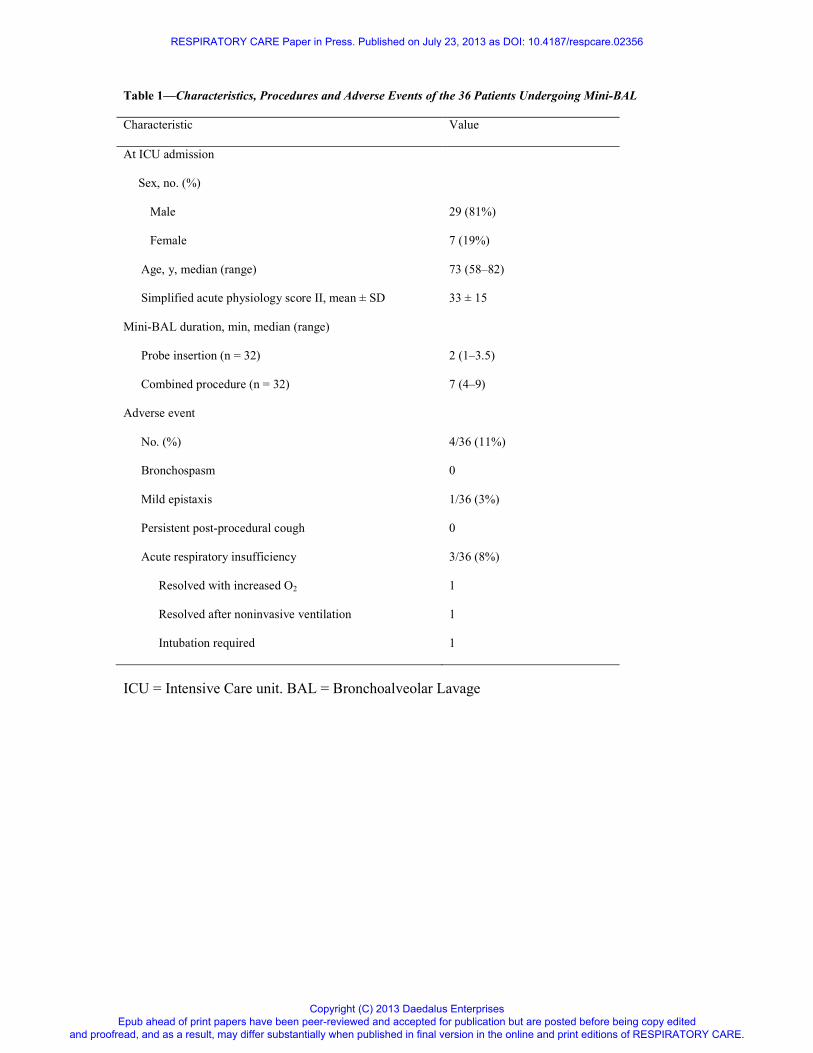
Table 1—Characteristics, Procedures and Adverse Events of the 36 Patients Undergoing Mini-BAL
Characteristic Value
At ICU admission
Sex, no. (%)
Male 29 (81%)
Female 7 (19%)
Age, y, median (range) 73 (58–82)
Simplified acute physiology score II, mean ± SD 33 ± 15
Mini-BAL duration, min, median (range)
Probe insertion (n = 32) 2 (1–3.5)
Combined procedure (n = 32) 7 (4–9)
Adverse event
No. (%) 4/36 (11%)
Bronchospasm 0
Mild epistaxis 1/36 (3%)
Persistent post-procedural cough 0
Acute respiratory insufficiency 3/36 (8%)
Resolved with increased O2 1
Resolved after noninvasive ventilation 1
Intubation required 1
ICU = Intensive Care unit. BAL = Bronchoalveolar Lavage
RESPIRATORY CARE Paper in Press. Published on July 23, 2013 as DOI: 10.4187/respcare.02356
Copyright (C) 2013 Daedalus Enterprises Epub ahead of print papers have been peer-reviewed and accepted for publication but are posted before being copy edited
and proofread, and as a result, may differ substantially when published in final version in the online and print editions of RESPIRATORY CARE.
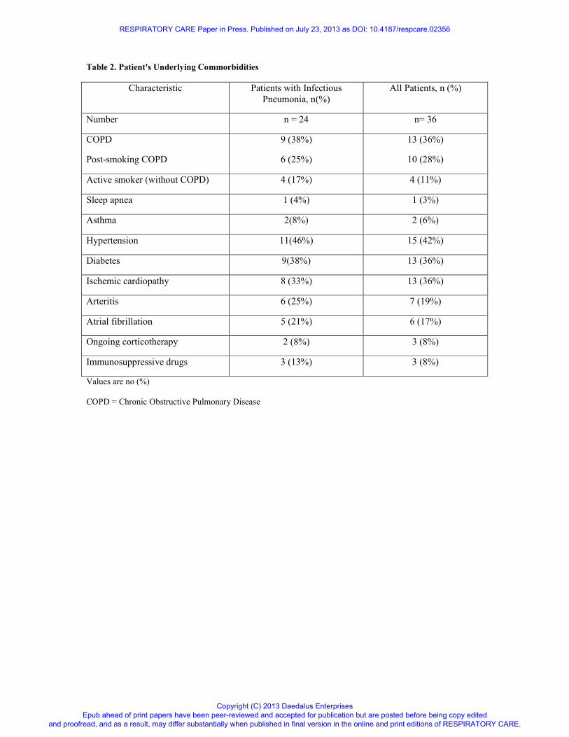
Table 2. Patient's Underlying Commorbidities
Characteristic Patients with Infectious
Pneumonia, n(%)
All Patients, n (%)
Number n = 24 n= 36
COPD
Post-smoking COPD
9 (38%)
6 (25%)
13 (36%)
10 (28%)
Active smoker (without COPD) 4 (17%) 4 (11%)
Sleep apnea 1 (4%) 1 (3%)
Asthma 2(8%) 2 (6%)
Hypertension 11(46%) 15 (42%)
Diabetes 9(38%) 13 (36%)
Ischemic cardiopathy 8 (33%) 13 (36%)
Arteritis 6 (25%) 7 (19%)
Atrial fibrillation 5 (21%) 6 (17%)
Ongoing corticotherapy 2 (8%) 3 (8%)
Immunosuppressive drugs 3 (13%) 3 (8%)
Values are no (%)
COPD = Chronic Obstructive Pulmonary Disease
RESPIRATORY CARE Paper in Press. Published on July 23, 2013 as DOI: 10.4187/respcare.02356
Copyright (C) 2013 Daedalus Enterprises Epub ahead of print papers have been peer-reviewed and accepted for publication but are posted before being copy edited
and proofread, and as a result, may differ substantially when published in final version in the online and print editions of RESPIRATORY CARE.

Table 3. Physiological Variables Before and 15 Min After the Procedure
Variable Before After Differences:
Mean ± SD or
Median (Q1;Q3)
Range Mean ± SD or
Median (Q1;Q3)
Range Median*/Mean
[95% CI]
Respiratory rate/min 25 (21;30) 13–40 25 (22;30) 15–40 0.5 [–0.5, 2]*†
Saturation O2 97 (95;99) 85–100 95 (92;99) 66–100 1 [0.5, 2.5]*‡
O2 flow (L/min) 6 (5;11) 1–15 6 (5;10) 1–15 0 [0, 0]*†
Heart rate, beats/min 101 ± 21 58–139 99 ± 19 60–132 –2.53 [–4.98, –0.08]†
Blood pressure
Systolic, mm Hg 128 ± 28 89–211 130 ± 33 77–231 1.47 [–5.09, 8.03]†
Diastolic, mm Hg 70 ± 16 43–98 66 ± 14 44–99 –4.41 [–8.07, –0.75]†
†Value after – Value before.
‡Value before – Value after. SD = standard deviation. Quartiles 1 and 3 =
Q1;Q3. CI = confidence interval.
All comparisons were non-significant.
RESPIRATORY CARE Paper in Press. Published on July 23, 2013 as DOI: 10.4187/respcare.02356
Copyright (C) 2013 Daedalus Enterprises Epub ahead of print papers have been peer-reviewed and accepted for publication but are posted before being copy edited
and proofread, and as a result, may differ substantially when published in final version in the online and print editions of RESPIRATORY CARE.
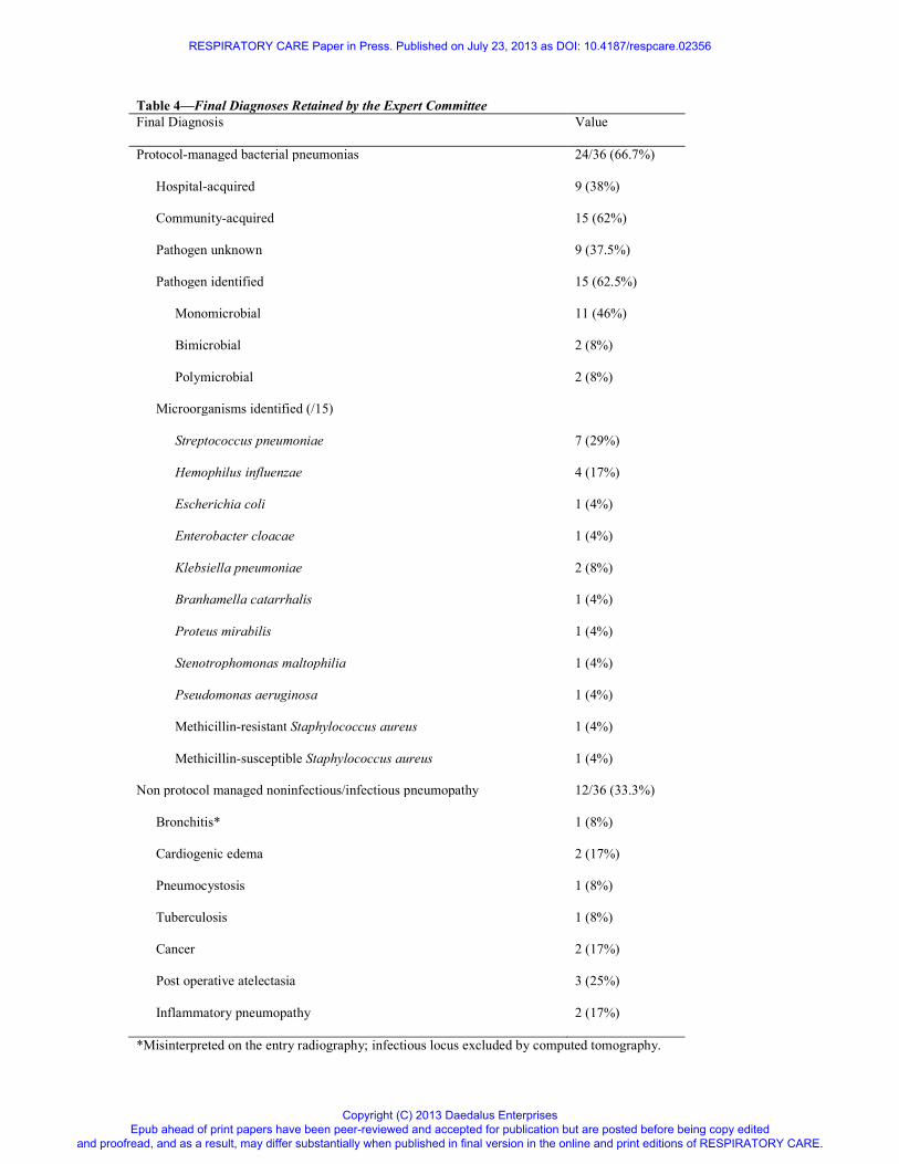
Table 4—Final Diagnoses Retained by the Expert Committee
Final Diagnosis Value
Protocol-managed bacterial pneumonias 24/36 (66.7%)
Hospital-acquired 9 (38%)
Community-acquired 15 (62%)
Pathogen unknown 9 (37.5%)
Pathogen identified 15 (62.5%)
Monomicrobial 11 (46%)
Bimicrobial 2 (8%)
Polymicrobial 2 (8%)
Microorganisms identified (/15)
Streptococcus pneumoniae 7 (29%)
Hemophilus influenzae 4 (17%)
Escherichia coli 1 (4%)
Enterobacter cloacae 1 (4%)
Klebsiella pneumoniae 2 (8%)
Branhamella catarrhalis 1 (4%)
Proteus mirabilis 1 (4%)
Stenotrophomonas maltophilia 1 (4%)
Pseudomonas aeruginosa 1 (4%)
Methicillin-resistant Staphylococcus aureus 1 (4%)
Methicillin-susceptible Staphylococcus aureus 1 (4%)
Non protocol managed noninfectious/infectious pneumopathy 12/36 (33.3%)
Bronchitis* 1 (8%)
Cardiogenic edema 2 (17%)
Pneumocystosis 1 (8%)
Tuberculosis 1 (8%)
Cancer 2 (17%)
Post operative atelectasia 3 (25%)
Inflammatory pneumopathy 2 (17%)
*Misinterpreted on the entry radiography; infectious locus excluded by computed tomography.
RESPIRATORY CARE Paper in Press. Published on July 23, 2013 as DOI: 10.4187/respcare.02356
Copyright (C) 2013 Daedalus Enterprises Epub ahead of print papers have been peer-reviewed and accepted for publication but are posted before being copy edited
and proofread, and as a result, may differ substantially when published in final version in the online and print editions of RESPIRATORY CARE.

Table 5—Invasive Procedure Outcomes for the 24 Patients with Bacterial Pneumonias
Culture Microbiological Diagnosis
Procedure n Positive Positive Negative Contaminated
Mini-BAL 21 16 (76%) 14 (67%) 7 (33%) 2 (10%)
NTS 21 9 (43%) 8 (38%) 13 (62%) 2 (10%)
Expectorated sputum 8 0 8 (100%)
BAL = bronchoalveolar lavage. NTS = nasotracheal suctioning
RESPIRATORY CARE Paper in Press. Published on July 23, 2013 as DOI: 10.4187/respcare.02356
Copyright (C) 2013 Daedalus Enterprises Epub ahead of print papers have been peer-reviewed and accepted for publication but are posted before being copy edited
and proofread, and as a result, may differ substantially when published in final version in the online and print editions of RESPIRATORY CARE.
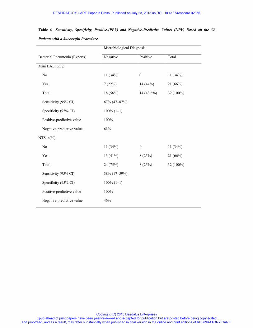
Table 6—Sensitivity, Specificity, Positive-(PPV) and Negative-Predictive Values (NPV) Based on the 32
Patients with a Successful Procedure
Microbiological Diagnosis
Bacterial Pneumonia (Experts) Negative Positive Total
Mini BAL, n(%)
No 11 (34%) 0 11 (34%)
Yes 7 (22%) 14 (44%) 21 (66%)
Total 18 (56%) 14 (43.8%) 32 (100%)
Sensitivity (95% CI) 67% (47–87%)
Specificity (95% CI) 100% (1–1)
Positive-predictive value 100%
Negative-predictive value 61%
NTS, n(%)
No 11 (34%) 0 11 (34%)
Yes 13 (41%) 8 (25%) 21 (66%)
Total 24 (75%) 8 (25%) 32 (100%)
Sensitivity (95% CI) 38% (17–59%)
Specificity (95% CI) 100% (1–1)
Positive-predictive value 100%
Negative-predictive value 46%
RESPIRATORY CARE Paper in Press. Published on July 23, 2013 as DOI: 10.4187/respcare.02356
Copyright (C) 2013 Daedalus Enterprises Epub ahead of print papers have been peer-reviewed and accepted for publication but are posted before being copy edited
and proofread, and as a result, may differ substantially when published in final version in the online and print editions of RESPIRATORY CARE.

FIG 1. Combicath. (A) The 2 catheters separated: white, the external tube, with movable
separator that maintains the internal mini-BAL catheter 5 cm above the opening of the
external tube. (B) After advancing the internal mini-BAL catheter, the end is protruded
allowing a sterile sample to be taken. The figures were kindly provided by Prodimed, Saint-
Leu La Forêt, France.
FIGURE 1.A
RESPIRATORY CARE Paper in Press. Published on July 23, 2013 as DOI: 10.4187/respcare.02356
Copyright (C) 2013 Daedalus Enterprises Epub ahead of print papers have been peer-reviewed and accepted for publication but are posted before being copy edited
and proofread, and as a result, may differ substantially when published in final version in the online and print editions of RESPIRATORY CARE.

FIGURE 1.B
RESPIRATORY CARE Paper in Press. Published on July 23, 2013 as DOI: 10.4187/respcare.02356
Copyright (C) 2013 Daedalus Enterprises Epub ahead of print papers have been peer-reviewed and accepted for publication but are posted before being copy edited
and proofread, and as a result, may differ substantially when published in final version in the online and print editions of RESPIRATORY CARE.