Embed Size (px)
Citation preview

Research ArticleErythrophagocytosis in Entamoeba histolytica andEntamoeba dispar: A Comparative Study
Daniel Talamás-Lara,1 Bibiana Chávez-Munguía,1
Arturo González-Robles,1 Patricia Talamás-Rohana,1 Lizbeth Salazar-Villatoro,1
Ángel Durán-Díaz,2 and Adolfo Martínez-Palomo1
1 Department of Infectomics and Molecular Pathogenesis, Center for Research and Advanced Studies, IPN,Avenida Instituto Politecnico Nacional No. 2508, Colonia San Pedro Zacatenco, Delegacion Gustavo A. Madero,07360 Mexico City, DF, Mexico
2 Faculty of Superior Studies Iztacala, Biology, UNAM, Los Reyes Iztacala, 54090 Tlalnepantla, MEX, Mexico
Correspondence should be addressed to Adolfo Martınez-Palomo; [email protected]
Received 28 February 2014; Revised 9 May 2014; Accepted 9 May 2014; Published 5 June 2014
Academic Editor: Abraham Landa-Piedra
Copyright © 2014 Daniel Talamas-Lara et al. This is an open access article distributed under the Creative Commons AttributionLicense, which permits unrestricted use, distribution, and reproduction in any medium, provided the original work is properlycited.
Entamoeba histolytica is the causative agent of human intestinal and liver amebiasis. The extraordinary phagocytic activityof E. histolytica trophozoites has been accepted as one of the virulence mechanisms responsible for their invasive capacity.The recognition of the noninvasive Entamoeba dispar as a different species has raised the question as to whether the lack ofpathogenic potential of this ameba correlates with a limited phagocytic capacity. We have therefore compared the process oferythrophagocytosis in both species by means of light and video microscopy, hemoglobin measurement, and the estimation ofreactive oxygen species (ROS). In the present study, we confirmed that E. dispar has lower erythrophagocytic capacity. We alsoobserved by video microscopy a new event of erythrocyte opsonization-like in both species, being more characteristic in E.histolytica. Moreover, E. dispar showed a lower capacity to produce ROS compared with the invasive species and also showed alarge population of amoebae that did not engulf any erythrocyte over time. Our results demonstrate that E. histolytica has a higherphagocytic capacity than E. dispar, including a higher rate of production of ROS in the course of ingesting red blood cells.
1. Introduction
Entamoeba histolytica, an enteric parasite capable of invadingintestinal mucosa and spreading to other organs, mainly theliver, is a significant source of morbidity and mortality indeveloping countries [1, 2]. The motile form of the parasite,the trophozoite, usually lives as a harmless commensal inthe lumen of the large intestine where it multiplies anddifferentiates into a cyst, the resistance form, responsiblefor transmission of the infection. Occasionally, trophozoiteswould invade the intestinal mucosa and produce dysenteryor amoeba and spread to other organs [3]. The existenceof two different species of Entamoeba, initially proposedby Brumpt in 1925, was approved at the XIII Seminar onAmebiasis, held in Mexico City [4]. At present, unequivocal
evidence for the existence of two morphologically similarand closely related species of Entamoeba in humans hasbeen substantiated by immunological, genetic and molecularstudies. As a result, E. histolytica, the invasive organism wasformally redescribed to separate it from the noninvasive, andmore common, Entamoeba dispar [5].
E. histolytica invasion starts when trophozoites residingin the colon deplete the mucus, interact with enterocytes,dismantle cell junctions, and lyse host cells [6], whereasE. dispar does not break down the mucus barrier or causeepithelial cell damage when in contact with cells on humancolonic explants [7].
Although erythrophagocytosis has been proposed as aqualitative pathogenicity indicator rather than a quantitativevirulence indicator [8], this process is widely considered as
Hindawi Publishing CorporationBioMed Research InternationalVolume 2014, Article ID 626259, 10 pageshttp://dx.doi.org/10.1155/2014/626259
2 BioMed Research International
one of the most prominent characteristics of E. histolyticavirulence [9–11].
Biochemical changes accompanying the process of endo-cytosis include increases of oxygen and glucose consumption,the activity of the pentose or hexose monophosphate cycle,and hydrogen peroxide production. Together, these changesconstitute the respiratory burst [12]. Once phagocytosisoccurs, the respiratory burst is carried out as part of themetabolic processes to remove endocytosed material.
Having the opportunity to compare invasive versus non-invasive parasites, we decided to analyze comparatively theerythrophagocytosis process to determine possible differ-ences between these two species of Entamoeba. The ery-throphagocytic process was registered with light microscopyby means of the Novikoff et al. [13] staining to quantify theamount of ingested erythrocytes and to determine whetherthe erythrophagocytic capacity of each species correlates withthe surface area of each amoeba as determined by lightmicroscopic measurements. We have also used spectropho-tometry to analyze the amount of ingested hemoglobin. Tocorroborate the percentage of nonphagocytic populations,video microscopy, flow cytometry, and confocal microscopywere used.Moreover, the extent of the respiratory burst, usingNBT, was measured for each amoebic species.
2. Materials and Methods
2.1. Cells. Amoebas were cultured in borosilicate glass tubesunder axenic conditions. E. histolytica trophozoites HM1-IMSS species were grown to logarithmic phase (72 h) inTYI-S-33 medium at 36∘C [14] and E. dispar trophozoitesin YI-S culture medium [15] for 72 h at 36∘C. Both culturemedia contained 10% bovine serum and a vitamin mixture.Parasites were harvested by chilling the culture tubes at4∘C in a water-ice bath for 10min and then they werecentrifuged at 900×g for 5min. Type B human erythrocytes(Rh+) were freshly obtained in Alsever’s solution (SigmaAldrich Company, UK) and washed 3 times in the samesolution to remove white blood cells. The erythrocytes werecounted and used in a 1 : 100 (trophozoites : erythrocytes)ratio in quantitative erythrophagocytosis assays and 1 : 5ratio for video microscopy analysis of erythrophagocytosis.Yeasts of the genus Candida albicans species CAI4 generatedfrom SC5314 (Clinical Systemic Isolate) [16] were routinelymaintained in YPD medium [17]. Minimal defined mediumconsisted of 2% glucose supplemented with yeast nitrogenbase (DIFCO). After that, Candida albicans were washed 2times and harvested in phosphate buffered saline solution(PBS) and centrifuged at 900×g for 5min and finally countedand used at a 1 : 100 (trophozoite : yeast) ratio in phagocytosisassays to measure the respiratory burst.
2.2. Erythrophagocytosis. E. histolytica and E. dispar tropho-zoites were washed in TYI-S-33 and YI-S without bovineserum, respectively. To establish the interaction, erythro-cytes were added, and the interaction was carried out for5, 10, and 15min at 37∘C without bovine serum, using
a 1 : 100 amoeba-erythrocyte ratio for quantitative studies and1 : 5 amoeba-erythrocyte ratio for video microscopy studies.Analysis was done with the AxioVision SE64 software withimages obtained with a Zeiss Axiophot photomicroscope.
2.3. Quantitative Erythrophagocytosis, Study, and Correlationof Ingestion/Area. For quantitative experiments, 900𝜇L (5 ×10⁵ amoebas) was incubated with 100 𝜇L (5 × 10⁷ erythro-cytes) at different times, at 37∘C. At the end of the incubationtime, amoebas were resuspended in 1mL of distilled water tolyse free erythrocytes and stop erythrophagocytosis. Tropho-zoites of both species can resist the osmotic shock withoutalterations of the plasma membrane permeability. Cells werecentrifuged at 900 g/min for 5min and pellets were fixed with2.5% glutaraldehyde in PBS. Phagocytosed erythrocytes werevisualized by phase contrast microscopy with the alkalinebenzidine method as described [13]. Amoebas were incu-bated for 30min at 37∘C in 2mL of 3,3-diaminobenzidine(Sigma) at a concentration of 2mg/mL, 0.2%H
2O2in 0.05M
2-amino-2-methyl-propanediol-HC1 (Merck-Schuchardt) atpHof 9.7. Afterwashingwith PBS, the number of erythrocytespresent in the cytoplasm of 100 amoebas, in each time, wascounted in triplicate. Considering that Entamoeba strainsdo not contain peroxisomes [18], cytoplasmic componentspositive to benzidine were considered only as ingested ery-throcytes. Correlation tests regarding ingestion/area wereconducted by measuring the surface area of each amoebaand the number of erythrocytes phagocytosed, using theAxioVision software SE64.
2.4. Indirect Determination of Erythrophagocytosis (Quantifi-cation of Hemoglobin). For a precise analysis of erythroph-agocytosis, a quantitative determination of hemoglobin wasdone. Nonfixed trophozoites were washed with Turk’s solu-tion to eliminate noningested erythrocytes; trophozoites werepelleted and lysed with 1mL of formic acid and the amountof hemoglobin was measured by spectrophotometric analysisat 400 nm.
2.5. Video Microscopy. Amoebas (1.25 × 10⁵) were placed oncoverslips resuspended in 100 𝜇L of the respective mediumand then erythrocytes (6.25 × 10⁵) were added in 5 𝜇L ofAlsever’s solution, maintaining slide temperature at 37∘C.Micrographs and video micrographs sequences were takenwith a Zeiss Axiophot microscope.
2.6. Fluorescent Labeling of Cells. To avoid manipulation oferythrocytes, 15𝜇L of whole blood were resuspended inAlsever’s solution (300 𝜇L) and labeled with 2.5𝜇L of Sytox9 through incubation for 1 h at room temperature underconstant stirring. After that, cells were washed twice at 600×gfor 10min and finally erythrocytes were resuspended in1.5mL Alsever’s solution.
2.7. Flow Cytometry and Confocal Microscopy. The existenceof a nonphagocytic population in both species was quantita-tive and qualitatively determined by flow cytometry and con-focal microscopy respectively. By flow cytometry the number

BioMed Research International 3
of amoebas that had ingested stained erythrocytes wasevaluated at different times, as mentioned. Once the timehad elapsed, cells were centrifuged at 900×g for 10min andthen fixed with freshly prepared 4% (v/v) paraformaldehyde,for 1 h. After that, cell suspensions were washed 4 timeswith PBS and then read in a flow cytometer FACS-Calibur(Becton Dickinson). Confocal microscopy was used to dis-tinguish ingested erythrocytes from free erythrocytes and toshow amoebas that had not phagocytosed. Coverslips weremounted with Vectashield (Vector Laboratories; Ontario,Canada) and analyzed by confocal microscopy in an LSM700microscope (Carl Zeiss Microimagin GmbH, Carl Zeiss,Germany).
2.8. Reduction of Nitroblue Tetrazolium (NBT) to Assess thePhagocytic Function. Amoebas (3.5 × 10⁵/400 𝜇L) previouslyadhered for 15min to coverslips were incubated with yeast(3.5 × 10⁷/100 𝜇L) for 30, 60, and 120min by the additionof 500𝜇L of NBT (1mg/mL in sterile PBS) [19]. At the endof interaction, the reaction was stopped by adding 1mL of70% methanol dissolved in PBS for 10min and washing oncewith PBS to remove excess of methanol; immediately after,0.5% safranin dissolved in water was added and incubatedfor 1min. The excess was removed with several washes untilthe sample was slightly stained. Coverslips were mounted,and yeast that had been reduced was counted using a phasecontrast microscope. Counts were done taking 100 amoebasrandomly, in triplicate, for each processed sample, and colorchanges that had occurred inside of cells were counted, takingas a positive value at least 1 single reduced yeast, and amoebasthat did not show a color change in their cytoplasm weretaken as a negative value. This method was performed intriplicate with only 2 possible variants (positive and negative)whereby only positive and negative cells where counted ineach time.
2.9. Statistical Analysis. In the data obtained concerningthe number of phagocytosed erythrocytes, absorbance ofhemoglobin, and amoebas that reduced NBT when exposedto pathogenic yeast at different times with each species ofEntamoeba, the following descriptive measures were cal-culated: average, minimum, and maximum standard devi-ation, quartiles (Q1, Q2, Q3), and coefficient of variation(Data not shown). Likewise, box and whisker diagrams weredeveloped to further describe the phenomena occurring inthe process of erythrophagocytosis and phagocytosis. Todetermine whether significant differences were present in thenumber of ingested erythrocytes, absorbance of hemoglobin,and amoeba showing NBT reduction, factorial analysis ofvariance was applied (ANOVA) with the transformed dataof these variables (data were transformed using the Box-Coxtechnique because the Anderson-Darling test indicated thatdata were not normal). Once the ANOVA test was performedto establish which conditions showed differences, the Tukey’stest was applied to measure the difference in the mean valuesbetween groups. These statistical analyses were carried outusing MiniTab software version 16.0.
a
a
a
b
b d f
b
c
c e
c
5 10 15 5 10 15Time (min)Strain Entamoeba dispar
Inge
sted
eryt
hroc
ytes
/am
oeba
Entamoeba histolytica
35
30
25
20
15
10
5
0
∗∗∗∗∗∗∗∗∗∗∗∗∗∗∗∗∗
∗∗∗∗∗∗∗
∗
∗∗∗
∗∗
∗
∗
∗
∗
∗
∗
∗∗
∗∗
∗∗∗∗
∗∗∗∗
∗∗∗∗
∗∗∗ ∗∗∗
∗∗∗∗∗∗∗∗
∗∗∗∗∗
∗∗∗∗
∗∗
∗∗∗
∗∗
Figure 1: Erythrocyte uptake performedwith E. dispar (blue) and E.histolytica (red) by light microscopy. The experiment was repeatedthree times independently in triplicates. The statistical comparisonsshowed significant differences in the erythrophagocytic capacitybetween species and among times (𝑃 < 0.001; different letters ontop). Moreover, species compared with their reciprocal times alsoshowed a significant difference (𝑃 < 0.001; bottom letters: a with b,c with d, and e with f) (∗). Outliers.
3. Results
A comparative analysis of the phagocytic capacity of E. his-tolytica andE. disparmayhelp to understand themechanismsinvolved in the virulence of E. histolytica. To date limitedinformation exists about the erythrophagocytic capacity ofE. dispar. Here we demonstrate basic differences in thephagocytic process between these two parasites.
3.1. Comparative Analysis of the Erythrophagocytic Capacitybetween E. dispar and E. histolytica
3.1.1. Determination of the Number of Ingested Erythrocytes.As a first step in the analysis of erythrophagocytosis, thenumber of ingested erythrocytes per amoeba was determinedafter 5, 10, and 15min of interaction. As shown in Figure 1,this is a time-dependent process for both E. dispar and E.histolytica, with a clear increase in the number of ingestederythrocytes with longer times of interaction. ANOVA testbetween species and time variables was carried out showingthat these variables, “species” and “time,” showed a significantdifference (𝑃 < 0.001). With Tukey’s test, each variable withrespect to all other variables of “species” and “time” was com-pared. Results show that, for each reciprocal combination,the 𝑃 value was less than 0.001 (𝑃 < 0.001). This confirmssignificant differences among all combinations (Figure 1).
3.1.2. Determination of Hemoglobin Content in Trophozoites.To corroborate the results obtained by direct assays, indirectmeasurements were carried out by spectrophotometry todetermine the absorbance produced by the hemoglobincontained in erythrocytes ingested by 5 × 10⁵ trophozoites ofeach species at previously described times. Therefore, it waspossible to obtain additional and more accurate information
4 BioMed Research International
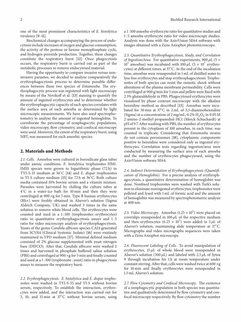
about the erythrophagocytosis process. In addition, ANOVAbetween different variables (species and time) was carried outto determine if differences found in absorbance between E.histolytica and E. dispar were significant.
As expected, there were clear differences in the ery-throphagocytic capacity of E. dispar versus E. histolytica(𝑃 < 0.001); however, in contrast to results found whencounting erythrocytes, hemoglobin determination did notshow significant differences among time in the same species.With Tukey’s test, each variable with respect to all other“species” and “time” variables were compared. Results showthat, for each reciprocal combination, the 𝑃 value was lessthan 0.001 (𝑃 < 0.001) (Figure 2).
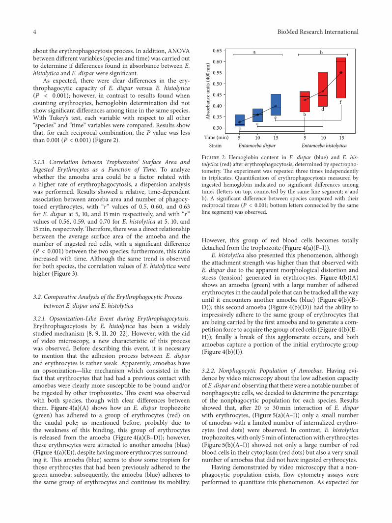
3.1.3. Correlation between Trophozoites’ Surface Area andIngested Erythrocytes as a Function of Time. To analyzewhether the amoeba area could be a factor related witha higher rate of erythrophagocytosis, a dispersion analysiswas performed. Results showed a relative, time-dependentassociation between amoeba area and number of phagocy-tosed erythrocytes, with “𝑟” values of 0.5, 0.60, and 0.63for E. dispar at 5, 10, and 15min respectively, and with “𝑟”values of 0.56, 0.59, and 0.70 for E. histolytica at 5, 10, and15min, respectively.Therefore, there was a direct relationshipbetween the average surface area of the amoeba and thenumber of ingested red cells, with a significant difference(𝑃 < 0.001) between the two species; furthermore, this ratioincreased with time. Although the same trend is observedfor both species, the correlation values of E. histolytica werehigher (Figure 3).
3.2. Comparative Analysis of the Erythrophagocytic Processbetween E. dispar and E. histolytica
3.2.1. Opsonization-Like Event during Erythrophagocytosis.Erythrophagocytosis by E. histolytica has been a widelystudied mechanism [8, 9, 11, 20–22]. However, with the aidof video microscopy, a new characteristic of this processwas observed. Before describing this event, it is necessaryto mention that the adhesion process between E. disparand erythrocytes is rather weak. Apparently, amoebas havean opsonization—like mechanism which consisted in thefact that erythrocytes that had had a previous contact withamoebas were clearly more susceptible to be bound and/orbe ingested by other trophozoites. This event was observedwith both species, though with clear differences betweenthem. Figure 4(a)(A) shows how an E. dispar trophozoite(green) has adhered to a group of erythrocytes (red) onthe caudal pole; as mentioned before, probably due tothe weakness of this binding, this group of erythrocytesis released from the amoeba (Figure 4(a)(B–D)); however,these erythrocytes were attracted to another amoeba (blue)(Figure 4(a)(E)), despite havingmore erythrocytes surround-ing it. This amoeba (blue) seems to show some tropism forthose erythrocytes that had been previously adhered to thegreen amoeba; subsequently, the amoeba (blue) adheres tothe same group of erythrocytes and continues its mobility.
a
a
b
b
df
ce
5 10 15 5 10 15Time (min)Strain Entamoeba dispar Entamoeba histolytica
Abso
rban
ce u
nits
(400
nm)
0.65
0.60
0.55
0.50
0.45
0.40
0.35
0.30
Figure 2: Hemoglobin content in E. dispar (blue) and E. his-tolytica (red) after erythrophagocytosis, determined by spectropho-tometry. The experiment was repeated three times independentlyin triplicates. Quantification of erythrophagocytosis measured byingested hemoglobin indicated no significant differences amongtimes (letters on top, connected by the same line segment; a andb). A significant difference between species compared with theirreciprocal times (𝑃 < 0.001; bottom letters connected by the sameline segment) was observed.
However, this group of red blood cells becomes totallydetached from the trophozoite (Figure 4(a)(F–I)).
E. histolytica also presented this phenomenon, althoughthe attachment strength was higher than that observed withE. dispar due to the apparent morphological distortion andstress (tension) generated in erythrocytes. Figure 4(b)(A)shows an amoeba (green) with a large number of adherederythrocytes in the caudal pole that can be tracked all the wayuntil it encounters another amoeba (blue) (Figure 4(b)(B–D)); this second amoeba (Figure 4(b)(D)) had the ability toimpressively adhere to the same group of erythrocytes thatare being carried by the first amoeba and to generate a com-petition force to acquire the group of red cells (Figure 4(b)(E–H)); finally a break of this agglomerate occurs, and bothamoebas capture a portion of the initial erythrocyte group(Figure 4(b)(I)).
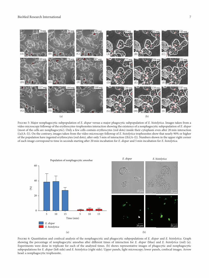
3.2.2. Nonphagocytic Population of Amoebas. Having evi-dence by video microscopy about the low adhesion capacityofE. dispar and observing that therewere a notable number ofnonphagocytic cells, we decided to determine the percentageof the nonphagocytic population for each species. Resultsshowed that, after 20 to 30min interaction of E. disparwith erythrocytes, (Figure 5(a)(A–I)) only a small numberof amoebas with a limited number of internalized erythro-cytes (red dots) were observed. In contrast, E. histolyticatrophozoites, with only 5min of interactionwith erythrocytes(Figure 5(b)(A–I)) showed not only a large number of redblood cells in their cytoplasm (red dots) but also a very smallnumber of amoebas that did not have ingested erythrocytes.
Having demonstrated by video microscopy that a non-phagocytic population exists, flow cytometry assays wereperformed to quantitate this phenomenon. As expected for

BioMed Research International 5
Entamoeba dispar
5minutesr = 0.50
Area (𝜇m2)
16
12
8
4
0Inge
sted
eryt
hroc
ytes
per
am
oeba
500 1000 1500 2000
10minutesr = 0.60
Area (𝜇m2)
16
12
8
4
0Inge
sted
eryt
hroc
ytes
per
am
oeba
500 750 1000 1250 1500
15minutes
r = 0.63
Area (𝜇m2)
16
12
8
4
0Inge
sted
eryt
hroc
ytes
per
am
oeba
500 1000 1500 2000
(a)
Entamoeba histolytica
5minutesr = 0.56
0 1200 2400 3600 4800
20
15
10
5
0Inge
sted
eryt
hroc
ytes
per
am
oeba
Area (𝜇m2)
10minutesr = 0.59
0 1000 2000 3000
20
15
10
5
0Inge
sted
eryt
hroc
ytes
per
am
oeba
Area (𝜇m2)
15minutesr = 0.70
1000 2000 3000 4000
20
15
10
5
0Inge
sted
eryt
hroc
ytes
per
am
oeba
Area (𝜇m2)
(b)
Figure 3: Scatter diagrams showing correlations between area and the number of ingested erythrocytes with respect to interaction time.There is a direct and significant relation (𝑃 < 0.001) for both strains, meaning that the larger is the area of the amoeba, these engulf moreerythrocytes. This ratio increases as time passes. The same trend for both strains was observed; however, correlations were higher for E.histolytica (“𝑟” value).

6 BioMed Research International
(A) (B) (C)
(D) (E) (F)
(G) (H) (I)
0 seg. 37 seg. 50 seg.
92 seg. 124 seg. 143 seg.
156 seg. 166 seg. 181 seg.
(a)
(A) (B) (C)
(D) (E) (F)
(G) (H) (I)
0 seg. 34 seg. 61 seg.
74 seg. 92 seg. 109 seg.
135 seg. 143 seg. 160 seg.
(b)
Figure 4: Real time video microscopy showing the opsonization-like event. (a) E. dispar: Figure 4(a)(A) shows a trophozoite (green) witha group of bound erythrocytes (red); then it can be seen how these erythrocytes completely detach from this amoeba (Figure 4(a)(B–D)),probably due to a low affinity binding. A new trophozoite (blue) appears in the scene (Figure 4(a)(D)) ready to bind the erythrocytes that hadbeen bound to another amoeba (Figure 4(a)(E and F)). Once again, these erythrocytes will detach from this blue amoeba (Figure 4(a)(G–I)). (b) E. histolytica: Figure 4(b)(A) shows a trophozoite (green) which binds erythrocytes (red) sending them to the caudal pole until itencounters another trophozoite (blue) (Figure 4(b)(B–D)); after both amoebas bound to the same group of erythrocytes, it is appreciatedhow they “fight” to keep the erythrocytes clump (Figure 4(b)(E–H)). Finally, Figure 4(b)(I) shows how every amoeba keeps a portion of theerythrocytes. Numbers shown in the upper right corner of each image correspond to time in seconds.
control assays, only 0.08% from 20,000 events of nonstainederythrocytes, showed some autofluorescence. In contrast,Sytox-stained erythrocytes showed that 99.87% of the cellswere positive for Sytox fluorescence; thus Sytox-positiveerythrocytes were then used for interaction experiments(data not shown). Figure 6(a) shows the summary of theseexperiments where there was a significant difference (𝑃 <0.001) between species; however, there were no differencesin the times measured. This confirms that nonphagocyticpopulation remains constant regardless of the time. It isimportant to mention that a viability test, with Trypan blue,was performed on trophozoites populations; they always hada 97% or higher viability (data not shown).
The previous results obtained by flow cytometry wereconfirmed by confocalmicroscopy. Figure 6(b) shows the twospecies, at 15min of interaction, and illustrates how the num-ber of ingested erythrocytes by E. dispar is smaller than thatin E. histolytica; also it shows an E. dispar trophozoite withoutany ingested erythrocytes (Figure 6(b), left side, arrowhead);as expected in the E. histolytica field, all trophozoites haveingested fluorescent erythrocytes (Figure 6(b), right side).
3.3. Comparative Analysis of ROS Production during Eryth-rophagocytosis by E. dispar and E. histolytica. The redoxcapability of each species by the NBT technique was analyzed
by light microscopy. The presence or the absence of blueformazan in yeast Candida albicans inside amoebas wasdetermined in 100 randomly chosen amoebas. Results regard-ing the variable “species” on the ability to produce reactiveoxygen species and the variable “time” required to producethem showed a significant difference (𝑃 < 0.001). WithTukey’s test, each variable with respect to all other variables“species” and “time” was compared. Results show that, foreach reciprocal combination, the 𝑃 value was less than 0.001(𝑃 < 0.001); however, at 1 and 2 h no significant differencesbetween times for each of the species were found becausethe maximum value of the respiratory burst occurs duringthe first hour [23] (Figure 7). Even though there was nosignificant difference, if we consider the slope, we can seethat the number of amoebas that reduce NBT increase withthe time much more rapidly in the case of E. histolytica incomparison with E. dispar.
4. Discussion
Phagocytosis is a central feature in the pathogenesis of inva-sive amebiasis, still an important public health problem [24].Considering that not all Entamoeba species have the samedegree of virulence, understanding of the various pathogenicmechanisms is an overriding goal. The unequivocal evidence
BioMed Research International 7
(A) (B) (C)
(D) (E) (F)
(G) (H) (I)
0 seg. 36 seg. 48 seg.
60 seg. 78 seg. 87 seg.
117 seg. 144 seg. 168 seg.
(a)
(A) (B) (C)
(D) (E) (F)
(G) (H) (I)
0 seg. 22 seg. 45 seg.
77 seg. 106 seg. 122 seg.
130 seg. 138 seg. 147 seg.
(b)
Figure 5: Major nonphagocytic subpopulation of E. dispar versus a major phagocytic subpopulation of E. histolytica. Images taken from avideo microscopy followup of the erythrocytes-trophozoites interaction showing the existence of a nonphagocytic subpopulation of E. dispar(most of the cells are nonphagocytic). Only a few cells contain erythrocytes (red dots) inside their cytoplasm even after 20min interaction((a)(A–I)). On the contrary, images taken from the video microscopy followup of E. histolytica trophozoites show that nearly 90% or higherof the population have ingested erythrocytes (red dots), after only 5min of interaction ((b)(A–I)). Numbers shown in the upper right cornerof each image correspond to time in seconds starting after 20min incubation for E. dispar and 5min incubation for E. histolytica.
E. disparE. histolytica
(%)
60
40
20
0
5 10 15 5 10 15
Time (min)
Population of nonphagocytic amoebae
(a)
E. dispar E. histolyticap y
20𝜇m 20𝜇m
20𝜇m20𝜇m
(b)
Figure 6: Quantitation and confocal analysis of the nonphagocytic and phagocytic subpopulations of E. dispar and E. histolytica. Graphshowing the percentage of nonphagocytic amoebas after different times of interaction for E. dispar (blue) and E. histolytica (red) (a).Experiments were done in triplicate for each of the analyzed times. (b) shows representative images of phagocytic and nonphagocyticsubpopulations for E. dispar (left side) and E. histolytica (right side). Upper panels, light microscopy; lower panels, confocal images. Arrowhead: a nonphagocytic trophozoite.

8 BioMed Research International
a
aa
b
b
b
d
f
ce
Time (min)Strain Entamoeba dispar Entamoeba histolytica
80
70
60
50
40
30
20
10
0
30 60 120 30 60 120
Amoe
bae t
hat r
educ
ed N
BT (%
)
Figure 7: Evaluation of the difference in the oxide-reduction abilitybetween E. dispar and E. histolytica using the NBT reductionassay. The experiment was repeated three times independently intriplicates. Results describing the oxide-reduction ability of bothstrains, measured by reduction of NBT, indicate that after 60minincubation, there is not a significant change in the amount offormazan produced (letters on top connected by the same linesegment); however, between 30 and 60min there is a significantdifference appreciated in both species (letters on top). Moreover,species compared with their reciprocal times also showed a signif-icant difference (𝑃 < 0.001) (different letters on bottom connectedby the same line segment).
for the existence of E. dispar, an amoeba closely relatedto E. histolytica, but not invasive, opens the way to studydifferences and similarities shared by the two species ofamoebas that colonize the human intestine [25–28].
In this work, we have addressed the study of ery-throphagocytosis from three different approaches, quanti-tative, qualitative, and biochemical, allowing presenting acomprehensive panorama of this phenomenon. We thereforewere able to demonstrate the existence of a nonphagocyticsubpopulation in each species, much larger, however, in E.dispar (40% inE. dispar versus 5% inE. histolytica).Moreover,a new event, similar to the two species, was the opsonization-like process of erythrocytes; this event was also stronger in E.histolytica than in E. dispar. E. histolytica has a larger capacityto induce phosphatidylserine exposure in the erythrocytessurface while E. dispar exhibits a deficient adhesion processwith red blood cells and also a very poor induction ofphosphatidylserine exposure, resulting in a less efficientphagocytosis [20]. The importance of phosphatidylserineexposure for E. histolytica engulfment of host cells has beensuggested by the work of Bailey et al., [22], who previouslydemonstrated that liposomes containing phosphatidylser-ine or synthetic negatively charged phospholipids, dicetylphosphate, stimulate E. histolytica actin polymerization, anecessary event for efficient phagocytosis.
4.1. Erythrophagocytosis. This process is highly asynchronouswith many variations that generate high standard deviations;furthermore, when values are analyzed in this way, it isnot easy to find statistically significant differences. For thisreason, statistical analysis of this study was performed using
box and whisker plots; this allowed us to collect informationabout individual amoebas and not to treat them as a jointpopulation. This gave us the opportunity to identify partic-ular events that usually are not detectable or distinguishableby other techniques, such as the presence of E. histolyticaamoebas that engulfed 20 or even uncountable erythrocytes;this could be due to the presence of amoebas that ingestederythrocytes more rapidly or to the presence of a subpopula-tion of larger size as shown by the graphs of scattering valuesof area versus number of phagocytosed erythrocytes. In thecase of E. dispar, the average population area ranges from 500to 1200𝜇m2, and very few amoebas fall out of range whilethe average for E. histolytica population falls in the range of1000 to 2000𝜇m2 with many more individual amoebas beingbigger than 3000 𝜇m2.
The spectrophotometric analysis of hemoglobin con-tent, a more sensitive technique used as a complementaryapproach for erythrophagocytosis evaluation, was usefulbecause samples were treated as total populations and indi-vidual variations were overcomed.This might explain why inthe graph showing results of hemoglobin content, significantdifferences among times of interaction were not foundwithinspecies.
Going back to results obtained by Trissl et al. [9] andcomparing their results with ours in terms of the number ofingested cells by the parasites obtained from an asymptomaticcarrier, with those obtained in this work, it is clear that theirvalues were similar to those obtained by us with E. dispar.
On the subject of the data obtained by flow cytometry,confocal microscopy, and video microscopy, we were ableto record a population of nonphagocytic amoebas for eachspecies and their behavior over time, supporting the resultsobtained by Sateriale et al. [29] who were able to separateamebic subpopulations of E. histolyticawith higher and lowerrates of phagocytosis. When microarrays tests were applied,these authors found that highly phagocytic amoeba showedoverexpression of at least 121 genes with respect to nonphago-cytic amoeba; therefore, additional studies with E. disparwill be necessary to differentiate sub- and overexpressedgenes in this species. This confirms the heterogeneity ofthe amoebic populations that might show different virulenceeven within the same population. Nowadays, the availabilityof the genomic sequences of E. histolytica and E. disparwouldallow the analysis of the genetic divergence and differentialgene expression between these two species and those genesand molecules associated with the erythrophagocytosis pro-cess (adherence, movement, endocytosis, etc.), to identifyvirulence mechanisms present in the virulent species andabsent or not expressed in the nonvirulent parasites [30].
4.2. Respiratory Burst. In mammals, the production of reac-tive oxygen species during phagocytosis has been widelystudied and has been associated with the killing capacity ofimmune cells to destroymicroorganisms [31]. In E. histolyticathere are only a few reports [32, 33] that describe a NBTreduction activity that implies that oxidoreduction activitiesare essential virulence components. It is known that E.histolytica is a highly phagocytic parasite and with respect

BioMed Research International 9
to E. dispar we have shown that these amoebas can alsophagocytose erythrocytes, however at a lower proportion;consequently we decided to analyze if the phagocytic pro-cess in both Entamoeba species would trigger an oxidativeburst, when feeding amoebas with pathogenic yeastsCandidaalbicans. Results showed that as low as 30% of E. dispartrophozoites were able to reduce NBT, in comparison withE. histolytica where as much as 80% of the cells reducedNBT. The conversion of NBT to formazan by the oxidativemetabolism of E. histolytica and E. dispar was used as aviability indicator, to measure their survival after challengewith various antiamoebic drugs [34, 35]. However, to datethere are no studies that associate the production of oxygenreactive species during phagocytosis by Entamoeba species.On the contrary, the ability of trophozoites to neutralizereactive oxygen species has been characterized and proposedas a virulence mechanism or as a mechanism that providesthe trophozoite with a major ability to survive to immunecells attack [36]. Therefore, the results here presented areconsistent with the fact that E. dispar, being considereda noninvasive amoeba, displays a smaller range of ROSproduction with respect to E. histolytica.
Recent studies have suggested that the generation ofROS and the presence of NADPH oxidase are necessaryin cancer cells for the formation of specialized structurescalled invadosomes (mechanosensory adhesive modules thatconsist of a dense core filamentous actin surrounded by aring of adhesion molecules able to infiltrate tissue underphysiological and pathological conditions) that allow for amore efficient tissue invasion process [37–40]. In this regard,the possible existence of invadosome-like structures in E.histolytica and its lack in E. dispar is worth pursuing.
5. Conclusions
We performed a comparative study of erythrophagocytosisbetween E. dispar, a noninvasive amoeba, and E. histolytica,a highly virulent and invasive parasite. Results demonstratethat the phenomenon is present in both Entamoeba speciesand that there are significant differences between the twoamoebas. Both, phagocytosis and the ability to producereactive oxygen species, are clearly more pronounced in E.histolytica in comparison to E. dispar.
Conflict of Interests
The authors declare that they have no conflict of interests.
Acknowledgments
The authors are grateful to Dr. Maritza Omana for helpfulcomments on this project; M. C. Vıctor Hugo Rosales forhis help with flow cytometry analysis; Veronica Hernandezand Ma. Magdalena Miranda for their help with confocalstudies; Anel Lagunes, Cesar Salas, Ranferi Andres, andJudith Escobedo for their technical support; Dr. Juan PedroLuna for the gift of Candida albicans strain. This researchwas supported by the PNPC 2013 Program from CONACyT,
Mexico; Daniel Talamas-Lara was a recipient of a CONACyTfellowship (262824).
References
[1] World Health Organization, “Amoebiasis,”Weekly Epidemiolog-ical Record, vol. 72, pp. 97–98, 1997.
[2] R. Haque, C. D. Huston, M. Hughes, E. Houpt, and W. A. PetriJr., “Amebiasis,”The New England Journal of Medicine, vol. 348,no. 16, pp. 1565–1573, 2003.
[3] A. Martınez-Palomo, “Parasitic amebas of the intestinal tract,”in Parasitic Protozoa, J. P. Kreier and J. R. Barker, Eds., pp. 65–141, Academic Press, New York, NY, USA, 2nd edition, 1993.
[4] “WHO/PAHO/UNESCO report. A consultation with expertson amoebiasis. Mexico City, Mexico 28-29 January, 1997,” inEpidemiological Bulletin, vol. 18, no. 1, pp. 13–14, 1997.
[5] L. S. Diamond and C. G. Clark, “A redescription of Entamoebahistolytica Schaudinn, 1903 (Emended Walker, 1911) separatingit from Entamoeba dispar Brumpt, 1925,”The Journal of Eukary-otic Microbiology, vol. 40, no. 3, pp. 340–344, 1993.
[6] I. Meza, “Extracellular matrix-induced signaling in Entamoebahistolytica: its role in invasiveness,” Parasitology Today, vol. 16,no. 1, pp. 23–28, 2000.
[7] D. Bansal, P. Ave, S. Kerneis et al., “An ex-vivo human intestinalmodel to study Entamoeba histolytica pathogenesis,” PLoSNeglected Tropical Diseases, vol. 3, no. 11, article e551, 2009.
[8] V. Tsutsumi, A. Ramirez-Rosales, H. Lanz-Mendoza et al.,“Entamoeba histolytica: erythrophagocytosis, collagenolysis,and liver abscess production as virulencemarkers,”Transactionsof the Royal Society of TropicalMedicine andHygiene, vol. 86, no.2, pp. 170–172, 1992.
[9] D. Trissl, A. Martinez-Palomo, M. de La Torre, R. de la Hoz,and E. Perez de Suarez, “Phagocytosis of human erythrocytes byEntamoeba histolytica. Quantitative study,” Archivos de Investi-gacion Medica, vol. 9, no. 1, pp. 219–222, 1978.
[10] E. Orozco, G. Guarneros, A. M. Palomo, and T. Sanchez,“Entamoeba histolytica. Phagocytosis as a virulence factor,”Journal of Experimental Medicine, vol. 158, no. 5, pp. 1511–1521,1983.
[11] S. Bhattacharya, A. Bhattacharya, and W. A. Petri Jr., “Examin-ing Entamoeba,” Trends in Parasitology, vol. 18, no. 5, pp. 196–197, 2002.
[12] N. Suzuki, G. Miller, J. Morales, V. Shulaev, M. A. Torres, andR. Mittler, “Respiratory burst oxidases: the engines of ROSsignaling,” Current Opinion in Plant Biology, vol. 14, no. 6, pp.691–699, 2011.
[13] A. B. Novikoff, P. M. Novikoff, C. Davis, and N. Quintana,“Studies on microperoxisomes. II. A cytochemical method forlight and electron microscopy,” Journal of Histochemistry andCytochemistry, vol. 20, no. 12, pp. 1006–1023, 1972.
[14] L. S. Diamond, D. R. Harlow, and C. C. Cunnick, “A newmedium for the axenic cultivation of Entamoeba histolytica andother Entamoeba,” Transactions of the Royal Society of TropicalMedicine and Hygiene, vol. 72, no. 4, pp. 431–432, 1978.
[15] L. S. Diamond, C. G. Clark, and C. C. Cunnick, “YI-S, a casein-free medium for axenic cultivation of Entamoeba histolytica,related Entamoeba, Giardia intestinalis and Trichomonas vagi-nalis,”The Journal of Eukaryotic Microbiology, vol. 42, no. 3, pp.277–278, 1995.
[16] W. A. Fonzi and M. Y. Irwin, “Isogenic strain construction andgene mapping in Candida albicans,” Genetics, vol. 134, no. 3, pp.717–728, 1993.

10 BioMed Research International
[17] J. B. Hicks, A. Hinnen, and G. R. Fink, “Properties of yeasttransformation,” Cold Spring Harbor Symposia on QuantitativeBiology, vol. 43, no. 2, pp. 1305–1313, 1979.
[18] A. Martınez-Palomo, A. Gonzalez-Robles, and B. C. deRamırez, “Ultraestructural study of various Entamoeba strains,”in Proceedings of the International Conference of Amebiasis, B.Sepulveda and L. S. Diamond, Eds., vol. 1, pp. 226–237, IMSS,1976.
[19] K. B. Hellum, “Standardization of the nitroblue tetrazoliumtest. Influence of pH, dye concentration and sample storage,”Scandinavian Journal of Infectious Diseases, vol. 9, no. 2, pp. 125–130, 1977.
[20] D. R. Boettner, C. D. Huston, J. A. Sullivan, and W. A. PetriJr., “Entamoeba histolytica and Entamoeba dispar utilize exter-nalized phosphatidylserine for recognition and phagocytosis oferythrocytes,” Infection and Immunity, vol. 73, no. 6, pp. 3422–3430, 2005.
[21] A. Chevez, I. Iturbe-Alessio, M. Segura, and D. Corona,“Phagocytosis of human erythrocytes byEntamoeba histolytica,”Archivos de Investigacion Medica, vol. 2, supplement 2, pp. 275–286, 1972.
[22] G. B. Bailey, D. B. Day, C. Nokkaew, and C. C. Harper, “Stimula-tion by target cell membrane lipid of actin polymerization andphagocytosis byEntamoeba histolytica,” Infection and Immunity,vol. 55, no. 8, pp. 1848–1853, 1987.
[23] J. A. Dıaz-Gandarilla, C. Osorio-Trujillo, V. I. Hernandez-Ramırez, and P. Talamas-Rohana, “PPAR activation inducesm1macrophage polarization via cPLA
2
-COX-2 inhibition, acti-vating ros production against Leishmania mexicana,” BioMedResearch International, vol. 2013, Article ID 215283, 13 pages,2013.
[24] C. D. Huston, D. R. Boettner, V. Miller-Sims, andW. A. Petri Jr.,“Apoptotic killing and phagocytosis of host cells by the parasiteEntamoeba histolytica,” Infection and Immunity, vol. 71, no. 2, pp.964–972, 2003.
[25] M. Espinosa-Cantellano, A. Gonzales-Robles, B. Chavez et al.,“Entamoeba dispar: ultrastructure, surface properties and cyto-pathic effect,” Journal of Eukaryotic Microbiology, vol. 45, no. 3,pp. 265–272, 1820.
[26] P. F. P. Pimenta, L. S. Diamond, and D. Mirelman, “Entamoebahistolytica Schaudinn, 1903 andEntamoeba dispar Brumpt, 1925:differences in their cell surfaces and in the bacteria-containingvacuoles,” Journal of Eukaryotic Microbiology, vol. 49, no. 3, pp.209–219, 2002.
[27] B. N. Mitra, T. Yasuda, S. Kobayashi, Y. Saito-Nakano, and T.Nozaki, “Differences in morphology of phagosomes and kinet-ics of acidification and degradation in phagosomes betweenthe pathogenic Entamoeba histolytica and the non-pathogenicEntamoeba dispar,” Cell Motility and the Cytoskeleton, vol. 62,no. 2, pp. 84–99, 2005.
[28] B. Chavez-Munguıa, P. Talamas-Rohana, G. Castanon, L.Salazar-Villatoro, V. Hernandez-Ramırez, and A. Martınez-Palomo, “Differences in cap formation between invasive Enta-moeba histolytica and non-invasive Entamoeba dispar,” Para-sitology Research, vol. 111, no. 1, pp. 215–221, 2012.
[29] A. Sateriale, A. Vaithilingam, L. Donnelly, P. Miller, and C.D. Huston, “Feed-forward regulation of phagocytosis by Enta-moeba histolytica,” Infection and Immunity, vol. 80, no. 12, pp.4456–4462, 2012.
[30] I.W.Wilson, G. D.Weedall, andN.Hall, “Host-Parasite interac-tions in Entamoeba histolytica and Entamoeba dispar: what have
we learned from their genomes?” Parasite Immunology, vol. 34,no. 2-3, pp. 90–99, 2012.
[31] L. F. Marchi, R. Sesti-Costa, M. D. C. Ignacchiti, S. Chedraoui-Silva, and B. Mantovani, “In vitro activation of mouse neu-trophils by recombinant human interferon-gamma: increasedphagocytosis and release of reactive oxygen species and pro-inflammatory cytokines,” International Immunopharmacology,vol. 18, no. 2, pp. 228–235, 2014.
[32] A. A. Kettis, C. Jarstrand, and T. Urban, “The nitrobluetetrazolium (NBT) reduction of Entamoeba histolytica duringendocytosis of E. coli and homologous antibodies,” Archivos deInvestigacion Medica, vol. 13, supplement 3, pp. 261–264, 1982.
[33] S. Kumar, L. M. Tripathi, and P. Sagar, “Oxido-reductivefunctions of Entamoeba histolytica in relation to virulence,”Annals of Tropical Medicine and Parasitology, vol. 86, no. 3, pp.239–248, 1992.
[34] R. M. Mukhopadhyay and S. K. Chaudhuri, “Rapid in vitro testfor determination of anti-amoebic activity,” Transactions of theRoyal Society of Tropical Medicine and Hygiene, vol. 90, no. 2,pp. 189–191, 1996.
[35] D. Bansal, R. Sehgal, Y. Chawla, R. C. Mahajan, and N. Malla,“In vitro activity of antiamoebic drugs against clinical isolates ofEntamoeba histolytica and Entamoeba dispar,”Annals of ClinicalMicrobiology and Antimicrobials, vol. 3, article 27, 2004.
[36] S. Sim, T.-S. Yong, S.-J. Park et al., “NADPH oxidase-derivedreactive oxygen species-mediated activation of ERK1/2 isrequired for apoptosis of human neutrophils induced by Enta-moeba histolytica,” Journal of Immunology, vol. 174, no. 7, pp.4279–4288, 2005.
[37] B. Diaz, G. Shani, I. Pass, D. Anderson, M. Quintavalle, andS. A. Courtneidge, “Tks5-dependent, nox-mediated generationof reactive oxygen species is necessary for invadopodia forma-tion,” Science Signaling, vol. 2, no. 88, article ra53, 2009.
[38] D. Gianni, N. Taulet, C. DerMardirossian, and G. M. Bokoch,“c-Src-mediated phosphorylation of NoxA1 and Tks4 inducesthe Reactive Oxygen Species (ROS)-dependent formation offunctional invadopodia in human colon cancer cells,”MolecularBiology of the Cell, vol. 21, no. 23, pp. 4287–4298, 2010.
[39] O. Destaing, M. R. Block, E. Planus, and C. Albiges-Rizo, “Inva-dosome regulation by adhesion signaling,” Current Opinion inCell Biology, vol. 23, no. 5, pp. 597–606, 2011.
[40] L. R. Boateng and A. Huttenlocher, “Spatiotemporal regulationof Src and its substrates at invadosomes,” European Journal ofCell Biology, vol. 91, no. 11-12, pp. 878–888, 2012.

Submit your manuscripts athttp://www.hindawi.com
Hindawi Publishing Corporationhttp://www.hindawi.com Volume 2014
Anatomy Research International
PeptidesInternational Journal of
Hindawi Publishing Corporationhttp://www.hindawi.com Volume 2014
Hindawi Publishing Corporation http://www.hindawi.com
International Journal of
Volume 2014
Zoology
Hindawi Publishing Corporationhttp://www.hindawi.com Volume 2014
Molecular Biology International
GenomicsInternational Journal of
Hindawi Publishing Corporationhttp://www.hindawi.com Volume 2014
The Scientific World JournalHindawi Publishing Corporation http://www.hindawi.com Volume 2014
Hindawi Publishing Corporationhttp://www.hindawi.com Volume 2014
BioinformaticsAdvances in
Marine BiologyJournal of
Hindawi Publishing Corporationhttp://www.hindawi.com Volume 2014
Hindawi Publishing Corporationhttp://www.hindawi.com Volume 2014
Signal TransductionJournal of
Hindawi Publishing Corporationhttp://www.hindawi.com Volume 2014
BioMed Research International
Evolutionary BiologyInternational Journal of
Hindawi Publishing Corporationhttp://www.hindawi.com Volume 2014
Hindawi Publishing Corporationhttp://www.hindawi.com Volume 2014
Biochemistry Research International
ArchaeaHindawi Publishing Corporationhttp://www.hindawi.com Volume 2014
Hindawi Publishing Corporationhttp://www.hindawi.com Volume 2014
Genetics Research International
Hindawi Publishing Corporationhttp://www.hindawi.com Volume 2014
Advances in
Virolog y
Hindawi Publishing Corporationhttp://www.hindawi.com
Nucleic AcidsJournal of
Volume 2014
Stem CellsInternational
Hindawi Publishing Corporationhttp://www.hindawi.com Volume 2014
Hindawi Publishing Corporationhttp://www.hindawi.com Volume 2014
Enzyme Research
Hindawi Publishing Corporationhttp://www.hindawi.com Volume 2014
International Journal of
Microbiology