Embed Size (px)
Citation preview

Research ArticleEffects of Water Models on Binding Affinity:Evidence from All-Atom Simulation of Binding ofTamiflu to A/H5N1 Neuraminidase
Trang Truc Nguyen,1 Man Hoang Viet,2 and Mai Suan Li2
1 Institute for Computational Science and Technology, Quarter 6, Linh Trung Ward, Thu Duc District,Ho Chi Minh City, Vietnam
2 Institute of Physics, Polish Academy of Sciences, Aleja Lotnikow 32/46, 02-668 Warsaw, Poland
Correspondence should be addressed to Mai Suan Li; [email protected]
Received 31 August 2013; Accepted 5 November 2013; Published 2 February 2014
Academic Editors: R. Luo and K. Spiegel
Copyright © 2014 Trang Truc Nguyen et al. This is an open access article distributed under the Creative Commons AttributionLicense, which permits unrestricted use, distribution, and reproduction in any medium, provided the original work is properlycited.
The influence of water models SPC, SPC/E, TIP3P, and TIP4P on ligand binding affinity is examined by calculating the bindingfree energyΔ𝐺bind of oseltamivir carboxylate (Tamiflu) to the wild type of glycoprotein neuraminidase from the pandemic A/H5N1virus. Δ𝐺bind is estimated by the Molecular Mechanic-Poisson Boltzmann Surface Area method and all-atom simulations withdifferent combinations of these aqueous models and four force fields AMBER99SB, CHARMM27, GROMOS96 43a1, and OPLS-AA/L. It is shown that there is no correlation between the binding free energy and the water density in the binding pocket inCHARMM. However, for three remaining force fields Δ𝐺bind decays with increase of water density. SPC/E provides the lowestbinding free energy for any force field, while the water effect is the most pronounced in CHARMM. In agreement with the popularGROMACS recommendation, the binding score obtained by combinations of AMBER-TIP3P, OPLS-TIP4P, and GROMOS-SPC isthe most relevant to the experiments. For wild-type neuraminidase we have found that SPC is more suitable for CHARMM thanTIP3P recommended by GROMACS for studying ligand binding. However, our study for three of its mutants reveals that TIP3P ispresumably the best choice for CHARMM.
1. Introduction
The determination of binding affinity is a central problem incomputer-aided drug design which is a useful tool to searchfor potential leads for various diseases. The accuracy ofestimation of the binding free energy Δ𝐺bind of ligand toreceptor depends on computational methods and modelingof receptor-ligand complex. The docking method is usuallyused for locating binding sites and virtual screening of poten-tial drug candidates from large data bases. This approachsuffers from low accuracy and its results usually have to berefined by more sophisticated methods based on the molec-ular dynamics (MD) simulation. In many cases MDmethodscan reproduce reliable results on binding free energy havingacceptable correlation with experimental data [1–5]. Amongthem Molecular Mechanic-Poisson Boltzmann Surface Area
(MM-PBSA) [1], thermodynamic integration (TI) [2], linearinteraction energy (LIE) [3], linear response approximation(LRA) [6, 7], free energy perturbation (FEP) [4], and steeredMD [8, 9] methods are widely used. Each method shouldbe considered carefully to compromise between CPU timeefficiency and accuracy level.
In modeling of biosystems in aqueous environment, itis important to develop appropriate force fields and watermodels. Force fields, which are given in the form of empiricalpotential energy functions, have been developed by differentgroups. Today OPLS, CHARMM, AMBER, and GROMOSare the most popular force fields for all-atom simulation ofbiomolecules. To describe aqueous environment, one can usevarious models such as SPC [10], SPC/E [11], TIP3P [12],and TIP4P [13]. The adjustment of parameters of these mod-els is based on their ability to reproduce the enthalpy of
Hindawi Publishing Corporatione Scientific World JournalVolume 2014, Article ID 536084, 14 pageshttp://dx.doi.org/10.1155/2014/536084

2 The Scientific World Journal
vaporization and the density of water. SPC/E is especiallyaccurate for capturing experimental properties of water suchas the diffusion coefficient and dielectric constant. BecauseSPC, SPC/E, TIP3P, and TIP4P are relatively simple and ableto provide reasonable results, they are often employed forsimulation of peptides [14, 15] and proteins [16].
Previous studies [17, 18] have revealed that differentforce fields provide different estimations forΔ𝐺bind. Recently,the role of water molecules in the binding process hasbeen considered [19–21]. GROMACS manual (http://www.gromacs.org/Support/Online Manual) suggests that for all-atom simulation of biomolecules water model TIP3P issuitable for AMBER and CHARMM, while TIP4P and SPCare more appropriate, respectively, for OPLS and GROMOSforce fields. However, what water model is the best fit for agiven force field in computation of Δ𝐺bind remains largelyunknown.
To shed light on this problem, in this paper we study theimpact of combinations of four main water models SPC [10],SPC/E [11], TIP3P [12], and TIP4P [13] with AMBER99SB[24], CHARMM27 [25], OPLS-AA [26], and GROMOS9643a1 [27] force fields on the binding affinity of Tamiflu tothe wild type (WT) of A/H5N1 neuraminidase (NA). Wehave chosen the NA-Tamiflu complex because A/H5N1 viruscauses great damage to live poultry markets [28], especiallybeing recognized as human transmitted virus [29]. Moreimportantly, the binding free energy of Tamiflu to NA hasbeen experimentally determined [23] and this gives us theopportunity to compare theoretical estimates with the exper-imental ones.
Using MM-PBSA method we have shown that combina-tions AMBER-TIP3P, OPLS-TIP4P, and GROMOS-SPC arethe best choice for estimation ofΔ𝐺bind of Tamiflu.This resultis in agreementwith theGROMACS recommendation,whichis followed from force field development [24–27]. Contraryto the GROMACS suggestion, it is shown that SPC is moresuitable for CHARMM than TIP3P but this conclusion isvalid for the wild type of NA. Our study of three mutantsH274Y, N294S, and Y252H reveals that TIP3P is presumablythe best choice for CHARMM as suggested by GROMACS.
It is found that Δ𝐺bind obtained by the OPLS force fieldis much less sensitive to water models compared to otherforce fields. The difference in water models seems to have thedrastic effect in CHARMM modulating both the receptor-ligand interaction energy and hydrogen bond network inbinding area. For all studied force fields, SPC/E is worse thanother aqueous models overestimating Δ𝐺bind.
2. Materials and Methods
2.1. Crystal Structure of A/H5N1 NA and Parametrization ofTamiflu. The initial structures of A/H5N1 WT and mutantsH274Y and N294S were obtained from Protein Data Bankwith PDB ID 2HU4, 3CL0, and 3CL2, respectively [23].Y252H was derived by the corresponding mutation inWT structure using the mutagenesis module, integrated inPyMOL package [30]. For Tamiflu we use the oseltamivir car-boxylate type. Its charges and atom types, used for MD
simulation, are described in detail in our previous work[18]. Namely, for united-atom GROMOS96 43a1 force field,charges and atom types of oseltamivir were fully parame-trized by Dundee PRODRG2.5 Server (Beta) [31]. For theremaining all-atom force fields, atomic partial charges forTamiflu were derived by ESP charge. To obtain optimalgeometry for electrostatic potential calculations, its structureis first optimized with the help of Gaussian98 [32] using theB3LYP/6-31G∗ level of theory. Fitting charges to the electro-static potential was subsequently done by the RESP method.Atom types for Tamiflu were derived from different modulesto get along with each force field. ACPYPE [33] and MKTOP[34] were adjusted to provide suitable atom types in OPLS-AA/L [26], while for AMBER99SB and CHARMM27 [25],atom types were named by ACPYPE and SwissParam [35],respectively.
2.2. Water Models. Water, known as an indispensable solventin almost chemical and biological reactions, has been built indifferent ways to obtain reasonable models for computationalstudy [10–13]. A water molecule is characterized by itsgeometrical parameters such as bond lengths and angleswhich could be kept rigid or flexible during simulation. Eachmodel is parametrized with atomic partial charges of oxygenand hydrogen and assigned with dispersion and repulsionforces approximated by Lennard-Jones potential [22]. Watermodels are categorized by the number of points used toshape them, by rigid or flexible structures, and by integrationor not with polarization effects [22]. There are 46 distinctmodels [36] that have from 3 to 6 sites. However, only 3-and 4-site models (Figure 1) are often used in simulationsof biological systems. For 3-site model like SPC [10] andTIP3P [12], a water molecule is constructed by one oxygenatom and two hydrogen atoms. Each atom is assigned withatomic partial charge, but only oxygen is allowed to havethe Lennard-Jones interaction with other atoms. The vander Waals (vdW) interaction among hydrogen atoms wasnot parametrized yet. Three-site models are known as rigidand have the experimental geometry of water, except SPCwhich has the ideal tetrahedral angle of water as 109.47, butnot 104.5∘. SPC/E [11], an updated version of SPC model,adds an average polarization correction to the potentialenergy function, resulting in the better density and diffusionconstant than SPC model.
The four-site model TIP4P [13] is a rigid planar four-siteinteraction potential for water (Figure 1), having a similargeometry to the Bernal and Fowler model [37]. Here thenegative charge is shifted from the oxygen atom to a point0.15 A along the bisector between hydrogen atoms. In thispaper, we just limit our study to only four frequently usedwater models SPC, SPC/E, TIP3P, and TIP4P (Figure 1).Their geometrical and physical characteristics are describedin Table 1. Here 𝑞H, 𝑞O, and 𝑞L are the partial charges ofhydrogen, oxygen, and lone pair, respectively. 𝜃 and 𝜙 are theH–O–H and lone pair-O–H angles, while 𝜀 and 𝜎 are the welldepth and vdW radius, respectively.
2.3. Molecular Dynamic Simulations. Complex of NA-Tam-iflu is placed in a triclinic box of around 12000 water

The Scientific World Journal 3
𝜃
𝜎
qH
qH
qO
l1
(a)
𝜃𝜎
𝜑
qH
qH
qL
l1
l2
(b)
Figure 1: The 3-site (a) and 4-site (b) water models [22]. The labels are explained in Table 1.
Table 1: Physical properties of water models [22]. All data is recorded at 25∘ and 1 atm. 𝑞H, 𝑞O, and 𝑞L are the partial charges of hydrogen,oxygen, and lone pair, respectively. 𝜃 and 𝜙 are the H–O–H and lone pair-O–H angles, respectively. 𝜀 and 𝜎 are the well depth and vdWradius, respectively.
𝜎 (A) 𝜀 (kJ/mol) 𝑙1(A) 𝑙
2𝑞H (e) 𝑞O/𝑞L (e) 𝜃 𝜙 Dipole moment Dielectric constant
SPC 3.166 0.65 1 0.41 −0.82 109.47 2.27 65SPC/E 3.166 0.65 1 0.423 −0.847 109.47 2.35 71TIP3P 3.150 0.636 0.957 0.417 −0.834 104.52 2.35 82TIP4P 3.153 0.648 0.957 0.15 0.52 −1.04 104.52 52.26 2.18 53Exp. 2.95 78.4
molecules with 1 nm distance between the solute andbox (a typical snapshot is shown in Figure S1 of sup-plementary material available online at http://dx.doi.org/10.1155/2014/536084). The receptor and ligand have 3832 and5749 atoms in the united atom and all-atom models, respec-tively. Periodic boundary condition is imposed in threedirections. We use 1.4 nm and 1.0 nm cut-off for vdW andelectrostatic interactions, respectively. Long range electro-static interaction was computed by the particle-mesh Ewaldsummation method [38]. Equations of motion were inte-grated using a leap-frog algorithm [39] with a time step1 fs. The nonbonded interaction pairlist was updated every10 fs with the cut-off of 1 nm. All systems were neutralizedby adding counter-ions and then minimized to remove thelocal strain in protein upon addition of all hydrogen atomsand to remove bad vdW contacts with water. By using theconjugate gradient method for every 50 steps of steepestdescent, minimization is converged when maximum forcebecomes smaller than 0.01 kJ/mol/nm. Then, all bonds ofprotein were restrained, leaving remaining parts to relax for100 ps to obtain evenly distributed system. The temperaturewas gradually heated to 300K during 100 ps with 5 kcal/molharmonic restraints in all systems.The equilibration was nextperformed, coupled with temperature and pressure. Constanttemperature 300K was enforced using Berendsen algorithm[40] under 50 ps NVT simulation with a damping coefficientof 0.1 ps. Parrinello-Rahman pressure coupling [41] was used
in 100 ps NPT run for 1 atm with the damping coefficientof 0.5 ps. Final NPT simulations of 20 ns were carried outfor all force fields. Each force field is combined with fourdifferent water models SPC, SPC/E, TIP3P, and TIP4P, exceptGROMOS, which uses only SPC and SPC/E models. In total,we have 14 different models for the NA-Tamiflu complex. Allsimulations have been carried out in theGROMACS suit withGromacs-4.5 package [42].
2.4. Binding Free Energy Calculation by MM-PBSA. Thedetails of MM-PBSA method are given in our previous work[18]. Overall, in this method the binding free energy of ligandto receptor is defined as follows:
Δ𝐺bind = Δ𝐸elec + Δ𝐸vdw + Δ𝐺sur + Δ𝐺PB − 𝑇Δ𝑆, (1)
where Δ𝐸elec and Δ𝐸vdw are contributions from electrostaticand vdW interactions, respectively. Δ𝐺sur and Δ𝐺PB are non-polar and polar solvation energies.The entropic contribution𝑇Δ𝑆 is estimated using the normal mode approximation [18].From 20 ns MD simulation output only snapshots collectedin equilibrium are used to compute the binding free energygiven by (1).
2.5. Measures Used in Data Analysis. The C𝛼root-mean-
square deviation (RMSD) is employed to measure the devi-ation of receptor structures from the initial configuration.

4 The Scientific World Journal
Table 2: List of 50 residues surrounding the binding site.
Ile117 Arg118 Glu119 Leu134 Thr135 Gln136 Thr148 Val149 Lys150 Asp151Arg152 Ser153 Arg156 Trp178 Ser179 Asp198 Asn221 Ile222 Leu223 Arg224Thr225 Glu227 Pro245 Ser246 Glu276 Glu277 Arg292 Asp293 Asn294 Asn325Pro326 Tyr347 Gly348 Val349 Lys350 Ser370 Arg371 Trp403 Ser404 Gly405Tyr406 Ile427 Arg428 Gly429 Arg430 Pro431 Lys432 Thr439 Ser440 Gly441
Figure 2: The construction of convex hull for the binding site. C𝛼
atoms of fifty residues which define the binding pocket are shownin blue ball. Not all sides of polyhedron are shown. Oseltamivir iscolored in green. Water molecules are also presented.
The hydrogen bond (HB) is assumed to be formed if thedistance between proton donor (D) and proton acceptor (A)is less than 0.35 nm and the angle H-D-A is also less than 30∘.
2.6. Definition of Binding Site. The binding pocket is definedas a space surrounded by 50 amino acids as shown in FigureS2. Our definition is compatible with that of Cheng et al. [43].The list of these amino acids is given in Table 2. Volume ofthe binding pocket is approximately estimated as volume ofsmallest convex hull which contains all of the fifty C
𝛼atoms
[44–46] (Figure 2).The number of watermolecules inside theconvex hull is considered as the number of water moleculesin the binding site. The binding pocket volume and numberof water molecules inside it are calculated byMatlab software[47].Thewater density in binding site is a ratio of the numberof water molecules to its volume.
3. Results and Discussion
In this section we present results obtained for WT NA if nototherwise stated.
3.1. Equilibration Time Scales Depend on Force Field andWater Model. The time dependence of C
𝛼-RMSD of NA in
complex with Tamiflu is shown in Figure 3 for different setsof force fields and water models. The equilibration of systemis reached when RMSD gets saturation. In AMBER theequilibration time 𝑡eq ≈ 15 ns for SPC, while ≈10 ns is neededto equilibrate the system in other water models (Figure 3).
SPC gives the largest RMSD in equilibrium state. In OPLSRMSD reaches saturation quite fast, after about 3 ns for TIP3Pand 5 ns for remaining models (Figure 3). TIP3P provides abit larger departure from the initial structure compared toremaining models.
For CHARMM 𝑡eq ≈ 8 ns for all sets (Figure 3).The effectof water on stability of the NA-Tamiflu complex is at mostpronounced in CHARMM, where TIP4P affects the stabilityto a greater extent than other models. In GROMOS one hasdifferent time scales for equilibration in SPC (𝑡eq ≈ 7 ns) andSPC/E (𝑡eq ≈ 10 ns), but in equilibrium the average valuesof RMSD are almost the same for both water models. Dueto united-atom nature GROMOS is the most unstable havingaverage value of RMSD ≈ 0.2 nm against 0.13 nm of otherforce fields (Figure 3).
3.2. Estimation of Binding Free Energy by MM-PBSA Method
3.2.1. Effect of Water Model on the Receptor-Ligand Inter-action Energy. The interaction energy 𝐸int between ligandand receptor is shown in Figure 4. The most pronounceddependence on water is observed for CHARMM as 𝐸intfluctuates at very different levels. Particularly, TIP3P andTIP4P models make the interaction energy highly unstableduring the first 7 ns, while it remains almost stable during thewhole MD run for SPC and SPC/E (Figure 4). In equilibriumTIP3P and TIP4P give lower interaction energy than SPC andSPC/E. Average interaction energy 𝐸int ≈ −259.2, −279.7,−228.5, and −216.9 kcal/mol for SPC, SPC/E, TIP3P, andTP4P, respectively.
In GROMOS 𝐸int, obtained by using SPC, is higherthan SPC/E. The effect of water modeling is also visiblefor AMBER, where SPC/E provides the lowest interactionenergy in equilibrium. 𝐸int is almost the same in TIP3P andTIP4P (Figure 4). As in the RMSD case (Figure 3), the results,obtained by the OPLS force field, are not affected much bywater models (Figure 4). The combination of GROMOS withSPC and SPC/E gives the highest receptor-ligand interactionenergy among four force fields, while the lowest 𝐸int isobtained by CHARMM-SPC and CHARMM-SPC/E.
3.2.2. Electrostatic Interaction Dominates over vdW Interac-tion in All Models. The separate contributions of these twointeractions are shown in Figures S3 and S4. Clearly, theelectrostatic interaction is far superior than vdW in bindingaffinity of Tamiflu to NA. This observation was reportedpreviously [18] for a few number of models, but the role ofwater has not been explored yet.

The Scientific World Journal 5
0 5 10 15 200
0.1
0.2
0.3
Time (ns)
AMBERC 𝛼
-RM
SD (n
m)
(a)
Time (ns)0 5 10 15 20
0
0.1
0.2
0.3OPLS
C 𝛼-R
MSD
(nm
)
(b)
SPCSPC/E
TIP3PTIP4P
0 5 10 15 20Time (ns)
0
0.1
0.2
0.3
CHARMM
C 𝛼-R
MSD
(nm
)
(c)
SPCSPC/E
TIP3PTIP4P
Time (ns)0 5 10 15 20
0
0.1
0.2
0.3GROMOS
C 𝛼-R
MSD
(nm
)
(d)
Figure 3: C𝛼-RMSD of wild-type NA when interacting with Tamiflu during 20 ns simulations with different combination of force fields and
water models. For AMBER equilibration time 𝑡eq ≈ 15 ns for SPC (black arrow) while 𝑡eq ≈ 10 ns for remaining water models (magentaarrow). In OPLS 𝑡eq ≈ 3 ns for TIP3P (green arrow) and 5 ns for other models (magenta arrow). In the CHARMM case all systems reachequilibrium after about 8 ns. For GROMOS 𝑡eq ≈ 7 ns (black arrow) and 10 ns (red arrow) for SPC and SPC/E, respectively.
As follows from Tables 3–6, the contribution of vdWinteraction to the binding free energy is not sensitive to watermodels for all force fields except CHARMM where SPCmakes markedly higher contribution compared to othermodels. The effect of environment on the electrostatic inter-action is weak in OPLS (Table 4) leaving Δ𝐸elec almost equalin 4 water models. For AMBER (Table 3) SPC/E gives thelowest estimation for Δ𝐸elec, while the drastic water effect isobserved in CHARMM (Table 5) and GROMOS (Table 6).In the latter case two models yield the difference in Δ𝐸elecof about 30 kcal/mol, but SPC/E and TIP4P in CHARMMprovide even larger difference of ≈61.5 kcal/mol.
3.2.3. Binding Free Energy Depends on Water Models. Apolarsolvation energy Δ𝐺sur and entropy contributions are notsensitive to force fields and water models (Tables 3–6). Δ𝐺suris about 4.5 kcal/mol, while −𝑇Δ𝑆 is 13–15 kcal/mol for allmodels. The dependence of Δ𝐺bind on water mainly comesfrom competition between the electrostatic energy and polarsolvation energy Δ𝐺PB. If they compensate each other as inthe case of AMBER and GROMOS, then the absolute valueof Δ𝐺bind is small (Tables 3 and 6). For OPLS Δ𝐺PB is farbelow the absolute value of Δ𝐸elec leading to large Δ𝐺bind.This result suggests that the charge parametrization of OPLSis not suitable for studying binding affinity of oseltamivir to

6 The Scientific World Journal
0 5 10 15 20Time (ns)
AMBERIn
tera
ctio
n en
ergy
(kca
l/mol
)
−50
−100
−150
−200
−250
−300
−350
(a)
0 5 10 15 20Time (ns)
OPLS
Inte
ract
ion
ener
gy (k
cal/m
ol)
−50
−100
−150
−200
−250
−300
−350
(b)
0 5 10 15 20Time (ns)
CHARMM
Inte
ract
ion
ener
gy (k
cal/m
ol)
−50
−100
−150
−200
−250
−300
−350
SPCSPC/E
TIP3PTIP4P
(c)
Time (ns)0 5 10 15 20
GROMOSIn
tera
ctio
n en
ergy
(kca
l/mol
)
−50
−100
−150
−200
−250
−300
−350
SPCSPC/E
TIP3PTIP4P
(d)
Figure 4: Time dependence of interaction energies of wild-type NAwith Tamiflu during 20 ns simulation with different combination of forcefields and water models.
NA and its mutations [18]. Since overestimation of Δ𝐺bindwas obtained by the MM-PBSA method, it remains unclearwhether other methods would change this conclusion. Thesimilar noncompensation effect is observed in CHARMM-SPC/E set (Table 5) where Δ𝐺bind is also far below theexperimental result (≈ −40.85 kcal/mol).
SPC/E generates the most negative values for both elec-trostatic and vdW interactions comparedwith other 3models(Figure 4).Therefore, thismodel provides the highest bindingaffinity in all studied force fields (Tables 3–6). This obser-vation agrees with the previous study of Hu and Jiang [16]that the Coulomb interaction between water and lysozymeis more negative in SPC/E than in SPC and TIP3P sinceSPC/E has weaker self-diffusion than others, but closer to
the experiment. InGROMOS force fields SPC and SPC/E giveΔ𝐺bind ≈ −11.79 and −18.56 kcal/mol, respectively (Table 6).Clearly, SPC result is closer to the experiment [23].
Averaging the binding free energy over water models,one has Δ𝐺bind = −18.36 ± 4.16, −66.96 ± 2.26, −28.33 ±8.62, and −15.18 ± 4.79 for AMBER, OPLS, CHARMM, andGROMOS, respectively. Thus, the strongest effect of watermodeling is observed inCHARMMas the departure from theaverage value Δ𝐺bind is about 8.6 kcal/mol, while the weakestinfluence is seen in OPLS with deviation of ≈2.3 kcal/mol.
3.3. Recommendation for the Best Sets of Force Field andWater Model. To recommend the best combination one hasto rely on the experimental results. The experiments of

The Scientific World Journal 7
Table 3: Binding free energies (in units of kcal/mol) of Tamiflu to WT of A/H5N1 NA calculated by MM-PBSA method and AMBER99SBforce field with different water models.
Δ𝐸elec Δ𝐸vdw Δ𝐺sur Δ𝐺PB −𝑇Δ𝑆 Δ𝐺bind
SPC −181.16 ± 0.176 −28.19 ± 0.029 −4.92 ± 0.005 183.09 ± 0.182 13.53 −17.65SPC/E −203.02 ± 0.125 −29.88 ± 0.035 −5.17 ± 0.005 197.59 ± 0.189 15.32 −25.16TIP3P −175.67 ± 0.176 −28.98 ± 0.029 −4.96 ± 0.005 181.50 ± 0.182 14.20 −13.91TIP4P −173.92 ± 0.164 −31.30 ± 0.031 −5.10 ± 0.004 179.92 ± 0.187 13.68 −16.72
Table 4: The same as in Table 3 but for OPLS-AA/L force field.
Δ𝐸elec Δ𝐸vdw Δ𝐺sur Δ𝐺PB −𝑇Δ𝑆 Δ𝐺bind
SPC −211.98 ± 0.105 −21.93 ± 0.011 −4.99 ± 0.002 155.68 ± 0.077 14.23 −68.99SPC/E −212.69 ± 0.107 −21.60 ± 0.011 −5.03 ± 0.002 155.01 ± 0.080 14.93 −69.38TIP3P −213.83 ± 0.105 −22.01 ± 0.011 −4.93 ± 0.002 160.32 ± 0.080 15.25 −65.20TIP4P −208.64 ± 0.105 −20.37 ± 0.011 −4.98 ± 0.002 153.48 ± 0.077 16.26 −64.25
Table 5: The same as in Table 3 but for CHARMM27 force field.
Δ𝐸elec Δ𝐸vdw Δ𝐺sur Δ𝐺PB −𝑇Δ𝑆 Δ𝐺bind
SPC −246.30 ± 0.429 −12.93 ± 0.164 −4.73 ± 0.005 230.70 ± 0.323 15.47 −17.79SPC/E −260.11 ± 0.294 −19.60 ± 0.154 −4.94 ± 0.005 229.01 ± 0.181 14.79 −40.85TIP3P −210.71 ± 0.429 −17.82 ± 0.164 −4.87 ± 0.005 195.15 ± 0.323 14.63 −23.62TIP4P −198.60 ± 0.538 −18.32 ± 0.186 −4.29 ± 0.005 175.36 ± 0.369 14.79 −31.06
Collin’s group [23] have shown that the binding free energyof Oseltaminir to A/H5N1 NA Δ𝐺bind = −13.12 kcal/mol.Clearly, in AMBER TIP3P is the best fit to the experimentsgiving Δ𝐺bind = −13.91 kcal/mol (Table 3). Thus, in accordwith the GROMACS recommendation, AMBER-TIP3P isthe best choice for studying ligand binding affinity. Theagreement with the GROMACS’s suggestion has been alsoobtained for GROMOS-SPC and OPLS-TIP4P (Tables 6 and4) having Δ𝐺bind closer to the experiments than other sets.It should be noted that OPLS-TIP4P is marginally betterthan OPLS-TIP3P because their difference in Δ𝐺bind is lessthan 1 kcal/mol. So OPLS-TIP3P may not be a bad choice forestimation of binding affinity.
For CHARMM the closest to experiment result (Δ𝐺bind =−17.79 kcal/mol) falls into SPC model (Table 5). TIP3P isranked second having Δ𝐺bind = −23.62 kcal/mol whichis far from the experimental estimate. Thus, based on theresults obtained for WT NA, one may recommend to useCHARMM-SPC instead of CHARMM-TIP3P suggested bythe GROMACS. Since this conclusion is obtained for onesystem, to ascertain that SPC is the best choice for CHARMMwe have computed Δ𝐺bind for three more systems includingmutants Y252H, N294S, andH274Y which have been studiedexperimentally [23]. We summarize the main results inTable 7 providing details of calculations for different watermodels in Supporting Information (SI) (Figure S5 and TablesS1–S4).The experiments show the ranking for binding affinityas Y252H >WT > N294S > H274Y. This ranking is correctlycaptured by TIP3P and TIP4P but not SPC as well as SPC/E(Table 7). Comparing absolute values of Δ𝐺bind with theexperiments one can see that SPC and SPC/E are the best
for WT and Y252H, while TIP3P is the best for both N294Sand H274Y. Taken together, in accord with GROMACS’ssuggestion, TIP3P is most suitable for CHARMM.
3.4. Hydrogen Bond Network at the Binding Site. From previ-ous MM-PBSA results, the hydrogen bonding, which mostlycontributes to the electrostatic energy, plays the key role inthe interaction between Tamiflu and A/H5N1 NA [18, 48].However, the role of water has not been explored yet. FigureS6 shows the time dependence of HBs obtained by differentforce fields and water models.TheHB number not only levelssignificantly among force fields but also depends on aqueousenvironments.
3.4.1. Amber Force Field. Typical HB networks of four setswith AMBER are shown in Figure 5, where one has 7, 7, 6, and7 HBs for SPC, SPC/E, TIP3P, and TIP4P, respectively. For allwater models oseltamivir has the strong hydrogen bondingwith residues E119, D151, R292, and R371 (lower panel ofFigure 5). Within SPC/E the H-bonding with R152 is weakerthan othermodels which have the population exceeding 80%.The strong interaction with E277 is observed only for thiswater model. Thus, in terms of individual contributions ofligand atoms SPC/E differs from other models. However, inequilibrium the average numbers of HBs are almost equal inall aqueous environments havingHB(𝑡) ≈ 6.6, 6.7, 7.0, and 7.0for SPC, SPC/E, TIP3P, and TIP4P, respectively (Figure S6).
3.4.2. OPLS Force Filed. ForOPLS four aqueousmodels shownearly the same HB networks (Figure S6) (in equilibrium

8 The Scientific World Journal
IIe222Arg152
Arg224
Ser246
Glu277
Glu276
Arg292
Tyr406
Tyr347
Arg371
Glu119
Trp178
Asp151
2.95
2.95
2.72
2.85
3.133.05
2.71
3.11
R118 E119 D151 R152 E227 E277 R292 Y347 R3710
20
40
60
80
100
H-b
ond
occu
patio
n (%
)
SPCSPC/E
TIP3TIP4
AMBER99SB
(a) (b)
(c) (d)
Ligand bondNonligand bondHydrogen bond
Nonligand residues involved in hydrophobic contact(s)Corresponding atoms involved in hydrophobic contact(s)and its length
Arg152
Glu227
Asp151
Trp178
Glu119
Arg371
Tyr406
Glu277
Glu276Asn294
2.90
2.62
2.77
2.843.03
3.233.08
Arg292
Ser246
Arg224
Glu277
Asn294
Arg292
Tyr347
Arg371
Tyr406
Asp151
Glu119
Arg152
Trp178
2.80
2.892.82
2.85
3.082.89 3.23
Arg152 IIe222
Glu276
Asn294
Arg292
Arg371
Glu277Glu119
Tyr406
Asp151
Trp178
3.07
2.96
3.03
2.74
2.902.78
3.0
His53Ligand bondNonligand bondHydrogen bond
Nonligand residues involved in hydrophobic contact(s)Corresponding atoms involved in hydrophobic contact(s)and its length
3.0
His53
Figure 5: Typical snapshots for hydrogen bond network between Tamiflu’s charged groups and residues of NA at the binding site obtainedby AMBER99SB force field with SPC (a), SPC/E (b), TIP3P (c), and TIP4P (d). Oseltamivir is hydrobonded with –COO− and –NH
2(R371,
R292); –OH (Y347); −NH3
+ and –COO− (D151, E119); NHAc and −NH2(R152) of NA. All hydrogen atoms are implicit.The lower panel refers
to the probability of formation of HBs between ligand and receptor.The results are averaged over the last 2 ns of simulation. Black, red, green,and blue refer to SPC, SPC/E, TIP3P, and TIP4P, respectively.

The Scientific World Journal 9
Table 6: The same as in Table 3 but for GROMOS96 43a1 force field.
Δ𝐸elec Δ𝐸vdw Δ𝐺sur Δ𝐺PB −𝑇Δ𝑆 Δ𝐺bind
SPC −116.24 ± 0.074 −25.03 ± 0.014 −4.22 ± 0.002 120.51 ± 0.074 13.19 −11.79SPC/E −147.34 ± 0.076 −28.33 ± 0.013 −4.72 ± 0.002 147.80 ± 0.076 14.03 −18.56
Table 7: The binding free energy Δ𝐺bind (in kcal/mol) obtained by using the combination of CHARMM 27 with different water models forWT and mutants Y252H, N294S, and H274Y. The experimental results are taken from Collins et al. [23].
SPC SCP/E TIP3P TIP4P ExperimentWT −17.79 −40.85 −23.62 −31.6 −13.12Y252H −39.82 −11.41 −27.30 −31.69 −14.50N294S −33.14 −29.2 −23.02 −26.42 −10.48H274Y −30.26 −28.85 −17.06 −24.34 −9.77
HB(𝑡) ≈ 6.2, 6.0, 6.0, and 7.2 for SPC, SPC/E, TIP3P, andTIP4P, resp.). This is not surprising because they also havelittle effect on the binding free energy as discussed in theprevious section (Table 4). HB patterns are quite similaramong various water models in AMBER and OPLS forcefields (Figure 5 and Figure S7) implying that the geometry ofligand and area around the binding pocket does not dependmuch on water models. A slight difference is in population atresidues R152 and E277 for two force fields.
3.4.3. CHARMM Force Filed. The situation becomes verydifferent in the case of CHARMMwhere water has the strongeffect on the HB network (Figure S8).The average number ofHB in equilibrium HB(𝑡) varies between 5.5 for SPC and 4.4for TIP4P (Figure S6). Contrary to AMBER and OPLS, onlyR371 remains the key residue for 4 aqueousmodels having thepopulation more than 75%. SPC gives also strong H-bondingwith E119 and D151 (population > 50%), while D151, R152,and R292 are H-bonded with Tamiflu for themost simulationtime with SPC/E (Figure S8). The ligand forms HB with R118and E277 if one uses TIP3P but not other models (Figure S8).TIP4P shows the modest HB population with E119, R152, andY347, while together with R371 residue R292 is conserved inthis watermodel.The diversity of HB networks in CHARMMpresumably causes strong variation of the binding free energyamong 4 aqueous models (Table 5).
3.4.4. GROMOS Force Filed. As follows from Figure S6, dueto the united-atom approximation used for this force fieldthe number of HBs is much lower (HB(𝑡) ≈ 0.7 and 1.6 forSPC and SPC/E) than other force fields. Consequently, HBnetworks are very poor (Figure S9). H-bonding in SPC/Eis stronger than SPC leading to its higher binding affinity(Table 6). Residue R152 has the substantial population in thiswatermodel. For SPCH-bonding isweak for all residues fromthe binding pocket.
3.5. Effect of Hydration on Binding Affinity
3.5.1. AMBER Force Field. Volume of the binding pocket,estimated by approximate polyhedron (Materials and Meth-ods), fluctuates during the course of MD simulation
(Figure 6) depending on types of hydration. It gets saturatedin equilibrium and the average volume is shown in TableS5. The largest volume is obtained for SPC/E, while TIP4Pprovides the smallest volume. The time fluctuation of thenumber of water molecules inside the pocket (Figure 7)indicates that water molecules keep going out and comingback (see Movie 1). The weak dependence on water modelsis observed for AMBER force field because in equilibriumthe binding pocket contains 42–46 water molecules (TableS5). SPC/E widens the binding site volume to a greater extentcompared to other models providing the largest number ofwater molecules.
Figure S10 shows the time dependence of the waterdensity in the binding space 𝜌bsw for all cases. As expected, 𝜌bsw(Table S5) is lower than the standard density of 1 kg/L of watersurrounding protein. It is well known that water weakens H-bonding leading to lower binding affinity than in vacuum. Ifthis is true, then SPC/E model, for example, would providethe lowest binding affinity having the highest 𝜌bsw . However,this is not the case as this model provides the highest bindingaffinity (Table 4). In general, one has the strong correlation(correlation level 𝑅 = 0.9) between 𝜌bsw and Δ𝐺bind (Figure 8)that the higher water density is, the higher binding affinityis. Since this correlation is at odds with the role of water inweakening H-bonding, one expects that HBs alone do notgovern ligand binding affinity.
Using parameters of watermodels (Table 1), one can showthatΔ𝐺bind is not correlatedwith either the dielectric constantor dipolemoment.Thus, one can notwork out a unique factorthat controls the binding affinity of Tamiflu toNA inAMBER.This is also true for other force fields.
3.5.2. OPLS Force Field. As in the AMBER case, binding sitevolume (Figure 6), number of water molecules (Figure 7),and water density (Figure S10) do not show much variationsamong water models. In equilibrium the volume fluctuatesaround 5000 A3 for all water models. SPC shows the highestwater density, while the lowest value of 𝜌bsw is given by TIP4P(Table S6).The latter model also has the smallest binding site.SPC/E and TIP3P have the same water density (Table S6)but different binding free energies (Table 4). For four water

10 The Scientific World Journal
0 5 10 15 204000
5000
6000
Time (ns)
AMBERVo
lum
e of b
indi
ng si
te (A
3)
(a)
Time (ns)0 5 10 15 20
4000
5000
6000OPLS
Volu
me o
f bin
ding
site
(A3)
(b)
0 5 10 15 20Time (ns)
4000
5000
6000
7000
8000
CHARMM
Volu
me o
f bin
ding
site
(A3)
SPCSPC/E
TIP3TIP4
(c)
Time (ns)0 5 10 15 20
4000
5000
6000GROMOS
Volu
me o
f bin
ding
site
(A3)
SPCSPC/E
TIP3TIP4
(d)
Figure 6: Time dependence of binding pocket volume in different force fields and water models.
models there is a modest correlation (𝑅 = 0.67) between 𝜌bswandΔ𝐺bind (Figure 8). Again, similar to the AMBER case, thiscorrelation cannot explain the binding affinity through theinfluence of water on HB network.
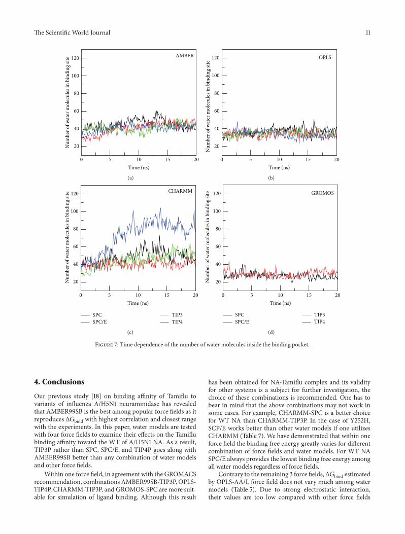
3.5.3. CHARMM Force Field. The situation becomes entirelydifferent in the case of CHARMM where the binding pocketvolume (Figure 6) and number of water molecules inside it(Figure 7) are substantially higher than other force fields.TIP4P, which has the lowest volume in AMBER and OPLS,widens the pocket to the largest extent in CHARMM (TablesS5–S7).The number of watermolecules in thismodel is abouttwofold larger than SPC/E.Thepacking of TIP4Pwater insidethe binding site is also much denser (Figure S10 and Table S7)
than other models having 𝜌bsw ≈ 0.37 kg/L. Nevertheless,the corresponding binding free energy remains higher thanSPC/E (Table 5). Overall, there is no correlation between 𝜌bswand Δ𝐺bind (Figure 8) in CHARMM.
3.5.4. GROMOSForce Field. InGROMOS the binding pocketvolume (Figure 6), the number of water molecules (Figure 7),and water density (Figure S10) are lower than other forcefields (Table S8). This presumably comes from united-atomapproximation. There is a pronounced difference in thebinding free energies obtained by SPC and SPC/E due todifferent water densities.Thus, similar to AMBER and OPLS,Δ𝐺bind decreases with 𝜌
bsw .
The Scientific World Journal 11
0 5 10 15 20
20
40
60
80
100
120
Time (ns)
Num
ber o
f wat
er m
olec
ules
in b
indi
ng si
te AMBER
(a)
Time (ns)
Num
ber o
f wat
er m
olec
ules
in b
indi
ng si
te
0 5 10 15 20
20
40
60
80
100
120 OPLS
(b)
0 5 10 15 20Time (ns)
20
40
60
80
100
120
Num
ber o
f wat
er m
olec
ules
in b
indi
ng si
te CHARMM
SPCSPC/E
TIP3TIP4
(c)
Time (ns)
Num
ber o
f wat
er m
olec
ules
in b
indi
ng si
te
0 5 10 15 20
20
40
60
80
100
120 GROMOS
SPCSPC/E
TIP3TIP4
(d)
Figure 7: Time dependence of the number of water molecules inside the binding pocket.
4. Conclusions
Our previous study [18] on binding affinity of Tamiflu tovariants of influenza A/H5N1 neuraminidase has revealedthat AMBER99SB is the best among popular force fields as itreproduces Δ𝐺bind with highest correlation and closest rangewith the experiments. In this paper, water models are testedwith four force fields to examine their effects on the Tamiflubinding affinity toward the WT of A/H5N1 NA. As a result,TIP3P rather than SPC, SPC/E, and TIP4P goes along withAMBER99SB better than any combination of water modelsand other force fields.
Within one force field, in agreement with the GROMACSrecommendation, combinations AMBER99SB-TIP3P, OPLS-TIP4P, CHARMM-TIP3P, and GROMOS-SPC are more suit-able for simulation of ligand binding. Although this result
has been obtained for NA-Tamiflu complex and its validityfor other systems is a subject for further investigation, thechoice of these combinations is recommended. One has tobear in mind that the above combinations may not work insome cases. For example, CHARMM-SPC is a better choicefor WT NA than CHARMM-TIP3P. In the case of Y252H,SCP/E works better than other water models if one utilizesCHARMM (Table 7). We have demonstrated that within oneforce field the binding free energy greatly varies for differentcombination of force fields and water models. For WT NASPC/E always provides the lowest binding free energy amongall water models regardless of force fields.
Contrary to the remaining 3 force fields,Δ𝐺bind estimatedby OPLS-AA/L force field does not vary much among watermodels (Table 5). Due to strong electrostatic interaction,their values are too low compared with other force fields

12 The Scientific World Journal
0.24 0.25 0.26Water density in binding pocket (kg/L)
Bind
ing
free e
nerg
y (k
cal/m
ol)
AMBER−30
−25
−20
−15
−10
R = 0.90
(a)
Water density in binding pocket (kg/L)
Bind
ing
free e
nerg
y (k
cal/m
ol)
0.19 0.2 0.21 0.22
OPLS−70
−68
−66
−64R = 0.67
(b)
0.2 0.25 0.3 0.35 0.4Water density in binding pocket (kg/L)
Bind
ing
free e
nerg
y (k
cal/m
ol)
CHARMM
−50
−40
−30
−20
−10
SPC TIP3P
TIP4PSPC/E
R = 0.03
(c)
Water density in binding pocket (kg/L)
Bind
ing
free e
nerg
y (k
cal/m
ol)
0.16 0.17 0.18
GROMOS−20
−18
−16
−14
−12
−10
SPC TIP3P
TIP4PSPC/E
(d)
Figure 8: Δ𝐺bind versus water density at the binding site in all force fields.
and experiments. It remains unclear if this is an artifact ofMM-PBSA approximation. Apolar solvation and entropycontributions are not affected by either force fields or watermodels (Tables 3–6), but other terms are sensitive to them.TheHBnetwork between Tamiflu andNA changes little uponwater models in OPLS and AMBER force fields, while it doesstrongly in GROMOS. CHARMM is a medium case. Thepronounced influence of aqueous models on water densityinside binding pocket has been observed in CHARMM forcefield.
Conflict of Interests
The authors declare that there is not conflict of interests inwriting this paper.
Acknowledgments
The useful discussion with P. L. Chau is highly appreciated.The work was supported by Narodowe Centrum Nauki inPoland (Grant no. 2011/01/B/NZ1/01622) and Department ofScience and Technology at Ho Chi Minh city, Vietnam.
References
[1] P. A. Kollman, I. Massova, C. Reyes et al., “Calculating struc-tures and free energies of complex molecules: combiningmolecular mechanics and continuum models,” Accounts ofChemical Research, vol. 33, no. 12, pp. 889–897, 2000.
[2] J. G. Kirkwood, “Statistical mechanics of fluid mixtures,” TheJournal of Chemical Physics, vol. 3, no. 5, pp. 300–313, 1935.

The Scientific World Journal 13
[3] J. Aqvist, C. Medina, and J.-E. Samuelsson, “A new method forpredicting binding affinity in computer-aided drug design,”Protein Engineering, vol. 7, no. 3, pp. 385–391, 1994.
[4] R. W. Zwanzig, “High-temperature equation of state by a per-turbation method. I. Nonpolar gases,” The Journal of ChemicalPhysics, vol. 22, no. 12, pp. 1420–1426, 1954.
[5] M. Karplus and J. A. McCammon, “Molecular dynamics simu-lations of biomolecules,” Nature Structural Biology, vol. 9, no. 9,pp. 646–652, 2002.
[6] F. S. Lee, Z.-T. Chu, M. B. Bolger, and A. Warshel, “Calcula-tions of antibody-antigen interactions: microscopic and semi-microscopic evaluation of the free energies of binding ofphosphorylcholine analogs to McPC603,” Protein Engineering,vol. 5, no. 3, pp. 215–228, 1992.
[7] U. Bren, J. Lah,M. Bren, V.Martnek, and J. Florin, “DNAduplexstability: the role of preorganized electrostatics,” Journal ofPhysical Chemistry B, vol. 114, no. 8, pp. 2876–2885, 2010.
[8] H. Grubmuller, B. Heymann, and P. Tavan, “Ligand binding:molecular mechanics calculation of the streptavidin-biotinrupture force,” Science, vol. 271, no. 5251, pp. 997–999, 1996.
[9] B. K. Mai, M. H. Viet, and M. S. Li J, “Top leads for swineinfluenzaA/H1N1 virus revealed by steeredmolecular dynamicsapproach,” Journal of Chemical Information and Modeling, vol.50, no. 12, pp. 2236–2247, 2010.
[10] J. H. C. Berendsen, J. P. M. Postma, W. F. van Gunsteren, and J.Hermans, “Interaction models for water in relation to proteinhydration,” in Intermolecular Forces, B. Pullmann, Ed., pp. 331–342, 1981.
[11] H. J. C. Berendsen, J. R. Grigera, and T. P. Straatsma, “Themissing term in effective pair potentials,” Journal of PhysicalChemistry, vol. 91, no. 24, pp. 6269–6271, 1987.
[12] W. L. Jorgensen, J. Chandrasekhar, J. D. Madura, R. W. Impey,and M. L. Klein, “Comparison of simple potential functions forsimulating liquidwater,”The Journal of Chemical Physics, vol. 79,no. 2, pp. 926–935, 1983.
[13] W. L. Jorgensen and J.D.Madura, “Temperature and size depen-dence for Monte Carlo simulations of TIP4P water,” MolecularPhysics, vol. 56, no. 6, pp. 1381–1392, 1985.
[14] B. Hess and N. F. A. van der Vegt, “Hydration thermodynamicproperties of amino acid analogues: a systematic comparison ofbiomolecular force fields and water models,” Journal of PhysicalChemistry B, vol. 110, no. 35, pp. 17616–17626, 2006.
[15] P. Florova, P. Sklenovsky, P. Banas, and M. Otyepka, “Explicitwater models affect the specific solvation and dynamics ofunfolded peptides while the conformational behavior andflexibility of folded peptides remain intact,” Journal of ChemicalTheory and Computation, vol. 6, no. 11, pp. 3569–3579, 2010.
[16] Z. Hu and J. Jiang, “Assessment of biomolecular force fields formolecular dynamics simulations in a protein crystal,” Journal ofComputational Chemistry, vol. 31, no. 2, pp. 371–380, 2009.
[17] M. Almlof, B. O. Brandsdal, and J. Aqvist, “Binding affinityprediction with different force fields: examination of the linearinteraction energy method,” Journal of Computational Chem-istry, vol. 25, no. 10, pp. 1242–1254, 2004.
[18] T. T.Nguyen, B.K.Mai, andM. S. Li, “Study of tamiflu sensitivityto variants of A/H5N1 virus using different force fields,” Journalof Chemical Information and Modeling, vol. 51, no. 9, pp. 2266–2276, 2011.
[19] P. Cozzini, M. Fornabaio, A. Marabotti, D. J. Abraham, G. E.Kellogs, and A. Mozzarelli, “Free energy of ligand binding toprotein: evaluation of the contribution of water molecules by
computational methods,” Current Medicinal Chemistry, vol. 11,no. 23, pp. 3093–3118, 2004.
[20] J. Michel, J. Tirado-Rives, and W. L. Jorgensen, “Prediction ofthe water content in protein binding sites,” Journal of PhysicalChemistry B, vol. 113, no. 40, pp. 13337–13346, 2009.
[21] L. Wang, B. J. Berne, and R. A. Friesner, “Ligand binding toprotein-binding pockets with wet and dry regions,” Proceedingsof the National Academy of Sciences of the United States ofAmerica, vol. 108, no. 4, pp. 1326–1330, 2011.
[22] M. Chaplin, “Water structure and science,” 2000, http://www.lsbu.ac.uk/water/.
[23] P. J. Collins, L. F. Haire, Y. P. Lin et al., “Crystal structures ofoseltamivir-resistant influenza virus neuraminidase mutants,”Nature, vol. 453, no. 7199, pp. 1258–1261, 2008.
[24] V. Hornak, R. Abel, A. Okur, B. Strockbine, A. Roitberg, andC. Simmerling, “Comparison of multiple amber force fieldsand development of improved protein backbone parameters,”Proteins, vol. 65, no. 3, pp. 712–725, 2006.
[25] B. R. Brooks, C. L. Brooks, A. D. Mackerell et al., “CHARMM:the biomolecular simulation program,” Journal of Computa-tional Chemistry, vol. 30, no. 10, pp. 1545–1614, 2009.
[26] W. L. Jorgensen and J. Tirado-Rives, “The OPLS potentialfunctions for proteins. Energy minimizations for crystals ofcyclic peptides and crambin,” Journal of the American ChemicalSociety, vol. 110, no. 6, pp. 1657–1666, 1988.
[27] W. F. van Gunsteren, S. R. Billeter, A. A. Eising et al., Biomolec-ular Simulation: The GROMOS96 Manual and User Guide, VdfHochschulverlag AG an der ETH Zurich, Zurich, Switzerland,1996.
[28] R. G. Webster and E. A. Govorkova, “H5N1 influenza—con-tinuing evolution and spread,” The New England Journal ofMedicine, vol. 355, no. 21, pp. 2174–2177, 2006.
[29] The Writing Committee of the World Health Organization(WHO) Consultation on Human Influenza A/H5, “Avianinfluenza A (H5N1) infection in humans,” The New EnglandJournal of Medicine, vol. 353, no. 13, pp. 1374–1385, 2005.
[30] Schrodinger, PyMOL: The PyMOL Molecular Graphics System,Version 1.3, Schrodinger, 2010.
[31] D. M. F. van Aalten, R. Bywater, J. B. C. Findlay, M. Hendlich,R. W. W. Hooft, and G. Vriend, “PRODRG, a program for gen-erating molecular topologies and unique molecular descriptorsfrom coordinates of small molecules,” Journal of Computer-Aided Molecular Design, vol. 10, no. 3, pp. 255–262, 1996.
[32] M. J. Frisch, G. W. Trucks, H. B. Schlegel et al., Gaussian 03,Revision C.02, Gaussian, Wallingford, Conn, USA, 2004.
[33] A. W. S. D. Silva, W. F. Vranken, and E. D. Laue, “ACPYPE—AnteChamber PYthonParser interfacE,” submitted.
[34] A. S. T. R. Andre, A. C. H. Bruno, and B. A. Ricardo, “MKTOP:a program for automatic construction of molecular topologies,”Journal of the Brazilian Chemical Society, vol. 19, no. 7, pp. 1433–1435, 2008.
[35] V. Zoete, M. A. Cuendet, A. Grosdidier, and O. Michielin,“SwissParam: a fast force field generation tool for small organicmolecules,” Journal of Computational Chemistry, vol. 32, no. 11,pp. 2359–2368, 2011.
[36] B. Guillot, “A reappraisal of what we have learnt during threedecades of computer simulations onwater,” Journal ofMolecularLiquids, vol. 101, no. 1-3, pp. 219–260, 2002.
[37] J. D. Bernal and R. H. Fowler, “A theory of water and ionicsolution, with particular reference to hydrogen and hydroxylions,”The Journal of Chemical Physics, vol. 1, no. 8, pp. 515–548,1933.

14 The Scientific World Journal
[38] T. Darden, D. York, and L. Pedersen, “Particle mesh Ewald: anN⋅log(N) method for Ewald sums in large systems,”The Journalof Chemical Physics, vol. 98, no. 12, pp. 10089–10092, 1993.
[39] R. W. Hockney, S. P. Goel, and J. W. Eastwood, “Quiet high-resolution computer models of a plasma,” Journal of Computa-tional Physics, vol. 14, no. 2, pp. 148–158, 1974.
[40] H. J. C. Berendsen, J. P. M. Postma, W. F. van Gunsteren, A.Dinola, and J. R. Haak, “Molecular dynamics with coupling toan external bath,”The Journal of Chemical Physics, vol. 81, no. 8,pp. 3684–3690, 1984.
[41] M. Parrinello and A. Rahman, “Polymorphic transitions in sin-gle crystals: a new molecular dynamics method,” Journal ofApplied Physics, vol. 52, no. 12, pp. 7182–7190, 1981.
[42] B. Hess, C. Kutzner, D. van der Spoel, and E. Lindahl,“GRGMACS 4: algorithms for highly efficient, load-balanced,and scalable molecular simulation,” Journal of Chemical Theoryand Computation, vol. 4, no. 3, pp. 435–447, 2008.
[43] L. S. Cheng, R. E. Amaro, D. Xu, W. W. Li, P. W. Arzberger, andJ. A. McCammon, “Ensemble-based virtual screening revealspotential novel antiviral compounds for avian influenza neu-raminidase,” Journal of Medicinal Chemistry, vol. 51, no. 13, pp.3878–3894, 2008.
[44] C. B. Barber, D. P. Dobkin, and H. Huhdanpaa, “The quickhullalgorithm for convex hulls,”ACMTransactions onMathematicalSoftware, vol. 22, no. 4, pp. 469–483, 1996.
[45] K. L. Clarkson,K.Menlhorn, andR. Seidel, “Four results on ran-domized incremental constructions,” Computational Geometry,vol. 3, no. 4, pp. 185–212, 1993.
[46] P. L. Chau, “Water movement during Ligand Unbinding fromreceptor site,” Biophysical Journal, vol. 87, no. 1, pp. 121–128,2004.
[47] MATLAB Version 7.0.1 (R2007a), The MathWorks Inc., Natick,Mass, USA, 2007.
[48] O. Aruksakunwong, M. Malaisree, P. Decha et al., “On thelower susceptibility of oseltamivir to influenza neuraminidasesubtype N1 than those in N2 and N9,” Biophysical Journal, vol.92, no. 3, pp. 798–807, 2007.

Hindawi Publishing Corporation http://www.hindawi.com Volume 2013Hindawi Publishing Corporation http://www.hindawi.com Volume 2013
The Scientific World Journal
Impact Factor 1.73028 Days Fast Track Peer ReviewAll Subject Areas of ScienceSubmit at http://www.tswj.com