Embed Size (px)
Citation preview

Hindawi Publishing CorporationAnatomy Research InternationalVolume 2012, Article ID 951836, 9 pagesdoi:10.1155/2012/951836
Research Article
Cranial Osteology of Meiglyptini (Aves: Piciformes: Picidae)
Reginaldo Jose Donatelli
Laboratory of Ornithology, Department of Biological Sciences, Sciences College, Sao Paulo State University (UNESP),Caixa Postal 473, 17001-970 Bauru, SP, Brazil
Correspondence should be addressed to Reginaldo Jose Donatelli, [email protected]
Received 18 July 2011; Revised 22 September 2011; Accepted 22 September 2011
Academic Editor: Friedrich Paulsen
Copyright © 2012 Reginaldo Jose Donatelli. This is an open access article distributed under the Creative Commons AttributionLicense, which permits unrestricted use, distribution, and reproduction in any medium, provided the original work is properlycited.
The Meiglyptini comprise eight species grouped into three genera: Meiglyptes and Mulleripicus, with three species each, andHemicircus, with two species. The aim of the present study was to describe the cranial osteology of six species and three genera ofMeiglyptini and to compare them to each other, as well as with other species of woodpeckers and other bird groups. The cranialosteology varied among the investigated species, but the most markedly distinct characteristics were: (1) a frontal overhang is onlyobserved in the middle portion of the frontale of H. concretus; (2) the Proc. zygomaticus and suprameaticus are thick and longin species of the genus Mulleripicus, but short in other species; (3) the Pes pterygoidei is relatively larger in species of the genusMulleripicus, while it is narrow, thin and relatively smaller in species of the genus Meiglyptes and indistinct in H. concretus; (4) thebony projection of the ectethmoidale is relatively short and thin in species of Mulleripicus and more developed in H. concretus. Itappears that the greatest structural complexity of the cranial osteology is associated with the birds’ diet, with the frugivorous H.concretus being markedly different from the insectivorous species.
1. Introduction
Woodpeckers have been investigated scientifically for over acentury. These birds are notable for their colour, size, forag-ing mode, nest-building behaviour, instrumental signals, andthe way they climb vertical surfaces. Interestingly, many ofthe behavioural patterns of woodpeckers are closely relatedto their anatomical features. Thus, these aspects cannot bedissociated in a study [1].
The Meiglyptini comprise three genera of Old Worldwoodpeckers: Meiglyptes and Mulleripicus, with three specieseach, and Hemicircus, with two species [2]. These speciesare all arboreal and feed mainly upon the larvae and eggsof insects, ants and secondarily termites, beetles, caterpillars,and other arthropods. There is also a frugivorous species inthis group, H. concretus.
The foraging modes vary among these woodpeckers,irrespective of food type. The Meiglyptini primarily employgleaning, with probing and tapping being used secondarily;excavating and tonguing are less common.
The aim of the present study was to describe and comparethe mandibular apparatus of several species of Meiglyptini
and perform a morphofunctional analysis of the complexityof this apparatus, relating it with the species’ foraging mode.Specifically, the following questions were addressed:
Are the foraging mode and the structure of the mandibu-lar apparatus related among the Meiglyptini?; and is itpossible to establish any relationship between form andfunction based on structural differences between the jawapparatus and the foraging modes? Thus, there are at leasttwo hypotheses to be considered: (1) there is no relationshipbetween the structural complexity of the mandibular appa-ratus in Meiglyptini and the foraging mode, and (2) thereis a relationship between the structure of the mandibularapparatus and the foraging mode in Meiglyptini.
2. Materials
In this study, I described the osteological characters of theskull of 15 specimens of Meiglyptini belonging to threegenera and six species. The specimens are housed at theNational Museum of Natural History (USNM), SmithsonianInstitution, Washington DC, USA, the Museum Zoologicum

2 Anatomy Research International
ZFC
FP
Na
J
Q1 cm
Figure 1: Dorsal view of the skull of Meiglyptes tristis. F: frontal region; J: jugal arch; NA: nostril; P: parietal region; Q: quadrate bone; ZFC:craniofacial flexion zone.
ZFC
FP
Q
J
1 cmNa
Figure 2: Dorsal view of the skull of Meiglyptes tukki. F: frontal region; J: jugal arch; NA: nostril; P: parietal region; Q: quadrate bone; ZFC:craniofacial flexion zone.
Bogoriense (MZB), Indonesian Institute of Sciences (LIPI),and the Natural History Museum of the Indonesian Instituteof Sciences, Indonesia.
Bellow is a list of the species investigated after Winklerand Christie [2], museum abbreviations and the relativenumbers of specimens in the collections. All specimens pre-served in 70% ethanol were from the Museum ZoologicumBogoriense, Indonesian Institute of Sciences (LIPI), whereasthe others were osteological material: Hemicircus concretus(Temminck, 1821) LIPI MZB.Skt 125 Hc1; LIPI MZB.SKT126 HC2; Meiglyptes tristis (Horsfield, 1821) LIPI MZB.SktMtr1 123; MZB.Skt Mtr2 124; USNM 292,228 ♂; Meiglyptestukki (Lesson, 1839) LIPI MZB.Skt Mtu1 121; MZB.SktMtk1 122; USNM 489,269 ♀; Mulleripicus pulverulentus(Temminck, 1826) LIPI MZB.Skt Mp1 127; MZB.Skt 128Mp2; ♀ USNM 19201, USNM 562,042 ♀; Mulleripicus fulvus(Quoy and Gaimard, 1830) USNM 491,227 ♀, USNM226,191 ♀; Mulleripicus funebris (Valenciennes, 1826) ♂USNM 489,265.
3. Methods
The osteology of the skull and mandible was studied com-paratively, described, and drawn using a Zeiss Stemi SV11stereomicroscope (http://www.zeiss.com/) with magnifica-tion ranging from 4 to 66X. M. pulverulentus was usedas a reference for comparison of structures. All drawingsare accompanied by legends to facilitate the observation ofstructures.
The nomenclature used to describe the cranial osteologyfollows the Nomina Anatomica Avium (NAA; [3, 4]). Whenno reference to a particular structure was available, I usedletters and numbers to avoid the unnecessary creationof names. The species nomenclature follows Winkler andChristie [2].
4. Results
4.1. Osteology
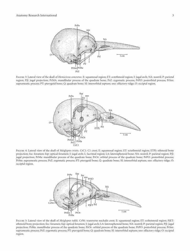
Ossa cranii. The Os frontale (F) articulates rostrally withthe Os nasalis through the craniofacial flexion zone (ZFC).This is more evident in M. tristis (Figure 1) and M. tukki(Figure 2) and less in H. concretus, whereas it is indistinguish-able in M. pulverulentus. Hemicircus concretus (Figure 3)bears a unique thin bony elevation (Be) at the middleportion of the Os frontale; there is no such elevation in otherspecies. Laterocaudally, the frontal region is connected withthe Proc. postorbitalis (PrPO). This process is short, withapproximately 1/6 of the length occurring between its originin the skull and the jugal arch in M. tristis (Figure 4), 1/5 inH. concretus (Figure 3), 1/4 in M. tukki (Figure 5), and 1/3in M. pulverulentus (Figure 6). The sutura frontolacrimalisis absent, and the Os lacrimale is fusioned with the frontalregion in all species.
The Os parietale is expanded laterally approximatelytwice the length of the lateral expansion of the Os frontale in
Anatomy Research International 3
P
QE
PrPo
Sl
NA
PrSm
PrZ
PrMa
PTJ
sno
OPrJ
ET
1 cm
Figure 3: Lateral view of the skull of Hemicircus concretus. E: squamosal region; ET: ectethmoid region; J: jugal arch; NA: nostril; P: parietalregion; PjJ: jugal projection; PrMA: mandibular process of the quadrate bone; PrZ: zygomatic process; PrPO: postorbital process; PrSm:suprameatic process; PT: pterygoid bone; Q: quadrate bone; SI: interorbital septum; sno: olfactory ridge; O: occiptal region.
PL
Q
E
PrPo
Sl
NA
PrSmPrZ PrMa
PT
PrJ
fop
focsno
LA
ETPj
CrC1
ET
O
PrOr1 cm
Figure 4: Lateral view of the skull of Meiglyptes tristis. CrC1: C1 crest; E: squamosal region; ET: ectethmoid region; ETPj: ethmoid bonyprojection; foc: foramen; fop: optical foramen; J: jugal arch; L: lacrimal region; LA: laterosphenoid bone; NA: nostril; P: parietal region; PjJ:jugal projection; PrMa: mandibular process of the quadrate bone; PrOr: orbital process of the quadrate bone; PrPO: postorbital process;PrSm: suprameatic process; PrZ: zygomatic process; PT: pterygoid bone; Q: quadrate bone; SI: interorbital septum; sno: olfactory ridge; O:occiptal region.
P
Q
E
PrPo
Sl
NA
PrSmPrMa
PT
J PrJ
fopfoc
sno
ETLA
PrSO
PjET
PrOr
CrNtO
1 cm
Figure 5: Lateral view of the skull of Meiglyptes tukki. CrNt: transverse nuchalis crest; E: squamosal region; ET: ecthetmoid region; PjET:ethmoid bony projection; foc: foramen; fop: optical foramen; J: jugal arch; LA: laterosphenoid bone; NA: nostril; P: parietal region; PjJ: jugalprojection; PrMa: mandibular process of the quadrate bone; PrOr: orbital process of the quadrate bone; PrPO: postorbital process; PrSm:suprameatic process; PrZ: zygomatic process; PT: pterygoid bone; Q: quadrate bone; SI: interorbital septum; sno: olfactory ridge; O: occiptalregion.

4 Anatomy Research International
P
L
Q
E
PrPo
Sl
NA
PrSmPrZ
PrMa
J
PrJ
sno
LA
PjET
ET
PrOtQ
PT
PrOr
1 cm
Figure 6: Lateral view of the skull of Mulleripicus pulverulentus. E: squamosal region; ET: ectethmoid region; PjET: ethmoid bony projection;J: jugal arch; L: lacrimal region; LA: aterosphenoid bone; NA: nostril; P: parietal region; PjJ: jugal projection; PrMa: mandibular process ofthe quadrate bone; PrOr: orbital process of the quadrate bone; PrOtQ: otic process of the quadrate bone; PrPO: postorbital process; PrSm:suprameatic process; PrZ: zygomatic process; PT: pterygoid bone; Q: quadrate bone; SI: interorbital septum; sno: olfactory ridge.
ZFC
FP
J
Na1 cm
Figure 7: Dorsal view of the skull of Hemicircus concretus. F: frontal region; J: jugal arch; NA: nostril; P: parietal region; ZFC: craniofacialflexion zone.
FP
NA
J
1 cm
Figure 8: Dorsal view of the skull of Mulleripicus pulverulentus. F: frontal region; J: jugal arch; NA: nostril; P: parietal region.
H. concretus (Figure 7), about 1.5 times in M. pulverulentus(Figure 8), and about 2.5 times in M. tristis (Figure 1) andM. tukki (Figure 2).
The values of these parameters indicate not only thefrontale/parietale relationship but also provide the skulldimensions in many species. The Os squamosum is con-nected anteromedially with the Os laterosphenoidale by thecrista laterosphenoidale (CrL–Figure 5) and anterocaudallywith the Os frontale by the Proc. postorbitalis. The Fossatemporalis is wider than long in all species.
The Os squamosum is projected rostrally, forming theProc. zygomaticus (PRZ), which articulates ventrally withthe Proc. oticus quadrati and clearly has dorsal, lateral,medial, and ventral faces. This is the region of origin of theaponeurosis of the M. adductor mandibulae externus ventralisand the M. adductor mandibulae externus rostralis lateralis(Donatelli, [5]). The Proc. zygomaticus (Proc. squamosal,[6]) is thick and long in species of the genus Mulleripicus(Figure 6) and short in the other species. The Os squamosumalso forms the Proc. suprameaticus (PrSm–Figure 6), and

Anatomy Research International 5
Occ PjPtac
aclacm
PtFp
BAPC
PrMFC
V
OcoeFog
LP
J FVCrV
PAL
FoM
Sul
1 cm
EX
Figure 9: Ventral view of the skull of Hemicircus concretus; BA: basioccipital region; CrV: ventral palatine crest; EX: exoccipital region;FC-; Fog-; FoM: foramen magnum; Fp-; FV: ventral fossa; J: jugal arch; LP: lamina parasphenoidalis; Occ-; Ocoe-; PAL: palatine; PC-;PjP: projection of the parasphenoid rostrum; PrM: maxilar process of the palatine; PrPA: paraoccipital process; Pt: pterygoid bone; RP:parasphenoidal rostrum; Sul: intercotylar sulcus; V: vomer; Occ: Ostium canalis carotici; Ocoe: Ostium canalis ophthalmici externus; PC:Proeminentia cerebellaris; Fog: Foramen nervi glossopharyngealis; FC: Fossa choanalis; Fp: Fossa parabasalis.
Sul Occ PjPacl
acm
PtFp
PC
tac
PrM
V
OcoeFog
FV
FoM BA LP
PAL
1 cm
EX
Figure 10: Ventral view of the skull of Meiglyptes tristis; BA: basioccipital region; EX: exoccipital region; Fog-; FOM: foramen magnum; Fp-;FV: ventral fossa; LP: lamina parasphenoidalis; Occ-; Ocoe-; PAL: palatine; PC-; PjP: projection of the parasphenoid rostrum; PrM: maxilarprocess of the palatine; Pt: pterygoid bone; Sul: intercotylar sulcus; V: vomer; Occ: Ostium canalis carotici; Ocoe: Ostium canalis ophthalmiciexternus; PC: Proeminentia cerebellaris; Fog: Foramen nervi glossopharyngealis; Fp: Fossa parabasalis.
this is present only in species of the genus Mulleripicus(Figure 6).
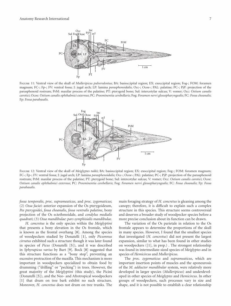
Ossa faciei. The Os pterygoideum has a well-developedanterior expansion, known as the Pes pterygoideus, whichextends rostrally and articulates with the ventral portionof the septum interorbitale, the rostrum parasphenoidale,and the pars palatina. The Pes pterygoideus is observedand well developed in species of Mulleripicus, but it isrelatively less developed, thin, and narrow in species ofMeiglyptes and absent in H. concretus. Posterodorsally to theOs pterygoideum lies the processus dorsalis, which includesthe aponeurosis of the M. protractor pterygoidei. This is onlypresent in species of Mulleripicus.
In the middle portion of the Os palatinum lies the fossachoanalis (FC), which is delimited by the crista ventralis(CrV), forming a gap. This gap varies in size amongthe species studied. It is relatively wider in H. concretus(Figure 9), decreases in width in species of Meiglyptes(Figure 10), and is relatively narrow in species of Mul-leripicus (Figure 11), in which the Os palatinum is curvedcaudally. Between the cristae ventralis et lateralis lies thefossa ventralis (FV), which is distinct and deep in M.tristis (Figure 10), relatively deep and shallow in species ofMulleripicus (Figure 11), and shallow in M. tukki (Figure 12)and H. concretus (Figure 9). This is an important regionbecause it is the origin of the aponeurosis and muscle fibresof the M. pterygoideus ventralis (Donatelli, [5]).

6 Anatomy Research International
The Os laterosphenoidale lies caudally to the facies orbita-les. It is connected laterally with the Os squamosum by thecrista laterosphenoidale (CrL–Figure 5), where the aponeu-rosis of the M. adductor mandibulae externus caudalis medi-alis originates (Donatelli, [5]). This crest is apparent only inspecies of the genus Mulleripicus and in M. tukki (Figure 5).It is common to observe a swelling in the connection betweenthe crista laterosphenoidale and the septum interorbitale inall species. The nerve foramen lies ventrolaterally.
The Os ectethmoidale has a bony projection (ETPj–Figures 4–6) reaching the dorsal surface of the arcus jugalis,though without fusion. The projection is relatively moreapparent in M. tristis (Figure 4) than in other species, butit is more developed in H. concretus (Figure 3). It assumesa triangular shape in M. tukki (Figure 5). In Mulleripicus(Figure 6), the projection is relatively short and thin uponthe arcus jugalis.
The Os quadratum has a corpus quadrati that connectswith the proc. oticus quadrati (PrOtQ), proc. orbitalis, andproc. mandibularis. The proc. oticus quadrati articulatesdorsally with the Os squamosum by the ventral surface of theproc. zygomaticus (PRZ), which protrudes dorsally upon theproc. oticus quadrati in the species of Mulleripicus (Figure 6).This process is relatively shorter in H. concretus and M. tukki.In species of Mulleripicus, there is a greater distance betweenthe proc. orbitalis quadrati and the posterior portion of theOs laterosphenoidale. In the proc. oticus quadrati, a small C1crest (C1Cr) can be observed in all species but represented inM. tristis (Figure 4), which originates from the aponeurosisof the M. adductor mandibulae externus caudalis lateralis(Donatelli, [5]). The proc. orbitalis quadrati (PrOr–Figures 4–6) protrudes anteromedially from the corpus quadrati. This isa short process, but its form varies greatly among species. Ingeneral, it is slender and has a length equivalent to two thirdsof the length of the Os pterygoideus, which lies medially. Thecondylus medialis is usually the most developed, comparedto other condyles, as observed in most species, and thisstructure is relatively more developed in M. tristis, in whichit acquires a protruding and pointed shape. The condyluscaudalis is an extension of the condylus lateralis in all species.
The maxilla is formed by the fusion of the Os premaxil-lare, maxillare, and nasale. It presents approximately half thetotal length of the skull in most species, except in species ofMulleripicus (Figure 8), in which it measures approximately65% of the total length of the skull.
Ossa Mandibulae. The pars symphisialis mandibulae (Psi)is short and measures slightly more than one third of thetotal length of the mandible only in species of Meiglyptes.In M. pulverulentus, the Psi measures approximately 40%of the total length of the mandible, compared to 45% inH. concretus.In the dorsal region of the mandible lies theproc. pseudocoronoideus 1 (= process of the M. adductormandibulae, [3]). This process is the insertion point of thetendon common to the M. adductor mandibulae externusrostralis temporalis and the M. adductor mandibulae externusrostralis medialis (Donatelli, [5]). It is indistinct in all species,except M. tristis and species of Mulleripicus. Additionally, theproc. pseudocoronoideus 2 is relatively indistinct in all species.
On the pars intermedia, there is a peculiar depression,the fossa lateralis mandibulae, in which the muscle fibres ofthe M. adductor mandibulae externus ventralis are inserted.This region is connected to the posterior portion by thecrista caudalis mandibulae. This crest reaches the dorsal Proc.pseudocoronoideus 2.
In the middle portion of the mandible lies the proc.medialis mandibulae (internal jaw process, angular medialprocess, [6, 7]), which protrudes dorsomedially. This processvaries greatly in length in the woodpeckers studied. Thisis one of the most important structures for the insertionof muscle fibres and their aponeurosis of the complexof the Os pterygoideum. The tuberculum pseudotemporale,which includes the aponeurosis of the M. pseudotemporalissuperficialis, is also noticeable. It is a conspicuous structure inmost species, except in H. concretus. All species bear a shallowfossa caudalis in the posterior portion of the mandible. Thisis the insertion point of the muscle fibres of the M. depressormandibulae.
Some of the structural differences observed in thecomponents of the cranial osteology of the Meiglyptini arenoteworthy due to their exclusivity, relative development(being larger or smaller) or unique features present in onegroup of species within a genus or a species, as follows:(1) there is a thin bony elevation (Be), referred to as thefrontal overhang by Bock [8], in the middle portion of theOs frontale in H. concretus; (2) the Os parietale is expandedlaterally and is equivalent to approximately twice the lengthof the lateral expansion of the Os frontale in H. concretus, 1.5times in M. pulverulentus, and 2.5 times in M. tristis and M.tukki; (3) the fossa temporalis is wider than long in all species;(4) the proc. zygomaticus is long and thick in species of thegenus Mulleripicus and short in the other species; (5) theproc. suprameaticus (PrSM) is apparent only in species of thegenus Mulleripicus; (6) the Pes pterygoidei is relatively largerin species of the genus Mulleripicus, relatively smaller, thin,and narrow in species of the genus Meiglyptes, and indistinctin H. concretus; (7) the fossa choanalis is relatively wider in H.concretus and becomes progressively narrower in species ofMeiglyptes and Mulleripicus; (8) the fossa ventralis palatinais deep in M. tristis and becomes progressively shallowerin Mulleripicus species, M. tukki, and H. concretus; (9) thebony projection of the Os ectethmoidale is relatively shortand slender in Mulleripicus species and more developed inH. concretus; (10) the condylus medialis is usually the mostdeveloped one in all species, and, in M. tristis, it is prominentand pointed; (11) the pars symphisialis mandibulae is shortand extends for slightly more than 1/3 of total length ofthe mandible only in species of Meiglyptes for approximately40% in M. Pulverulentus, and approximately 45% in H.concretus.
5. Discussion
This analysis of the cranial osteological structures of theMeiglyptini elucidated seven important mechanisms of oper-ation of the jaw apparatus: (1) Ossa cranii: frontal overhang,the extension of the Os parietale versus the Os frontale,
Anatomy Research International 7
OccPjP
tacacl
PTFp
PC
PrMFC
OcoeFog
JFV
PjP acl
PT
PrMFC
OcoeFog
JFV
Sul
BA LPFoMPAL
EX
1 cm
Figure 11: Ventral view of the skull of Mulleripicus pulverulentus; BA: basioccipital region; EX: exoccipital region; Fog-; FOM: foramenmagnum; FC-; Fp-; FV: ventral fossa; J: jugal arch; LP: lamina parasphenoidalis; Occ-; Ocoe-; PAL: palatine; PC-; PjP: projection of theparasphenoid rostrum; PrM: maxilar process of the palatine; PT: pterygoid bone; Sul: intercotylar sulcus; V: vomer; Occ: Ostium canaliscarotici; Ocoe: Ostium canalis ophthalmici externus; PC: Proeminentia cerebellaris; Fog: Foramen nervi glossopharyngealis; FC: Fossa choanalis;Fp: Fossa parabasalis.
Occ PjPtac
acl
Fp
PC
PrM
OcoeFog
J FV
Occ PjPacl
C
PrM
Ocoe
JJ FV
V
Sul
FoM BA LPPAL
PT
EX
1 cm
Figure 12: Ventral view of the skull of Meiglyptes tukki; BA: basioccipital region; EX: exoccipital region; Fog-; FOM: foramen magnum;FC-; Fp-; FV: ventral fossa; J: jugal arch; LP: lamina parasphenoidalis; Occ-; Ocoe-; PAL: palatine; PC-; PjP: projection of the parasphenoidrostrum; PrM: maxilar process of the palatine; PT: pterygoid bone; Sul: intercotylar sulcus; V: vomer; Occ: Ostium canalis carotici; Ocoe:Ostium canalis ophthalmici externus; PC: Proeminentia cerebellaris; Fog: Foramen nervi glossopharyngealis; FC: Fossa choanalis; Fp: Fossaparabasalis.
fossa temporalis, proc. suprameaticus, and proc. zygomaticus;(2) Ossa faciei: anterior expansion of the Os pterygoideum,Pes pterygoidei, fossa choanalis, fossa ventralis palatine, bonyprojection of the Os ectethmoidale, and condylus medialisquadrati; (3) Ossa mandibulae: pars symphisialis mandibulae.
H. concretus is the only species within the Meiglyptinithat presents a bony elevation in the Os frontale, whichis known as the frontal overhang [8]. Among the speciesof woodpeckers studied by Donatelli [1], only Picumnuscirratus exhibited such a structure though it was later foundin species of Picus (Donatelli [5]), and it was describedin Sphyrapicus varius by Burt [9]. Bock [8] suggested thatthis structure functions as a “bony stop”, preventing anexcessive protraction of the maxilla. This mechanism is moreimportant in woodpeckers specialized to obtain food bydrumming (“drilling” or “pecking”) in trees. However, thegreat majority of the Meiglyptini (this study), the Picini(Donatelli [5]), and the Neo- and Afrotropical woodpeckers[1] that drum on tree bark exhibit no such structure.Moreover, H. concretus does not drum on tree trunks. The
main foraging strategy of H. concretus is gleaning among thecanopy; therefore, it is difficult to explain such a complexstructure in this species. This structure seems controversialand deserves a broader study of woodpecker species before amore precise conclusion about its function can be drawn.
The variation of the Os parietale in relation to the Osfrontale appears to determine the proportions of the skullin many species. However, I found that the smallest speciesthat investigated (H. concretus) did not present the largestexpansion, similar to what has been found in other studieson woodpeckers ([1], in prep.) . The strongest relationshipwas found in intermediate-sized species of Meiglyptes and inspecies of Hemicircus and Mulleripicus.
The proc. zygomaticus and suprameaticus, which areimportant insertion points of muscles and the aponeurosisof the M. adductor mandibular system, were relatively moredeveloped in larger species (Mulleripicus) and underdevel-oped in other species of Meiglyptes and Hemicircus. In othergroups of woodpeckers, such processes vary in size andshape, and it is not possible to establish a clear relationship

8 Anatomy Research International
between their development and size ([1], in prep.) . Jollie[10] suggested that the Os squamosum articulates with theOs quadratum and a short proc. “zygomaticus” in chickens,which was also observed in all species of Picidae studied here.However, the proper term for this should be proc. squamosaland not “zygomaticus” [4], as the term “zygomatic” is charac-teristic of mammalian skulls.
The anterior expansion of the Os pterygoideum formsthe Pes pterygoidei. The length of this process increasesin associate with the size of Meiglyptini species, from thelargest to the smallest in the order Mulleripicus, Meiglyptes,and Hemicircus. This observation diverges from findingsof previous studies (e.g., Donatelli [1, 5]) that found thatthis structure is well developed and is a unique featureof all woodpeckers. Such a structure was not described inother groups of birds related to the Piciformes, such as theGalbulidae [11] or Coraciiformes [12]. In these birds, thePes pterygoidei is the insertion point of the fibres of the M.pterygoideus dorsalis medialis, which is an important muscleretractor of the upper jaw. Burton [7] described the Pespterygoidei in the Picidae, Picumninae, and Indicatoridae.The proc. pterygoideus dorsalis [13] is the insertion pointof the aponeurosis of the M. protractor pterygoideus. Thisprocess is conspicuous only in Mulleripicus among theMeiglyptini. In the Picidae (Donatelli [5]), this process islarger in B. rubiginosus and smaller in other species, butalways distinct. This process was not mentioned by Burton[7], perhaps because he considered it as only one muscleof the M. protractor quadrati system: the M. protractorpterygoidei et quadrati (with insertion in the dorsal portion ofthe articulation of the pterygoideum-quadratum). Donatelli[1, 5] described this muscle as two distinct muscles (M.protractor pterygoidei and M. protractor quadrati) becausethey had different origins and insertions. Bock ([14], p. 12)previously called attention to this structure, particularly inwoodpeckers.
The M. pterygoideus ventralis medialis, which lies in thefossa ventralis palatine, is very well developed in woodpeckersand represents a powerful retractor of the upper jaw inbirds. Gennip [15] is one of the few authors who describedthis fossa and related it to the origin and developmentof the M. pterygoideus ventralis medialis. Other authorsstudying the Columbidae [16, 17] did not mention thisstructure and only related the development of these musclesin species within the family. According to Bock [14], “themass and shape of the palate are correlated with the sizeand power of the upper jaw and with the strength of themuscles. Many of the exact details of this correlation muststill be ascertained.” According to Morioka [18], the deeperthe fossa ventralis palatina, the greater the developmentof the related muscle mass and the greater the power ofretraction of the upper jaw. However, he noted that thepoorly developed muscle mass in the Apodidae allowedthem to close their beak more rapidly, at the expense of a“powerful biting force.” Among the woodpeckers studied,this structure is relatively deep and conspicuous only in M.tristis, whereas the largest size and structural developmentof the M. pterygoideus ventralis medialis et lateralis werefound in M. pulverulentus.The size and shape of the proc.
orbitalis quadrati is prominent in Mulleripicus species relativeto other woodpeckers.The associated M. pseudotemporalisprofundus is also relatively more developed. Donatelli [5]reported that B. rubiginosus had the largest process amongthe Picini, and the associated muscle was relatively lessdeveloped than in other species. As pointed out by Bock[14], the condylus medialis mandibulae is the most developedamong the condyles of the Os quadratum. This is similar towhat was found in the Meiglyptini, especially in M. tristis, inwhich the condyle was even more distinguished in shape.
We found clear structural differences in the cranialosteology between the frugivorous H. concretus and otherinsectivorous species of Meiglyptini. Therefore, natural selec-tion appears to have shaped the jaw apparatus as a wholedifferently in species with different food types feedinglocations, irrespective of their foraging mode. This becomesclear when the development of these structures is consideredin species of Meiglyptes and Mulleripicus compared toHemicircus concretus. These aspects will be further discussedelsewhere based on the results of this study.
Acknowledgments
The author is very indebted to Martjan Lammertink, whocollected the woodpeckers and made them available for meto study in detail. I am very grateful to the curators of theMuseum Zoologicum Bogoriense, the Indonesian Instituteof Sciences (LIPI), and the Natural History Museum ofthe Indonesian Institute of Sciences (MZB), Indonesia forthe loan of the Picini for anatomical studies. I also thankthe curators of the National Museum of Natural History(USNM), Smithsonian Institution, Washington DC, USA forallowing me to visit the collection to study the Picidae.
References
[1] R. J. Donatelli, “The jaw apparatus of the neotropical andafrotropical woodpeckers (Aves: Piciformes),” Arqivos de Zoo-logia, vol. 33, pp. 1–70, 1996.
[2] H. Winkler and D. A. Christie, “Family Picidae (woodpeck-ers),” in Handbook of the Birds of the World, Vol. 7: Jacamarsto Woodpeckers, J. del Hoyo, A. Elliot, and J. Sargatal, Eds., pp.296–555, Lynx Editions, Barcelona, Spain, 2002.
[3] J. J. Baumel, A. S. King, J. E. Breazile, and H. E. Evans, NominaAnatomica Avium, Academic Press, London, UK, 1993.
[4] J. J. Baumel and L. M. Witmer, “Osteologia,” in NominaAnatomica Avium, J. Baumel, A. S. King, J. E. Breazile, andH. E. Evans, Eds., pp. 45–132, Academic Press, London, UK,1993.
[5] R. J. Donatelli, “The jaw musculature of the Meiglyptini (Aves:Piciformes: Picidae),” Acta Zoologica (Stockholm) (In press).
[6] E. Hofling and J. P. Gasc, “Biomecanique du crane etdu bec chez Ramphastos (Aves, Ramphastidae),” GegenbaursMorphologisches Jahrbuch, vol. 130, pp. 125–147, 1984.
[7] P. J. K. Burton, “Anatomy and evolution of the feedingapparatus in the avian orders Coraciiformes and Piciformes,”Bulletin of the British Museum of Natural History, Zoology, vol.47, pp. 331–443, 1984.
[8] W. J. Bock, “Functional and evolutionary morphology ofwoodpeckers,” Ostrich, vol. 70, no. 1, pp. 23–31, 1999.
Anatomy Research International 9
[9] W. H. Burt, “Adaptative modifications in the woodpeckers,”University of California Publications in Zoology, vol. 32, pp.455–524, 1930.
[10] M. Jollie, “The head skeleton of the chicken and remarkson the anatomy of this region in other birds,” Journal ofMorphology, vol. 100, pp. 389–436, 1957.
[11] R. J. Donatelli, “Cranial osteology and myology of the jawapparatus in the Galbulidae (Aves: Piciformes),” Arqivos deZoologia, vol. 32, pp. 1–32, 1992.
[12] M. C. Pascotto, E. Hofling, and R. J. Donatelli, “Cranialosteology of Coraciiformes (Aves) [Osteologia craniana deCoraciiformes (Aves)],” Revista Brasileira de Zoologia, vol. 23,no. 3, pp. 841–864, 2006.
[13] H. Hofer, “Untersuchungen uber den Bau des Vogelschadelsbesonders uber den der Spechte und Steinhuhner,” ZoologischeJahrbucher. Abteilung fur Anatomie und Ontogenie der Tiere,vol. 69, pp. 1–158, 1945.
[14] W. J. Bock, “Kinetics of the avian skull,” Journal of Morphology,vol. 114, pp. 1–52, 1964.
[15] E. M. S. J. van Gennip, “The osteology, arthrology andmyology of the jaw apparatus of the Pigeon (Columba liviaL.),” Netherlands Journal of Zoology, vol. 36, pp. 1–46, 1986.
[16] J. Rooth, “On the correlation between the jaw muscles andthe structure of the skull in Columba palumbus palumbusL.,” Proceedings of the Koninklijke Nederlandse Akademie vanWetenschappen, vol. 56, pp. 251–264, 1953.
[17] R. L. Merz, “Jaw musculature of the mourning and white-winged doves,” University of Kansas Publications, Museum ofNatural History, vol. 12, pp. 521–551, 1963.
[18] H. Morioka, “Jaw musculature of swifts (Aves, Apodidae),”Bulletin of the National Museum of Natural Science, vol. 17, pp.1–16, 1974.

Submit your manuscripts athttp://www.hindawi.com
Hindawi Publishing Corporationhttp://www.hindawi.com Volume 2014
Anatomy Research International
PeptidesInternational Journal of
Hindawi Publishing Corporationhttp://www.hindawi.com Volume 2014
Hindawi Publishing Corporation http://www.hindawi.com
International Journal of
Volume 2014
Zoology
Hindawi Publishing Corporationhttp://www.hindawi.com Volume 2014
Molecular Biology International
GenomicsInternational Journal of
Hindawi Publishing Corporationhttp://www.hindawi.com Volume 2014
The Scientific World JournalHindawi Publishing Corporation http://www.hindawi.com Volume 2014
Hindawi Publishing Corporationhttp://www.hindawi.com Volume 2014
BioinformaticsAdvances in
Marine BiologyJournal of
Hindawi Publishing Corporationhttp://www.hindawi.com Volume 2014
Hindawi Publishing Corporationhttp://www.hindawi.com Volume 2014
Signal TransductionJournal of
Hindawi Publishing Corporationhttp://www.hindawi.com Volume 2014
BioMed Research International
Evolutionary BiologyInternational Journal of
Hindawi Publishing Corporationhttp://www.hindawi.com Volume 2014
Hindawi Publishing Corporationhttp://www.hindawi.com Volume 2014
Biochemistry Research International
ArchaeaHindawi Publishing Corporationhttp://www.hindawi.com Volume 2014
Hindawi Publishing Corporationhttp://www.hindawi.com Volume 2014
Genetics Research International
Hindawi Publishing Corporationhttp://www.hindawi.com Volume 2014
Advances in
Virolog y
Hindawi Publishing Corporationhttp://www.hindawi.com
Nucleic AcidsJournal of
Volume 2014
Stem CellsInternational
Hindawi Publishing Corporationhttp://www.hindawi.com Volume 2014
Hindawi Publishing Corporationhttp://www.hindawi.com Volume 2014
Enzyme Research
Hindawi Publishing Corporationhttp://www.hindawi.com Volume 2014
International Journal of
Microbiology