Embed Size (px)
Citation preview
1 | Advances in Orthopedics and Sports Medicine, Volume 2021, Issue 01
www.kosmospublishers.com
DOI: 10.37722/AOASM.2021101
The Influence of Kyphosis on Kinematics and Kinetics during Turning in the Elderly
Wataru Yamazaki1, 2*, Yasuhiko Hatanaka1
1Graduate School of Health Science, Suzuka University of Medical Science, 1001-1 Kishioka-cyo, Suzuka-shi, Mie, Japan
2Department of Physical Therapy, Faculty of Health Sciences, Kansai University of Health Sciences, 2-11-1, Wakaba,
Kumatori-cyo, Osaka, Japan
Abstract
Elderly people with kyphosis are at increased risk of
falling when walking. In particular, there is a tendency to fall
when turning. Knowledge of strategies which the elderly with
kyphosis could use to turn without falling would be helpful for
preventing falls. Therefore, the purpose of this study was to
describe strategies of turning in elderly individuals with
kyphosis based on investigation of kinematics and kinetics by
motion analysis. Forty elderly participants (20 with kyphosis,
20 controls) aged 65 years and over were recruited for this
study. The participants were able to walk independently and
were attending outpatient rehabilitation or community day-
care centers. We measured ground reaction force and joint
coordination using force plates and a motion capture system
during participants’ turns. We found that hip extension,
adduction, and first metatarsophalangeal extension angle were
significantly smaller in the kyphosis group, moreover, walking
velocity before turning was also lower. Thus, turning in the
elderly with kyphosis changed into a short-arc and slow-
velocity pattern. Meanwhile, propulsive impulse and ankle
plantar flexion moment were significantly larger in the
kyphosis group, which was necessary to accelerate body
movement again after turning. We considered that short-arc
and slow-velocity turning enabled the elderly with kyphosis to
turn stably by reducing centrifugal force. Accelerating body
movement again after performing a slow turn was also
necessary to maintain stability in the turn. In this study, we
clarified the strategies of turning used by elderly people with
kyphosis to prevent falling, based on kinematics and kinetics.
The results of this study could be effectively utilized to prevent
falls when turning in patients who have difficulty in improving
their kyphosis.
Keywords: Biomechanics; Elderly; Kinematics; Kinetics;
Kyphosis; Turning
Introduction
It is important for elderly people to be able to perform turns
without risk of falling. Approximately 30% of community-
dwelling elderly people suffer falls at least once a year [1] and
falling results in hospitalization or outpatient rehabilitation
which reduces the daily activities of the elderly [2, 3]. In
particular, kyphosis, which is a characteristic posture of the
elderly, increases the risk of falls. Aging increases the rate of
kyphosis in the population [4] and it has been reported that the
estimated proportion of community-dwelling elderly with
kyphosis is 20% to 40% [5]. Kyphosis brings the center of
mass (COM) close to the limit of stability and reduces balance
and walking ability [6, 7]. Conditions that are related to
kyphosis in the elderly include reduced walking velocity,
stride length, range of motion limitations in the hip and the
knee, and decreased muscle strength in the hip and the knee
[7-10]. As a result, elderly people with kyphosis are more
likely to suffer falls during walking [11, 12]. When walking,
turning often causes falls in the elderly because of the greater
requirement for dynamic balance ability compared to straight
walking [13-15]. Turning has been divided into step turns and
spin turns by Hase [16]. The step turn involves a change in
direction opposite to the stance limb, while the spin turn
involves a change in direction toward the same side as the
stance limb. A spin turn is potentially destabilizing because the
COM will be outside the base of support [17, 18] and requires
more exertion of the lower extremity muscle strength
compared to a step turn [19, 20]. Therefore, the spin turn is a
motion with a particularly high risk of falls when turning.
However, the type of movement change which occurs in
the spin turn as a result of kyphosis has not been clarified.
Consequently, it is necessary to analyze the influence of
kyphosis during spin turns in the elderly based on kinematics
and kinetics. The rotation of the pelvis in the stance phase is
an important movement for turning the body[7], and body
movement needs to be decelerated in the stance phase during
spin turns[16,21,22]. We infer from the characteristics of
kyphosis that the posterior shift of the COM causes changes in
movement of the lower extremity and in the joint moment as
well as deceleration and acceleration of body movements.
Therefore, the purpose of this study was to clarify, from the
analysis of kinematics and kinetics, what kind of strategy was
adopted by the elderly with kyphosis to enable them to
perform a spin turn without falling. The results of this study
are expected to contribute to fall prevention in elderly patients
with kyphosis.
Methods
Participants
Twenty elderly volunteers with kyphosis, and 20 age-
matched control subjects participated in this study. All
participants were aged 65 years or over, were able to walk with
no assistance and were attending outpatient rehabilitation or
community day-care centers (Table 1). All participants could
perform a Timed up & Go test within 13.5 seconds, and we
confirmed using the method of Milne [23] that the elderly
participants with kyphosis had an index of kyphosis of 13 and
over. Exclusion criteria were; 1) severe pain during movement, 2)
Research Article Advances in Orthopedics and Sports Medicine AOASM-135
ISSN: 2641-6859
Received Date: January 11, 2021; Accepted Date: February 05, 2021; Published Date: February 15, 2021
*Corresponding author: Wataru Yamazaki, Graduate School of Health Science, Suzuka University of Medical Science,
1001-1 Kishioka-cyo, Suzuka-shi, Mie, Japan. / Department of Physical Therapy, Faculty of Health Sciences, Kansai
University of Health Sciences, 2-11-1, Wakaba, Kumatori-cyo, Osaka, Japan. Tel: +81593839208; Fax: +81593839666;
Email: [email protected]
2 | Advances in Orthopedics and Sports Medicine, Volume 2021, Issue 01
The Influence of Kyphosis on Kinematics and Kinetics during Turning in the
Elderly
Copyright: ©
2021 Wataru Yamazaki*
prominent deformation of the lower extremity, 3) history of
neurological disease, 4) cognitive disorder, or 5) a history of
orthopedic disease other than spinal column disease within the
previous 6 months. This study was conducted with the
approval of the Suzuka University of Medical Science and
Kansai University of Health Sciences ethics committee
(Authorization number: 405, 19-06), written informed consent
was obtained from all patients and the rights of the participants
were protected.
Control (n=20) Kyphosis 61=20)
Mean ± SD Mean ± SD
Age (years) 74.21±4.89 76.3±7.78
Gender (n (%))
Male 12(60) 7(35)
Female 8(40) 13(65)
Height (cm) 156.61±7.5 154.22±7.21
Mass (kg) 56.28±7.11 53.54±8.24
Index of kyphosis 7.12±2.55 16.44±2.33*
pelvis posterior tilt 7.32±3.22 13.82±6.78*
Table 1: Participant descriptive data.
Instrumentation
Three force plates (BP400600, 400 × 600 mm, AMTI, Inc.,
Watertown, MA, USA) installed in the walkway were used to
measure ground reaction force, and a ten-camera motion
capture system (Vicon Motion Systems Ltd., Oxford, UK) was
used to measure joint coordination during spin turns. At the
start of the measurement operation, the forward/backward
direction was Y+/Y-, the right/left direction was X+/X-, and
the upward/downward direction was Z+/Z-. The sampling
frequency was set at 100 Hz. Joint coordination was measured
by tracing infrared reflective markers with a diameter of 15
mm attached to the subject using 10 charge-coupled device
(CCD) cameras. Reflective markers were placed according to
plug-in gait model marker placement, as well as at the bilateral
medial malleolus, first metatarsal head, and midpoint of the
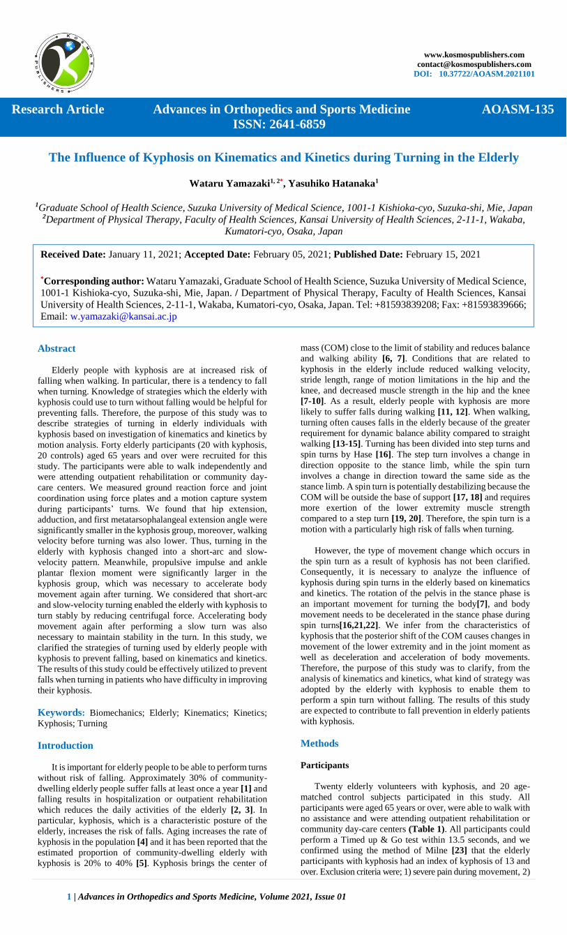
proximal phalange (Figure 1).
Figure 1: Marker placement.
RFHD : right forehead.
LFHD : left forehead.
RBHD : right back of head.
LBHD : left back of head.
C7 : 7th cervical vertebra.
T10 : 10th thoracic vertebra.
CLAV : clavicle.
TRN : sternum.
RBAK : right back of scapula.
RHSO : right shoulder.
LSHO : left shoulder.
RUPA : right midpoint of upper arm.
LUPA : left midpoint of upper arm.
RELB : right elbow.
LELB : left elbow.
RFRA : right midpoint of forearm.
LFRA : left midpoint of forearm.
RWRA : right styloid process of radius.
LWRA : left styloid process of radius.
RWRB : right styloid process of ulna.
LWRB : left styloid process of ulna.
RFIN : right 2ndmetacarpal head.
LFIN : left 2nd metacarpal head.
RASI : right superior anterior iliac spine.
LASI : left superior anterior iliac spine.
RPSI : right superior posterior iliac spine.
LPSI : left superior posterior iliac spine.
RTHI : right thigh. LTHI: left thigh.
RKNE : right lateral epicondyle of the femur.
LKNE : left lateral epicondyle of the femur.
RTIB : right midpoint of shank.
LTIB : left midpoint of shank.
RANA : right lateral malleolus.
LANA : left lateral malleolus.
RANB : right medial malleolus.
LANB : left medial malleolus.
3 | Advances in Orthopedics and Sports Medicine, Volume 2021, Issue 01
The Influence of Kyphosis on Kinematics and Kinetics during Turning in the
Elderly
Copyright: ©
2021 Wataru Yamazaki*
RHEE : right calcaneus ridge.
LHEE : left calcaneus ridge.
RTOE : right 2nd metatarsal head.
LTOE : left 2nd metatarsal head.
RFTA : right 1st metatarsal head.
LFTA : left 1st metatarsal head.
RFTB : right midpoint of proximal phalange.
LFTB : left midpoint of proximal phalange.
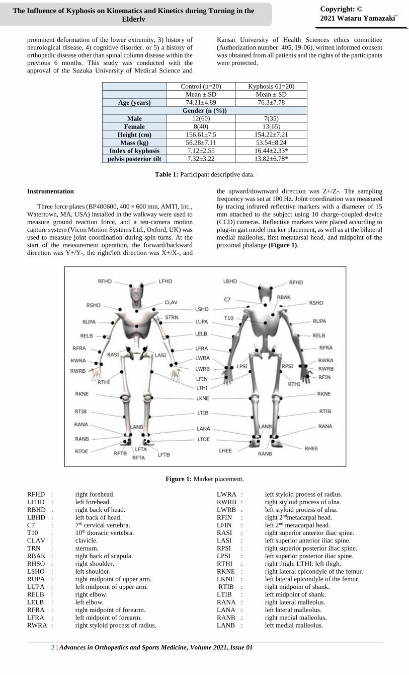
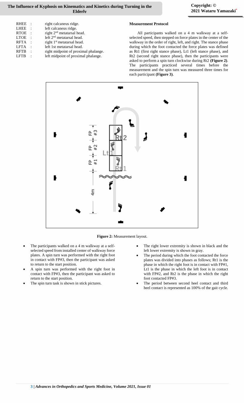
Measurement Protocol
All participants walked on a 4 m walkway at a self-
selected speed, then stepped on force plates in the center of the
walkway in the order of right, left, and right. The stance phase
during which the foot contacted the force plates was defined
as Rt1 (first right stance phase), Lt1 (left stance phase), and
Rt2 (second right stance phase), then the participants were
asked to perform a spin turn clockwise during Rt2 (Figure 2).
The participants practiced several times before the
measurement and the spin turn was measured three times for
each participant (Figure 3).
Figure 2: Measurement layout.
The participants walked on a 4 m walkway at a self-
selected speed from installed center of walkway force
plates. A spin turn was performed with the right foot
in contact with FP#3, then the participant was asked
to return to the start position.
A spin turn was performed with the right foot in
contact with FP#3, then the participant was asked to
return to the start position.
The spin turn task is shown in stick pictures.
The right lower extremity is shown in black and the
left lower extremity is shown in gray.
The period during which the foot contacted the force
plates was divided into phases as follows; Rt1 is the
phase in which the right foot is in contact with FP#1,
Lt1 is the phase in which the left foot is in contact
with FP#2, and Rt2 is the phase in which the right
foot contacted FP#3.
The period between second heel contact and third
heel contact is represented as 100% of the gait cycle.
4 | Advances in Orthopedics and Sports Medicine, Volume 2021, Issue 01
The Influence of Kyphosis on Kinematics and Kinetics during Turning in the
Elderly
Copyright: ©
2021 Wataru Yamazaki*
Figure 3: Spin turn task.
Data Processing
The trajectory distance of the COM, walking velocity,
centrifugal force, ground reaction forces, joint angles, and
joint moments were analyzed.
Trajectory Distance of COM
The trajectory distance of the COM was calculated from
the placement of reflective markers on each part of the body
during each stance phase using the analysis software Nexus2
ver.2.8.1 (Vicon Motion Systems Ltd.). Moreover, the
difference in trajectory distance of the COM between Rt1 and
Rt2 were compared between the two groups.
Walking Velocity
The walking velocity was the value of the trajectory
distance of the COM divided by the time of displacement
during each stance phase. Moreover, the difference in walking
velocity between Rt1 and Rt2, and Lt1 and Rt2 were compared
between the two groups.
Centrifugal Force
The magnitude of the centrifugal force F was defined as:
F=m v²/r
Where,
m: mass of object,
v: walking velocity,
r: the distance between COM and center of pressure (COP)
The centrifugal force in Rt2 was compared between the two
groups.
Ground Reaction Force
The braking impulse generated on deceleration of the
body was calculated from the posterior component of the
ground reaction force (Fy-) and its action time. The propulsive
impulse generated on acceleration of the body was calculated
from the anterior component of the ground reaction force
(Fy+) and its action time. The braking impulse and propulsive
impulse were normalized to each subject’s body mass, and
these values were compared between the two groups in the
same way as variation of walking velocity.
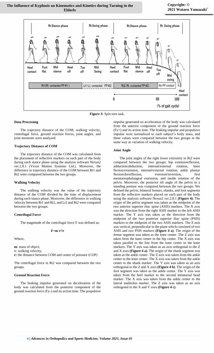
Joint Angle
The joint angles of the right lower extremity in Rt2 were
compared between the two groups; hip extension/flexion,
adduction/abduction, internal/external rotation, knee
flexion/extension, internal/external rotation, ankle plantar
flexion/dorsiflexion, eversion/inversion, first
metatarsophalangeal extension, and inside rotation of the
pelvis. Moreover, the posterior tilt angle of the pelvis in a
standing posture was compared between the two groups. We
defined the pelvis, bilateral femurs, shanks, and foot segments
from the reflective markers placed on each part of the body
using the analysis software Nexus2 ver.2.8.1 (Figure 4). The
origin of the pelvis segment was taken as the midpoint of the
two anterior superior iliac spine (ASIS) markers. The X axis
was the direction from the right ASIS marker to the left ASIS
marker. The Y axis was taken as the direction from the
midpoint of the two posterior superior iliac spine (PSIS)
markers to the midpoint of the two ASIS markers. The Z axis
was vertical, perpendicular to the plane which consisted of two
ASIS and two PSIS markers (Figure 4 a). The origin of the
femur segment was taken as the knee center. The Z axis was
taken from the knee center to the hip center. The X axis was
taken parallel to the line from the knee center to the knee
markers. The Y axis was taken as an axis orthogonal to the Z
and X axes (Figure 4 a). The origin of the shank segment was
taken as the ankle center. The Z axis was taken from the ankle
center to the knee center. The X axis was taken from the ankle
center to the shank marker. The Y axis was taken as an axis
orthogonal to the Z and X axes (Figure 4 b). The origin of the
foot segment was taken as the ankle center. The Y axis was
taken from the heel marker to the second metatarsal head
marker. The X axis was taken from the ankle center to the
lateral malleolus marker. The Z axis was taken as an axis
orthogonal to the X and Y axes (Figure 4 c).

5 | Advances in Orthopedics and Sports Medicine, Volume 2021, Issue 01
The Influence of Kyphosis on Kinematics and Kinetics during Turning in the
Elderly
Copyright: ©
2021 Wataru Yamazaki*
Figure 4: The segment definitions for pelvis, bilateral femurs, shanks, and feet. The segment definitions for pelvis, bilateral femurs,
shanks, and feet are shown. (a) pelvis and femur, (b) shank, (c) feet.
Next, the hip center, knee center and ankle center were
calculated according to Bell’s technique [24-26]. The relative
angle between these adjacent segments was calculated by
using the Cardan angle in which the rotation order is the Y-X-
Z. Then, rotation around the Y axis was defined as
adduction/abduction (foot undergoes eversion/inversion),
rotation around the X-axis was defined as flexion/extension
(dorsiflexion/plantar flexion of the ankle) and rotation around
the Z axis was defined as internal/external rotation in each
adjacent segment. The rotation of the pelvis was determined
between the Y axis of the pelvic segment and the Y axis in the
global coordinate system. Similarly, the tilting of the pelvis
posteriorly was determined by the angle formed by each Z
axis. The first metatarsophalangeal extension and flexion
angles were calculated using the arithmetic processing
software Vicon Body Builder ver3.6 (Vicon Motion Systems
Ltd.). These joint angles were calculated as a relative angle
between the following two axes: first, the axis connecting the
calcaneal ridge and the first metatarsal head through the
midpoint of the line connecting the medial malleolus and the
lateral malleolus. Second, the axis connecting the first
metatarsal head and the midpoint of the first phalange.
Joint Moment
The maximal values of the following joint moments of the
right lower extremity in Rt2 were compared in the two groups:
hip extension/flexion, adduction/abduction, internal/external
rotation, knee flexion/extension, internal/external rotation,
ankle plantar flexion/dorsiflexion, eversion/inversion. The
joint moments of the hip, knee, and ankle were calculated by
inverse dynamic analysis in which the moment of inertia,
mass, and location of the COM were substituted into the joint
coordinates and ground reaction force[27 -31] using the
analysis software Nexus2 ver.2.8.1 (Vicon Motion Systems
Ltd.) based on the technique of Winter[27]. The joint moments
were normalized to each subject’s body mass.
Statistical Analysis
The variation in trajectory distance of the COM, walking
velocity, and braking and propulsive impulse in each phase
was compared between the two groups, as well as centrifugal
force and the maximal joint angles and joint moments during
Rt2. The variation of rotation of the pelvis inside angle from
heel contact to toe off during Rt2 was compared between the
two groups because of the gradual increase in internal rotation
of the pelvis during Rt2. These data used the mean value of
three trials in each subject. Non-paired t-tests were used for
comparison between the two groups after confirming that the
data were normally distributed with equal variance. IBM SPSS
Statistical ver.24 statistical analysis software (SPSS, IBM
Corp., Armonk, NY, USA) was used for statistical analysis,
and the significance level was set at P < 0.05.
Results
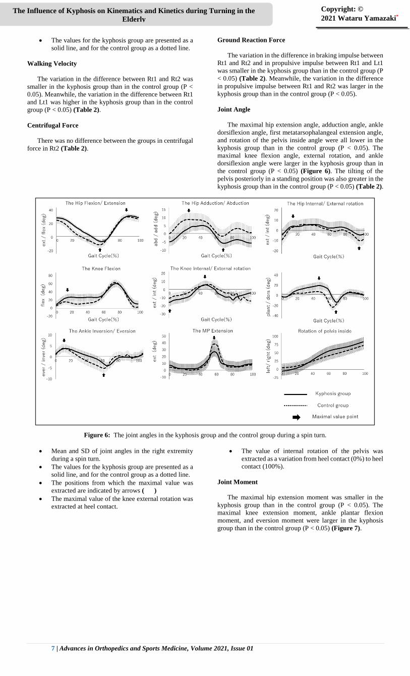
Trajectory Distance of the COM
The variation in the difference between Rt1 and Rt2
forward and inside was shorter in the kyphosis group than the
control group (P < 0.05) (Table 2). Trajectory distances of the
COM in forward and inside directions are shown, which was
from the right heel contact to the right toe off during the spin
turn (Figure 5).
Control (n=20) Mean ±SD Kyphosis (n=20) Mean ±SD
Trajectory Distance of COM (mm)
Forward Displacement (Variation from Rt1 to Rt2) -287.17±91.26 -374.30±74.91*
Inside Displacement (Variation from Rt1 to Rt2) 367.13±78.92 264.67±67.30*
Upward Displacement (Variation from Rt1 to Rt2) 3.27±1.65 4.8±2.98
Walking velocity (m /sec)
Variation from R t1 to R t2 -0.51±0.15 -0.34±0.12*
Variation from R t1 to Lt1 -0.29±0.10 -0.55±0.12*
Centrifugal force (N ) 824.81±219.17 858.12±294.33
Impulse (N .s/kg)
Braking impulse (variation from R t1 to Lt1) -0.23±0.14 -0.29±0.19
Braking impulse (variation from R t1 to R t2) 2.23±0.62 -0.99±0.27*
Propulsive impulse (variation from R t1 to Lt1) -1.13±0.31 -1.96±0.41*
Propulsive impulse (variation from R t1 to R t2 ) -1.43±0.57 -0.94±0.28*
6 | Advances in Orthopedics and Sports Medicine, Volume 2021, Issue 01
The Influence of Kyphosis on Kinematics and Kinetics during Turning in the
Elderly
Copyright: ©
2021 Wataru Yamazaki*
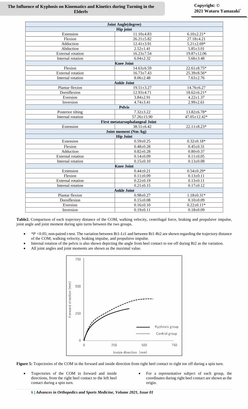
Joint Angle(degree)
Hip joint
Extension 11.10±4.83 6.10±2.21*
Flexion 26.21±5.82 27.18±4.21
Adduction 12.41±3.91 5.21±2.69*
Abduction 2.52±1.41 5.85±3.01
External rotation 16.23±7.54 19.87±12.06
Internal rotation 6.04±2.32 5.66±3.48
Knee Joint
Flexion 14.63±6.59 22.61±8.75*
External rotation 16.73±7.43 25.39±8.56*
Internal rotation 8.06±2.48 7.63±2.76
Ankle Joint
Plantar flexion 19.51±3.27 14.76±6.27
Dorsiflexion 12.93±4.71 18.62±6.21*
Eversion 3.84±2.91 4.22±1.37
Inversion 4.74±3.41 2.99±2.61
Pelvis
Posterior tilting 7.32±3.22 13.82±6.78*
Internal rotation 57.26±15.90 47.05±12.42*
First metatarsophalangeal Joint
Extension 38.51±6.42 22.11±8.23*
Joint moment (Nm /kg)
Hip Joint
Extension 0.59±0.25 0.32±0.18*
Flexion 0.48±0.28 0.45±0.31
Adduction 0.82±0.28 0.80±0.37
External rotation 0.14±0.09 0.11±0.05
Internal rotation 0.15±0.10 0.13±0.08
Knee Joint
Extension 0.44±0.21 0.54±0.29*
Flexion 0.11±0.09 0.13±0.11
External rotation 0.22±0.19 0.13±0.11
Internal rotation 0.21±0.15 0.17±0.12
Ankle Joint
Plantar flexion 0.98±0.27 1.18±0.31*
Dorsiflexion 0.15±0.08 0.10±0.09
Eversion 0.16±0.10 0.22±0.11*
Inversion 0.19±0.11 0.18±0.09
Table2. Comparison of each trajectory distance of the COM, walking velocity, centrifugal force, braking and propulsive impulse,
joint angle and joint moment during spin turns between the two groups.
*P <0.05; non-paired t-test. The variation between Rt1-Lt1 and between Rt1-Rt2 are shown regarding the trajectory distance
of the COM, walking velocity, braking impulse, and propulsive impulse.
Internal rotation of the pelvis is also shown depicting the angle from heel contact to toe off during Rt2 as the variation.
All joint angles and joint moments are shown as the maximal value.
Figure 5: Trajectories of the COM in the forward and inside direction from right heel contact to right toe off during a spin turn.
Trajectories of the COM in forward and inside
directions, from the right heel contact to the left heel
contact during a spin turn.
For a representative subject of each group, the
coordinates during right heel contact are shown as the
origin.
7 | Advances in Orthopedics and Sports Medicine, Volume 2021, Issue 01
The Influence of Kyphosis on Kinematics and Kinetics during Turning in the
Elderly
Copyright: ©
2021 Wataru Yamazaki*
The values for the kyphosis group are presented as a
solid line, and for the control group as a dotted line.
Walking Velocity
The variation in the difference between Rt1 and Rt2 was
smaller in the kyphosis group than in the control group (P <
0.05). Meanwhile, the variation in the difference between Rt1
and Lt1 was higher in the kyphosis group than in the control
group (P < 0.05) (Table 2).
Centrifugal Force
There was no difference between the groups in centrifugal
force in Rt2 (Table 2).
Ground Reaction Force
The variation in the difference in braking impulse between
Rt1 and Rt2 and in propulsive impulse between Rt1 and Lt1
was smaller in the kyphosis group than in the control group (P
< 0.05) (Table 2). Meanwhile, the variation in the difference
in propulsive impulse between Rt1 and Rt2 was larger in the
kyphosis group than in the control group (P < 0.05).
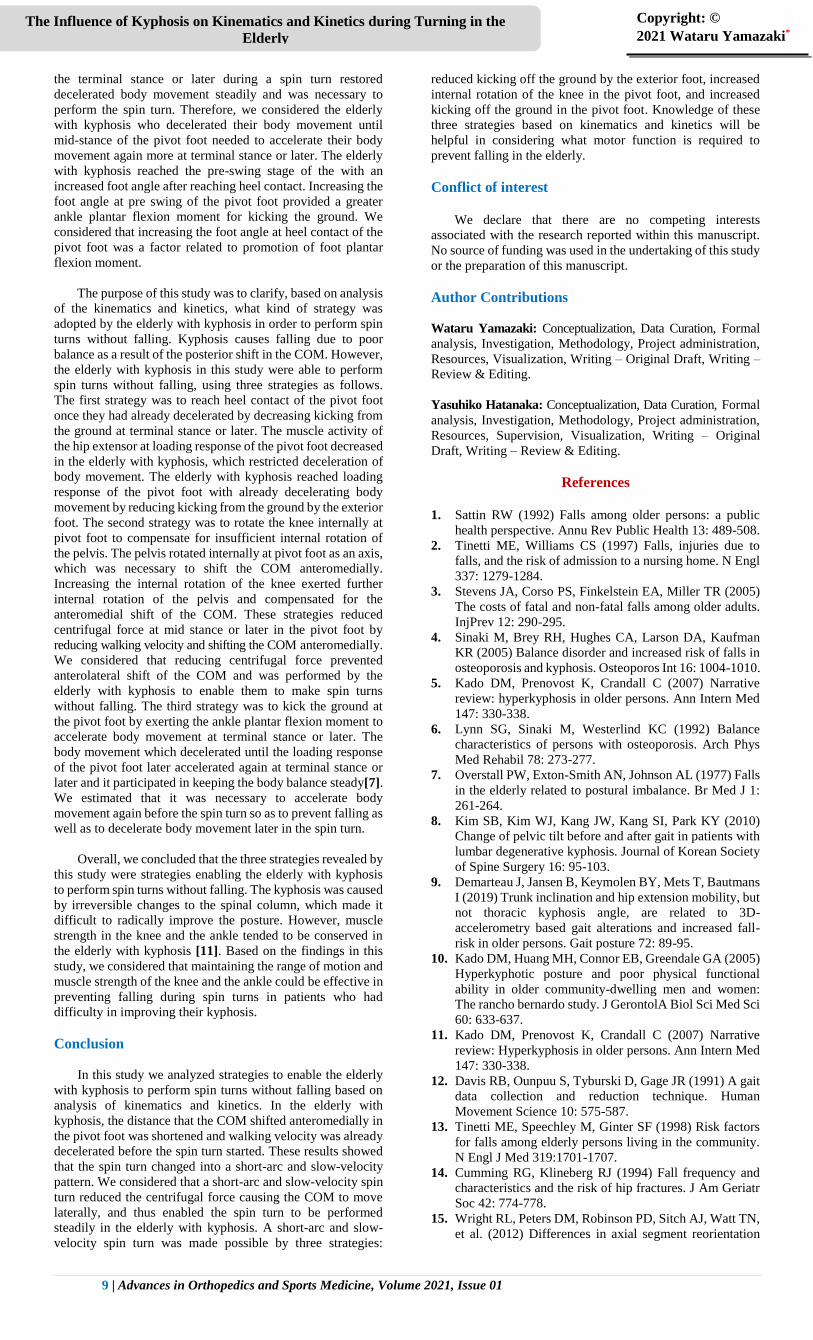
Joint Angle
The maximal hip extension angle, adduction angle, ankle
dorsiflexion angle, first metatarsophalangeal extension angle,
and rotation of the pelvis inside angle were all lower in the
kyphosis group than in the control group (P < 0.05). The
maximal knee flexion angle, external rotation, and ankle
dorsiflexion angle were larger in the kyphosis group than in
the control group (P < 0.05) (Figure 6). The tilting of the
pelvis posteriorly in a standing position was also greater in the
kyphosis group than in the control group (P < 0.05) (Table 2).
Figure 6: The joint angles in the kyphosis group and the control group during a spin turn.
Mean and SD of joint angles in the right extremity
during a spin turn.
The values for the kyphosis group are presented as a
solid line, and for the control group as a dotted line.
The positions from which the maximal value was
extracted are indicated by arrows ( )
The maximal value of the knee external rotation was
extracted at heel contact.
The value of internal rotation of the pelvis was
extracted as a variation from heel contact (0%) to heel
contact (100%).
Joint Moment
The maximal hip extension moment was smaller in the
kyphosis group than in the control group (P < 0.05). The
maximal knee extension moment, ankle plantar flexion
moment, and eversion moment were larger in the kyphosis
group than in the control group (P < 0.05) (Figure 7).

8 | Advances in Orthopedics and Sports Medicine, Volume 2021, Issue 01
The Influence of Kyphosis on Kinematics and Kinetics during Turning in the
Elderly
Copyright: ©
2021 Wataru Yamazaki*
Figure 7: The joint moments in the kyphosis group and control group during spin turns.
Mean and SD are shown in joint moments in the right extremity during spin turns.
The values for the kyphosis group are presented as a solid line, and for the control group as a dotted line.
The positions at which maximal value was extracted are indicated by arrows ( )
Discussion
Kyphosis caused a change in the pattern of a spin turn into
a short-arc and slow-velocity movement. The COM moved
away from the COP because of the shifting COM
anteromedially at mid stance or later in the pivot foot of the
spin turn. The shortening of the distance of the anterior shift
of the COM in the elderly with kyphosis indicated that a spin
turn was performed in a short arc. Moreover, the velocity of
the COM had already decelerated before the spin turn started
in the elderly with kyphosis. A short-arc and slow-velocity
spin turn reduced the centrifugal force acting on the COM at
mid stance or later. The centrifugal force could cause the COM
to move anterolaterally during a spin turn. Kyphosis caused
the COM to move posteriorly and increased the radius of
rotation of the COM around the COP; meanwhile, the
centrifugal force did not significantly differ between the two
groups. Therefore, we considered that elderly subjects with
kyphosis performed a spin turn with a short arc and slow
velocity, which reduced the centrifugal force to the same
magnitude as the control group.
Performing a spin turn with a short arc and slow velocity
influenced the movement of the pelvis and lower extremities.
Decreasing the maximal angle of hip extension, adduction, and
first metatarsophalangeal extension restricted the anterolateral
shift of the COM and changed the movement to a short-arc
spin turn in the elderly with kyphosis. In addition, internal
rotation of the pelvis decreased during the stance phase of the
pivot foot in the elderly subjects with kyphosis. The internal
rotation of the pelvis in stance phase of the pivot foot was
driven by rotation of the foot on the floor with the ball of the
foot as the axis as well as by hip internal rotation and knee
internal rotation [32]. The first metatarsophalangeal extension
at mid stance or later in the pivot foot involved lifting the heel
off the ground due to rotation of the foot on the floor with the
ball of the foot as the axis. However, tilting of the pelvis
posteriorly, which occurs in kyphosis, restricted extension of
the hip and the first metatarsophalangeal joint [31, 33].
Therefore, we considered that internal rotation of the pelvis
was restricted by the limitation of heel lifting, and then, tilting
of the pelvis posteriorly, caused by kyphosis, restricted the
ability to take the heel off the ground. On the other hand,
internal rotation of the pelvis in the elderly with kyphosis was
compensated for by knee internal rotation at heel contact or
later in the pivot foot. The differences in the maximal angle of
the knee during external rotation and internal rotation in the
two groups indicated that the knee internal rotation occurred
significantly at heel contact or later of the pivot foot in elderly
subjects with kyphosis. The knee internal rotation meant
rotation of the tibia internally against the femur or rotation of
the femur externally against the tibia. The knee internal
rotation in the pivot foot of the spin turn was rotation of the
femur externally against the tibia and rotation of the pelvis on
the same side with external rotation of the femur. The knee
internal rotation compensated for the decrease of internal
rotation of the pelvis in the pivot foot and then elderly subjects
with kyphosis were able to perform a short-arc spin turn.
The mechanism of deceleration in the pivot foot of the
spin turn changed the movement to a slow-velocity spin turn
in the elderly with kyphosis. The elderly with kyphosis had
already entered the loading response of the pivot foot with
deceleration by reducing the kicking off from the ground on
the exterior foot at terminal stance or later. It was necessary to
decelerate body movement in the stance phase of the pivot foot
to perform a short-arc spin turn and reduce the centrifugal
force in the spin turn. Comparing the propulsive impulse on
the exterior foot between the two groups, we found that it
decreased in the elderly with kyphosis. Hase et al. reported that
it was necessary to decelerate due to the posterior shift of the
COM in the pivot foot of the spin turn[16], however, the hip
extension moment was not been affected because the body
movement had already decelerated at the exterior foot in the
elderly with kyphosis. From the above, it can be seen that the
elderly with kyphosis decelerated body movement during the
terminal stance or later of the exterior foot before heel contact
of the pivot foot more than the control group. Moreover, the
ankle inversion moment and the knee extension moment both
increased in the elderly with kyphosis because decreasing the
shifting COM anteromedially on the pivot foot caused the
ground reaction force vector on the pivot foot to pass inside
the ball of the foot and posterior to the knee. Increasing the
knee flexion and ankle dorsiflexion angles needed to
compensate for shifting the COM posteriorly, a characteristic
posture of the elderly with kyphosis. Meanwhile, the elderly
with kyphosis showed accelerated body movement compared
to the control group at the terminal stance or later in the pivot
foot because of increased walking velocity, propulsive
impulse, and ankle plantar flexion moment. Acceleration in
9 | Advances in Orthopedics and Sports Medicine, Volume 2021, Issue 01
The Influence of Kyphosis on Kinematics and Kinetics during Turning in the
Elderly
Copyright: ©
2021 Wataru Yamazaki*
the terminal stance or later during a spin turn restored
decelerated body movement steadily and was necessary to
perform the spin turn. Therefore, we considered the elderly
with kyphosis who decelerated their body movement until
mid-stance of the pivot foot needed to accelerate their body
movement again more at terminal stance or later. The elderly
with kyphosis reached the pre-swing stage of the with an
increased foot angle after reaching heel contact. Increasing the
foot angle at pre swing of the pivot foot provided a greater
ankle plantar flexion moment for kicking the ground. We
considered that increasing the foot angle at heel contact of the
pivot foot was a factor related to promotion of foot plantar
flexion moment.
The purpose of this study was to clarify, based on analysis
of the kinematics and kinetics, what kind of strategy was
adopted by the elderly with kyphosis in order to perform spin
turns without falling. Kyphosis causes falling due to poor
balance as a result of the posterior shift in the COM. However,
the elderly with kyphosis in this study were able to perform
spin turns without falling, using three strategies as follows.
The first strategy was to reach heel contact of the pivot foot
once they had already decelerated by decreasing kicking from
the ground at terminal stance or later. The muscle activity of
the hip extensor at loading response of the pivot foot decreased
in the elderly with kyphosis, which restricted deceleration of
body movement. The elderly with kyphosis reached loading
response of the pivot foot with already decelerating body
movement by reducing kicking from the ground by the exterior
foot. The second strategy was to rotate the knee internally at
pivot foot to compensate for insufficient internal rotation of
the pelvis. The pelvis rotated internally at pivot foot as an axis,
which was necessary to shift the COM anteromedially.
Increasing the internal rotation of the knee exerted further
internal rotation of the pelvis and compensated for the
anteromedial shift of the COM. These strategies reduced
centrifugal force at mid stance or later in the pivot foot by
reducing walking velocity and shifting the COM anteromedially.
We considered that reducing centrifugal force prevented
anterolateral shift of the COM and was performed by the
elderly with kyphosis to enable them to make spin turns
without falling. The third strategy was to kick the ground at
the pivot foot by exerting the ankle plantar flexion moment to
accelerate body movement at terminal stance or later. The
body movement which decelerated until the loading response
of the pivot foot later accelerated again at terminal stance or
later and it participated in keeping the body balance steady[7].
We estimated that it was necessary to accelerate body
movement again before the spin turn so as to prevent falling as
well as to decelerate body movement later in the spin turn.
Overall, we concluded that the three strategies revealed by
this study were strategies enabling the elderly with kyphosis
to perform spin turns without falling. The kyphosis was caused
by irreversible changes to the spinal column, which made it
difficult to radically improve the posture. However, muscle
strength in the knee and the ankle tended to be conserved in
the elderly with kyphosis [11]. Based on the findings in this
study, we considered that maintaining the range of motion and
muscle strength of the knee and the ankle could be effective in
preventing falling during spin turns in patients who had
difficulty in improving their kyphosis.
Conclusion
In this study we analyzed strategies to enable the elderly
with kyphosis to perform spin turns without falling based on
analysis of kinematics and kinetics. In the elderly with
kyphosis, the distance that the COM shifted anteromedially in
the pivot foot was shortened and walking velocity was already
decelerated before the spin turn started. These results showed
that the spin turn changed into a short-arc and slow-velocity
pattern. We considered that a short-arc and slow-velocity spin
turn reduced the centrifugal force causing the COM to move
laterally, and thus enabled the spin turn to be performed
steadily in the elderly with kyphosis. A short-arc and slow-
velocity spin turn was made possible by three strategies:
reduced kicking off the ground by the exterior foot, increased
internal rotation of the knee in the pivot foot, and increased
kicking off the ground in the pivot foot. Knowledge of these
three strategies based on kinematics and kinetics will be
helpful in considering what motor function is required to
prevent falling in the elderly.
Conflict of interest
We declare that there are no competing interests
associated with the research reported within this manuscript.
No source of funding was used in the undertaking of this study
or the preparation of this manuscript.
Author Contributions
Wataru Yamazaki: Conceptualization, Data Curation, Formal
analysis, Investigation, Methodology, Project administration,
Resources, Visualization, Writing – Original Draft, Writing –
Review & Editing.
Yasuhiko Hatanaka: Conceptualization, Data Curation, Formal
analysis, Investigation, Methodology, Project administration,
Resources, Supervision, Visualization, Writing – Original
Draft, Writing – Review & Editing.
References
1. Sattin RW (1992) Falls among older persons: a public
health perspective. Annu Rev Public Health 13: 489-508.
2. Tinetti ME, Williams CS (1997) Falls, injuries due to
falls, and the risk of admission to a nursing home. N Engl
337: 1279-1284.
3. Stevens JA, Corso PS, Finkelstein EA, Miller TR (2005)
The costs of fatal and non-fatal falls among older adults.
InjPrev 12: 290-295.
4. Sinaki M, Brey RH, Hughes CA, Larson DA, Kaufman
KR (2005) Balance disorder and increased risk of falls in
osteoporosis and kyphosis. Osteoporos Int 16: 1004-1010.
5. Kado DM, Prenovost K, Crandall C (2007) Narrative
review: hyperkyphosis in older persons. Ann Intern Med
147: 330-338.
6. Lynn SG, Sinaki M, Westerlind KC (1992) Balance
characteristics of persons with osteoporosis. Arch Phys
Med Rehabil 78: 273-277.
7. Overstall PW, Exton-Smith AN, Johnson AL (1977) Falls
in the elderly related to postural imbalance. Br Med J 1:
261-264.
8. Kim SB, Kim WJ, Kang JW, Kang SI, Park KY (2010)
Change of pelvic tilt before and after gait in patients with
lumbar degenerative kyphosis. Journal of Korean Society
of Spine Surgery 16: 95-103.
9. Demarteau J, Jansen B, Keymolen BY, Mets T, Bautmans
I (2019) Trunk inclination and hip extension mobility, but
not thoracic kyphosis angle, are related to 3D-
accelerometry based gait alterations and increased fall-
risk in older persons. Gait posture 72: 89-95.
10. Kado DM, Huang MH, Connor EB, Greendale GA (2005)
Hyperkyphotic posture and poor physical functional
ability in older community-dwelling men and women:
The rancho bernardo study. J GerontolA Biol Sci Med Sci
60: 633-637.
11. Kado DM, Prenovost K, Crandall C (2007) Narrative
review: Hyperkyphosis in older persons. Ann Intern Med
147: 330-338.
12. Davis RB, Ounpuu S, Tyburski D, Gage JR (1991) A gait
data collection and reduction technique. Human
Movement Science 10: 575-587.
13. Tinetti ME, Speechley M, Ginter SF (1998) Risk factors
for falls among elderly persons living in the community.
N Engl J Med 319:1701-1707.
14. Cumming RG, Klineberg RJ (1994) Fall frequency and
characteristics and the risk of hip fractures. J Am Geriatr
Soc 42: 774-778.
15. Wright RL, Peters DM, Robinson PD, Sitch AJ, Watt TN,
et al. (2012) Differences in axial segment reorientation
10 | Advances in Orthopedics and Sports Medicine, Volume 2021, Issue 01
The Influence of Kyphosis on Kinematics and Kinetics during Turning in the
Elderly
Copyright: ©
2021 Wataru Yamazaki*
during standing turns predict multiple falls in older adults.
Gait Posture 36: 541-545.
16. Hase K, Stein RB (1999) Turning strategies during human
walking. J Neurophysiol 81: 2914-2922.
17. Akram SB, Frank JS, Chenouri S (2010) Turning behavior
in healthy older adults: is there a preference for step
versus spin turns? Gait Posture 31: 23-26.
18. England SE, Verghese J, Mahoney JR, Trantzas C,
Holtzer R (2010) Three-level rating of turns while
walking. Gait Posture 41: 300-303.
19. Kasukawa Y (2017) Age-related changes in muscle
strength and spinal kyphosis angles in an elderly Japanese
population. Clinical interventions in aging 12: 413-420.
20. Yamada M, Higuchi T, Mori S, Uemura K, Nagai K, et al.
(2012) Maladaptive turning and gaze behavior induces
impaired stepping on multiple footfall targets during gait
in older individuals who are at high risk of falling. Arch
Gerontol Geriatr 54: 102-108.
21. Glaister BC, Orendurff MS, Schoen JA, Bernatz GC,
Klute GK (2008) Ground reaction forces and impulses
during a transient turning maneuver. J Biomech 41: 3090-
3093.
22. Orendurff MS, Bernatz GC, Schoen JA, Klute GK (2008)
Kinetic mechanisms to alter walking speed. Gait Posture
27: 603-610.
23. Milne JS, Lauder IJ (1974) Age effects in kyphosis and
lordosis in adult. Ann Hum Biol 1- 327-337.
24. Winter DA, Patla AE, Prince F, Ishac M, Gielo-Perczak
K (1998) Stiffness control of balance in quiet standing. J
Neurophysiol 80: 1211-1221.
25. Bell AL, Pedersen DR, Brand RA (1990) A comparison
of the accuracy of several hip center location prediction
methods. J Biomech 23: 617-621.
26. Davis R, Ounpuu S, Tyburski T, Gage JR (1991) A gait
analysis data collection and reduction technique. Human
Movement Sciences 10: 575-587.
27. Kadaba MP, Ramakrishnan HK, Wootten ME (1987)
Lower extremity joint moments and ground reaction
torque in adult gait. Biomechanics of normal and
prosthetic gait 4-87-92.
28. Kadaba MP, Ramakrishnan HK, Wootten ME, Gainey J,
Gorton G, et al. (1989) Repeatability of kinematic,
kinetics and electromyographic data in normal adult gait.
Journal of Orthopaedic Research 7: 849-860.
29. Kadaba, MP, Ramakrishnan HK, Wooten ME (1990)
Lower extremity kinematics during level walking. Journal
of Orthopaedic Research 8: 383-392.
30. Ramakrishnan HK, Masiello G, Kadaba MP (1991) On
the estimation of the three dimensional joint moments in
gait. Biomechanics Symposium, ASME 120: 333-339.
31. Winter DA (2002) Biomechanics and motor control of
human movement. John Wiley & Sons. Print
ISBN:9780470398180 |Online ISBN:9780470549148
32. Thigpen MT, Light KE, Creel GL, Flynn SM (2000)
Turning difficulty characteristics of adults aged 65 years
or older. Phys Ther 80: 1174-1187.
33. Maki BE and McIlroy WE (2006) Control of rapid limb
movements for balance recovery: age-related changes and
implications for fall prevention. Age and ageing 35: 12-
18.
Citation: Yamazaki W, Hatanaka Y (2021) The Influence of Kyphosis on Kinematics and Kinetics during Turning in the
Elderly. Adv Ortho and Sprts Med: AOASM-135.